Experiments on the Mechanism of Silver Staining - Journal of Cell
20
Experiments on the Mechanism of Silver Staining Part I. Impregnation By A. PETERS {Department of Zoology, University of Bristol) With i plate (fig. 3) SUMMARY In the impregnating stage of silver staining, there are essentially two different reactions taking place between the silver ions and the sections of fixed tissue. A sub- stantial part of the silver is combined with the tissue in the unreduced form, and a smaller part is reduced to produce silver nuclei which form centres on which the de- veloping agents can deposit additional silver derived from the combined (reducible) silver fraction. A study of the effect of pH and time of impregnation, as well as of the specific blocking of chemically active protein end-groups upon the reducible silver fraction, suggests that this is chiefly combined with histidine and that it is not limited to the sites of the final differential staining. Except for the cell nuclei, the reducible silver shows a uniform distribution throughout the section; the amount taken up is in equilibrium with the silver ion concentration in the impregnating bath. The fraction present as silver nuclei increases progressively with time, with in- creasing pH, and with the temperature of the impregnating bath. Silver ions appear to be converted to silver nuclei by a process of physical reduction, and once formed, the nuclei are quite stable. Deposition of silver nuclei, necessary for differentiation of the stain, only occurs after impregnation between pH 7-5 and 9-0. In addition, it is only within this range that a sufficient amount of reducible silver is taken up to produce deep staining after development. CONTENTS I INTRODUCTION . . . . . . . . . . . . 85 II METHOD OF STAINING . . . . . . . . . . 85 III COMBINATION OF SILVER WITH THE TISSUE 86 (a) Method 86 (fe) The effect of pH 86 (c) The effect of duration of impregnation . . . . . . 87 (rf) The effect of the concentration of silver . . . . . . 87 (e) The effect of blocking of protein and other end-groups . . . . 88 (/) The role of histidine in silver combination . . . . . . go IV T H E FORMATION OF SILVER NUCLEI . . . . . . . • 91 (a) Factors affecting the formation of silver nuclei . . . . . 92 (6) The process of the formation of silver nuclei . . . . -95 V GENERAL DISCUSSION . . . . . . . . . . . 100 REFERENCES . . . . . . . . . . . 101 [Quarterly Journal of Microscopical Science, Vol. 96, part 1, pp. 84-102, March 1955.]
Transcript of Experiments on the Mechanism of Silver Staining - Journal of Cell

Experiments on the Mechanism of Silver Staining
Part I. Impregnation
By A. PETERS
{Department of Zoology, University of Bristol)
With i plate (fig. 3)
S U M M A R Y
In the impregnating stage of silver staining, there are essentially two differentreactions taking place between the silver ions and the sections of fixed tissue. A sub-stantial part of the silver is combined with the tissue in the unreduced form, and asmaller part is reduced to produce silver nuclei which form centres on which the de-veloping agents can deposit additional silver derived from the combined (reducible)silver fraction.
A study of the effect of p H and time of impregnation, as well as of the specificblocking of chemically active protein end-groups upon the reducible silver fraction,suggests that this is chiefly combined with histidine and that it is not limited to thesites of the final differential staining. Except for the cell nuclei, the reducible silvershows a uniform distribution throughout the section; the amount taken up is inequilibrium with the silver ion concentration in the impregnating bath.
The fraction present as silver nuclei increases progressively with time, with in-creasing pH, and with the temperature of the impregnating bath. Silver ions appear tobe converted to silver nuclei by a process of physical reduction, and once formed, thenuclei are quite stable.
Deposition of silver nuclei, necessary for differentiation of the stain, only occursafter impregnation between p H 7-5 and 9-0. In addition, it is only within this rangethat a sufficient amount of reducible silver is taken up to produce deep staining afterdevelopment.
C O N T E N T S
I INTRODUCTION . . . . . . . . . . . . 85
II METHOD OF STAINING . . . . . . . . . . 85
III COMBINATION OF SILVER WITH THE TISSUE 86
(a) Method 86(fe) The effect of pH 86(c) The effect of duration of impregnation . . . . . . 87(rf) The effect of the concentration of silver . . . . . . 87(e) The effect of blocking of protein and other end-groups . . . . 88(/) The role of histidine in silver combination . . . . . . go
IV T H E FORMATION OF SILVER NUCLEI . . . . . . . • 91
(a) Factors affecting the formation of silver nuclei . . . . . 92(6) The process of the formation of silver nuclei . . . . - 9 5
V GENERAL DISCUSSION . . . . . . . . . . . 1 0 0
REFERENCES . . . . . . . . . . . 101
[Quarterly Journal of Microscopical Science, Vol. 96, part 1, pp. 84-102, March 1955.]
/ . Impregnation 85
I. INTRODUCTION
THE silver staining methods are very important for the study of nervoustissue but, as with many other methods of histological staining, the
methods used are generally empirical, and the principles involved little under-stood. The present work is an attempt to further an understanding of theseprinciples.
The first theoretical approach to the subject was that made by Liesegang(1911), who postulated that during the impregnation of the tissue in the silversolution, nuclei of reduced silver are produced, which, on development of thestain, act as centres for the further deposition of silver reduced by the develop-ing agent, with the result that a visible staining is produced. This theory in-volves many parallels with photography, and the silver nuclei may be likenedto the latent image produced on exposure of a photographic emulsion.Liesegang's theory has formed the basis of many later theories on silverstaining, and has been greatly extended by the work of Silver (1942), Holmes(1943), Palmgren (1948), Romanes (1950), and Samuel (1953a, b). The recentwork on silver staining has confirmed that there are two forms of silver con-tained in impregnated nervous tissue, namely, silver which has been reducedto form nuclei, and developable silver which is combined with the componentsof the tissue. The developable silver is reduced by the developer and probablydeposited in relation to the silver nuclei.
II. METHOD OF STAINING
Before either the factors affecting silver staining or the mechanism ofstaining could be investigated, it was necessary to evolve a simple method ofstaining which could be consistently repeated. The techniques generally usedfor silver staining of paraffin sections are complex, and the reagents variable;especially when the method involves the use of protargol or other silver pro-teinates. However, three recent techniques, namely, those of Holmes (1943and 1947), Romanes (1950), and Samuel (19536), have made use of dilutesolutions of simple silver salts. Holmes used a 1/10,000-1/100,000 solution ofsilver nitrate buffered at pH 7-8 or 8-4 by boric acid/borax buffer, and Samuela modification of Holmes's technique employing a 1/10,000 solution of silvernitrate. Romanes used a 1/30,000 solution of silver chloride, the pH of whichwas adjusted by ammonia to 7-8, as indicated by comparator papers; this isequivalent to pH 9 as shown by a glass electrode (Romanes, personal com-munication). I found, as did Romanes, that the optimum pH for impregnationbefore chemical development was pH 9, and unless otherwise stated thefollowing method of staining was employed throughout the present work:
(1) Place pieces of fresh tissue in 70 per cent, alcohol for a sufficient timeto allow fixation, dehydrate in alcohol, and embed in paraffin wax.
(2) Mount paraffin sections on slide with albumen; remove the paraffinwax.

86 Peters—Experiments on Silver Staining
(3) Impregnate in the following solution in an incubator at 370 C. for 16hours: 1 ml. of 1 percent, silver nitrate, 180 ml. of distilled water, and 20 ml.of o-1 M boric acid/borax buffer at pH 9 (final concentration of silver nitrateis 1/20,000). A standard buffer of pH 9 is made up by mixing solutions of0 ' i M borax and o-i M boric acid until the pH value, as indicated by the glasselectrode, is 9.
(4) Transfer the slides to the developing solution, after rinsing in distilledwater.
(5) Wash in tap water for 10 minutes.(6) If toning is required, pass through distilled water to 0-2 per cent, gold
chloride for 3 minutes.(7) Transfer to 2 per cent, oxalic acid for 2 minutes.(8) Wash in tap water.(9) Transfer to 5 per cent, sodium thiosulphate for 10 minutes.(10) Wash well in tap water, followed by distilled water, dehydrate, and
mount.
III. COMBINATION OF SILVER WITH THE TISSUE
To understand the mechanism of silver staining it is clearly necessary todetermine how the silver, which can be subsequently reduced by the developer,is combined with the sections. It seems unlikely that the silver ions will com-bine with the fats and carbohydrates to any great extent, and therefore onlythe proteins will be considered. Recent work on the combination of metal ionswith proteins suggests that the combination may be stoichiometric (Klotz,1952), and the following investigation appears to support this view.
(a) Method
The tissues used in this investigation consisted of transverse sections offrog spinal cord, frog sciatic nerve, and rat cerebellum. These had been fixedin alcohol, which is assumed not to combine with the proteins to any extentduring fixation. After treatment, the sections were stained by the methodpreviously outlined.
(b) The effect of pH
The pH of the impregnating solution in these experiments was adjustedfrom pH 4-5 to 5-5 by acetate-acetic acid buffers, and over the range pH 6-3to 9-0 by boric acid/borax buffers. The final concentration of the buffer ineach case was o-oi M. Using silver electrodes, it was found that the concentra-tion of free ionic silver in the impregnating solution was fairly constant overthe whole of the pH range used.
After development with a hydroquinone-sulphite developer, there was adeeper staining at the higher pH values; this agreed with the results ofRomanes (1950) and Samuel (19536). There was little staining below pH 7,although there was a rapid increase in the intensity of staining up to pH 9.If, however, the silver combined with the section was precipitated by more

/ . Impregnation 87
active agents, such as an acid solution of hydrogen sulphide or a 2 per cent,solution of hydroquinone, which precipitate the silver more rapidly than thesulphite-containing developing solutions, there was some precipitate on thesections below pH 7. The amount of silver precipitated after impregnationat pH values below pH 7 was small, but these experiments showed that withan increase in the pH value the combined silver increased in a much moreuniform manner than the results with the developer had suggested.
The marked increase in the depth of staining above pH 7, which occurredafter development with the hydroquinone-sulphite solution, might suggestthat the silver combines with the carboxyl groups, which would be expectedto ionize at pH 5 to 6. A more probable explanation is that below pH 7 therewere not enough silver nuclei formed in the section during impregnation toact as centres for the reduction of the combined silver by the developer. Theprecipitation of the silver by more active reagents suggests that the combina-tion of the silver with the section involves some weak group rather than thecarboxyl groups.
(c) The effect of duration of impregnation
The effect of time on the silver uptake was determined at pH 9. Sectionswere impregnated, rapidly rinsed in distilled water, and, as above, immersedin either an acid solution of hydrogen sulphide or a 2 per cent, solution ofhydroquinone. At 370 C. the total uptake of silver by the section appeared tobe almost complete at the end of 10 minutes, although there was a slightincrease in the amount combined up to 30 minutes. At 190 C. the rate of com-bination with the silver was slower, but was almost complete at the end of30 minutes.
When similar sections were developed in the hydroquinone-sulphite de-veloper, staining was only slight up to 1 hour, but was almost complete at theend of 2 hours; there was very little further increase in the density of stainingup to 17 hours, although there was an improvement in the details of staining.Samuel (19536), also using a hydroquinone-sulphite developer, found thatat 560 C. staining occurred at pH 9-1 after 15 minutes' impregnation, and thatgood staining was obtained at pH 6-8 after z\ hours' impregnation. In noneof the present experiments was the staining so rapid.
Thus the density of differential staining obtained by the use of a normaldeveloping solution takes about 2 hours to reach an optimum. This is probablydue to the relatively longer time necessary for the formation of a sufficientnumber of silver nuclei to produce normal development, because the experi-ments with more active reagents showed that the combination of reduciblesilver with the section was almost complete at the end of 10 minutes.
(d) The effect of the concentration of silver
Sections were impregnated in solutions of 1/20,000, 1/40,000, 1/100,000,and 1/200,000 silver nitrate for 3 hours at pH 9. The combined silver wasdeposited by immersing the sections in either a 2 per cent, solution of hydro-

88 Peters—Experiments on Silver Staining
quinone (fig. 3, H) or an acid solution of hydrogen sulphide. The results showedthat the amount of reducible silver combined with the section increases withan increased concentration of silver in the impregnating bath. There is noquestion of a removal of all the silver ions from the solution; this result there-fore means that there is an equilibrium between the silver ions in the impreg-nating bath and the silver combined with the section. This suggests that thecombination of silver with the section is reversible and probably due to aweak grouping; otherwise, provided that there are sufficient silver ions in theimpregnating bath, the amount of silver combined with the section would beexpected to be constant.
(e) The effect of blocking of protein and other end-groups
The above experiments gave some indication of the type of combinationtaking place between the silver ions and the section, but to determine morespecifically which amino-acid was involved in the combination, the end-groups of the amino-acids in the sections were treated with specific reagentsbefore impregnation. The effect that the end-group reagents had on the im-pregnation was used to determine which amino-acids were involved in thecombination of silver with the sections.
In these experiments some of the protein reagents suggested by Olcott andFraenkel-Conrat (1947) and Danielli (1950) were employed. The results, andthe conditions used for blocking the groups, are shown in table 1, whichindicates the specificity of the stain after blocking together with the intensityof the staining (see also Peters, 1953).
The results show that while the carboxyl groups appear to play a part indetermining the specificity of the stain (fig. 3, B and c) they are not thegroups responsible for the general non-specific uptake of silver; this is alsoindicated by the effect of pH. That the carboxyl groups were blocked afteresterification was proved by treating the sections with methylene blue, a basicdye, which did not stain the treated sections although control sections werefully stained. The effect of diazomethane is difficult to interpret, since thiscompound is an active methylating agent and probably reacts with othergroups as well as the carboxyl groups.
The possibility of the aldehyde groups playing a part in the reactions of thesections can be eliminated, because the compounds containing these groupswould be extracted by the alcohol fixative used (Danielli, 1949); Schiff's reac-tion was negative on the sections. This point will be considered in more detaillater. Extraction of the nucleic acids by trichloracetic acid (Schneider, 1945)did not affect the staining apart'from reducing the staining of the nucleolus.
Hydrolysis of the phosphate and sulphate esters by 1 M hydrochloric acidat 37° C. had no effect.
There remain to be considered the ring-containing amino acids—trypto-phane, tyrosine, and histidine. Two reagents used were 2:4 dinitrofluoro-benzene (DNFB) (Danielli, 1947), and benzoyl chloride (Danielli, 1950).Both greatly reduce the intensity of staining, DNFB to a greater extent than

/ . Impregnation 89
benzoyl chloride. DNFB blocks tyrosine and histidine (Porter, 1950), whilebenzoyl chloride blocks all three ring-containing compounds. Performic acidwas also used to block tryptophane but was of little use, for although it re-duced the intensity of staining (fig. 3, D), a similar effect was brought about by
TABLE I
The effect of blocking agents on staining
Reagent
ControlAmino-groups:
Nitrous acid
FormaldehydePicric acid .Iodoacetic acid
Carboxyl groups:Esterification
Diazomethane
Sulphydryl groups:Iodoacetic acid
Hydrogen peroxide
Periodic acid
Performic acid
Disulphide links:Thioglycollic acid.
followed byIodoacetic acid
Ring compounds:DNFB
Benzoyl chloride .
Conditions of blockingreaction
i M NaNO2—HC1; o ° C ;2 hrs.
Neutral fixative; 4 per cent.Fixative; saturated solutiono-i M ;pH 9-6-10-6; 370 C ;
7 hrs.
o-i M HC1 in abs. MeOH;370 C.; 48-65 hrs.
Ethereal solution; 18° C ;4 hrs.
o-i M; pH 9-6-10-6; 37° C ;7 hrs.
005 M; pH 7; 18° C ; 45-72 hrs.
1 per cent. aq. soln.; 18° C.;2 hrs.
90 per cent, formic acid—H2O2; i8°C.; 10-15 min.
o-i M ; p H 8-4-7-7; 180 C ;12-15 hrs.
o-i M ; p H 9-6-10-6; 37° C ;7 hrs.
015 M DNFB in 90 percent. EtOH saturated withNaHCO3; 180 C ; 24 hrs.
10 per cent, in dry pyridine;18° C ; 17 hrs.
Specificityof staining
+ +-)--f-
+ ++ +
0
+ -|-+ ++ +
+
+ +
+
+
Intensityof im-
pregnation
+ 4-
-f-
+ ++ +
-f-
+ ++
+ +o(+)
+ +
-f
+
Refer-ence
( 1 )
(0(z)
(3)
(4)
(5)
(6)
(7)
(1) Michaelis and Schubert (1934).(2) Olcott and Fraenkel-Conrat (1947).(3) Dempsey, Singer, and Wislocki (1950).(4) Sanger (1945)-(5) Goddard and Michaelis (1934).(6) Danielli (1947).(7) Danielli (1950).
formic acid alone; this acid is necessarily present in large quantities in anypreparation of performic acid. Another oxidizing agent, acidified potassium
90 Peters—Experiments on Silver Staining
permanganate, also reduced staining; it is known to disrupt the imidazolering of histidine (Karrer, 1947, p. 773). The reaction which takes place in thepresence of formic acid is unknown. Evidence against the extensive participa-tion of tryptophane is suggested by the fact that if frozen sections of gelatinare stained by the present method, they stain quite deeply, although gelatincontains no tryptophane.
Since staining begins as low as pH 7, it suggests that the silver probablycombines with the imidazole group of histidine. This has a pKA of 7-97 (Hilland Branch, 1940); the pK value of the —OH group of tyrosine is 10. Furtherevidence is obtained from the silver staining of smears of the followingproteins:
Haemoglobin > Fibrinogen > Pepsin > Protamine.
The intensity of staining shows a descending order which is paralleled bytheir histidine content (Tristram, 1949).
(/) The role of histidine in silver combination
The results suggest that reducible silver is taken up by the tissue in com-bination with histidine, and this confirms the recent work of Wormser (1951),who showed that in dialysed solutions of serum-albumen the histidine wasresponsible for silver combination. Grassmann and Kusch (1952) also con-cluded that since the amount of silver combined at pH 6-2 with a series ofcollagen-type proteins was proportional to their histidine content, the silvercombined with this amino acid. The imidazole nucleus is known to formcomplexes with silver and thought to combine with silver bromide grains inphotographic emulsions (Mees, 1944, p. 191). Mees suggests that the com-bination occurs in the following manner:
Ag+ / A g
CH 1NL C H N-v a. - rearrangement CH—N<
I > > > ><
> g - • II >CHX > || >CH + HX.CH—NH/ CH—NH / CH—N^
He also states that there may be a combination of the silver with theguanidyl grouping of arginine, since this contains the group —CH = NH,which is considered to act in the same way as the imidazole nucleus of histidine:
NH. /NHj Ag—NHN y +Ag+x- —> I
I X- +C—NH2.
The above results by no means exclude the possibility of combinationbetween silver and arginine, but this could not be investigated owing to thelack of specific blocking reagents for guanidyl groups. Arginase cannot beemployed because the enzyme only attacks free arginine. Staining experimentson protein smears appear to exclude the possibility of an extensive combina-tion between silver and arginine because a smear of protamine showed littleuptake of silver, though the arginine content of this protein is about 87per cent.; furthermore, the pK value of the guanidyl group is 12-13. A further

/ . Impregnation 91
possibility is the sulphydryl group, for although the above experiments giveonly a slight suggestion of combination, the —SH group is known to formcompounds with silver; the pK value is 9-11.
Gorriz (see Weber, 1947) suggested that formalin used as a fixative methy-lates the neurofibrils and thus mordants the tissue. The fact that methylationof the tissue either by diazomethane or methyl alcohol reduces that stainingsuggests that Gorriz's inference is incorrect.
M. L. Silver (1942) states that the amount of silver combined with thesections increases with time, but Romanes (1950), has shown that after a giventime there is little increase in the amount of silver taken up by the sectionsfrom the impregnating bath; we have confirmed Romanes' result. Romanesattributed this to a saturation of the reducing groups in the tissue, and believedthat the groups involved were the aldehyde groups. However, we have pointedout that these groups will be absent after alcohol fixation, and suggest thatthe groups which are combined with the reducible silver are the imidazolegroups of histidine, together with any, other groups which may combine withsilver. Thus combination between the sections and silver is assumed to bestoichiometric and chemical; further, it has been shown that there is anequilibrium between the silver combined with the section and the silver ionsin the impregnating solution.
If the combination of the silver is with histidine then, before the slides aredeveloped, silver will not be specifically confined to the nerve elements; itwould be expected that the distribution of the silver-combining groups isgeneral. Therefore an attempt was made to determine the site of histidine inthe sections, but no specific test for histidine could be found. A modified formof the Knoop test (Hunter, 1951) was attempted on the sections, but withoutsuccess. In all of the colour tests available the reagents react with tyrosine ortyrosine and tryptophane. However, sections were condensed with diazotizedsulphanilic acid at 0° C. for 15 minutes, washed, and mounted in glycerine.This reaction produces a yellow colour with tyrosine and histidine which, asfar as could be seen, was not confined to any specific elements of the tissue.
To determine the position of silver before development, a number of slideswere impregnated, washed to remove any traces of uncombined silver, andthen immersed in an acid solution of hydrogen sulphide to precipitate thereducible silver in situ. Although the cell nuclei were blackened more thanthe rest of the section, there was no specific deposition of silver sulphide on thefibres, and the precipitate was fairly uniform. Therefore, before the silver isdeveloped, it appears that the combination of the unreduced silver with thesection is relatively unspecific. Since development leads to specific staining,the combined silver is not reduced in situ by the developer; the specificity ofthe silver preparation is therefore determined in the actual process of develop-ment.
IV. THE FORMATION OF SILVER NUCLEI
Holmes (1943) claimed that during the immersion of formol-fixed sectionsin the impregnating bath, nuclei of reduced silver are formed in them. He
92 Peters—Experiments on Silver Staining
impregnated paraffin sections, washed them in distilled water for 6 hours, thendeveloped and toned with gold. He states that although the sections showedlittle or no darkening after development, on toning there was an almost com-plete staining of the tissue elements; Romanes (1950), who used no develop-ing agent, found that the staining was both light and incomplete.
A similar experiment to that of Holmes was carried out with tissues fixedin alcohol, picric acid, chloral hydrate, and formol. Slides were impregnatedand washed in several changes of distilled water for 24 hours before develop-ment in chloroquinol; hardly any staining was found after toning except alight but incomplete staining of the formol-fixed sections. This staining wasprobably due to a slight initial reduction of the silver in the impregnatingbath. Sections from the other fixatives showed no reduction at all, whichsuggests that this method gives no true indication of the presence of silvernuclei in these sections.
Two types of developing solution have been used in silver staining. Thosewhich contain free silver ions in the developing solution initially will be re-ferred to as 'physical developers', and those which contain no free silver ionswill be referred to as 'chemical developers' (see Peters, 1955). In photographythe deposition of silver from a physical developing solution is controlled bythe 'latent image' in the emulsion. Similarly, in sections of tissue the presenceof silver nuclei should lead to a deposition of silver from such a developingsolution. Thus, in physical development the developed silver is provided bythe developing solution and, in the main, is not derived from the unreducedsilver combined with the proteins of the section, as it is in chemical develop-ment.
Samuel (19536) used the physical developer suggested by Pearson andO'Neill (1946) to demonstrate the presence of silver nuclei in impregnatedsections. To remove the reducible silver he immersed the sections in a 2-5per cent, solution of sodium sulphite for 5 minutes before development. Inthe present experiments, a glycin-containing physical developer was used todetermine the presence of silver nuclei (Peters, 1955).
Sections which had been impregnated were washed and placed in thedeveloping solution; in every case a complete development of the stain re-sulted (fig. 3, E). Consequently, sections fixed in fixatives other than formol,although they may not show any visible reduction of silver on removal fromthe impregnating bath, do contain silver nuclei. Sections which had not beenimpregnated were placed in the physical developer, but in no case was thereany specific staining, and the deposition of silver on to the section was onlyslight.
(a) Factors affecting the formation of silver nuclei
The factors affecting the formation of silver nuclei in the impregnatingbath were investigated by the use of the glycin physical developer. In theseexperiments the assumption is made that the depth of staining produced bythe physical developer in a given time is dependent on the number of silver

/ . Impregnation 93
nuclei in the tissue. Whether these nuclei act as centres for the catalytic re-duction of silver or as centres for the deposition of silver, the greater theirnumber the greater will be the silver deposition, provided that the conditionsof development are constant. This assumption appears to be fully valid, sinceJames (1950, p. i n ) found that silver sol particles accelerate the reduction ofsilver ions by developing agents; the rate of reduction was proportional to theamount of silver sol added.
In all cases sections of alcohol-fixed frog spinal cord, frog sciatic nerve, andrat cerebellum were used.
(1) To determine the stability of the silver nuclei five slides were impreg-nated for 16 hours, then transferred to distilled water, which was changedfive times during the first day and subsequently once every day. Slides wereremoved from the distilled water after 1, 2, 3, 5, and 7 days, and developed.The pH of the distilled water used to wash the slides was 6. Although the timenecessary for development at room temperature increased from 16 to 18minutes during the first 5 days, development was normal. After 7 days, how-ever, 32 minutes was required for development and, in contrast to the previousslides, resulted in a very deep staining of the nerve-cell nuclei, relative to thatof the nerve fibres. Nevertheless, the silver nuclei were still present in thetissue after 7 days' washing, and consequently they appear to be quite stable.
(2) Impregnation in 1/40,000, 1/20,000, 1/5,000, and 1/2,000 silver nitrate,followed by washing for 24 hours before development, showed that the rate offormation of silver nuclei increased with the concentration of silver in theimpregnating bath. After impregnation in 1/2,000 silver nitrate the stainingwas less specific than when more dilute solutions were used.
(3) Impregnation was carried out in 1/20,000 silver nitrate at 0° C , io° C ,14° C , and 370 C. for 16 hours and followed by washing the sections in dis-tilled water for 24 hours. The results showed that the rate of formation ofnuclei increases with temperature. Very little staining was obtained, evenafter 18 minutes' development, in the sections impregnated at o° C. and10° C , while sections stained at 370 C. gave quite normal staining.
(4) Silver nuclei increase progressively with time. They begin to appearsoon after immersion of the sections in the impregnating bath. With longperiods of immersion, of the order of 8 days, the reduction of silver on to thesections can be appreciated more fully. After such a period of time the sectionsare quite brown in colour, and many of the structural details of the sectionsare visible as though the section had been impregnated and developed normally,although the details are not as clear. This is probably due to an initial forma-tion of nuclei which act as centres for the further deposition of silver from thesolution, so that a very slow type of physical development occurs. The reasonfor the lack of detail in the sections after such a long impregnation is probablythat silver nuclei are formed in other tissue elements as well as the nervouselements. A similar effect is noticed when sections are impregnated for 2 or3 days before development; here again there is some loss of specificity of thestaining.
94 Peters—Experiments on Silver Staining
(5) The effect of pH was investigated by impregnating the sections in a1/20,000 solution of silver nitrate for 16 hours at 370 C. The pH of the solu-tions was adjusted at 4-7 and 5-5 by acetate-acetic acid buffers, and at7-0, 8-o, and 9-0 by borate/boric acid buffers. The final concentration ofthe buffer was o-oi M.
In the first experiments the reducible silver was removed from the sectionsby washing them in several changes of distilled water for 24 hours beforedevelopment in either the glycin physical developer or in a modification ofPearson and O'Neill's developer (Samuel, 1953&). A similar set of experimentswas also carried out in which the reducible silver was removed from thesections by immersing them in either 2-5 per cent, sodium sulphite for 5minutes or in a citrate buffer at pH 3-2 for 1 hour before development (Samuel,1953^). In all cases where the glycin physical developer was used, the deepeststaining was obtained at pH 8-o, indicating that there was a maximum forma-tion of silver nuclei at this pH value. On either side of this value the depth ofstaining was less, and at pH 5-5 and 4-7 only very faint and unspecific stainingoccurred (fig. 3, G). The staining was specific at pH 7-0, 8-o, and 9-0, but thebest differentiation of fibres was found at pH 8-o.
Using Pearson and O'Neill's developer there was a maximum depth ofstaining at pH 8.0 when the reducible silver was removed by continuouswashing, and the maximum varied between pH 7-0 and 8-o when the reduciblesilver was removed by sodium sulphite treatment. Samuel (1953^), in ex-periments carried out over the range pH 6-8 to 9-1, found an optimum for theformation of nuclei at pH 6-8, and that as the pH rose the amount of silvernuclei per unit volume of the tissue decreased. With Pearson and O'Neill'sdeveloper the staining was only specific at pH 7-0 and 8-o; outside this rangethe staining was coarse and unspecific. A similar result was found by Samuel
(1953*)-The results indicate an optimum formation of nuclei at pH 7-8; on each
side of this range there is a decrease in the rate of formation of nuclei.(6) Since very little staining was produced by chemical development at
pH 4-5, the formation of silver nuclei in higher concentrations of silver nitrate,namely, 1/100, 1/500, 1/1,000, and 1/5,000, was investigated at this pH value.Impregnation was for 18 hours at 37° C. As in the experiments carried out atpH 9, there was an increase in the staining with concentration, so that thestaining was quite deep after impregnation in the 1/100 silver nitrate. How-ever, the staining was completely unspecific and coarse, and showed nostructural details. Thus, although formation of nuclei takes place at pH 4-5in higher concentrations of silver nitrate, these nuclei do not lead to specificstaining after physical development (fig. 3, G).
(7) Three of the blocking agents which were found to affect silver staining,namely, DNFB, performic acid, and methyl alcohol with hydrochloric acid,were investigated. The sections were pretreated with the reagents as previouslydescribed (table 1) and washed thoroughly in several changes of distilled waterbefore impregnation. They were then washed in distilled water for 24 hours

/ . Impregnation 95
and developed in the physical developer. The effects of these blocking agentson the formation of silver nuclei are shown in table 2.
These experiments show that the silver nuclei are quite stable, and thattheir formation increases with time, temperature, and the concentration ofsilver in the impregnating bath. There appears to be an optimum formation ofsilver nuclei at pH 7-8. When the silver nuclei are formed at low pH values,or if the sections are pretreated with certain blocking agents, the silvernuclei which are formed do not give rise to specific staining on development.
TABLE 2
Slocking agent
Control (none)MeOH plus HC1 (fig. 3, B) .DNFBPerformic acid (fig. 3, D)
Formation ofsilver nuclei
specificunspecificunspecificunspecific
Amount ofreducible
silver present
normalnormal
decreaseddecreased
(b) The process of the formation of silver nucleiRomanes (1950) considered that the reduction of silver salts in the im-
pregnating stage to produce silver nuclei was brought about either by sul-phydryl groups, ascorbic acid, or aldehydes. The results of blocking thesulphydryl groups suggests that they do not participate appreciably in theformation of nuclei. To test the effects of ascorbic acid, Romanes immersedsections in solutions of ascorbic acid before impregnation, but he states thatalthough in some cases there appeared to be an improvement in the staining,the results were not conclusive. It is doubtful if ascorbic acid does reduce thesilver ions to form nuclei, since the acid is unstable in alkaline solutionsthough stable in acid solutions. In the present method of staining silver nucleiare not formed to any extent in acid solutions, and even when they are formedthey do not lead to specific staining (fig. 3, G). Other evidence has beenobtained on this point from the fact that alcohol-fixed sections of scorbuticguinea pig spinal cord give quite normal staining.
If the silver salts in the impregnating bath are reduced chemically, then, assuggested by Romanes, reduction of the silver is probably due to the aldehydegroups. To some extent this appears to be in keeping with the brown colourshown by formol-fixed tissues on removal from the impregnating bath, forsections of tissue from other fixatives do not show the same brown colourafter impregnation at 370 C , even though it has been demonstrated thatsilver nuclei are present. Danielli (1949) and Chu (1950) consider that thealdehyde groups producing the Feulgen's test are those contained in fattycompounds; these are not appreciably affected by formol fixation, which alsoprotects them from later extraction by alcohol. Consequently, these fattycompounds will remain in formol-fixed tissue, and further aldehyde groupsmight be expected to be derived from the fixative. Most of the aldehyde groupsmust in fact be derived from the fixative, because sections of frozen tissue

96 Peters—Experiments on Silver Staining
show only a slight reduction of silver on removal from the impregnatingsolution. On the other hand, alcohol extracts these fatty compounds from freshtissue (Danielli, 1949), so that the amount of aldehyde remaining in the tissueafter alcohol extraction will be small. Such tissue shows no appreciable re-duction of silver on removal from the impregnating bath, and can be shown tocontain fewer silver nuclei than frozen-dried tissue.
Lhotka and Myhre (1953) showed that treatment of sections with 4 percent, periodic acid for 2 hours produces an increase in the depth of stainingof connective tissue using the method of Foot (1924). They attributed theincrease in staining to the freeing of aldehyde groups from 1:2 glycols.Lhotka, Myhre, and Combs (1953) continued these oxidation studies andtreated nervous tissue with potassium permanganate, periodic acid, chromicacid, lead tetra-acetate, and sodium bismuthate prior to silver impregnationby a series of different techniques. With two exceptions they found that theoxidation had no effect on the affinity of silver for nerve fibres, and concludedthat it is unlikely that the 1:2 glycol radical is present in amounts demon-strable by the procedure used. We pretreated sections of frog sciatic nerveand rat cerebellum with 4 per cent, periodic acid for 2 hours at 25° C. Thesections were brown on removal from the impregnating bath, showing thatreduction of some silver had been effected, but development produced ratherunspecific staining, so that the myelin sheaths of the sciatic nerve and thefibre interspaces were stained. In the rat cerebellum sections staining was alsounspecific; in this case few fibres were visible, but staining of capillaries re-sulted (fig. 3, F). In spite of this different result the general conclusion is thesame as that reached by Lhotka, Myhre, and Combs (1953), that the aldehydegroups play no part in the staining of nerves because the release of extra alde-hyde groups leads to the staining of connective tissue and not to a strengthen-ing of the nerve staining. As pointed out previously, acidified potassiumpermanganate reduced the intensity of staining.
Other facts are inconsistent with the idea of reduction of the silver by alde-hyde groups. The aldehyde groups were condensed with hydroxylamine byimmersing the sections in a solution of 2 gm. NH2OH. HC1, 4 gm. sodiumacetate, and 6 ml. distilled water for 80 minutes (Danielli, 1949). After treat-ment the sections were washed and impregnated. The formation of silvernuclei was found to be unaffected. Similarly, treatment of sections with o-i Mpotassium cyanide before impregnation did not produce any effect.
If the aldehyde groups are responsible for the reduction of silver, then sucha reaction could only take place once, because the aldehyde groups would beoxidized to carboxyl groups in the process. It was found that the silver nucleicould be removed from the sections by either 5 per cent, thiosulphate or dilutenitric acid. Sections of alcohol-fixed material were therefore impregnated,treated to remove the nuclei, and then reimpregnated. The reimpregnatedsections developed normally, thus indicating the reformation of nuclei.Control slides developed without reimpregnation showed that the nuclei hadbeen removed.
/ . Impregnation 97
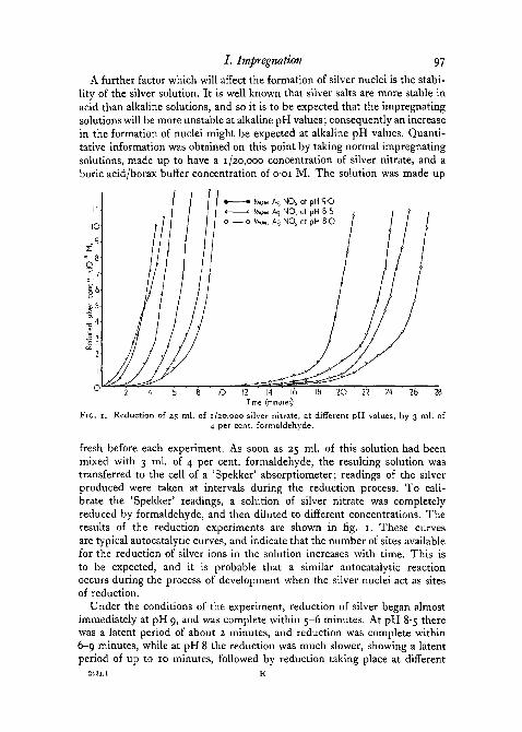
A further factor which will affect the formation of silver nuclei is the stabi-lity of the silver solution. It is well known that silver salts are more stable inacid than alkaline solutions, and so it is to be expected that the impregnatingsolutions will be more unstable at alkaline pH values; consequently an increasein the formation of nuclei might be expected at alkaline pH values. Quanti-tative information was obtained on this point by taking normal impregnatingsolutions, made up to have a 1 /2o,ooo concentration of silver nitrate, and aboric acid/borax buffer concentration of o-oi M. The solution was made up
II;
10-
.9.21 8-Q
g 5-
10 2O 22 24 26 28
FIG.
12 14 16Time (minutes)
Reduction of 25 ml. of 1/20,000 silver nitrate, at different pH values, by 3 ml. of4 per cent, formaldehyde.
fresh before each experiment. As soon as 25 ml. of this solution had beenmixed with 3 ml. of 4 per cent, formaldehyde, the resulting solution wastransferred to the cell of a 'Spekker' absorptiometer; readings of the silverproduced were taken at intervals during the reduction process. To cali-brate the 'Spekker' readings, a solution of silver nitrate was completelyreduced by formaldehyde, and then diluted to different concentrations. Theresults of the reduction experiments are shown in fig. 1. These curvesare typical autocatalytic curves, and indicate that the number of sites availablefor the reduction of silver ions in the solution increases with time. This isto be expected, and it is probable that a similar autocatalytic reactionoccurs during the process of development when the silver nuclei act as sitesof reduction.
Under the conditions of the experiment, reduction of silver began almostimmediately at pH 9, and was complete within 5-6 minutes. At pH 8-5 therewas a latent period of about 2 minutes, and reduction was complete within6-9 minutes, while at pH 8 the reduction was much slower, showing a latentperiod of up to 10 minutes, followed by reduction taking place at different
98 Peters—Experiments on Silver Staining
rates. Reduction was even slower at lower pH values, so that at pH 7-5 therewas no indication of reduction in the solution even after 75 minutes. In spiteof the small pH range covered by the experiment, there is an appreciabledifference in the stability of the different solutions towards reduction. Aspointed out by Kubie and Davidson (1928), one reason for this pH effect isthat the nitric acid and formic acid produced as a result of the reduction tendto inhibit further reduction. At higher pH values these acids are effectivelyneutralized.
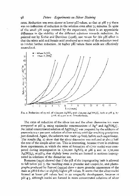
2 IOb 7
Time (minutes)
FIG. 2. Reduction of 25 ml. of 1/20,000 AgNO3 and 1/20,000 Ag(NH3)t, both at pH 9, by3 ml. of 4 per cent, formaldehyde.
The rates of reduction of the silver ion and the silver diammine ion werecompared at pH 9, using equimolar concentrations of Ag+ and Ag(NH3)J.An initial concentrated solution of Ag(NH3)£ was prepared by the addition ofammonia to a 1 per cent, solution of silver nitrate until the resulting precipitatejust dissolved. Again, the solution was made up fresh before each experiment.The results (fig. 2) show that the silver diammine was reduced at about halfthe rate of the simple silver ion. This is interesting, because there is evidencefrom experiments, in which the rates of formation of silver nuclei were com-pared during impregnation in 1/20,000 AgNO3 at pH 9 and in 1/20,000Ag(NH3)2' atpH 9, that slightly fewer nuclei are formed in sections impreg-nated in solutions of the diammine ion.
Romanes (1950) showed that if the pH of the impregnating bath is allowedto fall below pH 7, the resulting stain is granular and unspeciflc, and photo-graphs produced by Samuel (1953a) show a more granular appearance of thestain at pH6-8 than at slightly higher pH values. It seems that the silver nucleiformed at lower pH values lead to an unspecific development, because atpH 4-5, although nuclei are formed in more concentrated solutions of silver

5OH
sow .
FIG. 3
A. PETERS
f . - •••• ' . . • • ••:. A

/ . Impregnation 99
nitrate, they do not lead to specific physical development (fig. 3, G). It isprobable then, that the nuclei only lead to a specific development when theyhave been formed at higher pH values (fig. 3, E). However, a similar effect isbrought about by the use of a 1 /2,ooo solution of silver nitrate at pH 9, but inthis case there is only a slight loss of specificity due to connective-tissuestaining. This is probably brought about by an excessive formation of silvernuclei, with the result that there is some loss of specificity on development.
The process by which nuclei are formed in still in doubt. The results donot distinguish between a physical or chemical reduction of the silver, if infact such a distinction does exist, because the process may be a combination ofboth. In the case of formation of nuclei by chemical reduction the aldehydegroups might be responsible, while for physical reduction the silver would bereduced at certain sites by virtue of the redox potential existing there. Factorsknown to affect the impregnation, such as pH, concentration, and temperature,can support either case. The slow rate of formation of nuclei may be explainedeither by an initial physical reduction of silver or on the basis that the nucleihave to attain a certain size before they can act as centres for the developmentof the reducible silver. Similarly, the effect of pH on the reducibility of thesilver ions is compatible with either postulate except that there is no explana-tion for an optimum formation of nuclei at pH 7 to 8. However, whether theinitial reduction of the silver is chemical or physical, any further deposition ofsilver in the impregnating bath on to the existing nuclei is probably due tophysical reduction.
The changes brought about by some of the blocking agents which lead toan unspecific development are unknown, but this again points to a physicalreduction of the silver in the formation of nuclei, because these blocking agentsattack different end-groups in the proteins.
Palmgren (1948) suggests that the power of tissues to reduce silver dependson the fixative employed. Thus he suggests that if the tissue is fixed inindifferent chemical fixatives such as alcohol, then the reducing power of the
FIG. 3 (plate), A, transverse section of alcohol-fixed frog spinal cord showing ventral horncells. Impregnated in 1/20,000 silver nitrate at pH 9. Developed in hydroquinone-sulphite10 /i.
B, transverse section of alcohol-fixed frog spinal cord showing ventral horn cells. Esterifiedbefore impregnation. Compare with fig. 3, A. 10 fi.
c, transverse section of alcohol-fixed frog spinal cord showing ventral horn cells. Treatedwith diazomethane before impregnation. Compare with fig. 3, A. 10 /x.
D, transverse section of alcohol-fixed frog spinal cord showing ventral horn cells. Treatedwith performic acid before impregnation. Compare with fig. 3, A. 10 /1.
E, transverse section of rat cerebellum. Purkinje cell region. Chloral hydrate fixed. Impreg-nated at pH 9, developed in glycin physical developer. 10 p.
F, transverse section of rat cerebellum. Purkinje cell region. Alcohol fixed. Pretreated withperiodic acid before impregnation at pH 9. Note staining of capillaries and lack of staining ofnerve fibres. Compare with fig. 3, E. 10 /j..
G, transverse section of rat cerebellum. Impregnated at pH 47. Developed in glycine physicaldeveloper. Note lack of differential staining, iop.
H, transverse section of rat cerebellum. Impregnated at pH 90. Silver reduced in 2 per cent,hydroquinone. Note lack of differential staining. 10 p..

ioo Peters—Experiments on Silver Staining
tissue proteins is less than if they had been fixed in formol, and further, thatmany compounds such as chloral hydrate, pyridine, and ammoniated alcoholproduce a general increase in the reducing power of the tissues. If Palmgren'shypothesis is correct, then this points to a physical reduction of silver ionsto form nuclei in the tissue.
The weight of evidence points to the initial formation of silver nuclei inthe tissue by a physical reduction of the silver.
V. GENERAL DISCUSSION
Two processes take place in the impregnating bath—the fairly rapid com-bination of the free silver ions with the histidine and other amino-acids in thesection to form compounded, but unreduced, silver, and the slower formationof silver nuclei. For extensive combination of the silver ions with the histidineit is necessary that the pH value of the impregnating bath should be in theregion of pH 9. The effect of pH on the combination of silver ions withhistidine has been described by Haarmann and Friihauf-Heilmann (1941),who showed that at pH 7 only 0-41 equivalents of silver are combined withhistidine, while at pH 9 the number is increased to 1-12. The process ofcombination with reduced silver is complete within about 15 minutes at370 C , but to obtain good staining it has been shown that a longer period ofimpregnation is necessary because the formation of silver nuclei is a slowerprocess. Hence, although deep staining may be obtained at the end of 2 hours,the details are poor. It is only within the pH range 7-5-9-0 that a balanceis obtained between the specific formation of silver nuclei and a sufficientamount of combined silver to produce a deep enough chemical developmentwith developers such as hydroquinone-sulphite.
Willis (1945) believes that combination of silver is at the sites of accumula-tion of basic proteins, and that these combine with the silver to form com-plexes. This is in agreement with the hypothesis that it is the histidine whichcombines chiefly with the silver. Willis does not mention silver nuclei, butdoes point out that such co-ordination complexes that silver might form wouldbe readily reducible.
The formation of silver nuclei has been assumed since the early paper ofLiesegang (1911), and has been confirmed more recently by the work ofHolmes (1943), Romanes (1950), and Samuel (19530,6). To a large extent itseems that the factors controlling the combination of unreduced silver with histi-dine are the same ones that govern the formation of silver nuclei. If the silveris reduced to form nuclei by virtue of the redox potential of the tissues (andthis appears to be suggested by the experiments), then it is probably necessaryfor the silver ions to be combined with the section before they can be reducedto form nuclei.
The position of the silver nuclei is still in question, and their presence isonly assumed from indirect evidence. However, the 'latent image' in a photo-graphic emulsion has never been seen, and it is only necessary for relatively

/ . Impregnation 101
few atoms of silver to be present to form a nucleus for development. If thenuclei form the centres for development, so that the developed silver is de-posited on to them, then they must be in the same positions as the developedsilver in the finally stained preparation.
Liesegang (1911) thought that the characteristics of the silver nuclei deter-mined the specificity of the stain; thus deeper staining occurred where morenuclei were present. Clearly, to some extent this is true, because without theirpresence it has been shown that no development can take place. Samuel(19536) has criticized Liesegang's theory because Liesegang failed to removethe reducible silver from the section before development. In his experimentsSamuel found that compared with %\ hours' impregnation, when more nucleishould be present, 15 minutes' impregnation at pH 9-1 resulted in intenseaxonal staining, although very few silver nuclei should be present. He goes onfurther to say that one should expect some comparable degree of nucleardeposition after z\ hours' impregnation at pH 6-8 and 15 minutes' impregna-tion at pH 9-1, if there is any quantitative correlation between staining and thesilver nuclei. To some extent this is true, but there is evidence that the silvernuclei are not formed in the same positions in the section after impregnationat different pH values. Thus it was shown that unspecific staining occurredafter the formation of silver nuclei at pH 4-5 (fig. 3, G). It is clear however, thatvery little is known about the characteristics of the silver nuclei, and it isdoubtful if much progress can be made in this direction until the silvernuclei and their positions in the sections can be determined directly.
To summarize, it has been shown that during the impregnating stage acomplex series of reactions takes place between the silver ions and the fixedtissue of the sections. Essentially, however, there are two processes taking place,a rapid combination of unreduced silver with the histidine and other amino-acids of the section, and a slower formation of nuclei of reduced silver. To alarge extent these two processes are controlled by the same factors of time, pH,temperature, and the concentration of free silver ions in the impregnating bath.
I wish to express sincere thanks to my supervisor, Professor J. E. Harris,for his interest and advice during the course of this work. I am indebted toProfessor J. F. Danielli of King's College, London, for advice on the use ofprotein reagents, and to Dr. J. W. Mitchell of the Physics Department fordiscussions about the mechanism of photography.
The photographs were taken for me by Mr. K. J. Wood of the ZoologyDepartment.
This work was carried out during the tenure of a graduate scholarship in theUniversity of Bristol.
REFERENCESCHU, C. H. U., 1950. 'A histochemical study of staining the axis cylinder with Fuchsin-
sulphurous acid (Schiff's reagent).' Anat. Rec, 168, 723.DANIELLI, J. F., 1947. 'A study of techniques for the cyto-chemical demonstration of nucleic
acids and some components of proteins.' Symposium Soc. exp. Biol., I, 101.
IOZ Peters—Experiments on Silver Staining
DANIELLI, J. F., 1949. 'A critical study of techniques for the demonstration of aldehydes.'Quart. J. micr. Sci., 90, 67.1950. 'Studies on the cytochemistry of proteins.' Cold. Spr. Harb. Sym. quant. Biol.,
14, 32.DEMPSEY, E. W., SINGER, M., and WISLOCKI, G. B., 1950. 'The increased basophilia of
tissue proteins after oxidation with periodic acid.' Stain Tech., 25, 73.FOOT, N. C, 1924. 'A technique for demonstrating reticulum fibres in Zenker-fixed paraffin
sections.' J. Lab. & Clin. Med., 9, 777.GODDARD, D. R., and MICHAELIS, L., 1934. 'Studies on keratin.' J. Biol. Chem., 106, 608.GRASSMANN, W., and KUSCH, D., 1952. 'Ober die Bindung von Ag- und Pb-Ionen durch
Kollagen.' Hoppe-Seyl Z., 290, 216.HAAHMANN, W., and FRUHAUF-HEILMANN, E., 1941. 'OberdieKomplexaffinitatvonSchwer-
metallen und EiweiBstoffen.' Biochem. Z., 309, 13.HILL, T. L., and BRANCH, G. E. K., 1940. 'Resonance and the chemistry of histidine.'
Science, 91, 145.HOLMES, W., 1943. 'Silver staining of nerve axons in paraffin sections.' Anat. Rec, 86, 157.
'947, in Recent advances in clinical pathology. London (Churchill).HUNTER, A., 1951. 'A modified Knoop test for histidine.' Fed. Proa, 10, 201.JAMES, T. H., 1950. 'Catalytic phenomena related to photographic development.' Advances
in Catalysis, 2, 105 (Academic Press Inc., N.Y.).KARRER, P., 1947. Organic chemistry. Elsevier Pub. Co. Inc.KLOTZ, I. H., 1952. 'Some metal complexes with proteins and other large molecules' in
Trends in Physiology and Biochemistry (Academic Press Inc. N.Y.).KUBIE, L., and DAVIDSON, D., 1928. 'The ammonia silver solutions used in neuropathology.
Their staining properties and methods of preparation.' Arch. Neurol. Psychiat.(Chicago), 19, 888.
LHOTKA, J. F., MYHRE, B. A., and COMBS, C. M., 1953. 'Effects of oxidation on neurofibrillarargyrophilia.' Stain Tech., 28, 101.
LIESEGANG, R. E., 1911. 'Die Kolloidchemie der histologischen Silberfarbungen.' Kolloid.,Beih. 3, i.
MEES, C. E. K., 1944. The theory of the photographic process. New York (Macmillan).MICHAELIS, L., and SCHUBERT, M. P. P., 1934. 'The reactions of iodoacetic acid on mer-
captans and amines.' J. Biol. Chem., 106, 331.OLCOTT, H. S., and FRAENKEL-CONRAT, H., 1947. 'Specific group reagents for proteins.'
Chem. Rev., 41, 151.PALMGREN, A., 1948. 'A rapid method for selective silver staining of nerve fibres and nerve
endings in mounted paraffin sections.' Acta zool. Stockh., 29, 377.PEARSON, A. A., and O'NEILL, S. L., 1946. 'A silver gelatin method for staining nerve
fibres.' Anat. Rec, 95, 297.PETERS, A., 1953. 'Silver impregnation of nerve fibres.' Nature, 171,613.
1955. 'Experiments on the mechanism of silver staining. II. Development.' Quart.J. micr. Sci., 96, 103.
POKTER, R. R., 1950. 'Reactivity of the imidazole ring in proteins.' Biochem. J., 46, 304.ROMANES, G. J., 1950. 'The staining of nerve fibres in paraffin sections with silver.' J.
Anat. Lond., 84, 104.SAMUEL, E. P., 1953a. 'Impregnation and development in silver staining.' Ibid., 87, 268.
19536. 'The mechanism of silver staining.' Ibid., 278.SANGER, F., 1945. 'The free amino groups of insulin.' Biochem J., 39, 507.SCHNEIDER, W. C , 1945. 'Phosphorus compounds in animal tissues. The extraction and
estimation of desoxypentose nucleic acid and of pentose nucleic acid.' J. Biol. Chem.,161, 293.
SILVER, M. L., 1942. 'Colloidal factors controlling silver staining.' Anat. Rec, 82, 507.TRISTRAM, G. R., 1949. 'Amino-acid content of purified proteins.' Advances in protein
Chemistry, 5, 83 (Academic Press Inc. N. Y.).WEBER, A., 1947. 'Analysis des phases successives de l'imprdgnation neurofibrillaire par
l'argent r£duit.' Bull, histol. appl., 24, 49.WILLIS, A. G., 1945. 'A new method for staining neurofibrillae and axis cylinders.' J. R.
micv. Soc, 15, 29.WORMS™, Y., 195I. 'Recherches sur la dissimulation de l'ion Ag, applique'es a l'e'tude d'une
protiSine.' J. Chim. Phys., 48, 344.