Experimental nonalcoholic fatty liver disease in mice ... · b College of Animal Sciences and...
8
Research Paper Experimental nonalcoholic fatty liver disease in mice leads to cytochrome p450 2a5 upregulation through nuclear factor erythroid 2-like 2 translocation Yizhe Cui a,b,1 , Qiuju Wang b,1 , Xiaochong Li a , Xiuying Zhang a,n a College of Veterinary Medicine, Northeast Agricultural University, No. 59 Mucai Street, Xiangfang District, Harbin 150030, Heilongjiang, China b College of Animal Sciences and Technology, Heilongjiang Bayi Agricultural University, 2# Xinyang Road, New Development District, Daqing 163319, Heilongjiang, China article info Article history: Received 17 July 2013 Received in revised form 15 August 2013 Accepted 16 August 2013 Keywords: Coumarin 7-hydroxylase Oxidative stress glutathione S-transferases High-fat diet Knockout mice abstract Mouse cytochrome P450 2A5 (CYP2A5) is upregulated in various liver diseases and a putative common feature for all of these conditions is altered cellular redox status. Nuclear factor erythroid 2-like 2 (Nrf2) is a transcription factor that is post-translationally regulated by oxidative stress and controls the transcription of protective target genes. In the present study, we have characterized the regulation of CYP2A5 by Nrf2 and evaluated gene expression, protein content and activity of anti-oxidant enzymes in the Nrf2 þ/ þ and Nrf2 / mice model of non-alcoholic fatty liver (NAFLD). After eight weeks of feeding on a high-fat diet, livers from Nrf2 / mice showed a substantial increase in macro and microvesicular steatosis and a massive increase in the number of neutrophil polymorphs, compared to livers from wild- type mice treated similarly. Livers of Nrf2 / mice on the high-fat diet exhibited more oxidative stress than their wild-type counterparts as assessed by a significant depletion of reduced glutathione that was coupled with increases in malondialdehyde. Furthermore, results in Nrf2-deficient mice showed that CYP2A5 expression was significantly attenuated in the absence of Nrf2, as was found with the conventional target genes of Nrf2. The treatment of wild-type mice with high-fat diet leaded to nuclear accumulation of Nrf2, and co-immunoprecipitation experiments showed that Nrf2 was bound to Cyp2a5. These findings suggest that the high-fat diet induced alteration in cellular redox status and induction of CYP2A5 was modulated through the redox-sensitive transcription Nrf2. Crown Copyright & 2013 Published by Elsevier B.V. Introduction Nonalcoholic fatty liver disease (NAFLD) is composed of a spectrum of pathologies ranging from simple fatty liver (steatosis) to the more severe nonalcoholic steatohepatitis (NASH). The proposed mechanism for progression of NAFLD involves a two- hit theory where lipid accumulation in hepatocytes (the“first hit”) is followed by a “second hit,” including insulin resistance, oxida- tive stress, and cytokine production [1]. The capacity of a cell to manage oxidative stress is primarily mediated through antioxidant response elements or electrophile response elements (AREs), which are largely under the control of the transcription factor nuclear factor erythroid 2-like 2 (Nrf2) [2,3]. Nrf2 is primarily a cytosolic protein due to proteasomal degradation regulated by Keap1 [4]. When a cell is undergoing oxidative stress, Nrf2 escapes Keap1- mediated degradation, translocates to the nucleus, actives a battery of redox-homeostasis regulatory genes such as γ-glutamyl cysteine ligase (γ-GCS), and phase II genes such as glutathione S-transferases (GST) and NAD(P)H:quinone oxidoreductase1 (NQO1) [5]. Activation of Nrf2 plays a crucial role in regulating the cytoprotective responses to a variety of drugs, toxicants, and cellular stresses. CYP2A5 belongs to the group of P450 enzymes involved in the metabolism of xenobiotics, such as drugs and environmental tox- icants [6]. Murine CYP2A5 and its human orthologue CYP2A6 are the main catalysts of coumarin and nicotine metabolism but have also been shown to participate in metabolic activation of procarcinogens, such as aflatoxin B1 and nitrosamines [7,8]. A number of studies have indicated high expression of CYP2A5 in mouse hepatomas and preneoplastic liver lesions; therefore, CYP2A5 has been suggested to Contents lists available at ScienceDirect journal homepage: www.elsevier.com/locate/redox Redox Biology 2213-2317 Crown Copyright & 2013 Published by Elsevier B.V. http://dx.doi.org/10.1016/j.redox.2013.08.003 Abbreviations: ARE, Antioxidant response element; Nrf2, Nuclear factor erythroid 2-like 2; KO, Knockout; WT, Wild-type; P450, Cytochrome P450; NASH, Nonalco- holic steatohepatitis; NAFLD, Nonalcoholic fatty liver disease; co-IP, Co- immunoprecipitation n Corresponding author. Tel/fax.: þ86 451 55190674. E-mail addresses: [email protected], [email protected] (X. Zhang). 1 Qiuju Wang and Yizhe Cui had contributed equallly to this paper, and hence both are first authors. Redox Biology 1 (2013) 433–440 Open access under CC BY-NC-SA license. Open access under CC BY-NC-SA license.
Transcript of Experimental nonalcoholic fatty liver disease in mice ... · b College of Animal Sciences and...
Research Paper
Experimental nonalcoholic fatty liver disease in mice leadsto cytochrome p450 2a5 upregulation through nuclear factorerythroid 2-like 2 translocation
Yizhe Cui a,b,1, Qiuju Wang b,1, Xiaochong Li a, Xiuying Zhang a,n
a College of Veterinary Medicine, Northeast Agricultural University, No. 59 Mucai Street, Xiangfang District, Harbin 150030,Heilongjiang, Chinab College of Animal Sciences and Technology, Heilongjiang Bayi Agricultural University, 2# Xinyang Road, New Development District, Daqing 163319,Heilongjiang, China
a r t i c l e i n f o
Article history:Received 17 July 2013Received in revised form15 August 2013Accepted 16 August 2013
Keywords:Coumarin 7-hydroxylaseOxidative stressglutathione S-transferasesHigh-fat dietKnockout mice
a b s t r a c t
Mouse cytochrome P450 2A5 (CYP2A5) is upregulated in various liver diseases and a putative commonfeature for all of these conditions is altered cellular redox status. Nuclear factor erythroid 2-like 2 (Nrf2)is a transcription factor that is post-translationally regulated by oxidative stress and controls thetranscription of protective target genes. In the present study, we have characterized the regulation ofCYP2A5 by Nrf2 and evaluated gene expression, protein content and activity of anti-oxidant enzymes inthe Nrf2þ /þ and Nrf2� /� mice model of non-alcoholic fatty liver (NAFLD). After eight weeks of feedingon a high-fat diet, livers from Nrf2� /� mice showed a substantial increase in macro and microvesicularsteatosis and a massive increase in the number of neutrophil polymorphs, compared to livers from wild-type mice treated similarly. Livers of Nrf2� /� mice on the high-fat diet exhibited more oxidative stressthan their wild-type counterparts as assessed by a significant depletion of reduced glutathione that wascoupled with increases in malondialdehyde. Furthermore, results in Nrf2-deficient mice showed thatCYP2A5 expression was significantly attenuated in the absence of Nrf2, as was found with theconventional target genes of Nrf2. The treatment of wild-type mice with high-fat diet leaded to nuclearaccumulation of Nrf2, and co-immunoprecipitation experiments showed that Nrf2 was bound to Cyp2a5.These findings suggest that the high-fat diet induced alteration in cellular redox status and induction ofCYP2A5 was modulated through the redox-sensitive transcription Nrf2.
Crown Copyright & 2013 Published by Elsevier B.V.
Introduction
Nonalcoholic fatty liver disease (NAFLD) is composed of aspectrum of pathologies ranging from simple fatty liver (steatosis)to the more severe nonalcoholic steatohepatitis (NASH). Theproposed mechanism for progression of NAFLD involves a two-hit theory where lipid accumulation in hepatocytes (the“first hit”)is followed by a “second hit,” including insulin resistance, oxida-tive stress, and cytokine production [1]. The capacity of a cell tomanage oxidative stress is primarily mediated through antioxidant
response elements or electrophile response elements (AREs), whichare largely under the control of the transcription factor nuclearfactor erythroid 2-like 2 (Nrf2) [2,3]. Nrf2 is primarily a cytosolicprotein due to proteasomal degradation regulated by Keap1 [4].When a cell is undergoing oxidative stress, Nrf2 escapes Keap1-mediated degradation, translocates to the nucleus, actives a batteryof redox-homeostasis regulatory genes such as γ-glutamyl cysteineligase (γ-GCS), and phase II genes such as glutathione S-transferases(GST) and NAD(P)H:quinone oxidoreductase1 (NQO1) [5]. Activationof Nrf2 plays a crucial role in regulating the cytoprotective responsesto a variety of drugs, toxicants, and cellular stresses.
CYP2A5 belongs to the group of P450 enzymes involved in themetabolism of xenobiotics, such as drugs and environmental tox-icants [6]. Murine CYP2A5 and its human orthologue CYP2A6 are themain catalysts of coumarin and nicotine metabolism but have alsobeen shown to participate in metabolic activation of procarcinogens,such as aflatoxin B1 and nitrosamines [7,8]. A number of studieshave indicated high expression of CYP2A5 in mouse hepatomas andpreneoplastic liver lesions; therefore, CYP2A5 has been suggested to
Contents lists available at ScienceDirect
journal homepage: www.elsevier.com/locate/redox
Redox Biology
2213-2317 Crown Copyright & 2013 Published by Elsevier B.V.http://dx.doi.org/10.1016/j.redox.2013.08.003
Abbreviations: ARE, Antioxidant response element; Nrf2, Nuclear factor erythroid2-like 2; KO, Knockout; WT, Wild-type; P450, Cytochrome P450; NASH, Nonalco-holic steatohepatitis; NAFLD, Nonalcoholic fatty liver disease; co-IP, Co-immunoprecipitation
n Corresponding author. Tel/fax.: þ86 451 55190674.E-mail addresses: [email protected], [email protected] (X. Zhang).1 Qiuju Wang and Yizhe Cui had contributed equallly to this paper, and hence
both are first authors.
Redox Biology 1 (2013) 433–440
Open access under CC BY-NC-SA license.
Open access under CC BY-NC-SA license.

be a useful marker for hepatocarcinogenesis [9–12]. Additionally,CYP2A5 has been found to be up-regulated in several types ofhepatic infections of viral, bacterial, and parasitic origin, which areassociated with an increased risk of hepatocarcinogenesis [13–16].The mechanistic basis behind these elevated expression levels hasremained enigmatic. A possible explanation for these apparentlyunrelated effects on CYP2A5 expression could be involvement of acommon indirect mechanism.
We previously found that mice fed with high-fat diet resultedin an increase in expression of Nrf2 and CYP2A5 in liver. Wehypothesized that feeding a high-fat diet would cause an elevationof ROS, which increases Nrf2 levels, and the elevated Nrf2 up-regulates CYP2A5. To test this hypothesis, we have characterized ahigh-fat diet induced model of NAFLD and evaluated gene expres-sion and protein activity of anti-oxidant enzymes in Nrf2þ /þ andNrf2� /� mice.
Materials and methods
Chemicals and antibodies
All reagents were obtained from Sigma Chemical Company(St. Louis, MO, USA) unless otherwise indicated. Rabbit IgG(sc-2027) and rabbit polyclonal anti-Nrf2 (sc-13032), were fromSanta Cruz Biotechnology Inc. (Santa Cruz, CA). Polyclonal chickenCyp2a5 antiserum was a kind gift from Dr. H. Raunio (University ofKuopio, Kuopio, Finland).
Animal treatment
Six to eight-week-old, ICR male wild-type (Nrf2þ /þ) and Nrf2knockout mice (Nrf2� /�) were purchased from the Animal Experi-ment Center of General Hospital of Nanjing Military Area Com-mand [Certification no. SCXK (JUN) 2007-012]. They were dividedinto four groups of three mice in each group and housed in filter-top polycarbonate cages containing wood chip bedding andmaintained in a 12-h light/dark cycle with free access to standardmouse chow and tap water. After a one-week adaptive period, thefour groups of mice were treated for eight consecutive weeks. TheNrf2þ /þ and Nrf2� /� control groups were fed with the standarddiet. The Nrf2þ /þ and Nrf2� /� high-fat model groups were fedwith a high-fat diet (68.5% standard diet, 15% lard, 1% cholesterol,0.5% bile and 15% dextrin) which induces NAFLD (data not shown).The mice in all groups were sacrificed by CO2 overdose. At the timeof death, blood was collected from the thoracic aorta. The serumwas separated from the cellular elements by centrifugation. Thelivers were removed, rinsed in ice-cold saline, and divided forvarious assays, as outlined below. All the experimental procedureswere approved by, and conducted in accordance with, the Institu-tional Animal Use and Care Committee of the Northeast Agricul-tural University.
Liver histological examination
Fresh liver tissue pieces from four groups of mice were fixed in10% neutral formalin, embedded in paraffin, sliced and stainedwith H&E. The slides were examined by a pathologist to detect thepresence of fat, necrosis, fibrosis and inflammation. The patholo-gist scored them from 0 to 4 using the standards proposed byDixon for assessing changes in fat and inflammation [17]. The sumof the fat, necrosis, inflammation and fibrosis scores was termedthe total histological score. The presence of NAFLD in mice wasdiagnosed using standard criteria [18].
Biochemistry analysis
Serum alanine aminotransferase (ALT), aspartate aminotransferase(AST), alkaline phosphatase (ALP), triglycerides (TG), glucose (GLU),total protein (TP), high-density lipoprotein (HDL), low-density lipo-protein (LDL) and total cholesterol (TC) were determined using aBeckman CX4 automatic biochemical analyzer (Beckman Colter,Inc., USA).
Measurement of reduced glutathione levels
Liver homogenate was mixed with trichloroacetic acid (TCA)to a final concentration of 5%, and the mixture was incubated at4 1C for 30 min to extract glutathione (GSH). The TCA extracts(10 μl) were added to 200 μl of methanol containing 1 mg/mlo-phthaldehyde and then were incubated for 15 min at 37 1C inthe dark. Fluorescence was measured at 350/420 nm (excitation/emission). The concentration of GSH was determined from a GSHstandard curve [19].
Lipid peroxidation in livers
Lipid peroxidation in liver was monitored by quantifyingthiobarbituric acid reactive substances (TBARS). About 25 mg ofliver was homogenized in 2 fold volumes of ice-cold PBS, andTBARS were determined via the OXltek TBARS kit (ZeptoMetrix,Buffalo, NY). Sample homogenates, as well as malondialdehyde(MDA) standards, were incubated with sodium dodecyl sulfateand thiobarbituric acid at 95 1C for 1 h. After incubation, sampleswere cooled on ice and centrifuged at 1800g for 15 min. Super-natants (200 μl) were transferred to a 96-well plate and quantifiedat 532 nm. MDA equivalents were expressed as nmol of MDAequivalents per gram liver.
Preparation of hepatic microsomes.
Hepatic microsomes were prepared by placing liver aliquots in0.15 M KCl and homogenized in a polytron homogenizer for 10strokes. The homogenate was centrifuged at 9000g for 20 min,and then the resulting supernatant fraction was centrifugedfurther at 105,000g for 60 min. The resulting pellets (microsomes)were re-suspended in 50 mM sodium phosphate buffer (pH 7.4).All procedures were carried out on ice.
Real-time qPCR
Differences in gene expression levels were quantitated usingBrilliant SYBR Green QPCR Master Mix (Stratagene, La Jolla, CA)and Mx3005P Q-PCR system (Stratagene). Analyses were done bythe comparative Ct (ΔΔCt) method, wih fluorescence correction bythe passive reference dye ROX and subsequent normalization toreference gene GAPDH. Sequences for primers used in quantitationof gene expression are listed in Table 1.
Isolation of nuclear and cytoplasmic proteins from mouse liver
Nuclear and cytoplasmic extracts were prepared with nuclearand cytoplasmic protein extraction kit according to their protocols(Beyotime Institute of Biotechnology, Beijing, China). Protein con-centrations were determined with the BCA Assay Kit from Beyo-time Biotechnology. Briefly, approximately 0.2 g of mouse liverwas homogenated with 2 ml of ice cold hypotonic buffer A andbuffer B (20:1). The homogenate was incubated at 4 1C for 15 minand centrifuged at 12,000g for 10 min at 4 1C. The supernatant wasobtained as cytoplasmic extract and the residue was added with0.2 ml of hypertonic buffer C. The pellet suspension was shaken
Y. Cui et al. / Redox Biology 1 (2013) 433–440434

gently for 30 min at 4 1C and then centrifuged at 12,000g 10 min at4 1C. The supernatant was obtained as nuclei extract. PMSF andleupeptin were added into buffer A and buffer C before use.
Immunoprecipitation
Liver fractions were extracted as described above. Sufficientamount of Nrf2 antibody or Cyp2a5 antibody or mouse IgG negativecontrol was added to 200 μg protein and gently rotated at 4 1Covernight. The immunocomplex was captured by adding 25 μlprotein AþG agarose beads (Beyotime, Beijing, China) and gentlyrotating at 4 1C for 3 h. Then the mixture was centrifuged at 2500gfor 5 min at 4 1C and the supernatant was discarded. The precipitatewas washed for three times with ice-cold RIPA buffer, resuspended in3� sample buffer and boiled for 5 min to dissociate the immuno-complex from the beads. The supernatant was collected by centrifu-gation and subjected to SDS-PAGE as descriped followed by westernblotting with Cyp2a5 or Nrf2 antibody.
Western blot
Protein levels were determined by Western blotting. Solubilizedsample suspensions were boiled for 5 min, and proteins (20 μg ofliver microsomal proteins for detection of CYP2A5 proteins; 15 μg ofcytosolic or nuclear protein for detection of Nrf2 protein) wereloaded and separated using SDS-polyacrylamide gel electrophoresis(12%), electrophoretically transferred to nitrocellulose/polyvinylidenedifluoride membranes (Pierce Biotechnology, Rockford, IL/Bio-RadLaboratories, Hercules, CA), and blocked for 1 h in phosphate-buffered saline containing Tween 20 (0.1%) and nonfat milk (5%).Blots were incubated with CYP2A5 (1:1000 dilution), or Nrf2 (1:500dilution) antibody for 3 h. Membranes were then incubated for 1 hwith horseradish peroxidase-conjugated goat anti-rabbit (1:5000dilution) or goat anti-mouse (1:5000 dilution) antibody. After furtherwashing with phosphate-buffered saline, blots were incubated incommercial chemoluminescence reagents (Amersham Biosciences).
Band intensities were measured using Quantity One software (Bio-Rad, Hercules, CA).
Data analysis
Data was analyzed by the SAS 13.0 (The SAS Institute, Cary, NC).Differences in all of the measured endpoints between the controland the treatment groups were compared by ANOVA and GLMModel of the SAS.
Results
Serological profiles
Experiments were performed to determine the effect of eightweeks high-fat diet on hepatocellular integrity in Nrf2þ /þ miceand in the Nrf2� /� mice (Table 2). The mice (Nrf2þ /þ or Nrf2� /�)with high-fat diet had significantly higher TC, LDL, GLU, ALT, AST(Po0.01), and ALP levels (Po0.05) than did the mice in thecontrol group. However, the TP levels were significantly lower inthe mice with high-fat diet than mice in the control (Po0.01).Mice in the Nrf2� /�control group exhibited significantly differentlevels of TC, HDL, TP, ALT and ALP (Po0.05 or Po0.01) than themice in the Nrf2þ /þ control group. Similarly, the mice in theNrf2� /� high-fat model group exhibited significantly differentlevels of TG, TC, HDL, LDL, TP, ALT, AST and ALP (Po0.05 orPo0.01) compared to the mice in the Nrf2þ /þ high-fat group.
Histological profile
All of the sections in the experimental groups exhibited diffusehepatic steatosis (Fig. 1) under a light microscope, whereas nofatty liver was observed in the control group. The relative sizes ofthe hepatic cell nuclei were uneven. Hepatic steatosis (mostlymicrovesicular and macrovesicular mixed steatosis) was most
Table 1Primer sequences used in mRNA quantitation by reverse transciption-polymerase chain reaction.
Gene Forward primers Reverse primers
Cyp2a5 5′–GGACAAAGAGTTCCTGTCACTGCTTC-3′ 5′–GTGTTCCACTTTCTTGGTTATGAAGTCC-3′Gclc 5′–TTTGAGAACTCTGCCTATGTGGT-3′ 5′–ATAAAACATCCCCTGCAAGACA-3′r-GCS 5′–ATGCGGTGGTGCTACTGATTG-3′ 5′–CATCTCGGAAGTACACCACAG-3′Catalase 5′–ACATGGTCTGGGACTTCTGG-3′ 5′–CAAGTTTTTGATGCCCTGGT-3′Ngo1 5′–CGCCTCGAGGCCTCTGAATACTTTCAACAA-3′ 5′–GCGAAGCTTTCGGAGAGATCCTTAGGGCTG-3′Gsta1 5′–CCCCTTTCCCTCTGCTGAAG-3′ 5′–TGCAGCTTCACTGAATCTTGAAAG-3′Ho-1 5′–CACGCATATACCCGCTACCT-3′ 5′–CCAGAGTGTTCATTCGAGCA-3′GAPDH 5′–GCACCACCAACTGCTTAGCCCCCCTG-3′ 5′–CACAAACATGGGGGCATCGGCAGAAG-3′
Table 2Serum characteristics of WT and KO mice with or without NAFLD.
Parameters Controls(Nrf2þ /þ) NAFLD(Nrf2þ /þ) Controls(Nrf2� /�) NAFLD(Nrf2� /�)
TG (mmo/L) 0.9170.14 0.7870.15 1.0670.11 1.2370.23†TC (mmo/L) 2.3970.39 11.3371.39nn 5.3070.42†† 7.8170.23nn†HDL (mmo/L) 1.2970.20 1.4470.10 2.7570.51† 2.8170.18††LDL (mmo/L) 0.2670.6 2.1270.28nn 0.270.04 0.8870.05nn††TP (g/L) 1.9070.10 1.0170.19nn 1.5670.05†† 1.0470.07nn
GLU (mmo/L) 4.9070.40 8.5370.46nn 5.1670.15 8.5770.30nn
ALT (IU/L) 71.33710.21 135.67710.97nn 45.6778.08† 165.33714.98nn†AST (IU/L) 238.33715.89 439744.16nn 223.67713.32 336.33713.65nn†ALP (IU/L) 101.33710.02 192.33719.13n 20577.21†† 430.67722.03nn††
The data are expressed as the mean 7SD (n¼3 per treatment group). †Statistically different from wild type on the same diet; nstatistical difference caused by the high-fatdiet within each genotype. nPo0.05, nnPo0.01; †Po0.05, ††Po0.01. ALT, alanine aminotransferase; AST, aspartate aminotransferase; ALP, alkaline phosphatase; TG,triglyerides; TC, total cholesterol; TP, total protein; HDL, high-density lipoprotein; LDL, low-density lipoprotein; NAFLD, nonalcoholic fatty liver disease.
Y. Cui et al. / Redox Biology 1 (2013) 433–440 435
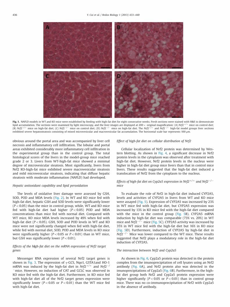
obvious around the portal area and was accompanied by liver cellnecrosis and inflammatory cell infiltration. The lobular and portalareas exhibited considerably more inflammatory cell infiltration inthe experimental group than in the control group. The totalhistological scores of the livers in the model-group mice reachedgrade 2 or 3. Livers from WT-high-fat mice showed a minimaldegree of microvesicular steatosis. Most significantly, livers fromNrf2 KO-high-fat mice exhibited severe macrovesicular steatosisand mild microvesicular steatosis, indicating that diffuse hepaticsteatosis with moderate inflammation (NAFLD) had developed.
Hepatic antioxidant capability and lipid peroxidation
The levels of oxidative liver damage were assessed by GSH,SOD, POD and MDA levels (Fig. 2). In WT and KO mice fed withhigh-fat diet, hepatic GSH and SOD levels were significantly lower(Po0.05) than the mice in control group, while, WT and KO micefed with high-fat diet had higher (Po0.05) POD and MDAconcentrations than mice fed with normal diet. Compared withWT mice, KO mice MDA levels increased by 49% when fed withhigh-fat diet (Po0.05). GSH, SOD and POD levels in WT and KOmice were not significantly changed when fed with high-fat diet,while fed with normal diet, SOD, POD and MDA levels in KO micewere significantly higher (Po0.05 or Po0.01) than in WT mice,but GSH was significantly lower (Po0.01).
Effects of the high-fat diet on the mRNA expression of Nrf2 targetgenes
Messenger RNA expression of several Nrf2 target genes isshown in Fig. 3. The expression of γ-GCS, Nqo1, GSTA1and HO-1mRNA was induced by the high-fat diet in Nrf2þ /þ and Nrf2� /
�mice. However, no induction of CAT and GCLC was observed inKO mice fed with the high-fat diet. Furthermore, in KO mice fedwith high-fat diet all of the Nrf2 target genes expression weresignificantly lower (Po0.05 or Po0.01) than the WT mice fedwith high-fat diet.
Effect of high-fat diet on cellular distribution of Nrf2
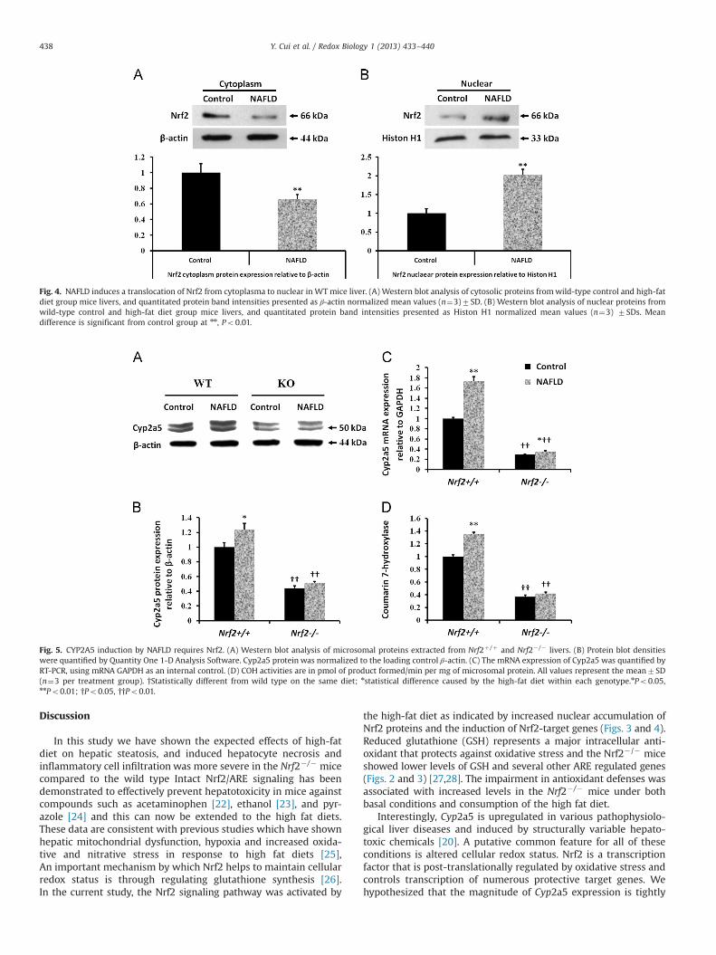
Cellular localization of Nrf2 protein was determined by Wes-tern blotting. As shown in Fig. 4, a significant decrease in Nrf2protein levels in the cytoplasm was observed after treatment withhigh-fat diet. However, Nrf2 protein levels in the nucleus werehigher in high-fat diet group mice livers than that in control micelivers. These results suggested that the high-fat diet induced atranslocation of Nrf2 from the cytoplasm to the nucleus.
Effects of high-fat diet on Cyp2a5 expression in Nrf2þ /þ and Nrf2� /�
mice
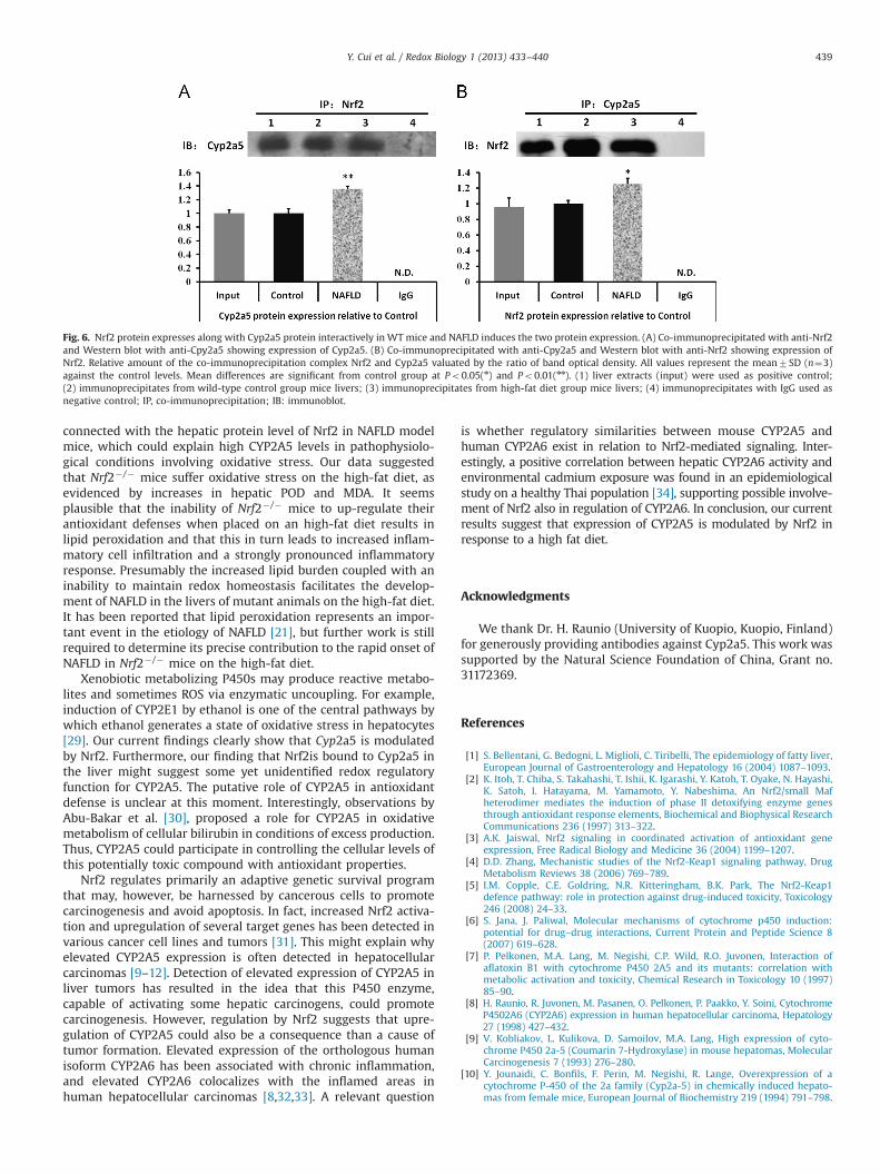
To evaluate the role of Nrf2 in high-fat diet induced CYP2A5,levels and activities of CYP2A5 in livers from WT and KO micewere assayed (Fig. 5). Expression of CYP2A5 was increased by 23%in WT mice fed with high-fat diet, but CYP2A5 expression wasincreased by 13% in KO mice fed with the high-fat diet comparedwith the mice in the control group (Fig. 5B). CYP2A5 mRNAinduction by high-fat diet was comparable (73% vs. 20%) in WTmice and Nrf2� /� mice (Fig. 5C). CYP2A5 activity was increased by35% in WT mice fed with the high-fat diet but 16% in KO mice(Fig. 5D). Furthermore, induction of CYP2A5 by high-fat diet inNrf2� /� Mice was lower compared to the WT mice. These resultssuggested that Nrf2 plays a modulatory role in the high-fat dietinduction of CYP2A5.
The interaction between Nrf2 and Cyp2a5
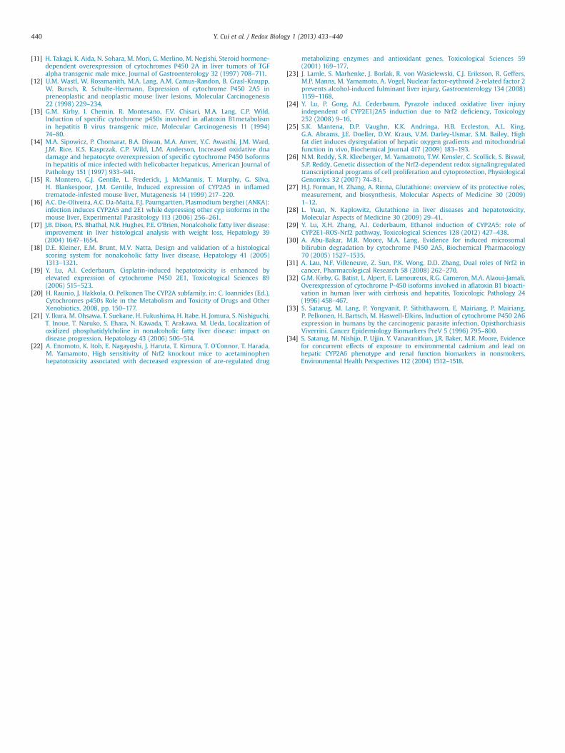
As shown in Fig. 6, Cyp2a5 protein was detected in the proteincomplex from the imunoprecipitation of cell lysates using an Nrf2antibody (Fig. 6A), and Nrf2 protein also was detected in theimunoprecipitations of Cyp2a5 (Fig. 6B). Furthermore, in the high-fat diet group both Nrf2 and Cyp2a5 protein expression werehigher significantly (Po0.05 or Po0.01) than in control groupmice. There was no co-immunoprecipitation of Nrf2 with Cyp2a5in the absence of antibody.
Fig. 1. NAFLD models in WT and KO mice were established by feeding with high-fat diet for eight consecutive weeks. Fresh sections were stained with H&E to demonstratelipid accumulation. The sections were examined by light microscopy, and the liver images are displayed at 200� original magnification: (A) Nrf2þ /þ mice on control diet;(B) Nrf2þ /þ mice on high-fat diet; (C) Nrf2� /� mice on control diet; (D) Nrf2� /� mice on high-fat diet. The Nrf2þ /þ and Nrf2� /� high-fat model groups liver sectionsexhibited severe hepatosteatosis consisting of mixed microvesicular and macrovesicular fat accumulation. The horizontal scale bar represents 100 μm.
Y. Cui et al. / Redox Biology 1 (2013) 433–440436
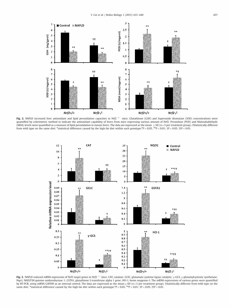
Fig. 2. NAFLD increased liver antioxidant and lipid peroxidation capacities in Nrf2� /� mice. Glutathione (GSH) and Superoxide dismutase (SOD) concentrations werequantified by colorimetric method to indicate the antioxidant capability of livers from mice expressing various amount of Nrf2. Peroxidase (POD) and Malondialdehyde(MDA) levels were quantified as a measure of lipid peroxidation in mouse livers. The data are expressed as the mean 7SD (n¼3 per treatment group). †Statistically differentfrom wild type on the same diet; nstatistical difference caused by the high-fat diet within each genotype.nPo0.05, nnPo0.01; †Po0.05, ††Po0.01.
Fig. 3. NAFLD reduced mRNA expression of Nrf2 target genes in Nrf2� /�mice. CAT, catalase; GClC, glutamate cysteine ligase catalytic; γ-GCS, γ-glutamylcysteine synthetase;Nqo1, NAD(P)H:quinine oxidoreductase 1; GSTA1, glutathione S-transferase alpha 1 gene; HO-1, heme oxygense-1. The mRNA expressions of various genes were quantifiedby RT-PCR, using mRNA GAPDH as an internal control. The data are expressed as the mean7SD (n¼3 per treatment group). †Statistically different from wild type on thesame diet; nstatistical difference caused by the high-fat diet within each genotype.nPo0.05, nnPo0.01; †Po0.05, ††Po0.01.
Y. Cui et al. / Redox Biology 1 (2013) 433–440 437
Discussion
In this study we have shown the expected effects of high-fatdiet on hepatic steatosis, and induced hepatocyte necrosis andinflammatory cell infiltration was more severe in the Nrf2� /� micecompared to the wild type Intact Nrf2/ARE signaling has beendemonstrated to effectively prevent hepatotoxicity in mice againstcompounds such as acetaminophen [22], ethanol [23], and pyr-azole [24] and this can now be extended to the high fat diets.These data are consistent with previous studies which have shownhepatic mitochondrial dysfunction, hypoxia and increased oxida-tive and nitrative stress in response to high fat diets [25],An important mechanism by which Nrf2 helps to maintain cellularredox status is through regulating glutathione synthesis [26].In the current study, the Nrf2 signaling pathway was activated by
the high-fat diet as indicated by increased nuclear accumulation ofNrf2 proteins and the induction of Nrf2-target genes (Figs. 3 and 4).Reduced glutathione (GSH) represents a major intracellular anti-oxidant that protects against oxidative stress and the Nrf2� /� miceshowed lower levels of GSH and several other ARE regulated genes(Figs. 2 and 3) [27,28]. The impairment in antioxidant defenses wasassociated with increased levels in the Nrf2� /� mice under bothbasal conditions and consumption of the high fat diet.
Interestingly, Cyp2a5 is upregulated in various pathophysiolo-gical liver diseases and induced by structurally variable hepato-toxic chemicals [20]. A putative common feature for all of theseconditions is altered cellular redox status. Nrf2 is a transcriptionfactor that is post-translationally regulated by oxidative stress andcontrols transcription of numerous protective target genes. Wehypothesized that the magnitude of Cyp2a5 expression is tightly
Fig. 4. NAFLD induces a translocation of Nrf2 from cytoplasma to nuclear in WT mice liver. (A) Western blot analysis of cytosolic proteins fromwild-type control and high-fatdiet group mice livers, and quantitated protein band intensities presented as β-actin normalized mean values (n¼3)7SD. (B) Western blot analysis of nuclear proteins fromwild-type control and high-fat diet group mice livers, and quantitated protein band intensities presented as Histon H1 normalized mean values (n¼3) 7SDs. Meandifference is significant from control group at nn, Po0.01.
Fig. 5. CYP2A5 induction by NAFLD requires Nrf2. (A) Western blot analysis of microsomal proteins extracted from Nrf2þ /þ and Nrf2� /� livers. (B) Protein blot densitieswere quantified by Quantity One 1-D Analysis Software. Cyp2a5 protein was normalized to the loading control β-actin. (C) The mRNA expression of Cyp2a5 was quantified byRT-PCR, using mRNA GAPDH as an internal control. (D) COH activities are in pmol of product formed/min per mg of microsomal protein. All values represent the mean7SD(n¼3 per treatment group). †Statistically different from wild type on the same diet; nstatistical difference caused by the high-fat diet within each genotype.nPo0.05,nnPo0.01; †Po0.05, ††Po0.01.
Y. Cui et al. / Redox Biology 1 (2013) 433–440438
connected with the hepatic protein level of Nrf2 in NAFLD modelmice, which could explain high CYP2A5 levels in pathophysiolo-gical conditions involving oxidative stress. Our data suggestedthat Nrf2� /� mice suffer oxidative stress on the high-fat diet, asevidenced by increases in hepatic POD and MDA. It seemsplausible that the inability of Nrf2� /� mice to up-regulate theirantioxidant defenses when placed on an high-fat diet results inlipid peroxidation and that this in turn leads to increased inflam-matory cell infiltration and a strongly pronounced inflammatoryresponse. Presumably the increased lipid burden coupled with aninability to maintain redox homeostasis facilitates the develop-ment of NAFLD in the livers of mutant animals on the high-fat diet.It has been reported that lipid peroxidation represents an impor-tant event in the etiology of NAFLD [21], but further work is stillrequired to determine its precise contribution to the rapid onset ofNAFLD in Nrf2� /� mice on the high-fat diet.
Xenobiotic metabolizing P450s may produce reactive metabo-lites and sometimes ROS via enzymatic uncoupling. For example,induction of CYP2E1 by ethanol is one of the central pathways bywhich ethanol generates a state of oxidative stress in hepatocytes[29]. Our current findings clearly show that Cyp2a5 is modulatedby Nrf2. Furthermore, our finding that Nrf2is bound to Cyp2a5 inthe liver might suggest some yet unidentified redox regulatoryfunction for CYP2A5. The putative role of CYP2A5 in antioxidantdefense is unclear at this moment. Interestingly, observations byAbu-Bakar et al. [30], proposed a role for CYP2A5 in oxidativemetabolism of cellular bilirubin in conditions of excess production.Thus, CYP2A5 could participate in controlling the cellular levels ofthis potentially toxic compound with antioxidant properties.
Nrf2 regulates primarily an adaptive genetic survival programthat may, however, be harnessed by cancerous cells to promotecarcinogenesis and avoid apoptosis. In fact, increased Nrf2 activa-tion and upregulation of several target genes has been detected invarious cancer cell lines and tumors [31]. This might explain whyelevated CYP2A5 expression is often detected in hepatocellularcarcinomas [9–12]. Detection of elevated expression of CYP2A5 inliver tumors has resulted in the idea that this P450 enzyme,capable of activating some hepatic carcinogens, could promotecarcinogenesis. However, regulation by Nrf2 suggests that upre-gulation of CYP2A5 could also be a consequence than a cause oftumor formation. Elevated expression of the orthologous humanisoform CYP2A6 has been associated with chronic inflammation,and elevated CYP2A6 colocalizes with the inflamed areas inhuman hepatocellular carcinomas [8,32,33]. A relevant question
is whether regulatory similarities between mouse CYP2A5 andhuman CYP2A6 exist in relation to Nrf2-mediated signaling. Inter-estingly, a positive correlation between hepatic CYP2A6 activity andenvironmental cadmium exposure was found in an epidemiologicalstudy on a healthy Thai population [34], supporting possible involve-ment of Nrf2 also in regulation of CYP2A6. In conclusion, our currentresults suggest that expression of CYP2A5 is modulated by Nrf2 inresponse to a high fat diet.
Acknowledgments
We thank Dr. H. Raunio (University of Kuopio, Kuopio, Finland)for generously providing antibodies against Cyp2a5. This work wassupported by the Natural Science Foundation of China, Grant no.31172369.
References
[1] S. Bellentani, G. Bedogni, L. Miglioli, C. Tiribelli, The epidemiology of fatty liver,European Journal of Gastroenterology and Hepatology 16 (2004) 1087–1093.
[2] K. Itoh, T. Chiba, S. Takahashi, T. Ishii, K. Igarashi, Y. Katoh, T. Oyake, N. Hayashi,K. Satoh, I. Hatayama, M. Yamamoto, Y. Nabeshima, An Nrf2/small Mafheterodimer mediates the induction of phase II detoxifying enzyme genesthrough antioxidant response elements, Biochemical and Biophysical ResearchCommunications 236 (1997) 313–322.
[3] A.K. Jaiswal, Nrf2 signaling in coordinated activation of antioxidant geneexpression, Free Radical Biology and Medicine 36 (2004) 1199–1207.
[4] D.D. Zhang, Mechanistic studies of the Nrf2-Keap1 signaling pathway, DrugMetabolism Reviews 38 (2006) 769–789.
[5] I.M. Copple, C.E. Goldring, N.R. Kitteringham, B.K. Park, The Nrf2-Keap1defence pathway: role in protection against drug-induced toxicity, Toxicology246 (2008) 24–33.
[6] S. Jana, J. Paliwal, Molecular mechanisms of cytochrome p450 induction:potential for drug–drug interactions, Current Protein and Peptide Science 8(2007) 619–628.
[7] P. Pelkonen, M.A. Lang, M. Negishi, C.P. Wild, R.O. Juvonen, Interaction ofaflatoxin B1 with cytochrome P450 2A5 and its mutants: correlation withmetabolic activation and toxicity, Chemical Research in Toxicology 10 (1997)85–90.
[8] H. Raunio, R. Juvonen, M. Pasanen, O. Pelkonen, P. Paakko, Y. Soini, CytochromeP4502A6 (CYP2A6) expression in human hepatocellular carcinoma, Hepatology27 (1998) 427–432.
[9] V. Kobliakov, L. Kulikova, D. Samoilov, M.A. Lang, High expression of cyto-chrome P450 2a-5 (Coumarin 7-Hydroxylase) in mouse hepatomas, MolecularCarcinogenesis 7 (1993) 276–280.
[10] Y. Jounaidi, C. Bonfils, F. Perin, M. Negishi, R. Lange, Overexpression of acytochrome P-450 of the 2a family (Cyp2a-5) in chemically induced hepato-mas from female mice, European Journal of Biochemistry 219 (1994) 791–798.
Fig. 6. Nrf2 protein expresses along with Cyp2a5 protein interactively in WT mice and NAFLD induces the two protein expression. (A) Co-immunoprecipitated with anti-Nrf2and Western blot with anti-Cpy2a5 showing expression of Cyp2a5. (B) Co-immunoprecipitated with anti-Cpy2a5 and Western blot with anti-Nrf2 showing expression ofNrf2. Relative amount of the co-immunoprecipitation complex Nrf2 and Cyp2a5 valuated by the ratio of band optical density. All values represent the mean7SD (n¼3)against the control levels. Mean differences are significant from control group at Po0.05(n) and Po0.01(nn). (1) liver extracts (input) were used as positive control;(2) immunoprecipitates from wild-type control group mice livers; (3) immunoprecipitates from high-fat diet group mice livers; (4) immunoprecipitates with IgG used asnegative control; IP, co-immunoprecipitation; IB: immunoblot.
Y. Cui et al. / Redox Biology 1 (2013) 433–440 439
[11] H. Takagi, K. Aida, N. Sohara, M. Mori, G. Merlino, M. Negishi, Steroid hormone-dependent overexpression of cytochromes P450 2A in liver tumors of TGFalpha transgenic male mice, Journal of Gastroenterology 32 (1997) 708–711.
[12] U.M. Wastl, W. Rossmanith, M.A. Lang, A.M. Camus-Randon, B. Grasl-Kraupp,W. Bursch, R. Schulte-Hermann, Expression of cytochrome P450 2A5 inpreneoplastic and neoplastic mouse liver lesions, Molecular Carcinogenesis22 (1998) 229–234.
[13] G.M. Kirby, I. Chemin, R. Montesano, F.V. Chisari, M.A. Lang, C.P. Wild,Induction of specific cytochrome p450s involved in aflatoxin B1metabolismin hepatitis B virus transgenic mice, Molecular Carcinogenesis 11 (1994)74–80.
[14] M.A. Sipowicz, P. Chomarat, B.A. Diwan, M.A. Anver, Y.C. Awasthi, J.M. Ward,J.M. Rice, K.S. Kasprzak, C.P. Wild, L.M. Anderson, Increased oxidative dnadamage and hepatocyte overexpression of specific cytochrome P450 Isoformsin hepatitis of mice infected with helicobacter hepaticus, American Journal ofPathology 151 (1997) 933–941.
[15] R. Montero, G.J. Gentile, L. Frederick, J. McMannis, T. Murphy, G. Silva,H. Blankespoor, J.M. Gentile, Induced expression of CYP2A5 in inflamedtrematode-infested mouse liver, Mutagenesis 14 (1999) 217–220.
[16] A.C. De-Oliveira, A.C. Da-Matta, F.J. Paumgartten, Plasmodium berghei (ANKA):infection induces CYP2A5 and 2E1 while depressing other cyp isoforms in themouse liver, Experimental Parasitology 113 (2006) 256–261.
[17] J.B. Dixon, P.S. Bhathal, N.R. Hughes, P.E. O'Brien, Nonalcoholic fatty liver disease:improvement in liver histological analysis with weight loss, Hepatology 39(2004) 1647–1654.
[18] D.E. Kleiner, E.M. Brunt, M.V. Natta, Design and validation of a histologicalscoring system for nonalcoholic fatty liver disease, Hepatology 41 (2005)1313–1321.
[19] Y. Lu, A.I. Cederbaum, Cisplatin-induced hepatotoxicity is enhanced byelevated expression of cytochrome P450 2E1, Toxicological Sciences 89(2006) 515–523.
[20] H. Raunio, J. Hakkola, O. Pelkonen The CYP2A subfamily, in: C. Ioannides (Ed.),Cytochromes p450s Role in the Metabolism and Toxicity of Drugs and OtherXenobiotics, 2008, pp. 150–177.
[21] Y. Ikura, M. Ohsawa, T. Suekane, H. Fukushima, H. Itabe, H. Jomura, S. Nishiguchi,T. Inoue, T. Naruko, S. Ehara, N. Kawada, T. Arakawa, M. Ueda, Localization ofoxidized phosphatidylcholine in nonalcoholic fatty liver disease: impact ondisease progression, Hepatology 43 (2006) 506–514.
[22] A. Enomoto, K. Itoh, E. Nagayoshi, J. Haruta, T. Kimura, T. O'Connor, T. Harada,M. Yamamoto, High sensitivity of Nrf2 knockout mice to acetaminophenhepatotoxicity associated with decreased expression of are-regulated drug
metabolizing enzymes and antioxidant genes, Toxicological Sciences 59(2001) 169–177.
[23] J. Lamle, S. Marhenke, J. Borlak, R. von Wasielewski, C.J. Eriksson, R. Geffers,M.P. Manns, M. Yamamoto, A. Vogel, Nuclear factor-eythroid 2-related factor 2prevents alcohol-induced fulminant liver injury, Gastroenterology 134 (2008)1159–1168.
[24] Y. Lu, P. Gong, A.I. Cederbaum, Pyrazole induced oxidative liver injuryindependent of CYP2E1/2A5 induction due to Nrf2 deficiency, Toxicology252 (2008) 9–16.
[25] S.K. Mantena, D.P. Vaughn, K.K. Andringa, H.B. Eccleston, A.L. King,G.A. Abrams, J.E. Doeller, D.W. Kraus, V.M. Darley-Usmar, S.M. Bailey, Highfat diet induces dysregulation of hepatic oxygen gradients and mitochondrialfunction in vivo, Biochemical Journal 417 (2009) 183–193.
[26] N.M. Reddy, S.R. Kleeberger, M. Yamamoto, T.W. Kensler, C. Scollick, S. Biswal,S.P. Reddy, Genetic dissection of the Nrf2-dependent redox signalingregulatedtranscriptional programs of cell proliferation and cytoprotection, PhysiologicalGenomics 32 (2007) 74–81.
[27] H.J. Forman, H. Zhang, A. Rinna, Glutathione: overview of its protective roles,measurement, and biosynthesis, Molecular Aspects of Medicine 30 (2009)1–12.
[28] L. Yuan, N. Kaplowitz, Glutathione in liver diseases and hepatotoxicity,Molecular Aspects of Medicine 30 (2009) 29–41.
[29] Y. Lu, X.H. Zhang, A.I. Cederbaum, Ethanol induction of CYP2A5: role ofCYP2E1-ROS-Nrf2 pathway, Toxicological Sciences 128 (2012) 427–438.
[30] A. Abu-Bakar, M.R. Moore, M.A. Lang, Evidence for induced microsomalbilirubin degradation by cytochrome P450 2A5, Biochemical Pharmacology70 (2005) 1527–1535.
[31] A. Lau, N.F. Villeneuve, Z. Sun, P.K. Wong, D.D. Zhang, Dual roles of Nrf2 incancer, Pharmacological Research 58 (2008) 262–270.
[32] G.M. Kirby, G. Batist, L. Alpert, E. Lamoureux, R.G. Cameron, M.A. Alaoui-Jamali,Overexpression of cytochrome P-450 isoforms involved in aflatoxin B1 bioacti-vation in human liver with cirrhosis and hepatitis, Toxicologic Pathology 24(1996) 458–467.
[33] S. Satarug, M. Lang, P. Yongvanit, P. Sithithaworn, E. Mairiang, P. Mairiang,P. Pelkonen, H. Bartsch, M. Haswell-Elkins, Induction of cytochrome P450 2A6expression in humans by the carcinogenic parasite infection, OpisthorchiasisViverrini. Cancer Epidemiology Biomarkers PreV 5 (1996) 795–800.
[34] S. Satarug, M. Nishijo, P. Ujjin, Y. Vanavanitkun, J.R. Baker, M.R. Moore, Evidencefor concurrent effects of exposure to environmental cadmium and lead onhepatic CYP2A6 phenotype and renal function biomarkers in nonsmokers,Environmental Health Perspectives 112 (2004) 1512–1518.
Y. Cui et al. / Redox Biology 1 (2013) 433–440440