Experimental infection of hamsters with avian paramyxovirus
12
RESEARCH Open Access Experimental infection of hamsters with avian paramyxovirus serotypes 1 to 9 Arthur S Samuel 1 , Madhuri Subbiah 1 , Heather Shive 2 , Peter L Collins 3 , Siba K Samal 1* Abstract Avian paramyxoviruses (APMVs) are frequently isolated from domestic and wild birds throughout the world and are separated into nine serotypes (APMV-1 to -9). Only in the case of APMV-1, the infection of non-avian species has been investigated. The APMVs presently are being considered as human vaccine vectors. In this study, we evaluated the replication and pathogenicity of all nine APMV serotypes in hamsters. The hamsters were inoculated intranasally with each virus and monitored for clinical disease, pathology, histopathology, virus replication, and seroconversion. On the basis of one or more of these criteria, each of the APMV serotypes was found to replicate in hamsters. The APMVs produced mild or inapparent clinical signs in hamsters except for APMV-9, which produced moderate disease. Gross lesions were observed over the pulmonary surface of hamsters infected with APMV-2 & -3, which showed petechial and ecchymotic hemorrhages, respectively. Replication of all of the APMVs except APMV-5 was confirmed in the nasal turbinates and lungs, indicating a tropism for the respiratory tract. Histologically, the infection resulted in lung lesions consistent with bronchointerstitial pneumonia of varying severity and nasal turbinates with blunting or loss of cilia of the epithelium lining the nasal septa. The majority of APMV-infected hamsters exhibited transient histological lesions that self resolved by 14 days post infection (dpi). All of the hamsters infected with the APMVs produced serotype-specific HI or neutralizing antibodies, confirming virus replication. Taken together, these results demonstrate that all nine known APMV serotypes are capable of replicating in hamsters with minimal disease and pathology. Introduction Paramyxoviridae is a large and diverse family whose members have been isolated from many species of avian, terrestrial, and aquatic animal species around the world [1]. Paramyxoviruses are pleomorphic, enveloped, cytoplasmic viruses that have a non-segmented, nega- tive-sense RNA genome. The family is divided into two subfamilies, Paramyxovirinae and Pneumovirinae, based on their structure, genome organization, and sequence relatedness [2]. The subfamily Paramyxovirinae contains five genera: Respirovirus, Rubulavirus, Morbillivirus, Henipavirus, and Avulavirus, while the subfamily Pneu- movirinae contains two genera, Pneumovirus and Metapneumovirus [3]. All paramyxoviruses that have been isolated to date from avian species can be segre- gated into two genera based on the taxonomic criteria mentioned above: genus Avulavirus, whose members are called the avian paramyxoviruses (APMV), and genus Metapneumovirus , whose members are called avian metapneumoviruses. The APMV of genus Avulavirus are separated into nine serotypes (APMV-1 through -9) based on Hemagglutination Inhibition (HI) and Neura- minidase Inhibition (NI) assays [4]. Various strains of APMV-1, which is also called Newcastle disease virus (NDV), have been analyzed in detail by biochemical ana- lysis, genome sequencing, and pathogenesis studies, and important molecular determinants of virulence have been identified [5-9]. As a first step in characterizing the other APMV serotypes, complete genome sequences of one or more representative strains of APMV serotypes 2 to 9 were recently determined, expanding our knowl- edge about these viruses [10-18]. APMV-1 comprises all strains of NDV and is the best characterized serotype because of the severity of disease caused by virulent NDV strains in chickens. NDV strains vary greatly in their pathogenicity to chickens and are grouped into three pathotypes: highly virulent (velogenic) strains, which cause severe respiratory and * Correspondence: [email protected] 1 Virginia-Maryland Regional College of Veterinary Medicine, University of Maryland, College Park, Maryland, USA Full list of author information is available at the end of the article Samuel et al. Veterinary Research 2011, 42:38 http://www.veterinaryresearch.org/content/42/1/38 VETERINARY RESEARCH © 2011 Samuel et al; licensee BioMed Central Ltd. This is an Open Access article distributed under the terms of the Creative Commons Attribution License (http://creativecommons.org/licenses/by/2.0), which permits unrestricted use, distribution, and reproduction in any medium, provided the original work is properly cited.
Transcript of Experimental infection of hamsters with avian paramyxovirus
RESEARCH Open Access
Experimental infection of hamsters with avianparamyxovirus serotypes 1 to 9Arthur S Samuel1, Madhuri Subbiah1, Heather Shive2, Peter L Collins3, Siba K Samal1*
Abstract
Avian paramyxoviruses (APMVs) are frequently isolated from domestic and wild birds throughout the world and areseparated into nine serotypes (APMV-1 to -9). Only in the case of APMV-1, the infection of non-avian species hasbeen investigated. The APMVs presently are being considered as human vaccine vectors. In this study, weevaluated the replication and pathogenicity of all nine APMV serotypes in hamsters. The hamsters were inoculatedintranasally with each virus and monitored for clinical disease, pathology, histopathology, virus replication, andseroconversion. On the basis of one or more of these criteria, each of the APMV serotypes was found to replicatein hamsters. The APMVs produced mild or inapparent clinical signs in hamsters except for APMV-9, whichproduced moderate disease. Gross lesions were observed over the pulmonary surface of hamsters infected withAPMV-2 & -3, which showed petechial and ecchymotic hemorrhages, respectively. Replication of all of the APMVsexcept APMV-5 was confirmed in the nasal turbinates and lungs, indicating a tropism for the respiratory tract.Histologically, the infection resulted in lung lesions consistent with bronchointerstitial pneumonia of varyingseverity and nasal turbinates with blunting or loss of cilia of the epithelium lining the nasal septa. The majority ofAPMV-infected hamsters exhibited transient histological lesions that self resolved by 14 days post infection (dpi). Allof the hamsters infected with the APMVs produced serotype-specific HI or neutralizing antibodies, confirming virusreplication. Taken together, these results demonstrate that all nine known APMV serotypes are capable ofreplicating in hamsters with minimal disease and pathology.
IntroductionParamyxoviridae is a large and diverse family whosemembers have been isolated from many species ofavian, terrestrial, and aquatic animal species around theworld [1]. Paramyxoviruses are pleomorphic, enveloped,cytoplasmic viruses that have a non-segmented, nega-tive-sense RNA genome. The family is divided into twosubfamilies, Paramyxovirinae and Pneumovirinae, basedon their structure, genome organization, and sequencerelatedness [2]. The subfamily Paramyxovirinae containsfive genera: Respirovirus, Rubulavirus, Morbillivirus,Henipavirus, and Avulavirus, while the subfamily Pneu-movirinae contains two genera, Pneumovirus andMetapneumovirus [3]. All paramyxoviruses that havebeen isolated to date from avian species can be segre-gated into two genera based on the taxonomic criteriamentioned above: genus Avulavirus, whose members are
called the avian paramyxoviruses (APMV), and genusMetapneumovirus, whose members are called avianmetapneumoviruses. The APMV of genus Avulavirusare separated into nine serotypes (APMV-1 through -9)based on Hemagglutination Inhibition (HI) and Neura-minidase Inhibition (NI) assays [4]. Various strains ofAPMV-1, which is also called Newcastle disease virus(NDV), have been analyzed in detail by biochemical ana-lysis, genome sequencing, and pathogenesis studies, andimportant molecular determinants of virulence havebeen identified [5-9]. As a first step in characterizing theother APMV serotypes, complete genome sequences ofone or more representative strains of APMV serotypes 2to 9 were recently determined, expanding our knowl-edge about these viruses [10-18].APMV-1 comprises all strains of NDV and is the best
characterized serotype because of the severity of diseasecaused by virulent NDV strains in chickens. NDVstrains vary greatly in their pathogenicity to chickensand are grouped into three pathotypes: highly virulent(velogenic) strains, which cause severe respiratory and
* Correspondence: [email protected] Regional College of Veterinary Medicine, University ofMaryland, College Park, Maryland, USAFull list of author information is available at the end of the article
Samuel et al. Veterinary Research 2011, 42:38http://www.veterinaryresearch.org/content/42/1/38 VETERINARY RESEARCH
© 2011 Samuel et al; licensee BioMed Central Ltd. This is an Open Access article distributed under the terms of the Creative CommonsAttribution License (http://creativecommons.org/licenses/by/2.0), which permits unrestricted use, distribution, and reproduction inany medium, provided the original work is properly cited.
neurological disease in chickens; moderately virulent(mesogenic) strains, which cause mild disease; and non-pathogenic (lentogenic) strains, which cause inapparentinfections. In contrast, very little is known about thecomparative disease potential of APMV-2 to APMV-9in domestic and wild birds. APMV-2 strains have beenisolated from chickens, turkeys and wild birds across theglobe [4,19-22]. APMV-2 infections in turkeys havebeen found to cause mild respiratory disease, decreasesin egg production, and infertility [23,24]. APMV-3strains have been isolated from wild and domestic birds[25]. APMV-3 infections have been associated withencephalitis and high mortality in caged birds [26-28].APMV-4 strains have been isolated from chickens,ducks and geese [29]. Experimental infection of chickenswith APMV-4 resulted in mild interstitial pneumoniaand catarrhal tracheitis [30]. APMV-5 strains have onlybeen isolated from budgerigars (Melopsittacus undula-tus) and cause depression, dyspnoea, diarrhea, torticollis,and acute fatal enteritis in immature budgerigars, lead-ing to very high mortality [31]. APMV-6 was first iso-lated from a domestic duck and was found to causemild respiratory disease and drop in egg production inturkeys, but was avirulent in chickens [10,30,32].APMV-7 was first isolated from a hunter-killed doveand has also been isolated from a natural outbreak ofrespiratory disease in turkeys. APMV-7 infection in tur-keys caused respiratory disease, mild multifocal nodularlymphocytic airsacculitis, and decreased egg production[33]. APMV-8 was isolated from a goose and a feral pin-tail duck [34,35]. APMV-9 strains have been isolatedfrom ducks around the world [36,37]. APMV types -2,-3, and -7 have been associated with mild respiratorydisease and egg production problems in domestic chick-ens [33]. There are no reports of isolation of APMV-5,-8 and -9 from poultry [32]. But recent serosurveillanceof commercial poultry farms in USA indicated the possi-ble prevalence of all APMV serotypes excluding APMV-5 in chickens [38].APMV-1 (NDV) is known to replicate in non-avian
species including humans [39-44], although its only nat-ural hosts are birds. APMV-1 infections in non-avianspecies are usually asymptomatic or mild. Clinical signsin human infections commonly involve conjunctivitis,which usually is transient and self-limiting. Presently,APMV-1 is being evaluated as a vaccine vector againsthuman pathogens [45]. When administered to therespiratory tract of non-human primates, NDV is highlyrestricted in replication, but foreign antigens expressedby recombinant NDV vectors are moderately to highlyimmunogenic. One of the major advantages of thisapproach is that most humans do not have pre-existingimmunity to APMV-1. Pre-existing immunity is apotential drawback to using vectors derived from
common human pathogens, and also can be a concernfor any vector if two or more doses are necessary to eli-cit protective immunity. Therefore, we are investigatingAPMV types 2 to 9, which are antigenically distinctfrom APMV-1, as alternative human vaccine vectors.Also, some of these additional APMV types likely willhave differences in replication, attenuation, and immu-nogenicity compared to APMV-1 that may be advanta-geous. However, the replication and pathogenicity ofAPMV-2 to -9 in non-avian species has not been stu-died. As a first step, we have evaluated the replicationand pathogenicity of APMV-2 to -9 in hamsters. In thisstudy, groups of hamsters were infected with a proto-type strain of each APMV serotype by the intranasalroute and monitored for virus replication, clinical symp-toms, histopathology, and seroconversion. Our resultsshowed that each of the APMV serotypes replicated inhamsters without causing adverse clinical signs of ill-ness, although histopathologic evidence of disease wasobserved in some cases, and also induced high neutraliz-ing antibody titers.
Materials and methodsViruses and cellsThe following nine prototype strains of APMV serotypes1 to 9 were used, APMV-1, NDV lentogenic strainLaSota/46; APMV-2, APMV-2/Chicken/California/Yucaipa/56; APMV-3, APMV-3/PKT/Netherland/449/75; APMV-4, APMV-4/duck/HongKong/D3/75; APMV-5, APMV-5/budgerigar/Kunitachi/74; APMV-6, APMV-6/duck/HongKong/18/199/77; APMV-7, APMV-7/dove/Tennessee/4/75; APMV-8, APMV-8/goose/Delaware/1053/76; APMV-9, APMV-9/duck/New York/22/1978.All of the viruses were grown in 9-day-old embryonatedspecific-pathogen-free (SPF) chicken eggs inoculated bythe allantoic route, except for APMV-5, which wasgrown in African green monkey kidney (Vero) cells(ATCC, Manassas, VA, USA) [17]. The allantoic fluidsfrom infected eggs were collected 96 h post-inoculationand virus titers were determined by hemagglutination(HA) assay with 0.5% chicken RBC except in the case ofAPMV-5, which was titrated by plaque assay on Verocells. The APMV-5 samples were inoculated in triplicateonto 24-well plates of Vero cells at 80% confluency,incubated for 1 h, washed with PBS, overlaid with 0.8%methylcellulose, and observed for plaque production till7 days post infection (dpi). The cells were fixed withmethanol and stained with 1% crystal violet. Values foreach tissue sample were based on average plaque countfrom three wells. Vero cells were grown in Earle’s mini-mum essential medium (EMEM) with 10% fetal bovineserum (FBS). Chicken embryo fibroblast (DF-1) cellswere grown in Dulbecco’s minimal essential medium(DMEM) containing 10% FBS at 37°C with 5% CO2.
Samuel et al. Veterinary Research 2011, 42:38http://www.veterinaryresearch.org/content/42/1/38
Page 2 of 12
Preparation of hyperimmune antisera against viralnucleocapsid (N) proteins in rabbitsFor each of the prototype strains, virions were purifiedon discontinuous sucrose gradients [12]. The viral pro-teins were denatured and reduced and were separatedon 10% SDS-Polyacrylamide gels and negatively stainedusing E-Zinc ™reversible stain kit (Pierce, Rockford, IL,USA). Each serotype was analyzed on a separate gel toavoid cross contamination. The N protein bands wereexcised from the gels and destained with Tris-glycinebuffer, pH 8. The excised gel bands were minced in aclean pestle and mixed with elution buffer (50 mMTris-HCl buffer pH 8, 150 mM NaCl, 0.5 mM EDTA,5 mM DTT and 0.1% SDS) and transferred to the upperchambers of Nanosep centrifugal device (Pall LifeSciences, Ann Arbor, MI, USA). The proteins wereeluted by centrifugation at 13000 × g for 5 minutes, andthe eluted proteins were quantified and 200 μg of eachprotein was mixed in complete Freund’s adjuvant andinjected subcutaneously into a rabbit. After two weeks, abooster immunization was given with 200 μg of proteinin incomplete Freund’s adjuvant and two weeks later thehyperimmune sera were collected. The sera were testedby western blot and were found to recognize specificallythe homologous N protein (data not shown). The serawere stored at -80°C until further use.
Experimental infection of hamstersTo study viral replication and pathogenicity, fifty seven4-week-old Syrian golden hamsters (Charles RiverLaboratories Inc, Wilmington, MA, USA) were housedin negative-pressure isolators under Bio Safety Level(BSL)-2 conditions and provided feed and water ad libi-tum. The animals were cared for in accordance with theAnimal Welfare Act and the Guide for Care and Use ofLaboratory Animals and the protocols were approved bythe institution’s IACUC. Hamsters in groups of six wereinoculated intranasally with 100 μL of infectious allan-toic fluid containing 28 HAU of each individual APMV,except for APMV-5, which contained 3 × 10³ PFU/mL,under isoflurane anesthesia. A group of three hamstersserved as uninfected controls and were mock infectedwith normal allantoic fluid. The hamsters were observedthree times daily for physical activity and for any clinicalsigns of illness, and were weighed on day 0, 5 and14 dpi. Three hamsters from each group were eutha-nized at 3 dpi and the other three (as well as the threeanimals in the control group) at 14 dpi by rapid asphyx-iation in a CO2 chamber. Necropsies were performedimmediately postmortem and the following tissuesamples were collected for immunohistochemistry(IHC), histopathology, and virus isolation: brain, nasalturbinates, lung, spleen, kidney and small intestine.In addition serum samples were collected on 14 dpi
immediately prior to euthanasia, and seroconversion wasevaluated by HI assay [46].
Virus detection and quantification from tissue samplesHalf of each tissue sample was used for virus titration.These samples were collected aseptically in 1 mL ofDMEM in 10X antibiotic solution containing 2000units/mL penicillin, 200 ug/mL gentamicin sulfate, and4 ug/mL amphotericin B (Sigma chemical co., St. Louis,MO, USA). They were processed immediately to avoidany reduction in virus titers. Briefly, a 10% homogenateof the tissue samples were prepared by using a homoge-nizer and clarified by centrifugation at 420 × g for10 min. For all of the serotypes except APMV-5, thevirus titers in the clarified supernatants were determinedby end-point dilution on DF-1 cells. Tenfold dilutions oftissue supernatant were inoculated onto DF-1 cells in96-well plates and incubated for three days. The plateswere fixed in 10% formalin for 30 min and the cellswere permeabilized using 2% Triton X-100 for 2 min.The plates were washed five times with PBS to removeany residual formalin in the wells. The cells wereblocked using 2% normal goat serum for 60 min andthe plates were washed twice with PBS- Tween-20(PBS-T). The cells were incubated with primary rabbitantiserum raised against the N protein of the homolo-gous APMV serotype at room temperature for 1 h. Theplates were washed with PBS twice, 5 min each. Thecells were incubated with anti rabbit FITC antibody assecondary antibody for 45 min and washed finally withPBS twice, 5 min each. The slides were visualized andthe virus titers were determined by end point titrationmethod using the Reed and Muench formula, and werephotomicrographed using a fluorescent microscope(Zeiss Axioshop 2000, Zeiss, Göttingen, Germany) [47].The virus titers in the supernatants of tissue homoge-nates from APMV-5 infected hamsters were determinedby plaque assay in Vero cells as described above using10-fold dilution series. Values for each tissue samplewere based on average plaque count from three wells.
Immunohistochemistry (IHC) and histopathologyThe other half of each tissue sample was used for IHCand histopathology. The tissues were fixed in 10% neu-tral buffered formalin, held for approximately sevendays, and processed for IHC and histopathology. Paraffinembedded 5-micron sections of all the tissue sampleswere prepared at Histoserve, Inc. (Maryland, MD, USA).The sections were stained with hematoxylin and eosinfor histopathology. Sections were also immunostained todetect viral N protein using the following protocol.Briefly, the tissue sections were deparaffinized in twochanges of xylene for 5 min each, hydrated in twochanges of 100% ethanol for 3 min each, changes of
Samuel et al. Veterinary Research 2011, 42:38http://www.veterinaryresearch.org/content/42/1/38
Page 3 of 12
95% and 80% ethanol for 1 min each, and finally washedin distilled water. The sections were processed for anti-gen retrieval in a water bath containing sodium citratebuffer (10 mM citric acid, 0.05% Tween 20, pH 6.0), at95-100°C for 40 min and then allowed to cool to roomtemperature for another 60 min. The sections wererinsed in PBS-Tween 20, twice for 2 min each. The sec-tions were blocked with 2% BSA in PBS for 1 h at roomtemperature. The sections were then incubated with a1:500 dilution of the homologous primary N-specificrabbit antiserum in PBS for 1 h in a humidified cham-ber. After three washes in PBS, the sections were incu-bated with the secondary antibody (FITC conjugatedgoat anti-rabbit antibody) for 30 min. After a furtherwash cycle, the sections were mounted with glyceroland viewed under an immunofluorescence microscope.
Serological analysisSera were collected from all the hamsters on 14 dpiand evaluated for seroconversion by HI assay [46],except in the case of APMV-5. In addition, cross-HItests were performed to investigate cross reactivitywith other APMV serotypes. Since APMV-5 does notcause hemagglutination of chicken RBC, the antibodytiter was determined by plaque reduction neutraliza-tion assay. Briefly, the sera were heat inactivated at56°C for 30 min. Ten-fold dilutions of sera were madeand mixed with a constant amount of APMV-5 (3 ×10³ PFU), and incubated at room temperature for 1 hin a shaker. The antigen antibody mixtures were ana-lyzed by plaque assay as described above. The serumtiter that reduced plaque numbers by 70% was the endpoint titer.
ResultsClinical disease and gross pathologyFour-week-old hamsters in groups of six were inocu-lated by the intranasal route with 28 HA units of each
of the APMV serotypes except APMV-5, which wasadministered at a dose of 3 × 10³ PFU/mL. Three ani-mals were sacrificed at day 3 and the remaining threeanimals were sacrificed on day 14; following sacrifice,the animals were processed for gross pathology, histo-pathology, IHC, quantitative virology, and seroconver-sion. Three uninfected animals served as controls andwere sacrificed and processed on day 14. All of the ani-mals were observed three times daily and weighed daily.None of the hamsters infected with any of the APMVserotypes showed any visible clinical signs of diseaseexcept those infected with APMV-9. All the six ham-sters infected with APMV-9 serotype were dull and hadrough skin coat at 3 dpi. They appeared weak and lostweight (Table 1) till 5 dpi, but later gained body weightand appeared normal by 10 dpi. None of the hamstersdied of disease. The uninfected control hamstersappeared healthy and normal.Following sacrifice on days 3 and 14, the followings
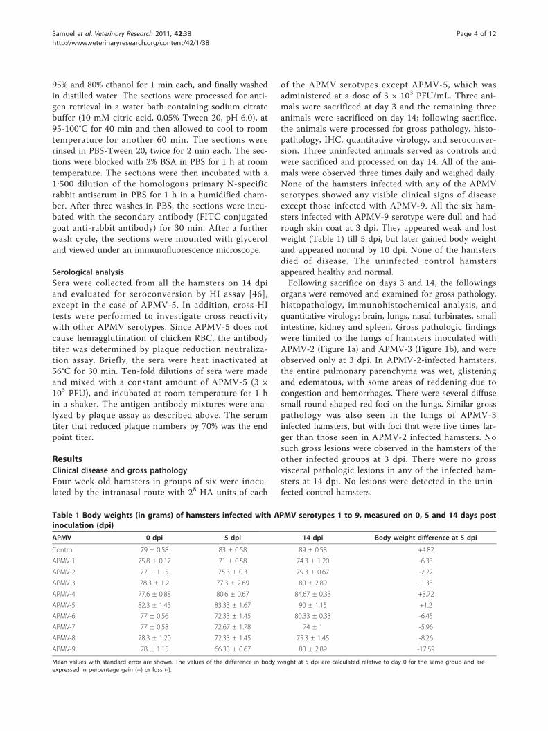
organs were removed and examined for gross pathology,histopathology, immunohistochemical analysis, andquantitative virology: brain, lungs, nasal turbinates, smallintestine, kidney and spleen. Gross pathologic findingswere limited to the lungs of hamsters inoculated withAPMV-2 (Figure 1a) and APMV-3 (Figure 1b), and wereobserved only at 3 dpi. In APMV-2-infected hamsters,the entire pulmonary parenchyma was wet, glisteningand edematous, with some areas of reddening due tocongestion and hemorrhages. There were several diffusesmall round shaped red foci on the lungs. Similar grosspathology was also seen in the lungs of APMV-3infected hamsters, but with foci that were five times lar-ger than those seen in APMV-2 infected hamsters. Nosuch gross lesions were observed in the hamsters of theother infected groups at 3 dpi. There were no grossvisceral pathologic lesions in any of the infected ham-sters at 14 dpi. No lesions were detected in the unin-fected control hamsters.
Table 1 Body weights (in grams) of hamsters infected with APMV serotypes 1 to 9, measured on 0, 5 and 14 days postinoculation (dpi)
APMV 0 dpi 5 dpi 14 dpi Body weight difference at 5 dpi
Control 79 ± 0.58 83 ± 0.58 89 ± 0.58 +4.82
APMV-1 75.8 ± 0.17 71 ± 0.58 74.3 ± 1.20 -6.33
APMV-2 77 ± 1.15 75.3 ± 0.3 79.3 ± 0.67 -2.22
APMV-3 78.3 ± 1.2 77.3 ± 2.69 80 ± 2.89 -1.33
APMV-4 77.6 ± 0.88 80.6 ± 0.67 84.67 ± 0.33 +3.72
APMV-5 82.3 ± 1.45 83.33 ± 1.67 90 ± 1.15 +1.2
APMV-6 77 ± 0.56 72.33 ± 1.45 80.33 ± 0.33 -6.45
APMV-7 77 ± 0.58 72.67 ± 1.78 74 ± 1 -5.96
APMV-8 78.3 ± 1.20 72.33 ± 1.45 75.3 ± 1.45 -8.26
APMV-9 78 ± 1.15 66.33 ± 0.67 80 ± 2.89 -17.59
Mean values with standard error are shown. The values of the difference in body weight at 5 dpi are calculated relative to day 0 for the same group and areexpressed in percentage gain (+) or loss (-).
Samuel et al. Veterinary Research 2011, 42:38http://www.veterinaryresearch.org/content/42/1/38
Page 4 of 12
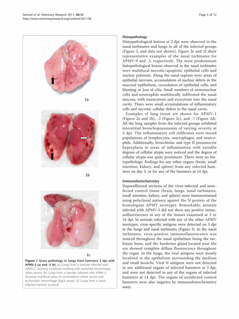
HistopathologyHistopathological lesions at 3 dpi were observed in thenasal turbinates and lungs in all of the infected groups(Figure 2, and data not shown). Figure 2e and 2f showrepresentative examples of the nasal turbinates forAPMV-9 and -3, respectively. The most predominanthistopathological lesions observed in the nasal turbinateswere multifocal necrotic/apoptotic epithelial cells andnuclear pyknosis. Along the nasal septum were areas ofepithelial necrosis, accumulation of nuclear debris in themucosal epithelium, vacuolation of epithelial cells, andblunting or loss of cilia. Small numbers of mononuclearcells and neutrophils multifocally infiltrated the nasalmucosa, with transcytosis and exocytosis into the nasalcavity. There were small accumulations of inflammatorycells and necrotic cellular debris in the nasal cavity.Examples of lung tissue are shown for APMV-1
(Figure 2a and 2b), -2 (Figure 2c), and -7 (Figure 2d).All the lung samples from the infected groups exhibitedinterstitial bronchopneumonia of varying severity at3 dpi. The inflammatory cell infiltrates were mixedpopulations of lymphocytes, macrophages, and neutro-phils. Additionally, bronchiolar and type II pneumocytehyperplasia in areas of inflammation with variabledegrees of cellular atypia were noticed and the degree ofcellular atypia was quite prominent. There were no his-topathologic findings for any other organs (brain, smallintestine, kidney, and spleen) from any infected ham-sters on day 3, or for any of the hamsters at 14 dpi.
ImmunohistochemistryDeparaffinized sections of the virus-infected and unin-fected control tissue (brain, lungs, nasal turbinates,small intestine, kidney, and spleen) were immunostainedusing polyclonal antisera against the N protein of thehomologous APMV serotypes. Remarkably, animalsinfected with APMV-5 did not show any positive immu-nofluorescence in any of the tissues examined at 3 or14 dpi. In animals infected with any of the other APMVserotypes, virus-specific antigens were detected on 3 dpiin the lungs and nasal turbinates (Figure 3). In the nasalturbinates, virus-positive immunofluorescence wasnoticed throughout the nasal epithelium lining the tur-binate bone, and the harderian gland located near theeye showed complete diffuse fluorescence throughoutthe organ. In the lungs, the viral antigens were mostlylocalized in the epithelium surrounding the mediumand small bronchi. Viral N antigens were not detectedin any additional organs of infected hamsters at 3 dpi,and were not detected in any of the organs of infectedhamsters at 14 dpi. The organs of uninfected controlhamsters were also negative by immunohistochemistryassay.
1b
1c
1a
Figure 1 Gross pathology in lungs from hamsters 3 dpi withAPMV-2 (a) and -3 (b). (a) Lungs from a hamster infected withAPMV-2, showing multifocal mottling with petechial hemorrhages(black arrow). (b) Lungs from a hamster infected with APMV-3,showing multifocal areas of consolidation (white arrow) andecchymotic hemorrhage (black arrow). (c) Lungs from a mockinfected hamster (control).
Samuel et al. Veterinary Research 2011, 42:38http://www.veterinaryresearch.org/content/42/1/38
Page 5 of 12
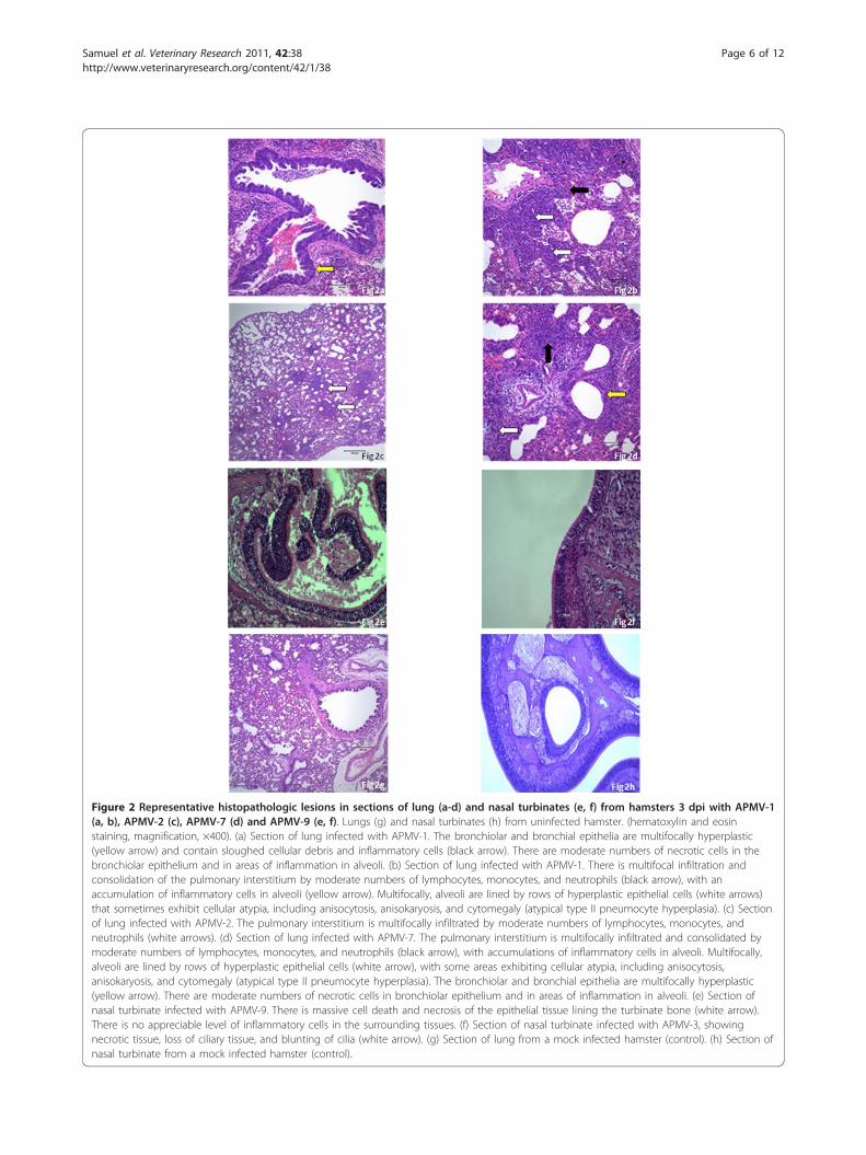
Figure 2 Representative histopathologic lesions in sections of lung (a-d) and nasal turbinates (e, f) from hamsters 3 dpi with APMV-1(a, b), APMV-2 (c), APMV-7 (d) and APMV-9 (e, f). Lungs (g) and nasal turbinates (h) from uninfected hamster. (hematoxylin and eosinstaining, magnification, ×400). (a) Section of lung infected with APMV-1. The bronchiolar and bronchial epithelia are multifocally hyperplastic(yellow arrow) and contain sloughed cellular debris and inflammatory cells (black arrow). There are moderate numbers of necrotic cells in thebronchiolar epithelium and in areas of inflammation in alveoli. (b) Section of lung infected with APMV-1. There is multifocal infiltration andconsolidation of the pulmonary interstitium by moderate numbers of lymphocytes, monocytes, and neutrophils (black arrow), with anaccumulation of inflammatory cells in alveoli (yellow arrow). Multifocally, alveoli are lined by rows of hyperplastic epithelial cells (white arrows)that sometimes exhibit cellular atypia, including anisocytosis, anisokaryosis, and cytomegaly (atypical type II pneumocyte hyperplasia). (c) Sectionof lung infected with APMV-2. The pulmonary interstitium is multifocally infiltrated by moderate numbers of lymphocytes, monocytes, andneutrophils (white arrows). (d) Section of lung infected with APMV-7. The pulmonary interstitium is multifocally infiltrated and consolidated bymoderate numbers of lymphocytes, monocytes, and neutrophils (black arrow), with accumulations of inflammatory cells in alveoli. Multifocally,alveoli are lined by rows of hyperplastic epithelial cells (white arrow), with some areas exhibiting cellular atypia, including anisocytosis,anisokaryosis, and cytomegaly (atypical type II pneumocyte hyperplasia). The bronchiolar and bronchial epithelia are multifocally hyperplastic(yellow arrow). There are moderate numbers of necrotic cells in bronchiolar epithelium and in areas of inflammation in alveoli. (e) Section ofnasal turbinate infected with APMV-9. There is massive cell death and necrosis of the epithelial tissue lining the turbinate bone (white arrow).There is no appreciable level of inflammatory cells in the surrounding tissues. (f) Section of nasal turbinate infected with APMV-3, showingnecrotic tissue, loss of ciliary tissue, and blunting of cilia (white arrow). (g) Section of lung from a mock infected hamster (control). (h) Section ofnasal turbinate from a mock infected hamster (control).
Samuel et al. Veterinary Research 2011, 42:38http://www.veterinaryresearch.org/content/42/1/38
Page 6 of 12
Figure 3 Immunofluorescence localization of viral N antigen in sections of nasal turbinates (a, b), harderian glands (c, d) and lungs(e, f) from hamsters 3 dpi with APMV-9 (a), APMV-1 (b), APMV-9 (c), APMV-8 (d), APMV-3 (e), and APMV-2 (f). Nasal turbinates (g) andlung (h) from a representative uninfected control hamster (magnification, ×400). (a) Section of nasal turbinate infected with APMV-9.Immunofluorescence was evident in the ciliated epithelium lining the turbinate bone (white arrows). (b) Section of nasal turbinate infected withAPMV-1. Immunofluorescence was evident primarily at the apical surface of the ciliated epithelial cells and in the cytoplasm (white arrows). (c)Section of harderian gland infected with APMV-9. Immunofluorescence was evident primarily in the collecting ducts of the gland and also in thecytoplasm of the infected cells (white arrows). (d) Section of harderian gland and nasal turbinate infected with APMV-8. Immunofluorescencewas evident primarily the cytoplasm of the infected cells in the harderian gland (white arrows) and in the epithelial cells lining the turbinates(yellow arrows). (e) Section of the lung infected with APMV-3. Immunofluorescence was evident around the bronchial epithelium (white arrows).(f) Section of lung infected with APMV-2. Immunofluorescence was mainly evident around the bronchiolar epithelium (white arrows). (g) Sectionof nasal turbinate from a mock infected hamster (control). (h) Section of lung from a mock infected hamster (control).
Samuel et al. Veterinary Research 2011, 42:38http://www.veterinaryresearch.org/content/42/1/38
Page 7 of 12
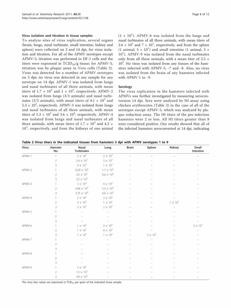
Virus isolation and titration in tissue samplesTo analyze sites of virus replication, several organs(brain, lungs, nasal turbinate, small intestine, kidney andspleen) were collected on 3 and 14 dpi, for virus isola-tion and titration. For all of the APMV serotypes exceptAPMV-5, titration was performed in DF-1 cells and thetiters were expressed in TCID50/g tissue; for APMV-5,titration was by plaque assay in Vero cells (Table 2).Virus was detected for a number of APMV serotypeson 3 dpi; no virus was detected in any sample for anyserotype on 14 dpi. APMV-1 was isolated from lungsand nasal turbinates of all three animals, with meantiters of 1.7 × 10³ and 1 × 10³, respectively. APMV-2was isolated from lungs (3/3 animals) and nasal turbi-nates (2/3 animals), with mean titers of 4.1 × 10³ and5.3 × 103, respectively. APMV-3 was isolated from lungsand nasal turbinates of all three animals, with meantiters of 5.3 × 10² and 3.6 × 10⁴, respectively. APMV-4was isolated from lungs and nasal turbinates of allthree animals, with mean titers of 1.7 × 10² and 4.2 ×10³, respectively, and from the kidneys of one animal
(1 × 101). APMV-6 was isolated from the lungs andnasal turbinates of all three animals, with mean titers of2.6 × 103 and 7 × 101, respectively, and from the spleen(1 animal, 3 × 101) and small intestine (1 animal, 3 ×101). APMV-9 was isolated from the nasal turbinatesonly from all three animals, with a mean titer of 2.5 ×10⁵. No virus was isolated from any tissues of the ham-sters infected with APMV-5, -7 and -8. Also, no viruswas isolated from the brain of any hamsters infectedwith APMV-1 to -9.
SerologyThe virus replication in the hamsters infected withAPMVs was further investigated by measuring serocon-version 14 dpi. Sera were analyzed by HI assay usingchicken erythrocytes (Table 3) in the case of all of theserotypes except APMV-5, which was analyzed by pla-que reduction assay. The HI titers of the pre-infectionhamsters were 2 or less. All HI titers greater than 8were considered positive. Our results showed that all ofthe infected hamsters seroconverted at 14 dpi, indicating
Table 2 Virus titers in the indicated tissues from hamsters 3 dpi with APMV serotypes 1 to 9
Virus HamsterNo.
NasalTurbinates
Lung Brain Spleen Kidney SmallIntestine
APMV-1 1 3 × 10³ 2 × 10³ – – – –
2 2.4 × 10³ 3 × 10³ – – – –
3 4 × 10³ 1 × 10² – – – –
APMV-2 1 9.29 × 10³ 1.7 × 10³ – – – –
2 3.4 × 10³ 6.6 × 10² – – – –
3 3.5 × 10³ - – – – –
APMV-3 1 3 × 10⁴ 4 × 10² – – – –
2 4.06 × 10⁴ 5.5 × 10² – – – –
3 3.75 × 10⁴ 6.6 × 10² – – – –
APMV-4 1 2 × 10³ 3 × 10² – – – –
2 2 × 10³ 1 × 101 – – 1 × 101 –
3 2 × 10² 2 × 10² – – – –
APMV-5 1 – – – – – –
2 – – – – – –
3 – – – – – –
APMV-6 1 1 × 10² 3 × 10³ – – – 3 × 101
2 1 × 101 4 × 10³ – – – –
3 1 × 10² 1 × 10³ – 3 × 101 – –
APMV-7 1 – – – – – –
2 – – – – – –
3 – – – – – –
APMV-8 1 – – – – – –
2 – – – – – –
3 – – – – – –
APMV-9 1 3 × 10⁵ – – – – –
2 1.5 × 10⁵ – – – – –
3 3.8 × 10⁵ – – – – –
The virus titer values are expressed in TCID50 per gram of the indicated tissue sample.
Samuel et al. Veterinary Research 2011, 42:38http://www.veterinaryresearch.org/content/42/1/38
Page 8 of 12
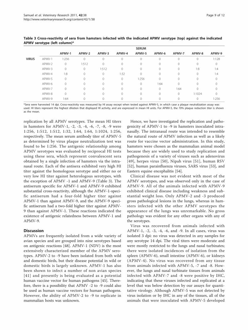
replication by all APMV serotypes. The mean HI titersin hamsters for APMV-1, -2, -3, -4, -6, -7, -8, -9 were1:256, 1:512, 1:512, 1:32, 1:64, 1:64, 1:1024, 1:256,respectively. The mean serum antibody titer of APMV-5as determined by virus plaque neutralization test wasfound to be 1:256. The antigenic relationship amongAPMV serotypes was evaluated by reciprocal HI testsusing these sera, which represent convalescent seraobtained by a single infection of hamsters via the intra-nasal route. Each of the antisera exhibited very high HItiter against the homologous serotype and either no orvery low HI titer against heterologous serotypes, withthe exception of APMV -1 and APMV-9 (Table 3). Theantiserum specific for APMV-1 and APMV-9 exhibitedsubstantial cross-reactivity, although the APMV-1-speci-fic antiserum had a four-fold higher titer againstAPMV-1 than against APMV-9, and the APMV-9 speci-fic antiserum had a two-fold higher titer against APMV-9 than against APMV-1. These reactions indicated theexistence of antigenic relatedness between APMV-1 andAPMV-9.
DiscussionAPMVs are frequently isolated from a wide variety ofavian species and are grouped into nine serotypes basedon antigenic reactions [48]. APMV-1 (NDV) is the mostextensively characterized member of the APMV sero-types. APMV-2 to -9 have been isolated from both wildand domestic birds, but their disease potential in wild ordomestic birds is largely unknown. APMV-1 has alsobeen shown to infect a number of non avian species[41] and presently is being evaluated as a potentialhuman vaccine vector for human pathogens [45]. There-fore, there is a possibility that APMV -2 to -9 could alsobe used as human vaccine vectors for human pathogens.However, the ability of APMV-2 to -9 to replicate inmammalian hosts was unknown.
Hence, we have investigated the replication and patho-genicity of APMV-1 to -9 in hamsters inoculated intra-nasally. The intranasal route was intended to resemblethe natural route of APMV infection as well as a likelyroute for vaccine vector administration. In this study,hamsters were chosen as the mammalian animal modelbecause they are widely used to study replication andpathogenesis of a variety of viruses such as adenovirus[49], herpes virus [50], Nipah virus [51], human RSV[52], human parainfluenza viruses, SARS virus [53], andEastern equine encephalitis [54].Clinical disease was not evident with most of the
APMV serotypes, and was observed only in the case ofAPMV-9. All of the animals infected with APMV-9exhibited clinical disease including weakness and sub-stantial weight loss. Only APMV-2 and -3 producedgross pathological lesions in the lungs, whereas in ham-sters infected with the other APMV serotypes theappearance of the lungs was unremarkable. No grosspathology was evident for any other organs with any ofthe serotypes.Virus was recovered from animals infected with
APMV-1, -2, -3, -4, -6, and -9. In all cases, virus wasisolated 3 dpi: no virus was detected in any samples forany serotype 14 dpi. The viral titers were moderate andwere mostly restricted to the lungs and nasal turbinates;there were isolated incidences of isolation from thespleen (APMV-6), small intestine (APMV-6), or kidneys(APMV-4). No virus was recovered from any tissuefrom animals infected with APMV-5, -7 and -8. How-ever, the lungs and nasal turbinate tissues from animalsinfected with APMV-7 and -8 were positive by IHC,indicating that these viruses infected and replicated at alevel that was below detection by our assays for quanti-tative virology. Although APMV-5 was not detected byvirus isolation or by IHC in any of the tissues, all of theanimals that were inoculated with APMV-5 developed
Table 3 Cross-reactivity of sera from hamsters infected with the indicated APMV serotype (top) against the indicatedAPMV serotype (left column)*
SERUM
APMV-1 APMV-2 APMV-3 APMV-4 APMV-5 APMV-6 APMV-7 APMV-8 APMV-9
VIRUS APMV-1 1:256 0 0 0 0 0 0 0 1:128
APMV-2 0 1:512 0 0 0 0 0 0 0
APMV-3 0 0 1:512 0 0 0 0 1:8 1:4
APMV-4 1:8 0 0 1:32 0 0 0 0 1:4
APMV-5 0 0 0 0 1:256 0 0 0 0
APMV-6 0 0 0 0 0 1:64 0 0 0
APMV-7 0 0 0 0 0 0 1:64 0 0
APMV-8 0 0 0 0 0 0 0 1:1024 0
APMV-9 1:64 0 0 0 0 0 0 0 1:256
*Sera were harvested 14 dpi. Cross-reactivity was measured by HI assay except when tested against APMV-5, in which case a plaque neutralization assay wasused. HI titers represent the highest dilution that displayed HI activity, and are expressed in mean HI units. For APMV-5, the 70% plaque reduction titer is shownas the mean.
Samuel et al. Veterinary Research 2011, 42:38http://www.veterinaryresearch.org/content/42/1/38
Page 9 of 12
titers of APMV-5 specific antibodies detected by virusplaque reduction neutralization assay, indicating a lowlevel of replication. By 14 dpi, no virus could bedetected in any of the tissues in any the infected ham-sters for any serotype, whether by histopathology, histo-chemistry, or virus isolation. This suggests that the viruswas cleared from all tissues and disease was resolved,indicating the self-limited nature of the infections. Simi-lar results have been obtained from chickens infectedwith APMV-2, -3 and -4, where no virus could be iso-lated at 14 dpi [30].Using IHC, viral N antigen was detected in the same
tissues that were positive by virus isolation. An interest-ing finding was the presence of large amounts of viralantigens at the epithelial cell linings, suggesting thatthese cells are highly permissive to APMV replication.In addition, the detection of viral antigens, and in mostcases infectious virus, in nasal turbinates and lungs ofthe hamsters indicate that APMV replication is mostlyrestricted to the respiratory tract. These results showthat all APMV serotypes are capable of infecting ham-sters using a nasal route of infection and the extensiveamount of virus replication in the respiratory tract wasnot accompanied by severe disease in hamsters.Serologic assays demonstrated a humoral response in
all the hamsters inoculated with APMV serotypes -1 to-9, a further indication of successful virus replication.Our results show that all APMVs (except APMV-5,which does not hemagglutinate) produced HI antibodytiters that varied between 1:32 to 1:1024. Based on theHI titers, APMV-8 (1:1024) produced maximum anti-body titer and APMV-4 (1:32) produced the least. Para-doxically, while APMV-8 induced the highest titer of HIantibodies, the virus could not be isolated from any ofthe infected hamsters. Conversely, APMV-4 replicatedefficiently in hamsters as observed by virus isolation innasal turbinates, lungs and kidneys, but produced lowlevels of virus-specific HI antibodies. Whether theseexamples of incongruity between levels of replicationand serum antibody responses are indicative of differ-ences in the antigenicity of the respective HN proteinsor to some other factor remains to be determined.Warke et al. [30] have also provided evidence of low
HI titers in the case of APMV-4, in chickens, which wastaken as evidence of a low level of replication of thisvirus in chickens. The HI test is the most commonlyused method to diagnose APMV infections and also isused to measure the antibody response. But chickenantiserum against NDV cross-reacts in HI tests withseveral of the other APMV serotypes, thus questioningthe specificity of HI test in field cases [55]. Warke et al.[38] indicated that the HI test lacks sensitivity fordetecting infection with these APMV serotypes. Theyalso observed that even in the case of a high infectious
dose and observed microscopic changes in the infectedorgans, high HI titers were not observed in the infectedbirds. On the contrary in our study, most of the APMVserotypes replicated well in hamsters, producing mildrespiratory pathology and high neutralizing antibodytiters. Our results indicated that the antibody developedin hamsters against these serotypes were very specificand no cross reaction was observed between serotypesexcept between APMV-1 and -9. Cross-reactivitybetween these two serotypes is not completely unantici-pated, since they share the highest level of genomenucleotide sequence identity (58%) among the APMVserotypes [12]. Hence, we conclude that the HI test withhamster serum is highly specific and can be used todiagnose different APMV serotypes in field cases.The replication of APMVs in hamsters produced mild
rhinitis and mild pathology that was mainly restricted tothe respiratory tract. There was a concern that APMVsmay also replicate in the intestine and shed in feces,which might act as a source of infection for the otheranimals. But our results indicated that none of theAPMVs replicate in the intestinal epithelial cells of ham-sters. Importantly, our results also suggest that theseviruses do not cross the blood-brain barrier and do notinduce neurological symptoms. Also, our previousexperimental studies of all the APMVs in chickensshowed that they were avirulent. Taken together, theseresults show that the APMVs replicate moderately inhamsters and produce either mild or no clinical signs,and elicit substantial antibody responses. Therefore, it ispossible that APMVs might replicate in other mamma-lian species including humans. In conclusion, this studyis the first comparative report on the replication andpathogenicity of prototype strains of all 9 APMV sero-types in hamsters. Our results lay the foundation for agood laboratory animal model for testing the replicationand pathogenicity of APMV strains.
AcknowledgementsWe thank Daniel Rockemann, Flavia Dias and all our laboratory members fortheir excellent technical assistance and help. “This research was supportedby NIAID contract no. N01A060009 (85% support) and NIAID, NIH IntramuralResearch Program (15% support). The views expressed herein do notnecessarily reflect the official policies of the Department of Health andHuman Services; nor does mention of trade names, commercial practices, ororganizations imply endorsement by the U.S. Government.”
Author details1Virginia-Maryland Regional College of Veterinary Medicine, University ofMaryland, College Park, Maryland, USA. 2Experimental Transplantation andImmunology Branch, National Cancer Institute/National Institute of Health,Bethesda, USA. 3Laboratory of Infectious Diseases, National Institute ofAllergy and Infectious Diseases, Bethesda, USA.
Authors’ contributionsAS carried out the pathogenesis studies, carried out the immunoassays,performed the analysis, and drafted the manuscript. MS carried out thepathogenesis studies. HS participated in the analysis, interpretation of
Samuel et al. Veterinary Research 2011, 42:38http://www.veterinaryresearch.org/content/42/1/38
Page 10 of 12
histopathological slides. PC and SKS, conceived the study, participated in itsdesign and coordination. All authors read and approved the finalmanuscript.
Competing interestsThe authors declare that they have no competing interests.
Received: 13 October 2010 Accepted: 22 December 2010Published: 23 February 2011
References1. Lamb R, Parks G: In Paramyxoviridae: the viruses and their replication. 5
edition. Edited by: Knipe DM, Howley PM, Griffin DE, Lamb RA, Martin MA,Roizman B, Straus SE. Philadelphia: Lippincott Williams 2007.
2. Lamb RA, Collins PL, Kolakofsky D, Melero JA, Nagai Y, Oldstone MBA,Pringle CR, Rima BK: In Family Paramyxoviridae. Virus Taxonomy: TheClassification and Nomenclature of Viruses. The Eighth Report of theInternational Committee in Taxonomy of Viruses Edited by: Fauquet CM 2005.
3. Mayo MA: A summary of taxonomic changes recently approved by ICTV.Arch Virol 2002, 147:1655-1663.
4. Alexander DJ: Avian paramyxoviruses-other than Newcastle disease virus.World’s Poul Sci J 1982, 38:97-104.
5. de Leeuw O, Peeters B: Complete nucleotide sequence of Newcastledisease virus: evidence for the existence of a new genus within thesubfamily Paramyxovirinae. J Gen Virol 1999, 80(Pt 1):131-136.
6. Huang Z, Panda A, Elankumaran S, Govindarajan D, Rockemann DD,Samal SK: The hemagglutinin-neuraminidase protein of Newcastle diseasevirus determines tropism and virulence. J Virol 2004, 78:4176-4184.
7. Krishnamurthy S, Samal SK: Nucleotide sequences of the trailer,nucleocapsid protein gene and intergenic regions of Newcastle diseasevirus strain Beaudette C and completion of the entire genomesequence. J Gen Virol 1998, 79(Pt 10):2419-2424.
8. Panda A, Huang Z, Elankumaran S, Rockemann DD, Samal SK: Role offusion protein cleavage site in the virulence of Newcastle disease virus.Microb Pathog 2004, 36:1-10.
9. Rout SN, Samal SK: The large polymerase protein is associated with thevirulence of Newcastle disease virus. J Virol 2008, 82:7828-7836.
10. Chang PC, Hsieh ML, Shien JH, Graham DA, Lee MS, Shieh HK: Completenucleotide sequence of avian paramyxovirus type 6 isolated from ducks.J Gen Virol 2001, 82:2157-2168.
11. Subbiah M, Xiao S, Collins PL, Samal SK: Complete sequence of thegenome of avian paramyxovirus type 2 (strain Yucaipa) and comparisonwith other paramyxoviruses. Virus Res 2008, 137:40-48.
12. Samuel AS, Kumar S, Madhuri S, Collins PL, Samal SK: Complete sequenceof the genome of avian paramyxovirus type 9 and comparison withother paramyxoviruses. Virus Res 2009, 142:10-18.
13. Nayak B, Kumar S, Collins PL, Samal SK: Molecular characterization andcomplete genome sequence of avian paramyxovirus type 4 prototypestrain duck/Hong Kong/D3/75. Virol J 2008, 5:124.
14. Jeon WJ, Lee EK, Kwon JH, Choi KS: Full-length genome sequence ofavain paramyxovirus type 4 isolated from a mallard duck. Virus Genes2008, 37:342-350.
15. Kumar S, Nayak B, Collins PL, Samal SK: Complete genome sequence ofavian paramyxovirus type 3 reveals an unusually long trailer region. VirusRes 2008, 137:189-197.
16. Paldurai A, Subbiah M, Kumar S, Collins PL, Samal SK: Complete genomesequences of avian paramyxovirus type 8 strains goose/Delaware/1053/76 and pintail/Wakuya/20/78. Virus Res 2009, 142:144-153.
17. Samuel AS, Paldurai A, Kumar S, Collins PL, Samal SK: Complete genomesequence of avian paramyxovirus (APMV) serotype 5 completes theanalysis of nine APMV serotypes and reveals the longest APMV genome.PLoS One 2010, 5:e9269.
18. Xiao S, Paldurai A, Nayak B, Subbiah M, Collins PL, Samal SK: Completegenome sequence of avian paramyxovirus type 7 (strain Tennessee) andcomparison with other paramyxoviruses. Virus Res 2009, 145:80-91.
19. Zhang GZ, Zhao JX, Wang HW, Yang AM, Bu CY, Wang M: Isolation,identification, and comparison of four isolates of avian paramyxovirusserotype 2 in China. Avian Dis 2006, 50:386-390.
20. Tumova B, Stumpa A, Janout V, Uvizl M, Chmela J: A further member ofthe Yucaipa group isolated from the common wren (Troglodytestroglodytes). Acta Virol 1979, 23:504-507.
21. Bankowski RA, Corstvet RE, Clark GT: Isolation of an unidentified agentfrom the respiratory tract of chickens. Science 1960, 132:292-293.
22. Bankowski RA, Conrad RD, Reynolde B: Avian influenza A and paramyxoviruses complicating respiratory disease diagnosis in poultry. Avian Dis1968, 12:259-278.
23. Lipkind MA, Weisman Y, Shihmanter E, Shoham D, Aronovici A: Theisolation of yucaipa-like paramyxoviruses from epizootics of arespiratory disease in turkey poultry farms in Israel. Vet Rec 1979,105:577-578.
24. Bankowski RA, Almquist J, Dombrucki J: Effect of paramyxovirus yucaipaon fertility, hatchability, and poult yield of turkeys. Avian Dis 1981,25:517-520.
25. Tumova B, Robinson JH, Easterday BC: A hitherto unreportedparamyxovirus of turkeys. Res Vet Sci 1979, 27:135-140.
26. Macpherson I, Watt RG, Alexander DJ: Isolation of avian paramyxovirusother than Newcastle disease virus from commercial poultry in GreatBritain. Vet Rec 1983, 112:479-480.
27. Alexander DJ, Collins MS: Pathogenecity of PMV-3/Parakeet/Netherland/449/75 for chickens. Avian Pathol 1982, 11:179-185.
28. Beck I, Gerlach H, Burkhardt E, Kaleta EF: Investigation of several selectedadjuvants regarding their efficacy and side effects for the production ofa vaccine for parakeets to prevent a disease caused by a paramyxovirustype 3. Vaccine 2003, 21:1006-1022.
29. Shortridge KF, Alexander DJ, Hu LY, Kam SL: Isolation of Newcastle diseasevirus from Phasianidae birds in Hong Kong. J Comp Pathol 1978,88:633-636.
30. Warke A, Stallknecht D, Williams SM, Pritchard N, Mundt E: Comparativestudy on the pathogenicity and immunogenicity of wild bird isolates ofavian paramyxovirus 2, 4, and 6 in chickens. Avian Pathol 2008,37:429-434.
31. Nerome K, Nakayama M, Ishida M, Fukumi H: Isolation of a new avianparamyxovirus from budgerigar (Melopsittacus undulatus). J Gen Virol1978, 38:293-301.
32. Alexander DJ: Avian paramyxoviruses 2-9. 11 edition. Ames: Iowa StateUniversity Press; 2003.
33. Saif YM, Mohan R, Ward L, Senne DA, Panigrahy B, Dearth RN: Natural andexperimental infection of turkeys with avian paramyxovirus-7. Avian Dis1997, 41:326-329.
34. Cloud SS, Rosenberger JK: Characterization of nine avianparamyxoviruses. Avian Dis 1980, 24:139-152.
35. Yamane N, Arikawa J, Odagiri T, Ishida N: Characterization of avianparamyxoviruses isolated from feral ducks in northern Japan: thepresence of three distinct viruses in nature. Microbiol Immunol 1982,26:557-568.
36. Sandhu T, Hinshaw V: Influenza A virus infection of domestic ducks firstint. symp. avian influenza. Beltsville, Maryland 1981, 93-99.
37. Capua I, De Nardi R, Beato MS, Terregino C, Scremin M, Guberti V: Isolationof an avian paramyxovirus type 9 from migratory waterfowl in Italy. VetRec 2004, 155:156.
38. Warke A, Appleby L, Mundt E: Prevalence of antibodies to different avianparamyxoviruses in commercial poultry in the United States. Avian Dis2008, 52:694-697.
39. Yates VJ, Fry DE, Henderson BW Jr: Isolation of Newcastle disease virusfrom a calf. J Am Vet Med Assoc 1952, 120:149-150.
40. Nelson CB PB, Schrall K, Park WE, Lindeman RJ: An outbreak ofconjunctivitis due to Newcastle disease virus (NDV) occurring in poultryworkers. Am J Public Health 1952, 42:672-678.
41. Hofstad MS: Experimental inoculation of swine and sheep withNewcastle disease virus. Cornell Vet 1950, 40:190-197.
42. Quinn RW, Hanson RP, Brown JW, Brandly CA: Newcastle disease virusin man; results of studies in five cases. J Lab Clin Med 1952,40:736-743.
43. Bukreyev A, Huang Z, Yang L, Elankumaran S, St Claire M, Murphy BR,Samal SK, Collins PL: Recombinant newcastle disease virus expressing aforeign viral antigen is attenuated and highly immunogenic in primates.J Virol 2005, 79:13275-13284.
44. Subbiah M, Yan Y, Rockemann D, Samal SK: Experimental infection ofcalves with Newcastle disease virus induces systemic and mucosalantibody responses. Arch Virol 2008, 153:1197-1200.
45. Bukreyev A, Collins PL: Newcastle disease virus as a vaccine vector forhumans. Curr Opin Mol Ther 2008, 10:46-55.
Samuel et al. Veterinary Research 2011, 42:38http://www.veterinaryresearch.org/content/42/1/38
Page 11 of 12
46. Alexander DJ: Newcastle disease diagnosis Boston: Kluwer AcademicPublishers; 1988.
47. Reed LJ, Muench H: A simple method of estimating fifty per centendpoints. Am J Hyg 1938, 27:493-497.
48. Alexander DJ: Newcastle disease and other avian paramyxoviruses. RevSci Tech 2000, 19:443-462.
49. Thomas MA, Spencer JF, Toth K, Sagartz JE, Phillips NJ, Wold WS:Immunosuppression enhances oncolytic adenovirus replication andantitumor efficacy in the Syrian hamster model. Mol Ther 2008,16:1665-1673.
50. van Ekdom LT, Herbrink P, Meddens MJ: Hamster model for herpessimplex virus infection of the central nervous system. Infection 1987,15:125-127.
51. Guillaume V, Contamin H, Loth P, Georges-Courbot MC, Lefeuvre A,Marianneau P, Chua KB, Lam SK, Buckland R, Deubel V, Wild TF: Nipahvirus: vaccination and passive protection studies in a hamster model.J Virol 2004, 78:834-840.
52. Byrd LG, Prince GA: Animal models of respiratory syncytial virus infection.Clin Infect Dis 1997, 25:1363-1368.
53. Schaecher SR, Stabenow J, Oberle C, Schriewer J, Buller RM, Sagartz JE,Pekosz A: An immunosuppressed Syrian golden hamster model forSARS-CoV infection. Virology 2008, 380:312-321.
54. Paessler S, Aguilar P, Anishchenko M, Wang HQ, Aronson J, Campbell G,Cararra AS, Weaver SC: The hamster as an animal model for easternequine encephalitis–and its use in studies of virus entrance into thebrain. J Infect Dis 2004, 189:2072-2076.
55. Lipkind M, Shihmanter E: Antigenic relationships between avianparamyxoviruses. I. Quantitative characteristics based onhemagglutination and neuraminidase inhibition tests. Arch Virol 1986,89:89-111.
doi:10.1186/1297-9716-42-38Cite this article as: Samuel et al.: Experimental infection of hamsterswith avian paramyxovirus serotypes 1 to 9. Veterinary Research 201142:38.
Submit your next manuscript to BioMed Centraland take full advantage of:
• Convenient online submission
• Thorough peer review
• No space constraints or color figure charges
• Immediate publication on acceptance
• Inclusion in PubMed, CAS, Scopus and Google Scholar
• Research which is freely available for redistribution
Submit your manuscript at www.biomedcentral.com/submit
Samuel et al. Veterinary Research 2011, 42:38http://www.veterinaryresearch.org/content/42/1/38
Page 12 of 12


![[MIKROBIOLOGI] IT 20 - Orthomyxovirus, Paramyxovirus - KHS](https://static.fdocuments.in/doc/165x107/55cf9193550346f57b8ea639/mikrobiologi-it-20-orthomyxovirus-paramyxovirus-khs.jpg)