Experimental evidence in support of single host...
11
Experimental evidence in support of single host maintenance of a multihost pathogen AMANDA L. J. DUFFUS, 1,2,3, RICHARD A. NICHOLS, 2 AND TRENTON W. J. GARNER 1 1 Institute of Zoology, Zoological Society of London, Regent’s Park, London NW1 4RY United Kingdom 2 School of Biological and Chemical Sciences, Queen Mary University of London, Mile End Road, London E1 4NS United Kingdom Citation: Duffus, A. L. J., R. A. Nichols, and T. W. J. Garner. 2014. Experimental evidence in support of single host maintenance of a multihost pathogen. Ecosphere 5(11):142. http://dx.doi.org/10.1890/ES14-00074.1 Abstract. The rate at which pathogens are emerging appears to be increasing. Therefore, it is important to determine the factors, such as virulence and host specificity, and how they have been affected by the emergence. In the United Kingdom, ranaviruses, which are double stranded DNA viruses (Family Iridoviridae) began to emerge in populations of common frogs (Rana temporaria) in the mid-to-late 1980s, followed closely by emergence in common toad (Bufo bufo) populations. Here we present experimental evidence that a single host may be able to maintain a multihost pathogen. We exposed common frog tadpoles and common toad tadpoles to ranavirus isolates from the mid-1990s at two different doses. Tadpole survival differed significantly between treatments and this was primarily driven by the dose of the exposure. However, at the low dose, common frog tadpoles exposed to isolates from common frogs experienced higher mortality (n ¼ 35/treatment, log-rank: P ¼ 0.0320, Wilcoxon: P ¼ 0.0835, df ¼ 1) than those exposed to common toad isolates. The high dose caused 75% mortality in common frog tadpoles, but common toads never experienced more than 40% mortality. This, and other evidence provided by this study, show that common frogs are likely to be the primary host of the ranavirus in the UK, and that single host maintenance of ranaviruses can occur in anurans. Key words: Bufo bufo; common frogs; common toads; experimental evidence infection; Iridoviridae; multihost pathogen; Rana temporaria; ranavirus; single host maintenance; United Kingdom. Received 6 March 2014; revised 7 July 2014; accepted 5 August 2014; final version received 4 October 2014; published 21 November 2014. Corresponding Editor: D. P. C. Peters. Copyright: Ó 2014 Duffus et al. This is an open-access article distributed under the terms of the Creative Commons Attribution License, which permits unrestricted use, distribution, and reproduction in any medium, provided the original author and source are credited. http://creativecommons.org/licenses/by/3.0/ 3 Present address: Department of Biology, Gordon College, Barnesville, Georgia 30204 USA. E-mail: [email protected] INTRODUCTION Infectious diseases are emerging at an in- creased rate (Woolhouse and Gowtage-Sequeria 2005, Jones et al. 2008) providing opportunities for pathogens to infect and cause disease in additional host species (Benmayor et al. 2009). Host range and virulence are dictated both by evolution and by ecology; being shaped by historical interactions and by the more contem- porary process of emergence (Woolhouse et al. 2005). Ascertaining if host range and virulence have increased during emergence, or arisen as a result of previous host/pathogen dynamics, can be challenging, particularly in systems where pathogens are re-emerging or in those where the history of the pathogen is uncertain. On the other hand, introduced pathogens offer opportunities to investigate the determinants of host range and virulence. The prospects for such studies have increased as the globalization of human activities has led to an increase in such novel introductions (Daszak et al. 2000, Hajek and Tobin 2011, Andreou et al. 2012, Fisher et al. v www.esajournals.org 1 November 2014 v Volume 5(11) v Article 142
Transcript of Experimental evidence in support of single host...

Experimental evidence in support of single host maintenanceof a multihost pathogen
AMANDA L. J. DUFFUS,1,2,3,� RICHARD A. NICHOLS,2 AND TRENTON W. J. GARNER1
1Institute of Zoology, Zoological Society of London, Regent’s Park, London NW14RY United Kingdom2School of Biological and Chemical Sciences, Queen Mary University of London, Mile End Road, London E1 4NS United Kingdom
Citation: Duffus, A. L. J., R. A. Nichols, and T. W. J. Garner. 2014. Experimental evidence in support of single host
maintenance of a multihost pathogen. Ecosphere 5(11):142. http://dx.doi.org/10.1890/ES14-00074.1
Abstract. The rate at which pathogens are emerging appears to be increasing. Therefore, it is important
to determine the factors, such as virulence and host specificity, and how they have been affected by the
emergence. In the United Kingdom, ranaviruses, which are double stranded DNA viruses (Family
Iridoviridae) began to emerge in populations of common frogs (Rana temporaria) in the mid-to-late 1980s,
followed closely by emergence in common toad (Bufo bufo) populations. Here we present experimental
evidence that a single host may be able to maintain a multihost pathogen. We exposed common frog
tadpoles and common toad tadpoles to ranavirus isolates from the mid-1990s at two different doses.
Tadpole survival differed significantly between treatments and this was primarily driven by the dose of the
exposure. However, at the low dose, common frog tadpoles exposed to isolates from common frogs
experienced higher mortality (n ¼ 35/treatment, log-rank: P ¼ 0.0320, Wilcoxon: P ¼ 0.0835, df ¼ 1) than
those exposed to common toad isolates. The high dose caused 75% mortality in common frog tadpoles, but
common toads never experienced more than 40% mortality. This, and other evidence provided by this
study, show that common frogs are likely to be the primary host of the ranavirus in the UK, and that single
host maintenance of ranaviruses can occur in anurans.
Key words: Bufo bufo; common frogs; common toads; experimental evidence infection; Iridoviridae; multihost pathogen;
Rana temporaria; ranavirus; single host maintenance; United Kingdom.
Received 6 March 2014; revised 7 July 2014; accepted 5 August 2014; final version received 4 October 2014; published 21
November 2014. Corresponding Editor: D. P. C. Peters.
Copyright: � 2014 Duffus et al. This is an open-access article distributed under the terms of the Creative Commons
Attribution License, which permits unrestricted use, distribution, and reproduction in any medium, provided the
original author and source are credited. http://creativecommons.org/licenses/by/3.0/3 Present address: Department of Biology, Gordon College, Barnesville, Georgia 30204 USA.
� E-mail: [email protected]
INTRODUCTION
Infectious diseases are emerging at an in-creased rate (Woolhouse and Gowtage-Sequeria2005, Jones et al. 2008) providing opportunitiesfor pathogens to infect and cause disease inadditional host species (Benmayor et al. 2009).Host range and virulence are dictated both byevolution and by ecology; being shaped byhistorical interactions and by the more contem-porary process of emergence (Woolhouse et al.2005). Ascertaining if host range and virulence
have increased during emergence, or arisen as aresult of previous host/pathogen dynamics, canbe challenging, particularly in systems wherepathogens are re-emerging or in those where thehistory of the pathogen is uncertain.
On the other hand, introduced pathogens offeropportunities to investigate the determinants ofhost range and virulence. The prospects for suchstudies have increased as the globalization ofhuman activities has led to an increase in suchnovel introductions (Daszak et al. 2000, Hajekand Tobin 2011, Andreou et al. 2012, Fisher et al.
v www.esajournals.org 1 November 2014 v Volume 5(11) v Article 142
2012). These introductions can be valuableexamples, because the ecological and evolution-ary dynamics between the hosts and introducedpathogens are newly established, rather thanhaving being shaped by historical interactions.The pathogens face immune responses that aretypically different from those of their historicalhosts; similarly the new hosts may be illequipped by their previous evolution, since few,if any, pathogen strategies for overcoming hostbarriers to infection are universal (Parrish et al.2008, but see Fisher et al. 2012).
Introduced pathogens can be capable ofinfecting multiple host species de novo, but theability to infect multiple hosts is not equivalent tothe ability to establish persistent infections andcause disease. Most novel communities of hostspecies present a heterogeneous distribution ofsusceptibilities due to variation of immuneresponses in different host species. A novelpathogen is most likely to exploit the mostsusceptible host species initially, and infectionsof other species often arise through infrequentspill-over events from this primary host (Wool-house and Gowtage-Sequeria 2005, Woolhouse etal. 2005). Spill-over or interactions with other, co-occurring pathogens can lead to evolutionarydynamics that result in pathogen divergence,where diverged lineages become adapted to, butnot necessarily restricted to, secondary hostspecies (Kawecki 1998, Crill et al. 2000, Nemirovet al. 2002, Parrish et al. 2008, Rouchet andVorburger 2012). The result of these evolutionarydynamics can manifest as decreased parasitismand virulence in the initial host species andincreased virulence in new hosts (Ebert 1998).Introduction need not lead to increased pathogenspecialization, though, since coevolution can alsofavour generalist strategies (Hall et al. 2011) andhost-switching does not always require adapta-tion by the pathogen (Nam et al. 2011).
Host range expansion by viruses has repeat-edly caused novel disease emergence in wildlife,and some of these cases can be attributed topathogen introduction (Roelke-Parker et al. 1996,Woolhouse et al. 2005, Harkonen et al. 2006,Bruemmer et al. 2010, Lawson et al. 2012). Thisappears to be the case for members of the genusRanavirus, iridoviruses that exploit an exception-ally diverse range of vertebrate hosts (Hedrick etal. 1992, Mao et al. 1999, Hyatt et al. 2002, De Voe
et al. 2004, Gray et al. 2009a, b, Jensen et al. 2009).Ranavirosis often affects multiple hosts simulta-neously at a single location (Gray et al. 2009a),but it is uncertain if multihost infection anddisease dynamics are possible during initialemergence events or require more prolongedhistory.
Ranaviruses may be transmitted by vectorscarrying them outside their native ranges, in-cluding introduced, commercially traded herpe-tofauna from which ranaviruses have frequentlybeen isolated (e.g., Une et al. 2009). Once arrivedat a new location the evidence suggests thatranaviruses are capable of producing infection,since they are experimentally transmittable tonovel hosts (Jankovich et al. 2001, Pearman et al.2004, Whittington et al. 2010). The introductionof virus to populations of a novel host species canresult in local adaptation by the virus (Storfer etal. 2007, Ridenhour and Storfer 2008).
In the UK, unusual common frog (Ranatemporaria) mortality events that were subse-quently shown to be caused by ranavirosis werefirst detected in 1985 (Cunningham et al. 1996).Infections in common toads (Bufo bufo) weredetected soon after the initial detection ofranavirosis in common frogs (Hyatt et al. 2000,Cunningham et al. 2007a). Since then, commonfrog mortality events in the UK caused byranavirosis have increased substantially in geo-graphic range. There is a serious impact on thecommon frog populations: approximately half ofranavirosis emergence events in frog populationsresult in substantial and persistent populationdeclines (Teacher et al. 2010). Diseased or deadtoads have rarely been detected and, apart fromcommon frogs, no mass mortality events associ-ated with ranavirosis emergence have beenreported in the UK involving other amphibians.This evidence suggests that ranaviruses in theUK are incapable of eliciting sustained disease inmultiple hosts, in contrast to the reports ofranaviruses affecting amphibian communities inNorth America (Duffus et al. 2008). It isconceivable that toad mass mortality events areoccurring but go undetected. Toads have experi-enced inexplicable and significant declines inareas where ranavirus emergence was firstdetected in British frogs (Cunningham et al.1996, Carrier and Beebee 2003, Teacher et al.2010). Ranavirus isolates from toads and frogs
v www.esajournals.org 2 November 2014 v Volume 5(11) v Article 142
DUFFUS ET AL.

are genetically similar (but not identical; Hyatt etal. 2000), and toad isolates have been experimen-tally transmitted to frog hosts, resulting in fataldisease (Cunningham et al. 2007a). These latterfindings suggest that amphibian ranaviruses inthe UK are effective at exploiting both frogs andtoads as hosts.
Here we report the results of experimentswhere we tested the potential for British ranavi-ruses to infect and cause disease in both R.temporaria and B. bufo. We used isolates derivedseveral years after the initial emergence ofranavirosis in the UK from both species. Ourgoal was to determine if isolates from each hostspecies had equivalent capacity to elicit disease inboth hosts, or if there was any evidence of hostspecialization.
METHODS
Ethics statementAll experiments were fully licensed by the
Home Office (License No. 07928346219) and allprocedures and designs were subject to fullethical review before implementation.
Virus culture and titrationWe used four viruses that were isolated from
visibly diseased, wild amphibians collected in theUK in the early 1990s (Cunningham et al.2007a, b). Two (BUK 2, BUK 3) were isolatedfrom common toads, and two (RUK 11 and RUK13) from common frogs (Hyatt et al. 2000,Cunningham et al. 2007a, b). Viruses isolatedfrom common frogs were associated with differ-ent disease syndromes: RUK 11 was isolatedfrom a R. temporaria adult presenting substantialinternal haemorrhages (haemorrhagic syndrome)and RUK 13 was isolated from a frog presentingsuperficial skin ulcers (ulcerative syndrome:Cunningham et al. 2007b), unlike those obtainedfrom B. bufo where only one disease syndromehas been observed (Hyatt et al. 2000).
Viruses were cultured in confluent lawns offathead minnow (Pimephales promelus) cells(FHM) purchased from the European Collectionof Cell Cultures (No. 88102401, ECACC, Oxford,UK). FHM cells were first propagated at 258C inEagle’s minimum essential media (EMEM: Sig-ma-Aldrich, Andover, UK), supplemented with1% L-glutamine (Sigma-Aldrich), 0.005% penicil-
lin-streptomycin (Sigma-Aldrich), 0.005% nysta-tin (Gibco, Invitrogen, Paislely, UK), and 10%research grade foetal bovine serum (Hyclone,Perbio Science, Northumberland, UK). Confluentflasks of FHM cells were then inoculated with1000lL of virus isolate. Twenty-five milliliters ofmaintenance media (EMEM supplemented with1% L-glutamine, 0.005% penicillin-streptomycin,0.005% nystatin, and 1% research grade fetalbovine serum, all suppliers as above) was addedto each flask and all flasks were then incubated at258C. Flasks were monitored daily for theformation of viral plaques and once plaqueswere observed and FHM cells ceased to adhere tothe flask, virus was harvested and stored at�808C. Each isolate was standardized by passag-ing three times before final harvest and titration.To generate experimental negative controls wemock-harvested FHM cell cultures that had notbeen exposed to virus. To do this, we substitutedmaintenance media for culture media in conflu-ent flasks of FHM cells and left flasks to incubatefor 3 days, which we observed to be the averagenumber of days required to bring virus culturesto harvest stage after the addition of maintenancemedia. After 3 days we scraped cells into themedia and stored the cell solution at�808C.
We determined the number of plaque formingunits per milliliter (PFU/mL) for each virus usingserial dilutions of harvested virus titrated induplicate into six well flasks with confluent FHMcells (10�3 to 10�8, 1 mL per well per dilution, oneisolate per plate, two plates per isolate; Duffus etal. 2008). Plates were incubated at 258C for 24hours, following which 2 mL of maintenancemedia was added to each well. Plates werereturned to 258C and monitored daily for plaqueformation. As soon as plaques were first detectedin the well with the highest concentration virus,we removed the media from all wells of the plateand fixed cells in 100% methanol. After 10minutes we removed the methanol, stained cellsusing a 0.05% crystal violet-20% methanolsolution for 20 minutes and rinsed the wellswith sterile water to removed excess stain.Plaques were counted by eye and we averagedplaque counts to generate PFU/mL for eachisolate harvest.
Experimental design and procedureRana temporaria eggs (10 clutches) were collect-
v www.esajournals.org 3 November 2014 v Volume 5(11) v Article 142
DUFFUS ET AL.
ed from a pond located in Faversham, Kent,England, and B. bufo eggs (10 clutches) from apond located in Cowden, Sussex, England, inMarch 2009. Infection with ranavirus and rana-virosis have not been detected at either site(Teacher et al. 2010, Duffus et al. 2013). Eggswere brought to the Institute of Zoology, Zoo-logical Society of London and transferred into 84-L plastic tubs (Really Useful Box Company,Normanton, UK) containing approximately 45 Lof tap water aged for 48 hrs. Clutches were splitamong tubs by species and no more than threeclutches were kept in any one box. We partiallychanged water every few days and removed anycomponents of clutches that did not exhibitdevelopment. Hatched tadpoles were combinedin single species tubs, where water was changedevery two days and Tetra Tabimin pellets (TetraFish, Southampton, UK) were provided adlibitum.
Tadpoles were allocated to the experiments,one per amphibian species, when the tadpoleshad reached Gosner stage 25 (Gosner 1960). Dueto time differences in egg availability, the twoexperiments were not run simultaneously. Ex-perimental treatments were the same in eachexperiment and included 35 individuals pertreatment, for a total of 315 animals perexperiment/amphibian species. Tadpoles werebath exposed in groups of five in Petri dishes(Nunc, Roskilde, Denmark) to one of ninedifferent treatments: negative control, 106 PFU(high dose) of one of the four isolates, or 104 PFUof one of the four isolates. All exposures werestandardized to 30 mL total volume and thevolume of harvested virus was kept constantacross all treatments. Tadpole groups wereexposed for 18 hrs, after which individualtadpoles were transferred either into Petri dishescontaining 30 mL aged tap water (R. temporaria)or 75 cm2 tissue culture flasks (Nunc) containing140 mL aged tap water (Bufo bufo). Tadpoles weremaintained on a three- (R. temporaria) or four-day(Bufo bufo) water change and frog tadpoles werefed 125 lL of a dilution of Tabimin pellets (6finely ground pellets suspended in 50 mL agedtap water) every second day until the end of theexperiment. Toad tadpoles were fed as per R.temporaria until the 10th day, when we reducedfood concentration to 3 pellets in 50mL andpipetted 100lL of food suspension into each flask
once every 2 days. To limit the possibility ofcontamination amongst experimental treatments,we kept animals separated by treatment andspecies, but rotated the position in the rearingroom of each treatment and each tadpole withina treatment daily. Initially we checked forevidence of disease and death daily, but oncemortality commenced checks were done twicedaily until mortality rate had declined signifi-cantly. We recorded date of death (or survival tothe end of the experiment) and visible signs ofdisease commonly reported for tadpoles suffer-ing from ranavirosis (e.g., Greer et al. 2005) foreach animal. Because our goal was to measureimpacts before metamorphosis, animals reachingGosner Stage 43–44 were euthanized and count-ed as survivors. Both experiments were complet-ed after 30 days and survivors were euthanizedusing an overdose of MS-2,2,2 [1g/L Tricanemethylsulphonate (Thompson and Joseph, Nor-wich, UK) buffered to pH 7 with sodiumbicarbonate]. All carcasses were stored in 2 mLmicrocentrifuge tubes in 100% ethanol for mo-lecular diagnosis of infection.
We selected from each treatment, wherepossible, the first 10 animals to die and the first5 animals that survived to the end of theexperiment; these individuals were euthanizedfor screening for the presence of the ranavirus. Intreatments where 10 animals did not die, allanimals that died were included and the rest ofthe sample was made up of euthanized individ-uals. We aseptically sampled a small triangularsection from the left side of every tadpole (eachsample contained viscera and skin) and extractedDNA from these tissues using the Wizard SV96Genomic DNA Purification System (Promega,Southampton, UK). We used extractions toamplify a 500-bp region of the major capsidprotein (MCP) of frog virus 3 (FV3) using theprimer set originally developed by Mao et al.(1996). All polymerase chain reactions (PCR)were completed using the Multiplex PCR kit(QIAGEN, Crawley, UK) and with the followingthermocycler settings: initial five minute dena-turing step at 958C, followed by 35 cycles of 958Cfor 45 sec, 528C for 45 sec, 728C for 45 sec. Positivecontrols and negative extraction controls wereincluded in all PCR plates. We screened everyextraction at least twice and any ambiguousamplifications or runs where positive PCR
v www.esajournals.org 4 November 2014 v Volume 5(11) v Article 142
DUFFUS ET AL.
controls failed were re-amplified: ambiguousamplifications or failed runs were exceptionallyrare. PCR products were visualized on 1%
agarose gels stained with ethidium bromide.Amplifications generating a 500-bp fragmentwere scored positive for infection with ranavirus.
Statistical analysisWe tested for differences in survival amongst
treatments in each experiment using both log-rank analysis and Wilcoxon tests, as in bothexperiments data were right-censored (Klein-baum and Klein 2005, Machin et al. 2006) andthe ratio of hazards was greater earlier in theexperiments (Kleinbaum and Klein 2005). Whenwe detected a significant difference using eitherapproach, we used Proportional Hazard Modelsto determine if dose or isolate type had thegreater impact on survival (Kleinbaum and Klein2005, Machin et al. 2006). We used a proportionalhazard model because the hazard was continu-
ous and the model allows the inclusion ofcategorical variables (Machin et al. 2006). Rela-tionships between infection prevalence and signsof disease were examined with the controlsremoved and the direction of the differenceswas established using a Fisher’s exact test. Allstatistics were performed with JMP 8.0 (SASInstitute, North Carolina, USA).
RESULTS
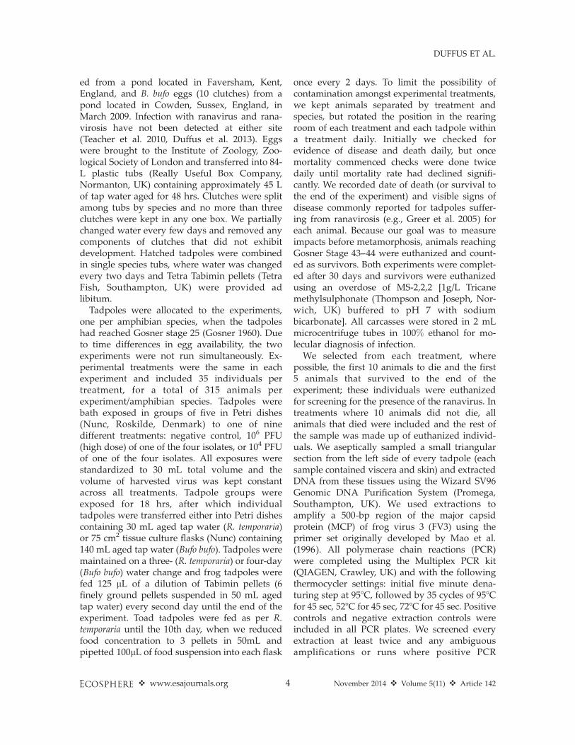
Frog survival differed significantly amongsttreatments (n ¼ 35/treatment, log-rank: P ,
0.0001, Wilcoxon: P , 0.0001, df ¼ 8; Fig. 1).This was primarily driven by the dose (propor-tional hazards, n ¼ 315, df ¼ 2, P , 0.0001).Further analysis on low dose treatments revealeda significant difference, with RUK isolates caus-ing significantly more mortality than BUKisolates in exposed tadpoles (n ¼ 35/treatment,log-rank: P¼ 0.0320, Wilcoxon: P¼ 0.0835, df¼ 1;
Fig. 1. Survivorship of common frog (Rana temporaria) tadpoles exposed to different doses and isolates of
ranaviruses from the UK (n ¼ 35/treatment).
v www.esajournals.org 5 November 2014 v Volume 5(11) v Article 142
DUFFUS ET AL.
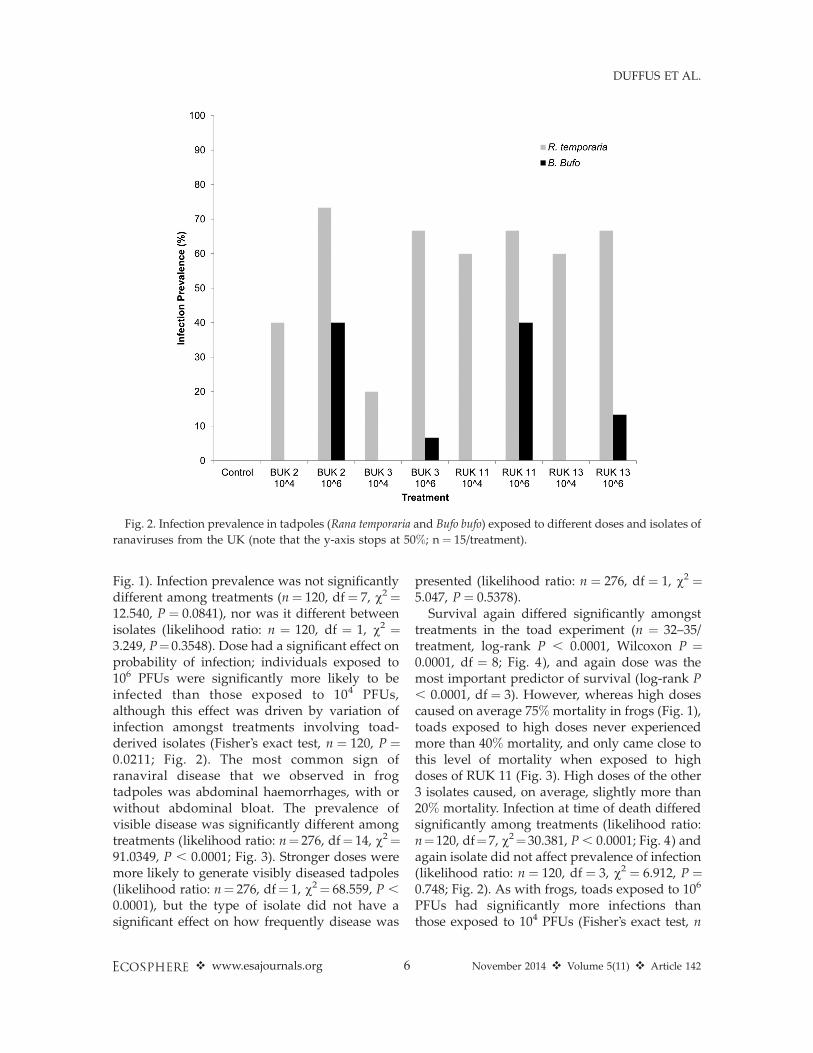
Fig. 1). Infection prevalence was not significantlydifferent among treatments (n¼ 120, df¼ 7, v2¼12.540, P ¼ 0.0841), nor was it different betweenisolates (likelihood ratio: n ¼ 120, df ¼ 1, v2 ¼3.249, P¼0.3548). Dose had a significant effect onprobability of infection; individuals exposed to106 PFUs were significantly more likely to beinfected than those exposed to 104 PFUs,although this effect was driven by variation ofinfection amongst treatments involving toad-derived isolates (Fisher’s exact test, n ¼ 120, P ¼0.0211; Fig. 2). The most common sign ofranaviral disease that we observed in frogtadpoles was abdominal haemorrhages, with orwithout abdominal bloat. The prevalence ofvisible disease was significantly different amongtreatments (likelihood ratio: n¼ 276, df¼ 14, v2¼91.0349, P , 0.0001; Fig. 3). Stronger doses weremore likely to generate visibly diseased tadpoles(likelihood ratio: n¼ 276, df¼ 1, v2¼ 68.559, P ,
0.0001), but the type of isolate did not have asignificant effect on how frequently disease was
presented (likelihood ratio: n ¼ 276, df ¼ 1, v2 ¼5.047, P ¼ 0.5378).
Survival again differed significantly amongsttreatments in the toad experiment (n ¼ 32–35/treatment, log-rank P , 0.0001, Wilcoxon P ¼0.0001, df ¼ 8; Fig. 4), and again dose was themost important predictor of survival (log-rank P, 0.0001, df ¼ 3). However, whereas high dosescaused on average 75% mortality in frogs (Fig. 1),toads exposed to high doses never experiencedmore than 40% mortality, and only came close tothis level of mortality when exposed to highdoses of RUK 11 (Fig. 3). High doses of the other3 isolates caused, on average, slightly more than20% mortality. Infection at time of death differedsignificantly among treatments (likelihood ratio:n¼120, df¼7, v2¼30.381, P , 0.0001; Fig. 4) andagain isolate did not affect prevalence of infection(likelihood ratio: n ¼ 120, df ¼ 3, v2 ¼ 6.912, P ¼0.748; Fig. 2). As with frogs, toads exposed to 106
PFUs had significantly more infections thanthose exposed to 104 PFUs (Fisher’s exact test, n
Fig. 2. Infection prevalence in tadpoles (Rana temporaria and Bufo bufo) exposed to different doses and isolates of
ranaviruses from the UK (note that the y-axis stops at 50%; n ¼ 15/treatment).
v www.esajournals.org 6 November 2014 v Volume 5(11) v Article 142
DUFFUS ET AL.

¼ 120, P , 0.0001), an effect driven by the lack ofdetectable infection in any toads exposed to lowdoses (Fig. 2). We did not observe abdominal orindeed any hemorrhages affecting toads andbloating was rare. We did commonly observeskin sloughing, and taken together, signs diddiffer significantly amongst treatments (likeli-hood ratio: n¼269, df¼ 7, v2¼ 23.523, P¼ 0.0014;Fig. 4), an effect driven by increasing dosestrength (Fisher’s exact test, n ¼ 269, P ,
0.0001). As with frogs, isolate did not have aneffect on the frequency of observable signs ofdisease (likelihood ratio: n ¼ 269, df ¼ 3, v2 ¼0.337, P ¼ 0.9529).
DISCUSSION
Highly virulent ranavirosis had been circulat-ing in UK common frog populations for close toten years when the ranaviruses used in this studywere cryobanked, after isolation from wild UKamphibians. This time period is more thanadequate for ranavirus evolution in response tohost immunity (Ridenhour and Storfer 2008) andR. temporaria in populations experiencing sus-tained ranavirosis exhibit the molecular signal of
selection at immunogenetic loci important forcombating infection with ranaviruses (Teacher etal. 2009a). Nevertheless, the responses of R.temporaria tadpoles we exposed to ranaviruseswere broadly congruent with those commonlyobserved when novel pathogens emerge inhighly susceptible hosts (Fig. 1; Pearman andGarner 2005, Warnecke et al. 2013): infection,disease and mortality were predominantly dose-dependent. We did detect divergence in virulenceamong isolates derived from different hostspecies when frog tadpoles were exposed tolow doses (Fig. 1), but any evidence of special-ization of viruses was swamped by force ofinfection, as all four UK ranavirus isolates causedapproximately 80% mortality in common frogtadpoles at high doses (Fig. 1).
The fact that frog-derived isolates inducedstronger mortality at low doses in R. temporariacould be explained as decreased virulence of toadisolates in the primary host (R. temporaria).Theory predicts lower virulence in the primaryhost in cases where novel pathogens diverge anda new pathogen lineage exploits a novel, second-ary host (Ebert 1998). This same theory predictsincreased virulence of divergent pathogen line-
Fig. 3. Frequencies of signs of ranavirosis in tadpoles (Rana temporaria and Bufo bufo) exposed to different doses
and isolates of ranaviruses from the UK (n ¼ 35/treatment).
v www.esajournals.org 7 November 2014 v Volume 5(11) v Article 142
DUFFUS ET AL.
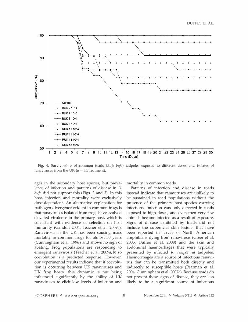
ages in the secondary host species, but preva-lence of infection and patterns of disease in B.bufo did not support this (Figs. 2 and 3). In thishost, infection and mortality were exclusivelydose-dependent. An alternative explanation forpathogen divergence evident in common frogs isthat ranaviruses isolated from frogs have evolvedelevated virulence in the primary host, which isconsistent with evidence of selection on hostimmunity (Gandon 2004, Teacher et al. 2009a).Ranavirosis in the UK has been causing massmortality in common frogs for almost 30 years(Cunningham et al. 1996) and shows no sign ofabating. Frog populations are responding toemergent ranavirosis (Teacher et al. 2009a, b) socoevolution is a predicted response. However,our experimental results indicate that if coevolu-tion is occurring between UK ranaviruses andUK frog hosts, this dynamic is not beinginfluenced significantly by the ability of UKranaviruses to elicit low levels of infection and
mortality in common toads.Patterns of infection and disease in toads
instead indicate that ranaviruses are unlikely tobe sustained in toad populations without thepresence of the primary host species carryinginfections. Infection was only detected in toadsexposed to high doses, and even then very fewanimals became infected as a result of exposure.Signs of disease exhibited by toads did notinclude the superficial skin lesions that havebeen reported in larvae of North Americanamphibians dying from ranavirosis (Greer et al.2005, Duffus et al. 2008) and the skin andabdominal haemorrhages that were typicallypresented by infected R. temporaria tadpoles.Haemorrhages are a source of infectious ranavi-rus that can be transmitted both directly andindirectly to susceptible hosts (Pearman et al.2004, Cunningham et al. 2007b). Because toads donot present these signs of disease, they are lesslikely to be a significant source of infectious
Fig. 4. Survivorship of common toads (Bufo bufo) tadpoles exposed to different doses and isolates of
ranaviruses from the UK (n ¼ 35/treatment).
v www.esajournals.org 8 November 2014 v Volume 5(11) v Article 142
DUFFUS ET AL.

ranavirus and rare infections in UK commontoads appear most likely to have arisen throughspill-over from diseased R. temporaria.
Given that toads are likely to be dead-endranavirus hosts, that ranavirosis is relativelyunknown in other UK amphibians, and persis-tent disease is reported in frog populationswhere other alternative hosts (e.g., fish, reptiles)are lacking, ranavirus may be maintained in theUK in a single, primary host species, R. tempora-ria. Precedence exists for highly virulent ranavi-rosis to be maintained in a single host species,even with significant mortality in that host(Brunner et al. 2004). Survival was still possiblein R. temporaria exposed to both dose concentra-tions of ranavirus and some survivors did exhibitdetectable infections. If these survivors couldmaintain infections while recruiting into later lifehistory stages and then return to their natal sitewith transmissible infections or transmit infec-tions to returning frogs before dying, infectionwould be sustained in manner comparable tosalamander species affected by persistent ranavi-rosis (Brunner et al. 2004). Alternatively, infectioncould be maintained in adult frogs that experi-ence infection but do not always succumb todisease. In support of this proposal, it has beenobserved that breeding common frogs presentingthe systemic haemorrhagic form of ranavirosisoften present healed skin ulcers that indicate about of disease the preceding year (Cunninghamet al. 2007b).
ACKNOWLEDGMENTS
This study was supported by a PhD studentshipawarded to A. J. L. Duffus by Queen Mary Universityof London, an Overseas Research Studentship, as wellas one provided by the National Science and Engi-neering Research Council of Canada. Additionalsupport was provided by a Convocation ResearchTrust Award, Amphibian Conservation Research TrustStudent Research Grant and a British Wildlife HealthAssociation Grant to A. L. J. Duffus and an RCUKFellowship awarded to T. W. J. Garner. We thankAndrew Cunningham for making isolates available forthis study.
LITERATURE CITED
Andreou, D., K. D. Arkush, J.-F. Guegan, and R. E.Gozlan. 2012. Introduced pathogens and nativefreshwater biodiversity: a case study of Spaerothe-
cum destruens. PLoS ONE 7:e36998.Benmayor, R., D. J. Hodgson, G. G. Perron, and A.
Buckling. 2009. Host mixing and disease emer-gence. Current Biology 19:764–767.
Bruemmer, C. M., S. P. Rushton, J. Gurnell, P. W. W.Lurz, P. Nettleton, A. W. Sainsbury, J. P. Duff, J.Gilray, J., and C. J. McInnes. 2010. Epidemiology ofsquirrel poxvirus in grey squirrels in the UK.Epidemiology and Infection 138:941–950.
Brunner, J. L., D. M. Schock, E. W. Davidson, and J. P.Collins. 2004. Intraspecific reservoirs: complex lifehistory and the persistence of a lethal ranavirus.Ecology 85:560–566.
Carrier, J.-A., and T. J. C. Beebee. 2003. Recent,substantial, and unexplained declines of the com-mon toad Bufo bufo in lowland England. BiologicalConservation 111:395–399.
Crill, W. D., H. A. Wichman, and J. J. Bull. 2000.Evolutionary reversals during viral adaptation toalternating hosts. Genetics 154:27–37.
Cunningham, A. A., A. D. Hyatt, P. Russell, and P. M.Bennett. 2007a. Experimental transmission of aranavirus disease of common toads (Bufo bufo) tocommon frogs (Rana temporaria). Epidemiology andInfection 135:1213–1216.
Cunningham, A. A., A. D. Hyatt, P. Russell, and P. M.Bennett. 2007b. Emerging epidemic diseases offrogs in Britain are dependent on the source ofranavirus agent and the route of exposure. Epide-miology and Infection 135:1200–1212.
Cunningham, A. A., T. E. S. Langton, P. M. Bennett,J. F. Lewin, S. E. N. Drury, R. E. Gough, and S. K.MacGregor. 1996. Pathological and microbiologicalfindings from incidents of unusual mortality of thecommon frog (Rana temporaria). PhilosophicalTransactions of the Royal Society B 351:1539–1557.
Daszak, P., A. A. Cunningham, and A. D. Hyatt. 2000.Wildlife ecology: Emerging infectious diseases ofwildlife—threats to biodiversity and human health.Science 287:443–449.
De Voe, R., K. Geissler, S. Elmore, D. Rotstein, G.Lewbart, and J. Guy. 2004. Ranavirus-associatedmorbidity and mortality in a group of captiveeastern box turtles (Terrapene carolina carolina).Journal of Zoo and Wildlife Medicine 35:534–543.
Duffus, A. L. J., R. A. Nichols, and T. W. J. Garner.2013. 2013 Investigations into the life history stagesof the common frog (Rana temporaria) affected by anamphibian ranavirus in the United Kingdom.Herpetological Review 44:260–263.
Duffus, A. L. J., B. D. Pauli, K. Wozney, C. R. Brunetti,and M. Berrill. 2008. Frog virus 3-like infections inaquatic amphibian communities. Journal of Wild-life Diseases 44:109–120.
Ebert, D. 1998. Experimental evolution of parasites.Science 282:1432–1435.
Fisher, M. C., D. A. Henk, C. J. Briggs, J. S. Brownstein,
v www.esajournals.org 9 November 2014 v Volume 5(11) v Article 142
DUFFUS ET AL.

L. C. Madoff, S. L. McCraw, and S. J. Gurr. 2012.Emerging fungal threats to animal, plant andecosystem health. Nature 484:186–194.
Gandon, S. 2004. Evolution of multihost parasites.Evolution 58:455–469.
Gosner, K. L. 1960. A simplified table for staginganuran embryos and larvae with notes on identi-fication. Herpetologica 16:183–190.
Gray, M. J., D. L. Miller, and J. T. Hoverman. 2009a.Ecology and pathology of amphibian ranaviruses.Diseases of Aquatic Organisms 87:243–266.
Gray, M. J., D. L. Miller, and J. T. Hoverman. 2009b.First report of Ranavirus infecting lungless sala-manders. Herpetological Review 40:316–319.
Greer, A. L., M. Berrill, and P. J. Wilson. 2005. Fiveamphibian mortality events associated with rana-virus infection in south central Ontario, Canada.Diseases of Aquatic Organisms 67:9–14.
Hajek, A. E., and P. C. Tobin. 2011. Introducedpathogens follow the invasion front of a spreadingalien host. Journal of Animal Ecology 80:1217–1226.
Hall, A. R., P. D. Scanlan, and A. Buckling. 2011.Bacteria-phage coevolution and the emergence ofgeneralist pathogens. American Naturalist 177:44–53.
Harkonen, T., R. Dietz, P. Reijnders, J. Teilmann, K.Harding, A. Hall, S. Brasseur, U. Siebert, S. J.Goodman, P. D. Jepson, T. D. Rasmussen, and P.Thompson. 2006. The 1988 and 2002 phocinedistemper virus epidemics in European harbourseals. Diseases of Aquatic Organisms 68:115–130.
Hedrick, R. P., T. S. McDowell, W. Ahne, C. Torhy, andP. Dekinkelin. 1992. Properties of three iridovirus-like agents associated with systemic infections offish. Diseases of Aquatic Organisms 13:203–209.
Hyatt, A. D., A. R. Gould, Z. Zupanovic, A. A.Cunningham, S. G. Hengstberger, R. J. Whitting-ton, and B. E. H. Coupar. 2000. Characterisation ofpiscine and amphibian iridoviruses. Archives ofVirology 145:301–331.
Hyatt, A. D., M. Williamson, B. E. H. Coupar, D.Middleton, S. G. Hengstberger, A. R. Gould, P.Selleck, T. G. Wise, J. Kattenbelt, A. A. Cunning-ham, and J. Lee. 2002. First identification of aranavirus from green pythons (Chondropythonviridis). Journal of Wildlife Diseases 38:239–252.
Jankovich, J. K., E. W. Davidson, A. Seiler, B. L. Jacobs,and J. P. Collins. 2001. Transmission of theAmbystoma tigrinum virus to alternative hosts.Diseases of Aquatic Organisms 46:159–163.
Jensen, B. B., A. K. Ersbøll, and E. Ariel. 2009.Susceptibility of pike Esox lucius to a panel ofRanavirus isolates. Diseases of Aquatic Organisms83:169–179.
Jones, K. E., N. G. Patel, M. A. Levy, A. Storeygard, D.Balk, J. L. Gittleman, and P. Daszak. 2008. Global
trends in emerging infectious diseases. Nature451:990–993.
Kawecki, T. J. 1998. Red Queen meets Santa Rosalia:arms races and the evolution of host specializationin organisms with parasitic lifestyles. AmericanNaturalist 152:635–651.
Kleinbaum, D. G., and M. Klein. 2005. Survivalanalysis: A self-learn text. Second edition. Springer,New York, New York, USA.
Lawson, B., S. Lachish, K. M. Colvile, C. Durrant, K. M.Peck, M. P. Toms, B. C. Sheldon, and A. A.Cunningham. 2012. Emergence of a novel avianpox disease in British tit species. PLoS ONE7:e40176.
Machin, D., Y. B. Cheung, and M. K. B. Parmar. 2006.Survival analysis, a practical approach. Secondedition. John Wiley and Sons, West Sussex, UK.
Mao, J., D. E. Green, G. Fellers, and V. G. Chinchar.1999. Molecular characterization of iridovirusesisolated from sympatric amphibians and fish. VirusResearch 63:45–52.
Mao, J., T. N. Tham, G. A. Gentry, A. Aubertin, andV. G. Chinchar. 1996. Cloning, sequence analysis,and expression of the major capsid protein of theIridovirus frog virus 3. Virology 216:431–436.
Nam, J.-H., E.-H. Kim, D. Song, Y. K. Choi, J.-K. Kim,and H. Poo. 2011. Emergence of mammalianspecies-infectious and -pathogenic avian influenzaH6N5 virus with no evidence of adaptation.Journal of Virology 85:13271–13277.
Nemirov, K., H. Henttonen, A. Vaheri, and A.Plyusnin. 2002. Phylogenetic evidence for hostswitching in the evolution of hantaviruses carriedby Apodemus mice. Virus Research 90:207–215.
Parrish, C. R., E. C. Holmes, D. M. Morens, E.-C. Park,D. S. Burke, C. H. Calisher, C. A. Laughlin, L. J.Saif, and P. Daszak. 2008. Cross-species virustransmission and the emergence of new epidemicdiseases. Microbiology and Molecular BiologyReviews 72:457–470.
Pearman, P. B., and T. W. J. Garner. 2005. Susceptibilityof Italian agile frog populations to an emergingRanavirus parallels population genetic diversity.Ecology Letters 8:401–408.
Pearman, P. B., T. W. J. Garner, M. Straub, and U. F.Greber. 2004. Response of Rana latastei to theranavirus FV3: a model for viral emergence in anaıve population. Journal of Wildlife Disease40:600–609.
Ridenhour, B. J., and A. T. Storfer. 2008. Geographi-cally variable selection in Ambystoma tigrinum virus(Iridoviridae) throughout the western USA. Journalof Evolutionary Biology 21:1151–1159.
Roelke-Parker, M. E., L. Munson, C. Packer, R. Kock, S.Cleaveland, M. Carpenter, S. J. O’Brien, A. Pos-pischil, R. Hofmann-Lehmann, H. Lutz, G. L. M.Mwamengele, et al. 1996. A canine distemper virus
v www.esajournals.org 10 November 2014 v Volume 5(11) v Article 142
DUFFUS ET AL.

epidemic in Serengeti lions (Panthera leo). Nature379:441–445.
Rouchet, R., and C. Vorburger. 2012. Strong specificityin the interaction between parasitoids and symbi-ont-protected hosts. Journal of Evolutionary Biolo-gy 25:2369–2375.
Storfer, A., M. E. Alfaro, B. J. Ridenhour, J. K.Jancovich, S. G. Mech, M. J. Parris, and J. P. Collins.2007. Phylogenetic concordance analysis shows anemerging pathogen is novel and endemic. EcologyLetters 10:1075–1083.
Teacher, A. G. F., A. A. Cunningham, and T. W. J.Garner. 2010. The impact of Ranavirus infection onwild common frog populations in the UK. AnimalConservation 13:514–522.
Teacher, A. G. F., T. W. J. Garner, and R. A. Nichols.2009a. Evidence for directional selection at a novelMajor Histocompatability Class 1 marker in wildcommon frogs (Rana temporaria) exposed to a viralpathogen. PLoS ONE 4:e4616.
Teacher, A. G. F., T. W. J. Garner, and R. A. Nichols.2009b. Population genetic patterns suggest abehavioural change in wild common frogs (Ranatemporaria) following disease outbreaks (Ranavirus).
Molecular Ecology 18:3163–3172.
Une, Y., A. Sakuma, H. Matsueda, K. Nakai, and M.Murakami. 2009. Ranavirus outbreak in NorthAmerican bullfrogs (Rana catesbeiana), Japan, 2008.Emerging Infectious Diseases 15:1146–1147.
Warnecke, L., J. M. Turner, T. K. Bollinger, J. M. Lorch,V. Misra, P. M. Cryan, G. Wibbelt, D. S. Blehert, andC. K. R. Willis. 2013. Inoculation of bats withEuropean Geomyces destructans supports the novelpathogen hypothesis for the origin of white-nosesyndrome. Proceedings of the National Academyof Sciences USA. doi: 10.1073/pnas.1200374109
Whittington, R. J., J. A. Becker, and M. M. Dennis.2010. Iridovirus infections in finfish—critical re-view with emphasis on ranaviruses. Journal of FishDiseases 33:95–122.
Woolhouse, M. E. J., and S. Gowtage-Sequeria. 2005.Host range and emerging and re-emerging patho-gens. Emerging Infectious Diseases 11:1842–1847.
Woolhouse, M. E. J., D. T. Haydon, and R. Antia. 2005.Emerging pathogens: the epidemiology and evolu-tion of species jumps. Trends in Ecology andEvolution 20:238–244.
v www.esajournals.org 11 November 2014 v Volume 5(11) v Article 142
DUFFUS ET AL.


![Parasitic (Protozoan) cardiomyopathy: A review and pooled ... · as the severity and pattern of heart failure (HF) [47,48]. Experimental animal models show a correlation between host](https://static.fdocuments.in/doc/165x107/5f399caca4a5805ece5f8cf5/parasitic-protozoan-cardiomyopathy-a-review-and-pooled-as-the-severity-and.jpg)