Experimental evidence for sparse firing in the neocortex evidence for sparse firing in the...
11
Experimental evidence for sparse firing in the neocortex Alison L. Barth 1 and James F.A. Poulet 2, 3 1 Department of Biological Sciences and Center for the Neural Basis of Cognition, Carnegie Mellon University, Pittsburgh, PA 15213, USA 2 Department of Neuroscience, Max Delbru ¨ ck Center for Molecular Medicine (MDC), Berlin-Buch, Germany 3 Neuroscience Research Center and Cluster of Excellence NeuroCure, Charite ´ –Universita ¨ tsmedizin Berlin, Germany The advent of unbiased recording and imaging techni- ques to evaluate firing activity across neocortical neu- rons has revealed substantial heterogeneity in response properties in vivo, and that a minority of neurons are responsible for the majority of spikes. Despite the computational advantages to sparsely firing popula- tions, experimental data defining the fraction of respon- sive neurons and the range of firing rates have not been synthesized. Here we review data about the distribution of activity across neuronal populations in primary sen- sory cortex. Overall, the firing output of granular and infragranular layers is highest. Although subthreshold activity across supragranular neurons is decidedly non- sparse, spikes are much less frequent and some cells are silent. Superficial layers of the cortex may employ spe- cific cell and circuit mechanisms to increase sparseness. Introduction The number of neurons in the cerebral cortex has grown throughout phylogeny via an increase in the number of columns that are distributed across the cortical mantle [1]. Given the selective pressure that must have driven the expansion of the cerebral cortex, it is surprising that many neocortical neurons show very low firing rates. The sparse firing of neocortical neurons in vivo was not anticipated by decades of extracellular recordings (but see [2]), where detection of spiking neurons was not difficult. However, accumulating experimental evidence, using non-selective methods to assess the activity of identified, individual neurons, indicates that traditional extracellular record- ings may have been strongly biased by selection of the most active cells. What are the biological mechanisms that underlie the sparse firing of neocortical neurons? Why are there so many neurons if many do not transmit information to a subsequent stage of processing? Are sparse population responses an artifact of anesthesia, a reflection of a quies- cent brain state, a consequence of impoverished laboratory animal experience, poor stimulus selection, or an essential feature of neural circuits? Because the spike is the primary mechanism by which information is transmitted in the central nervous system, understanding what factors deter- mine which neurons will spike is of crucial importance. The computational and energetic advantages of sparse- ly firing neurons for efficient representation of sensory stimuli have been discussed in other excellent reviews on the subject [3–9]. Sparse sensory-evoked firing has perhaps been most thoroughly investigated in insects [10,11] and has also been observed in other vertebrates, such as songbirds [12]. Here we focus on experimental evidence in the mammalian neocortex indicating that fir- ing neurons are rare, or that silent neurons are common, and evaluate possible mechanisms that produce or regu- late sparse firing. The most convincing experimental data – with post hoc identification of recorded neurons for cell- and layer-assignment – has been obtained from analysis of neurons in rodent primary sensory cortex (but see [13–15]); thus, that material will be reviewed in greatest depth. In addition, we will address how the laminar location of a neuron influences its activity, with special attention to the superficial layers that receive dense innervation from in- put layers of the neocortex but fire at lower rates. Critical issues required to evaluate hypotheses about sparse firing across neocortical networks will be identified, and experi- ments to resolve these issues will be proposed. Schemas for sparseness The term ‘sparse’ has been widely used to describe the response properties of neocortical neurons. We have sche- matized different scenarios that give rise to the firing of only a few neurons in the cortical network to provide a framework in which to evaluate experimental data that support these models. There are at least 4 significantly different scenarios that can give the appearance of small ensemble activation, or sparseness (Figure 1). First, trial- to-trial variability in firing output, caused by either short- term or long-lasting changes in synaptic function or neural excitability, can lead to the appearance of sparse firing (‘population sparseness’; Figure 1a). Trial-to-trial variabil- ity might arise from noise within the system. In both cases, neural responses appear to be sparsely distributed across the network at a given instant, but may average-out over longer periods of analysis (minutes to hours). A second possibility is that the size of the responding ensemble is small and relatively fixed across trials (‘life- time sparseness’; Figure 1b). Generally, the computational advantages of having few firing neurons derive from this model. In contrast to Figure 1a, the identity of these ensembles is relatively constant, although they may shift Review Corresponding authors: Barth, A.L. ([email protected]); Poulet, J.F.A. ([email protected]). Keywords: silent neurons; optimality; kurtosis; pyramidal neuron; coding. TINS-900; No. of Pages 11 0166-2236/$ – see front matter ß 2012 Elsevier Ltd. All rights reserved. http://dx.doi.org/10.1016/j.tins.2012.03.008 Trends in Neurosciences xx (2012) 1–11 1
Transcript of Experimental evidence for sparse firing in the neocortex evidence for sparse firing in the...

TINS-900; No. of Pages 11
Experimental evidence for sparse firingin the neocortexAlison L. Barth1 and James F.A. Poulet2,3
1 Department of Biological Sciences and Center for the Neural Basis of Cognition, Carnegie Mellon University, Pittsburgh, PA 15213,
USA2 Department of Neuroscience, Max Delbruck Center for Molecular Medicine (MDC), Berlin-Buch, Germany3 Neuroscience Research Center and Cluster of Excellence NeuroCure, Charite –Universitatsmedizin Berlin, Germany
Review
The advent of unbiased recording and imaging techni-ques to evaluate firing activity across neocortical neu-rons has revealed substantial heterogeneity in responseproperties in vivo, and that a minority of neurons areresponsible for the majority of spikes. Despite thecomputational advantages to sparsely firing popula-tions, experimental data defining the fraction of respon-sive neurons and the range of firing rates have not beensynthesized. Here we review data about the distributionof activity across neuronal populations in primary sen-sory cortex. Overall, the firing output of granular andinfragranular layers is highest. Although subthresholdactivity across supragranular neurons is decidedly non-sparse, spikes are much less frequent and some cells aresilent. Superficial layers of the cortex may employ spe-cific cell and circuit mechanisms to increase sparseness.
IntroductionThe number of neurons in the cerebral cortex has grownthroughout phylogeny via an increase in the number ofcolumns that are distributed across the cortical mantle [1].Given the selective pressure that must have driven theexpansion of the cerebral cortex, it is surprising that manyneocortical neurons show very low firing rates. The sparsefiring of neocortical neurons in vivo was not anticipated bydecades of extracellular recordings (but see [2]), wheredetection of spiking neurons was not difficult. However,accumulating experimental evidence, using non-selectivemethods to assess the activity of identified, individualneurons, indicates that traditional extracellular record-ings may have been strongly biased by selection of themost active cells.
What are the biological mechanisms that underlie thesparse firing of neocortical neurons? Why are there somany neurons if many do not transmit information to asubsequent stage of processing? Are sparse populationresponses an artifact of anesthesia, a reflection of a quies-cent brain state, a consequence of impoverished laboratoryanimal experience, poor stimulus selection, or an essentialfeature of neural circuits? Because the spike is the primarymechanism by which information is transmitted in thecentral nervous system, understanding what factors deter-mine which neurons will spike is of crucial importance.
Corresponding authors: Barth, A.L. ([email protected]); Poulet, J.F.A.([email protected]).Keywords: silent neurons; optimality; kurtosis; pyramidal neuron; coding.
0166-2236/$ – see front matter � 2012 Elsevier Ltd. All rights reserved. http://dx.doi.org/10.101
The computational and energetic advantages of sparse-ly firing neurons for efficient representation of sensorystimuli have been discussed in other excellent reviewson the subject [3–9]. Sparse sensory-evoked firing hasperhaps been most thoroughly investigated in insects[10,11] and has also been observed in other vertebrates,such as songbirds [12]. Here we focus on experimentalevidence in the mammalian neocortex indicating that fir-ing neurons are rare, or that silent neurons are common,and evaluate possible mechanisms that produce or regu-late sparse firing. The most convincing experimental data –with post hoc identification of recorded neurons for cell-and layer-assignment – has been obtained from analysis ofneurons in rodent primary sensory cortex (but see [13–15]);thus, that material will be reviewed in greatest depth. Inaddition, we will address how the laminar location of aneuron influences its activity, with special attention to thesuperficial layers that receive dense innervation from in-put layers of the neocortex but fire at lower rates. Criticalissues required to evaluate hypotheses about sparse firingacross neocortical networks will be identified, and experi-ments to resolve these issues will be proposed.
Schemas for sparsenessThe term ‘sparse’ has been widely used to describe theresponse properties of neocortical neurons. We have sche-matized different scenarios that give rise to the firing ofonly a few neurons in the cortical network to provide aframework in which to evaluate experimental data thatsupport these models. There are at least 4 significantlydifferent scenarios that can give the appearance of smallensemble activation, or sparseness (Figure 1). First, trial-to-trial variability in firing output, caused by either short-term or long-lasting changes in synaptic function or neuralexcitability, can lead to the appearance of sparse firing(‘population sparseness’; Figure 1a). Trial-to-trial variabil-ity might arise from noise within the system. In both cases,neural responses appear to be sparsely distributed acrossthe network at a given instant, but may average-out overlonger periods of analysis (minutes to hours).
A second possibility is that the size of the respondingensemble is small and relatively fixed across trials (‘life-time sparseness’; Figure 1b). Generally, the computationaladvantages of having few firing neurons derive from thismodel. In contrast to Figure 1a, the identity of theseensembles is relatively constant, although they may shift
6/j.tins.2012.03.008 Trends in Neurosciences xx (2012) 1–11 1

Trial 1 Trial 2 Trial 3 Sum of 3 trials
Firing rateHigh
Low
(a)
(b)
(c)
(d)
Trial-to-trial variability creates apparent sparseness
The size of the spiking ensemble is small compared to the total number of cells
The fraction of spiking neurons is small and relatively constant
The fraction of spiking neurons can be increased by changing brain state
The fraction of spiking neurons is highly stimulus-specific
Stimulus A Stimulus B Stimulus C Stimulus D
Anesthetized Alert, motivated
TRENDS in Neurosciences
Figure 1. Models of sparseness. (a) Variability across trials (from cell-intrinsic or network properties) creates the appearance of sparseness that is diminished with longer
time-intervals for analysis. (b) A small but constant subset of cells fires more reliably across many trials than the rest of the population. (c) The fraction of firing neurons can
be changed by brain state, for example, from sleep or anesthetized to waking. (d) Highly stimulus-specific neurons will create the appearance of sparseness. As stimulus
space expands, more neurons will fire.
Review Trends in Neurosciences xxx xxxx, Vol. xxx, No. x
TINS-900; No. of Pages 11
over very long time-scales (days to weeks). In both cases itis most likely that the most active cells receive strongerafferent drive for a given stimulus, or are intrinsicallymore excitable, and that these properties could not bemodified instantly. In such a scenario, increasing stimulusstrength might effectively recruit more cells into theresponding population, but there would be a core set ofcells that always show stronger firing output than theirneighbors.
A third and often-discussed scenario is that sparsenessis only apparent, a product of anesthesia or brain state(Figure 1c). Indeed, experimental evidence indicates thatanesthesia suppresses response output [16–18] and themajority of studies have employed anesthesia to evaluateneural firing across large populations of cells for practicalreasons. Brain state in unanesthetized animals mightcontrol the number of responsive neurons, such that thefraction of cells spiking in a quiet, resting animal could bevery different from that in an awake, behaving animal
2
performing a reward-driven task. This model does notspecify whether the low abundance of firing cells stemsfrom the depictions in Figures 1a or 1b, although it sug-gests that mean firing rates across the population mayhave been significantly underestimated. Direct experimen-tal data to address this model have not been easy to obtain,but are likely to become available in the near future.
A final possibility is that the subset of responsive neu-rons is determined by the specific stimulus applied(Figure 1d). This possibility is related to the ‘lifetimesparseness’ model in Figure 1b, but differs in suggestingthat the majority of neurons will reliably spike, given adiverse enough stimulus test-set. In such a case, the frac-tion of responsive cells may be underestimated because theexperimental stimulus applied may not be appropriate todrive the neurons tested. Indeed, early studies of responseproperties in the primary visual cortex (V1) took off when itwas discovered that the best stimulus to drive neural firingwas oriented bars. Our collective understanding of the

Review Trends in Neurosciences xxx xxxx, Vol. xxx, No. x
TINS-900; No. of Pages 11
optimal sensory stimulus to drive firing of neocorticalneurons is still in its infancy. Especially in cases wherethe neuron requires convergent sensory or motor input (i.e.it is multimodal), a highly constrained experimental stim-ulus may not be optimized to drive a cell.
In the following sections each possibility will be dis-cussed, with specific attention to different cortical layersand brain areas. We will focus on studies that have notused spike-sorting for cellular isolation because thesemethods can be biased by analysis of highly responsivecells.
Does sparseness exist? The distribution of firing acrosscortical layersCompared to sensory-evoked responses at earlier stages ofsensory processing, the fraction of stimulus-driven neu-rons in the neocortex is remarkably reduced. In the thala-mus, firing is highly reliable. Individual thalamic neuronsare innervated by only 1–2 ganglion cells from the retina ortrigeminal nucleus [19,20], and the overall strength ofinputs from a single fiber (�1 nA) is sufficient to drive aspike in the postsynaptic cell. Although this is a simplifi-cation of how inputs drive thalamic neurons, it is in starkcontrast to how inputs are organized in cortical layer 4. Themajority of thalamic inputs synapse within layer 4, themain input layer of the cortex. Terminals are highly dis-tributed (one thalamic neuron projects to many layer 4neurons) but weak (�50 pA) [21], a divergence thatrequires the firing of many thalamic neurons to drive aspike in a layer 4 neuron.
Mean evoked firing-rates across layer 4 neurons aretypically higher than in upper layers. For example, inthe somatosensory cortical area that represents the facialwhiskers (i.e. barrel cortex), averaged evoked firing-ratesfrom identified layer 4 neurons are 0.14–0.41 action poten-tials 100 ms after whisker deflection in anesthetized ani-mals (Table 1) [22,23]. These averaged rates do not revealwhat fraction of neurons were spiking, but analysis ofpublished data indicates that the fraction of layer 4 neu-rons that ever spike in response to whisker touch issubstantially less than the total population size inanesthetized animals [16,22,23]. Overall, in layer 4, overrecording periods that range from �5 to 60 min, approxi-mately half the cells may be silent. Because these esti-mates are taken from many trials, the fraction of neuronsspiking on a given trial is lower (e.g. Figure 1a).
A major challenge in assessing the sparseness ofresponding neurons is determining the size of the respond-ing ensemble. Typically, this has been approached byrecording individual neurons using whole-cell patch-clampor sharp-electrode recording techniques. Ca2+-imaging isperfectly suited to assess population size, assuming thatcells take up a threshold level of Ca2+ indicator to reportfaithfully even a single spike. Unfortunately, use of Ca2+-imaging to detect spiking activity across a large populationis difficult at the depth required for analysis of layer 4 (butsee [24]), so new techniques will be useful in determiningthe fraction of responsive cells. Such techniques might bethe development of brighter Ca2+-indicators that fluorescein the red range, enabling better depth resolution for theexamination of layer 4 firing.
Supragranular layers show many silent cellsLow evoked firing-rates in superficial layers have beenrecorded in a number of electrophysiological experiments(Table 1) [13,16,23,25–31]. For example, in primary audi-tory cortex of rat, acoustic stimuli failed to modulate firingin approximately half the neurons analyzed, and only 10%of neurons showed an increase in stimulus-evoked firing inawake animals [26].
The optical accessibility of superficial layers means thatCa2+-imaging experiments are feasible and highly infor-mative. Imaging studies in different sensory modalities,carried out in primary visual (V1) [17,32,33], somatosen-sory (S1) [34], olfactory [35,36], and gustatory cortex [37],confirm that only a small fraction of layer 2/3 neuronsexhibit stimulus-evoked firing.
What is the precise fraction of the population that fireson a given stimulus trial? In S1 (barrel) cortex, the size ofthe responding ensemble can vary considerably from trialto trial, and is approximately 20% of the total layer 2/3population in anesthetized animals [34]. This result iscorroborated by electrophysiological measurements inboth anesthetized and awake animals, which show thatmost of the spikes emanate from a small proportion (<20%)of the total recorded (e.g. [38–40]). Overall firing rates andthe fraction of responsive cells appear to be higher in V1(e.g. [41,42]). Although it is difficult to tease out firing ratesfor individual cells from many studies, one study reportedthat 63% of cells in layer 2/3 of V1 in anesthetized cat (61%in rat) show spiking activity in response to orientation bars[32] (Figure 2). In this case, it was suggested that theunresponsive cells might be due to poor indicator loading[32], and that the fraction of responsive cells has beensubstantially underestimated. Ca2+-imaging studies inanesthetized mouse V1 show a similar fraction of respon-sive neurons, with 50–66% of imaged cells showing a Ca2+
transient in response to a natural movie [33,106].The development of new techniques, such as genetically
encoded Ca2+ indicators, to monitor neural activity inawake and behaving animals over long time-scales willbe a crucial step towards understanding that parametersthat regulate the size of the responding ensemble undermore naturalistic conditions [38,39,43–45,107], as well ashow it may change over time. In addition, the specificlaminar mechanisms leading to especially sparse layer2/3 responses, as well as how they can be modified by brainstate or experience-dependent plasticity, are of particularinterest [108,109].
Infragranular layers are less sparseDespite the fact that the deeper cortical layers representthe major subcortical output layer, and appear to dominatethe spiking of the column, there are surprisingly fewanatomically identified recordings from deep layers. Layer5 cells fire more spontaneously during behavior and inresponse to sensory stimulation than other layers (Table 1)[16,39,46]. Within layer 5, clear electrophysiological differ-ences have been recorded with thick tufted (layer 5b) firingmore than thin tufted (layer 5a) cells in the anesthetizedand quiet waking state [16].
Even fewer recordings have been made from corticallayer 6. Those that have been made show a mix of silent
3

Table 1. Laminar analysis of spiking activity in sensory cortex
Behavioral state Spontaneous
rate (Hz)a,bEvoked rate (Hz)c,d Stimulus Recording methode Area Refsf
Layer 2/3
Anesthetized
(urethane)
0.32 +/� 0.49 Mean: 0.11 +/�0.14
(APs/100 ms post-stim)
Ramp + hold Juxta S1g [23]
Anesthetized
(ketamine/
xylazine)
0.32 +/� 0.03* – – Ca2+,h V1 [17]
Anesthetized
(urethane)
0.28 +/� 0.01*
0.24 +/� 0.04*
– – Ca2+
Juxta
S1 [34]
Anesthetized
(urethane)
0.068 +/� 0.21 Mean: 0.031 +/� 0.056
(APs/100 ms post-stim)
Ramp + hold WC S1 [48]
Awake 0.61 +/� 0.99
median 0.13
– – WC S1 [27]
Awake resting 0.31 +/� 0.21 – – Juxta S1 [16]
Awake resting 0.87 +/� 1.38 – – WC S1 [25]
Awake resting 1.1 +/� 0.3
0.9 +/� 0.5
– – WC
Juxta
S1 [43]
Awake resting 0.44 +/� 0.02* – – Ca2+ V1 [17]
Awake resting 0.2 +/� 0.2
0.1 median
0.0–0.5 range
– – WC S1 [38]
Awake behaving 0.3 +/� 0.9
0.04 median
0.0–3.9 range
1.7 +/� 5.0
0.2 median
0.0–20.8 range
(across touch sequence)
0.12 +/� 0.23
0.02 median
0.0–0.87 range
(probability of AP firing
within 50 ms of touch)
Active touch WC
Juxta
S1 [38]
Awake behaving 1.31 +/� 2.02 – – WC S1 [25]
Awake behaving 1.00 +/� 0.4 – – WC S1 [43]
Awake behaving 0.18 +/� 0.16 – – Juxta S1 [16]
Awake behaving 0.49 +/� 0.03* – – Ca2+ V1 [17]
Awake trained behavior – 3.04 +/� 7.36
0.18 +/� 1.58 median +/� IQR
(across touch sequence)
Active touch Juxta S1 [39]
Layer 4
Anesthetized
(Urethane)
0.58 +/� 0.36 0.41 +/� 0.41 (AP/100 ms
post-stim)
Ramp + hold Juxta S1 [23]
Anesthetized
(Urethane)
0.053 +/� 0.12 0.14 +/� 0.29
(AP/100 ms post-stim)
Ramp + hold WC S1 [22]
Awake resting 1.93 +/� 2.02 – – Juxta S1 [16]
Awake behaving 1.77 +/� 2.29 – – Juxta S1 [16]
Awake trained behavior – 11.96 +/� 16.50
3.48 +/� 11.57 median +/� IQR
(across touch sequence)
Active touch Juxta S1 [39]
Layer 4/5
Awake trained behavior Air 4.6 +/� 0.9 Rough 9.4 +/� 0.8
Smooth 7.3 +/� 0.7
Active touch Tetrode S1 [91]
Layer 5
Anesthetized
(Urethane)
1.08 +/� 0.38 slender-tufted
3.65 +/� 1.32 thick-tufted
0.15 +/� 0.35 slender-tufted
0.64 +/� 0.47 thick-tufted
(APs/100 ms stim)
Ramp + hold Juxta S1 [23]
Anesthetized
(Urethane)
Layer 5A: 0.39 +/� 0.14
Layer 5B: 0.77+/� 0.28
Layer 5A: 0.12 +/� 0.03
Layer 5B: 0.13 +/� 0.05
(APs/100 ms post-stim)
Ramp + hold WC S1 [46]
Awake resting 1.62 +/� 1.81
slender-tufted
4.12 +/� 3.22
thick-tufted
– – Juxta S1 [16]
Awake behaving 4.94 +/� 7.22
slender tuft
4.53 +/� 4.84
thick-tufted
– – Juxta S1 [16]
Review Trends in Neurosciences xxx xxxx, Vol. xxx, No. x
TINS-900; No. of Pages 11
4
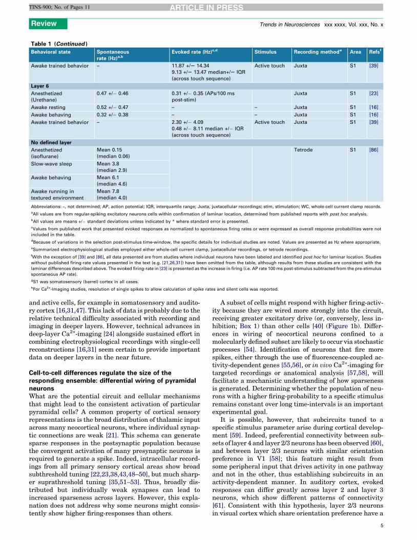
Table 1 (Continued )
Behavioral state Spontaneous
rate (Hz)a,bEvoked rate (Hz)c,d Stimulus Recording methode Area Refsf
Awake trained behavior – 11.87 +/S 14.34
9.13 +/S 13.47 median+/S IQR
(across touch sequence)
Active touch Juxta S1 [39]
Layer 6
Anesthetized
(Urethane)
0.47 +/� 0.46 0.31 +/� 0.35 (APs/100 ms
post-stim)
Juxta S1 [23]
Awake resting 0.52 +/� 0.47 – – Juxta S1 [16]
Awake behaving 0.32 +/� 0.38 – – Juxta S1 [16]
Awake trained behavior – 2.30 +/� 4.09
0.48 +/� 8.11 median +/� IQR
(across touch sequence)
Active touch Juxta S1 [39]
No defined layer
Anesthetized
(isoflurane)
Mean 0.15
(median 0.06)
Tetrode S1 [86]
Slow-wave sleep Mean 3.8
(median 2.9)
Awake behaving Mean 6.1
(median 4.6)
Awake running in
textured environment
Mean 7.8
(median 4.0)
Abbreviations: –, not determined; AP, action potential; IQR, interquartile range; Juxta, juxtacellular recordings; stim, stimulation; WC, whole-cell current clamp records.
aAll values are from regular-spiking excitatory neurons cells within confirmation of laminar location, determined from published reports with post hoc analysis.
bAll values are means +/� standard deviations unless indicated by * where standard error is presented.
cValues from published work that presented evoked responses as normalized to spontaneous firing rates or were expressed as overall response probabilities were not
included in the table.
dBecause of variations in the selection post-stimulus time-window, the specific details for individual studies are noted. Values are presented as Hz where appropriate.
eSummarized electrophysiological studies employed either whole-cell current clamp, juxtacellular recordings, or tetrode recordings.
fWith the exception of [39] and [86], all data presented are from studies where individual neurons have been labeled and identified post hoc for laminar location. Studies
without published firing-rate values presented in the text (e.g. [21,26,31]) have been omitted from the table, although results from these studies are consistent with the
laminar differences described above. The evoked firing-rate in [23] is presented as the increase in firing (i.e. AP rate 100 ms post-stimulus subtracted from the pre-stimulus
spontaneous AP rate).
gS1 was somatosensory (barrel) cortex in all cases.
hFor Ca2+-imaging studies, resolution of single spikes to allow calculation of spike rates and silent cells was reported.
Review Trends in Neurosciences xxx xxxx, Vol. xxx, No. x
TINS-900; No. of Pages 11
and active cells, for example in somatosensory and audito-ry cortex [16,31,47]. This lack of data is probably due to therelative technical difficulty associated with recording andimaging in deeper layers. However, technical advances indeep-layer Ca2+-imaging [24] alongside sustained effort incombining electrophysiological recordings with single-cellreconstructions [16,31] seem certain to provide importantdata on deeper layers in the near future.
Cell-to-cell differences regulate the size of theresponding ensemble: differential wiring of pyramidalneuronsWhat are the potential circuit and cellular mechanismsthat might lead to the consistent activation of particularpyramidal cells? A common property of cortical sensoryrepresentations is the broad distribution of thalamic inputacross many neocortical neurons, where individual synap-tic connections are weak [21]. This schema can generatesparse responses in the postsynaptic population becausethe convergent activation of many presynaptic neurons isrequired to generate a spike. Indeed, intracellular record-ings from all primary sensory cortical areas show broadsubthreshold tuning [22,23,38,43,48–50], but much sharp-er suprathreshold tuning [35,51–53]. Thus, broadly dis-tributed but individually weak synapses can lead toincreased sparseness across layers. However, this expla-nation does not address why some neurons might consis-tently show higher firing-responses than others.
A subset of cells might respond with higher firing-activ-ity because they are wired more strongly into the circuit,receiving greater excitatory drive (or, conversely, less in-hibition; Box 1) than other cells [40] (Figure 1b). Differ-ences in wiring of neocortical neurons confined to amolecularly defined subset are likely to occur via stochasticprocesses [54]. Identification of neurons that fire morespikes, either through the use of fluorescence-coupled ac-tivity-dependent genes [55,56], or in vivo Ca2+-imaging fortargeted recordings or anatomical analysis [57,58], willfacilitate a mechanistic understanding of how sparsenessis generated. Determining whether the population of neu-rons with a higher firing-probability to a specific stimulusremains constant over long time-intervals is an importantexperimental goal.
It is possible, however, that subcircuits tuned to aspecific stimulus parameter arise during cortical develop-ment [59]. Indeed, preferential connectivity between sub-sets of layer 4 and layer 2/3 neurons has been observed [60],and between layer 2/3 neurons with similar orientationpreference in V1 [58]; this feature might result fromsome peripheral input that drives activity in one pathwayand not in the other, thus establishing subcircuits in anactivity-dependent manner. In auditory cortex, evokedresponses can differ greatly across layer 2 and layer 3neurons, which show different patterns of connectivity[61]. Consistent with this hypothesis, layer 2/3 neuronsin visual cortex which share orientation preference have a
5

(a) (b)
(c) (e)
(d)
40
0˚ 90˚ 180˚ 270˚ 0˚ 90˚ 180˚ 270˚
0
0 ΔF = max
ΔF
/F (
%)
40
0
40
0
50
Depth = 400 μm
Depth = 85 μm
0
401
3 4
2
0
40
0
40
0
50
0
0 82 0
1
3
82Time (s) Time (s)
TRENDS in Neurosciences
Figure 2. Functional maps of selective responses in rat visual cortex with single-cell resolution. (a) In vivo images of cortical cells stained with a calcium indicator, OGB-1
AM. The top panel shows a volume of stained cells (depth of 85–400 mm including 3000 cells) reconstructed from images obtained with 1 mm spacing in depth; the bottom
panel is an anatomical image at 290 mm below the pia, averaged over all frames during the visual stimulation protocol. (b) Single-condition maps (DF) for eight directions
of visual stimuli (outer panels; the arrows indicate stimulus direction). Each map is the average of eight repeats. In this and subsequent figures, the scale bar (DF) only
applies to the outer panels. The central panel is redrawn from (a). (c) Pixel-based orientation map, in which hue is determined by the best orientation, overlaid with the
anatomical image in (a). (d) Time-courses of four orientation-selective cells (1–4) in (e). The upper traces show the average response to eight repeats (SEM in gray); lower
traces show three individual repeats. Visual stimulation periods for eight directions are indicated by grey bars. (e) Cell-based orientation map. Responsive cells (P = 0.01,
ANOVA; 45/115) are colored according to preferred orientation. Scale bars, 100 mm. Adapted, with permission, from [32].
Review Trends in Neurosciences xxx xxxx, Vol. xxx, No. x
TINS-900; No. of Pages 11
significantly higher probability of being connected to eachother than those cells with an orthogonal orientation [33].In layer 5, it has been observed that subgroups of neuronsmay share very strong connections, a feature that mightresult in correlated firing of these cells [62,63]. Experi-ments that address differential wiring and its impact onsparse firing in neocortical neurons are an importantpriority.
Cell-to-cell differences regulate the size of theresponding ensemble: intrinsic firing propertiesAssuming that the population of neurons within a givenclass or lamina is equivalent, differential firing of neocor-tical neurons might arise from intrinsic conductances thatfacilitate firing in some neurons and not others. This couldbe dynamic, regulated for example by prior activity (e.g.[64–66]), or might be entirely stochastic. Support for thisgeneral hypothesis has come from a long history of analysisof the intrinsic firing properties of neocortical neurons [67–69]. Differences in the intrinsic firing properties of other-wise equivalent pyramidal neurons could lead some cellsto spike consistently and others to be relatively silent.For example, brain-slice experiments comparing cellproperties across cortical layers have shown a more
6
hyperpolarized resting membrane potential in layer 2/3as compared to layer 5 pyramidal neurons that could resultin lower in vivo firing rates [70,71]. However, in a recentstudy from our labs, the increased spike-output of a class oflayer 2/3 pyramidal neurons was not associated withgreater intrinsic excitability [40] It is not yet clear whethersparseness could be generated by an identified subclass ofneocortical pyramidal neurons.
Behavioral state regulates the size of the respondingensemblePerforming an experiment under the appropriate behav-ioral conditions can have dramatic impact on the sensoryresponse and the size of the responding ensemble(Figure 1c). Animals process sensory information whilethey are awake and behaving, but the vast majority ofin vivo mammalian experiments have been performed onanesthetized or sedated animals. Furthermore, it is oftennot possible to determine the size of the ensemble becausein many cases mean firing-rates (not the fraction ofresponding cells) are reported.
Anesthesia alters both correlations between neuronsand reduces firing rates by approximately 30% in visualcortex [17]; this results in cell type-specific reductions in

Box 1. Inhibitory neurons and sparseness
In many studies, a small subset of neurons that respond with
multiple spikes and reliably across trials has been identified. Fewer
studies have attempted to classify these cells as excitatory or
inhibitory. In studies that distinguish between these two classes
(using genetically targeted fluorescent protein expression), it has
become clear that many of the high-firing neurons may in fact be
interneurons [38,43,58,92–95], a subpopulation that may constitute
20% of all neocortical neurons. Indeed, without genetic markers to
label specific cell types, or anatomical recovery and reconstruction
of the specific neuron recorded, it is difficult to interpret much of the
data that suggest sparse responses. Because many regular-spiking
interneuron subtypes are less stimulus-selective than excitatory
neurons (but have similar spike waveforms to pyramidal neurons
[43]) and show higher evoked firing rates, extracellular recordings
may have selectively focused on these cells. This bias would lead to
an overestimate of firing rates, and indicates that excitatory neuron
responses are even more sparse.
Inhibitory neurons will themselves generate sparse responses
across pyramidal cells. Sensory input strongly drives inhibitory
neuron firing and rapidly restricts the spread of excitation in the
cortex [96]. Several subtypes of inhibitory neurons show extremely
broad connectivity, where a single inhibitory neuron can be
synaptically connected to virtually all nearby pyramidal cells [97–
99]. Strong recurrent inhibition reduces overall firing output from
pyramidal cells, increasing the sparseness of response probabilities.
For example, a recent study found that increasing the size of the
visual stimulus generated more spikes in inhibitory interneurons
that reduce the spiking response of nearby pyramidal neurons [41].
Changing interneuron output by neuromodulation [100,101] could
alter the number of responding pyramidal cells, offering a point of
control to modulate sparseness.
Review Trends in Neurosciences xxx xxxx, Vol. xxx, No. x
TINS-900; No. of Pages 11
spontaneous firing rates in somatosensory cortex [16].Despite the relative increase in firing rates in awakeanimals, population analysis of firing activity in awakeanimals also suggests that sparseness may be maintained[16,39,44], especially in layer 2/3. A recent study hasconfirmed that sparse firing is present during active whis-ker-exploration [38], suggesting that it may result fromstrong inhibitory input (Box 1).
Some of the effects of anesthesia on firing rate maydepend upon the type of anesthesia administered, and on
(a)
0 418 588 890 1154
60
''L2/3'' ''L4'' ''L5'' ''L6''
50
40
30
Ove
rall
spik
e ra
te (
Hz)
20
10
0
Depth (μm)
Figure 3. Most spikes are fired by a small number of neurons. (a) Firing rate for each neu
epochs), as a function of cortical depth. Each circle corresponds to a single neuron (purp
vertical dashed lines. Note that some cells (black circles) have very low but non-zero firing r
‘silent neurons.’ Inset, cumulative histogram of the same data, both omitting (black) and
the extent of thalamic inactivation [72]. Unexpected pre-sentation of stimuli to awake, resting animals triggerslarge-amplitude spiking responses – perhaps reflecting asensory ‘wake-up call’ – whereas stimulation during atten-tive or active behaviors evokes a smaller amplitude re-sponse [25,73] (but see [74]).
The gold standard for examining sensory coding is torecord and manipulate neuronal activity while the animalis performing a task. Inspired by decades of primate work,rodents are now being trained to perform sophisticatedhead-restrained and freely moving behaviors while record-ing or manipulating neuronal activity with powerful bat-tery of novel techniques [13,39,75–77]. Interestingly, arecent study using juxtacellular recordings in all corticallayers in mice during a whisker-dependent tactile discrim-ination task showed firing rates with a higher thanexpected mean population firing-rate (and a higher frac-tion of responsive cells), especially in layer 5 [39] (Figure 3).Single-cell stimulation in infragranular layers may also besufficient to drive behavior in some cases (Box 2). Howeverthe data still support a sparse model for cortical layers 2/3and 6, where a minority (approximately 10%) of neuronsfired the majority of spikes (Figure 3). Studies in awake,motivated animals with careful cell identification willsignificantly add to our understanding of sparseness incortical responses.
Cortical states and the size of the responding ensembleNeocortical neurons are spontaneously active, displayingsub- and supra-threshold oscillations at a broad range offrequencies during different behavioral states [78]. At firstglance this activity appears to be large in amplitude (sub-threshold oscillations of 20 mV are common during periodsof quiet wakefulness in mouse [25,27,38,43,79] and rat[80,81] somatosensory cortex), highly variable, and strong-ly influences the spiking behavior of cortical neurons – notideal conditions for decoding small-amplitude sensory sti-muli. However, when mice enter active or attentive behav-ioral states, and slow, large-amplitude oscillations are
60
(b)
50
40
30
20
10
0
0 20 40 60Overall spike rate (Hz)
Cum
. fra
ctio
n of
neu
rons 1
.8
.6
.4
.20
0 10 20 30 40 50
Ove
rall
spik
e ra
te (
Hz)
Number of neurons
TRENDS in Neurosciences
ron during sensory-based task performance in mice (averaged across all behavioral
le, ‘silent neurons’). Laminar boundaries are indicated by colored bars at top and by
ates. (b) Histogram of the firing rate data in (a). The purple bar indicates the number of
including (purple) the silent neurons. Adapted, with permission, from [39].
7

Box 2. Sparse cortical stimulation for perception and behavior
Are a small number of firing neurons in the cortex sufficient for
sensory perception or motor behavior? Recent technical advances
have allowed stimulation of spikes in small numbers of cortical
neurons while simultaneously monitoring their effect on behavior.
Electrical stimulation of a small number of cortical neurons activates a
sparse and variable population of cortical neurons [102]. For example,
a 200 ms burst of 10 spikes elicited in only one neuron in rat motor
cortex can elicit small-amplitude whisker movements [103].
It is even possible to condition rats to report a small number of
spikes from very few neurons in sensory cortex [75,104]. Stimulat-
ing single neurons with trains of single spikes can have a small but
measurable impact on network activity [90] (Figure I). It is surprising
that effects from stimulating such a small population or even
single neurons are measurable at all at the network or behavioral
level, however in general these effects are relatively small (but see
[105]).
19
0
8
19
0
–1 0 1 Time (s)
Microstimulation
Catch trials
Single-cell stimulation
(c)
(b)
(a)
0
TRENDS in Neurosciences
Num
ber
of tr
ials
Figure I. Behavioral response to stimulation of a single cortical neuron. (a) Juxtacellular stimulation (at 0 s) of a single layer 5b cortical pyramidal neuron in an awake rat
increases the firing rate of the cell (each spike is one tick on the raster plot) in each of the 19 trials. In 47% of trials, stimulation also triggers a lick (marked by red square)
to obtain a water reward [90]. (b) Catch trials with no stimulation show few behavioral responses. (c) Microstimulation of many cells in the same cortical region drives
behavioral responses (in 71% of trials in this example) [90]. Adapted, with permission, from [90].
Review Trends in Neurosciences xxx xxxx, Vol. xxx, No. x
TINS-900; No. of Pages 11
replaced by smaller-amplitude, higher-frequency oscilla-tions, evoked firing-rates are reduced. It will be exciting toinvestigate whether similar changes in membrane poten-tial and firing rates are present in different corticalregions, and in different species, during trained behavioraltasks.
Such changes in cortical state can dramatically altersparseness, creating small time-windows where corticalneurons are more likely to fire [25,73,74,81,82]. In audi-tory cortex, extracellular recordings show that spikingresponses in awake, attending animals are suppressedcompared to passive listening [73]. In somatosensorycortex, deflection of a single whisker in an awake animalcan lead to spike suppression associated with decreasedexcitatory postsynaptic potential (EPSP) amplitude [25].
8
In V1, on the other hand, animal movement triggers anincrease in evoked spiking responses of individual neu-rons [74]. These effects could be due to depression of thethalamocortical synapse [83] due to high thalamic firing-rates [79], or to subcortical neuromodulatory input fromcholinergic or noradrenergic centers [84,85]. Further-more, attentional processes – release of neuromodula-tory factors – may also alter firing rates and sparseness[78].
Maybe responses are not sparse?It is important to consider that the relatively small fractionof responsive cells, especially in superficial layers, might bemerely an artifact of non-optimal stimulus presentation orimpoverished behavioral conditions (Figure 1d). This is

Review Trends in Neurosciences xxx xxxx, Vol. xxx, No. x
TINS-900; No. of Pages 11
more likely in whisker somatosensory cortex, where atypical stimulus consists of deflection of single whiskeror of all whiskers by an airpuff – a far cry from how thewhiskers might be using during behavior.
Consistent with this, extracellular recordings that usespike-sorting to record individual neurons in freely movinganimals have reported higher firing-rates than have stud-ies using head-fixed or immobilized animals [86]. However,cells that fire few spikes are still nearly impossible todetect in these studies. Although firing rates observed inS1 during awake behavior [39] are higher, these studiesconfirm that superficial layers contain a substantial num-ber of silent cells. Visual stimuli typically engage a largerfraction of the network. In addition, in auditory cortex,primate recording studies using naturalistic vocalizationstypically report higher firing-rates (and potentially moredense population responses) [87]. Stimulus optimization isa crucial parameter in assessing sparseness.
Why is it important to determine the size of the re-sponse ensemble? Across primary sensory cortex, synapticconnections are typically small, from 0.02 to 1 mV[70,88,89], such that spiking from a single cell is insuffi-cient to drive a spike in a downstream neuron (but see[90]). With spike thresholds typically �30 mV above theresting membrane potential, near-synchronous (i.e. with-in 10–50 ms) activation of tens to hundreds of cells mightbe required to drive downstream neurons. Too few neuronsthat spike in response to a stimulus effectively ends thetransfer of information across the chain. There is probablya balance between the computational advantages ofsparse neural activity (which can carry more informationthan more dense representations [7]) and the disadvan-tages of representations that are too sparse (which carryno information if they cannot drive downstream spikes).To evaluate computational models that rely on sparsefiring of large populations of neocortical neurons, it willbe important to perform experimental recordings withhigh temporal and spatial resolution from identified neu-rons, preferably in the context of an awake, attentive, andmotivated animal.
Concluding remarksIn summary, it is crucial to confirm that sparse represen-tations are used by the cerebral cortex, before determiningthe computational advantages of this phenomenon as acoding strategy (Box 3). Significant confusion betweeninstantaneous or population sparseness (Figure 1a) andlifetime sparseness (Figure 1b) must be addressed by long-
Box 3. Outstanding questions
� Why does sparseness change across cortical layers, with many
more silent neurons being observed in supragranular layers?
� How can high-throughput measures to address sparseness be
developed for analysis of layer 4 and deep cortical layers?
� Are the cells that show higher firing rates a stable subset of the
population?
� What kind of synaptic connectivity might underlie differential
firing across populations of neurons within a layer?
� Can neuromodulation alter sparseness?
� Does motivated behavior with a naturalistic stimulus reduce the
apparent sparseness of neocortical responses?
term recording or imaging studies, where individual neu-rons can be reliably monitored in the context of richsensory experience. Few studies have directly addressedthe long-term firing activity of neocortical neurons, andextracting data about silent or very low firing-rate neuronsfrom published work is fraught with difficulty. Despitethis, supragranular layers in particular exhibit a higherfraction of silent or nearly silent neurons. The broad rangeof firing rates of neurons, especially in supragranularlayers of primary sensory cortex, may reflect the differen-tial wiring of cortical circuits for distinct forms of sensa-tion, perception, and learning.
AcknowledgmentsSupported by grants from the National Institutes of Health (DA0171-88to A.L.B.), the Humboldt Foundation (A.L.B.), NeuroCure (A.L.B. andJ.F.A.P.), the Swiss–German Research Unit/Barrel Cortex FunctionDeutsche Forschungs Gemeinschaft, Forschergruppe 134 (J.F.A.P.), anda European Research Council starting grant (J.F.A.P.) We thank MichaelBrecht and members of the Barth and Poulet labs for helpful discussionand comments.
References1 Herculano-Houzel, S. (2009) The human brain in numbers: a linearly
scaled-up primate brain. Front. Hum. Neurosci. 3, 312 Dykes, R.W. and Lamour, Y. (1988) An electrophysiological study of
single somatosensory neurons in rat granular cortex serving thelimbs: a laminar analysis. J. Neurophysiol. 60, 703–724
3 Attwell, D. and Laughlin, S.B. (2001) An energy budget for signalingin the grey matter of the brain. J. Cereb. Blood Flow Metab. 21, 1133–1145
4 Barlow, H.B. (1972) Single units and sensation: a neuron doctrine forperceptual psychology? Perception 1, 371–394
5 Laughlin, S.B. and Sejnowski, T.J. (2003) Communication in neuronalnetworks. Science 301, 1870–1874
6 Lennie, P. (2003) The cost of cortical computation. Curr. Biol. 13,493–497
7 Olshausen, B.A. and Fields, D.J. (2004) Sparse coding of sensoryinputs. Curr. Opin. Neurobiol. 14, 481–487
8 Shoham, S. et al. (2006) How silent is the brain: is there a ‘dark matter’problem in neuroscience? J. Comp. Physiol. 192, 777–784
9 Wolfe, J. et al. (2010) Sparse and powerful cortical spikes. Curr. Opin.Neurobiol. 20, 306–312
10 Perez-Orive, J. et al. (2002) Oscillations and sparsening of odorrepresentations in the mushroom body. Science 297, 359–365
11 Papadopoulou, M. et al. (2011) Normalization for sparse encoding ofodors by a wide-field interneuron. Science 332, 721–725
12 Hahnloser, R.H. et al. (2002) An ultra-sparse code underlies thegeneration of neural sequences in a songbird. Nature 419, 65–70
13 Beloozerova, I.N. et al. (2003) Activity of different classes of neuronsof the motor cortex during locomotion. J. Neurosci. 23, 1087–1097
14 Burgalossi, A. et al. (2011) Microcircuits of functionally identifiedneurons in the rat medial entorhinal cortex. Neuron 70, 773–786
15 Epsztein, J. et al. (2011) Intracellular determinants of hippocampalCA1 place and silent cell activity in a novel environment. Neuron 70,109–120
16 de Kock, C.P. and Sakmann, B. (2009) Spiking in primarysomatosensory cortex during natural whisking in awake head-restrained rats is cell-type specific. Proc. Natl. Acad. Sci. U.S.A.106, 16446–16450
17 Greenberg, D.S. et al. (2008) Population imaging of ongoing neuronalactivity in the visual cortex of awake rats. Nat. Neurosci. 11, 749–751
18 Vincis, R. et al. (2012) Dense representation of natural odorants in themouse olfactory bulb. Nat. Neurosci. 15, 537–539
19 Arsenault, D. and Zhang, Z.W. (2006) Developmental remodelling ofthe lemniscal synapse in the ventral basal thalamus of the mouse. J.Physiol. 573, 121–132
20 Chen, C. and Regehr, W.G. (2000) Developmental remodeling of theretinogeniculate synapse. Neuron 28, 955–966
9

Review Trends in Neurosciences xxx xxxx, Vol. xxx, No. x
TINS-900; No. of Pages 11
21 Bruno, R.M. and Sakmann, B. (2006) Cortex is driven by weak butsynchronously active thalamocortical synapses. Science 312, 1622–1627
22 Brecht, M. and Sakmann, B. (2002) Dynamic representation ofwhisker deflection by synaptic potentials in spiny stellate andpyramidal cells in the barrels and septa of layer 4 ratsomatosensory cortex. J. Physiol. 543, 49–70
23 de Kock, C.P. et al. (2007) Layer- and cell-type-specific suprathresholdstimulus representation in rat primary somatosensory cortex. J.Physiol. 581, 139–154
24 Mittmann, W. et al. (2011) Two-photon calcium imaging of evokedactivity from L5 somatosensory neurons in vivo. Nat. Neurosci. 14,1089–1093
25 Crochet, S. and Petersen, C.C. (2006) Correlating whisker behaviorwith membrane potential in barrel cortex of awake mice. Nat.Neurosci. 9, 608–610
26 Hromadka, T. et al. (2008) Sparse representation of sounds in theunanesthetized auditory cortex. PLoS Biol. 6, e16
27 Poulet, J.F.A. and Petersen, C.C. (2008) Internal brain state regulatesmembrane potential synchrony in barrel cortex of behaving mice.Nature 454, 881–885
28 Swadlow, H.A. (1989) Efferent neurons and suspected interneurons inS-1 vibrissa cortex of the awake rabbit: receptive fields and axonalproperties. J. Neurophysiol. 62, 288–308
29 Swadlow, H.A. (1988) Efferent neurons and suspected interneurons inbinocular visual cortex of the awake rabbit: receptive fields andbinocular properties. J. Neurophysiol. 59, 1162–1187
30 Swadlow, H.A. (1990) Efferent neurons and suspected interneurons inS-1 forelimb representation of the awake rabbit: receptive fields andaxonal properties. J. Neurophysiol. 63, 1477–1498
31 Sakata, S. and Harris, K.D. (2009) Laminar structure of spontaneousand sensory-evoked population activity in auditory cortex. Neuron 64,404–418
32 Ohki, K. et al. (2005) Functional imaging with cellular resolutionreveals precise micro-architecture in visual cortex. Nature 433, 597–603
33 Ko, H. et al. (2011) Functional specificity of local synaptic connectionsin neocortical networks. Nature 473, 87–91
34 Kerr, J.N. et al. (2007) Spatial organization of neuronal populationresponses in layer 2/3 of rat barrel cortex. J. Neurosci. 27, 13316–13328
35 Poo, C. and Isaacson, J.S. (2009) Odor representations in olfactorycortex: ‘sparse’ coding, global inhibition, and oscillations. Neuron 62,850–861
36 Stettler, D.D. and Axel, R. (2009) Representations of odor in thepiriform cortex. Neuron 63, 854–864
37 Chen, X. et al. (2011) A gustotopic map of taste qualities in themammalian brain. Science 333, 1262–1266
38 Crochet, S. et al. (2011) Synaptic mechanisms underlying sparsecoding of active touch. Neuron 69, 1160–1175
39 O’Connor, D.H. et al. (2010) Neural activity in barrel cortexunderlying vibrissa-based object localization in mice. Neuron 67,1048–1061
40 Yassin, L. et al. (2010) An embedded subnetwork of highly activeneurons in the neocortex. Neuron 68, 1043–1050
41 Haider, B. et al. (2010) Synaptic and network mechanisms of sparseand reliable visual cortical activity during nonclassical receptive fieldstimulation. Neuron 65, 107–121
42 Vinje, W.E. and Gallant, J.L. (2000) Sparse coding and decorrelationin primary visual cortex during natural vision. Science 287, 1273–1276
43 Gentet, L.J. et al. (2010) Membrane potential dynamics of GABAergicneurons in the barrel cortex of behaving mice. Neuron 65, 422–435
44 Sawinski, J. et al. (2009) Visually evoked activity in cortical cellsimaged in freely moving animals. Proc. Natl. Acad. Sci. U.S.A. 106,19557–19562
45 Minderer, M. et al. (2011) Chronic imaging of cortical sensory mapdynamics using a genetically encoded calcium indicator. J. Physiol.590, 99–107
46 Manns, I.D. et al. (2004) Sub- and suprathreshold receptive fieldproperties of pyramidal neurones in layers 5A and 5B of ratsomatosensory barrel cortex. J. Physiol. 556, 601–622
10
47 Zhou, Y. et al. (2010) Preceding inhibition silences layer 6 neurons inauditory cortex. Neuron 65, 706–717
48 Brecht, M. et al. (2003) Dynamic receptive fields of reconstructedpyramidal cells in layers 3 and 2 of rat somatosensory barrel cortex. J.Physiol. 553, 243–265
49 Moore, C.I. and Nelson, S.B. (1998) Spatio-temporal subthresholdreceptive fields in the vibrissa representation of rat primarysomatosensory cortex. J. Neurophysiol. 80, 2882–2892
50 Varga, Z. et al. (2011) Dendritic coding of multiple sensory inputs insingle cortical neurons in vivo. Proc. Natl. Acad. Sci. U.S.A. 108,15420–15425
51 Jia, H. et al. (2010) Dendritic organization of sensory input to corticalneurons in vivo. Nature 464, 1307–1312
52 Carandini, M. and Ferster, D. (2000) Membrane potential and firingrate in cat primary visual cortex. J. Neurosci. 20, 470–484
53 Bringuier, V. et al. (1999) Horizontal propagation of visual activity inthe synaptic integration field of area 17 neurons. Science 283, 695–699
54 Zariwala, H.A. et al. (2011) Visual tuning properties of geneticallyidentified layer 2/3 neuronal types in the primary visual cortex of cre-transgenic mice. Front. Syst. Neurosci. 4, 162
55 Barth, A.L. et al. (2004) Alteration of neuronal firing properties afterin vivo experience in a FosGFP transgenic mouse. J. Neurosci. 24,6466–6475
56 Wang, K.H. et al. (2006) In vivo two-photon imaging reveals a role ofarc in enhancing orientation specificity in visual cortex. Cell 126, 389–402
57 Bock, D.D. et al. (2011) Network anatomy and in vivo physiology ofvisual cortical neurons. Nature 471, 177–182
58 Hofer, S.B. et al. (2011) Differential connectivity and responsedynamics of excitatory and inhibitory neurons in visual cortex.Nat. Neurosci. 14, 1045–1052
59 Schubert, D. et al. (2007) Mapping functional connectivity in barrel-related columns reveals layer- and cell type-specific microcircuits.Brain Struct. Funct. 212, 107–119
60 Yoshimura, Y. et al. (2005) Excitatory cortical neurons form fine-scalefunctional networks. Nature 433, 868–873
61 Oviedo, H.V. et al. (2011) The functional asymmetry of auditory cortexis reflected in the organization of local cortical circuits. Nat. Neurosci.13, 1413–1420
62 Song, S. et al. (2005) Highly nonrandom features of synapticconnectivity in local cortical circuits. PLoS Biol. 3, e68
63 Perin, R. et al. (2011) A synaptic organizing principle for corticalneuronal groups. Proc. Natl. Acad. Sci. U.S.A. 108, 5419–5424
64 Bernard, C. et al. (2004) Acquired dendritic channelopathy intemporal lobe epilepsy. Science 305, 532–535
65 Desai, N.S. et al. (1999) Plasticity in the intrinsic excitability ofcortical pyramidal neurons. Nat. Neurosci. 2, 515–520
66 Shruti, S. et al. (2008) A seizure-induced gain-of-function in BKchannels is associated with elevated firing activity in neocorticalpyramidal neurons. Neurobiol. Dis. 30, 323–330
67 Connors, B.W. et al. (1982) Electrophysiological properties ofneocortical neurons in vitro. J. Neurophysiol. 48, 1302–1320
68 Gray, C.M. and McCormick, D.A. (1996) Chattering cells: superficialpyramidal neurons contributing to the generation of synchronousoscillations in the visual cortex. Science 274, 109–113
69 Nowak, L.G. et al. (2003) Electrophysiological classes of cat primaryvisual cortical neurons in vivo as revealed by quantitative analyses. J.Neurophysiol. 89, 1541–1566
70 Lefort, S. et al. (2009) The excitatory neuronal network of the C2barrel column in mouse primary somatosensory cortex. Neuron 61,301–316
71 Mason, A. and Larkman, A. (1990) Correlations between morphologyand electrophysiology of pyramidal neurons in slices of rat visualcortex. II. Electrophysiology. J. Neurosci. 10, 1415–1428
72 Hirata, A. and Castro-Alamancos, M.A. (2010) Neocortex networkactivation and deactivation states controlled by the thalamus. J.Neurophysiol. 103, 1147–1157
73 Otazu, G.H. et al. (2009) Engaging in an auditory task suppressesresponses in auditory cortex. Nat. Neurosci. 12, 646–654
74 Niell, C.M. and Stryker, M.P. (2010) Modulation of visual responsesby behavioral state in mouse visual cortex. Neuron 65, 472–479
75 Houweling, A.R. and Brecht, M. (2008) Behavioural report of singleneuron stimulation in somatosensory cortex. Nature 451, 65–68

Review Trends in Neurosciences xxx xxxx, Vol. xxx, No. x
TINS-900; No. of Pages 11
76 Stuttgen, M.C. and Schwarz, C. (2008) Psychophysical andneurometric detection performance under stimulus uncertainty.Nat. Neurosci. 11, 1091–1099
77 Komiyama, T. et al. (2010) Learning-related fine-scale specificityimaged in motor cortex circuits of behaving mice. Nature 464,1182–1186
78 Harris, K.D. and Thiele, A. (2011) Cortical state and attention. Nat.Rev. Neurosci. 12, 509–523
79 Poulet, J.F.A. et al. (2012) Thalamic control of cortical states. Nat.Neurosci. 15, 370–372
80 Okun, M. et al. (2010) The subthreshold relation between cortical localfield potential and neuronal firing unveiled by intracellularrecordings in awake rats. J. Neurosci. 30, 4440–4448
81 Petersen, C.C. et al. (2003) Interaction of sensory responses withspontaneous depolarization in layer 2/3 barrel cortex. Proc. Natl.Acad. Sci. U.S.A. 100, 13638–13643
82 Haider, B. and McCormick, D.A. (2009) Rapid neocortical dynamics:cellular and network mechanisms. Neuron 62, 171–189
83 Castro-Alamancos, M.A. (2004) Absence of rapid sensory adaptation inneocortex during information processing states. Neuron 41, 455–464
84 Goard, M. and Dan, Y. (2009) Basal forebrain activation enhancescortical coding of natural scenes. Nat. Neurosci. 12, 1444–1449
85 Constantinople, C.M. and Bruno, R.M. (2011) Effects and mechanismsof wakefulness on local cortical networks. Neuron 69, 1061–1068
86 Vijayan, S. et al. (2010) Activity in the barrel cortex during activebehavior and sleep. J. Neurophysiol. 103, 2074–2084
87 Wang, X. et al. (2005) Sustained firing in auditory cortex evoked bypreferred stimuli. Nature 435, 341–346
88 Hardingham, N.R. et al. (2010) Quantal analysis reveals a functionalcorrelation between presynaptic and postsynaptic efficacy inexcitatory connections from rat neocortex. J. Neurosci. 30, 1441–1451
89 Oswald, A.M. and Reyes, A.D. (2008) Maturation of intrinsic andsynaptic properties of layer 2/3 pyramidal neurons in mouse auditorycortex. J. Neurophysiol. 99, 2998–3008
90 London, M. et al. (2010) Sensitivity to perturbations in vivo implies highnoise and suggests rate coding in cortex. Nature 466, 123– 127
91 Jadhav, S.P. et al. (2009) Sparse temporal coding of elementary tactilefeatures during active whisker sensation. Nat. Neurosci. 12, 792–800
92 Kerlin, A.M. et al. (2010) Broadly tuned response properties of diverseinhibitory neuron subtypes in mouse visual cortex. Neuron 67, 858–871
93 Kuhlman, S.J. et al. (2011) Fast-spiking interneurons have an initialorientation bias that is lost with vision. Nat. Neurosci. 14, 1121–1123
94 Sohya, K. et al. (2007) GABAergic neurons are less selective tostimulus orientation than excitatory neurons in layer II/III ofvisual cortex, as revealed by in vivo functional Ca2+ imaging intransgenic mice. J. Neurosci. 27, 2145–2149
95 Runyan, C.A. et al. (2010) Response features of parvalbumin-expressing interneurons suggest precise roles for subtypes ofinhibition in visual cortex. Neuron 67, 847–857
96 Isaacson, J.S. and Scanziani, M. (2011) How inhibition shapes corticalactivity. Neuron 72, 231–243
97 Fino, E. and Yuste, R. (2011) Dense inhibitory connectivity inneocortex. Neuron 69, 1188–1203
98 Silberberg, G. and Markram, H. (2007) Disynaptic inhibition betweenneocortical pyramidal cells mediated by Martinotti cells. Neuron 53,735–746
99 Kapfer, C. et al. (2007) Supralinear increase of recurrent inhibitionduring sparse activity in the somatosensory cortex. Nat. Neurosci. 10,743–753
100 Froemke, R.C. et al. (2007) A synaptic memory trace for corticalreceptive field plasticity. Nature 450, 425–429
101 Letzkus, J.J. et al. (2011) A disinhibitory microcircuit for associativefear learning in the auditory cortex. Nature 480, 331–335
102 Histed, M.H. et al. (2009) Direct activation of sparse, distributedpopulations of cortical neurons by electrical microstimulation.Neuron 63, 508–522
103 Brecht, M. et al. (2004) Whisker movements evoked by stimulation ofsingle pyramidal cells in rat motor cortex. Nature 427, 704–710
104 Huber, D. et al. (2008) Sparse optical microstimulation in barrelcortex drives learned behaviour in freely moving mice. Nature 451,61–67
105 Li, C.Y.T. et al. (2009) Burst spiking of a single cortical neuronmodifies global brain state. Science 324, 643–646
106 Kampa, B.M. et al. (2011) Representation of visual scenes by localneuronal populations in layer 2/3 of mouse visual cortex. Front.Neural Circuits 5, 18
107 Huber, D. et al. (2012) Multiple dynamic representations in the motorcortex during sensorimotor learning. Nature 484, 473–478
108 Avermann, M. et al. (2012) Microcircuits of excitatory and inhibitoryneurons in layer 2/3 of mouse barrel cortex. J. Neurophysiol. http://dx.doi.org/10.1152/jn.00917.2011
109 Mateo, C. et al. (2011) In vivo optogenetic stimulation of neocorticalexcitatory neurons drives brain-state-dependent inhibition. CurrBiol. 21, 1593–1602
11