Experimental arthritis C57black/6 normal and beige ...
7
Annals of the Rheumatic Diseases, 1988; 47, 940-946 Experimental arthritis in C57black/6 normal and beige (Chediak-Higashi) mice: in vivo and in vitro observations on cartilage degradation JOOST SCHALKWIJK, LEO A B JOOSTEN, WIM B VAN DEN BERG, AND LEVINUS B A VAN DE PUTTE From the Department of Rheumatology, University Hospital St Radboud, University of Nijmegen, Geert Grooteplein Zuid 8, 6500 HB Nijmegen, The Netherlands SUMMARY Mice with the beige mutation are known to be deficient for polymorphonuclear leucocyte (PMN) elastase and cathepsin G and can therefore be used as a model for protease dependence of tissue destruction in inflammatory conditions. The in vitro and in vivo effect of PMN activation on cartilage damage in C57black/6 normal and beige mice was measured. In vitro it was found that stimulation of normal PMNs with chemotactic peptide caused degradation of articular cartilage matrix owing to an elastase dependent mechanism; PMNs of beige mice did not induce degradation of cartilage. In vivo, using zymosan induced arthritis, which is a model characterised by a PMN-rich infiltrate and exudate, no significant differences were found between the two strains with respect to (a) joint oedema formation as measured by technetium-99m uptake; (b) matrix degradation as measured quantitatively and with histology; (c) chondrocyte proteoglycan synthesis as measured by radiosulphate uptake. At day 28 after induction of arthritis, when inflammation is waning, no differences in end stage irreversible damage to joint tissues were found. The relevance of these observations to the supposed role of PMN derived neutral proteases in joint inflammation is discussed. Key words: elastase, cathepsin G. Lysosomal proteases secreted by inflammatory cells such as polymorphonuclear leucocytes (PMNs) and macrophages have been strongly implicated in the pathological degradation of tissues in various dis- orders, including lung emphysema,' 2 nephritis,3 and arthritis." In particular, neutral proteases such as elastase, cathepsin G, and collagenase could be of interest as it has been shown (largely in vitro) that they have the potency to degrade a variety of connective tissue molecules at physiological pH.49 On the other hand, the role of these proteases has been questioned as there is a large amount of protease inhibitor present in body fluids such as plasma and synovial fluid.'0 Recently, however, we have shown that elastase can escape inactivation by a, proteinase inhibitor, thereby causing cartilage Accepted for publication 24 March 1988. Correspondence to Dr Joost Schalkwijk, Department of Rheuma- tology, University Hospital St Radboud, Geert Grooteplein Zuid 8, 6500 HB Nijmegen, The Netherlands. degradation even in the presence of a physiological concentration of at proteinase inhibitor.6 As there is at the moment no conclusive evidence for a role of elastase in cartilage degradation in vivo we examined the effect of experimental joint inflammation in C57black/6 normal and beige mice, the latter being deficient for PMN elastase and cathepsin G."1 Using quantitative methods we showed that in vitro normal PMNs can degrade cartilage, whereas the PMNs from beige mice are without effect. In vivo, however, using an experimental arthritis model, there is no difference in the rate of cartilage degradation between the two mice strains. Materials and methods ANIMALS Male, normal C57BI/6 and beige C57B1/6 (bg/bg) mice were obtained from Harlan OLAC Ltd, UK. Mice were fed a standard diet and tap water freely. They were used between the age of 10 and 16 weeks. 940 copyright. on November 23, 2021 by guest. Protected by http://ard.bmj.com/ Ann Rheum Dis: first published as 10.1136/ard.47.11.940 on 1 November 1988. Downloaded from
Transcript of Experimental arthritis C57black/6 normal and beige ...

Annals of the Rheumatic Diseases, 1988; 47, 940-946
Experimental arthritis in C57black/6 normal andbeige (Chediak-Higashi) mice: in vivo and in vitroobservations on cartilage degradationJOOST SCHALKWIJK, LEO A B JOOSTEN, WIM B VAN DEN BERG,AND LEVINUS B A VAN DE PUTTE
From the Department of Rheumatology, University Hospital St Radboud, University of Nijmegen, GeertGrooteplein Zuid 8, 6500 HB Nijmegen, The Netherlands
SUMMARY Mice with the beige mutation are known to be deficient for polymorphonuclearleucocyte (PMN) elastase and cathepsin G and can therefore be used as a model for protease
dependence of tissue destruction in inflammatory conditions. The in vitro and in vivo effect ofPMN activation on cartilage damage in C57black/6 normal and beige mice was measured. In vitroit was found that stimulation of normal PMNs with chemotactic peptide caused degradation ofarticular cartilage matrix owing to an elastase dependent mechanism; PMNs of beige mice did not
induce degradation of cartilage. In vivo, using zymosan induced arthritis, which is a modelcharacterised by a PMN-rich infiltrate and exudate, no significant differences were foundbetween the two strains with respect to (a) joint oedema formation as measured bytechnetium-99m uptake; (b) matrix degradation as measured quantitatively and with histology;(c) chondrocyte proteoglycan synthesis as measured by radiosulphate uptake. At day 28 afterinduction of arthritis, when inflammation is waning, no differences in end stage irreversibledamage to joint tissues were found. The relevance of these observations to the supposed role ofPMN derived neutral proteases in joint inflammation is discussed.
Key words: elastase, cathepsin G.
Lysosomal proteases secreted by inflammatory cellssuch as polymorphonuclear leucocytes (PMNs) andmacrophages have been strongly implicated in thepathological degradation of tissues in various dis-orders, including lung emphysema,' 2 nephritis,3and arthritis." In particular, neutral proteases suchas elastase, cathepsin G, and collagenase could be ofinterest as it has been shown (largely in vitro) thatthey have the potency to degrade a variety ofconnective tissue molecules at physiological pH.49On the other hand, the role of these proteases hasbeen questioned as there is a large amount ofprotease inhibitor present in body fluids such asplasma and synovial fluid.'0 Recently, however, wehave shown that elastase can escape inactivation bya, proteinase inhibitor, thereby causing cartilage
Accepted for publication 24 March 1988.Correspondence to Dr Joost Schalkwijk, Department of Rheuma-tology, University Hospital St Radboud, Geert Grooteplein Zuid8, 6500 HB Nijmegen, The Netherlands.
degradation even in the presence of a physiologicalconcentration of at proteinase inhibitor.6 As there isat the moment no conclusive evidence for a role ofelastase in cartilage degradation in vivo we examinedthe effect of experimental joint inflammation inC57black/6 normal and beige mice, the latter beingdeficient for PMN elastase and cathepsin G."1 Usingquantitative methods we showed that in vitronormal PMNs can degrade cartilage, whereas thePMNs from beige mice are without effect. In vivo,however, using an experimental arthritis model,there is no difference in the rate of cartilagedegradation between the two mice strains.
Materials and methods
ANIMALSMale, normal C57BI/6 and beige C57B1/6 (bg/bg)mice were obtained from Harlan OLAC Ltd, UK.Mice were fed a standard diet and tap water freely.They were used between the age of 10 and 16 weeks.
940
copyright. on N
ovember 23, 2021 by guest. P
rotected byhttp://ard.bm
j.com/
Ann R
heum D
is: first published as 10.1136/ard.47.11.940 on 1 Novem
ber 1988. Dow
nloaded from
CHEMICALSZymosan, cytochrome c, superoxide dismutase, N-formyl - methionyl - leucyl - phenylalanine (FMLP),Ellman's reagent, cytochalasin B, and phorbolmyristate acetate (PMA) were obtained from SigmaChemicals Co, USA. Methoxysuccinyl-alanyl-alanyl-prolyl-valyl-chloromethylketone (MAAPV-CMK),methoxysuccinyl-alanyl- alanyl-prolyl-valyl - amino -
methylcoumarin, and succinyl-phenylalanyl-leucyl-thiobenzylester were obtained from Bachem,Bubendorf, Switzerland.
ENZYME MEASUREMENTS
Elastase was measured fluorimetrically usingmethoxysuccinyl-alanyl -alanyl-prolyl-valyl-amino -
methylcoumarin as substrate. 12 In this assay7-amino-4-methylcoumarin is released from the pep-tide and measured at 370 nm (excitation wavelength)and 460 nm wavelength (emission wavelength).CathepsinGwasmeasured using succingl-phenylalanyl-leucyl-thiobenzylester as substrate.1 In this assaycathepsin G acts as an esterase that specificallycleaves off the benzylthiol which could then bedetected colorimetrically in a coupled assay withEllman's reagent. Enzyme measurements were per-formed on extracts of mouse peritoneal exudates,which were obtained by the injection of 0-5 mlparaffin oil. After 24 hours the cells (90-95% PMNs)were collected from the peritoneal cavity, washed,and extracted in 0.5 M NaCl, 0-1 M trometamol(TRIS), 0.4% cetrimide, 0*1% Triton X-100 pH 8-0.Values for the enzyme content of mouse PMNs
were obtained as the mean (SD) of three separatemeasurements on pools of peritoneal exudate cellsof four mice in each experiment.
SUPEROXIDE MEASUREMENT
Superoxide production was measured by thecytochrome c reduction method as describedpreviously. 14 We used peritoneal exudate cellswhich were obtained as described above. Cells weresuspended in Dulbecco's phosphate buffered saline(DPBS) and stimulated with PMA (100 [ig/l). Thesuperoxide dismutase inhibitable cytochrome c re-duction was converted to nmoles of superoxideproduced per 106 cells per minute, assumin~a molarextinction coefficient of 21 000 M-1 cm- (differ-ence between reduced and oxidised forms).
Values for superoxide production by beige andnormal mice (mean (SD)) were obtained from sixanimals per strain.
DEGRADATION OF ARTICULAR CARTILAGEIN VITROThe capacity of PMNs to degrade articular cartilagein vitro was measured as described previously for
Cartilage degradation in beige mice 941
human peripheral blood PMNs.6 Briefly, PMNssuspended in DPBS (5 x 10⁶ cells/ml) were added tomurine patellar cartilage which was prelabelled with[35S]sulphate. The PMNs were stimulated withFMLP (10-6 M) in the presence of cytochalasin B(5 mg/l). Incubations were performed at 37°C inmicrotitre plates. After a two hour incubationperiod the cartilage samples were washed and theresidual radioactivity in the cartilage measured byliquid scintillation counting and expressed as apercentage of the initial radioactivity. As shown inprevious reports15 over 90% of the [35S]sulphate isassociated with the glycosaminoglycan fraction.Loss of 35S labelled material from the cartilage istherefore taken to indicate breakdown of proteo-glycans.
ARTHRITIS INDUCTION: MEASUREMENTOF INFLAMMATIONJoint inflammation was induced in beige and normalmice in the right knee joint by the intra-articularinjection of 180 ig of zymosan as previouslydescribed.16 Arthritis was measured by technetium-99m (99mTc) uptake as described before.16 17Briefly, the accumulation of 99'Tc is measured inthe right (R) and left (L) control knee joint andexpressed as a ratio (RJL). This ratio is taken as ameasure of joint swelling. Ratios exceeding 1-1 wereconsidered to indicate inflammation of the rightknee.
HISTOLOGYMouse knee joints (six animals per group) wereprocessed for histology as described before. His-tology of zymosan induced arthritis was carried outon days 2, 7, and 28 after induction of arthritis.Arthritis was scored on serial sections (haematoxy-lin and eosin staining) using a combined score ofexudate in the joint space and synovial infiltration.'8Proteoglycan depletion of cartilage matrix wasscored semiquantitatively on Saffranin 0 stainedsections.
MEASUREMENT OF IN VIVO CARTILAGEDEGRADATIONCartilage of beige and normal mice was labelled invivo with [35S]sulphate by the intrapentoneal injec-tion of 1.85 MBq radiosulphate in saline. After 24hours, when labelling was completed (the bulk ofthe radiosulphate is cleared from the circulation),arthritis was induced as described above. At 12, 24,and 48 hours after induction of arthritis the patellarcartilage of the right (arthritic) and left (control)knee joint was dissected and the 35S contentmeasured by liquid scintillation counting (five miceat each time). Radiolabel in the patellar cartilage
copyright. on N
ovember 23, 2021 by guest. P
rotected byhttp://ard.bm
j.com/
Ann R
heum D
is: first published as 10.1136/ard.47.11.940 on 1 Novem
ber 1988. Dow
nloaded from

942 Schalkwijk, Joosten, van den Berg, van de Putte
was shown to be associated with the glycosaminogly-can fraction.15 Cartilage degradation is expressed as
the percentage of radiolabelled proteoglycans lostfrom the arthritic patella compared with the control.
INHIBITION OF CHONDROCYTEPROTEOGLYCAN SYNTHESIS
At several intervals after the induction of arthritismice were killed, and the patellae of the right(inflamed) and left (control) joint were dissected.[35S]Sulphate incorporation in vitro was performedas described in earlier reports.13 14 The incorpora-tion into the right patella was compared with thatinto the control and taken as a measure forproteoglycan synthesis.
Results
PMN FUNCTION IN BEIGE ANDNORMAL MICEBoth oxygen metabolites and lysosomal proteasesare known to affect cartilage integrity in vitro.7 14
Therefore we measured the superoxide productionof PMNs from beige and normal mice and checkedthe elastase and cathepsin G content, which wasreported to be lower in the beige mice."' Mouseperitoneal exudate cells were obtained by theinjection of paraffin oil and harvested 24 hours later.The cells were washed and suspended in DPBS.Superoxide production by PMNs after stimulationwith PMA was not significantly different for PMNsfrom beige and normal mice: 3-4 (0-5) and 2-9 (0.7)nmol 02/min/106 cells respectively. We could con-
firm earlier observations that PMNs from beigemice are lower in cathepsin G and elastase thanthose from normal mice, though the values were
somewhat higher than reported by others."1 Wefound that the elastase and cathepsin G content ofPMNs from beige mice were 14 (3%) and 11 (5%)respectively, compared with PMNs from normalmice (mean (SD) of three measurements on pools offive peritoneal exudates).
IN VITRO CARTILAGE DEGRADATION BY
ACTIVATED PMNsMouse patellar cartilage, prelabelled with[35Slsulphate in vitro, was exposed to PMNs frombeige and normal mice. Table 1 shows that restingPMNs from either strain did not induce measurablecartilage degradation in a two hour incubation.Upon stimulation with FMLP in the presence ofcytochalasin B, agents that are known to inducesuperoxide production and degranulation, normalPMNs caused nearly 70% degradation of cartilageproteoglycans, whereas no significant cartilagebreakdown was seen with PMNs from beige mice.
Table 1 In vitro cartilage degradation by PMNs frombeige and normal mice. Values are mean (SD)
Treatment of [35S]Sulphate content ofcartilage patellar cartilage (cpm)
Dulbecco's 961 (197)Normal PMNs non-stimulated 845 (246)Normal PMNs stimulated 295 (71)*Beige PMNs non-stimulated 857 (127)Beige PMNs stimulated 909 (124)tNormal PMNs stimulated in the
presence of MAAPV-CMK§ 940 (104)f
Cartilage, prelabelled with [35Sjsulphate (six patellae a group) wasincubated with 2x 10⁷ mouse PMNs/ml in Dulbecco's medium.PMNs were stimulated with FMLP (10'6 M) and cytochalasin B(10 mg/l). After two hours the 35S content of the cartilage wasmeasured and compared with that of cartilage kept in medium orexposed to non-stimulated cells. The specific elastase inhibitorMAAPV-CMK was added at 200 tsmol/l.*p<O-005 compared with non-stimulated normal PMNs, two tailedMann-Whitney U test.tNot significantly different from non-stimulated PMNs from beigemice.1p<O-005, significantly different from stimulated normal PMNs inthe absence of MAAPV-CMK.§MAAPV - CMK = methoxysuccinyl - alanyl - alanyl - prolyl - valyl -chloromethylketone.
The mechanism of cartilage breakdown was shownto be elastase dependent as the specific elastaseinhibitor MAAPV-CMK completely prevented car-tilage degradation. MAAPV-CMK did not interferewith PMN stimulation. The results for cartilagedegradation by PMNs from normal mice coincidewith data we recently published on the same testsystem with human PMNs.6
EXPERIMENTAL ARTHRITIS IN BEIGEAND NORMAL MICEArthritis was induced by the injection of 180 ,ugzymosan into the right knee joint. At days 2 and 7arthritis was measured by 99mTc uptake, which is ameasure for joint oedema. Table 2 shows 99mTc
Table 2 Measurement of arthritis in beige and normalmice. Values are mean (SD)
Mice 99mTc ratio
Day 2 Day 7
Beige 1-91 (0-19) 1-26 (0-14)Normal 1-90 (0-16) 1-24 (0-08)
Six mice per group were injected with 180 tsg of zymosan at day 0.At days 2 and 7 arthritis was measured by the 99mTc uptakemethod. The ratio of '"Tc uptake in the right and left knee jointis taken as a measure for joint oedema.No significant differences were measured, two tailed Mann-Whitney U test.
copyright. on N
ovember 23, 2021 by guest. P
rotected byhttp://ard.bm
j.com/
Ann R
heum D
is: first published as 10.1136/ard.47.11.940 on 1 Novem
ber 1988. Dow
nloaded from

uptake for both mouse strains. No significantdifferences were noted at any time.At days 2, 7, and 28 animals were killed for
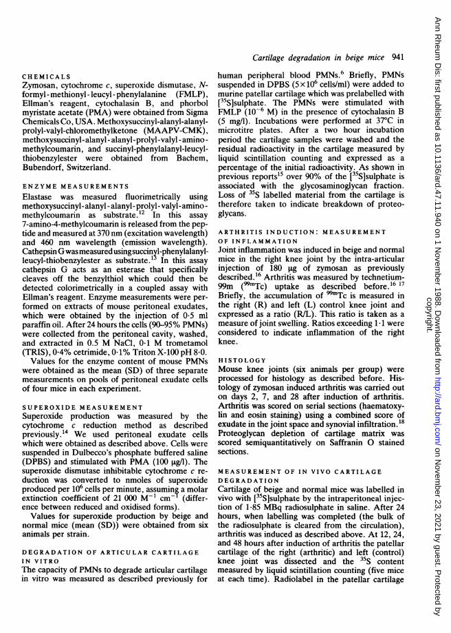
histology. Inflammation was scored on serial sec-tions using cellular exudate in the joint space,synovial infiltration, and depletion of cartilagematrix as seen on Saffranin 0 stained sections. Nodifferences in histological parameters were seen.Beige mice showed a strong inflammatory responseagainst zymosan (Figs 1, 2, and 3). The acute phaseof arthritis was characterised by a PMN-rich exudatein the joint space and a heavily infiltrated synovium.At day 28 chondrocyte death and bone apposition
Cartilage degradation in beige mice 943
could be seen in some of the animals. The histo-logical picture of zymosan induced arthritis in beigemice was similar to that of normal C57black mice asdescribed in earlier reports.'6 18 19
QUANTITATIVE MEASUREMENT OFCARTILAGE DAMAGETo investigate possible differences in cartilage de-gradation and inhibition of chondrocyte metabolismbetween beige and normal mice we used[35S]sulphate labelling of cartilage proteoglycans.Cartilage was labelled in vivo by [35S]sulphate, andafter 24 hours, when labelling was complete, arthri-
t.'. ;
tR
'..'
I A4Fig. 1 Normal knee joint ofC57BI/6 beige mouse. Section ofnormaljointfrom a C57B1/6 beigemouse. Haematoxylin and eosin(H&E) staining. P=patella;F=femur.
Fig. 2 Arthritic knee joint ofC57BI16 beige mouse. Section ofinflamed knee joint two days afterinduction ofarthritis with 180 pgzymosan. Note the exudate in thejoint space and the cellularinfiltration in the synovium. H&Estaining. P=patella; F=femur;S=synovium.
copyright. on N
ovember 23, 2021 by guest. P
rotected byhttp://ard.bm
j.com/
Ann R
heum D
is: first published as 10.1136/ard.47.11.940 on 1 Novem
ber 1988. Dow
nloaded from
944 Schalkwijk, Joosten, van den Berg, van de Putte
' Lir*B r
4*A
s. i5.
4dr t40 1.
.,.. ikft-Oo"',*21wl
.ct
leOi
*44
tis was induced in the right knee joint as describedabove. At 12, 24, and 48 hours after induction ofarthritis the patellar cartilage of the right (arthritic)and the left (control) knee joints was dissected and
40
*0-
(o
E0 20
12 24 48
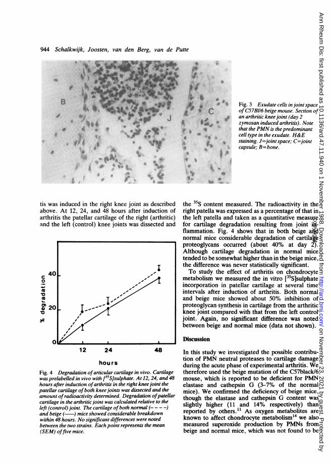
hoursFig. 4 Degradation ofarticular cartilage in vivo. Cartilagewas prelabelled in vivo with l3S]sulphate. At 12, 24, and 48hours after induction ofarthritis in the right knee joint thepatellar cartilage ofboth knee joints was dissected and theamount ofradioactivity determined. Degradation ofpatellarcartilage in the arthritic joint was calculated relative to theleft (control) joint. The cartilage ofboth normal (-- - -)and beige (-) mice showed considerable breakdownwithin 48 hours. No significant differences were notedbetween the two strains. Each point represents the mean(SEM) offive mice.
4p.
pi.
I
0;0 4
A $
'I
S.. I
r4
.4
4 e C
IC*
494't
^ Fig. 3 Exudate cells in joint spaceof C57BI16 beige mouse. Section ofan arthritic knee joint (day 2zymosan induced arthritis). Notethat the PMN is the predominantcell type in the exudate. H&E
4 staining. J=joint space; C=jointcapsule; B=bone.
I..S
* 4
Is*
the 35S content measured. The radioactivity in theright patella was expressed as a percentage of that inthe left patella and taken as a quantitative measurefor cartilage degradation resulting from joint in-flammation. Fig. 4 shows that in both beige andnormal mice considerable degradation of cartilageproteoglycans occurred (about 40% at day 2).Although cartilage degradation in normal micetended to be somewhat higher than in the beige mice,the difference was never statistically significant.To study the effect of arthritis on chondrocyte
metabolism we measured the in vitro [35S]sulphateincorporation in patellar cartilage at several timeintervals after induction of arthritis. Both normaland beige mice showed about 50% inhibition ofproteoglycan synthesis in cartilage from the arthriticknee joint compared with that from the left controljoint. Again, no significant difference was notedbetween beige and normal mice (data not shown).
Discussion
In this study we investigated the possible contribu-tion of PMN neutral proteases to cartilage damageduring the acute phase of experimental arthritis. Wetherefore used the beige mutation of the C57black/6mouse, which is reported to be deficient for PMNelastase and cathepsin G (3-7% of the normalmice). We confirmed the deficiency of beige mice,though the elastase and cathepsin G content wasslightly higher (11 and 14% respectively) thanreported by others.11 As oxygen metabolites areknown to affect chondrocyte metabolism"4 we alsomeasured superoxide production by PMNs frombeige and normal mice, which was not found to be
b
to .5b
OF0 AgaAm'
.....-*r
AL -
copyright. on N
ovember 23, 2021 by guest. P
rotected byhttp://ard.bm
j.com/
Ann R
heum D
is: first published as 10.1136/ard.47.11.940 on 1 Novem
ber 1988. Dow
nloaded from

significantly different. In vitro experiments usingmouse patellar cartilage as a target showed thatstimulated PMNs from normal mice readily de-graded cartilage proteoglycans by an elastase depen-dent mechanism, whereas the PMNs from beigemice did not. To ascertain the relevance of thesefindings in vivo we measured cartilage damage in amodel of experimental arthritis. To this end we
chose zymosan induced arthritis, a model which ischaracterised by an abundant PMN infiltrate andexudate in the acute phase of inflammation.18 Theacute inflammatory response induces inhibition ofchondrocyte proteoglycan synthesis and degradationof cartilage matrix.18 19 If it is assumed that neutralproteases contribute to these phenomena one wouldexpect a difference in cartilage damage between thetwo strains. We did not, however, find significantdifferences, either with quantitative measurements(35S labelling) or at the histological level.
Neutral proteases have been strongly implicatedin the pathogenesis of a number of disorders,including lung emphysema,' 2 nephritis,3 andarthritis." In vitro it was shown that enzymes suchas elastase4 and cathepsin G7 purified from humanPMNs could degrade articular cartilage. On theother hand, it was argued that the large amount ofprotease inhibitors in body fluids would preventthese proteases from degrading the extracellularmatrix. 10 It was shown that elastase can escapeinactivation by a, proteinase inhibitor by oxidativeinactivation of the inhibitor.' Recently we showedthat activated PMNs in vitro can degrade articularcartilage even in the presence of physiological levelsof a1 proteinase inhibitor owing to exclusion of theinhibitor from the matrix.6 Therefore elastase wasconsidered likely to cause cartilage damage in vivoduring active arthritis. This would then justify thesearch for elastase inhibitors that could be applied inrheumatic diseases. Recently, a number of syntheticlow molecular weight elastase inhibitors have beenreported, which may be useful in such diseases as
lung emphysema, adult respiratory distress syn-
drome, and rheumatoid arthritis.20The results given in this report can be interpreted
in several ways. Firstly, one could assume thatduring acute arthritis there is a substantial 'overkill'by other mediators (oxygen metabolites, enzymes,
prostanoids, cytokines). Therefore, the absence oftwo enzymes (elastase and cathepsin G) would notmake much difference. We have shown that oxygen
metabolites, though they suppress chondrocytemetabolism in vitro, do not cause cartilage break-down in experimental models of arthritis.'4 On theother hand, cytokines such as interleukin 1 andtumour necrosis factor are likely candidates for theobserved cartilage damage in this arthritis model.2'
Cartilage degradation in beige mice 945
Both degradation of cartilage matrix and inhibitionof proteoglycan synthesis can be explained by theaction of cytokines. The contribution of interleukin1 in experimental arthritis would be difficult toverify as there are no specific inhibitors of inter-leukin 1 available at the moment. Apart fromelastase and cathepsin G, cartilage damage couldalso be induced by other PMN derived proteases,such as cathepsin D, plasminogen activator, andcollagenase. These three enzymes were reported tobe normal in the beige mice.1' In addition, proteasesderived from synovial cells, such as collagenase,22rather than from PMNs could also be involved.A second explanation could be that in beige mice
the residual neutral protease activity (11-14%)would still be enough to induce cartilage break-down. We performed dose-response experimentswith varying degrees of inflammation and did notnotice significant differences in cartilage breakdown,which one would expect if there were a criticalconcentration of elastase in the joint with respect tocartilage degradation. A third possibility would be ahigher turnover of PMNs from beige mice in theinflamed joint. Discharge of elastase and cathepsinG from senescent PMNs could thus increase theprotease 'load' in the joint. There are no publisheddata indicating that this is the case, nor do we havedata on this point. Finally, it is possible that the roleof certain mediators in tissue damage depends onthe model of inflammation or arthritis. The apparentlack of neutral protease involvement in zymosaninduced arthritis could be caused by the action ofother mediators that are pertinent to this type ofinflammation. We are, therefore, currently under-taking studies on the role of neutral proteases inseveral other models of arthritis and inflammation.Once the data of these studies are available a moredefinitive statement on the role of elastase andcathepsin G in tissue damage can be given.There are a few other reports on the possible role
of neutral proteases in animal models of inflamma-tion using beige mice. It has been shown that in lunginjury following systemic complement activationneutral proteases were of limited importance, asbeige mice showed no differences from controls.23Recently, it was suggested that collagen type IIarthritis could not be transferred passively (withantibodies) to C57black/6 beige mice as opposed toC57black/6 normal mice.24 These experiments,however, do not provide us with a clue for the roleof neutral proteases in cartilage destruction asinjection of anticollagen type II antibodies intobeige mice failed to elicit PMN infiltration andexudation in the joint.
In conclusion we suggest that in vitro experimentsusing purified cells or mediators should be inter-
copyright. on N
ovember 23, 2021 by guest. P
rotected byhttp://ard.bm
j.com/
Ann R
heum D
is: first published as 10.1136/ard.47.11.940 on 1 Novem
ber 1988. Dow
nloaded from

946 Schalkwijk, Joosten, van den Berg, van de Putte
preted with caution. In this report we describe anapparent discrepancy between in vivo and in vitroresults. On the basis of in vitro results-6 one wouldpredict that elastase is an important mediator forcartilage damage. The in vivo experiments, however,contradict (or at least weaken) this conclusion.
This study was supported by the Dutch League against Rheuma-tism and the Netherlands Organisation for the Advancement ofPure Research (ZWO-Medigon). Liduine van den Bersselaar andthe staff of the Central Animal Laboratory are acknowledged fortechnical assistance.
References
1 Carp H, Janoff A. In vitro suppression of serum elastase-inhibitory capacity by reactive oxygen species generated byphagocytosing polymorphonuclear leukocytes. J Clin Invest1979; 63: 793-7.
2 Johnson D, Travis J. The oxidative inactivation of humana-1-proteinase inhibitor. Further evidence for methionine at thereactive center. J Biol Chem 1979; 254: 4022-6.
3 Jenette J C, Tidwell R R, Geratz J D, Bing B H, Falk R J.Amelioration of immune complex-mediated glomerulonephritisby synthetic protease inhibitors. Am J Pathol 1987; 127:499-506.
4 Janoff A, Feinstein G, Malemud C J, Elias J M. Degradation ofcartilage proteoglycan by human leukocyte granule neutralproteases a model of joint injury. 1. Penetration of enzymeinto rabbit articular cartilage and release of 35S04-labelledmaterial from the tissue. J Clin Inivest 1976; 57: 615-24.
5 Bartholomew J, Lowther D A, Handley C J. Changes inproteoglycan biosynthesis following leukocyte elastase treat-ment of bovine articular cartilage in culture. Arthritis Rheum1984; 27: 905-11.
6 Schalkwijk J, van den Berg W B, van de Putte L B A, JoostenL A B. Elastase secreted by activated polymorphonuclearleukocytes causes chondrocyte damage and matrix degradationin intact articular cartilage: escape from inactivation by alpha-1-proteinase inhibitor. Br J Exp Pathol 1987; 68: 81-8.
7 Keiser H, Greenwald R, Feinstein G. Janoff A. Degradation ofcartilage proteoglycan by human leukocyte granule neutralproteases-a model of joint injury. II. Degradation of isolatedbovine nasal cartilage proteoglycan. J Clin Invest 1976; 57:625-32.
8 Weiss S J, Regiani S. Neutrophils degrade subendothelialmatrices in the presence of alpha-1-proteinase inhibitor. Co-operative use of lysosomal proteinases and oxygen metabolites.J Clin Invest 1984; 73: 1297-303.
9 Campbell E J, Senior R M, McDonald J A, Cox D L.Proteolysis by neutrophils. Relative importance of cell-substratecontact and oxidative inactivation of proteinase inhibitors invitro. J Clin Invest 1982; 70: 845-52.
10 Hadler N M, Spitznagel J K, Quinet R J. Lysosomal enzymes ininflammatory synovial effusions. J Immunol 1979; 123: 572-7.
11 Takeuchi K, Wood H. Swank R T. Lysosomal elastase andcathepsin G in beige mice. Neutrophils of beige (Chediak-Higashi) mice selectively lack lysosomal elastase and cathepsinG. J Exp Med 1986; 163: 665-77.
12 Lammers A M, van de Kerkhof P C M, Schalkwijk J, Mier P D.Elastase, a marker for neutrophils in skin infiltrates. Br JDermatol 1986; 115: 181-6.
13 Harper J W, Ramirez G. Powers J C. Reaction of peptidethiobenzyl esters with mammalian chymotrypsin-like enzymes:a sensitive assay method. Anal Biochem 1981; 118: 382-7.
14 Schalkwijk J, van den Berg W B, van de Putte L B A, JoostenL A B. Hydrogen peroxide suppresses the proteoglycansynthesis of intact articular cartilage. J Rheumatol 1985; 12:205-10.
15 De Vries B J, van den Berg W B, van de Putte L B A. Salicylateinduced depletion of endogenous inorganic sulfate: its potentialrole in the suppression of sulfated glycosaminoglycan synthesisin murine articular cartilage. Arthritis Rheum 1985; 28: 922-9.
16 Schalkwijk J, van den Berg W B, van de Putte L B A, JoostenL A B. Cationization of catalase, peroxidase and superoxidedismutase. Effect of improved intraarticular retention onexperimental arthritis in mice. J Clin Invest 1985; 76: 198-205.
17 van den Berg W B, van de Putte L B A, Zwarts W A, JoostenL A B. Electrical charge of the antigen determines intraarticularantigen handling and chronicity of arthritis in mice. J Clin Invest1984; 74: 1850-9.
18 Schalkwijk J. van den Berg W B, van de Putte L B A, JoostenL A B, van der Sluis M. Effects of experimental jointinflammation on bone marrow and periarticular bone. A studyof two types of arthritis, using variable degrees of inflammation.Br J Exp Pathol 1985; 66: 435-44.
19 van den Berg W B, Kruijsen M W M, van de Putte L B A, vanBeusekom H J, van der Sluis-van der Pol M, Zwarts W A.Antigen-induced and zymosan-induced arthritis in mice: studieson in vivo cartilage proteoglycan synthesis and chondrocytcdeath. Br J Exp Pathol 1981; 62: 308-16.
20 Doherty J B, Ashe B M, Argenbright L W, et al. Cephalosporinantibiotics can be modified to inhibit human leukocyte elastase.Nature 1986; 322: 192-4.
21 Saklatvala J. Tumour necrosis factor alpha stimulates resorp-tion and inhibits synthesis of proteoglycan in cartilage. Nature1986; 322: 547-9.
22 Woolley D E, Brinckerhoff C E, Mainardi C L, Vater C A,Evanson J M, Harris E D. Collagenase production by rheuma-toid synovial cells: morphological and immunohistochemicalstudies of the dendritic cell: Ann Rheum Dis 1979; 38: 262-70.
23 Tvedten H W, Till G 0, Ward P A. Mediators of lung injury inmice following systemic activation of complement. Am J Pathol1985; 119: 92-100.
24 Watson W C, Brown P S, Pitcock J A, Townes A S. Passivetransfer studies with collagen type II antibody in B10.D2/oldand new line and C57bl/6 normal and beige (Chediak-Higashi)strains: evidence of important roles for C5 and multipleinflammatory cell types in the development of erosive arthritis.Arthritis Rheum 1987; 30: 460-5.
copyright. on N
ovember 23, 2021 by guest. P
rotected byhttp://ard.bm
j.com/
Ann R
heum D
is: first published as 10.1136/ard.47.11.940 on 1 Novem
ber 1988. Dow
nloaded from