Experience-Dependent Gene Expression in the Rat ... · Experience-Dependent Gene Expression in the...
10
Experience-Dependent Gene Expression in the Rat Hippocampus after Spatial Learning: A Comparison of the Immediate-Early Genes Arc, c-fos, and zif268 John F. Guzowski, 1 Barry Setlow, 2 Edward K. Wagner, 3 and James L. McGaugh 4 1 Arizona Research Laboratories, Division of Neural Systems, Memory and Aging, University of Arizona, Tucson, Arizona 85724-5115, 2 Department of Psychology, Johns Hopkins University, Baltimore, Maryland 21218, 3 Department of Molecular Biology and Biochemistry, University of California, Irvine, California 92697-3900, and 4 Department of Neurobiology and Behavior and Center for the Neurobiology of Learning and Memory, University of California, Irvine, California 92697-3800 Neuronal immediate-early gene (IEG) expression is regulated by synaptic activity and plays an important role in the neuroplastic mechanisms critical to memory consolidation. IEGs can be divided into two functional classes: (1) regulatory transcription factors (RTFs), which can broadly influence cell function de- pending on the “downstream” genes they regulate, and (2) “effector” proteins, which may directly modulate specific cellu- lar functions. The objective of the current study was to deter- mine whether the expression of an effector IEG (Arc) was similar to, or different from, that of two well characterized RTF IEGs (c-fos and zif268) after learning. IEG RNA levels from rats trained in spatial and nonspatial water tasks were determined using RNase protection assays and in situ hybridization. Over- all, the regulation of the three IEGs was similar in the hippocam- pus and the entorhinal and primary visual cortices. Conse- quently, IEG RNA levels were positively correlated within a structure. By contrast, Arc and zif268 RNA levels were not correlated or only weakly correlated across structures, although c-fos RNA levels were moderately correlated across structures. Arc RNA expression differed from that of zif268 and c-fos in two regards: (1) hippocampal Arc RNA levels were correlated with learning of the hippocampal-dependent spatial, but not hippocampal-independent cued response, water task, and (2) Arc RNA levels in the hippocampus and entorhinal cortex in- creased after spatial reversal learning relative to an asymptotic performance group. Thus, although the expression of Arc, zif268, and c-fos exhibited many similarities, Arc was most responsive to differences in behavioral task demands. Key words: immediate-early; gene; spatial learning; memory; water maze; Arc; c-fos; zif268; hippocampus; entorhinal cortex; RNase protection assay; in situ hybridization The expression of specific immediate-early genes (IEGs) is in- duced by neural activity that produces stable changes in synaptic strength (Abraham et al., 1993; Worley et al., 1993) and by behavioral training (Hess et al., 1995; Nagahara and Handa, 1995; Seeds et al., 1995; Vann et al., 2000). This has led to the hypoth- esis that IEG expression plays a role in the neuroplastic mecha- nisms required for memory consolidation processes (Robertson, 1992; Kaczmarek, 1993; Dragunow, 1996; Tischmeyer and Grimm, 1999). Consistent with this, antisense oligonucleotide- mediated suppression of the IEG proteins c-Fos (Lamprecht and Dudai, 1996; Grimm et al., 1997; Morrow et al., 1999) or Arc (Guzowski et al., 2000) impairs long-term memory consolidation without affecting task acquisition or short-term memory. Further- more, studies using transgenic and knock-out mice indicate a role for the IEGs zif268 (Jones et al., 2001) and tissue plasminogen activator (Madani et al., 1999; Calabresi et al., 2000) in memory consolidation. IEGs can be divided into two functional classes. One class encodes regulatory transcription factors (RTFs), which may indi- rectly influence cellular physiology by increasing expression of specific “downstream” genes (Herdegen and Leah, 1998; O’Donovan et al., 1999; Tischmeyer and Grimm, 1999). The second class encodes a diverse range of biological “effector” proteins, which have more defined and direct effects on cellular function than RTFs (Lanahan and Worley, 1998). Using subtrac- tive hybridization techniques, Lanahan and Worley (1998) esti- mate that ;30 – 40 genes constitute the total neuronal I EG re- sponse and that ;10–15 genes encode RTF IEGs and the rest encode effector IEGs. Experiments using artificial synaptic stimulation demonstrate that different IEGs have different stimulus thresholds for tran- scriptional induction (Abraham et al., 1993; Worley et al., 1993). However, the degree to which neuronal IEGs are coordinately regulated in response to physiological stimuli is not well charac- terized. Furthermore, the exact nature of the IEG response to synaptic input could have distinct consequences on defined neu- roplastic processes. Therefore, it will be important to determine whether IEGs are expressed coordinately as discrete subsets of the total neuronal I EGs under defined physiological conditions or whether there is a generic “IEG response.” The activity-regulated cytoskeletal-associated (Arc) gene, also termed Arg3.1 (Link et al., 1995), is an effector IEG, the RNA and protein products of which are localized to neuronal soma and dendrites (Lyford et al., 1995). As detailed in Discussion, Arc Received Jan. 5, 2001; revised April 30, 2001; accepted May 1, 2001. This research was supported by U.S. Public Health Service Research Grants M H60123 (J.F.G.) and M H12526 (J.L.M.). We thank Dr. Paul Worley for providing the Arc cDNA plasmid and Dr. Carol Barnes for valuable input during the writing of this manuscript. Correspondence should be addressed to J. F. Guzowski, Arizona Research Laboratories, Division of Neural Systems, Memory and Aging, University of Ari- zona, Tucson, AZ 85424-5115. E-mail: [email protected]. Copyright © 2001 Society for Neuroscience 0270-6474/01/215089-10$15.00/0 The Journal of Neuroscience, July 15, 2001, 21(14):5089–5098
Transcript of Experience-Dependent Gene Expression in the Rat ... · Experience-Dependent Gene Expression in the...
Experience-Dependent Gene Expression in the Rat Hippocampusafter Spatial Learning: A Comparison of the Immediate-Early GenesArc, c-fos, and zif268
John F. Guzowski,1 Barry Setlow,2 Edward K. Wagner,3 and James L. McGaugh4
1Arizona Research Laboratories, Division of Neural Systems, Memory and Aging, University of Arizona, Tucson, Arizona85724-5115, 2Department of Psychology, Johns Hopkins University, Baltimore, Maryland 21218, 3Department ofMolecular Biology and Biochemistry, University of California, Irvine, California 92697-3900, and 4Department ofNeurobiology and Behavior and Center for the Neurobiology of Learning and Memory, University of California, Irvine,California 92697-3800
Neuronal immediate-early gene (IEG) expression is regulated bysynaptic activity and plays an important role in the neuroplasticmechanisms critical to memory consolidation. IEGs can bedivided into two functional classes: (1) regulatory transcriptionfactors (RTFs), which can broadly influence cell function de-pending on the “downstream” genes they regulate, and (2)“effector” proteins, which may directly modulate specific cellu-lar functions. The objective of the current study was to deter-mine whether the expression of an effector IEG (Arc) was similarto, or different from, that of two well characterized RTF IEGs(c-fos and zif268) after learning. IEG RNA levels from ratstrained in spatial and nonspatial water tasks were determinedusing RNase protection assays and in situ hybridization. Over-all, the regulation of the three IEGs was similar in the hippocam-pus and the entorhinal and primary visual cortices. Conse-quently, IEG RNA levels were positively correlated within a
structure. By contrast, Arc and zif268 RNA levels were notcorrelated or only weakly correlated across structures, althoughc-fos RNA levels were moderately correlated across structures.Arc RNA expression differed from that of zif268 and c-fos in tworegards: (1) hippocampal Arc RNA levels were correlated withlearning of the hippocampal-dependent spatial, but nothippocampal-independent cued response, water task, and (2)Arc RNA levels in the hippocampus and entorhinal cortex in-creased after spatial reversal learning relative to an asymptoticperformance group. Thus, although the expression of Arc,zif268, and c-fos exhibited many similarities, Arc was mostresponsive to differences in behavioral task demands.
Key words: immediate-early; gene; spatial learning; memory;water maze; Arc; c-fos; zif268; hippocampus; entorhinal cortex;RNase protection assay; in situ hybridization
The expression of specific immediate-early genes (IEGs) is in-duced by neural activity that produces stable changes in synapticstrength (Abraham et al., 1993; Worley et al., 1993) and bybehavioral training (Hess et al., 1995; Nagahara and Handa, 1995;Seeds et al., 1995; Vann et al., 2000). This has led to the hypoth-esis that IEG expression plays a role in the neuroplastic mecha-nisms required for memory consolidation processes (Robertson,1992; Kaczmarek, 1993; Dragunow, 1996; Tischmeyer andGrimm, 1999). Consistent with this, antisense oligonucleotide-mediated suppression of the IEG proteins c-Fos (Lamprecht andDudai, 1996; Grimm et al., 1997; Morrow et al., 1999) or Arc(Guzowski et al., 2000) impairs long-term memory consolidationwithout affecting task acquisition or short-term memory. Further-more, studies using transgenic and knock-out mice indicate a rolefor the IEGs zif268 (Jones et al., 2001) and tissue plasminogenactivator (Madani et al., 1999; Calabresi et al., 2000) in memoryconsolidation.
IEGs can be divided into two functional classes. One class
encodes regulatory transcription factors (RTFs), which may indi-rectly influence cellular physiology by increasing expression ofspecific “downstream” genes (Herdegen and Leah, 1998;O’Donovan et al., 1999; Tischmeyer and Grimm, 1999). Thesecond class encodes a diverse range of biological “effector”proteins, which have more defined and direct effects on cellularfunction than RTFs (Lanahan and Worley, 1998). Using subtrac-tive hybridization techniques, Lanahan and Worley (1998) esti-mate that ;30–40 genes constitute the total neuronal IEG re-sponse and that ;10–15 genes encode RTF IEGs and the restencode effector IEGs.
Experiments using artificial synaptic stimulation demonstratethat different IEGs have different stimulus thresholds for tran-scriptional induction (Abraham et al., 1993; Worley et al., 1993).However, the degree to which neuronal IEGs are coordinatelyregulated in response to physiological stimuli is not well charac-terized. Furthermore, the exact nature of the IEG response tosynaptic input could have distinct consequences on defined neu-roplastic processes. Therefore, it will be important to determinewhether IEGs are expressed coordinately as discrete subsets ofthe total neuronal IEGs under defined physiological conditions orwhether there is a generic “IEG response.”
The activity-regulated cytoskeletal-associated (Arc) gene, alsotermed Arg3.1 (Link et al., 1995), is an effector IEG, the RNAand protein products of which are localized to neuronal soma anddendrites (Lyford et al., 1995). As detailed in Discussion, Arc
Received Jan. 5, 2001; revised April 30, 2001; accepted May 1, 2001.This research was supported by U.S. Public Health Service Research Grants
MH60123 (J.F.G.) and MH12526 (J.L.M.). We thank Dr. Paul Worley for providingthe Arc cDNA plasmid and Dr. Carol Barnes for valuable input during the writingof this manuscript.
Correspondence should be addressed to J. F. Guzowski, Arizona ResearchLaboratories, Division of Neural Systems, Memory and Aging, University of Ari-zona, Tucson, AZ 85424-5115. E-mail: [email protected] © 2001 Society for Neuroscience 0270-6474/01/215089-10$15.00/0
The Journal of Neuroscience, July 15, 2001, 21(14):5089–5098

possesses many properties indicating that Arc may play a rolestabilizing activity-dependent changes in synaptic efficacy. Theaim of the current study was to determine whether the physiolog-ical regulation of Arc was similar to, or different from, that of twoRTF IEGs, c-fos and zif268, which have been the focus of manystudies of neuroplasticity (for review, see Herrera and Robertson,1996; Tischmeyer and Grimm, 1999). The presented findingsshow that the behavioral regulation of Arc RNA expression sharesboth similarities and differences with c-fos and zif268, but that Arcgene expression was most sensitive to changes in behavioral taskdemands.
MATERIALS AND METHODSAnimalsMale Sprague Dawley rats (250 gm at arrival; Charles River BreedingLaboratories) were used. The rats were individually housed in atemperature- (22°C) and light-controlled vivarium (12 hr light / dark cyclewith the lights on at 7:00 A.M.), with food and water available ad libitum,and acclimatized to laboratory conditions for ;1 week before any han-dling or behavioral training.
Behavioral training proceduresSpatial water-task training. The apparatus used for all water tasks was ablack tank (diameter 1.83 m, height 0.58 m) filled to a depth of ;20 cmwith water (24 6 2°C). For the spatial task, a submerged Plexiglasplatform (20 3 25 cm; 2 cm below the surface of the water) was locatedat a fixed position throughout the training sessions. A training sessionconsisted of a series of six trials with a 20 sec inter-trial interval (ITI). Oneach trial, the rat started from one of five positions along the side of thetank. The rat was given 60 sec to find the submerged platform. If a rat didnot mount the platform within the 60 sec, it was guided to the platformby hand. The time to mount the platform was recorded as training latencyfor each trial. The rat was allowed to remain on the platform for 20 secbefore being removed to a holding cage for the ITI.
Cued water-task training. The cued task training consisted of a series ofsix trials with a 40 sec ITI. Each trial consisted of the rat starting fromone of five random positions along the side of the water tank. The rat wasgiven 60 sec to find the visible platform, which was marked with a blackand white striped ball protruding above the surface of the water. If the ratdid not mount the platform within the 60 sec, it was guided to theplatform by hand. The time to mount the platform was recorded astraining latency for that trial. After mounting the platform, the rat wasimmediately removed to a holding cage for the ITI. The visible platformwas moved to different locations between each trial, so that the rat’sstarting position and the platform location were unique between trials.
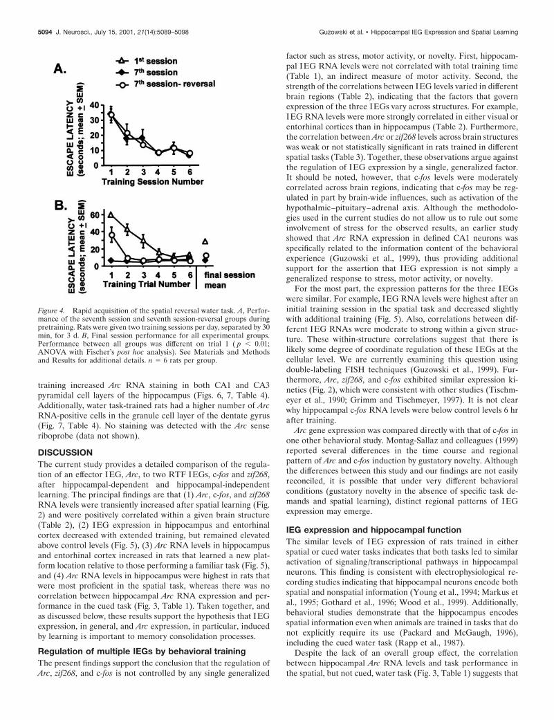
Spatial reversal water-task training. Rats were trained in the spatialwater maze (see above) over 3 d with the submerged platform located inone fixed position. On each day, the rats received two training sessions ofsix trials per session, separated by 30 min. On the final experimental day,one group (seventh session; n 5 6) received training exactly as on the sixprevious sessions. For a second group (seventh session reversal; n 5 6),the submerged platform was moved to a new location in the pool; theserats were familiar with the task demands but had to learn the new spatiallocation of the platform. A third group was also included in this exper-iment (first session; n 5 6); these rats were trained in the spatial watertask for the first time. To minimize the stress on the seventh sessionreversal and other two groups, all rats were placed on the platform for 10sec before the start of each training session for this experiment.
RNA detection methodsBrain areas analyzed. The dorsal hippocampus [approximately 23.6 mmfrom bregma (Paxinos and Watson, 1986)] was the focus of the currentstudy because this region has been shown to be required for acquisitionand consolidation of training for the spatial water task (E. Moser et al.,1993; M. B. Moser et al., 1995; Guzowski and McGaugh, 1997; Guzowskiet al., 2000). IEG RNA levels in the primary visual cortex and lateralentorhinal cortex [both at approximately 26.0 mm from bregma (Paxinosand Watson, 1986)] were also examined in one experiment. The ento-rhinal cortex was examined because it provides a major input to thehippocampus (for review, see Johnson and Amaral, 1998) and is impor-tant for spatial learning (Nagahara et al., 1995; Cho and Kesner, 1996).The primary visual cortex was examined to compare IEG regulation in
a primary sensory cortical area (engaged during task performance) toareas of higher order and polymodal processing (i.e., the hippocampusand entorhinal cortex).
Tissue dissection and RNA extraction for RNase protection assays.Water task-trained and caged control rats were anesthetized with sodiumpentobarbital, transcardially perfused with 0.1 M phosphate buffer, anddecapitated. The brains were removed rapidly, and 1-mm-thick coronalsections were prepared using a tissue matrix (Ted Pella Instruments).The sections were frozen on dry ice, and hippocampal and cortical tissuewas removed using a punch technique (Palkovits and Brownstein, 1988)with a glass pipette (inner diameter 1.0 mm). Single or multiple puncheswere taken to remove the region of interest. Hippocampal punchesremoved the entire hippocampus and dentate gyrus. Punches of theprimary visual or entorhinal cortices removed all neuronal layers withinthat region of cortex. Punches from both hemispheres were pooled for agiven structure for each rat. Total RNA was prepared using TRIzolaccording to the manufacturer’s instructions (Life Technologies-BRL).RNA concentrations were determined spectrophotometrically, and RNAaliquots were stored at 270°C until RNase protection assay (RPA)analysis.
Preparation of brain sections for fluorescent in situ hybridization. Thirtyminutes after training, the rats were anesthetized with sodium pentobar-bital and decapitated. The brains were removed rapidly, flash frozen inisopentane equilibrated in a dry ice/ethanol slurry, and stored at 270°Cfor further processing. Coronal brain sections (2 mm thick) were pre-pared with a tissue matrix and used to create tissue blocks. Sectionscontaining the dorsal hippocampus from one caged control rat and onewater task-trained rat were placed in a plastic mold, covered with OCTmedia (VWR), and frozen on dry ice. This was repeated for the othercaged control and water task-trained rats. Sections of tissue blocks (8 mmthick) were prepared using a cryostat and collected on Superfrost Plusslides (VWR). Slides were stored at 270°C until processing for fluores-cent in situ hybridization (FISH).
Riboprobe preparation. For the RNase protection assays, 32P-labeledactin, c-fos, and zif268 antisense riboprobes were prepared from com-mercially available plasmid templates (Ambion, Austin, TX). The Arcriboprobe was generated from a modified Arc cDNA plasmid. A nearlyfull-length cDNA pBluescript clone (Lyford et al., 1995) was restrictedwith XhoI and NdeI, treated with Klenow enzyme to blunt the DNAends, and closed with T4 DNA ligase. These steps removed all but 195bases of Arc 59 sequence from bases 33–228 of the published sequence(GenBank accession number U19866). The resulting plasmid was treatedwith XbaI to generate a linearized template for the synthesis of the Arcantisense riboprobe. Radioactively labeled riboprobes were generated byin vitro transcription with T7 or T3 RNA polymerases (Maxiscript kits;Ambion) and a-[ 32P] UTP (Amersham Pharmacia; 800 Ci/mM) at 15°C.Unlabeled UTP was added during the synthesis of the actin riboprobebut was not added during the synthesis of the IEG riboprobes. Theunlabeled UTP lowered the specific activity of actin riboprobes 25-foldrelative to the IEG riboprobes. Riboprobes were purified on G-50 spincolumns (Amersham Pharmacia, Arlington Heights, IL).
Antisense and sense Arc riboprobes for FISH were prepared from anearly full-length cDNA pBluescript clone (Lyford et al., 1995), asdescribed previously (Guzowski et al., 1999). The plasmid was treatedwith XbaI to generate the linearized template for the antisense ribo-probe and with XhoI to generate the linearized template for the senseriboprobe. Digoxigenin-labeled riboprobes were generated by in vitrotranscription with T7 (antisense) or T3 (sense) RNA polymerases(Maxiscript kit; Ambion) and digoxigenin RNA labeling mix (RocheMolecular Biochemicals). Riboprobes were purified on G-50 spin col-umns (Amersham Pharmacia).
RNase protection assays. RNase protection assays were performedusing a commercial kit (RPAII; Ambion) with minor modifications. TotalRNA (10 mg) and 32P-labeled riboprobes (30,000 cpm of each IEG probeplus 15,000 cpm of low specific activity actin probe) were mixed andevaporated to near dryness with a vacuum concentrator. Ten microlitersof hybridization buffer were added to each tube, and samples wereresuspended by repeated heating (85°C for 3 min) and vortexing. Onceresuspended, samples were denatured at 85°C for 5 min and then incu-bated at 42°C overnight. The following day, reaction tubes were incu-bated with a 1:100 dilution of RNase A/T1 mix (provided with the RPAIIkit) for 1 hr at room temperature. The RNase digestion reaction wasterminated, and the protected RNA species were precipitated with ad-dition of the RNase inactivation/precipitation solution (provided withthe RPAII kit). The protected RNA species were separated on 5%
5090 J. Neurosci., July 15, 2001, 21(14):5089–5098 Guzowski et al. • Hippocampal IEG Expression and Spatial Learning
polyacrylamide gels containing 8 M urea with the full-length probes and32P-labeled DNA markers (HinfI digested fX714 DNA; Promega, Mad-ison, WI).
Quantifying RNA levels by RNase protection assay. RPA gels were driedon Whatman 3mm paper. Dried gels were exposed to a phosphor screenfor 16–36 hr and scanned using a PhosphorImager 445SI (MolecularDynamics). Volume analysis was performed using ImageQuant software(version 1.1; Molecular Dynamics). Data values were obtained for eachIEG RNA band and for the actin band, which served as an internalcontrol. IEG values were normalized to actin values for each sample.
Fluorescent in situ hybridization. Fluorescent in situ hybridization(FISH) was performed on slide-mounted brain sections using adigoxigenin-labeled Arc antisense riboprobe as described in detail else-where (Guzowski et al., 1999). Arc riboprobe was detected with anantidigoxigenin–horseradish peroxidase conjugate (Roche MolecularBiochemicals) and the TSA-Direct Cyanine-3 kit (NEN Life Sciences).Nuclei were counterstained with YOYO-1 (Molecular Probes), and slideswere mounted with an anti-fade medium (Vectashield; Vector Labora-tories, Burlingame, CA). Four slides containing the dorsal hippocampus(approximately 23.6 mm from bregma) were analyzed for each rat.
Images (1024 3 1024 pixels) were acquired using a Leica TCS-4Dlaser scanning confocal microscope with a krypton/argon laser. A 103objective was used for analysis of CA1 and CA3 regions. The field of viewusing this objective was 1000 3 1000 mm. Confocal microscope settings(pinhole settings, and voltage settings and offset) were carefully adjustedto ensure that the full grayscale range was used and to minimize theoccurrence of saturated pixels. RGB TIFF images were analyzed inAdobe Photoshop 5.5. First, the pyramidal cell layer was selected byvisualizing only the color channel containing the YOYO-1 informationand using the “Lasso” tool. Then, the mean pixel density value for theregion of interest was determined for the color channel containing theCY3 information (Arc RNA staining). Finally, the values for the trainedrats were normalized using the values from the caged controls. Thisnormalization procedure minimizes artifact caused by slide-to-slide vari-ation in signal intensity and background.
For analysis of Arc-positive cells in the dentate gyrus granule cell layer,a modified approach was taken because of the different pattern of Arcstaining observed in the dentate. Overall, a much lower percentage ofdentate gyrus granule cells express Arc as compared with CA1 and CA3neurons (Guzowski et al., 1999). Because of this low density of Arc-positive cells in the dentate, it is more informative to count Arc-positivecells. Images were acquired with the same confocal microscope, but witha 53 objective, which yielded a field of view of 2000 3 2000 mm.Arc-positive cells were counted for each field. The total number ofArc-positive cells was determined for all four slides for each trained ratand each control rat. As done for analysis of CA1 and CA3 regions, thevalue obtained for each trained rat was normalized to the value for thecontrol rat in that tissue block (see Preparation of brain sections forfluorescent in situ hybridization, above).
StatisticsFor behavioral data, ANOVA or repeated measures ANOVA was usedto analyze individual trials or trial sessions, respectively. Fischer’s posthoc tests were used for pairwise comparisons. In some experiments, theIEG RNA data violated a principal assumption of parametric statistictests. Specifically, the variances between groups were often different asdetermined using an equality of variances F test. For this reason, non-parametric tests were used in the analysis of the RNA data. For com-parisons among three or more groups, the Kruskal–Wallis test was used;if a , 0.05, then pairwise comparisons were made using the Mann–Whitney U test. For correlational analyses, the Spearman correlationcoefficient, Rs, was determined. In all instances, a probability level of,0.05 was accepted as statistically significant.
RESULTSArc, zif268, and c-fos RNA levels are rapidly andtransiently increased in the dorsal hippocampus byspatial water task trainingTo quantify changes in IEG RNA levels from brain RNA sam-ples, a multiple probe RNase protection assay for the simulta-neous detection of Arc, c-fos, and zif268 mRNAs was developedand used for most of the experiments described here. In additionto the IEG riboprobes, a low specific activity riboprobe for
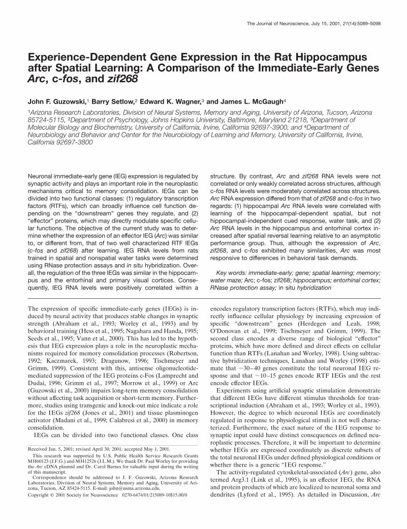
b-actin RNA was included in the RPA to normalize IEG signalsbetween samples. The rapid induction of Arc, c-fos, and zif268RNAs by neural activity was demonstrated in rats treated withthe convulsant pentylenetetrazole (50 mg/kg, i.p.), which inducedbehavioral seizures. The rats were killed 45 min after the onset ofseizures, hippocampi were removed, and total RNA was pre-pared. As expected (Morgan et al., 1987; Saffen et al., 1988;Lyford et al., 1995), hippocampal IEG RNA levels were increasedby seizure activity relative to controls (Fig. 1a). RPA autoradio-graphs of RNA from the hippocampi and the visual cortices ofrats trained in different spatial water tasks demonstrate the utilityof the RPA for behavioral studies (Fig. 1B); these data are part ofa larger experiment shown in Figures 4 and 5 and Tables 2 and 3.
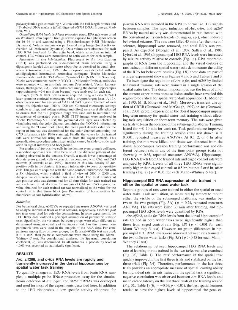
To investigate the regulation of Arc, c-fos, and zif268 by limitedbehavioral training, rats were trained in a single session of thespatial water task. The dorsal hippocampus was the focus of all ofthe current experiments because lesion studies have revealed thisregion to be critical for spatial water-task acquisition (E. Moser etal., 1993; M. B. Moser et al., 1995). Moreover, transient disrup-tion of CREB (Guzowski and McGaugh, 1997) or Arc (Guzowskiet al., 2000) protein expression in the dorsal hippocampus impairslong-term memory for spatial water-task training without affect-ing task acquisition or short-term memory. The rats were givensix trials to learn the location of the submerged platform. Traininglasted for ;8–10 min for each rat. Task performance improvedsignificantly during the training session (data not shown; p ,0.0001, repeated measures ANOVA). At 0.5, 2, or 6 hr aftertraining, the rats were killed, and tissue was dissected from thedorsal hippocampus. Session training performance was not dif-ferent between rats in any of the time point groups (data notshown; p 5 0.85, repeated measures ANOVA). HippocampalIEG RNA levels from the trained rats and caged control rats wereanalyzed by RPA. Levels of all three IEG RNAs were signifi-cantly higher than caged controls at 0.5 hr, but not 2 or 6 hr, aftertraining (Fig. 2) ( p , 0.05, for each Mann–Whitney U test).
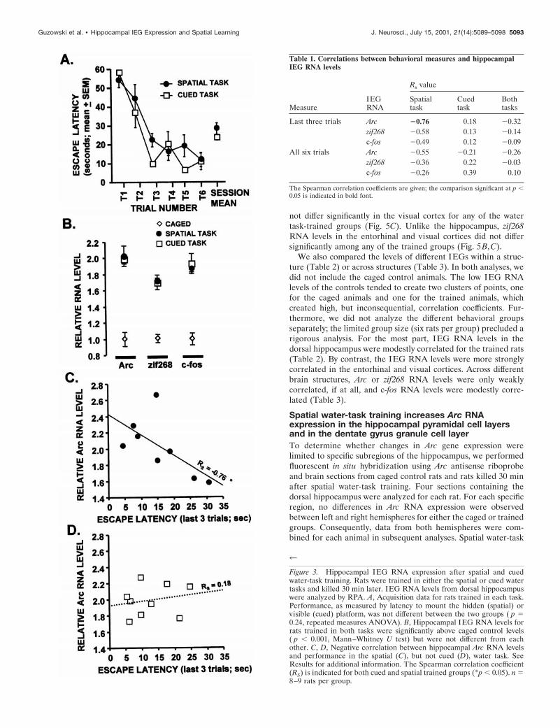
Hippocampal IEG RNA expression of rats trained ineither the spatial or cued water taskSeparate groups of rats were trained in either the spatial or cuedwater tasks. Task acquisition, as measured by latency to mounteither the visible or the submerged platforms, was similar be-tween the two groups (Fig. 3A) ( p 5 0.24, repeated measuresANOVA). The rats were killed 30 min after training, and hip-pocampal IEG RNA levels were quantified by RPA.
Arc, zif268, and c-fos RNA levels from the dorsal hippocampi ofrats trained in both water tasks were significantly higher thanthose from caged control rats (Fig. 3B) ( p , 0.001 for eachMann–Whitney U test). However, no group differences in hip-pocampal IEG RNA levels were observed between rats trained inthe two different water tasks (Fig. 3B) ( p . 0.45 for each Mann–Whitney U test).
The relationship between hippocampal IEG RNA levels andrate of learning in rats trained in the two tasks was also examined(Fig. 3C, Table 1). The rats’ performance in the spatial taskquickly improved in the first three trials and stabilized on the lastthree trials (Fig. 3A). Therefore, performance on the last threetrials provides an appropriate measure of spatial learning abilityfor individual rats. In rats trained in the spatial task, a significantnegative correlation was observed between Arc RNA levels andmean escape latency on the last three trials of the training session(Fig. 3C, Table 1) (Rs 5 20.76; p , 0.05): the best spatial learnerstended to have the highest levels of hippocampal Arc gene ex-
Guzowski et al. • Hippocampal IEG Expression and Spatial Learning J. Neurosci., July 15, 2001, 21(14):5089–5098 5091
pression. Although zif268 and c-fos were also negatively corre-lated with learning in the spatial task, these correlations did notreach statistical significance. By contrast, there was no correlationbetween IEG RNA levels and escape latency on the final threetrials of cued-task trained rats, nor was there a correlation be-tween IEG RNA levels and mean escape latency for all six trialsof either the spatial or cued tasks (Table 1).
Arc RNA expression in the hippocampus andentorhinal cortex is increased by spatialreversal trainingSeparate groups of rats were trained in one of three variations ofthe spatial water task (Fig. 4, 1st session, 7th session, or 7thsession-reversal) (see Materials and Methods for training detailsof the individual groups). Each group consisted of six rats.Trained rats were killed 30 min after the final training trial, andan equal number of rats were killed directly from their homecages to determine baseline levels of IEG expression. RNA wasprepared from dissected tissue (dorsal hippocampus, entorhinalcortex, and primary visual cortex) of the trained rats and cagedcontrol rats and was analyzed by RPA.
The seventh session group performed the familiar task at anasymptotic level of performance (Fig. 4B). The seventh session-reversal group learned the new platform location in one trial andperformed at levels similar to the seventh session group for theremaining five training trials. The first session group learned thetask rapidly and performed at the level of the other two groups bythe fifth and sixth trials.
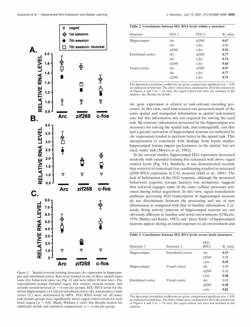
IEG RNA levels for each of the trained groups were signifi-cantly higher than those of the caged controls in the examinedbrain regions (Fig. 5). In the dorsal hippocampus, RNA levels forall IEGs were significantly lower in the seventh session group ascompared with the first session group (Fig. 5A). Arc RNA levelsof the seventh session-reversal group were significantly higherthan those of the seventh session group, but comparable to thoseof the first session group (Fig. 5A). The behavioral expressionprofile for c-fos RNA was similar in the dorsal hippocampus,entorhinal cortex, and visual cortex (Fig. 5). In each structure,c-fos RNA levels were highest in the first session group, but didnot differ significantly between the seventh session and the sev-enth session-reversal groups. In short, the pattern was as follows:caged , seventh session 5 seventh session reversal , first session.This pattern was also observed for zif268 RNA expression in thehippocampus (Fig. 5A).
Arc and zif268 RNA expression in the entorhinal (Fig. 5B) andvisual (Fig. 5C) cortices exhibited both similarities and differ-ences compared with that observed in the hippocampus. In theentorhinal cortex, as in the hippocampus, Arc RNA levels of theseventh session-reversal group were significantly higher thanthose of the seventh session group, but not different from those ofthe first session group (Fig. 5B). By contrast, Arc RNA levels did
Figure 1. Multiple probe RNase protection assay for the simultaneousdetection of Arc, c-fos, and zif268 RNAs. a, Hippocampal RNA levels forall three IEGs increased 45 min after systemic injection of pentylenetet-razole (PTZ) (50 mg/kg, i.p.) relative to caged controls. b, Detection ofIEG RNAs in the hippocampus and primary visual cortex of rats trainedin different versions of the spatial water task. The data shown here arefrom the experiment detailed in Results (Figs. 4, 5). See Materials andMethods and Results for full details of the assay and the behavioralgroups. The asterisks in a and b indicate a small amount of full-lengthactin probe that was consistently seen in all samples.
Figure 2. Rapid and transient increase in hippocampal IEG RNA levelsafter spatial water-task training. IEG RNA levels from rats killed at 0.5,2, and 6 hr after spatial water-task training were compared with cagedcontrol levels using the multiple probe RPA. RNA levels for all threeIEGs were significantly above control levels at 0.5 hr after training, andc-fos RNA levels were lower than caged control levels 6 hr after training.*p , 0.05 relative to caged (0 hr) group, Mann–Whitney U test. n 5 3–5rats per group.
5092 J. Neurosci., July 15, 2001, 21(14):5089–5098 Guzowski et al. • Hippocampal IEG Expression and Spatial Learning
not differ significantly in the visual cortex for any of the watertask-trained groups (Fig. 5C). Unlike the hippocampus, zif268RNA levels in the entorhinal and visual cortices did not differsignificantly among any of the trained groups (Fig. 5B,C).
We also compared the levels of different IEGs within a struc-ture (Table 2) or across structures (Table 3). In both analyses, wedid not include the caged control animals. The low IEG RNAlevels of the controls tended to create two clusters of points, onefor the caged animals and one for the trained animals, whichcreated high, but inconsequential, correlation coefficients. Fur-thermore, we did not analyze the different behavioral groupsseparately; the limited group size (six rats per group) precluded arigorous analysis. For the most part, IEG RNA levels in thedorsal hippocampus were modestly correlated for the trained rats(Table 2). By contrast, the IEG RNA levels were more stronglycorrelated in the entorhinal and visual cortices. Across differentbrain structures, Arc or zif268 RNA levels were only weaklycorrelated, if at all, and c-fos RNA levels were modestly corre-lated (Table 3).
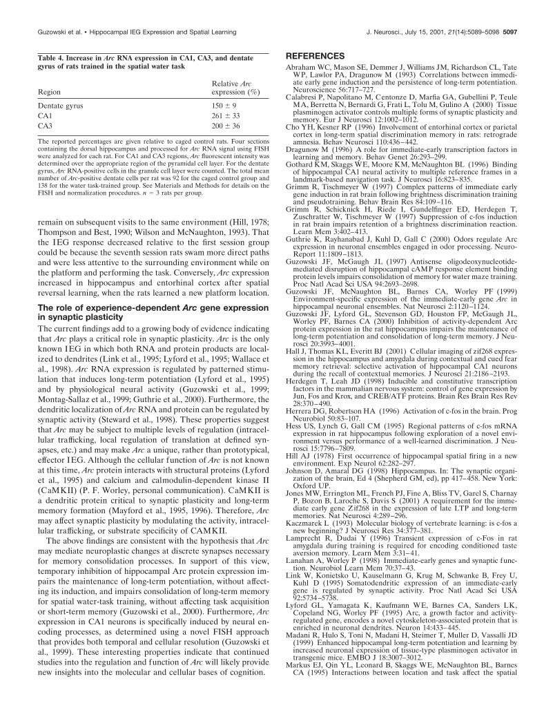
Spatial water-task training increases Arc RNAexpression in the hippocampal pyramidal cell layersand in the dentate gyrus granule cell layerTo determine whether changes in Arc gene expression werelimited to specific subregions of the hippocampus, we performedfluorescent in situ hybridization using Arc antisense riboprobeand brain sections from caged control rats and rats killed 30 minafter spatial water-task training. Four sections containing thedorsal hippocampus were analyzed for each rat. For each specificregion, no differences in Arc RNA expression were observedbetween left and right hemispheres for either the caged or trainedgroups. Consequently, data from both hemispheres were com-bined for each animal in subsequent analyses. Spatial water-task
4
Figure 3. Hippocampal IEG RNA expression after spatial and cuedwater-task training. Rats were trained in either the spatial or cued watertasks and killed 30 min later. IEG RNA levels from dorsal hippocampuswere analyzed by RPA. A, Acquisition data for rats trained in each task.Performance, as measured by latency to mount the hidden (spatial) orvisible (cued) platform, was not different between the two groups ( p 50.24, repeated measures ANOVA). B, Hippocampal IEG RNA levels forrats trained in both tasks were significantly above caged control levels( p , 0.001, Mann–Whitney U test) but were not different from eachother. C, D, Negative correlation between hippocampal Arc RNA levelsand performance in the spatial (C), but not cued (D), water task. SeeResults for additional information. The Spearman correlation coefficient(RS) is indicated for both cued and spatial trained groups (*p , 0.05). n 58–9 rats per group.
Table 1. Correlations between behavioral measures and hippocampalIEG RNA levels
MeasureIEGRNA
Rs value
Spatialtask
Cuedtask
Bothtasks
Last three trials Arc 20.76 0.18 20.32zif268 20.58 0.13 20.14c-fos 20.49 0.12 20.09
All six trials Arc 20.55 20.21 20.26zif268 20.36 0.22 20.03c-fos 20.26 0.39 0.10
The Spearman correlation coefficients are given; the comparison significant at p ,0.05 is indicated in bold font.
Guzowski et al. • Hippocampal IEG Expression and Spatial Learning J. Neurosci., July 15, 2001, 21(14):5089–5098 5093
training increased Arc RNA staining in both CA1 and CA3pyramidal cell layers of the hippocampus (Figs. 6, 7, Table 4).Additionally, water task-trained rats had a higher number of ArcRNA-positive cells in the granule cell layer of the dentate gyrus(Fig. 7, Table 4). No staining was detected with the Arc senseriboprobe (data not shown).
DISCUSSIONThe current study provides a detailed comparison of the regula-tion of an effector IEG, Arc, to two RTF IEGs, c-fos and zif268,after hippocampal-dependent and hippocampal-independentlearning. The principal findings are that (1) Arc, c-fos, and zif268RNA levels were transiently increased after spatial learning (Fig.2) and were positively correlated within a given brain structure(Table 2), (2) IEG expression in hippocampus and entorhinalcortex decreased with extended training, but remained elevatedabove control levels (Fig. 5), (3) Arc RNA levels in hippocampusand entorhinal cortex increased in rats that learned a new plat-form location relative to those performing a familiar task (Fig. 5),and (4) Arc RNA levels in hippocampus were highest in rats thatwere most proficient in the spatial task, whereas there was nocorrelation between hippocampal Arc RNA expression and per-formance in the cued task (Fig. 3, Table 1). Taken together, andas discussed below, these results support the hypothesis that IEGexpression, in general, and Arc expression, in particular, inducedby learning is important to memory consolidation processes.
Regulation of multiple IEGs by behavioral trainingThe present findings support the conclusion that the regulation ofArc, zif268, and c-fos is not controlled by any single generalized
factor such as stress, motor activity, or novelty. First, hippocam-pal IEG RNA levels were not correlated with total training time(Table 1), an indirect measure of motor activity. Second, thestrength of the correlations between IEG levels varied in differentbrain regions (Table 2), indicating that the factors that governexpression of the three IEGs vary across structures. For example,IEG RNA levels were more strongly correlated in either visual orentorhinal cortices than in hippocampus (Table 2). Furthermore,the correlation between Arc or zif268 levels across brain structureswas weak or not statistically significant in rats trained in differentspatial tasks (Table 3). Together, these observations argue againstthe regulation of IEG expression by a single, generalized factor.It should be noted, however, that c-fos levels were moderatelycorrelated across brain regions, indicating that c-fos may be reg-ulated in part by brain-wide influences, such as activation of thehypothalmic–pituitary–adrenal axis. Although the methodolo-gies used in the current studies do not allow us to rule out someinvolvement of stress for the observed results, an earlier studyshowed that Arc RNA expression in defined CA1 neurons wasspecifically related to the information content of the behavioralexperience (Guzowski et al., 1999), thus providing additionalsupport for the assertion that IEG expression is not simply ageneralized response to stress, motor activity, or novelty.
For the most part, the expression patterns for the three IEGswere similar. For example, IEG RNA levels were highest after aninitial training session in the spatial task and decreased slightlywith additional training (Fig. 5). Also, correlations between dif-ferent IEG RNAs were moderate to strong within a given struc-ture. These within-structure correlations suggest that there islikely some degree of coordinate regulation of these IEGs at thecellular level. We are currently examining this question usingdouble-labeling FISH techniques (Guzowski et al., 1999). Fur-thermore, Arc, zif268, and c-fos exhibited similar expression ki-netics (Fig. 2), which were consistent with other studies (Tischm-eyer et al., 1990; Grimm and Tischmeyer, 1997). It is not clearwhy hippocampal c-fos RNA levels were below control levels 6 hrafter training.
Arc gene expression was compared directly with that of c-fos inone other behavioral study. Montag-Sallaz and colleagues (1999)reported several differences in the time course and regionalpattern of Arc and c-fos induction by gustatory novelty. Althoughthe differences between this study and our findings are not easilyreconciled, it is possible that under very different behavioralconditions (gustatory novelty in the absence of specific task de-mands and spatial learning), distinct regional patterns of IEGexpression may emerge.
IEG expression and hippocampal functionThe similar levels of IEG expression of rats trained in eitherspatial or cued water tasks indicates that both tasks led to similaractivation of signaling/transcriptional pathways in hippocampalneurons. This finding is consistent with electrophysiological re-cording studies indicating that hippocampal neurons encode bothspatial and nonspatial information (Young et al., 1994; Markus etal., 1995; Gothard et al., 1996; Wood et al., 1999). Additionally,behavioral studies demonstrate that the hippocampus encodesspatial information even when animals are trained in tasks that donot explicitly require its use (Packard and McGaugh, 1996),including the cued water task (Rapp et al., 1987).
Despite the lack of an overall group effect, the correlationbetween hippocampal Arc RNA levels and task performance inthe spatial, but not cued, water task (Fig. 3, Table 1) suggests that
Figure 4. Rapid acquisition of the spatial reversal water task. A, Perfor-mance of the seventh session and seventh session-reversal groups duringpretraining. Rats were given two training sessions per day, separated by 30min, for 3 d. B, Final session performance for all experimental groups.Performance between all groups was different on trial 1 ( p , 0.01;ANOVA with Fischer’s post hoc analysis). See Materials and Methodsand Results for additional details. n 5 6 rats per group.
5094 J. Neurosci., July 15, 2001, 21(14):5089–5098 Guzowski et al. • Hippocampal IEG Expression and Spatial Learning
Arc gene expression is related to task-relevant encoding pro-cesses. In this view, cued task-trained rats processed much of thesame spatial and nonspatial information as spatial task-trainedrats, but this information was not required for solving the cuedtask. By contrast, information processed by the hippocampus wasnecessary for solving the spatial task, and consequently, rats thathad a greater activation of hippocampal neurons (as indicated byArc expression) tended to perform better in the spatial task. Thisinterpretation is consistent with findings from lesion studies:hippocampal lesions impair performance in the spatial, but notcued, water task (Morris et al., 1982).
In the current studies, hippocampal IEG expression decreasedmodestly with extended training but remained well above cagedcontrol levels (Fig. 5A). Similarly, it was demonstrated recentlythat retrieval of contextual fear conditioning resulted in increasedzif268 RNA expression in CA1 neurons (Hall et al., 2001). Thelack of habituation of the IEG response, although the measuredbehavioral response (escape latency) was asymptotic, suggeststhat retrieval engages some of the same cellular processes acti-vated during initial acquisition. In this view, signal transductionpathways governing IEG transcription in hippocampal neuronsdo not discriminate between the processing and use of newinformation as compared with that of familiar information. Cer-tainly, firing activity patterns of hippocampal neurons are notobviously different in familiar and novel environments (O’Keefe,1976; Muller and Kubie, 1987), and “place fields” of hippocampalneurons appear during an initial exposure to an environment and
Table 3. Correlations between IEG RNA levels across brain structures
Structure 1 Structure 2IEGRNA Rs value
Hippocampus Entorhinal cortex Arc 0.53zif268 0.28c-fos 0.54
Hippocampus Visual cortex Arc 0.40zif268 0.26c-fos 0.68
Entorhinal cortex Visual cortex Arc 0.45zif268 0.58c-fos 0.81
The Spearman correlation coefficients are given; comparisons significant at p , 0.05are indicated in bold font. The above values were calculated for all of the trained ratsof Figures 4 and 5 (n 5 18 rats); the caged control rats were not included in theanalyses.
Figure 5. Spatial reversal training increases Arc expression in hippocam-pus and entorhinal cortex. Rats were trained in one of three spatial watertasks (for behavioral data, see Fig. 4) and were killed 30 min later. Theexperimental groups included caged, first session, seventh session, andseventh session-reversal (n 5 6 rats per group). IEG RNA levels for thedorsal hippocampus (A), lateral entorhinal cortex (B), and primary visualcortex (C) were determined by RPA. IEG RNA levels for all watertask-trained groups were significantly above caged control levels for eachbrain region ( p , 0.02; Mann–Whitney U test). See Results section foradditional details and statistical comparisons. n 5 6 rats per group.
Table 2. Correlations between IEG RNA levels within a structure
Structure IEG 1 IEG 2 Rs value
Hippocampus Arc zif268 0.67Arc c-fos 0.46zif268 c-fos 0.55
Entorhinal cortex Arc zif268 0.77Arc c-fos 0.74zif268 c-fos 0.64
Visual cortex Arc zif268 0.97Arc c-fos 0.77zif268 c-fos 0.75
The Spearman correlation coefficients are given; comparisons significant at p , 0.05are indicated in bold font. The above values were calculated for all of the trained ratsof Figures 4 and 5 (n 5 18 rats); the caged control rats were not included in theanalyses. See Results for details.
Guzowski et al. • Hippocampal IEG Expression and Spatial Learning J. Neurosci., July 15, 2001, 21(14):5089–5098 5095
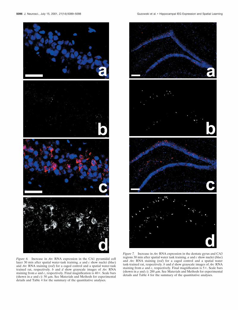
Figure 6. Increase in Arc RNA expression in the CA1 pyramidal celllayer 30 min after spatial water-task training. a and c show nuclei (blue)and Arc RNA staining (red) for a caged control and a spatial water-tasktrained rat, respectively. b and d show grayscale images of Arc RNAstaining from a and c, respectively. Final magnification is 403. Scale bars(shown in a and c): 50 mm. See Materials and Methods for experimentaldetails and Table 4 for the summary of the quantitative analyses.
Figure 7. Increase in Arc RNA expression in the dentate gyrus and CA3regions 30 min after spatial water task training. a and c show nuclei (blue)and Arc RNA staining (red) for a caged control and a spatial watertask-trained rat, respectively. b and d show grayscale images of Arc RNAstaining from a and c, respectively. Final magnification is 53. Scale bars(shown in a and c): 200 mm. See Materials and Methods for experimentaldetails and Table 4 for the summary of the quantitative analyses.
5096 J. Neurosci., July 15, 2001, 21(14):5089–5098 Guzowski et al. • Hippocampal IEG Expression and Spatial Learning
remain on subsequent visits to the same environment (Hill, 1978;Thompson and Best, 1990; Wilson and McNaughton, 1993). Thatthe IEG response decreased relative to the first session groupcould be because the seventh session rats swam more direct pathsand were less attentive to the surrounding environment while onthe platform and performing the task. Conversely, Arc expressionincreased in hippocampus and entorhinal cortex after spatialreversal learning, when the rats learned a new platform location.
The role of experience-dependent Arc gene expressionin synaptic plasticityThe current findings add to a growing body of evidence indicatingthat Arc plays a critical role in synaptic plasticity. Arc is the onlyknown IEG in which both RNA and protein products are local-ized to dendrites (Link et al., 1995; Lyford et al., 1995; Wallace etal., 1998). Arc RNA expression is regulated by patterned stimu-lation that induces long-term potentiation (Lyford et al., 1995)and by physiological neural activity (Guzowski et al., 1999;Montag-Sallaz et al., 1999; Guthrie et al., 2000). Furthermore, thedendritic localization of Arc RNA and protein can be regulated bysynaptic activity (Steward et al., 1998). These properties suggestthat Arc may be subject to multiple levels of regulation (intracel-lular trafficking, local regulation of translation at defined syn-apses, etc.) and may make Arc a unique, rather than prototypical,effector IEG. Although the cellular function of Arc is not knownat this time, Arc protein interacts with structural proteins (Lyfordet al., 1995) and calcium and calmodulin-dependent kinase II(CaMKII) (P. F. Worley, personal communication). CaMKII isa dendritic protein critical to synaptic plasticity and long-termmemory formation (Mayford et al., 1995, 1996). Therefore, Arcmay affect synaptic plasticity by modulating the activity, intracel-lular trafficking, or substrate specificity of CAMKII.
The above findings are consistent with the hypothesis that Arcmay mediate neuroplastic changes at discrete synapses necessaryfor memory consolidation processes. In support of this view,temporary inhibition of hippocampal Arc protein expression im-pairs the maintenance of long-term potentiation, without affect-ing its induction, and impairs consolidation of long-term memoryfor spatial water-task training, without affecting task acquisitionor short-term memory (Guzowski et al., 2000). Furthermore, Arcexpression in CA1 neurons is specifically induced by neural en-coding processes, as determined using a novel FISH approachthat provides both temporal and cellular resolution (Guzowski etal., 1999). These interesting properties indicate that continuedstudies into the regulation and function of Arc will likely providenew insights into the molecular and cellular bases of cognition.
REFERENCESAbraham WC, Mason SE, Demmer J, Williams JM, Richardson CL, Tate
WP, Lawlor PA, Dragunow M (1993) Correlations between immedi-ate early gene induction and the persistence of long-term potentiation.Neuroscience 56:717–727.
Calabresi P, Napolitano M, Centonze D, Marfia GA, Gubellini P, TeuleMA, Berretta N, Bernardi G, Frati L, Tolu M, Gulino A (2000) Tissueplasminogen activator controls multiple forms of synaptic plasticity andmemory. Eur J Neurosci 12:1002–1012.
Cho YH, Kesner RP (1996) Involvement of entorhinal cortex or parietalcortex in long-term spatial discrimination memory in rats: retrogradeamnesia. Behav Neurosci 110:436–442.
Dragunow M (1996) A role for immediate-early transcription factors inlearning and memory. Behav Genet 26:293–299.
Gothard KM, Skaggs WE, Moore KM, McNaughton BL (1996) Bindingof hippocampal CA1 neural activity to multiple reference frames in alandmark-based navigation task. J Neurosci 16:823–835.
Grimm R, Tischmeyer W (1997) Complex patterns of immediate earlygene induction in rat brain following brightness discrimination trainingand pseudotraining. Behav Brain Res 84:109–116.
Grimm R, Schicknick H, Riede I, Gundelfinger ED, Herdegen T,Zuschratter W, Tischmeyer W (1997) Suppression of c-fos inductionin rat brain impairs retention of a brightness discrimination reaction.Learn Mem 3:402–413.
Guthrie K, Rayhanabad J, Kuhl D, Gall C (2000) Odors regulate Arcexpression in neuronal ensembles engaged in odor processing. Neuro-Report 11:1809–1813.
Guzowski JF, McGaugh JL (1997) Antisense oligodeoxynucleotide-mediated disruption of hippocampal cAMP response element bindingprotein levels impairs consolidation of memory for water maze training.Proc Natl Acad Sci USA 94:2693–2698.
Guzowski JF, McNaughton BL, Barnes CA, Worley PF (1999)Environment-specific expression of the immediate-early gene Arc inhippocampal neuronal ensembles. Nat Neurosci 2:1120–1124.
Guzowski JF, Lyford GL, Stevenson GD, Houston FP, McGaugh JL,Worley PF, Barnes CA (2000) Inhibition of activity-dependent Arcprotein expression in the rat hippocampus impairs the maintenance oflong-term potentiation and consolidation of long-term memory. J Neu-rosci 20:3993–4001.
Hall J, Thomas KL, Everitt BJ (2001) Cellular imaging of zif268 expres-sion in the hippocampus and amygdala during contextual and cued fearmemory retrieval: selective activation of hippocampal CA1 neuronsduring the recall of contextual memories. J Neurosci 21:2186–2193.
Herdegen T, Leah JD (1998) Inducible and constitutive transcriptionfactors in the mammalian nervous system: control of gene expression byJun, Fos and Krox, and CREB/ATF proteins. Brain Res Brain Res Rev28:370–490.
Herrera DG, Robertson HA (1996) Activation of c-fos in the brain. ProgNeurobiol 50:83–107.
Hess US, Lynch G, Gall CM (1995) Regional patterns of c-fos mRNAexpression in rat hippocampus following exploration of a novel envi-ronment versus performance of a well-learned discrimination. J Neu-rosci 15:7796–7809.
Hill AJ (1978) First occurrence of hippocampal spatial firing in a newenvironment. Exp Neurol 62:282–297.
Johnson D, Amaral DG (1998) Hippocampus. In: The synaptic organi-zation of the brain, Ed 4 (Shepherd GM, ed), pp 417–458. New York:Oxford UP.
Jones MW, Errington ML, French PJ, Fine A, Bliss TV, Garel S, CharnayP, Bozon B, Laroche S, Davis S (2001) A requirement for the imme-diate early gene Zif268 in the expression of late LTP and long-termmemories. Nat Neurosci 4:289–296.
Kaczmarek L (1993) Molecular biology of vertebrate learning: is c-fos anew beginning? J Neurosci Res 34:377–381.
Lamprecht R, Dudai Y (1996) Transient expression of c-Fos in ratamygdala during training is required for encoding conditioned tasteaversion memory. Learn Mem 3:31–41.
Lanahan A, Worley P (1998) Immediate-early genes and synaptic func-tion. Neurobiol Learn Mem 70:37–43.
Link W, Konietsko U, Kauselmann G, Krug M, Schwanke B, Frey U,Kuhl D (1995) Somatodendritic expression of an immediate-earlygene is regulated by synaptic activity. Proc Natl Acad Sci USA92:5734–5738.
Lyford GL, Yamagata K, Kaufmann WE, Barnes CA, Sanders LK,Copeland NG, Worley PF (1995) Arc, a growth factor and activity-regulated gene, encodes a novel cytoskeleton-associated protein that isenriched in neuronal dendrites. Neuron 14:433–445.
Madani R, Hulo S, Toni N, Madani H, Steimer T, Muller D, Vassalli JD(1999) Enhanced hippocampal long-term potentiation and learning byincreased neuronal expression of tissue-type plasminogen activator intransgenic mice. EMBO J 18:3007–3012.
Markus EJ, Qin YL, Leonard B, Skaggs WE, McNaughton BL, BarnesCA (1995) Interactions between location and task affect the spatial
Table 4. Increase in Arc RNA expression in CA1, CA3, and dentategyrus of rats trained in the spatial water task
RegionRelative Arcexpression (%)
Dentate gyrus 150 6 9CA1 261 6 33CA3 200 6 36
The reported percentages are given relative to caged control rats. Four sectionscontaining the dorsal hippocampus and processed for Arc RNA signal using FISHwere analyzed for each rat. For CA1 and CA3 regions, Arc fluorescent intensity wasdetermined over the appropriate region of the pyramidal cell layer. For the dentategyrus, Arc RNA-positive cells in the granule cell layer were counted. The total meannumber of Arc-positive dentate cells per rat was 92 for the caged control group and138 for the water task-trained group. See Materials and Methods for details on theFISH and normalization procedures. n 5 3 rats per group.
Guzowski et al. • Hippocampal IEG Expression and Spatial Learning J. Neurosci., July 15, 2001, 21(14):5089–5098 5097
and directional firing of hippocampal neurons. J Neurosci15:7079–7094.
Mayford M, Wang J, Kandel ER, O’Dell TJ (1995) CaMKII regulatesthe frequency-response function of hippocampal synapses for the pro-duction of both LTD and LTP. Cell 81:891–904.
Mayford M, Bach ME, Huang YY, Wang L, Hawkins RD, Kandel ER(1996) Control of memory formation through regulated expression ofa CaMKII transgene. Science 274:1678–1683.
Montag-Sallaz M, Welzl H, Kuhl D, Montag D, Schachner M (1999)Novelty-induced increased expression of immediate-early genes c-fosand arg 3.1 in the mouse brain. J Neurobiol 38:234–246.
Morgan JI, Cohen DR, Hempstead JL, Curran T (1987) Mapping pat-terns of c-fos expression in the central nervous system after seizure.Science 237:192–197.
Morris RG, Garrud P, Rawlins JN, O’Keefe J (1982) Place navigationimpaired in rats with hippocampal lesions. Nature 297:681–683.
Morrow BA, Elsworth JD, Inglis FM, Roth RH (1999) An antisenseoligonucleotide reverses the footshock-induced expression of fos in therat medial prefrontal cortex and the subsequent expression of condi-tioned fear-induced immobility. J Neurosci 19:5666–5673.
Moser E, Moser MB, Andersen P (1993) Spatial learning impairmentparallels the magnitude of dorsal hippocampal lesions, but is hardlypresent following ventral lesions. J Neurosci 13:3916–3925.
Moser MB, Moser EI, Forrest E, Andersen P, Morris RG (1995) Spatiallearning with a minislab in the dorsal hippocampus. Proc Natl Acad SciUSA 92:9697–9701.
Muller RU, Kubie JL (1987) The effects of changes in the environmenton the spatial firing of hippocampal complex-spike cells. J Neurosci7:1951–1968.
Nagahara AH, Handa RJ (1995) Fetal alcohol exposure alters the induc-tion of immediate early gene mRNA in the rat prefrontal cortex afteran alternation task. Alcohol Clin Exp Res 19:1389–1397.
Nagahara AH, Otto T, Gallagher M (1995) Entorhinal-perirhinal lesionsimpair performance of rats on two versions of place learning in theMorris water maze. Behav Neurosci 109:3–9.
O’Donovan KJ, Tourtellotte WG, Millbrandt J, Baraban JM (1999) TheEGR family of transcription-regulatory factors: progress at the inter-face of molecular and systems neuroscience. Trends Neurosci22:167–173.
O’Keefe J (1976) Place units in the hippocampus of the freely movingrat. Exp Neurol 51:78–109.
Packard MG, McGaugh JL (1996) Inactivation of hippocampus or cau-date nucleus with lidocaine differentially affects expression of place andresponse learning. Neurobiol Learn Mem 65:65–72.
Palkovits M, Brownstein MJ (1988) Maps and guide to microdissectionof the rat brain. New York: Elsevier.
Paxinos G, Watson C (1986) The rat brain in stereotaxic coordinates, Ed2. San Diego: Academic.
Rapp PR, Rosenberg RA, Gallagher M (1987) An evaluation of spatialinformation processing in aged rats. Behav Neurosci 101:3–12.
Robertson HA (1992) Immediate-early genes, neuronal plasticity, andmemory. Biochem Cell Biol 70:729–737.
Saffen DW, Cole AJ, Worley PF, Christy BA, Ryder K, Baraban JM(1988) Convulsant-induced increase in transcription factor messengerRNAs in rat brain. Proc Natl Acad Sci USA 85:7795–7799.
Seeds NW, Williams BL, Bickford PC (1995) Tissue plasminogen acti-vator induction in Purkinje neurons after cerebellar motor learning.Science 270:1992–1994.
Steward O, Wallace CS, Lyford GL, Worley PF (1998) Synaptic activa-tion causes the mRNA for the IEG Arc to localize selectively nearactivated postsynaptic sites on dendrites. Neuron 21:741–751.
Thompson LT, Best PJ (1990) Long-term stability of the place-fieldactivity of single units recorded from the dorsal hippocampus of freelybehaving rats. Brain Res 509:299–308.
Tischmeyer W, Grimm R (1999) Activation of immediate early genesand memory formation. Cell Mol Life Sci 55:564–574.
Tischmeyer W, Kaczmarek L, Strauss M, Jork R, Matthies H (1990)Accumulation of c-fos mRNA in rat hippocampus during acquisition ofa brightness discrimination. Behav Neural Biol 54:165–171.
Vann SD, Brown MW, Erichsen JT, Aggleton JP (2000) Fos imagingreveals differential patterns of hippocampal and parahippocampal sub-field activation in rats in response to different spatial memory tests.J Neurosci 20:2711–2718.
Wallace CS, Lyford GL, Worley PF, Steward O (1998) Differential in-tracellular sorting of immediate early gene mRNAs depends on signalsin the mRNA sequence. J Neurosci 18:26–35.
Wilson MA, McNaughton BL (1993) Dynamics of the hippocampal en-semble code for space. Science 261:1055–1058.
Wood ER, Dudchenko PA, Eichenbaum H (1999) The global record ofmemory in hippocampal neuronal activity. Nature 397:613–616.
Worley PF, Bhat RV, Baraban JM, Erickson CA, McNaughton BL,Barnes CA (1993) Thresholds for synaptic activation of transcriptionfactors in hippocampus: correlation with long-term enhancement.J Neurosci 13:4776–4786.
Young BJ, Fox GD, Eichenbaum H (1994) Correlates of hippocampalcomplex-spike cell activity in rats performing a nonspatial radial mazetask. J Neurosci 14:6553–6563.
5098 J. Neurosci., July 15, 2001, 21(14):5089–5098 Guzowski et al. • Hippocampal IEG Expression and Spatial Learning