Expansion of High-Quality Human Pluripotent Stem Cells ...
1
B TOLL-FREE PHONE 1 800 667 0322 • PHONE 1 604 877 0713 • IN[email protected] • T[email protected] FOR GLOBAL CONTACT DETAILS VISIT WWW.STEMCELL.COM FOR RESEARCH USE ONLY. NOT INTENDED FOR HUMAN OR ANIMAL DIAGNOSTIC OR THERAPEUTIC USES. STEMCELL TECHNOLOGIES INC.’S QUALITY MANAGEMENT SYSTEM IS CERTIFIED TO ISO 13485 MEDICAL DEVICE STANDARDS. INTRODUCTION METHODS Expansion of High-Quality Human Pluripotent Stem Cells (hPSCs) Using a Novel Animal Origin-Free and Stabilized hPSC Maintenance Medium Kimberly A. Snyder¹, Olivia J. Neef¹, Thuy T. Hoang¹, Allen C. Eaves 1,2 , Sharon A. Louis¹, and Arwen L. Hunter¹ ¹STEMCELL Technologies Inc., Vancouver BC, Canada; ²Terry Fox Laboratory, BC Cancer, Vancouver BC, Canada As the number of clinical trials and approved therapies in the field of regenerative medicine increases, it is important to ensure that hPSC culture media are not only compliant with current required manufacturing and quality control processes but also ease the path toward regulatory approval. To simplify traceability and viral safety concerns, we have developed an animal origin-free (AOF) hPSC maintenance medium—TeSR™-AOF—using animal origin-free raw materials with traceability to the secondary level of manufacturing. hPSCs require specialized culture media to promote expansion while maintaining self-renewal and pluripotency. TeSR™-AOF, based on the TeSR™ formulations, was developed to ensure versatile feeding schedules while maintaining high-quality hPSC cultures. We investigated key cell quality parameters of hPSCs cultured for at least 10 passages in TeSR™-AOF, and found that hPSCs cultured in TeSR™-AOF have higher expansion and plating efficiency without affecting cell quality or downstream applications. Human embryonic stem (ES) cells (H9 & H1) and induced pluripotent stem (iPS) cells (STiPS-F016 & STiPS-M001) were cultured on Vitronectin XF™ for up to 10 passages in TeSR™-AOF with restricted feeding schedules or in TeSR™-E8™ with daily feeding. In addition, H9 ES cells were maintained on Corning ® Matrigel ® for 10 passages in the above conditions. hPSC cultures were passaged as clumps using ReLeSR™ passaging reagent on a 6- or 7-day passaging schedule. FIGURE 1. hPSCs Cultured in TeSR™-AOF with Restricted Feeding Maintain Excellent Colony Morphology hPSCs maintained in TeSR™-AOF exhibit hPSC-like morphology forming densely packed, round colonies with smooth edge morphology. Homogeneous cell morphology characteristic of hPSCs are observed, including large nucleoli and scant cytoplasm. FIGURE 5. hPSCs Maintained in TeSR™-AOF Have Improved Attachment and Higher Overall Expansion Compared to Low-Protein Medium (A) hPSCs cultured in TeSR™-AOF demonstrate a higher plating efficiency compared to hPSCs maintained in low-protein medium (TeSR™-E8™). Plating efficiency is calculated by seeding a known number of aggregates and comparing to the number of established colonies on day 7. (B) hPSCs maintained in TeSR™-AOF exhibit a higher average fold expansion per passage compared to TeSR™-E8™. (C) hPSCs cultured in TeSR™-AOF demonstrate consistent expansion and minimal cell line-to-cell line variability among ES and iPS cell lines assessed. Cumulative fold expansion was measured from passage 1 to 5. Data represented as mean plating efficiency or fold expansion across 10 passages ± SD. MG = Matrigel ® ; VN = Vitronectin XF™. ES (H9 & H1) and iPS (STiPS-M001 & STiPS-F016) cell lines cultured in TeSR™-AOF were screened for chromosomal abnormalities using the hPSC Genetic Analysis Kit and by G-banding at ≥ 10 passages. Representative data are shown for (A) H9 ES cultures at passage 10; no common chromosomal abnormalities were detected using the hPSC Genetic Analysis Kit, and (B) H9 ES cultures; these displayed a normal karyotype by G-banding at passage 13. FIGURE 2. Native bFGF Levels are Stabilized at 37°C in TeSR™-AOF TeSR™-AOF and TeSR™-E8™ were incubated at 37°C for 24, 48, and 72 hours. FGF2 levels were measured by Meso Scale Discovery (MSD) immuno- assay; data were normalized to t = 0 levels for TeSR™-E8™ and TeSR™-AOF, respectively. FGF2 levels in TeSR™-AOF remain at 36.7 ± 5.61% of t = 0 levels at 72 hours when incubated at 37°C. Data representative of n = 3 biological replicates ± SD. FIGURE 3. hPSCs Maintained in TeSR™-AOF with Daily and Restricted Feed Schedules Exhibit Comparable Colony Morphology hPSCs were maintained on Vitronectin XF™ for five passages. Phase-contrast images were taken on day 7 after seeding. For restricted feeds, hPSCs were fed with a double volume (4 mL) of medium on day 2 after passage, followed by two consecutive skipped days of feeds, with a final single-volume feed (2 mL) on day 5, prior to passaging on day 6 or 7. FIGURE 4. hPSCs Cultured in TeSR™-AOF Express Markers of the Undifferentiated State hPSCs maintained in TeSR™-AOF exhibit high levels of TRA-1-60 and OCT4 by flow cytometry at passage 5 and 10. Across n = 4 cell lines, the average TRA-1-60 expression was 92.8 ± 3.77%, and percent OCT-4 positive cells were 98.1 ± 1.79%. Data shown represent an average of passage 5 and 10 flow results for each cell line. MG = Matrigel ® ; VN = Vitronectin XF™. TeSR™-AOF is an animal origin-free hPSC culture medium, manufactured with raw material traceability to the secondary level of manufacturing. hPSCs maintained in TeSR™-AOF exhibit increased attachment and more consistent expansion when compared with hPSCs maintained in low-protein medium. Cell quality is maintained in TeSR™-AOF, even when using reduced feeding schedules. Gene and marker expression, genetic stability, and differentiation potential are unaltered when compared with hPSCs maintained in low-protein medium. Higher cloning efficiency is observed when hPSCs are seeded in TeSR™-AOF with CloneR™ at clonal density compared to competitor media. Summary Medium & Culture Performance Cell Quality Differentiation and Cloning Efficiency ES (H9) ES (H1) iPS (STiPS-F016) iPS (STiPS-M001) 500 µm 100 µm ES (H9) Matrigel® Vitronectin XF™ 0 20 40 60 80 0 25 50 75 100 Time at 37° C (hours) % FGF2 Relative to t=0 TeSR TM -E8 TM TeSR TM -AOF Daily Feeds Restricted Feeds iPS (STiPS- F016) ES (H1) 500 µm B C B C FIGURE 6. hPSCs Cultured in TeSR™-AOF with Restricted Feeding Maintain a Normal Karyotype Efficient differentiation to the three germ layers was demonstrated in one ES and one iPS cell line maintained for > 5 passages in TeSR™-AOF compared to mTeSR™1 or TeSR™-E8™ controls. (A) Cultures were processed for flow cytometry and assessed for PAX6 + /Nestin + cells on day 7 following monolayer differentiation using STEMdiff™ Neural Induction Medium. (B) Cultures were processed for flow cytometry and assessed for Brachyury (T) + /OCT4 - cells on day 5 following differentiation in STEMdiff™ Mesoderm Induction Medium. (C) Cultures were processed for flow cytometry and assessed for CXCR4 + /SOX17 + cells on day 5 following differentiation using STEMdiff™ Definitive Endoderm Kit. FIGURE 7. hPSCs Maintained in TeSR™-AOF with Restricted Feeding Differentiate to the Three Germ Layers hPSCs were seeded at clonal density (20 cells/cm 2 ) in TeSR™-E8™, mTeSR™1, TeSR™-AOF, and a GMP competitor hPSC maintenance medium (Competitor 1) onto Vitronectin XF™. H1 ES cultures cloned in TeSR™-AOF had significantly higher cloning efficiencies compared to TeSR™-E8™ and Competitor 1 (p < 0.05; Paired Student’s t test). WLS-1C iPS cells seeded in TeSR™ media trended toward having a higher cloning efficiency compared to Competitor 1. Furthermore, reduced variability in cloning efficiency was observed across the H1 ES and WLS-1C iPS cell lines cloned in TeSR™-AOF. Data representative of n = 2 biological replicates ± SD. FIGURE 8. hPSCs Exhibit Enhanced Cloning Efficiency in TeSR™-AOF Compared to GMP Competitor Medium
Transcript of Expansion of High-Quality Human Pluripotent Stem Cells ...
B
TOLL-FREE PHONE 1 800 667 0322 • PHONE 1 604 877 0713 • [email protected] • [email protected] FOR GLOBAL CONTACT DETAILS VISIT WWW.STEMCELL.COM
FOR RESEARCH USE ONLY. NOT INTENDED FOR HUMAN OR ANIMAL DIAGNOSTIC OR THERAPEUTIC USES. STEMCELL TECHNOLOGIES INC.’S QUALITY MANAGEMENT SYSTEM IS CERTIFIED TO ISO 13485 MEDICAL DEVICE STANDARDS.
INTRODUCTION
METHODS
Expansion of High-Quality Human Pluripotent Stem Cells (hPSCs) Using a Novel Animal Origin-Free and Stabilized hPSC Maintenance MediumKimberly A. Snyder¹, Olivia J. Neef¹, Thuy T. Hoang¹, Allen C. Eaves1,2, Sharon A. Louis¹, and Arwen L. Hunter¹
¹STEMCELL Technologies Inc., Vancouver BC, Canada; ²Terry Fox Laboratory, BC Cancer, Vancouver BC, Canada
As the number of clinical trials and approved therapies in the field of regenerative
medicine increases, it is important to ensure that hPSC culture media are not only
compliant with current required manufacturing and quality control processes but also
ease the path toward regulatory approval. To simplify traceability and viral safety
concerns, we have developed an animal origin-free (AOF) hPSC maintenance
medium—TeSR™-AOF—using animal origin-free raw materials with traceability to the
secondary level of manufacturing. hPSCs require specialized culture media to promote
expansion while maintaining self-renewal and pluripotency. TeSR™-AOF, based on the
TeSR™ formulations, was developed to ensure versatile feeding schedules while
maintaining high-quality hPSC cultures. We investigated key cell quality parameters of
hPSCs cultured for at least 10 passages in TeSR™-AOF, and found that hPSCs cultured
in TeSR™-AOF have higher expansion and plating efficiency without affecting cell
quality or downstream applications.
Human embryonic stem (ES) cells (H9 & H1) and induced pluripotent stem (iPS) cells
(STiPS-F016 & STiPS-M001) were cultured on Vitronectin XF™ for up to
10 passages in TeSR™-AOF with restricted feeding schedules or in TeSR™-E8™ with
daily feeding. In addition, H9 ES cells were maintained on Corning® Matrigel® for
10 passages in the above conditions. hPSC cultures were passaged as clumps using
ReLeSR™ passaging reagent on a 6- or 7-day passaging schedule.
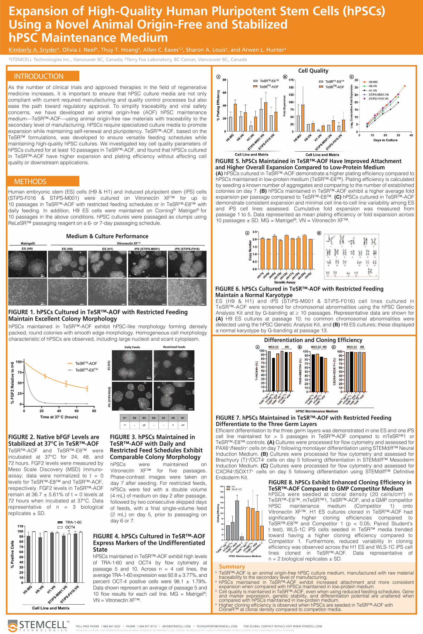
FIGURE 1. hPSCs Cultured in TeSR™-AOF with Restricted Feeding Maintain Excellent Colony MorphologyhPSCs maintained in TeSR™-AOF exhibit hPSC-like morphology forming densely
packed, round colonies with smooth edge morphology. Homogeneous cell morphology
characteristic of hPSCs are observed, including large nucleoli and scant cytoplasm.
FIGURE 5. hPSCs Maintained in TeSR™-AOF Have Improved Attachment and Higher Overall Expansion Compared to Low-Protein Medium (A) hPSCs cultured in TeSR™-AOF demonstrate a higher plating efficiency compared to hPSCs maintained in low-protein medium (TeSR™-E8™). Plating efficiency is calculated by seeding a known number of aggregates and comparing to the number of established colonies on day 7. (B) hPSCs maintained in TeSR™-AOF exhibit a higher average fold expansion per passage compared to TeSR™-E8™. (C) hPSCs cultured in TeSR™-AOF demonstrate consistent expansion and minimal cell line-to-cell line variability among ES and iPS cell lines assessed. Cumulative fold expansion was measured from passage 1 to 5. Data represented as mean plating efficiency or fold expansion across 10 passages ± SD. MG = Matrigel®; VN = Vitronectin XF™.
ES (H9 & H1) and iPS (STiPS-M001 & STiPS-F016) cell lines cultured in TeSR™-AOF were screened for chromosomal abnormalities using the hPSC Genetic Analysis Kit and by G-banding at ≥ 10 passages. Representative data are shown for (A) H9 ES cultures at passage 10; no common chromosomal abnormalities were detected using the hPSC Genetic Analysis Kit, and (B) H9 ES cultures; these displayed a normal karyotype by G-banding at passage 13.
FIGURE 2. Native bFGF Levels are Stabilized at 37°C in TeSR™-AOF TeSR™-AOF and TeSR™-E8™ were
incubated at 37°C for 24, 48, and
72 hours. FGF2 levels were measured by
Meso Scale Discovery (MSD) immuno-
assay; data were normalized to t = 0
levels for TeSR™-E8™ and TeSR™-AOF,
respectively. FGF2 levels in TeSR™-AOF
remain at 36.7 ± 5.61% of t = 0 levels at
72 hours when incubated at 37°C. Data
representative of n = 3 biological
replicates ± SD.
FIGURE 3. hPSCs Maintained in TeSR™-AOF with Daily and Restricted Feed Schedules Exhibit Comparable Colony Morphology hPSCs were maintained on
Vitronectin XF™ for five passages.
Phase-contrast images were taken on
day 7 after seeding. For restricted feeds,
hPSCs were fed with a double volume
(4 mL) of medium on day 2 after passage,
followed by two consecutive skipped days
of feeds, with a final single-volume feed
(2 mL) on day 5, prior to passaging on
day 6 or 7.
FIGURE 4. hPSCs Cultured in TeSR™-AOF Express Markers of the Undifferentiated StatehPSCs maintained in TeSR™-AOF exhibit high levels
of TRA-1-60 and OCT4 by flow cytometry at
passage 5 and 10. Across n = 4 cell lines, the
average TRA-1-60 expression was 92.8 ± 3.77%, and
percent OCT-4 positive cells were 98.1 ± 1.79%.
Data shown represent an average of passage 5 and
10 flow results for each cell line. MG = Matrigel®;
VN = Vitronectin XF™.
TeSR™-AOF is an animal origin-free hPSC culture medium, manufactured with raw material traceability to the secondary level of manufacturing.hPSCs maintained in TeSR™-AOF exhibit increased attachment and more consistent expansion when compared with hPSCs maintained in low-protein medium.Cell quality is maintained in TeSR™-AOF, even when using reduced feeding schedules. Gene and marker expression, genetic stability, and differentiation potential are unaltered when compared with hPSCs maintained in low-protein medium.Higher cloning efficiency is observed when hPSCs are seeded in TeSR™-AOF with CloneR™ at clonal density compared to competitor media.
Summary
Medium & Culture Performance
Cell Quality
Differentiation and Cloning Efficiency
ES (H9) ES (H1) iPS (STiPS-F016) iPS (STiPS-M001)
500
µm
100
µm
ES (H9)
Matrigel® Vitronectin XF™
0 20 40 60 80
0
25
50
75
100
Time at 37°C (hours)
% F
GF
2 R
ela
tiv
e t
o t
=0
TeSRTM
-E8TM
TeSRTM
-AOF
Daily Feeds Restricted Feeds
iPS
(ST
iPS-
F0
16
)
ES
(H
1)
500 µm
B C
B C
FIGURE 6. hPSCs Cultured in TeSR™-AOF with Restricted Feeding Maintain a Normal Karyotype
Efficient differentiation to the three germ layers was demonstrated in one ES and one iPS cell line maintained for > 5 passages in TeSR™-AOF compared to mTeSR™1 or TeSR™-E8™ controls. (A) Cultures were processed for flow cytometry and assessed for PAX6+/Nestin+ cells on day 7 following monolayer differentiation using STEMdiff™ Neural Induction Medium. (B) Cultures were processed for flow cytometry and assessed for Brachyury (T)+/OCT4- cells on day 5 following differentiation in STEMdiff™ Mesoderm Induction Medium. (C) Cultures were processed for flow cytometry and assessed for CXCR4+/SOX17+ cells on day 5 following differentiation using STEMdiff™ Definitive Endoderm Kit.
FIGURE 7. hPSCs Maintained in TeSR™-AOF with Restricted Feeding Differentiate to the Three Germ Layers
hPSCs were seeded at clonal density (20 cells/cm2) in TeSR™-E8™, mTeSR™1, TeSR™-AOF, and a GMP competitor hPSC maintenance medium (Competitor 1) onto Vitronectin XF™. H1 ES cultures cloned in TeSR™-AOF had significantly higher cloning efficiencies compared to TeSR™-E8™ and Competitor 1 (p < 0.05; Paired Student’s t test). WLS-1C iPS cells seeded in TeSR™ media trended toward having a higher cloning efficiency compared to Competitor 1. Furthermore, reduced variability in cloning efficiency was observed across the H1 ES and WLS-1C iPS cell lines cloned in TeSR™-AOF. Data representative of n = 2 biological replicates ± SD.
FIGURE 8. hPSCs Exhibit Enhanced Cloning Efficiency in TeSR™-AOF Compared to GMP Competitor Medium


![10000005505-Maintenance of Human Pluripotent Stem Cells …€¦ · The maintenance and expansion of human pluripotent stem cells (human embryonic stem [ES] cells and human induced](https://static.fdocuments.in/doc/165x107/6033bf7fdddc672302645fcf/10000005505-maintenance-of-human-pluripotent-stem-cells-the-maintenance-and-expansion.jpg)