Exosomes for Repair, Regeneration and Rejuvenation
48
Full Terms & Conditions of access and use can be found at http://www.tandfonline.com/action/journalInformation?journalCode=iebt20 Download by: [University of Nebraska, Lincoln] Date: 19 December 2015, At: 02:19 Expert Opinion on Biological Therapy ISSN: 1471-2598 (Print) 1744-7682 (Online) Journal homepage: http://www.tandfonline.com/loi/iebt20 Exosomes for Repair, Regeneration and Rejuvenation Joydeep Basu PhD & John W. Ludlow PhD To cite this article: Joydeep Basu PhD & John W. Ludlow PhD (2015): Exosomes for Repair, Regeneration and Rejuvenation, Expert Opinion on Biological Therapy, DOI: 10.1517/14712598.2016.1131976 To link to this article: http://dx.doi.org/10.1517/14712598.2016.1131976 Accepted author version posted online: 12 Dec 2015. Submit your article to this journal Article views: 4 View related articles View Crossmark data
Transcript of Exosomes for Repair, Regeneration and Rejuvenation

Full Terms & Conditions of access and use can be found athttp://www.tandfonline.com/action/journalInformation?journalCode=iebt20
Download by: [University of Nebraska, Lincoln] Date: 19 December 2015, At: 02:19
Expert Opinion on Biological Therapy
ISSN: 1471-2598 (Print) 1744-7682 (Online) Journal homepage: http://www.tandfonline.com/loi/iebt20
Exosomes for Repair, Regeneration andRejuvenation
Joydeep Basu PhD & John W. Ludlow PhD
To cite this article: Joydeep Basu PhD & John W. Ludlow PhD (2015): Exosomes forRepair, Regeneration and Rejuvenation, Expert Opinion on Biological Therapy, DOI:10.1517/14712598.2016.1131976
To link to this article: http://dx.doi.org/10.1517/14712598.2016.1131976
Accepted author version posted online: 12Dec 2015.
Submit your article to this journal
Article views: 4
View related articles
View Crossmark data
1
Publisher: Taylor & Francis
Journal: Expert Opinion on Biological Therapy
DOI: 10.1517/14712598.2016.1131976
Exosomes for Repair, Regeneration and Rejuvenation
Joydeep Basu1, PhD and John W. Ludlow, PhD
RegenMed TX, LLC
3929 Westpoint Blvd., Ste G
Winston-Salem, NC 27103, USA
Tel: (336) 448 2843
Fax: (336) 448 2882
(1) Author for correspondence: [email protected]
Keywords: exosome, micro-vesicle, extra-cellular vesicle, cell therapy, tissue
engineering, regenerative medicine, stem cell, progenitor cell, organogenesis,
morphogen, manufacturing, quality control, regulatory
Abstract
Introduction: Application of regenerative medicine strategies for repair of organs/tissue
impacted by chronic disease is an active subject for product development. Such
methodologies emphasize the role of stem cells as the active biological ingredient.
However, recent developments in elucidating mechanism of action of these therapies
have focused on the role of paracrine, “action-at-a-distance” modus operandi in
mediating the ability to catalyze regenerative outcomes without significant site-specific
Dow
nloa
ded
by [
Uni
vers
ity o
f N
ebra
ska,
Lin
coln
] at
02:
19 1
9 D
ecem
ber
2015
2
engraftment. A salient component of this secreted regenerative milieu are exosomes:
40-100nm intraluminal vesicles that mediate transfer of proteins and nucleic acids
across cellular boundaries.
Areas covered: Here, we synthesize recent studies from PubMed and Google Scholar
highlighting how cell-based therapeutics and cosmeceutics are transitioning towards the
secretome generally and exosomes specifically as a principal modulator of regenerative
outcomes.
Expert Opinion: Exosomes contribute to organ development and mediate regenerative
outcomes in injury and disease that recapitulate observed bioactivity of stem cell
populations. Encapsulation of the active biological ingredients of regeneration within
non-living exosome carriers may offer process, manufacturing and regulatory
advantages over stem cell-based therapies.
1. Introduction
The arc of biopharmaceutical development over the past decades has been
characterized by a movement away from the application of the cell as a mere
manufacturing platform for medicinal proteins. Instead, there is increasing recognition of
the cell itself as the active biological ingredient for catalyzing regeneration and repair of
diseased tissue. Currently however, strategies for application of stem and progenitor
cell populations for tissue engineering and regenerative medicine are being significantly
influenced by a new mechanistic understanding. The activity of secreted cell-derived
byproducts acting at a distance, rather than site-specific integration and directed
differentiation, represents the principal mechanism of action by which these cell
populations mediate regenerative outcomes [1, 2]. This secretome represents a
regenerative milieu broadly composed of proteins, nucleic acids and membrane-bound
vesicles of a variety of sizes. These may be capable of independently triggering
regeneration and repair as well as mediating the de novo organogenesis of tissue
engineered organs ex vivo [3, 4]. These observations signal a transitional return
towards leveraging the cell as a medicinal factory with the secretome rather than the
Dow
nloa
ded
by [
Uni
vers
ity o
f N
ebra
ska,
Lin
coln
] at
02:
19 1
9 D
ecem
ber
2015
Edit
Kiemelés
Edit
Kiemelés
Edit
Kiemelés
Edit
Kiemelés
Edit
Kiemelés
3
cell itself now representing the active biological ingredient [5]. Here, through synthesis
of the salient recent literature, we attempt to capture this paradigm shift away from the
manufacture and application of cells to cell-derived regenerative by-products such as
exosomes and conditioned media. Specific examples documenting regeneration of
heart, kidney, skin and the nervous system through application of exosome-based
therapies will be examined. Although no industrial scale manufacturing pipeline specific
to exosomes yet exists, we will leverage lessons learnt from cell-based systems to
illustrate process development, scale-up, manufacturing, quality control, regulatory and
intellectual property issues associated with exosome-based therapies (Figure 1).
2. Exosome Biology
Although extracellular particles described as “platelet dust” were observed in normal
plasma in the first half of the 20th century [6, 7], a functional role for exosomes was first
established in the form of MHC-II (Major Histocompatibility Class-II) presenting vesicles
secreted by B-lymphocytes and capable of inducing a specific T-cell proliferative
response [8]. While no rigorous and universally accepted definition has yet been
established [9], the term exosome is generally understood to reference a specific class
of lipid-membrane bound extra-cellular vesicle (EV) characterized by a diameter of 40-
150nm and density of 1.09-1.18 g/ml (Figure 2). Additionally shown to be secreted from
tumor cells [10], exosomes were first characterized by EM (Electron Microscope)-
methodologies upon their release via invagination of endosomal membranes as multi-
vesicular bodies during differentiation of erythrocyte progenitors [11]. Within the
endosome compartment, exosomes and their payloads are released into the
extracellular milieu upon fusion of the endosome with the plasma membrane. Exosomes
participate in a variety of cellular activities and have been shown to be isolatable from
multiple body fluids including saliva, urine, plasma, serum, breast milk and amniotic
fluid, as well as from the conditioned media of cultured cells, with yields typically 0.5 g
exosomal protein per 106 cells over a 24 hour culture period [12]. However, from a
regenerative medicine perspective, the salient function of exosomes is in inter-cell
communication through transport of protein, mRNAs and micro-RNAs [13]. Such
Dow
nloa
ded
by [
Uni
vers
ity o
f N
ebra
ska,
Lin
coln
] at
02:
19 1
9 D
ecem
ber
2015
4
signaling typically takes place within an organism, as was shown by the ability of ESC
sourced exosomes to reprogram murine hematopoietic progenitors towards acquisition
of a more pluripotent, ESC-like, phenotype [14]. However, the nematode parasite
Heligmosmoides has been shown to manipulate the innate immune response of its
mouse host through secretion of exosomal particles, thereby establishing exosomes as
a mechanism for inter-species transfer of RNA [15]. Other examples of immune
modulation by exosomes include potent anti-tumor bioactivity observed in tumor bearing
mice treated with dendritic cell-derived exosomes, which present MHC Class I and II
glycoproteins [16]. Key protein markers that have been associated with exosomes
include CD9, CD63, CD81, HSP70, HSP90 (see Figure 3), actin and annexin [17, 18]. A
systematic review of exosome composition and functionality in broad range of biological
fluids has recently been presented [19].
3. Regeneration leverages developmental signaling mechanisms: exosome-
mediated transfer of morphogens
Organ regeneration technologies aim to restore the original structure and functionality of
a diseased organ. In general, healing responses within mammals are characterized by
fibrosis and scar tissue formation, not regeneration. Nevertheless, developing
mammalian fetuses during the first trimester will typically present wound healing without
fibrosis and scar tissue formation [20]. Additionally, compensatory hyperplasia of
mammalian kidney or liver secondary to partial nephrectomy or hepatectomy,
remodeling of epidermis or bone consequent to injury and regeneration of limb digit tips
in humans and mice post-amputation are all examples of regenerative outcomes in
adult mammals indicative of an innate regenerative potential within adult mammals
(reviewed by Roy and Gatien [21]).
The mechanistic link between developmental and regenerative biology predicts that
potential regenerative therapies may leverage or manipulate the fundamental signaling
pathways governing cellular self-organization during embryonic organogenesis. For
example, the highly convoluted nature of developing epithelia mandates the existence
of an efficient mechanism for morphogen transport across the plasma membrane to
Dow
nloa
ded
by [
Uni
vers
ity o
f N
ebra
ska,
Lin
coln
] at
02:
19 1
9 D
ecem
ber
2015
5
establish the short and long range morphogen gradients central to assembly of the
developing embryo. To this end, the observation that morphogens including Wingless
and Hedgehog are closely associated with the plasma membrane, as opposed to freely
diffusing across the cytosol, strongly suggests the existence of a membrane-based
trans-cytotic vesicular mechanism for establishment of the morphogen gradient.
Evidence from the developing Drosophila embryo demonstrates that the establishment
of gradients of the morphogen Wingless during pattern formation of the imaginal disc
epithelium occurs at least in part through membrane bound exosome-like particles
called “argosomes” [22]. In C.elegans, an apical secretion pathway mediated by the
membrane bound V0 sector of the vacuolar H+-ATPase controls secretion of
Hedgehog-like proteins within exosomes [23]. Finally, the specification of left/right
asymmetry in the developing mouse requires the exosome-mediated transport of the
morphogens Sonic Hedgehog and retinoic acid in response to FGF (Fibroblast Growth
Factor)-signaling [24]. Vertebrate Sonic Hedgehog has been reported to be secreted
within two overlapping populations of exosomes, presenting distinctive accessory
signaling proteins. Co-expression of integrins was required together with Sonic
Hedgehog to activate certain Sonic Hedgehog target genes during differentiation of
mouse ESCs (Embryonic Stem Cells), suggesting the existence of a mechanism for
fine-tuning exosome-based morphogen gradients by presentation of distinctive sub-
categories of morphogen presenting exosomes [25]. Finally, the Xenopus cleavage
stage blastocoel is bridged by multiple arrays of parallel filopodia, that facilitate direct
interaction between nonadjacent blastomeres; these filopodia in turn fragment into
micro-vesicles (including exosomes) whose subsequent resorption identifies a specific
mechanism for the potential transfer of morphogens across the developing embryo [26].
4. Selection and delivery of cargo
Exosomes also contain the protein TSG101 (see Figure 3), a component of the
endosomal sorting complexes required for transport (ESCRT)-I, which regulates
vesicular trafficking processes. The ESCRT machinery is made up of several cytosolic
Dow
nloa
ded
by [
Uni
vers
ity o
f N
ebra
ska,
Lin
coln
] at
02:
19 1
9 D
ecem
ber
2015
6
protein complexes, known as ESCRT-0, ESCRT-I, ESCRT-II, and ESCRT-III. Together
with a number of accessory proteins, ESCRT enables a unique mode of membrane
remodeling that results in membrane bending and budding away from the cytoplasm. In
this regard, TSG101 binds to ubiquitinated cargo proteins and is required for the sorting
of endocytic, ubiquitinated cargos into multi-vesicular bodies (MVBs). ESCRT mediated
selection is not the only mechanism for getting protein cargo into exosomes: proteins
may also be recruited into exosomes by virtue of their association with chaperones such
as HSP70 and HSP90 [27].
The precise mechanism by which RNA species are selected for recruitment into
exosomes is less well defined. One possible mechanism involves specific sequence
motifs that may function as cis-acting elements for targeting RNAs to EV [28]. Another
possibility involves specific post-transcriptional modifications such as 3’ adenylation and
uridylation that may serve to separate cellular small RNAs from secreted RNA
populations packaged into exosomes [29]. The discovery that ESCRT-II is an RNA
binding complex [30] suggests that it may also function to select RNA for incorporation
into EVs. Finally, the observations that MVBs are sites of miRNA-loaded RISC (RNA-
induced silencing complex) accumulation [31] and that exosome-like vesicles are
considerably enriched in GW182 and AGO2 implicate functional roles of these proteins
in RNA sorting to exosomes.
In order for exosomes to transfer their cargo (nucleic acid, protein), they must somehow
be incorporated into the recipient cell. Fluorescently labeled lipophilic dye transferred
from exosomes and incorporated into cultured cells has been used to show that
exosomes are taken up by recipient cells but does not distinguish whether the
incorporated exosomes are dissociated or degraded in the endosomal/lysosomal
pathway [32]. A fusion event between exosomes and the cell membrane will transfer
fluorescence from the labeled exosomes to the recipient cells. The molecular basis
underlying the mechanism by which exosomes recognize, bind to and fuse with the
intended target cells remains to be elucidated. However, minor differences in the
Dow
nloa
ded
by [
Uni
vers
ity o
f N
ebra
ska,
Lin
coln
] at
02:
19 1
9 D
ecem
ber
2015
7
exosomal tetraspanin-integrin complex translate into marked alterations in exosome
target cell selection in vitro and in vivo, indicative of one potential mechanism of action
[33]. Other targeting proteins may include galactin 5, galactin 9, integrins, MHC-II and
ICAM1 [34]. Upon binding, exosomes remain attached to the plasma membrane or are
internalized by endocytosis and subsequent fusion with the endosome or channeled to
the lysosomal pathway for degradation. Such observations identify exosomes as
vectors for information transfer between cells, and highlight a specific mechanism by
which one cell population may manipulate another. To this end, stable modification of
cell fate by exosomes has been observed in rodent models, where lung derived
exosomes and micro-vesicles were shown to reprogram bone marrow cells towards a
pulmonary phenotype in vitro and in vivo [35]. Similarly, exosomes sources from hepatic
cells can promote acquisition of a hepatic phenotype in bone marrow cells [36]. In
addition, exosomes derived from cancer cells can promote the development of
tumorigenesis [37, 38]. Finally, in the brain, exosome mediated transfer of toxic protein
aggregates including amyloid- and prions may represent an important mechanism for
the onset of pathology [39].
5. Exosomes, not cells, may be sufficient as cell-based therapies
A number of studies suggest that the cell itself may ultimately be superfluous in
mediating observed regenerative bioactivity from cell-sourced therapeutic product
candidates. To this end, therapeutic bioactivity associated with stem and progenitor cell
populations can be recapitulated by conditioned media isolated from the culture,
maintenance and expansion of those populations [40]. The conditioned media
represents the complete regenerative milieu of cell-sourced secretomic and vesicular
elements, including soluble proteins, growth factors, cytokines, nucleic acid and small
molecules as well as micro-vesicles of varying size, composition and functionality
present in suspension. The soluble secretomic component may be separated from the
micro-vesicle fraction by centrifugation, filtration or polymer precipitation-based
methodologies, reviewed by [41].
Dow
nloa
ded
by [
Uni
vers
ity o
f N
ebra
ska,
Lin
coln
] at
02:
19 1
9 D
ecem
ber
2015
Edit
Kiemelés

8
(5.1) Regenerative bioactivity of conditioned media: Paracrine-based bioactivity
constitutes an important if not sole component of MSC (Mesenchymal Stem Cell)
therapeutic mechanism of action in addition to or instead of site specific engraftment
and directed differentiation [42, 43]. It has been demonstrated that MSC-derived
conditioned media is sufficient to significantly improve multiple biomarkers of renal
pathophysiology in rodent models of chronic kidney disease [44]. Mechanistically, it is
now understood that MSCs do not repair organ defects by differentiating into the
desired tissue type, but rather function more in a regulatory role. This paradigm shift
followed the demonstration that MSCs can inhibit apoptosis, stimulate angiogenesis,
promote endogenous cell proliferation, and inhibit inflammation during tissue
regeneration [45]. Such bioactivity is mediated through growth factor and cytokine
secretion (paracrine effects) in addition to cell-cell interactions and has been directly
leveraged in multiple models of regeneration. For example, the intramuscular injection
of conditioned media derived from endothelial progenitor cells into a rat model of
chronic hind-limb ischemia was shown to be as effective as transplantation of the
endothelial progenitor cells themselves for promotion of angiogenesis [46]. Similar
observations were made in a porcine acute myocardial infarction model, where
functional equivalence between intracoronary delivery of endothelial progenitors and
conditioned media derived from the same was demonstrated [47]. Endothelial cell-
derived conditioned media has been shown to promote brain microvascular cell viability
in vitro following ischemic insult in a manner contingent on the presence of both protein
and lipidic elements [48]. In addition, conditioned media from MSCs was shown to
resolve pulmonary inflammation in mouse models of lung injury in a manner similar to
MSCs [49]. Finally, in a study of bone tissue engineering in a rat model, MSC-sourced
conditioned media could actually outperform MSCs in regenerating bone [50].
Secretomic components of MSC-sourced conditioned media may also be leveraged for
regenerative applications in orthopedics and cartilage repair and regeneration, including
osteoarthritis and rheumatoid arthritis [51, 52].
Dow
nloa
ded
by [
Uni
vers
ity o
f N
ebra
ska,
Lin
coln
] at
02:
19 1
9 D
ecem
ber
2015
9
(5.2) Regenerative bioactivity of micro-vesicles, including exosomes: In a
systematic review of the literature presenting preclinical animal data on the therapeutic
potential of MSC-derived micro-vesicles including exosomes, all 13 reported studies
indicated that treatment improved at least one clinically relevant parameter associated
with organ functionality [53]. Purified exosomes from MSC-sourced conditioned media
were first shown to decrease infarct size in mouse models of myocardial
ischemia/reperfusion injury [54]. MSC-sourced micro-vesicles have also been shown to
mediate reno-protective effects in rat models of acute kidney disease by promoting
tubular epithelial cell proliferation and blocking the onset of apoptosis [55]. Examples of
cell populations other than MSCs shown to secrete therapeutically bioactive exosomes
include iPSCs (induced Pluripotent Stem Cells), where iPSC-derived exosomes have
been demonstrated to protect against myocardial ischemia/reperfusion injury upon intra-
myocardial injection into mouse ischemic myocardium prior to reperfusion [56]. Many of
the regenerative properties previously credited to stem cells are being shown to be
mediated through secreted exosomes, through mechanisms of action common to
organogenesis in the developing embryo. If valid, innovative approaches to wound
healing, tissue engineering, and regenerative medicine, whereby live cell therapies
could be replaced with exosomes as an active biologic, would be enabled.
The regenerative potential of exosomes may be modulated or tuned by prior exposure
of the originating cell population to external stimuli. For example, inflammatory
conditioning of human umbilical cord blood derived MSCs with IFN-(interferon-)
results in MSCs less able than unconditioned MSCs to protect against acute ischemic
renal injury in vivo [57]. Modulation of exosome bioactivity may also be achieved by the
ectopic expression of therapeutically bioactive RNA or protein [58]. Both ex vivo and in
vitro strategies may be employed to load therapeutically bioactive cargo molecules into
exosomes [59]. Additional methodologies for exosome tuning may incorporate defined
cell-biomaterial and 3D cell-cell interactions to modulate cargo loading and exosome
biogenesis [60].
Dow
nloa
ded
by [
Uni
vers
ity o
f N
ebra
ska,
Lin
coln
] at
02:
19 1
9 D
ecem
ber
2015
10
CD34+ cell populations are known to promote neo-vascularization in preclinical studies
and Phase I/II clinical trials. In in vitro and in vivo functional bioassays of angiogenesis,
exosomes sourced from CD34+ cells were shown to recapitulate the effects of cells
themselves, in some cases with increased potency relative to cells [61]. Additionally,
treatment with MSC-sourced exosomes was shown to be capable of blocking activation
of hypoxic signaling that triggers pulmonary inflammation and development of
pulmonary hypertension in rodent models [62]. For skin, iPSC-MSC sourced exosomes,
upon injection in and around the wound bed of rodent skin wounds, were found to
significantly promote wound healing, collagen synthesis and revascularization of the
wound site [63]; see illustrative example Figure 4. In proof of concept studies of
myocardial infarct in the rat, exosomes sourced from cardiosphere-derived cells
(CDCs), were shown to enhance cardiac functionality, decrease scar mass, increase
viable tissue mass and infarct wall thickness relative to exosomes sourced from dermal
fibroblasts or media controls. Importantly, injection of CDC-exosomes at 21 days post-
infarct, a time-point with a well-established scarification profile, resulted in significant
growth of new myocardial tissue as well as functional improvements consistent with a
true regenerative outcome. miRNA profiling of CDC-sourced and fibroblast-sourced
exosomes identified mir-146a as a potential active biological ingredient mediating
observed functional activity. Aspects of CDC-exosome bioactivity could be recapitulated
by treatment with mir-146a [64]. A summary of these studies is presented in Table 1.
(5.3) Exosomes in neurodegeneration and neuroregeneration
The potential of exosomes to mediate the transfer of neuropathogenic proteins between
neuronal and glial cell populations may be counterbalanced by their development as
therapeutic agents for the delivery of small molecules, proteins or nucleic acids for
amelioration of inflammatory and degenerative diseases of the nervous system.
Neurotoxic variants of key proteins including A42 (amyloid beta 42), huntingtin, -
synuclein and certain prion proteins have been observed to transfer across cell
populations via exosome-mediated inter-cellular communication [65]. It remains
possible that the catalysis of disease is an inadvertent consequence of neurons ejecting
Dow
nloa
ded
by [
Uni
vers
ity o
f N
ebra
ska,
Lin
coln
] at
02:
19 1
9 D
ecem
ber
2015
11
toxic protein into the extracellular space via the multi-vesicular body/exosome pathway
[66]. Conversely, exosome mediated secretion of myelin-associated glycoprotein (MAG)
and stress-protective proteins by oligodendrocytes may provide trophic support and
protection for neuronal cells [67]. Other proteins associated with neuroregeneration
observed to be transferred between glial and neuronal cell populations following injury
include galectin-3 [68] and AMPA (Alpha-amino-3-hydroxy-5-methyl-4-
isoxazolepropionic acid) receptor subunits [69]. The presence of proteins and nucleic
acid associated with neuroregeneration packaged into exosomes sourced from
differentiating neuronal progenitor cells has been leveraged to promote the
differentiation of MSCs into neuron-like cells. miRNA profiling of these exosomes
confirmed the presence of miRNAs previously established to participate in neuronal
differentiation [70]. Conversely, MSCs engineered to express certain miRNA elements
were observed to promote neuronal differentiation of neural progenitor cells in vitro in an
MSC-derived exosome dependent and cell contact independent manner [71].
The ability of exosomes to package small molecules, proteins or genetic material for
transfer across the Blood Brain Barrier (BBB) in a low immunogenicity package is a
critical advantage. Encapsulation of the anti-inflammatory small molecule curcumin into
exosomes was found to increase bioavailability of curcumin in vitro and in vivo [72].
Intranasal administration of curcumin carrying exosomes into three independent mouse
models of neuroinflammatory disease provides proof-of-concept for a clinically relevant
exosome-based product prototype. Following lipopolysaccharide (LPS)-induced
inflammation in mice, curcumin carrying exosomes reduced activated inflammatory
microglial cells within 2 hours of delivery. In a mouse model of myelin oligodendrocyte
glycoprotein–induced experimental allergic encephalomyelitis, mice treated with
curcumin carrying exosomes for 31 days had significantly reduced disease outcomes
relative to controls treated with unloaded exosomes. Finally, in a mouse glioblastoma
model, mice treated with exosome-encapsulated STAT3 inhibitor significantly delayed
tumor growth and extended mouse survival over controls treated with unloaded
exosomes [73]. In other rodent models of stroke, intravenous administration of MSC-
Dow
nloa
ded
by [
Uni
vers
ity o
f N
ebra
ska,
Lin
coln
] at
02:
19 1
9 D
ecem
ber
2015
12
sourced exosomes was shown to improve functional recovery while promoting neurite
remodeling, neurogenesis and angiogenesis [74-76]. This observed neuroregenerative
effect has been linked to the exosome-mediated transfer of certain key microRNAs such
as miR-133b [77].
(5.4) Identification of exosome-based mechanistic pathways of regenerative
bioactivity: As discussed previously, a number of methodological approaches may be
leveraged to dissect the specific contributions of the soluble, paracrine component of
conditioned media as opposed to the suspended, insoluble, micro-vesicle fraction,
including exosomes, to the mechanism(s) of action of observed, clinically active, cell-
based therapies. The soluble secretomic component may be separated from the micro-
vesicle fraction by centrifugation, filtration or polymer precipitation-based
methodologies, reviewed by [41]. As a specific example of how such techniques may be
applied, Selected Renal Cells (SRCs) represent a heterogenous, tubular epithelial cell
enriched renal cell population currently under evaluation in Phase I/II clinical trials of
chronic kidney disease patients [78]. Studies to identify key mechanistic pathways
leveraged by SRCs have highlighted attenuation of NFB (Nuclear Factor B) and PAI-
1 (Plasminogen Activator Inhibitor-1) signalling pathways in vivo with concomitant
promotion of host tubular cell expansion [78]. In vitro, SRC-derived conditioned media
attenuated TNF(Tumor Necrosis Factor-)-induced NFB response, TGF
(Transforming Growth Factor-) mediated PAI-1 response, and increased expression of
transcripts associated with cell cycle regulation [78]. Observed bioactive responses
were from vesicle and non-vesicle associated factors, including specific miRNAs. This
was shown by the use of ultracentrifugation techniques to isolate soluble, paracrine
factors from vesicular elements in suspension. The latter could be further size-
fractionated by systematic passage through membranes with differential molecular
weight cutoffs ranging from 5-100 kDa [78].
Dow
nloa
ded
by [
Uni
vers
ity o
f N
ebra
ska,
Lin
coln
] at
02:
19 1
9 D
ecem
ber
2015
13
(5.5): Specific advantages of using exosomes instead of conditioned media for
regenerative therapy:
Several clear advantages of exosome-based therapeutics over cell-based therapeutics
have been identified. These include the following:
1) Encapsulation of therapeutically relevant molecules (protein and nucleic acid) means
that the active biological ingredients (ABI) are protected from degradation, unlike the
cytokines, growth factors and nucleic acid that are present as soluble factors in
conditioned media. Such factors are rapidly degraded [20].
2) From a manufacturing perspective, the durability of exosomes as highlighted in (1)
above means that large amounts of exosomes may be derived simply by extended
culture of a producer cell line. This is not the case for soluble elements of conditioned
media which are subject to continued degradation while in extended culture.
3) Again from a manufacturing perspective, exosomes may be stored in a much smaller
volume compared to conditioned medium. This significantly impacts the cost of goods
associated with the product candidate.
These observations notwithstanding, delivery of combinations of growth factors and or
cell-derived micro-vesicles do not recapitulate the sustained, physiologically relevant
expression of these regenerative molecules from living cells. In circumstances where
the delivery of regenerative factors as discrete boluses in single or even multi-dose
units is inadequate to achieve clinical relevance, the application of living cells may be
unavoidable. Nevertheless, micro-vesicles in general and exosomes more specifically
may represent an ideal, generally non-cytotoxic and well-tolerated “off-the-shelf”
regenerative therapy, delivering most of the potential of cell-based therapies while
considerably streamlining process development and manufacturing.
6. Advantages of Exosomes over Cells as Therapeutic Agents
Exosome-based therapeutics have very clear advantages over their cell-based
counterparts. Application of exosomes resolves several safety considerations
Dow
nloa
ded
by [
Uni
vers
ity o
f N
ebra
ska,
Lin
coln
] at
02:
19 1
9 D
ecem
ber
2015
14
associated with transplantation of living, proliferative cell populations. Using naturally
occurring secreted vesicles such as exosomes might allow overcoming toxicity or
immunogenicity associated with other developed carrying agents like liposomes or
nanoparticles [79]. Compared with cells, MSC-sourced exosomes are more stable and
storable, have no risk of aneuploidy, a lower possibility of immune rejection following in
vivo allogeneic administration, and may provide an alternative therapy for various
diseases [80]. In short, MSC-sourced exosomes share the immune-privileged properties
of their origin cells. Like MSCs, their utility as a therapeutic is vastly expanded over
non-immune-privileged cell types since MSC-sourced exosomes may be applied
allogeneically. This greatly facilitates the development of allogeneic therapies that can
be stored and used directly “off the shelf”. It is likely that exosomes derived from antigen
presenting cell populations will induce an immune response. For example, exosomes
derived from glioblastoma cultures trigger an immune response in mice and are
recognized by sera from glioblastoma patients. These observations underlie the
potential application of exosome-based vaccines for cancer immunotherapy [81].
Exosomes also present less of a health and safety risk for such adverse events as
tumor or emboli formation. These are often major concerns for cellular therapies, since
exosomes are both non-viable and much smaller in size compared to live cells. Unlike
cellular therapeutics, exosomes may be evaluated for safety, dosage and potency in a
manner analogous to conventional pharmaceutical agents. Finally, exosomes may be
stored without application of potentially toxic cryopreservative agents for up to 6 months
at -20 without loss of product potency [82; 41]. Clinical application of a stem cell-derived
exosome product prototype has been reported [83]. Increasing dosage of MSC-
exosomes into a patient presenting with severe therapy-refractory cutaneous and
intestinal graft-versus-host disease grade IV was well tolerated and led to a significant
and sustainable amelioration of symptoms. In the USA, the fact that no clinical trial for
exosome-based therapy has been approved by FDA merely reflects the fact that this is
a much more recent product concept with a clinical development pipeline about 5-10
years behind that of stem cell therapies. Given that multiple clinical trials involving
MSCs have been approved by FDA, it is perfectly reasonable to expect approval of
conditioned media or conditioned media-derived biologics sourced from MSCs or other
Dow
nloa
ded
by [
Uni
vers
ity o
f N
ebra
ska,
Lin
coln
] at
02:
19 1
9 D
ecem
ber
2015

15
stem cell populations. To this end, a number of clinical trials have been reported
globally investigating the clinical potential of exosome-based therapeutics [8-88].
Although the composition of matter of exosome-based products is indeed complex, this
has never been a reason in and of itself for FDA to refuse approval of a regenerative
product. For example, amniotic fluid (AF) and platelet rich plasma (PRP) is routinely
used as a regenerative therapy for multiple applications in wound healing and
orthopedics [89]. The composition of matter of AF and PRP is highly complex, and
includes numerous growth factors and exosomes that remain poorly characterized.
Nevertheless, minimally manipulated AF and PRP are accepted by FDA for clinical use
in humans under section 21CFR, subsections 1270, 1271 dealing with HCT/Ps (Human
Cells, Tissues, and Cellular and Tissue-Based Products) and Section 361 of the Public
Health Services Act. Finally, exosomes sourced from dendritic cells are in clinical trials
for immunotherapy of certain cancers [90-93].
7. Exosomes as vectors for repair: skin
The skin is frequently injured by acute and chronic wounds, such as diabetic skin
ulcerations or extensive burns. In a recent study, exosomes from iPSC-MSCs were
found to exert beneficial effects on granulation tissue formation and angiogenesis,
which are two critical phases of the wound-healing process [63]. In addition, exosomes
from these cells facilitated a significant therapeutic effect during cutaneous wound
healing, supporting the notion that exosomes may be used as therapeutic tools in
wound healing. Mechanistically, WNT4 delivered by exosomes appears to be the key
mediator in this type of skin healing and repair [94], see illustrative example Figure 4.
Keloids represent the most extreme example of cutaneous scarring as a pathological
response to wound healing. Enhanced STAT3 expression and phosphorylation has
been observed in keloid scar tissue and in cultured keloid fibroblasts [95]. This type of
scarring has an overabundance of collagen deposition, contributing to its lack of
softening, flattening, and remodeling over time. In vitro inhibition of STAT3
Dow
nloa
ded
by [
Uni
vers
ity o
f N
ebra
ska,
Lin
coln
] at
02:
19 1
9 D
ecem
ber
2015
Edit
Kiemelés
16
phosphorylation has been shown to contribute to the loss of collagen production in
these cells. This raises the interesting possibility that inhibitors of STAT3
phosphorylation may be useful in prospectively treating burn wounds in vivo to reduce
keloid scar formation. Indeed, treatment with mouse exosomes or exosomes derived
from MSCs isolated from human umbilical cord stroma completely abrogated STAT3
phosphorylation due to hypoxia [96].
8. Exosomes as Cosmeceuticals; vectors of rejuvenation
Cosmeceuticals are cosmetic products with biologically active ingredients purporting to
have medical or drug-like benefits. The "cosmeceutical" label applies only to products
applied topically, such as creams, lotions and ointments. Liposomes are well-known
vesicular cosmetic delivery systems [97]. For example, liposomes may potentially be
used to deliver avobenzone (a sunscreen) and arbutin (a skin whitening agent) in a
differential manner such that the sunscreen is retained at the skin surface while the
whitening agent is delivered further into the dermal strata [98]. Nebulized liposomes
have also been evaluated for delivery of vitamin K1 into the skin [99]. Their topical
application offers several advantages including increased moisturization, restoring
action, biodegradability, biocompatibility and extended and slow dermal release. Their
similar structure to biological membranes allows penetration into the epidermal barrier
[100]. Given the structural similarities between liposomes and exosomes, it seems
reasonable to expect that exosomes will find their place in the cosmeceutical industry
much like liposomes and other cell-derived products have. Although initially potentially
cost-prohibitive due to the relative tediousness in isolating significant quantities of
exosomes (see below), profitability may be achieved by clever marketing as a
“regenerative” product of boutique interest to a niche market of high-net value
consumers. While it is expected that exosomes sourced from immune-privileged cell
populations like MSC will not trigger risks such as skin rashes or related immune
responses, this must be confirmed with the appropriate preclinical animal models.
Dow
nloa
ded
by [
Uni
vers
ity o
f N
ebra
ska,
Lin
coln
] at
02:
19 1
9 D
ecem
ber
2015
Edit
Kiemelés
17
Although data demonstrating the impact of stem cell or cell-sourced products in
modulating the biologic and biophysical properties of aging skin does not in itself prove
that exosomes may be useful or relevant in these applications, such a role may
reasonably be extrapolated based on regenerative outcomes associated with exosomes
in other systems. To this end, cell-derived secretomic extracts have demonstrated value
as cosmeceuticals for rejuvenation of aging skin as well as for the promotion of hair
growth. Evidence for a direct impact of stem cell-derived secretomic factors in
promoting skin rejuvenation is provided by randomized, investigator blinded “split-face”
studies where cell derived secretomic extracts are delivered by micro-needle to one half
of a subject’s face. A control, mock procedure using just the micro-needle is applied to
the other half. In such studies, a statistically significant improvement in skin
pigmentation, wrinkling and roughness was noted in the presence of the cell-derived
secretome [101, 102]. Similarly, the intradermal injection of GCSF (Granulocyte Colony
Stimulating Factor)-mobilized PBMCs (Peripheral Blood Mononuclear Cells) from young
pig could rejuvenate cheek skin of aged pigs as shown by increased levels of collagen,
elastin, hyaluronic acid and CD44, involucrin, integrin as well as increases in
proliferative capacity in the basal layer [103]. In mouse model of wrinkling created by
UV-B irradiation of hairless mice, wrinkling, dermal thickness and collagen content were
all improved by injection of adipose-derived stem cells. In vitro studies implicated
secretomic factors sourced from the adipose stem cells as potentially important in
mediating their anti-aging properties [104]. Conditioned media derived from human
dermal fibroblasts were shown to ameliorate the UV-A induced up-regulation of MMP1
(Matrix Metalloproteinase 1) and associated down-regulation of collagen and TIMP1
(Tissue Inhibitor of Metalloproteinase 1) transcripts as well as promoting migration and
inhibiting apoptosis in vitro [105]. Regenerative cycling of hair waves has been studied
in mouse skin. In this model, such cycling slows down with increasing age; however,
this behavior is non-cell autonomous, such that transplantation of aged mouse skin into
a young host rescues regenerative cycling, thus implicating secretomic factors as
inductive for hair follicle regeneration [106]. Conditioned media from adipose stem cells,
upon intradermal injection into alopecia patients using a “split-scalp” study design, has
been reported to significantly promote hair growth [107]. Finally, the observation that
Dow
nloa
ded
by [
Uni
vers
ity o
f N
ebra
ska,
Lin
coln
] at
02:
19 1
9 D
ecem
ber
2015
18
miR-214,acting through the WNT pathway, regulates both skin morphogenesis and hair
follicle development, opens the possibility of leveraging discrete populations of defined
microRNAs delivered through exosomes for triggering the regeneration of hair [108].
These observations notwithstanding, cosmeceutical products simply are not subject to
the same degree of regulatory scrutiny and oversight as therapeutic products are. This
provides an alternative pathway for exosome-based products to reach the marketplace
that is largely independent of any requirement to demonstrate product stability or
potency, beyond a simple demonstration of product biosafety.
9. Potential risks associated with exosome-based therapies
The role of exosomes in mediating horizontal transfer of genetic information within and
even across species boundaries [13, 15] raises the potential of risk associated with the
uncontrolled transfer of genetic information between cell populations. Rigorous,
genome-wide definition of all miRNA and other genetic elements incorporated within
candidate exosome therapeutics is therefore a prerequisite for clinical application. In
addition, it is now understood that cancer cells leverage exosome-mediated
communication pathways to signal to cancerous and non-cancerous cells in the local
environment, potentially catalyzing transformation of the latter [109]. Exosomes sourced
from breast cancer cells of increasing metastatic potential secrete exosomes with
proportionately greater potential to induce cell migration in in vitro cell migration assays
[110]. Furthermore, tumor supportive miRNA and other bioactive factors have been
shown to be present in MSC secretome [111]. The identification of specific biomarkers
associated with cancer such as claudin-4 which is increased in patients presenting with
ovarian carcinoma [112] may assist in risk mitigation of producer cell lines.
Exosomes can modulate the immune response through transport and presentation of
key antigens. For example, expression of FAS ligand and TRAIL (TNF-related
apoptosis-inducing ligand) in human placental-sourced exosomes can trigger apoptosis
in activated PBMCs in a dose-dependent manner [113]. Although it has been
Dow
nloa
ded
by [
Uni
vers
ity o
f N
ebra
ska,
Lin
coln
] at
02:
19 1
9 D
ecem
ber
2015
19
demonstrated that B-lymphocyte sourced exosomes present MHC Class-II antigen [8],
the T-cell stimulatory activity of free exosomes is significantly less than that of the
producer cell line [114] and free exosomes present significantly lowered ability to
activate naïve T-cells in vitro [115], suggesting that potency assays for exosome
immunogenicity in vitro may not adequately predict behavior in vivo. Taken together,
these observations indicate that immune-privileged producer cell lines may be a
prerequisite for clinical-grade exosome production.
Another aspect of the evaluation of potential toxicities associated with administration of
exosome-based therapies is bio-distribution- understanding the dynamics of exosome
bio-distribution post-delivery is key to ameliorating risk associated with the uncontrolled
localization of exosomes at sites other than the intended target site. To this end,
bioluminescence analysis of intravenously delivered, luciferase labelled exosomes in
mice showed strong localization to spleen, liver, lung, kidney with detection also
possible in brain, heart and muscle within 30 minutes of injection, prior to spiking in the
urine at 60 minutes post-delivery [116]. A clear understanding of the relationships
between delivery site, dosage and bio-distribution and clearing dynamics is essential for
ensuring product biosafety.
10. Scalable Production of Exosomes
Pre-clinically, the use of MSC-derived exosomes is strongly associated with improved
organ function following injury and may be useful for inhibiting tumor growth [53].
Exosomes have already been tested as a cancer vaccine in the clinic [91, 92, 117].
These studies were limited to particles produced during short-term ex vivo culture of
autologous dendritic cells. While limited in scope, this work is significant because the
exosomes were deemed safe in the small clinical trials conducted [117]. As with any
biologic, scalable production of the active ingredient must be achieved to have
relevance as a readily available and commercially feasible therapeutic. Unfortunately,
the process by which these exosomes were manufactured for these studies provides
Dow
nloa
ded
by [
Uni
vers
ity o
f N
ebra
ska,
Lin
coln
] at
02:
19 1
9 D
ecem
ber
2015
20
little guidance for large-scale cGMP (current Good Manufacturing Practice)
manufacturing of exosomes needed for more comprehensive clinical trials. In addition,
hundreds of micrograms to milligram quantities of exosomes may be needed to treat
many patients in a clinical trial. Senescence of the cells from which exosomes are
being manufactured represents an intrinsic limitation on final absolute amounts. Loss of
actively growing cells will most certainly effect exosome production, which in turn would
jeopardize trial outcomes. One approach to address the growth arrest/ senescence
issue is cell immortalization. Indeed, MYC transformation may represent a practical
strategy in ensuring an infinite supply of cells for production of exosomes in the
milligram range as a therapeutic agent [118]. In addition, the increased proliferative rate
of cells should reduce time for cell production, thus reducing production costs.
Another hurdle to overcome is how to culture a sufficient number of cells to produce
enough conditioned medium from which milligram quantities of exosomes may be
isolated. Creation of exosomes is straightforward-exosomes are isolatable from the
conditioned media of most cell populations. As discussed earlier, producer cell
populations may be selectively tuned to promote the overexpression of certain proteins
within the exosome fraction. Alternatively, genetic manipulation of the producer cell
miRNAome can modulate the expression of clinically relevant miRNA in the resulting
exosome product [119]. Broadly, methodologies for the isolation of exosomes from
conditioned media are based on ultracentrifugation, ultrafiltration or polymer-mediated
precipitation. The latter, while most straightforward and amenable to rapid isolation of
exosomes from small volumes of material, is not appropriate for large scale process
manufacture or for clinical application owing to the presence of the precipitating polymer
within the final exosome pellet [120]. Methodologies based on ultracentrifugation are
currently most typically applied to the preparation of exosomes from larger volumes of
conditioned media. Here, a preliminary spin of <10K g is used to remove larger
vesicular materials, cellular debris etc. from the conditioned media prior to centrifugation
of the crude exosome fraction at up to 100K g. Although amenable to the preparation of
large scale amounts of exosome, ultracentrifugation is a time-consuming option, usually
Dow
nloa
ded
by [
Uni
vers
ity o
f N
ebra
ska,
Lin
coln
] at
02:
19 1
9 D
ecem
ber
2015
21
requiring spin times of at least 10-12 hours. The forced filtration of conditioned media
through membranes of variable molecular weight cut-off may also be applied to
exosome isolation, as described in [78]. Perhaps the technology most relevant to
process manufacture of exosomes is immuno-affinity purification of exosomes from
conditioned media with antibodies targeting exosome-specific surface markers
(CD81/CD9/CD63) that are conjugated to magnetic beads or other matrix.
Combinations of these methodologies may also be applied, for example,
ultracentrifugation with an added filtration or immuno-affinity step to achieve both scale
and purity. For additional details, please see [41].
From a cGMP standpoint, cell culturing in a closed system is preferred. One approach
may be the use of hollow-fiber cell bioreactors, as a cGMP-compliant closed culture
system, for culturing large numbers of cells to produce large quantities of exosomes. A
bioreactor approach should also abolish the need to continually passage cells during a
production run, alleviating the need for huge numbers of plastic tissue culture vessels
while reducing medium volume. In the long term, use of bioreactors has the potential to
increase efficiency of exosome production while simultaneously reducing cost-of-goods.
Culture of placental derived MSCs in a hollow fiber bioreactor is a useful guide for
starting to address the scalable production of exosomes [121]. Preliminary results have
shown the bioreactor yield is in milligrams, approximately 10-fold greater than cultures
grown in T-flasks and cell factories, while simultaneously resulting in a higher
concentration/mL conditioned medium [121]. Finally, an additional factor for
consideration is that any therapeutically relevant bioactivity may be a function of an
exosome-mediated secretory milieu that is by definition heterogeneous and not
necessarily associated with any single molecule or medicinal agent. As a precedent, a
heterogeneous population of renal cells has been developed as a cell-based therapeutic
for chronic kidney disease- no single, definable cell population is understood to mediate
observed regenerative outcomes [122, 123]. It is likely that exosome-based therapeutics
and cosmeceutics catalyze their bioactivity as a function of their difficult to define,
heterogeneous nature as admixtures of medicinal agents.
Dow
nloa
ded
by [
Uni
vers
ity o
f N
ebra
ska,
Lin
coln
] at
02:
19 1
9 D
ecem
ber
2015
22
11. Regulatory requirements for manufacturing and quality control
The regulatory requirements placed upon the biotechnology industry for production of
medicinal products are quite demanding. Manufacturing of exosomes for therapeutic
applications needs to take place in a tightly controlled and qualified setting. Quality
systems must be in place to control the manufacturing environment, validation of
equipment, material and operational controls. Process controls and validation are critical
to meeting regulatory agency standards for product approval. For therapeutic
development, it is anticipated that exosomes will fall under the purview of the Center for
Biologics Evaluation and Research (CBER) -vaccines, blood, and biologics- of the FDA
[124] This Center reviews a wide range of products such as vaccines, blood and blood
components, allergenics, somatic cells, gene therapy, tissues, and recombinant
therapeutic proteins. Such agents can be composed of sugars, proteins, or nucleic
acids or complex combinations of these substances (exosomes fall into this category),
or may be living entities such as cells and tissues. These agents are isolated from a
variety of natural sources - human, animal, or microorganism - and may be produced by
biotechnology methods and other cutting-edge methodologies.
Below is a potential example, based on our experiences in developing several cell
therapeutic and tissue engineered products, of a flow-diagram for development of
exosomes as a therapeutic illustrating what the FDA might look for in a manufacturing
scheme (Figure 5). At left – cells are isolated, cultured, expanded, and exosomes
isolated from conditioned medium. This schematic assumes that cells will be extracted
from a specific tissue type for use in exosome isolation; for cells already isolated, the
steps will begin at the cell expansion stage. The quality tasks, which FDA is most
interested in during the manufacturing process, are in boxes at right. Notice that they
are heavily focused on testing for contamination by micro-organisms, cell number, and
cell viability during multiple steps of the process. Testing of the final product, the
exosomes, also includes testing for micro-organism contamination. In addition, the
exosomes must be characterized, which will include the determination of
Dow
nloa
ded
by [
Uni
vers
ity o
f N
ebra
ska,
Lin
coln
] at
02:
19 1
9 D
ecem
ber
2015
23
physicochemical properties, biological activity, immunochemical properties, purity and
impurities. This is necessary to establish the safety and efficacy profile of the product.
12. Synthetic exosomes and exosome mimetics
Exosomes by their nature represent a heterogeneous, incompletely characterized
biologic product. In addition, it remains to be established whether comparable lots of
exosome preparations are routinely and consistently isolatable at large scale. Together
with the somewhat tedious and time consuming nature of the exosome isolation and
manufacturing process [125], see also above, these factors have triggered attempts to
design and synthesize exosome-like particles or exosome mimetics that could
potentially be made at much larger scale. Such particles have a potentially significant
advantage in being fully definable at the lipidomic, proteomic and transcriptomic levels
[126]. In a separate example, ESC derived nano-vesicles that mimic exosomes have
been created by extruding living ESCs through micro-filters and shown to promote
proliferation of primary murine skin fibroblasts [127]. Such nano-vesicles are of course
not fully definable in the manner that a truly synthetic exosome would be. Other
methodologies currently under development include exosomes as vectors for
microRNAs, siRNAs or other defined protein cargo [128].
13. Exosomes as biomarkers for disease and regeneration
Finally, the presence of exosomes in multiple body secretions and fluids may be
leveraged as a mechanism to monitor disease phenotypes or regenerative outcomes
associated with a therapy. For example, the presence of certain microRNA biomarkers
in urine sourced exosomes may be leveraged to evaluate development of renal fibrosis.
Conversely, the presence of exosomes expressing CD133 or other stem and progenitor
cell proteins may be an indicator of regenerative activity within the kidney. Molecular
assays have been proposed to facilitate the rapid assessment of renal regeneration
associated with application of cell-based therapies [123]. However, the ability to monitor
Dow
nloa
ded
by [
Uni
vers
ity o
f N
ebra
ska,
Lin
coln
] at
02:
19 1
9 D
ecem
ber
2015
24
such outcomes merely by measurement of certain defined urinary exosomes would
represent a significant improvement [128].
14. Expert Opinion
Our understanding of the biological significance of exosomes has matured considerably
since their initial characterization and dismissal as platelet associated dust to our
current appreciation that exosome-mediated transfer of proteins and nucleic acid
represents a central and universal mechanism of cell-cell communication at a distance.
Exosomes are isolatable from multiple species and from most if not all biological fluids
examined to date. Exosomes have already been leveraged clinically as an agent for
vaccination. However, from a regenerative medicine perspective, the ability of
exosomes to mediate regenerative and reparative responses typically associated with
stem and progenitor cell bioactivity is most relevant. Such regenerative bioactivity may
be directly related to the role of exosomes as agents of morphogenesis during
embryonic development, pattern formation and organogenesis. Evidence from multiple
experimental systems is implicating the secretome in general and vesicular components
such as exosomes in particular as principal mechanistic agents that catalyze the
observed regenerative bioactivity of cell-based products. Specific examples of such
regenerative potential include observed regenerative outcomes from MSC-sourced
exosomes in multiple diseased or injured organ systems including kidney, heart and
skin as well as the ability of exosomes to reprogram targeted cell populations towards
acquisition of a cellular phenotype associated with that of the donor cell population. A
parallel emphasis on product development is transitioning to increasingly focus on
exosome-based therapies over cell-based therapies. Exosomes may present
considerable potential advantages over cells for manufacturing, storage, handling,
product shelf life and their potential as a ready to go biologic. This is a direct reflection
of the fact that processing, transport and storage of non-living biologics will always be
more cost-effective than delivery of cell-based therapeutics from a product development
perspective. Therefore, exosome-based therapies have the potential to mature as a
new class of regenerative therapeutic biologicals. Globally, at least one clinical trial of a
Dow
nloa
ded
by [
Uni
vers
ity o
f N
ebra
ska,
Lin
coln
] at
02:
19 1
9 D
ecem
ber
2015
Edit
Kiemelés
Edit
Kiemelés
Edit
Kiemelés
Edit
Kiemelés
Edit
Kiemelés
Edit
Kiemelés
Edit
Kiemelés
Edit
Kiemelés
Edit
Kiemelés
Edit
Kiemelés
Edit
Kiemelés
25
MSC-sourced exosomes for improvement of -cell mass in type 1 diabetes patients has
been reported [83-88]. We anticipate many more studies will be initiated in the next 1-5
years [83-88]. In addition, exosome-based cosmeceuticals may see development as a
boutique “regenerative” product in the near future. However, from the point of view of
the biotechnology entrepreneurial community, a number of key scientific, process
manufacture and business development questions remain to be resolved.
Mechanistically, identification of the specific proteins or nucleic acids being transported
by exosomes that mediate observed regenerative outcomes will be a primary focus of
continued research. Alternatively, if regeneration is a function of a heterogeneous
composite of multiple bioactive exosome sub-populations, a clear demonstration of this
will also be of significance. In parallel, further clarification of the role of exosomes in
mediating development of disease conditions such as cancer and neurodegenerative
disorders will be required. Investment activity into new companies developing exosome-
based therapeutics is contingent upon a clear intellectual property landscape securing
such technologies into defendable portfolios. To this end, the principal intellectual
property claims surrounding exosomes and their applications for regenerative therapies
remain to be resolved. From a process development and manufacturing perspective, a
commonly agreed upon framework for the establishment of exosome
identity/composition, purity and potency in reliable and reproducibly quantifiable manner
remains to be established. In addition, recapitulation of exosome bioactivity by
synthetic, exosome-like particles will considerably simplify manufacturing by facilitating
a more robust definition of product identity. The regenerative potential of exosomes may
be also modulated or tuned by prior exposure of the originating cell population to
external stimuli. Further research is needed to identify preconditioning methodologies
best suited to achieve a desired regenerative outcome. Finally, the ability to obtain
exosomes sourced from non-stem cell populations that can also catalyze clinically
relevant regenerative outcomes will considerably simplify manufacturing regimens by
removing the requirements to maintain and monitor populations of stem and progenitor
cells in their undifferentiated, proliferative condition.
Dow
nloa
ded
by [
Uni
vers
ity o
f N
ebra
ska,
Lin
coln
] at
02:
19 1
9 D
ecem
ber
2015
Edit
Kiemelés
Edit
Kiemelés
26
Declaration of interest
This work has been funded by ZenBio, Inc. The authors have no other relevant
affiliations or financial involvement with any organization or entity with a financial
interest in or financial conflict with the subject matter or materials discussed in the
manuscript. This includes employment, consultancies, honoraria, stock ownership or
options, expert testimony, grants or patents received or pending, or royalties.
Acknowledgement
The authors thank Randal McKenzie for producing the graphics for figure 1.
Article highlights
1) Exosomes participate in key mechanistic pathways in development, organogenesis,
wound healing and regeneration in adults by mediating inter-cell communication of key
developmental morphogens and other signaling elements.
2) Exosomes can reprogram target cells towards acquisition of characteristics
associated with the donor cell, including differentiated or stem cell-like phenotypes.
3) Regenerative bioactivity associated with stem and progenitor cell populations can be
recapitulated by conditioned media isolated from the culture, maintenance and
expansion of those populations. At least part of this bioactivity is specific to micro-
vesicles, including exosomes.
4) Purified exosomes have been demonstrated to have clinically relevant therapeutic
bioactivity across multiple in vitro and in vivo models.
5) Compared with cells, exosomes are more stable and storable, have no risk of
aneuploidy, a lower possibility of immune rejection following in vivo allogeneic
administration, and may provide an alternative therapy for various diseases.
6) Secretomic products including exosomes are being developed as cosmeceuticals.
Dow
nloa
ded
by [
Uni
vers
ity o
f N
ebra
ska,
Lin
coln
] at
02:
19 1
9 D
ecem
ber
2015
27
7) Methodologies for the industrial scale manufacture of exosome-based therapeutics
and the associated regulatory and quality control infrastructure remain generally
undefined.
8) Tunable exosomes, synthetic exosomes and exosome mimetics, as well as
exosomes engineered to overexpress or knockdown signaling pathways associated with
disease pathology represent the next generation of exosome-based product candidates
to be developed.
Figure legends
Figure 1: Exosomes for repair and regeneration. Regeneration leverages
mechanisms of organogenesis.
(A) Exosome mediated morphogen gradients are one such mechanism of action active
in the developing embryo. In this illustrative example, exosome gradients are instructive
in establishment of axial symmetry in the developing embryo. Similar instructive
signaling mediates exosome mechanism of action in regeneration.
(B) Although exosomes may be isolated from any cell type or bodily secretion, in this
example, exosomes are being sourced from MSC-like cell populations derived from
adipose or bone marrow (pelvis).
(C, D) Close-up illustration of cells showing genesis of exosomes through invagination
of endosomal membranes and ultimate secretion by fusion with plasma membrane.
(E) Manufacture of a clinically relevant dose will involve cell expansion in bioreactors
and may include tuning or modulation of specific exosome sub-populations carrying
defined payloads as illustrated below: in (F). Importantly, exosomes may be sourced
allogeneically as a storable, immune-privileged, “off-the-shelf” product that can be
delivered to a broad patient population.
Dow
nloa
ded
by [
Uni
vers
ity o
f N
ebra
ska,
Lin
coln
] at
02:
19 1
9 D
ecem
ber
2015

28
(G) Examples of organs potentially treatable with exosome-based therapeutics as
suggested by preclinical data include the brain, heart, kidneys, and skin.
Dow
nloa
ded
by [
Uni
vers
ity o
f N
ebra
ska,
Lin
coln
] at
02:
19 1
9 D
ecem
ber
2015
29
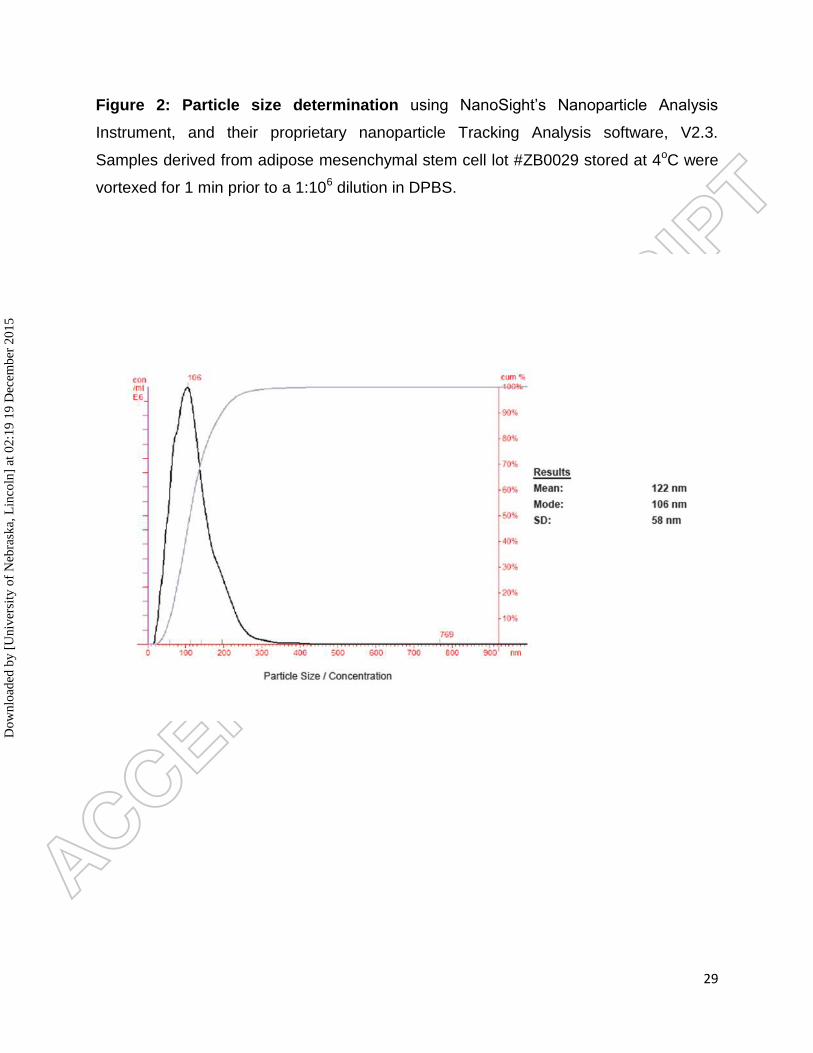
Figure 2: Particle size determination using NanoSight’s Nanoparticle Analysis
Instrument, and their proprietary nanoparticle Tracking Analysis software, V2.3.
Samples derived from adipose mesenchymal stem cell lot #ZB0029 stored at 4oC were
vortexed for 1 min prior to a 1:106 dilution in DPBS.
Dow
nloa
ded
by [
Uni
vers
ity o
f N
ebra
ska,
Lin
coln
] at
02:
19 1
9 D
ecem
ber
2015
30
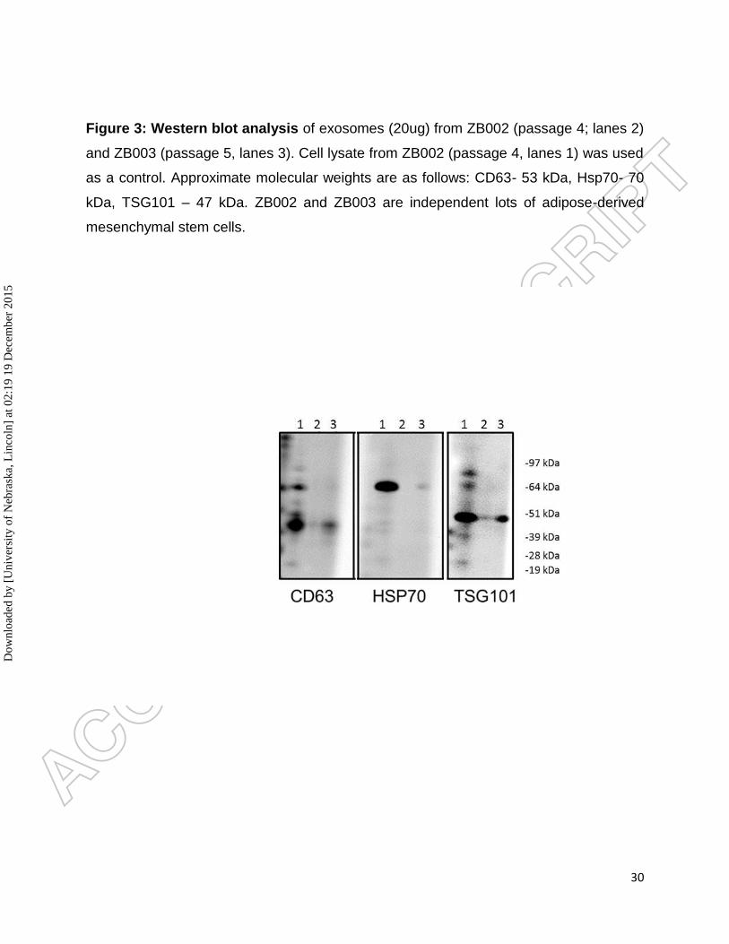
Figure 3: Western blot analysis of exosomes (20ug) from ZB002 (passage 4; lanes 2)
and ZB003 (passage 5, lanes 3). Cell lysate from ZB002 (passage 4, lanes 1) was used
as a control. Approximate molecular weights are as follows: CD63- 53 kDa, Hsp70- 70
kDa, TSG101 – 47 kDa. ZB002 and ZB003 are independent lots of adipose-derived
mesenchymal stem cells.
Dow
nloa
ded
by [
Uni
vers
ity o
f N
ebra
ska,
Lin
coln
] at
02:
19 1
9 D
ecem
ber
2015
31
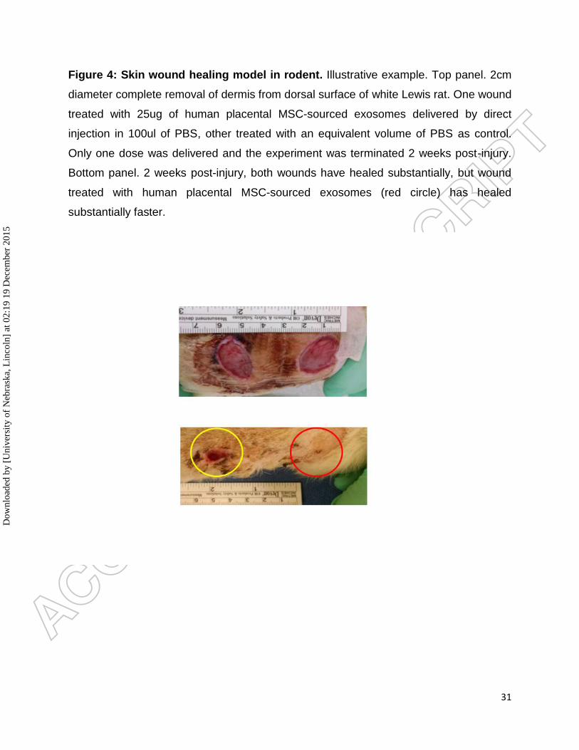
Figure 4: Skin wound healing model in rodent. Illustrative example. Top panel. 2cm
diameter complete removal of dermis from dorsal surface of white Lewis rat. One wound
treated with 25ug of human placental MSC-sourced exosomes delivered by direct
injection in 100ul of PBS, other treated with an equivalent volume of PBS as control.
Only one dose was delivered and the experiment was terminated 2 weeks post-injury.
Bottom panel. 2 weeks post-injury, both wounds have healed substantially, but wound
treated with human placental MSC-sourced exosomes (red circle) has healed
substantially faster.
Dow
nloa
ded
by [
Uni
vers
ity o
f N
ebra
ska,
Lin
coln
] at
02:
19 1
9 D
ecem
ber
2015
32
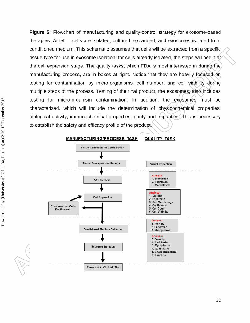
Figure 5: Flowchart of manufacturing and quality-control strategy for exosome-based
therapies. At left – cells are isolated, cultured, expanded, and exosomes isolated from
conditioned medium. This schematic assumes that cells will be extracted from a specific
tissue type for use in exosome isolation; for cells already isolated, the steps will begin at
the cell expansion stage. The quality tasks, which FDA is most interested in during the
manufacturing process, are in boxes at right. Notice that they are heavily focused on
testing for contamination by micro-organisms, cell number, and cell viability during
multiple steps of the process. Testing of the final product, the exosomes, also includes
testing for micro-organism contamination. In addition, the exosomes must be
characterized, which will include the determination of physicochemical properties,
biological activity, immunochemical properties, purity and impurities. This is necessary
to establish the safety and efficacy profile of the product.
Dow
nloa
ded
by [
Uni
vers
ity o
f N
ebra
ska,
Lin
coln
] at
02:
19 1
9 D
ecem
ber
2015
33
Acknowledgements
We are grateful to Randal McKenzie (McKenzie Illustrations, [email protected]) for the
graphics in Figure 1.
References
References
1. Basu, J. and Ludlow, J.W. Cell-based therapeutic products: potency assay
development and application. Regen Med 2014; 9: 497-512 ** First systematic definition
of potency assay development strategies for cell-based therapies.
2. Guthrie K, Bruce A, Sangha N, et al. Potency evaluation of tissue engineered and
regenerative medicine products. Trends Biotechnol 2013; 31: 505-14 ** First systematic
definition of potency assay development strategies for tissue engineered product
candidates.
3. Maguire G. Stem cell therapy without the cells. Commun Integr Biol 2013; 6: e26631
4. Justewicz DM, Shokes JE, Reavis B, et al. Characterization of the human smooth
muscle cell secretome for regenerative medicine. Tissue Eng Part C Methods 2012; 18:
797-816
5. Caplan AI and Correa D. The MSC: an injury drugstore. Cell Stem Cell 2011; 9: 11-
15
6. Chargaff E and West R. The biological significance of the thromboplastic protein of
blood. J Biol Chem 1946; 166: 189-97
7. Wolf P. The nature and significance of platelet products in human plasma. Br J
Hematol 1967; 13: 269-88
8. Raposo G, Nijman HW, Stoorvogel W, et al. B lymphocytes secrete antigen
presenting vesicles. J Exp Med 1996; 183: 1161-1172
Dow
nloa
ded
by [
Uni
vers
ity o
f N
ebra
ska,
Lin
coln
] at
02:
19 1
9 D
ecem
ber
2015
34
9. Gould SJ and Raposo G. As we wait: coping with an imperfect nomenclature for
extracellular vesicles. J Extracell Vesicles 2013; 2: 10.3402
10. Taylor DD, Homesley HD, Doellgast GJ. Binding of specific peroxidase-labeled
antibody to placental type phosphatase on tumor-derived membrane fragments. Cancer
Res 1980; 40: 4064-9
11. Johnstone RM, Adam M, Hammond JR, et al. Vesicle formation during reticulocyte
maturation. Association of plasma membrane activities with released vesicles
(exosomes). J Biol Chem 1987; 262: 9412-20
12. Thery C, Clayton A, Amigorena S, Raposo G. Isolation and characterization of
exosomes from cell culture supernatants and biological fluids. Curr. Prot. Cell Biol.
2006; 3.22.1-3.22.29
13. Valadi H, Ekstrom K, Bossios A, et al. Exosome mediated transfer of mRNAs and
microRNAs is a novel mechanism of genetic exchange between cells. Nature Cell Biol
2007; 9: 654-659 * Demonstration of exosome mediated communication between cells
during development
14. Ratajczak J, Miekus K, Kucia M, et al. Embryonic stem cell derived micro-vesicles
reprogram hematopoietic progenitors: evidence for horizontal transfer of mRNA and
protein delivery. Leukemia 2006; 20: 847-56
15. Buck AH, Coakley G, Simbari F, et al. Exosomes secreted by nematode parasites
transfer small RNAs to mammalian cells and modulate innate immunity. Nat Commun
2014; 5: 5488
16. Zitvogel L, Regnault A, Lozier A, et al. Eradication of established murine tumors
using a novel cell-free vaccine: dendritic cell-derived exosomes. Nature Med 1998; 4:
594-600
17. http://exocarta.org/exosome_markers (last accessed 11/04/15)
18. http://microvesicles.org/index.html (last accessed 11/04/15)
Dow
nloa
ded
by [
Uni
vers
ity o
f N
ebra
ska,
Lin
coln
] at
02:
19 1
9 D
ecem
ber
2015
35
19. Yanez-Mo M, Siljander PR, Andreu Z, et al. Biological properties of extracellular
vesicles and their physiological functions. J Extracell Vesicles 4: 10.3402/jev.v4.27066
20. Adzick NS and Lorenz HP. Cells, matrix, growth factors and the surgeon. The
biology of scarless fetal wound repair. Ann Surg 1994; 200: 10-18
21. Roy S, and Gatien S. Regeneration in axolotls: a model to aim for! Exp Gerontol
2008; 43: 968-973
22. Greco V, Hannus M, Eaton S. Argosomes: a potential vehicle for the spread of
morphogens through epithelia. Cell 2001; 5: 633-645
23. Liegeouis S, Benedetto A, Garnier JM, et al. The V0-ATPase mediates apical
secretion of exosomes containing Hedgehog-related proteins in Caenorhabditis
elegans. J Cell Biol 2006; 173: 949-961
24. Tanaka Y, Okada Y, Hirokawa N. FGF-induced vesicular release of Sonic hedgehog
and retinoic acid in leftward nodal flow is critical for left-right determination. Nature
2005; 435: 172-177
25. Vyas N, Walvekar A, Tate D, et al. Vertebrate Hedgehog is secreted on two types of
extracellular vesicles with different signaling properties. Sci Rep 2014; 4: 7357
26. Danilchik M, Williams M, Brown E. Blastocoel spanning filopodia in cleavage stage
Xenopus laevis: potential roles in morphogen distribution and detection. Dev Biol 2013;
382: 70-81
27. Buschow SI, van Balkom BW, Aalberts M, et al. MHC class II-associated proteins in
B-cell exosomes and potential functional implications for exosome biogenesis. Immunol
Cell Biol 2010; 88: 851-856
28. Batagov AO, Kuznetsov VA, Kurochkin IV. Identification of nucleotide patterns
enriched in secreted RNAs as putative cis-acting elements targeting them to exosome
nano-vesicles. BMC Genomics 2011; 12 Suppl 3:S18:
Dow
nloa
ded
by [
Uni
vers
ity o
f N
ebra
ska,
Lin
coln
] at
02:
19 1
9 D
ecem
ber
2015

36
29. Koppers-Lalic D, Hackenberg M, Bijnsdorp IV, et al. Nontemplated nucleotide
additions distinguish the small RNA composition in cells from exosomes. Cell Rep 2014;
8: 1649-58
30. Irion U. and St Johnston D. bicoid RNA localization requires specific binding of an
endosomal sorting complex. Nature 2007; 445:554-558
31. Gibbings DJ, Ciado C, Erhardt M, et al. Multivesicular bodies associate with
components of miRNA effector complexes and modulate miRNA activity. Nat Cell Biol
2009; 11: 1143-1149
32. Deregibus MC, Cantaluppi V, Calogero R, et al. Endothelial progenitor cell derived
microvesicles activate an angiogenic program in endothelial cells by a horizontal
transfer of mRNA. Blood 2007; 110: 2440-2448
33. Rana S, Yue S, Stadel D, et al., 2012. Towards tailored exosomes: the exosomal
tetraspanin web contributes to target cell selection. Int J Biochem Cell Biol. 44: 1574-84
34. Raposo G and Stoorvogel W. 2013. Extracellular vesicles: exosomes, microvesicles
and friends. J Cell Biol. 200: 373-83
35. Aliotta JM, Pereira M, Li M, et al. Stable cell fate changes in marrow cells induced
by lung-derived microvesicles. J Extracell Vesicles 2012; 10.3402
36. Quesenberry PJ, Goldberg LR, Aliotta JM, et al. Cellular phenotype and
extracellular vesicles: basic and clinical considerations. Stem Cells Dev 2014; 23: 1429-
1436
37. Melo SA, Sugimoto H, O'Connell JT, et al. Cancer exosomes perform cell-
independent microRNA biogenesis and promote tumorigenesis. Cancer Cell 2014; 26:
707-721
38. Zhang HG and Grizzle WE. Exosomes: a novel pathway of local and distant
intercellular communication that facilitates the growth and metastasis of neoplastic
lesions. Am J Pathol 2014; 184: 28-41
Dow
nloa
ded
by [
Uni
vers
ity o
f N
ebra
ska,
Lin
coln
] at
02:
19 1
9 D
ecem
ber
2015
37
39. Gupta A. and Pulliam L. Exosomes as mediators of neuroinflammation. J
Neuroinflammation 2014; 11: 68
40. Teixeira FG, Carvalho MM, Sousa N, et al. Mesenchymal stem cells secretome: a
new paradigm for central nervous system regeneration? Cell Mol Life Sci. 2013;
70:3871-3882.
41. Vishnubhatla I, Corteling R, Stevanato L, et al. The Development of Stem Cell-
derived Exosomes as a Cell-free Regenerative Medicine. J Circ Biomark, 2014, 3:2. doi:
10.5772/58597
42. Murphy MB, Moncivais K, Caplan AI. Mesenchymal stem cells: environmentally
responsive therapeutics for regenerative medicine. Exp Mol Med 2013; 45: e54
43. Liang X, Ding Y, Zhang Y. et al. Paracrine mechanisms of mesenchymal stem cell-
based therapy: current status and perspectives. Cell Transplantation 2013; 9: 1045-59
44. van Koppen A, Joles JA, van Balkom BW. et al. Human embryonic mesenchymal
stem cell-derived conditioned medium rescues kidney function in rats with established
chronic kidney disease. PLoS ONE 2012; 7:e38746
45. Ratajczak MZ, Kucia M, Jadczyk T. et al. Pivotal role of paracrine effects in stem
cell therapies in regenerative medicine: can we translate stem cell-secreted paracrine
factors and microvesicles into better therapeutic strategies? Leukemia 2012; 26: 1166-
1173
46. Di Santo S, Yang Z, Wyler von Ballmoos M. et al. Novel cell free strategy for
therapeutic angiogenesis: in vitro generated conditioned medium can replace progenitor
cell transplantation. PLoS One 2009; e5643
47. Doyle B, Sorajja P, Hynes B. Progenitor cell therapy in a porcine acute myocardial
infarction model induces cardiac hypertrophy mediated by paracrine secretion of
cardiotrophic factors including TGFbeta1. Stem Cells Dev 2008; 17: 941-51
48. Di Santo S, Yang Z, Wyler von Ballmoos M et al., 2015. Endothelial progenitor cell
derived conditioned medium promotes brain microvascular cell viability after ischemic
insult in vitro. Cell Transplant 4: 755
Dow
nloa
ded
by [
Uni
vers
ity o
f N
ebra
ska,
Lin
coln
] at
02:
19 1
9 D
ecem
ber
2015
38
49. Ionescu L, Byrne RN, van Haaften T, et al. Stem cell conditioned medium improves
acute lung injury in mice: in vivo evidence for stem cell paracrine action. Am J Physiol
Lung Cell Mol Physiol 2012; 303: L967-77
50. Osugi M, Katagiri W, Yoshimi R. et al. Conditioned media from mesenchymal stem
cells enhanced bone regeneration in rat calvarial bone defects. Tissue Eng Part A 2012;
18: 1479-89
51. Maumus M, Jorgensen C, Noël D. Mesenchymal stem cells in regenerative
medicine applied to rheumatic diseases: role of secretome and exosomes. Biochimie.
2013; 95: 2229-2234;
52. Toh WS, Foldager CB, Pei M, Hui JH. Advances in mesenchymal stem cell-based
strategies for cartilage repair and regeneration. Stem Cell Reviews and Reports. 2014;
10: 686-696
53. Akyurekli C, Le Y, Richardson RB, et al. A systematic review of preclinical studies
on the therapeutic potential of mesenchymal stromal cell-derived microvesicles. Stem
Cell Rev. 2015; 11:150-160 * Review of key preclinical studies associated with MSC-
sourced exosomes
54. Lai RC, Arslan F, Lee MM. et al. Exosome secreted by MSC reduces myocardial
ischemia/reperfusion injury. Stem Cell Res 2010; 4: 214-22
55. Gatti S, Bruno S, Deregibus MC, et al. Microvesicles derived from human adult
mesenchymal stem cells protect against ischaemia reperfusion induced acute and
chronic kidney injury. Nephrol Dial Transplant 2011; 26: 1474-83
56. Wang Y, Zhang L, Li Y. et al. Exosomes/microvesicles from induced pluripotent
stem cells deliver cardioprotective miRNAs and prevent cardiomyocyte apoptosis in the
ischemic myocardium. Int J Cardiol 2015; 192: 61-9
57. Kilpinen L, Impola U, Sankkila L, et al. Extracellular membrane vesicles from
umbilical cord blood derived MSC protect against ischemic acute kidney injury, a feature
that is lost after inflammatory conditioning. J Extracell Vesicles 2013; 10.3402
Dow
nloa
ded
by [
Uni
vers
ity o
f N
ebra
ska,
Lin
coln
] at
02:
19 1
9 D
ecem
ber
2015
39
58. Lee Y, El Andaloussi S, Wood MJ. Exosomes and microvesicles: extracellular
vesicles for genetic information transfer and gene therapy. Hum Mol Genet 2012; 21:
R125-34
59. György B, Hung ME, Breakefield XO, et al. Therapeutic applications of extracellular
vesicles: clinical promise and open questions. Ann Rev Pharmacol Toxicol 2015; 55:
439-464
60. Lamichhane TN, Sokic S, Schardt JS, et al. Emerging roles for extracellular
vesicles in tissue engineering and regenerative medicine. Tissue Engineering Part B
Rev 2015; 21: 45-54
61. Sahoo S, Klychko E, Thorne T, et al. Exosomes from human CD34+ stem cells
mediate their proangiogenic paracrine activity. Circ Res 2011; 109: 724-728
62. Lee C, Mitsialis SA, Aslam M, et al. Exosomes mediate the cytoprotective action of
mesenchymal stromal cells on hypoxia induced pulmonary hypertension. Circulation
2012; 126: 2601-2611
63. Zhang J, Guan J, Niu X, et al. Exosomes released from human induced pluripotent
stem cells derived MSCs facilitate cutaneous wound healing by promoting collagen
synthesis and angiogenesis. J Trans Med 2015; 13: 49
64. Ibrahim AG, Cheng K, Marbán E. Exosomes as critical agents of cardiac
regeneration triggered by cell therapy. Stem Cell Reports 2014; 2: 606-619
65. Kalani A, Tyagi A, Tyagi N. 2014. Exosomes: mediators of neurodegeneration,
neuroprotection and therapeutics. Mol Neurobiol 49: 590-600
66. Lai CP and Breakefield XO. 2012. Role of exosomes/microvesicles in the nervous
system and use in emerging therapies. Frontiers Physiol doi: 10.3389/fphys.2012.00228
67. Krämer-Albers EM, Bretz N, Tenzer S, et al., 2007. Oligodendrocytes secrete
exosomes containing major myelin and stress-protective proteins: trophic support for
axons? Proteomics Clin Appl 1, 1446–1461
Dow
nloa
ded
by [
Uni
vers
ity o
f N
ebra
ska,
Lin
coln
] at
02:
19 1
9 D
ecem
ber
2015
40
68. Bosse F. 2012. Extrinsic cellular and molecular mediators of peripheral axonal
regeneration. Cell Tissue Res. 2012; 349:5–14
69. Fruhbeis C, Frohlich D, Kuo WP et al. 2013. Neurotransmitter-triggered transfer of
exosomes mediates oligodendrocyte-neuron communication. PLoS Biol, e1001604
70. Takeda YS and Xu Q. 2015. Neuronal differentiation of human mesenchymal stem
cells using exosomes derived from differentiating neuronal cells. PLoS One e0135111
71. Lee HK, Finniss S, Cazacu S, et al., 2014. Mesenchymal stem cells deliver
exogenous miRNAs to neural cells and induce their differentiation and glutamate
transporter expression. Stem Cells Dev 23: 2851-61
72. Sun D, Zhuang X, Xiang X, et al., 2010. A Novel Nanoparticle Drug Delivery
System: The Anti-inflammatory Activity of Curcumin Is Enhanced When Encapsulated in
Exosomes. Mol Ther 18: 1606-1614
73. Zhuang X, Xiang X, Grizzle W, et al., 2011. Treatment of brain inflammatory
diseases by delivering exosome-encapsulated anti-inflammatory drugs from the nasal
region to the brain. Mol Ther 19: 1769–1779
74. Xin H, Li Y, Liu Z, et al. Systematic administration of exosomes released from
mesenchymal stromal cells promote functional recovery and neurovascular plasticity
after stroke in rats. J. Cereb Blood Flow Metab 2013; 33: 1711-1715
75. Zhang Y, Chopp M, Meng Y, et al. Effect of exosomes derived from multipluripotent
mesenchymal stromal cells on functional recovery and neurovascular plasticity in rats
after traumatic brain injury. J Neurosurg 2015; 122: 856-867
76. Doeppner TR, Herz J, Görgens A, et al., 2015. Extracellular vesicles improve post-
stroke neuroregeneration and prevent postischemic immunosuppression. Stem Cells
Transl Med 4: 1131-43
77. Xin H, Li Y, Liu Z, et al., 2013. MiR-133b promotes neural plasticity and functional
recovery after treatment of stroke with multipotent mesenchymal stromal cells in rats via
transfer of exosome-enriched extracellular particles. Stem Cells 31: 2737-46
Dow
nloa
ded
by [
Uni
vers
ity o
f N
ebra
ska,
Lin
coln
] at
02:
19 1
9 D
ecem
ber
2015
41
78. Bruce A, Ilagan R, Guthrie K, et al., 2015. Selected renal cells modulate disease
progression in rodent models of chronic kidney disease via NFB and TGF1 pathways.
Regenerative Medicine 10: 815-839
79. Fleury A, Martinez MC, Le Lay S. Extracellular vesicles as therapeutic tools in
cardiovascular diseases. Front Immunol 2014; 5: 370
80. Yu B, Zhang X, Li X. Exosomes derived from mesenchymal stem cells. Int J Mol Sci
2014; 15: 4142-4157
81. Harshyne LA, Hooper KM, Andrews EG. et al. Glioblastoma exosomes and IGF-
1R/AS-ODN are immunogenic stimuli in a translational research immunotherapy
paradigm. Cancer Immunol Immunother 2015; 64: 299-309
82. Vlassov AV, Magdaleno S, Setterquist R, et al. (2012) Exosomes: current
knowledge of their composition, biological functions, and diagnostic and therapeutic
potentials. Biochimica et Biophysica Acta 1820: 940-948
83. Kordelas L, Rebmann V, Ludwig A-K, et al. (2013) Successful treatment of therapy-
refractory acute graft-versus-host disease with mesenchymal stem cell-derived
exosomes. 39th Annual Meeting of the European Group for Blood and Marrow
Transplantation
84. https://clinicaltrials.gov/ct2/show/NCT01294072?term=exosome&rank=1 (last
accessed 11/04/15)
85. https://clinicaltrials.gov/ct2/show/NCT02565264?term=exosome&rank=3 (last
accessed 11/04/15)
86. https://clinicaltrials.gov/ct2/show/NCT02138331?term=exosome&rank=4 (last
accessed 11/04/15)
87. https://clinicaltrials.gov/ct2/show/NCT01668849?term=exosome&rank=8 (last
accessed 11/04/15)
88. https://clinicaltrials.gov/ct2/show/NCT01159288?term=exosome&rank=11 (last
accessed 11/04/15)
Dow
nloa
ded
by [
Uni
vers
ity o
f N
ebra
ska,
Lin
coln
] at
02:
19 1
9 D
ecem
ber
2015

42
89. www.appliedbiologics.com (last accessed 11/04/15)
90. Viaud S, Thery C, Ploix S, et al. (2010) Dendritic cell-derived exosomes for cancer
immunotherapy: what's next? Cancer Research 70: 1281-1285;
91. Morse MA, Garst J, Osada T, et al. (2005) A phase I study of dexosome
immunotherapy in patients with advanced non-small cell lung cancer. Journal of
Translational Medicine 3: 9
92. Dai S, Wei D, Wu Z, et al. (2008) Phase I clinical trial of autologous ascites-derived
exosomes combined with GM-CSF for colorectal cancer. Molecular Therapy 16: 782-
790;
93. Escudier B, Dorval T, Chaput N, et al. (2005) Vaccination of metastatic melanoma
patients with autologous dendritic cell (DC) derived-exosomes: results of the first phase
I clinical trial. Journal of Translational Medicine 3: 10
94. Zhang B, Wang M, Gong A, et al. HucMSC-exosome mediated -Wnt4 signaling is
required for cutaneous wound healing. Stem Cells 2014; 10.1002/stem.1771
95. Lim CP, Phan TT, Lim IJ, et al. Stat3 contributes to keloid pathogenesis via
promoting collagen production, cell proliferation, and migration. Oncogene 2006; 25:
5416-5425
96. Lee C, Mitsialis SA, Aslam M, et al. Exosomes mediate the cytoprotective action of
mesenchymal stromal cells on hypoxia-induced pulmonary hypertension. Circulation
2012; 126: 2601-2611
97. Madsen JT and Andersen KE. Microvesicle formulations used in topical drugs and
cosmetics affect product efficiency, performance and allergenicity. Dermatitis 2010; 21:
243-247
98. Liu JJ, Nazzal S, Chang TS, et al. Preparation and characterization of
cosmeceutical liposomes loaded with avobenzone and arbutin. J Cosmet Sci 2013; 64:
9-17
Dow
nloa
ded
by [
Uni
vers
ity o
f N
ebra
ska,
Lin
coln
] at
02:
19 1
9 D
ecem
ber
2015

43
99. Campani V, Marchese D, Pitaro MT, et al. Development of a liposome based
formulation for vitamin K1 nebulization on the skin. Int J Nanomedicine 2014; 9: 1823-
1832
100. Rahimpour Y. and Hamishehkar H. Liposomes in cosmeceutics. Expert Opin Drug
Deliv 2012; 9:443-455
101. Seo KY, Kim DH, Lee SE, et al. Skin rejuvenation by microneedle fractional
radiofrequency and a human stem cell conditioned medium in Asian skin: a randomized
controlled investigator blinded split face study. J. Cosmet Laser Ther 2013; 15: 25-33
102. Lee HJ, Lee EG, Kang S, et al. Efficacy of microneedling plus human stem cell
conditioned medium for skin rejuvenation: a randomized, controlled, blinded split face
study. Ann Dermatol 2014; 26: 584-591
103. Harn HJ, Huang MH, Huang CT, et al. Rejuvenation of aged pig facial skin by
transplanting allogeneic granulocyte colony stimulating factor induced peripheral blood
stem cells from a young pig. Cell Transplant 2013; 22: 755-765
104. Kim WS, Park BS, Park SH, et al. Antiwrinkle effect of adipose-derived stem cell:
activation of dermal fibroblast by secretory factors. J Dermatol Sci 2009; 53: 96-102
105. Shim JH, Park JY, Lee MG, et al. Human dermal stem/progenitor cell-derived
conditioned medium ameliorates ultraviolet a induced damage of normal human dermal
fibroblasts. PLoS One 2013; e67604
106. Chen CC, Murray PJ, Jiang TX, et al. Regenerative hair waves in aging mice and
extra-follicular modulators Follistatin, Dkk1 and Sfrp4. J Invest Dermatol 2014; 134:
2086-2096
107. Fukuoka H. and Suga H. Hair regeneration treatment using adipose-derived stem
cell conditioned medium: follow-up with trichograms. Eplasty 2015; 15:e10
108. Ahmed MI, Alam M, Emelianov VU, et al. MicroRNA-214 controls skin and hair
follicle development by modulating the activity of the Wnt pathway. J Cell Biol 2014;
207: 549-567
Dow
nloa
ded
by [
Uni
vers
ity o
f N
ebra
ska,
Lin
coln
] at
02:
19 1
9 D
ecem
ber
2015
44
109. Zhang GE, Grizzle WE. Exosomes: a novel pathway of local and distant
intercellular communication that facilitates the growth and metastasis of neoplastic
lesions. Am J Pathol 2014; 184: 28-41
110. Harris DA, Patel SH, Gucek M, et al. Exosomes released from breast cancer
carcinomas stimulate cell movement. PLoS One 2015; e0117495
111. Vallabhaneni KC, Penfornis P, Dhule S, et al. Extracellular vesicles from bone
marrow mesenchymal stem/stromal cells transport tumor regulatory microRNA, proteins
and metabolites. Oncotarget 2015; 6: 4953-67
112. Li J, Sherman-Baust CA, Tsai-Turton M, et al. Claudin containing exosomes in the
peripheral circulation of women with ovarian cancer. BMC Cancer 2009; 9: 244
113. Stenqvist AC, Nagaeva O, Baranov V, et al. Exosomes secreted by human
placenta carry functional Fas ligand and TRAIL molecules and convey apoptosis in
activated immune cells, suggesting exosome-mediated immune privilege of the fetus. J
Immunol 2013; 191: 5515-23
114. Vincent-Schneider H, Stumptner-Cuvelette P, Lankar D, et al. Exosomes bearing
HLA-DR1 molecules need dendritic cells to efficiently stimulate specific T cells. Int
Immunol 2002; 14: 713-22
115. Théry C, Duban L, Segura E, et al. Indirect activation of naïve CD4+ T cells by
dendritic cell derived exosomes. Nat Immunol 2002; 3: 1156-1162
116. Lai CP, Mardini O, Ericsson M, et al. Dynamic bio-distribution of extracellular
vesicles in vivo using a multimodal imaging reporter. ACS Nano 2014; 8: 483-94
117. Andre F, Escudier B, Angevin E, et al. Exosomes for cancer immunotherapy. Ann
Oncol 2004;15 Suppl 4:iv14, 1-4
118. Chen TS, Arslan F, Yin Y, et al. Enabling a robust scalable manufacturing process
for therapeutic exosomes through oncogenic immortalization of human ESC-derived
MSCs. J Trans Med 2011; 9: 47
Dow
nloa
ded
by [
Uni
vers
ity o
f N
ebra
ska,
Lin
coln
] at
02:
19 1
9 D
ecem
ber
2015
45
119. Mittelbrunn M, Gutiérrez-Vázquez C, Villarroya-Beltri C, et al., 2011. Unidirectional
transfer of microRNA-loaded exosomes from T cells to antigen-presenting cells. Nature
Communications 2: 282
120. www.systembio.com, last accessed 11/04/15
121. Basu J and Ludlow JW. MSC sourced exosomes as therapeutic agents for wound
healing and skin regeneration: from scaled production to functional regenerative
outcomes in vitro and in vivo. International Society for Stem Cell Research Annual
Meeting, 2015, Stockholm.
122. Basu J and Ludlow JW. Developmental engineering the kidney: leveraging
principles of morphogenesis for renal regeneration. Birth Defects Res C Embryo Today
2012; 96: 30-38
123. Genheimer CW, Ilagan RM, Spencer T, et al., 2012. Molecular characterization of
the regenerative response induced by intrarenal transplantation of selected renal cells in
a rodent model of chronic kidney disease. Cells Tissues Organs 2012; 196: 374-384
124. www.fda.gov/BiologicsBloodVaccines/default.htm (last accessed 11/04/15)
125. Marcus ME and Leonard JN. FedExosomes: engineering therapeutic biological
nanoparticles that truly deliver. Pharmaceuticals 2013; 6: 659-680
126. Kooijmans SA, Vader P, van Dommelen SM, et al. Exosome mimetics: a novel
class of drug delivery systems. Int J Nanomedicine 2012; 7: 1525-1541
127. Jeong D, Jo W, Yoon J, et al. Nanovesicles engineered from ES cells for enhanced
cell proliferation. Biomaterials 2014; 35: 9302-9310
128. Ranghino A, Dimuccio V, Papadimitriou E, et al. Extracellular vesicles in the urine:
markers and mediators of tissue damage and regeneration. Clin Kidney J 2015; 8: 23-
30
Dow
nloa
ded
by [
Uni
vers
ity o
f N
ebra
ska,
Lin
coln
] at
02:
19 1
9 D
ecem
ber
2015
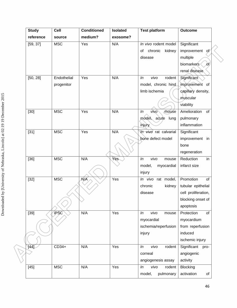
46
Study
reference
Cell
source
Conditioned
medium?
Isolated
exosome?
Test platform Outcome
[59, 37] MSC Yes N/A In vivo rodent model
of chronic kidney
disease
Significant
improvement of
multiple
biomarkers of
renal disease
[50, 28] Endothelial
progenitor
Yes N/A In vivo rodent
model, chronic hind
limb ischemia
Significant
improvement of
capillary density,
muscular
viability
[30] MSC Yes N/A In vivo mouse
model, acute lung
injury
Amelioration of
pulmonary
inflammation
[31] MSC Yes N/A In vivo rat calvarial
bone defect model
Significant
improvement in
bone
regeneration
[36] MSC N/A Yes In vivo mouse
model, myocardial
injury
Reduction in
infarct size
[32] MSC N/A Yes In vivo rat model,
chronic kidney
disease
Promotion of
tubular epithelial
cell proliferation,
blocking onset of
apoptosis
[39] iPSC N/A Yes In vivo mouse
myocardial
ischemia/reperfusion
injury
Protection of
myocardium
from reperfusion
induced
ischemic injury
[44] CD34+ N/A Yes In vivo rodent
corneal
angiogenesis assay
Significant pro-
angiogenic
activity
[45] MSC N/A Yes In vivo rodent
model, pulmonary
Blocking
activation of
Dow
nloa
ded
by [
Uni
vers
ity o
f N
ebra
ska,
Lin
coln
] at
02:
19 1
9 D
ecem
ber
2015
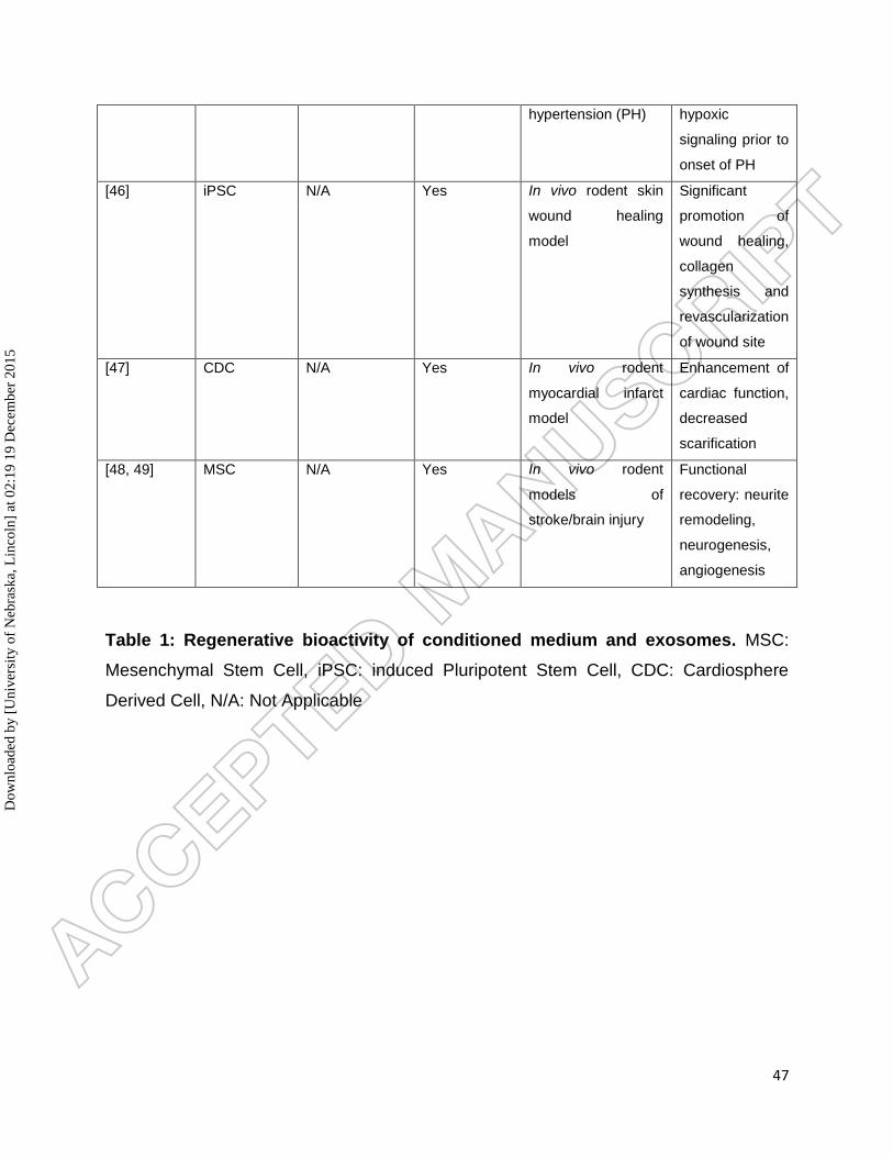
47
hypertension (PH) hypoxic
signaling prior to
onset of PH
[46] iPSC N/A Yes In vivo rodent skin
wound healing
model
Significant
promotion of
wound healing,
collagen
synthesis and
revascularization
of wound site
[47] CDC N/A Yes In vivo rodent
myocardial infarct
model
Enhancement of
cardiac function,
decreased
scarification
[48, 49] MSC N/A Yes In vivo rodent
models of
stroke/brain injury
Functional
recovery: neurite
remodeling,
neurogenesis,
angiogenesis
Table 1: Regenerative bioactivity of conditioned medium and exosomes. MSC:
Mesenchymal Stem Cell, iPSC: induced Pluripotent Stem Cell, CDC: Cardiosphere
Derived Cell, N/A: Not Applicable
Dow
nloa
ded
by [
Uni
vers
ity o
f N
ebra
ska,
Lin
coln
] at
02:
19 1
9 D
ecem
ber
2015