EXOSOMES AND THE NKG2D RECEPTOR- LIGAND ...358014/FULLTEXT01.pdfEXOSOMES AND THE NKG2D...
64
EXOSOMES AND THE NKG2D RECEPTOR- LIGAND SYSTEM IN PREGNANCY AND CANCER: USING STRESS FOR SURVIVAL By Malin Hedlund Department of Clinical Immunology Umeå University Umeå 2010
Transcript of EXOSOMES AND THE NKG2D RECEPTOR- LIGAND ...358014/FULLTEXT01.pdfEXOSOMES AND THE NKG2D...
EXOSOMES AND THE NKG2D RECEPTOR-LIGAND SYSTEM IN PREGNANCY AND
CANCER: USING STRESS FOR SURVIVAL
By
Malin Hedlund
Department of Clinical Immunology Umeå University
Umeå 2010

Cover: Electron micrograph of isolated exosomes from human early placenta. Courtesy Dr.Vladimir Baranov.
All previously published papers were reproduced with permission from the publisher Copyright © Malin Hedlund ISBN: 978-91-7459-072-2 ISSN: 0346-6612 New series: 1375 Printed by: Print Media Umeå, Sweden 2010

Nu ska en tornado gå fram
Gerd Lundquist
To my family and my Love

4
TABLE OF CONTENTS
ABSTRACT 7
SAMMANFATTNING PÅ SVENSKA 9
ORIGINAL PAPERS 11
LIST OF ABBREVIATIONS 12
INTRODUCTION 15 BACKGROUND 17 1. The immune system in general and in pregnancy 17
1.1 Definitions and general properties 171.2 Cells of the immune system 17
1.2.1 B lymphocytes 17 1.2.2 Antigen-presenting cells 18 1.2.3 ���T lymphocytes 19 1.2.4 Regulatory T cells 20 1.2.5 ���T lymphocytes 20 1.2.6 NKT cells 21 1.2.7 NK cells 211.3 Antigen-presenting molecules 22
1.3.1 Major histocompatibility complex 22 1.3.1.1 Classical HLA molecules 22 1.3.1.2 Non Classical HLA molecules 231.4 Toll-like receptors 231.5 Cytokines 241.6 Complement system 241.7 Immunosurveillance 251.8 The NKG2D receptor-ligand system 26
1.8.1 The NKG2D (KLRK1) receptor 261.8.2 The NKG2D ligands 271.8.3 The lytic machinery and the cytotoxic hit mediated by the NKG2D receptor 28
2. Mammalian pregnancy 29
2.1 Placenta is a unique temporary organ for pregnancy success 29 2.1.1 General description 29
2.1.2 Morphological organisation of human early pregnancy 29 placenta
2.2 Human trophoblasts – the main cells of the placenta with key importance in pregnancy 31
2.2.1 Phenotypic and functional characteristics of villous 31 trophoblast
5
2.2.2 Phenotypic and functional characteristics of 32 extravillous trophoblast
3. Immune escape – a common strategy for pregnancy and cancer 33
3.1 Trophoblast and cancer cells share many biological features 333.2 Trophoblast and cancer use similar immune escape mechanisms 343.3 Exosome secretion is a way of intercellular communication and generation of “soluble” bioactive ligands 35
3.3.1 Definition of exosomes 36 3.3.2 Biogenesis of exosomes 373.3.3 Secreted exosomes - general characteristics and functions 38
AIMS OF THE INVESTIGATION 40
RESULTS AND DISCUSSION 414. Methodological considerations 41
4.1 Isolation of villous trophoblast cells from human early pregnancy placenta 41 4.1.1 Description and advantages of our isolation procedure 424.2 Placental explant cultures 424.3 Isolation of exosomes 434.4 Quantification of exosome secretion 43
5. The NKG2D receptor-ligand system in human pregnancy 445.1 Expression of NKG2D ligands by human placenta 445.2 The NKG2D ligand molecules are processed, stored and
secreted through the late endosomal compartment of syncytiotrophoblast 44
5.3 Placenta explant cultures secrete NKG2D ligand-bearing exosomes that impair NK cell cytotoxicity 46
6. NKG2D ligand expression and secretion by cancer cells 486.1 Stress-induced up-regulation of NKG2D ligands in leukemia cell lines 486.2 Thermal- and oxidative stress up-regulates secretion and expression of NKG2D ligand-bearing exosomes that enhance the suppression of NK cell mediated cytotoxicity 49
CONCLUSIONS 50
ACKNOWLEDGEMENTS 51
REFERENCES 53
6
ABSTRACT
Although not obvious at first sight, several parallels can be drawn between pregnancy and
cancer. Many proliferative, invasive and immune tolerance mechanisms that support
normal pregnancy are also exploited by malignancies to establish a nutrient supply and
evade or edit the immune response of the host. The human placenta, of crucial importance
for pregnancy success, and its main cells, the trophoblast, share several features with
malignant cells such as high cell proliferation rate, lack of cell-contact inhibition and
invasiveness. Both in cancer and in pregnancy, the immune defense mechanisms,
potentially threatening the survival of the tumor or the fetus, are progressively blunted or
even turned into tumor- or pregnancy-promoting players.
Amongst immune mechanisms that are meant to protect the host from cancer and can be a
potential threat to the fetus, the NKG2D receptor-ligand system stands out as the most
powerful, stress-inducible “danger detector” system that comprises the activating NK cell
receptor NKG2D and its ligands, the MIC (MHC class I Chain-related proteins A and B)
and ULBP (UL-16 Binding Proteins) families. It is the major cytotoxic mechanism in the
body promoting surveillance and homeostasis. In the present thesis we investigate the
NKG2D receptor-ligand system in human early normal pregnancy and in the
leukemia/lymphoma cell lines Jurkat and Raji and ask the questions “How is the NKG2D
receptor-ligand system functioning in pregnancy and tumor? How is the danger of cytotoxic
attack of the fetus avoided? Why is the immunosurveillance function compromised in
cancer patients?”
We developed a method to isolate and culture villous trophoblast from early human normal
placenta and used it to study the NKG2D receptor-ligand system. We discovered that the
NKG2D ligand families of molecules MICA/B and ULBP1-5 are constitutively expressed
by the syncytiotrophoblast of the chorionic villi. Using immnunoelectron microscopy, we
studied the expression of these molecules at the subcellular level and could show for the
first time that they are preferably expressed on microvesicles in multivesicular bodies
(MVB) of the late endosomal compartment and are secreted as exosomes. Exosomes are
nanometer sized microvesicles of endosomal origin, produced and secreted by a great
7
variety of normal and tumor cells. The exosomes are packages of proteins and ribonucleic
acids that function as “mail” or “messengers” between cells conveying different biological
information. We isolated and studied exosomes from placental explant cultures. We found
that they carry NKG2D ligands on their surface and are able to bind and down-regulate the
cognate receptor on NK-, CD8+ and ���T cells. The down-regulation selectively caused
impairment of the cytotoxic response of the cells but did not affect their lytic ability as
measured by perforin content and gene transcription. Thus, the NKG2D ligand-bearing
exosomes suppress the cytotoxic activity of the cells in the vicinity of the placenta, leaving
their cytolytic machinery intact, ready to function when the cognate receptor is
restored/recycled. These findings highlight the role of placental exosomes in the fetal-
maternal immune escape and support the view of placenta as an unique immunomodulatory
organ.
Next, we studied the expression and exosomal release of NKG2D ligands by tumor cells
using the leukemia cell lines Jurkat and Raji as a tumor model. We found that NKG2D
ligand-bearing exosomes with similar immunosuppressive properties as placental exosomes
are constitutively secreted by the tumor cells, as a mechanism to blunt the cytotoxic
response of the immune cells and thus protect themselves from cytotoxic attack by the host.
Interestingly, we found that thermal- and oxidative stress up-regulates the exosome
secretion and the amount of exosome-secreted NKG2D ligands. Our results imply that
tumor therapies that cause stress-induced damage, such as thermotherapy and stripping of
oxygen supply to the tumor, might have a previously unrecognized side effect causing
enhanced exosome production and secretion, which in turn suppresses the natural anti-
tumor immune response and thus should be taken into account when designing an optimal
therapy of cancer patients.
In conclusion, we describe a novel stress-inducible mechanism shared by placenta and
tumors as an immune escape strategy. We found that placenta- and tumor-derived NKG2D
ligand-bearing exosomes can suppress immune responses to promote the survival and well
being of the fetus or the tumor. Our work comprises an important contribution to the
elucidation of the NKG2D ligand-receptor system and its mode of operation in the human
body and opens new perspectives for designing novel therapies for infertility and cancer.
8
SAMMANFATTNING PÅ SVENSKA
Även om det kan verka paradoxalt, utmanar graviditet och cancer immunsystemet på
liknande sätt. Dessa två så diametralt olika tillstånd representerar två mycket speciella
situationer: graviditeten innebär utveckling och tillväxt av ett foster, en egen individ olik
modern, tillfälligt ”transplanterad” i hennes kropp; cancersjukdomen innebär att
kroppsegna celler, som har blivit olika/förändrade genom en process kallad malignifiering,
växer och sprids genom att ”transplantera” dottersvulster i olika organ. Vid båda dessa
tillstånd har fostret och cancern ett gemensamt mål, att överleva och parasitera i en annan
kropp och de har utvecklat liknande strategier för att undvika angrepp från värdens
immunsystem. Moderkakan, placentan, och dess trofoblastceller är livsviktiga för
graviditeten och delar många egenskaper med många olika cancerceller såsom
okontrollerad celldelning, tillväxt och invasion.
I vår strävan att förstå på vilket sätt tumörer och placenta lyckas undvika en immunologisk
attack har vi valt att studera NKG2D receptor-ligand systemet. Detta system är ett mycket
viktigt immunologiskt verktyg med vilket alla förändrade, infekterade och på olika sätt
biologiskt stressade celler, inklusive cancerförändrade celler, avlägsnas från kroppen med
hjälp av ”mördarceller”, så kallade cytotoxiska T celler och NK celler. Mördarcellerna
uttrycker NKG2D receptorn på sin yta som binder till sina ligander, MIC och ULBP1-6,
uttryckta på förändrade kroppsceller. När en bindning har skett överförs en
aktiveringssignal till mördarcellen som dödar t.ex. cancercellen, märkt med MIC och/eller
ULBP molekyler på sin yta. Vi har ställt oss frågorna: ”Hur fungerar NKG2D receptor-
ligand systemet vid graviditet och cancer? Hur undviker fostret att attackeras av mammans
immunförsvar? Varför lyckas inte NKG2D receptor-ligand systemet eliminera de
förändrade tumörcellerna hos cancerpatienter?”
Vi upptäckte att moderkakans syncytiotrofoblaster utsöndrar liganderna till NKG2D
receptorn, MICA/B och ULBP1-5, bundna till ytan av mycket små (nanometer-stora)
membranomgivna blåsor som kallas exosomer, avbildade från en elektronmikroskopisk bild
på omslaget av denna avhandling. Exosomerna är 30-100 nm stora, uttrycker många olika
proteiner både på ytan och inuti och kan produceras och utsöndras i blodet av många olika
9
celler. Exosomerna används som ett sätt att kommunicera och kan betraktas som cellernas
”brev” till varandra. Vi upptäckte att moderkakans syncytiotrofoblastceller producerar
exosomer som bär NKG2D liganderna MIC och ULBP på sin yta. Dessa exosomer binder
med sina MIC och ULBP molekyler till NKG2D receptorn, trycker ner den från cellytan
och på så sätt förstör mördarcellens avdödande förmåga. De NKG2D ligand-bärande
exosomerna som moderkakan, placenta, producerar och utsöndrar används för att undvika
attack från moderns immunsystem och på så sätt skyddas fostrets överlevnad och
utveckling. Liknande mekanism används även av cancerceller för att etablera och sprida sig
i värdens kropp.
I vår nästa studie undersökte vi NKG2D ligand-bärande tumörexosomer från leukemi och
lymfomceller. En viktig upptäckt var att cancercellerna ökade sin exosomutsöndring och
därmed sin nedreglering av mördarcellernas avdödande förmåga när de utsattes för cellulär
stress. Våra resultat antyder att cancerbehandling som verkar genom stressinducerande
mekanismer, såsom kemoterapi och/eller termoterapi och strypning av syretillförseln till
cancerceller kan ha tidigare okända bieffekter som trycker ner patienternas immunförsvar
via ökad produktion av tumörexosomer - en viktig aspekt som bör övervägas när man
planerar optimal anti-cancer behandling.
Sammanfattningsvis använder placenta och cancerceller liknande strategi för fostrets
överlevnad och cancerns etablering, tillväxt och spridning, baserad på utsöndring av
NKG2D ligand-bärande immunosuppressiva exosomer. Våra resultat ökar förståelsen av
NKG2D receptor-ligand systemets och exosomernas funktion och kan bidra till
utvecklingen av nya strategier i behandlingen av infertilitet och cancer.
10
ORIGINAL PAPERS
Paper I
Ann-Christin Stenqvist, Ting Chen, Malin Hedlund, Tanya Dimova, Olga Nagaeva,
Lennart Kjellberg, Eva Innala, Lucia Mincheva-Nilsson. An efficient optimized method for
isolation of villous trophoblast cells from human early pregnancy placenta suitable for
functional and molecular studies. American Journal of Reproductive Immunology, 2008;
60(1): 33-42.
Paper II
Malin Hedlund, Ann-Christin Stenqvist, Olga Nagaeva, Lennart Kjellberg, Marianne
Wulff, Vladimir Baranov, Lucia Mincheva-Nilsson. Human placenta expresses and secretes
NKG2D ligands via exosomes that down-modulate the cognate receptor expression:
evidence for immunosuppressive function. The Journal of Immunology, 2009;183(1):340-
351.
Paper III
Malin Hedlund, Olga Nagaeva, Dominic Kargl, Vladimir Baranov, Lucia-Mincheva-
Nilsson. Thermal- and oxidative stress cause enhanced release of NKG2D ligand-bearing
immunosuppressive exosomes in leukemia/lymphoma T and B cells. Submitted.
11
LIST OF ABBREVIATIONS
APCs antigen-presenting cells ATM ataxia telangiectasia mutated ATR ataxia telangiectasia and Rad 3 related protein BCR B cell receptor CD cluster of differentiation Chk1 checkpoint kinase 1 CTB cytotrophoblast CTLs cytotoxic T cells DCs dendritic cells ECM extracellular matrix ESCRT endosomal sorting complex required for transport EVT extravillous trophoblast FasL Fas-ligand Foxp3 transcribing forkhead box protein 3 GH growth hormone GPI glycosylphospatidylinositol hCG human chorionic gonadotropin hCS human chorionic somatomammotropic hormone HLA human leukocyte antigen hPL human placental lactogen HSP heat shock protein IDO indoleamine 2, 3-dioxygenase IEM immnunoelectron microscopy IFN interferon Ig immunoglobulin IGF insulin growth factor IHC immunohistochemistry IL interleukin ILV intraluminal vesicles JAK janus kinase KIR killer cell-Ig-like receptorsLIF leukemia inhibitory factor M� macrophage MHC major histocompatibility complex MIC MHC class I Chain-related proteins MTOR oxygen-sensitive mammalian target of rapamycin MULT-1 murine UL16-binding-protein-like transcripts-1 MVB multivesicular body NK natural killer NKT natural killer TPAMP pathogen-associated molecular patternPECAM platelet endothelial cell adhesion molecule PLAP placental alkaline phophatase RAET retinoic acid early transcript STAT signal transducers and activators of transcription STB syncytiotrophoblast
12
ULBP UL-16 Binding Proteins uNK uterine natural killer TCR T cell receptor TGF transforming growth factor Th T helper TLRs toll-like receptor TNF tumor necrosis factors TRAIL tumor necrosis related apoptosis inducing ligand Tregs regulatory T cells TUN trophuteronectin VCAM vascular cell adhesion molecule VEGF vascular endothelial growth factor VT villous trophoblast
13

14
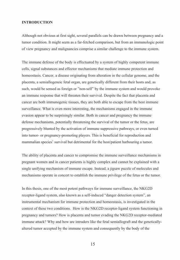
INTRODUCTION
Although not obvious at first sight, several parallels can be drawn between pregnancy and a
tumor condition. It might seem as a far-fetched comparison, but from an immunologic point
of view pregnancy and malignancies comprise a similar challenge to the immune system.
The immune defense of the body is effectuated by a system of highly competent immune
cells, signal substances and effector mechanisms that mediate immune protection and
homeostasis. Cancer, a disease originating from alteration in the cellular genome, and the
placenta, a semiallogeneic fetal organ, are genetically different from their hosts and, as
such, would be sensed as foreign or ”non-self” by the immune system and would provoke
an immune response that will threaten their survival. Despite the fact that placenta and
cancer are both immunogenic tissues, they are both able to escape from the host immune
surveillance. What is even more interesting, the mechanisms engaged in the immune
evasion appear to be surprisingly similar. Both in cancer and pregnancy the immune
defense mechanisms, potentially threatening the survival of the tumor or the fetus, are
progressively blunted by the activation of immune suppressive pathways, or even turned
into tumor- or pregnancy-promoting players. This is beneficial for reproduction and
mammalian species’ survival but detrimental for the host/patient harbouring a tumor.
The ability of placenta and cancer to compromise the immune surveillance mechanisms in
pregnant women and in cancer patients is highly complex and cannot be explained with a
single unifying mechanism of immune escape. Instead, a jigsaw puzzle of molecules and
mechanisms operate in concert to establish the immune privilege of the fetus or the tumor.
In this thesis, one of the most potent pathways for immune surveillance, the NKG2D
receptor-ligand system, also known as a self-induced “danger detection system”, an
instrumental mechanism for immune protection and homeostasis, is investigated in the
context of these two conditions. How is the NKG2D receptor-ligand system functioning in
pregnancy and tumors? How is placenta and tumor evading the NKG2D receptor-mediated
immune attack? Why and how are intruders like the fetal semiallograft and the genetically-
altered tumor accepted by the immune system and consequently by the body of the
15

pregnant woman and the succumbing body of the tumor host? We found an intricate way of
using the ligands of the NKG2D receptor and involvement of placental and tumor
exosomes. Initially, a brief background of the immune system, pregnancy, cancer and
exosomes is given as a background to the discussion of the results obtained in the present
study.
16
BACKGROUND
1. The immune system in general and in pregnancy
1.1 Definitions and general properties
The immune system of mammals is a defense system that provides protection against
invading microorganisms and, by immunosurveillance, promotes homeostasis and prevents
development of tumors. It comprises of two branches - the innate, antigen-non-specific, and
the acquired, antigen-specific immune system. Different cell types and signal molecules act
together to eliminate an unlimited variety of foreign invaders and preserve the homeostasis
of the body.
The innate branch of the immune defense involves phagocytic cells, such as macrophages
(M�), granulocytes, antigen-presenting cells (APCs)/dendritic cells (DCs), natural killer
(NK) cells, natural killer T (NKT) cells,����T cells, and soluble proteins like complement
factors, acute phase proteins, cytokines and chemokines. The acquired branch of the
immune defense comprises of T- and B cells, plasma cells and antibodies. It is an adaptive
process, characterized by specificity, memory and self/non-self discrimination based on
recognition of antigens presented by the major histocompatibility complex (MHC) class I
and II proteins. Two types of immune responses are generally evoked: a humoral response
resulting in specific antibodies and a cellular response resulting in activation of cytotoxic
effector cells such as cytotoxic T- and NK cells. In the different phases of an immune
response, cells from both the innate and the adaptive immunity co-operate with each other
to induce cell proliferation and differentiation leading to various effector functions. Thus,
the innate and adaptive defense mechanisms are intimately connected with each other in
their role to protect the organism from intruders.
1.2 Cells of the immune system
1.2.1 B lymphocytes
The receptor of B lymphocytes (BCR) consists of a membrane bound immunoglobulin (Ig)
molecule that works as an antigen-binding unit. The receptor consists of two heavy chains
and two light chains that are composed of a variable and a constant region. The constant
17
region of the heavy chain is responsible for the biological function and the variable region
determines the antigen specificity. Moreover, B cells have a co-receptor complex consisting
of cluster of differentiation (CD) 19, CD81 and CD21 that is activated by binding a protein
that is part of the complement system. Naïve B cells express IgM and IgD. While activated,
by direct binding of the BCR to epitopes of unprocessed antigens, the B cells may change
the constant part of the Ig molecule, a term called isotype switching and produce IgG, IgA
or IgE. Activated B cells can differentiate into plasma cells that produce antibodies.
Moreover, B cells can present antigens or turn into memory B cells [1]. B lymphocytes are
very rare or absent in the maternal-fetal interface [2].
1.2.2 Antigen-presenting cells
Antigen presenting cells present antigens to T and B cells and in this way initialize the
adaptive immune response. Major histocompatibility complex molecules class I and II also
called human leukocyte antigens (HLA) I and II, are involved in the antigen presentation.
MHC class I presents intracellular proteins and is expressed on all nucleated cells in the
body. MHC class II presents extracellular antigens and is expressed on APCs, including
monocytes/M�, DCs and B cells [1]. Monocytes have chemokine- and adhesion receptors
mediating migration from the blood flow to the tissue during infection and inflammation.
They secrete inflammatory cytokines and are able to differentiate to M� or DCs [3].
Macrophages are equipped with pattern recognition receptors making them sufficient at
phagocytosis and clearing of infected or transformed cells and cellular debris. Additionally,
M� produce inflammatory cytokines such as interferon (IFN)-��and interleukin (IL)-12 [1].
Dendritic cells are migratory cells distributed in the tissue, and when activated, they
migrate to lymphoid organs. There are three types of DCs in humans: lymphoid, non-
hematopoietic or myeloid. Besides their antigen presenting abilities, they display a
phagocytic capacity in their immature stage and cytokine producing competence in their
mature stage [1].
In pregnancy, the maternal mucosa-associated M� comprise 10-15 % of the leukocytes in
the decidua. They engulf microorganisms and immune complexes, and play an important
role in removal of apoptotic cells. Decidual M� may present maternal and/or fetal antigens
to resident T lymphocytes. The maternal M� produce cytokines and have been shown to
18

enhance IFN-� secretion by uterine NK cells (uNK) when cultured together. Dendritic cells
in the maternal interface have myeloid origin and can be immunomodulatory [2, 4].
�������T lymphocytes
T cells can be divided into diverse subsets according to their receptors, the markers they
express and their functions. Depending on the T cell receptor (TCR), T cells are divided
into TCR���or TCR�� cells. Additionally, ���T cells are divided into two subclasses
defined by the expression of CD4 or CD8 molecules. The TCR of �� T cells are composed
of the ��and ��chain that forms the antigen-specific binding unit and the CD3 complex of
molecules which transports signals into the cell upon activation. To be able to bind the
TCR, the proteins need to be presented as peptides in a complex with class I or class II
MHC proteins on the surface of an APC [5, 6].
CD4+ T cells, also called T helper (Th) cells, hold a central position in the immune system.
By producing a specific set of cytokines they promote humoral or cellular immune
response. Naïve CD4+ T cells can become two different types of Th cells: those who secrete
IFN-� and IL-2, called Th1 cells which evoke cellular immune response and those who
secrete IL-4 and IL-5, called Th2 cells which evoke humoral immune response [5].
CD8+ T cells, also called cytotoxic T cells (CTLs), mainly kill infected or transformed
cells. Their activation and differentiation are promoted by IL-2 and IFN-� i.e. by the Th1
immune response [5, 6]. The CTLs lyse their targets by cytolytic granule exocytosis or by
apoptosis induced by cross linking of Fas/Fas-ligand (FasL) [7]. In the cytolytic granule
exocytosis pathway, cytoplasmic granules containing perforin, granzymes and granulysin
are secreted. Perforin forms pores in the plasma membrane, allowing the granzymes to
enter in to the cell and cause cell death by apoptosis [8]. Apoptosis can also be induced by
ligation of FasL on CTLs with the cell-death transducing receptor Fas on target cells [9].
Additionally, CTLs secrete cytokines and thus contribute to regulation of the immune
response [6].
19
There are contradictory results concerning the amount of ���T cells in blood during
pregnancy. In pregnant women, the amount of ���T cells is decreased or unchanged. In
mice, silencing of antigen-specific T cell response towards paternal antigens has been
reported. Various mechanisms are suggested for the control of the amount of maternal T
cells at the fet -maternal interface, i.e. expression of indoleamine 2,3-dioxygenase (IDO) in
placenta which inhibits T cell proliferation, and/or clonal deletion of fetus-specific CD8
al+ T
cells through Fas-FasL system [2]. There is a reversal in the CD4:CD8 T lymphocyte ratio
in the endometrium and decidua compared with peripheral blood, suggesting a suppression
of Th cells. The role of the decidual ���T cells in pregnancy is currently not completely
understood [2].
1.2.4 Regulatory T cells
Another group of CD4+ T cells expressing CD25 protein and transcribing forkhead box
protein 3 (Foxp3) is called regulatory T cells (Tregs). The natural Tregs are generated in the
thymic medulla and express and secrete the immunosuppressive cytokine TGF-�. Another
group of Tregs, called induced- or adaptive Tregs develop in the periphery in response to
stimulation with specific antigens and secretes TGF-� (Th3 cells) or IL-10 (Treg1 cells)
[10]. It is not completely clear how adaptive Tregs inhibit T cell proliferation. Many
mechanisms have been proposed, including cross talk with immature DCs and triggering
DCs to produce IDO. Regulatory T cells have been found in human decidua during early
pregnancy. In mice, maternal Tregs comprise approximately 20 % of decidual CD4+ T cells
and were able to suppress proliferation of autologous stimulated T cells and rescue
pregnancy in abortion prone mice [11, 12]. In human pregnancy, it has been shown that the
amount of Tregs was reduced in decidual samples from recurrent abortions [13].
��������T lymphocytes
�� T cells with a � chain and a � chain in their receptor have the capacity to respond quickly
without the need of expansion of their specific clone. In contrast to ���T cells, most of the
�� T cells do not express CD4 or CD8 molecules. The ���T cells respond to cell stress and
can kill transformed or infected cells through the FasL, tumor necrosis factor (TNF)-related
apoptosis-inducing ligand (TRAIL) or NKG2D receptor-ligand system. In addition, they
20
produce immunomodulatory cytokines that can work both suppressive and stimulatory on
the immune system [14]. �� T cells are present in decidua of all mammals and are increased
during pregnancy in the decidua as well as in the peripheral blood. The expression of
TCR�� in the pregnant uterus is hormonally controlled. �� T cells in the peripheral blood of
pregnant women express progesterone receptors [2, 15].
1.2.6 NKT cells
Natural killer T cells are a unique group of T lymphocytes that express both the
TCR���chain and the NK cell receptors. A special group of NKT cells that was the first to
be discovered and described is the NKT cells carrying an invariant ��chain in their TCR,
V�14 in mice and V�24 in humans. These cells are usually CD4 and CD8 negative,
although some express CD4. They produce a huge amount of cytokines, such as IL-4, TNF-
� and IFN-�� and have cytotoxic ability [16]. NKT cells are present both in peripheral blood
and the decidua of pregnant women. The NKT cells recognize antigens in the context of
CD1d. CD1d is expressed by VT and EVT [2, 4].
1.2.7 NK cells
The NK cells, one of the major cellular components of the innate branch of the immune
system, possess the ability to lyse target cells in a MHC-independent manner and to
produce cytokines and chemokines. They participate in the early innate immune responses
and, by immunosurveillance, may play an important role in homeostasis and anti-tumor
defense. The NK cells recognize and kill abnormal cells, like virally infected- and tumor
cells by the “cytotoxic hit”. The recognition of targets by NK cells is described by the so-
called “missing self” hypothesis proposed by Kärre et al. [17]. According to this hypothesis
the NK cells recognize and react to the presence/absence of MHC molecules on the target
cells. Recognition of intact MHC molecules inhibits NK cell cytotoxicity, while absence or
abnormal MHC stimulates their killing capacity. Thus, stressed, infected and transformed
cells that down-regulate their MHC class I antigen expression to escape detection by
cytotoxic CD8+ T cells, will instead be recognized and killed by activated NK cells [18].
21
NK cells recognize their targets by a set of activating and inhibitory NK cell receptors that
regulate their lytic and/or cytokine producing capability. There are two major types of NK
receptors that include activating and inhibitory receptors: the immunoglobulin superfamily
and the C-type lectin-like family. There are inhibitory receptor subfamilies: the killer cell-
Ig-like receptors (KIR), the CD94/NKG2A lectin-like receptor and, the murine ly49 lectin-
like receptors that are not found in humans. The activating receptor subfamilies that trigger
NK cell-mediated cytotoxicity consist of activating members of KIR, CD94/NKG2C and
the activating killer cell receptor NKG2D [19].
The uNK cells, CD56+bright/CD16-, in contrast to peripheral blood CD56+dim/CD16+ NK
cells, are the dominating leukocyte population in the fetal-maternal interface during early
pregnancy and in the endometrium before implantation. Uterine NK cell population
decreases during pregnancy and is absent at pregnancy termination. Their cytotoxic
granules containing perforin, FasL and granzymes, suggest a cytotoxic potential. Although
the CD56+bright/CD16- uNK cells have dominated the reproductive immunology research for
many decades their precise function is still not known. The role of NK cells in peripheral
blood of pregnant women is not clear. There are contradictory reports, showing decreased
or increased number of NK cells in the peripheral blood of pregnant women and in women
with pregnancy failure [2].
1.3 Antigen-presenting molecules
1.3.1 Major histocompatibility complex
The major histocompatibility complex, also called HLA in humans, encodes 2 types of
polymorphic proteins, the MHC class I and class II molecules. Class I and II function in
antigen presentation to T cells. In general, class I molecules, expressed on nucleated cells,
present processed endogenous antigens to CD8+ CTLs, while class II molecules, expressed
on APCs, including M�, DCs and B cells, present processed exogenous antigens to CD4+
Th cells [1].
1.3.1.1 Classical HLA molecules
The class I molecules consist of a large glycoprotein ��chain and �2 microglobulin and are
encoded by three different loci on human chromosome 6, HLA-A, HLA-B and HLA-C.
22
The endogenous antigens presented by class I molecules are degraded into peptides
intracellularly, assembled together with the class I molecule in the endoplasmatic reticulum,
transported through the complex of Golgi and presented on the cellular surface of nucleated
cells together with �2 microglobulin. The MHC class II molecule, expressed on B cells,
M� and DCs are composed of two glycoproteins, the ��and ��chains. There are three major
class II proteins designated HLA-DR, HLA-DQ and HLA-DP encoded by their
polymorphic loci on chromosome 6. Exogenous peptide presentation by class II molecules
involves: i) internalization and degradation of proteins within the endosomes and lysosome
of the cell by digestive enzymes, and ii) binding of the exogenous peptides with class II
molecules, for subsequent presentation to CD4+ T cells, that takes place in the endosome
[1]. The VT in human placenta does not express MHC class I and II molecules [2].
1.3.1.2 Non classical HLA molecules
The non classical MHC class I molecules are a diverse group of proteins including HLA-G,
HLA-E, HLA-F and CD1. The HLA-G, E and F, structurally related to the classical MHC
class I molecules, are not polymorphic as the classical ones [20]. The CD1 molecules do
not pair with �2-microglobulin. The EVT cells express a unique combination of HLA-E,
HLA-C, HLA-G and CD1d that has an important immunomodulatory function in the
human placenta [21-23].
1.4 Toll-like receptors
Toll-like receptors (TLRs) are transmembrane proteins with extracellular domains. Today,
there are ten TLRs known to be expressed in humans and they are mainly expressed on M�
but can also be found on neutrophils, eosinophils, epithelial cells and keratinocytes. The
TLRs recognize pathogen-associated molecular patterns (PAMP) such as LPS on bacteria,
peptidoglycans, bacterial flagellar proteins and viral double-stranded RNA. Toll-like
receptors also recognize endogenous molecules such as heat shock proteins (HSP) and
dsDNA [24]. Activation of most TLRs programs CD4+ T cells to Th1 response, although
they can also induce Th2 response [25]. All ten TLRs are expressed by trophoblast cells in
human placenta. The expression pattern varies by gestational age and trophoblast type. In
first trimester placenta, the cytotrophoblast (CTB) and EVT express TLR-2 and TLR-4. In
23
contrast, the syncytiotrophoblast (STB) lacks expression, indicating that the placenta will
respond to invading microorganisms only if they pass through the STB [25].
1.5 Cytokines
Cytokines are small proteins of low-molecular weight produced and secreted by a variety of
cells. They play a major role in the induction and regulation of different cellular responses
by activating intracellular signal transduction pathways that lead to various functions. There
is a high number of different cytokines and most of them fall into one of the following
families: hematopoietins, interferons, interleukins, chemokines or tumor necrosis factor
family. The cytokines are receptor dependent and can only act on cells that have their
cognate receptors. Most cytokine receptors signal through the Janus kinase (JAK) and the
signal transducer and activator of transcription (STAT) proteins. Antigen-stimulation of Th
cells in the presence of cytokines can stimulate cellular immunity and Th1 response,
including IFN-�, IL-2, TNF-�, TNF-�, IL-12 and IL-15 secretion, or humoral immunity
and Th2 response, including IL-4, IL-5, IL-9, IL-10 and IL-13 secretion [1]. During
pregnancy, there is a shift towards anti-inflammatory Th2 response in the systemic maternal
immunity, although there is no consensus as to whether it is due to an increase in Th2
cytokine production or a decrease in Th1 cytokine production [2].
1.6 Complement system
The complement system consists of a series of plasma and cell surface proteins with
important effector functions in innate and adaptive immune responses. Activation of the
complement cascade results in cell lysis, opsonization of bacteria, inflammation and
clearance of immune complexes. The complement system is induced by three different
pathways: the classical, the alternative or the lectin pathway that all activate the same attack
membrane complex of proteins [1]. During pregnancy, a complement-attack of the
semiallogeneic placenta is prevented by expression of complement regulatory proteins on
trophoblast cells [2].
24
1.7 Immunosurveillance
The immunosurveillance theory was first described by Burnet and Thomas [26, 27], who
proposed that tumor cell-specific antigens provoke an effective immunological reaction that
would eliminate cancer development. Today the immunosurveillance theory is suggested as
a process consisting of three phases: elimination, equilibrium and escape. Elimination
represents the classical concept of immunosurveillance. The equilibrium phase refers to the
immune-mediated latency after incomplete killing of cancer cells when the remaining
cancer cells continue to proliferate. The escape phase is the period when the cells that have
avoided the immune system expand and become clinically detectable [28].
Both the innate- and the adaptive immune responses are involved in immunosurveillance.
They are modulated by the cellular origin of the tumor, mode of transformation, anatomical
location, natural immunogenicity and the tumors ability to produce cytokines. The anti-
tumor immune response occurs as a consequence of activation of DCs,����T cells, ���T
cells, NK cells and NKT cells. IFN-��secretion by these cells is an important factor in anti-
tumor defense. This secretion has two effects: i) increased expression of MHC class I on
cancer cells enhancing their immunogenicity and ii) promotion of the cytotoxic immune
response thus eliminating cancer cells. Regulatory T cells are immunosuppressive by nature
and thus have the capacity to protect cancer cells from immune attack via secretion of
inhibitory cytokines such as TGF-� and IL-10 [28, 29].
Other effector mechanisms involved in immunosurveillance are the inducers of apoptosis:
TRAIL and the Fas-FasL system. It has been shown that TRAIL expression protects from
cancer development and that the protective effect was dependent on IFN-� [30]. “Soluble”
FasL can be found in two different biological forms - a soluble form produced by
proteinase-cleavage of its membranal form, and a membranal “soluble” form on secreted
exosomes. Microvesicles and exosomes bearing FasL, shed by human placenta and cancer
cells, have been shown to promote a state of immune privilege and induce apoptosis of
immune cells through Fas-FasL interactions [9, 31-34]. However, the NKG2D receptor-
ligand system, which is in focus of this thesis, plays the most central role in
immunosurveillance.
25
1.8 The NKG2D receptor-ligand system
NKG2D receptor-ligand interaction is one of the major cytotoxic effector mechanisms
critically important in elimination of infected, stressed and/or transformed cells. The
importance of the NKG2D receptor in NK cell activation is illustrated by the fact that
engagement of NKG2D with one of its many ligands bypasses any inhibitory signal from
other NK receptors, leading to killing of the NK cell target [35]. Moreover, in mice,
NKG2D receptor-ligand system deficiency promotes the development of spontaneous
tumors [36].
1.8.1 The NKG2D (KLRK1) receptor
NKG2D was first identified in 1991 as “natural killer group 2, member D”. It has a low
homology to the other receptors in this group with only 21 % sequence identity. In contrast
to the other members, which are heterodimers and pair with CD94, the NKG2D receptor
forms a homodimer at the cell surface [37, 38].
In humans, NKG2D is expressed on all NK cells, some �� T cells and CD8+ �� T cells [39].
NKG2D serves as a primary activating receptor of NK cells triggering cytotoxicity and
cytokine production. On T cells, it acts as a co-stimulatory receptor that can stimulate the
activation of naïve T cells or trigger cytotoxicity [40]. In contrast to murine NKG2D, which
expression is not affected by cytokines, human NKG2D is up-regulated by IL-15, IL-10,
IL-12, IFN-� and downregulated by TGF-� and IL-21 [41].
NKG2D is a type II transmembrane glycoprotein and a member of the C-type (calcium-
dependent) lectin family [39]. In mammals, the signalling of the receptor is mediated by
signaling adaptors, DAP10 and DAP 12. Human NKG2D uses DAP10 as the only adaptor.
Mouse NKG2D can use both DAP10 and DAP12. This is determined by alternative
splicing which generates two different transcripts, a long and a short isoform of NKG2D.
The long isoform, NKG2D-L, is related with DAP10 and is constitutively expressed on all
human NK cells while the short form, NKG2D-S, is related to DAP10 and DAP12 and
expressed only on murine activated NK cells [35, 42, 43]. Engagement of the NKG2D
receptor with its ligand causes cellular internalization of the receptor-ligand complex which
leads to down-modulation of NK cell cytotoxicity [44].
26
1.8.2 The NKG2D ligands
Although related to the MHC class I antigens, NKG2D ligands do not present antigens but
serve as antigen themselves, and upon cross linking to the NKG2D receptor trigger a range
of immune effector functions such as cytotoxicity, cytokine production and cell
proliferation [45]. In mammals, the NKG2D receptor recognizes groups of ligands that are
distantly related to the MHC class I molecules. In humans there are eight proteins that serve
as ligands of the NKG2D receptor, they are grouped into two families: the MHC class I
Chain-related proteins A (MICA) and B (MICB) and the cytomegalovirus UL16-binding
proteins (ULBP) 1-6, named so because some of the ULBP molecules have the ability to
bind cytomegalovirus UL16 protein. The ULBP are also known as retinoic acid early
transcript1 (RAET1) proteins [39, 46-51]. In mice, NKG2D binds to five retinoic acid early
transcripts (RAE-1�-�), three H60 minor histocompatibility antigen and murine UL16-
binding-protein-like transcripts-1 (MULT1) [52]. The NKG2D ligands share little sequence
similarity, only 20-25 % between the two families, and they vary in expression pattern,
domain structure, cellular localization and binding affinity to NKG2D [53, 54]. NKG2D
ligands are highly polymorphic, particularly MICA and MICB, for which 70 and 31
different alleles have been described, respectively [55, 56].
MIC proteins A and B comprise three extracellular domains (�1-3), a transmembrane
region and a cytoplasmic tail, like classical HLA class I heavy chains, but do not associate
with �2-microglobulin or carry peptides [49]. ULBP1-3 are GPI anchored proteins while
ULBP4/RAET1E and ULBP5/RAET1G are type I transmembrane proteins. In contrast to
MIC the ULBP family lacks the �3 domain and contains only MHC class I-like �1 and �2
domains [45].
MIC molecules are stress-induced molecules since it has been shown that they could be
induced by heat shock [57]. MICA and MICB have heat shock promoter elements, in
contrast to ULBP that lack these motifs. MIC and ULBP molecules are up-regulated by
DNA damage, oxidative stress, inflammation (autoimmune diseases, viral and bacterial
infections) as well as in a broad range of different cancers [58]. MIC and ULBP are
expressed in normal bronchial cells, intestinal epithelium, placenta, muscle cells and the
skin [47, 57, 59-62].
27
A discrepancy between mRNA and protein expression of NKG2D ligands suggest
transcriptional- and post transcriptional regulation. For example, microRNAs, which have
been shown to bind 3'UTRs of MICA and MICB, effectively suppress the expression levels
of MIC molecules [63]. The regulation of expression of NKG2D ligands in response to
stress is mediated, in part, through the DNA damage pathway, which involves: i) ataxia
telangiectasia and Rad 3 related (ATR) protein, primarily involved in sensing cells that do
not proliferate appropriate; ii) ataxia telangiectasia mutated (ATM) protein, detecting
double stranded DNA breaks; and iii) checkpoint kinase 1 (Chk1), a kinase involved in the
signal cascade triggered by these two molecules [64]. NKG2D ligands are expressed both
on the cell surface as well as intracellularly. For example, in normal bronchial epithelium,
MIC and ULBP1-4 were expressed intracellularly. However, when the cells were
stimulated by oxidative stress, the NKG2D ligands were expressed on the surface. These
results suggest that there is a post translational regulation of the NKG2D ligand expression
[60]. The intracellular fate of the NKG2D ligands is not well known. Recently it was shown
that NKG2D ligand proteins in mice undergo ubiquitination, resulting in rapid degradation
[65].
1.8.3 The lytic machinery and the cytotoxic hit mediated by the NKG2D receptor
As mentioned before, activation of NKG2D in NK cells results in cytokine secretion and/or
killing by the “cytotoxic hit”. In the cytolytic granule exocytosis pathway, after NKG2D
receptor-ligand ligation, cytoplasmic granules containing perforin, granzymes and
granulysin are secreted. Perforin assists the granzymes to enter the target cells where they
cleave different targets, including procaspases, inducing cell death by apoptosis [8, 66].
28
2. Mammalian pregnancy
2.1 Placenta is a unique temporary organ for pregnancy success
2.1.1 General description
Placenta is essential for mammalian pregnancy. It is a temporary organ, produced during
embryogenesis that mediates the physiological and functional connection between the
mother and the fetus. Placenta functions as a nutrition and waste exchanger and has
important endocrine properties crucial for the pregnancy success. The placenta contains
both maternal and paternal genes. However, due to genomic imprinting, the paternal genes
are preferentially expressed in placenta [67-69].
The fetus is never in direct contact with the maternal blood or tissue, instead the placenta
works as a barrier between the fetal and maternal blood. The mammalian placentas are
classified into four categories: 1) epitheliochorial, 2) synepitheliochorial,
synepitheliochorial placenta, present in sheep, cows, goats and deer, the maternal
epithelium is partly preserved and the uterine mucosa is in contact with the invasive
trophoblasts. The placenta of dogs and cats is called endotheliochorial because of direct
apposition between the trophoblast and the endothelial cells of the maternal blood vessels.
The most invasive placenta is the hemochorial, present in humans, rodent and primates. The
trophoblast cells invade the maternal tissue, reaching the decidua, the inner third of the
myometrium and the maternal spiral arteries [70].
3) endotheliochorial and 4) hemochorial. In the epitheliochorial placenta, present in pigs
and horses, the trophoblast is in direct contact with uterine epithelium. In the
2.1.2 Morphological organisation of human early pregnancy placenta
The chorionic villi, irrigated by maternal blood, are the main functional unit of the human
placenta (fig.1). The pluripotent CTB differentiates into two distinct cell types: i) the EVT
invading the maternal pregnant uterine mucosa, the decidua, and participating in the
endovascular remodelling of the spiral arteries during placenta formation, ii) the STB,
lining the outermost layer of the chorionic villi and in contact with the maternal blood.
Extravillous trophoblast differentiates into two subpopulations: interstitial EVT that invades
as far as the inner third of the myometrium and differentiates into giant multinucleated
29
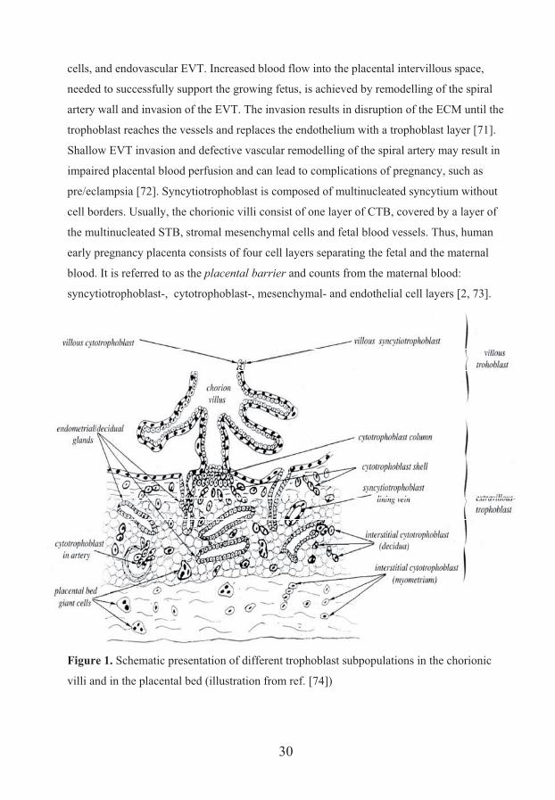
cells, and endovascular EVT. Increased blood flow into the placental intervillous space,
needed to successfully support the growing fetus, is achieved by remodelling of the spiral
artery wall and invasion of the EVT. The invasion results in disruption of the ECM until the
trophoblast reaches the vessels and replaces the endothelium with a trophoblast layer [71].
Shallow EVT invasion and defective vascular remodelling of the spiral artery may result in
impaired placental blood perfusion and can lead to complications of pregnancy, such as
pre/eclampsia [72]. Syncytiotrophoblast is composed of multinucleated syncytium without
cell borders. Usually, the chorionic villi consist of one layer of CTB, covered by a layer of
the multinucleated STB, stromal mesenchymal cells and fetal blood vessels. Thus, human
early pregnancy placenta consists of four cell layers separating the fetal and the maternal
blood. It is referred to as the placental barrier and counts from the maternal blood:
syncytiotrophoblast-, cytotrophoblast-, mesenchymal- and endothelial cell layers [2, 73].
Figure 1. Schematic presentation of different trophoblast subpopulations in the chorionic
villi and in the placental bed (illustration from ref. [74])
30
2.2 Human trophoblast - the main cells of the placenta with key importance in pregnancy
2.2.1 Phenotypic and functional characteristics of villous trophoblast
All trophoblast cells express cytokeratines 7, 8 and 18, indicating their epithelial origin
[75]. Adhesion molecules are widely expressed by the trophoblast cells. The calcium
dependent molecule E-cadherin, mediating homotypic adhesion between cells, is specific
for CTB. Other adhesion molecules such as the integrins are expressed by different subsets
of trophoblast cells and seem to be associated with their invasive behaviour. The CTB
expresses integrins �v�5 and �6�4 [76-78].
As previously described, in humans, the chorionic villi covered by the STB are in direct
contact with the maternal blood and participate in the fetal nutrition, gas- and waste
exchange. Respiratory gases diffuse from the maternal blood through the entire STB plasma
membrane. This diffusion is dependent on the flow rates of the umbilical- and uterine
circulation [79]. The STB is the major producer of placental hormones and, thus is an
important factor for pregnancy success. Villous trophoblast secretes polypeptide hormones,
like human chorionic gonadotropin (hCG), human placental lactogen (hPL) (also called
human chorionic somatomammotropic hormone (hCS)), placental growth hormone (GH),
and the steroid hormones progesterone and estrogen [80, 81].
Human chorionic gonadotropin is crucial for human pregnancy and works as an agonist for
luteinizing hormone (LH), rescues the corpus luteum from involution ensuring the
maintenance of ovarian progesterone secretion [80]. Besides being essential for the
successful pregnancy, progesterone has a suppressive effect on the immune response [82].
Interestingly, it has been shown that progesterone has other effects as well, such as
modifying the GABAA receptor in the central nervous system [83, 84]. The role of hPL in
the placenta is not completely clear. Normal pregnancies have been described in the
absence of hPL secretion. Growth hormones, produced by the placenta, are proposed to
have a metabolic role on the maternal organism during pregnancy, e.g. involvement in the
insulin resistance [80].
The villous trophoblast cells are semiallogeneic and should be attacked by the maternal
immune cells. To avoid the immune response, the STB cells do not express classical MHC
31
molecules, but express important molecules that are involved in immune modulation. Two
of them are PD-L1, also called CD240 and B7-H1, which is detected on the STB, and the
related type I membrane protein CD200 [22]. Moreover, STB expresses a variety of
complement regulatory proteins such as decay accelerating factor, CD46 and CD59,
protecting placenta from complement attack [22]. In addition, STB expresses FasL. In
humans, FasL are expressed within STB as endosomal vesicles [9, 31].
2.2.2 Phenotypic and functional characteristics of extravillous trophoblast
Extravillous trophoblast invasion is dependent on detachment of CTB cells from the
basement membrane. The CTB cells undergo a proliferative burst and differentiate into
cells of the CTB column, anchoring the peripheral villi to the decidual bed. This anchoring
cell subpopulation and the transition from CTB to the migratory EVT are mediated by
contact between the migratory EVT and the decidual extracellular matrix (ECM). This
adhesion is due to fibronectin-mediated extracellular matrix binding. Fetal fibronectin or
trophuteronectin (TUN) are produced by the EVT. Transforming growth factor ��and
leukemia inhibitory factor (LIF) have been shown to inhibit the trophoblast-differentiation
into an invasive phenotype. However, there are other reports showing that LIF increases the
invasion of first trimester EVT and mediates adhesion to ECM. Extravillous trophoblast
expresses receptors for laminin, fibronectin and integrins. The integrin expression changes
when CTB differentiates from a villous to an extravillous phenotype, with down-regulation
of �6�4 and up-regulation of �5�1 integrin [85-87]. Interestingly, when the EVT has invaded
the spiral arteries, it mimics the endothelium by expression of the vascular cell adhesion
molecule 1 (VCAM-1) and platelet endothelial cell adhesion molecule 1 (PECAM-1) [76,
88]. As described earlier, EVT expresses a unique combination of HLA-E, HLA-C and
HLA-G [21-23]. Like STB, EVT expresses the immune modulatory protein PD-L1 [22].
32
3. Immune escape - a common strategy for pregnancy and cancer
Although the placenta is a normal tissue, its principal cells, the trophoblasts, share several
features with malignant cells. Cancer is a disease originating from alteration in the cellular
genome resulting in an invasive and proliferating tumor that, like placenta, moulds its own
environment to favour its survival and expansion. Despite the fact that trophoblast and
cancer cells are both immunogenic tissues, they are both able to escape from the host
immunosurveillance [2].
3.1 Trophoblast and cancer cells share many biological features
In common, cancer cells and trophoblast share many biological characteristics, including
their capacity for proliferation, migration, invasion and establishment of blood supply
(table 1). Both cancer cells and trophoblast cells have increased telomerase activity,
reflecting their high proliferative capacity [89, 90]. Other mediators that promote
proliferation and inhibit apoptosis, are survivin, which is overexpressed by cancer- and
trophoblast cells [91, 92], and the insulin growth factor (IGF). Additionally, IGF protects
cancer cells from destructive effects of chemotherapy and radiation [93, 94]. Several proto-
oncogenes encoding growth-factors are expressed by cancer- and trophoblast cells [95].
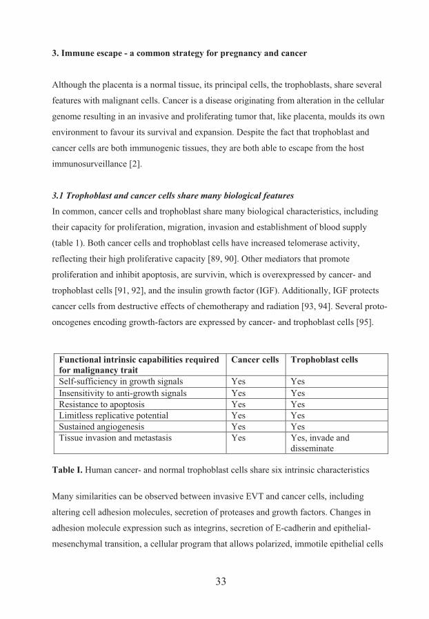
Functional intrinsic capabilities required for malignancy trait
Cancer cells Trophoblast cells
Self-sufficiency in growth signals Yes Yes Insensitivity to anti-growth signals Yes Yes Resistance to apoptosis Yes Yes Limitless replicative potential Yes Yes Sustained angiogenesis Yes Yes Tissue invasion and metastasis Yes Yes, invade and
disseminate Table I. Human cancer- and normal trophoblast cells share six intrinsic characteristics
Many similarities can be observed between invasive EVT and cancer cells, including
altering cell adhesion molecules, secretion of proteases and growth factors. Changes in
adhesion molecule expression such as integrins, secretion of E-cadherin and epithelial-
mesenchymal transition, a cellular program that allows polarized, immotile epithelial cells
33
to convert to motile mesenchymal cells, are mechanisms that trophoblast cells and cancer
cells use to lose polarity and enhance motility [96-98]. Epidermal growth factor (EGF) and
the Wnt signaling pathway are involved in switching cancer cells and trophoblast cells from
proliferative to invasive phenotype [99-102].
Blood supply is crucial for survival of the cancer and the fetus. The process that is
responsible for this is called vasculogenic mimicry, in which cells, other than endothelial
cells, form vascular structures [103, 104]. Vascular endothelial growth factor (VEGF),
angiopoetins and the oxygen-sensitive mammalian target of rapamycin (MTOR)-pathway
are other substances and mechanisms important for the angiogenesis in many tumors and
crucial for the spiral artery remodelling during placenta formation [105-108].
3.2 Trophoblast and cancer use similar immune escape mechanisms
Trophoblast and cancer cells do not only share many proliferative and invasive features,
additionally they actively modulate the host immune response. Uterine NK cells are the
most abundant immune cells at the fetal-maternal interface. One mechanism of recruitment
of uNK cells from the blood is IL-15 secretion by endometrial stromal cells [4]. The same
mechanism has been shown in numerous malignancies where NK cells infiltrate in response
to IL-15 [109].
Cells that infiltrate the fetal-maternal interface and play important roles in pregnancy and
cancers are M�, Tregs and DCs. Macrophages in the decidua secrete IL-10 and contribute
to a tolerogenic Th2 milieu [4] while M� associated with cancer can be both
immunosuppressive and inflammatory [110]. The amount of Tregs, expressing CD4, CD25
and FOXP3, are significantly increased in decidua [4, 11]. A similar expansion of Tregs can
be seen in cancer, contributing to impaired antitumor immunity [111].
HLA-G expression on EVT suppresses killing by both NK- and cytotoxic T cells, regulates
cytokine production in blood mononuclear cells, induces apoptosis of immune cells, and
impairs maturation of DCs. The immune inhibitory effect of HLA-G is due to binding to
the inhibitory receptors, immunoglobulin-like transcripts (ILT-2 and ILT-4), expressed on
myeloid and lymphoid cells. There are several reports showing HLA-G expression in a
34
wide variety of cancers, although there are some controversies about these findings [21, 23,
112, 113]. A similar immune-inhibitory effect of HLA-G in cancer has been suggested. A
soluble form of HLA-G has been found in peripheral blood of pregnant women impairing
NK/DC cross-talk, promoting inflammation and apoptosis. Similarly, soluble HLA-G has
been reported in serum of cancer patients. Additionally, HLA-G has also been found on
exosomes in melanoma patients [23, 114-117]. Another soluble immunomodulator, CD30,
a marker for Th2 polarization, is overexpressed by B cells in pregnant women as well as by
B cells in cancer patients. Reduced expression of CD30 is related to pathological
pregnancies suggesting a role in immunomodulation during pregnancy [118, 119].
3.3 Exosome secretion is a way of intercellular communication and generation of
”soluble” bioactive ligands
Membrane vesicles are classified based on their cellular origin, shape, and presence of a
surrounding membrane. Membrane vesicles are produced by a vast majority of cells such as
reticulocytes, mast cells, T and B cells, platelets, DCs, neurons and microglia, intestinal
epithelia, uroepithelia, bronchial epithelia, hepatocytes, syncytiotrophoblast and tumor cells
[61, 120-134]. Furthermore, membrane vesicles have been found in physiological fluids,
such as saliva, urine, plasma, synovial fluid, amniotic fluid, malignant effusions, bronchial
lavage fluid and breast milk [135-142]. There is a number of different types of membrane
vesicles: plasma membrane microvesicles/microparticles, shed microvilli, apoptotic bodies,
and exosomes. A summary of some of their properties is given in table 2 [143].
35
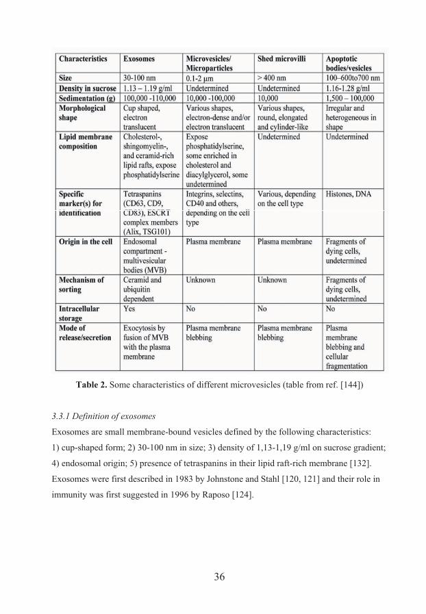
Table 2. Some characteristics of different microvesicles (table from ref. [144])
3.3.1 Definition of exosomes
Exosomes are small membrane-bound vesicles defined by the following characteristics:
1) cup-shaped form; 2) 30-100 nm in size; 3) density of 1,13-1,19 g/ml on sucrose gradient;
4) endosomal origin; 5) presence of tetraspanins in their lipid raft-rich membrane [132].
Exosomes were first described in 1983 by Johnstone and Stahl [120, 121] and their role in
immunity was first suggested in 1996 by Raposo [124].
36
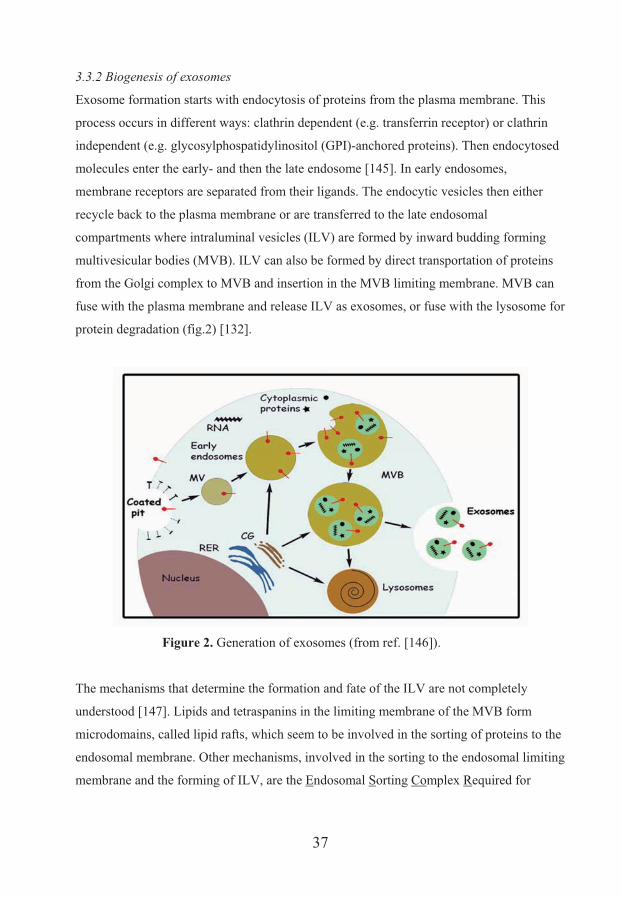
3.3.2 Biogenesis of exosomes
Exosome formation starts with endocytosis of proteins from the plasma membrane. This
process occurs in different ways: clathrin dependent (e.g. transferrin receptor) or clathrin
independent (e.g. glycosylphospatidylinositol (GPI)-anchored proteins). Then endocytosed
molecules enter the early- and then the late endosome [145]. In early endosomes,
membrane receptors are separated from their ligands. The endocytic vesicles then either
recycle back to the plasma membrane or are transferred to the late endosomal
compartments where intraluminal vesicles (ILV) are formed by inward budding forming
multivesicular bodies (MVB). ILV can also be formed by direct transportation of proteins
from the Golgi complex to MVB and insertion in the MVB limiting membrane. MVB can
fuse with the plasma membrane and release ILV as exosomes, or fuse with the lysosome for
protein degradation (fig.2) [132].
Figure 2. Generation of exosomes (from ref. [146]).
The mechanisms that determine the formation and fate of the ILV are not completely
understood [147]. Lipids and tetraspanins in the limiting membrane of the MVB form
microdomains, called lipid rafts, which seem to be involved in the sorting of proteins to the
endosomal membrane. Other mechanisms, involved in the sorting to the endosomal limiting
membrane and the forming of ILV, are the Endosomal Sorting Complex Required for
37
Transport (ESCRT) proteins. The ubiquitinylation process is tagging proteins of the
endosomal membrane targeting them for the ESCRT machinery. The ESCRT complex
which includes four protein complexes: ESCRT-0, ESCRT-I, ESCRT-II and ESCRT-III,
sorts ubiquitinated transmembrane proteins into ILV. Additional mechanisms for sorting of
non-ubiquitinated proteins through the ESCRT machinery has also been suggested [44, 129,
146, 148-150]. Recently, another sorting mechanism, independent of the ESCRT proteins,
was described, involving sphingolipid ceramide [147, 151].
,,
The mechanisms responsible for exocytosis of MVB and release of ILV as exosomes into
the extracellular environment are not well known, although it has been shown that Rab11,
Rab27a and Rab27b are involved in the docking of the MVB to the plasma membrane in a
Ca2+ dependent manner. Additionally, the p53 protein and the transmembrane TSAP6 have
been suggested to be involved in the regulation of exosome secretion [152-155].
3.3.3 Secreted exosomes - general characteristics and functions
Electron microscopy is still the most reliable method to study exosome morphology and
biogenesis. Isolated exosomes are cup-shaped and heterogeneous in size, varying between
30-100 nm. Their membrane is composed of cholesterol, sphingolipids and tetraspanins.
Today, there is no specific marker for exosomes, although they are typically enriched in
proteins from endosomes such as CD63, Alix, TSG101, CD9, CD81 and CD82 [156, 157].
In addition, exosomes contain mRNAs, microRNAs and a high variety of different
membranous and cytoskeletal proteins [156, 158]. Exosomes also contain cell specific
proteins which enable tracking of the producing cells [122, 125, 126, 129, 146, 159].
The secretion of exosomes is a potent way of communication between cells. The benefits of
secretion of proteins via exosomes are numerous: i) preservation of the three-dimensional
structure of the protein, and thus their biological activity; ii) independence from cell-to-cell
contact for signal delivery; iii) lower mobility and higher concentration of the carried
molecules; iv) independence from de novo protein synthesis; v) biological effects at a
distance [146].
38

Exosomes can be divided into immune activating and immune suppressive. Most of the
exosomes produced by immune cells such as DCs, M� or B cells are immune activating,
i.e. antigen presenting, acting directly or indirectly to activate immune effector mechanisms
such as cytokine and antibody production, cytotoxicity and activation of T cells [143].
Whether exosomes can stimulate T cells directly or need the presence of DCs has been
debated. Several studies have demonstrated that exosomes can stimulate T cells directly
[160, 161] while others have shown that exosomes exert their effect through APCs [162].
Recent publications show that antigen-loaded exosomes derived from DCs alone augment
the specific T cell response and that this effect was depending on B cells [160, 161].
Exosomes have been suggested to be an immune stimulatory factor in allergic immune
response. B cell-derived exosomes that present peptides causing allergy can stimulate
peptide-specific T cells to produce Th2-like cytokines [163].
The majority of exosomes, released by normal intestinal epithelia and cancer cells are
immune suppressive. Exosomes produced by human intestinal epithelium have been
suggested to play a role in oral tolerance and referred to as tolerosomes [134, 164].
Exosomes, most likely released from intestinal epithelia of antigen-fed rats can induce
tolerance to the antigen when injected in naïve recipients and this tolerance is MHC class II
dependent [134, 165, 166]. Cancer exosomes and placental exosomes suppress the host’s
immune defense by decoy-mechanisms of receptor down-regulation, apoptosis and Treg
induction [9, 31, 33, 61, 134, 167-170]. Taylor et al. have shown that the amount of
placenta-derived exosomes found in sera of pregnant women was significantly higher in
those delivering at term compared to those delivering preterm. These exosomes, carrying
biologically active components, such as FasL, induce T cell suppression via CD3-� and
JAK3 [33]. We and others have shown that placenta releases exosomes, carrying immune
suppressive molecules, e.g. NKG2D ligands, and FasL that suppress the maternal immune
system [9, 31, 61, 171].
39

AIMS OF THE INVESTIGATION
The overall objective of this investigation was to study the expression, regulation and function of the NKG2D receptor-ligand system in pregnancy and cancer. We hypothesized that placenta and tumors escape NKG2D receptor-mediated immunosurveillance by generation of an exosomal form of NKG2D ligands. The specific aims were:
To investigate the mRNA transcription and protein expression of the NKG2D ligands MIC and ULBPs in normal human placenta and tumors.
To isolate and characterise NKG2D ligand-bearing exosomes secreted from
human placental explant cultures and T and B leukemia/lymphoma cells.
To examine the effect of biological stress on the production and secretion of NKG2D ligand-bearing exosomes in cancer cells.
To study the effect of NKG2D ligand-bearing placental and tumor exosomes on
the down-modulation of the NKG2D receptor and its consequence for NK cytotoxicity.
As a prerequisite and a corollary of the above investigation, an optimized
technique for isolation of human early villous trophoblast was developed as a potential method for in vitro production of exosomes.
40

RESULTS AND DISCUSSION
In this section, I will present and discuss the main results of my work. The papers are
referred to in the thesis by their Roman numbers (I-III).
4. Methodological considerations
4.1 Isolation of villous trophoblast cells from human early pregnancy placenta
Human placenta is a unique organ that governs pregnancy and as such a focus for research
in reproduction. Studies of human placenta must be done on the organ itself and/or cells
isolated from the placenta and cannot be replaced by animal models for biochemical and
functional studies. The most important cell type responsible for nutrition and gas exchange
to the fetus, and production of bioactive molecules such as pregnancy-related hormones,
cytokines, chemokines and other immunomodulatory molecules, metalloproteases,
adhesion molecules and growth factors is the STB that comprises the outermost cell layer
of the placental villi which is in direct contact with the maternal blood. To isolate STB from
early pregnancy placenta for our molecular and functional studies was a prerequisite for our
further studies.
There are several reports describing methods for isolation of trophoblast cells from term
placenta [172]. In these methods, using combinations of digestive enzymes and density
gradient centrifugation, term CTB cells were obtained. However, there were very few
methods for isolation of VT from human early placenta and these methods gave yields of
throphoblast cells heavily contaminated with leukocytes [173]. Thus, there was a need to
optimize a method for isolation of VT from early pregnancy placenta to get a pure
trophoblast. At the same time, we needed a gentle isolation method that could allow us to
(1) use isolated STB in molecular studies for transcription and expression of different
genes; (2) obtain CTB for establishment of primary human trophoblast cultures; and
(3) establish long term trophoblast cultures for harvesting of exosomes for future studies.
In paper I we present an optimized and easy technique for isolation of VT from human
early (8-14 weeks) normal placenta.
41
4.1.1 Description and advantages of our isolation procedure
The procedure includes three steps: (1) tissue disruption by treatment with a mild enzymatic
cocktail, (2) Percoll gradient centrifugation for enrichment of trophoblast cells and
(3) depletion of contaminating leukocytes using immunomagnetic beads coated with anti
CD45 andibodies. The isolation procedure is illustrated in fig.1 paper I. Our isolation
method gives a good yield of isolated trophoblast cells with preserved morphology and high
viability (fig.2 and fig.4 paper I). The trophoblastic origin of the isolated cells was proved
by cytokeratin 7 staining. We found that more than 95% of them were positively stained,
showing a high purity of isolated cells, composed of both CTB and STB (fig.3 paper I). To
obtain a single CTB population, a negative selection with magnetic beads coated with
specific antibodies to surface molecules expressed on the STB cells, e.g. anti-MICA [61] or
anti-PLAP, can be used. The positively selected STB, bound on magnetic beads can be used
for RNA or DNA extraction and molecular studies. In summary, we have developed an
easy and time-saving method that give us good yield of pure VT cells with preserved
morphology, well suited for phenotypic, morphological and functional studies of the VT
cells in early human placenta. Isolated VT cells were used in our molecular studies with
quantitative RT-PCR technique, in immunoflow cytometry experiments for assessing the
expression of NKG2D ligands and for western blot analyses (papers I and II).
4.2 Placental explant cultures
Culture of placental explants was used to obtain supernatants from which placental
exosomes were isolated. Our placental explant cultures were performed for two reasons.
From one side, we wanted to get exosomes produced by placenta only, but we could not use
blood from pregnant women because several organs contribute to exosome secretion in
peripheral blood. From another side, we wanted to obtain exosomes from an experimental
setting that resembles, or comes as near as possible, to the in vivo situation. Although far
from perfect, placental explant cultures are so far the only way to “mimic” an in vivo
situation where placenta secreted substances can be collected in a culture medium.
Dissected chorion villi of 5-10 mg wet weight from early normal human placenta were
cultured in RPMI 1640 supplemented with 0.5 % BSA and antibiotics at 37oC in 5 % CO2
and humidified air. The supernatant was collected after 24-hour culture and kept frozen
until exosome isolation. Since we were interested in isolation of secreted exosomes,
42
precautions were taken to avoid microvesicles released by dead cells. To minimize cell
death, the time between extraction of placenta and setting of explant cultures was kept very
short, the tissue was handled with great care, the explant dissection was done with gentle
techniques and the culture time was limited to 24 hours. All isolated placental exosomes
used in our experiments were produced by explant cultures (paper II).
4.3 Isolation of exosomes
Exosomes are present in human blood, saliva, urine, breast milk and other bodily effusions
together with other microvesicles, shed from the cellular plasma membrane, and apoptotic
bodies produced by dying cells. An important issue when studying exosomes is to be able
to obtain a pure population of exosomes separated from other contaminating microvesicles.
Many physical and chemical properties of exosomes and shed microvesicles are close to
each other (table 2) and this demands stringent purification procedures to ensure that pure
exosome population is obtained. This is even more important in studies of placental
exosomes since it is known that the STB constitutively releases not only exosomes but also
large amounts of microvesicles/microparticles shed from the apical part of the plasma
membrane. Our method for exosome isolation, described in paper II and III, comprises a
combination of ultracentrifugation and a continuous sucrose gradient (floating density
1.02–1.19 g/ml) or a sucrose cushion, thus ensuring exosome purity and minimizing
contamination by other microvesicles. We have also continuously examined the purity of
our exosome isolations by electron microscopy. Further, in all immunoflow cytometric
work presented here, exosomes loaded on latex beads directly or via antibody capture, are
used according to recommended protocol [174].
4.4 Quantification of exosome secretion
Today, there is no well-established and reliable method for exosome quantification. The
most frequently used methods are based on total exosomal protein measurements by BCA-
or Bradford assays and densitometric analysis of western blot bands for exosomal markers
[175]. Recently, fluorescence intensity measurements of exosomes labelled with lipophilic
fluorescence dyes has also been used [176]. To enhance the reliability of the quantification
measurements of isolated exosomes we used these three different methods; BCA protein
43
measurement, lipid staining with Vybrant DiI and densitometric analysis of western blot
bands of the exosomal marker CD63 (paper III).
5. The NKG2D receptor-ligand system in human pregnancy
We studied the NKG2D receptor-ligand system in human pregnancy for two main reasons:
1) the interaction of the activating NK cell receptor NKG2D and its inducible ligands is a
central perforin-mediated cytotoxic pathway by which damaged-, transformed-, or infected
cells are eliminated. Therefore, the NKG2D receptor-ligand system might be a potential
threat to the fetus [177, 178], and 2) in tumors, soluble NKG2D ligands can bind to
NKG2D and systemically down-regulate its expression on cytotoxic T cells and NK cells,
providing a mechanism for tumor immune escape [40, 168, 179, 180]. Therefore, we asked
the question: “Does placenta, similarly to tumors, generate and secrete soluble NKG2D
ligands for immune escape?”
5.1 Expression of NKG2D ligands by human placenta
We investigated NKG2D ligand expression in human placenta by quantitative real time-
PCR, flow cytometry, immunohistochemistry (IHC) and immunoelectron microscopy
(IEM). Our molecular studies showed a constitutive gene expression of all NKG2D ligands
(MICA/B and ULBP1-5). Moreover, all mRNA transcripts were translated into proteins.
Flow cytometry analysis of isolated VT cells revealed MICA/B expression on the surface
and intracellularly while ULBP1-5 expression was solely inside the cytoplasm (fig.1 paper
II). These results were confirmed and further extended by IEM, which showed that all
NKG2D ligands were exclusively expressed by STB (fig.2 paper II).
5.2 The NKG2D ligand molecules are processed, stored and secreted through the late
endosomal compartment of syncytiotrophoblast
Our novel findings of NKG2D ligand expression in placenta raise the question: “Why are
these molecules expressed in placenta and how are they processed, stored and secreted?”
The precise mechanism that regulates the NKG2D ligand expression is still unknown. The
shedding of NKG2D ligands from the cells as soluble molecules represents an additional
level of complexity in this system. Recent reports demonstrate that the expression of MICB
44
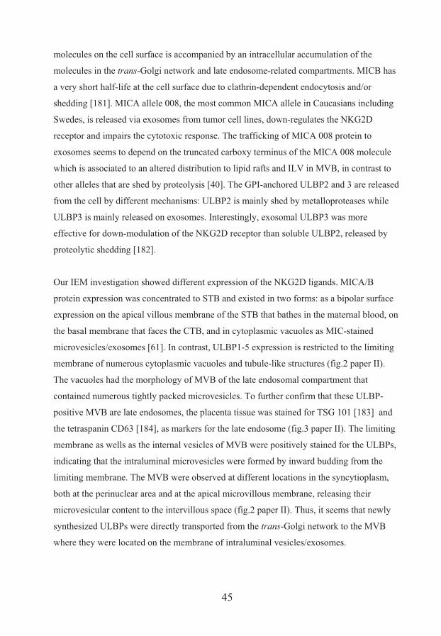
molecules on the cell surface is accompanied by an intracellular accumulation of the
molecules in the trans-Golgi network and late endosome-related compartments. MICB has
a very short half-life at the cell surface due to clathrin-dependent endocytosis and/or
shedding [181]. MICA allele 008, the most common MICA allele in Caucasians including
Swedes, is released via exosomes from tumor cell lines, down-regulates the NKG2D
receptor and impairs the cytotoxic response. The trafficking of MICA 008 protein to
exosomes seems to depend on the truncated carboxy terminus of the MICA 008 molecule
which is associated to an altered distribution to lipid rafts and ILV in MVB, in contrast to
other alleles that are shed by proteolysis [40]. The GPI-anchored ULBP2 and 3 are released
from the cell by different mechanisms: ULBP2 is mainly shed by metalloproteases while
ULBP3 is mainly released on exosomes. Interestingly, exosomal ULBP3 was more
effective for down-modulation of the NKG2D receptor than soluble ULBP2, released by
proteolytic shedding [182].
Our IEM investigation showed different expression of the NKG2D ligands. MICA/B
protein expression was concentrated to STB and existed in two forms: as a bipolar surface
expression on the apical villous membrane of the STB that bathes in the maternal blood, on
the basal membrane that faces the CTB, and in cytoplasmic vacuoles as MIC-stained
microvesicles/exosomes [61]. In contrast, ULBP1-5 expression is restricted to the limiting
membrane of numerous cytoplasmic vacuoles and tubule-like structures (fig.2 paper II).
The vacuoles had the morphology of MVB of the late endosomal compartment that
contained numerous tightly packed microvesicles. To further confirm that these ULBP-
positive MVB are late endosomes, the placenta tissue was stained for TSG 101 [183] and
the tetraspanin CD63 [184], as markers for the late endosome (fig.3 paper II). The limiting
membrane as wells as the internal vesicles of MVB were positively stained for the ULBPs,
indicating that the intraluminal microvesicles were formed by inward budding from the
limiting membrane. The MVB were observed at different locations in the syncytioplasm,
both at the perinuclear area and at the apical microvillous membrane, releasing their
microvesicular content to the intervillous space (fig.2 paper II). Thus, it seems that newly
synthesized ULBPs were directly transported from the trans-Golgi network to the MVB
where they were located on the membrane of intraluminal vesicles/exosomes.
45
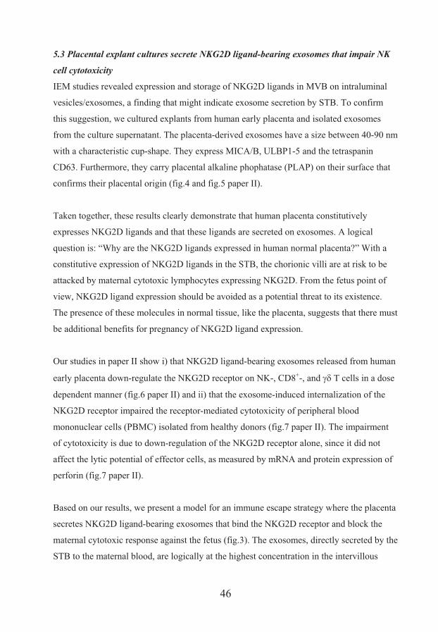
5.3 Placental explant cultures secrete NKG2D ligand-bearing exosomes that impair NK
cell cytotoxicity
IEM studies revealed expression and storage of NKG2D ligands in MVB on intraluminal
vesicles/exosomes, a finding that might indicate exosome secretion by STB. To confirm
this suggestion, we cultured explants from human early placenta and isolated exosomes
from the culture supernatant. The placenta-derived exosomes have a size between 40-90 nm
with a characteristic cup-shape. They express MICA/B, ULBP1-5 and the tetraspanin
CD63. Furthermore, they carry placental alkaline phophatase (PLAP) on their surface that
confirms their placental origin (fig.4 and fig.5 paper II).
Taken together, these results clearly demonstrate that human placenta constitutively
expresses NKG2D ligands and that these ligands are secreted on exosomes. A logical
question is: “Why are the NKG2D ligands expressed in human normal placenta?” With a
constitutive expression of NKG2D ligands in the STB, the chorionic villi are at risk to be
attacked by maternal cytotoxic lymphocytes expressing NKG2D. From the fetus point of
view, NKG2D ligand expression should be avoided as a potential threat to its existence.
The presence of these molecules in normal tissue, like the placenta, suggests that there must
be additional benefits for pregnancy of NKG2D ligand expression.
Our studies in paper II show i) that NKG2D ligand-bearing exosomes released from human
early placenta down-regulate the NKG2D receptor on NK-, CD8+-, and ���T�cells in a dose
dependent manner (fig.6 paper II) and ii) that the exosome-induced internalization of the
NKG2D receptor impaired the receptor-mediated cytotoxicity of peripheral blood
mononuclear cells (PBMC) isolated from healthy donors (fig.7 paper II). The impairment
of cytotoxicity is due to down-regulation of the NKG2D receptor alone, since it did not
affect the lytic potential of effector cells, as measured by mRNA and protein expression of
perforin (fig.7 paper II).
Based on our results, we present a model for an immune escape strategy where the placenta
secretes NKG2D ligand-bearing exosomes that bind the NKG2D receptor and block the
maternal cytotoxic response against the fetus (fig.3). The exosomes, directly secreted by the
STB to the maternal blood, are logically at the highest concentration in the intervillous
46
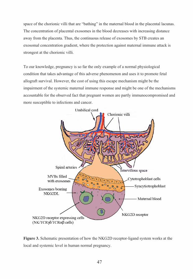
space of the chorionic villi that are “bathing” in the maternal blood in the placental lacunas.
The concentration of placental exosomes in the blood decreases with increasing distance
away from the placenta. Thus, the continuous release of exosomes by STB creates an
exosomal concentration gradient, where the protection against maternal immune attack is
strongest at the chorionic villi.
To our knowledge, pregnancy is so far the only example of a normal physiological
condition that takes advantage of this adverse phenomenon and uses it to promote fetal
allograft survival. However, the cost of using this escape mechanism might be the
impairment of the systemic maternal immune response and might be one of the mechanisms
accountable for the observed fact that pregnant women are partly immunocompromised and
more susceptible to infections and cancer.
Figure 3. Schematic presentation of how the NKG2D receptor-ligand system works at the
local and systemic level in human normal pregnancy.
47
6. NKG2D ligand expression and secretion by cancer cells
We have used Jurkat and Raji cell lines as a cancer model for studies of exosome secretion
and NKG2D ligand expression by T- and B cell leukemia/lymphoma cells under normal
and stress conditions. This model was chosen for the following reasons: i) these rapidly
progressing malignancies have poor prognosis and documented NK cell dysfunction [185],
ii) exosome secretion is a constitutive feature of many human malignancies and tumor-
derived exosomes are known to express NKG2D ligands and interfere with the NKG2D
receptor causing impairment of NK cell cytotoxicity, iii) NKG2D ligands are induced by
biologic stress. The treatment regimens of these leukemia/lymphoma malignancies include
heavy cytostatic treatment, sometimes combined with thermotherapy both of which expose
the body to massive cellular stress, and iv) a comprehensive clinical study by Nückel et al
[186] showed that soluble MICA, MICB and ULBP2 were present in the peripheral blood
of patients with chronic B cell leukemia and related to a prognostic significance.
6.1 Stress-induced up-regulation of NKG2D ligands in leukemia cell lines
As mentioned earlier, NKG2D ligands are poorly expressed by normal cells but up-
regulated in transformed cells. NKG2D ligands are also up-regulated by diverse cellular
stress signals, such as heat shock, oxidative stress, irradiation, anti-cancer drugs and
genotoxic agents, all of those causing DNA damage [187]. We stressed the
leukemia/lymphoma cell lines Jurkat and Raji with thermal- and oxidative stress, and used
up-regulation of HSP70 transcripts as a positive control to estimate the effectiveness of
stress induction. Despite the accumulated reports about the nature of stress signals inducing
NKG2D ligand expression only limited information about the precise mechanisms that lead
to up-regulation of the ligands in cancer are available. The promoter elements for
transcriptional control of these ligands are not yet fully understood. MICA/B molecule
expression is regulated by promoter elements similar to those of heat shock HSP70 gene,
while the transcriptional regulation of other NKG2D ligands remains unclear. It has been
reported that heat shock-induced transcriptional activation has not been observed for
ULBPs [177, 187]. We found mRNA and protein expression up-regulation for MICA/B and
ULBP1-2 in response to thermal- and oxidative stress by H2O2. ULBP3 was not expressed
in the cell lines tested (fig.1 paper III).
48

6.2 Thermal- and oxidative stress up-regulates secretion and expression of NKG2D
ligand-bearing exosomes that enhance the suppression of NK cell mediated cytotoxicity
We isolated exosomes from supernatants of Jurkat and Raji cell cultures and found that
leukemia/lymphoma T and B cells constitutively secreted exosomes and that the secretion
was significantly increased after thermal- and oxidative stress (fig.2 paper III). Our IEM,
dot blot and flow cytometric analysis revealed that the cell-derived exosomes carry the
NKG2D ligands MICA/B and ULBP1 and 2 on their surface (fig.3, 4A, B paper III).
Moreover, thermal- and oxidative stress significantly increased the total amount of secreted
exosomes and thus, NKG2D ligands (fig.3 paper III).
As a next step, we investigated if tumor exosomes secreted under normal- and stress
conditions could alter NKG2D receptor-mediated killing in vitro. Our results show that
NKG2D ligand-bearing exosomes secreted from leukemia/lymphoma cell lines had a
suppressive effect on the NK cell-mediated cytotoxicity and that this suppressive effect was
enhanced by stress culture conditions (fig.4C paper III).
Our investigation (paper III) demonstrates that thermal- and oxidative stress can enhance
the exosome secretion, thus generating an increased amount of soluble exosomal NKG2D
ligands. As a consequence, the suppression of NKG2D mediated cytotoxicity is aggravated,
which might promote immune escape of the leukemia/lymphoma cells. This suggestion is
supported by Nückel et al. who have found that soluble MICA/B and ULBP2 were
present in serum of patients with chronic B lymphocytic leukemia and all of them,
especially ULBP2, were strongly associated with poor survival of patients [186]. Oxidative
stress is induced by cancer chemotherapy, and hyperthermia is usually used as an
adjunctive therapy alongside conventional cancer treatments. It has recently been reported
that hyperthermia can suppress the lytic potential of NK cells via down-regulation of
perforin/granzyme B expression [188]. We suggest that in addition to the suppressed
cytolytic machinery of the effector cells, thermal stress might further augment the
dysfunction of the NK cells by down-regulating their killing ability via increased secretion
of immunosuppressive NKG2D ligand-bearing exosomes, and this should be taken into
account when designing cytostatic and hyperthermal anti-cancer therapy.
49
CONCLUSIONS
The MICA/B and ULBP1-5 proteins, stress-inducible ligands of the NKG2D
receptor, are constitutively expressed by the syncytiotrophoblast of human normal
early placenta. Immunoelectron microscopy studies of syncytiotrophoblast
demonstrated expression of ULBP1-5 proteins exclusively in the multivesicular
bodies on internal vesicles/exosomes. Placental explant cultures revealed secretion
of NKG2D ligand-bearing exosomes.
The NKG2D ligand-bearing placental exosomes down-regulate the cognate
NKG2D receptor on peripheral blood NK- and T cells and impair NK cell-
mediated cytotoxicity in vitro without altering of activation status or lytic potency.
Leukemia/lymphoma cells constitutively express the NKG2D ligands MICA/B
and ULBP1 and 2 and release them on exosomes. Interestingly, the exosome
release could be up-regulated by thermal- and oxidative stress. The increased
tumor exosome secretion provided an abundant moiety of soluble, membrane-
bound form of NKG2D ligands, which interfered and suppressed the NK cell-
mediated cytotoxicity in vitro.
The exosome-mediated release of ULBP and MIC molecules by placenta and
tumor is a novel way to generate a soluble form of NKG2D ligands.
We suggest that syncytiotrophoblast- and leukemia/lymphoma cell-generated,
NKG2D ligand-bearing exosomes are protective for placenta and tumors. This
protection seems to be based, at least partly, on immune suppression of T- and NK
cells by systemic down-regulation of the activating NKG2D receptor.
50
ACKNOWLEDGEMENTS
I wish to express my warmest gratitude to my supervisor Lucia Mincheva-Nilsson for inviting me to investigate the field of immunology. Thank you for your endless optimism, for teaching me the expression “half success” and for teaching me to appreciate when I have wind in my sails. Thank you for your enthusiastic support and for believing in my potential. Vladimir Baranov, thank you for teaching me how to think and write scientifically and for interesting discussions about private and scientific matters. Thank you for providing me with a never ending amount of scientific articles and for critically reading this book. Olga Nagaeva, thank you for taking care of me in the lab, your invaluable help and your methodological skilfulness. Thank you for caring about me and for encouraging me when I needed it. Ann-Christin Stenqvist, thank you for being my friend and for all good times we have spent together! I am so happy for having you in the lab, being a great travel companion and for making work and everyday life much more fun. I wish to express my gratitude to all donors of placental samples, all the staff of the department of Obstetric and Gynecology and the operating theatre for providing them. Without you this work would not have been possible. Many thanks to my co-authors Lennart Kjellberg, Marianne Wulff, Eva Innala, Tanya Dimova, Ting Chen and Dominic Kargl. Eva Dehlin, thank you for creating such a friendly atmosphere in the lab, for always being helpful, for sharing interesting ideas and for teaching me how they drink wine in Austria. Gunborg Eriksson, thank you for always helping me with all kinds of administrative matters and for your support. Thanks to former students and colle g es in our lab: Sofia, Giovanni, Stina, Agnes and Sara. ua I would like to thank all the people in Clinical Immunology, Anna, Agneta, Marléne, Marianne, Susanne K, Susanne B, Niklas, Janne, Karin, Catharina and Igna io for creating such a nice atmosphere, intense discussions and a lot of fika.
c
Thanks to the Virology department for the use of the ultracentrifuge, many thanks to Karin, Kickan, Ya-Fang and Rolf for helping me with all kinds of matters. All the people in the Immunology corridor: Marie-Louise, Sten, Torgny, Kristina, Mari, Lena, Marianne, Anne, David, Theres, Mia, Lina, Veronika, Rifat, Aziz, Greg, Olena thank you for help, nice dinners and fun parties. Thanks to the people in Molecular biology for inviting me to parties, dinners and beer corner: Edmund, Teresa, Karolis, Barbro, Jörgen, Maria and Patrik.
51
Tack till alla mina fantastiska vänner i Umeå: Anna tack för att du alltid finns där för mig, för att vi alltid har så kul tillsammans, oavsett om vi äger dansgolvet eller ser en skräckis och för våra dagliga samtal om livet. Sandra, min första vän i Umeå, vi hittade varandra direkt (eller jag hittade dig). Alice, min älskade vän och den perfekta sambon. Louise, min enda vän hemifrån här i Umeå och André för roliga fester och sing starkvällar. Isabell för att du är en så fin vän som alltid ställer upp. Malin, Veronika, Åsa, och Ylva mina kära vänner som välkomnade mig när jag kände mig lite vilsen och nästan gjorde tentaplugg till något att se fram emot, tack för intressanta diskussioner, roliga middagar och vild dans. Tack till Gurra för middagar och häng. Sabine, tack för att du är världens bästa vän som alltid delar med dig av din kärlek och lyfter mig när jag behöver det. Sofia, Ullis, Sanna, Susanne och Malin, mina älskade vänner hemifrån, tack för att ni finns och att vi alltid har så kul trots att jag bor 65 mil bort och att vi inte ses så ofta.
Britt-Inger, Jörgen, Peppe, Matte, Linda, Nova och Morris, tack för att ni välkomnat mig i familjen, för skidturer, båtturer och välbehövliga pauser från forskningslivet. Fredde Lijana och Leah för zeppelinikvällar och härliga middagar i Umeå. Lasse, Milli, Björn, Marit, Johanna och Nicke för fina kvällar på Bergholmen, världens godaste stekmiddagar och mysiga juldagar.
Marie, Thomas och Kurre Majsan för fina stunder i Stockholm och Skåne, de bästa jularna, gåsmiddagar, långa promenader längs havet och intressanta diskussioner. Storfamiljen: Moa, Theo, Martin, Max, Gunilla, Klara, Matilda, Sissel, PA, Jonna, Petter, Kicki, Totte, Jenny, Ellen. Tack för alla skidturer, fester, oförglömliga resor, äventyr och umgänge. Ni gör mig så glad! Mina älskade syskon Mårten och Anna, tack för att ni alltid finns där för mig, för att vi alltid har kul tillsammans och för fin vänskap. Mamma och Pappa, tack för ert otroliga stöd, för att ni alltid tror på mig, alltid står på min sida och för all kärlek ni ger mig. Min stora kärlek Jonas, tack för att du finns i mitt liv, för att du alltid peppar mig när jag behöver det och för kärleken.
52
REFERENCES
1. Chaplin DD. Overview of the immune response. Journal of Allergy and Clinical Immunology 2010;125(2):S3-S23.
2. Mincheva-Nilsson L. Immune cells and molecules in pregnancy:friends or foes to the fetus? Expert Reviews in Clinical Immunology 2006;3(2):457-70.
3. Geissmann F, Manz MG, Jung S, Sieweke MH, Merad M, Ley K. Development of Monocytes, Macrophages, and Dendritic Cells. Science 2010;327(5966):656-61.
4. Bulmer JN, Williams PJ, Lash GE. Immune cells in the placental bed. International Journal of Developmental Biology 2010;54(2-3):281-94.
5. Wan YSY. Multi-tasking of helper T cells. Immunology 2010;130(2):166-71. 6. Lefrancois L, Obar JJ. Once a killer, always a killer: from cytotoxic T cell to
memory cell. Immunological Reviews 2010;235(1):206-18. 7. Barry M, Bleackley RC. Cytotoxic T lymphocytes: All roads lead to death. Nature
Reviews Immunology 2002;2(6):401-9. 8. Hoves S, Trapani JA, Voskoboinik I. The battlefield of perforin/granzyme cell
death pathways. Journal of Leukocyte Biology 2009;87(2):237-43. 9. Frangsmyr L, Baranov V, Nagaeva O, Stendahl U, Kjellberg L, Mincheva-Nilsson
L. Cytoplasmic microvesicular form of Fas ligand in human early placenta: switching the tissue immune privilege hypothesis from cellular to vesicular level. Molecular Human Reproduction 2005;11(1):35-41.
10. Sakaguchi S, Miyara M, Costantino CM, Hafler DA. FOXP3+ regulatory T cells in the human immune system. Nature Reviews Immunology 2010;10(7):490-500.
11. Aluvihare VR, Kallikourdis M, Betz AG. Regulatory T cells mediate maternal tolerance to the fetus. Nature Immunology 2004;5(3):266-71.
12. Zenclussen AC, Gerlof K, Zenclussen ML, et al. Abnormal T-cell reactivity against paternal antigens in spontaneous abortion - Adoptive transfer of pregnancy-induced CD4+CD25+ T regulatory cells prevents fetal rejection in a murine abortion model. American Journal of Pathology 2005;166(3):811-22.
13. Sasaki Y, Sakai M, Miyazaki S, Higuma S, Shiozaki A, Saito S. Decidual and peripheral blood CD4+CD25+ regulatory T cells in early pregnancy subjects and spontaneous abortion cases. Molecular Human Reproduction 2004;10(5):347-53.
14. Bonneville M, O'Brien RL, Born WK. Gamma delta T cell effector functions: a blend of innate programming and acquired plasticity. Nature Reviews Immunology 2010;10(7):467-78.
15. Szekeres-Bartho J, Barakonyi A, Miko E, Polgar B, Palkovics T. The role of gamma/delta T cells in the feto-maternal relationship. Seminars in Immunology 2001;13(4):229-33.
16. Ruffell B, DeNardo DG, Affara NI, Coussens LM. Lymphocytes in cancer development: Polarization towards pro-tumor immunity. Cytokine & Growth Factor Reviews 2010;21(1):3-10.
17. Ljunggren HG, Karre K. In search of the missing self- MHC molecules and NK cell recognition. Immunology Today 1990;11(7):237-44.
18. Long EO. Tumor cell recognition by natural killer cells. Seminars in Cancer Biology 2002;12(1):57-61.
19. Cheent K, Khakoo SI. Natural killer cells: integrating diversity with function. Immunology 2009;126(4):449-57.
53
20. Gomes AQ, Correia DV, Silva-Santos B. Non-classical major histocompatibility complex proteins as determinants of tumour immunosurveillance. Embo Reports 2007;8(11):1024-30.
21. Hunt JS, Petroff MG, McIntire RH, Ober C. HLA-G and immune tolerance in pregnancy. Faseb Journal 2005;19(7):681-93.
22. Riley JK. Trophoblast immune receptors in maternal-fetal tolerance. Immunological Investigations 2008;37(5-6):395-426.
23. Apps R, Gardner L, Moffett A. A critical look at HLA-G. Trends in Immunology 2008;29(7):313-21.
24. Kawai T, Akira S. The role of pattern-recognition receptors in innate immunity: update on Toll-like receptors. Nature Immunology;11(5):373-84.
25. Koga K, Aldo PB, Mor G. Toll-like receptors and pregnancy: Trophoblast as modulators of the immune response. Journal of Obstetrics and Gynaecology Research 2009;35(2):191-202.
26. Burnet M. Cancer - a biological approach .1. The processes of control. British Medical Journal 1957;1(5022):779-86.
27. Thomas L. On immunosurveillance in human cancer. Yale Journal of Biology and Medicine 1982;55(3-4):329-33.
28. Sengupta N, MacFie TS, MacDonald TT, Pennington D, Silver AR. Cancer immunoediting and "spontaneous" tumor regression. Pathology Research and Practice 2010;206(1):1-8.
29. Gao YF, Yang WC, Pan M, Scully E, Girardi M, Augenlicht LH, Craft J, Yin ZN. Gamma delta T cells provide an early source of interferon gamma in tumor immunity. Journal of Experimental Medicine 2003;198(3):433-42.
30. Smyth MJ, Takeda K, Hayakawa Y, Peschon JJ, van den Brink MRM, Yagita H. Nature's TRAIL - On a path to cancer immunotherapy. Immunity 2003;18(1):1-6.
31. Abrahams VM, Straszewski-Chavez SL, Guller S, Mor G. First trimester trophoblast cells secrete Fas ligand which induces immune cell apoptosis. Molecular Human Reproduction 2004;10(1):55-63.
32. Abusamra AJ, Zhong ZH, Zheng XF, Li M, Ichim TE, Chin JL, Min WP. Tumor exosomes expressing Fas ligand mediate CD8+ T-cell apoptosis. Blood Cells Molecules and Diseases 2005;35(2):169-73.
33. Taylor DD, Akyol S, Gercel-Taylor C. Pregnancy-associated exosomes and their modulation of T cell signaling. Journal of Immunology 2006;176(3):1534-42.
34. Reilly LAO, Tai L, Lee L, et al. Membrane-bound Fas ligand only is essential for Fas-induced apoptosis. Nature 2009;461(7264):659-U106.
35. Diefenbach A, Raulet DH. The innate immune response to tumors and its role in the induction of T-cell immunity. Immunological Reviews 2002;188:9-21.
36. Guerra N, Tan YX, Joncker NT, et al. NKG2D-deficient mice are defective in tumor surveillance in models of spontaneous malignancy. Immunity 2008;28(4):571-80.
37. Houchins JP, Yabe T, McSherry C, Bach FH. DNA-sequence analysis of NKG2, a family of related cDNA clones encoding type-II integral membrane-proteins on human natural-killer-cells. Journal of Experimental Medicine 1991;173(4):1017-20.
38. Houchins JP, Yabe T, McSherry C, Miyokawa N, Bach FH. Isolation and characterzation of NK cell or NK/T cell-specific cDNA clones. Journal of Molecular and Cellular Immunology 1990;4(6):295-306.
54
39. Bauer S, Groh V, Wu J, Steinle A, Phillips JH, Lanier LL, Spies T. Activation of NK cells and T cells by NKG2D, a receptor for stress-inducible MICA. Science 1999;285(5428):727-9.
40. Ashiru O, Boutet P, Fernandez-Messina L, Aguera-Gonzalez S, Skepper JN, Vales-Gomez M, Reyburn HT. Natural Killer Cell Cytotoxicity Is Suppressed by Exposure to the Human NKG2D Ligand MICA*008 That Is Shed by Tumor Cells in Exosomes. Cancer Research 2010;70(2):481-9.
41. Sutherland CL, Chalupny NJ, Schooley K, VandenBos T, Cosman D. UL16-Binding proteins, novel MHC class I-Related proteins, bind to NKG2D and activate multiple signaling pathways in primary NK cells. Journal of Immunology 2002;168(2):671-9.
42. Rosen DB, Araki M, Hamerman JA, Chen T, Yamamura T, Lanier LL. A structural basis for the association of DAP12 with mouse, but not human, NKG2D. Journal of Immunology 2004;173(4):2470-8.
43. Wu J, Song YL, Bakker ABH, Bauer S, Spies T, Lanier LL, Phillips JH. An activating immunoreceptor complex formed by NKG2D and DAP10. Science 1999;285(5428):730-2.
44. Roda-Navarro P, Vales-Gomez M, Chisholm SE, Reyburn HT. Transfer of NKG2D and MICB at the cytotoxic NK cell immune synapse correlates with a reduction in NK cell cytotoxic function. Proceedings of the National Academy of Sciences of the United States of America 2006;103(30):11258-63.
45. Mistry AR, O'Callaghan CA. Regulation of ligands for the activating receptor NKG2D. Immunology 2007;121(4):439-47.
46. Bacon L, Eagle RA, Meyer M, Easom N, Young NT, Trowsdale J. Two human ULBP/RAET1 molecules with transmembrane regions are ligands for NKG2D. Journal of Immunology 2004;173(2):1078-84.
47. Chalupny NJ, Sutherland CL, Lawrence WA, Rein-Weston A, Cosman D. ULBP4 is a novel ligand for human NKG2D. Biochemical and Biophysical Research Communications 2003;305(1):129-35.
48. Cosman D, Mullberg J, Sutherland CL, Chin W, Armitage R, Fanslow W, Kubin M, Chalupny NJ. ULBPs, novel MHC class I-related molecules bind to CMV glycoprotein UL16 and stimulate NK cytotoxicity through the NKG2D receptor. Immunity 2001;14(2):123-33.
49. Bahram S, Bresnahan M, Geraghty DE, Spies T. A 2nd lineage of mammalian Major Histocompatibility Complex Class-I-genes. Proceedings of the National Academy of Sciences of the United States of America 1994;91(14):6259-63.
50. Eagle RA, Traherne JA, Hair JR, Jafferji I, Trowsdale J. ULBP6/RAET1L is an additional human NKG2D ligand. European Journal of Immunology 2009;39(11):3207-16.
51. Eagle RA, Flack G, Warford A, et al. Cellular Expression, Trafficking, and Function of Two Isoforms of Human ULBP5/RAET1G. Plos One 2009;4(2):1-14.
52. Takada A, Yoshida S, Kajikawa M, et al. Two novel NKG2D ligands of the mouse H60 family with differential expression patterns and binding affinities to NKG2D. Journal of Immunology 2008;180(3):1678-85.
53. Eagle RA, Trowsdale J. Promiscuity and the single receptor: NKG2D. Nature Reviews Immunology 2007;7(9):737-44.
54. McFarland BJ, Kortemme T, Yu SF, Baker D, Strong RK. Symmetry recognizing asymmetry: Analysis of the interactions between the C-type lectin-like
55

immunoreceptor NKG2D and MHC class I-like ligands. Structure 2003;11(4):411-22.
55. Eagle RA, Traherne JA, Ashiru O, Wills MR, Trowsdale J. Regulation of NKG2D ligand gene expression. Human Immunology 2006;67(3):159-69.
56. Steinle A, Li PW, Morris DL, Groh V, Lanier LL, Strong RK, Spies T. Interactions of human NKG2D with its ligands MICA, MICB, and homologs of the mouse RAE-1 protein family. Immunogenetics 2001;53(4):279-87.
57. Groh V, Bahram S, Bauer S, Herman A, Beauchamp M, Spies T. Cell stress-regulated human major histocompatibility complex class I gene expressed in gastrointestinal epithelium. Proceedings of the National Academy of Sciences of the United States of America 1996;93(22):12445-50.
58. Raulet DH. Roles of the NKG2D immunoreceptor and its ligands. Nature Reviews Immunology 2003;3(10):781-90.
59. Backstrom E, Chambers BJ, Ho EL, et al. Natural killer cell-mediated lysis of dorsal root ganglia neurons via RAE1/NKG2D interactions. European Journal of Immunology 2003;33(1):92-100.
60. Borchers MT, Harris NL, Wesselkamper SC, Vitucci M, Cosman D. NKG2D ligands are expressed on stressed human airway epithelial cells. American Journal of Physiology-Lung Cellular and Molecular Physiology 2006;291(2):L222-L31.
61. Mincheva-Nilsson L, Nagaeva O, Chen T, Stendahl U, Antsiferova J, Mogren I, Hernestal J, Baranov V. Placenta-derived soluble MHC class I chain-related molecules down-regulate NKG2D receptor on peripheral blood mononuclear cells during human pregnancy: A possible novel immune escape mechanism for fetal survival. Journal of Immunology 2006;176(6):3585-92.
62. Schreiner B, Voss J, Wischhusen JR, et al. Expression of toll-like receptors by human muscle cells in vitro and in vivo: TLR3 is highly expressed in inflammatory and HIV myopathies, mediates IL-8 release, and up-regulation of NKG2D-ligands. Faseb Journal 2006;20(1):118-20.
63. Stern-Ginossar N, Gur C, Biton M, Horwitz E, Elboim M, Stanietsky N, Mandelboim M, Mandelboim O. Human microRNAs regulate stress-induced immune responses mediated by the receptor NKG2D. Nature Immunology 2008;9(9):1065-73.
64. Gasser S, Raulet DH. Activation and self-tolerance of natural killer cells. Immunological Reviews 2006;214:130-42.
65. Nice TJ, Coscoy L, Raulet DH. Posttranslational regulation of the NKG2D ligand Mult1 in response to cell stress. Journal of Experimental Medicine 2009;206(2):287-98.
66. Topham NJ, Hewitt EW. Natural killer cell cytotoxicity: how do they pull the trigger? Immunology 2009;128(1):7-15.
67. Cross JC, Werb Z, Fisher SJ. Implantation of the placenta: key pieces of the development puzzle. Science 1994;266(5190):1508-18.
68. Tanaka TS, Jaradat SA, Lim MK, et al. Genome-wide expression profiling of mid-gestation placenta and embryo using a 15,000 mouse developmental cDNA microarray. Proceedings of the National Academy of Sciences of the United States of America 2000;97(16):9127-32.
69. Tilghman SM. The sins of the fathers and mothers: Genomic imprinting in mammalian development. Cell 1999;96(2):185-93.
56
70. Meeusen ENT, Bischof RJ, Lee CS. Comparative T-cell responses during pregnancy in large animals and humans. American Journal of Reproductive Immunology 2001;46(2):169-79.
71. Harris LK. Trophoblast-Vascular Cell Interactions in Early Pregnancy: How to Remodel a Vessel. Placenta 2010;31:S93-S8.
72. Pringle KG, Kind KL, Sferruzzi-Perri AN, Thompson JG, Roberts CT. Beyond oxygen: complex regulation and activity of hypoxia inducible factors in pregnancy. Human Reproduction Update 2010;16(4):415-31.
73. Vitiello D, Patrizio P. Implantation and early embryonic development: Implications for pregnancy. Seminars in Perinatology 2007;31(4):204-7.
74. Mincheva-Nilsson L. Immune cells in pregnant uterine mucosa-functional properties, cellular composition and tissue organiztion. Umeå University Medical Dissertation New series 1993(384):1.
75. Graham CH, Hawley TS, Hawley RG, Macdougall JR, Kerbel RS, Khoo N, Lala PK. Establishment and characterization of 1st trimester human trophoblast cells with extended life-span. Experimental Cell Research 1993;206(2):204-11.
76. Redman CWG. Cytotrophoblasts: Masters of disguise. Nature Medicine 1997;3(6):610-1.
77. Damsky CH, Librach C, Lim KH, et al. Intergin switching regulates normal trophoblast invasion. Development 1994;120(12):3657-66.
78. Zhou Y, Damsky CH, Fisher SJ. Preeclampsia is associated with failure of human cytotrophoblasts to mimic a vascular adhesion phenotype - One cause of defective endovascular invasion in this syndrome? Journal of Clinical Investigation 1997;99(9):2152-64.
79. Desforges M, Sibley CP. Placental nutrient supply and fetal growth. International Journal of Developmental Biology 2010;54(2-3):377-90.
80. Guibourdenche J FT, Malassiné A, Evain-Brion D. Development and hormonal functions of human placenta. Folia histochemica et cytobiologica 2009;47(5):35-43.
81. Tarrade A, Kuen RL, Malassine A, Tricottet V, Blain P, Vidaud M, Evain-Brion D. Characterization of human villous and extravillous trophoblasts isolated from first trimester placenta. Laboratory Investigation 2001;81(9):1199-211.
82. Szekeres-Bartho J, Halasz M, Palkovics T. Progesterone in pregnancy; receptor-ligand interaction and signaling pathways. Journal of Reproductive Immunology 2009;83(1-2):60-4.
83. Andreen L, Sundstrom-Poromaa I, Bixo M, Andersson A, Nyberg S, Backstrom T. Relationship between allopregnanolone and negative mood in postmenopausal women taking sequential hormone replacement therapy with vaginal progesterone. Psychoneuroendocrinology 2005;30(2):212-24.
84. Andreen L, Sundstrom-Poromaa I, Bixo M, Nyberg S, Backstrom T. Allopregnanolone concentration and mood - a bimodal association in postmenopausal women treated with oral progesterone. Psychopharmacology 2006;187(2):209-21.
85. Tapia A, Salamonsen LA, Manuelpillai U, Dimitriadis E. Leukemia inhibitory factor promotes human first trimester extravillous trophoblast adhesion to extracellular matrix and secretion of tissue inhibitor of metalloproteinases-1 and -2. Human Reproduction 2008;23(8):1724-32.
57
86. Aplin JD, Haigh T, Jones CJP, Church HJ, Vicovac L. Development of cytotrophoblast columns from explanted first-trimester human placental villi: Role of fibronectin and integrin alpha 5 beta 1. Biology of Reproduction 1999;60(4):828-38.
87. Morrish DW, Dakour J, Li HS. Functional regulation of human trophoblast differentiation. Journal of Reproductive Immunology 1998;39(1-2):179-95.
88. Fisher SJ, Cui TY, Li Z, Hartman L, Grahl K, Zhang GY, Tarpey J, Damsky CH. Adhesive and degradative properties of human placental cytotrophoblast cells- Invitro. Journal of Cell Biology 1989;109(2):891-902.
89. Kim NW, Piatyszek MA, Prowse KR, et al. Specific association of human telomerase activity with immortal cells and cancer. Science 1994;266(5193):2011-5.
90. Kyo S, Takakura M, Tanaka M, et al. Expression of telomerase activity in human chorion. Biochemical and Biophysical Research Communications 1997;241(2):498-503.
91. Li FZ, Ambrosini G, Chu EY, Plescia J, Tognin S, Marchisio PC, Altieri DC. Control of apoptosis and mitotic spindle checkpoint by survivin. Nature 1998;396(6711):580-4.
92. Lehner R, Bobak J, Kim NW, Shroyer AL, Shroyer KR. Localization of telomerase hTERT protein and survivin in placenta: Relation to placental development and hydatidiform mole. Obstetrics and Gynecology 2001;97(6):965-70.
93. Tao Y, Pinzi V, Bourhis J, Deutsch E. Mechanisms of disease: signaling of the insulin-like growth factor I receptor pathway-therapeutic perspectives in cancer. Nature Clinical Practice Oncology 2007;4(10):591-602.
94. Forbes K, Westwood M, Baker PN, Aplin JD. Insulin-like growth factor I and II regulate the life cycle of trophoblast in the developing human placenta. American Journal of Physiology-Cell Physiology 2008;294(6):1313-22.
95. Ferrett C BL, Dangles-Marie V, Pecking AP, Bellet D. Molecular circuits shared by placental and cancer cells, and their implications in proliferative, invasive and migratory capacities of trophoblasts. Human Reproduction Update 2006;13(2):121-41.
96. Yang J, Weinberg RA. Epithelial-mesenchymal transition: At the crossroads of development and tumor metastasis. Developmental Cell 2008;14(6):818-29.
97. Vicovac L, Aplin JD. Epithelial-mesenchymal transition during trophoblast differentiation. Acta Anatomica 1996;156(3):202-16.
98. Blechschmidt K, Mylonas I, Mayr D, Schiessl B, Schulze S, Becker KF, Jeschke U. Expression of E-cadherin and its repressor Snail in placental tissue of normal, preeclamptic and HELLP pregnancies. Virchows Archiv 2007;450(2):195-202.
99. Bass KE, Morrish D, Roth I, Bhardwaj D, Taylor R, Zhou Y, Fisher SJ. Human cytotrophoblast invasion is up-regulated by epidermal growth-factor- evidence that paracrine factors modify this process. Developmental Biology 1994;164(2):550-61.
100. Binker MG, Binker-Cosen AA, Richards D, Oliver B, Cosen-Binker LI. EGF promotes invasion by PANC-1 cells through Racl/ROS-dependent secretion and activation of MMP-2. Biochemical and Biophysical Research Communications 2009;379(2):445-50.
58

101. Polakis P. The many ways of Wnt in cancer. Current Opinion in Genetics & Development 2007;17(1):45-51.
102. Pollheimer J, Loregger T, Sonderegger S, et al. Activation of the canonical wingless/T-cell factor signaling pathway promotes invasive differentiation of human trophoblast. American Journal of Pathology 2006;168(4):1134-47.
103. Zhou Y, Fisher SJ, Janatpour M, Genbacev O, Dejana E, Wheelock M, Damsky CH. Human cytotrophoblasts adopt a vascular phenotype as they differentiate - A strategy for successful endovascular invasion? Journal of Clinical Investigation 1997;99(9):2139-51.
104. Robertson GP. Mig-7 linked to vasculogenic mimicry. American Journal of Pathology 2007;170(5):1454-6.
105. Schiessl B, Innes BA, Bulmer JN, Otun HA, Chadwick TJ, Robson SC, Lash GE. Localization of Angiogenic Growth Factors and Their Receptors in the Human Placental Bed Throughout Normal Human Pregnancy. Placenta 2009;30(1):79-87.
106. Cao YH, Liu Q. Therapeutic targets of multiple angiogenic factors for the treatment of cancer and metastasis. Advances in Cancer Research, Vol. 97. San Diego:Elsevier Academic Press Inc, 2007:203-24.
107. Guertin DA, Sabatini DM. Defining the role of mTOR in cancer. Cancer Cell 2007;12(1):9-22.
108. Wen HY, Abbasi S, Kellems RE, Xia Y. mTOR: A placental growth signaling sensor. Placenta 2005;26:S63-S9.
109. Wittnebel S, Da Rocha S, Giron-Michel J, et al. Membrane-bound interleukin (IL)-15 on renal tumor cells rescues natural killer cells from IL-2 starvation-induced apoptosis. Cancer Research 2007;67(12):5594-9.
110. Hagemann T, Biswas SK, Lawrence T, Sica A, Lewis CE. Regulation of macrophage function in tumors: the multifaceted role of NF-kappa B. Blood 2009;113(14):3139-46.
111. Beyer M, Schultze JL. Regulatory T cells in cancer. Blood 2006;108(3):804-11. 112. Sheu J, Shih LM. HLA-G and Immune Evasion in Cancer Cells. Journal of the
Formosan Medical Association 2010;109(4):248-57. 113. Menier C, Rouas-Freiss N, Favier B, LeMaoult J, Moreau P, Carosella ED. Recent
advances on the non-classical major histocompatibility complex class I HLA-G molecule. Tissue Antigens 2009;75(3):201-6.
114. Rebmann V, Regel J, Stolke D, Grosse-Wilde H. Secretion of sHLA-G molecules in malignancies. Seminars in Cancer Biology 2003;13(5):371-7.
115. Hunt JS, Langat DL. HLA-G: a human pregnancy-related immunomodulator. Current Opinion in Pharmacology 2009;9(4):462-9.
116. Riteau B, Faure F, Menier C, Viel S, Carosella ED, Amigorena S, Rouas-Freiss N. Exosomes bearing HLA-G are released by melanoma cells. Human Immunology 2003;64(11):1064-72.
117. van der Meer A, Lukassen HGM, van Cranenbroek B, Weiss EH, Braat DDM, van Lierop MJ, Joosten I. Soluble HLA-G promotes Th1-type cytokine production by cytokine-activated uterine and peripheral natural killer cells. Molecular Human Reproduction 2007;13(2):123-33.
118. Kusanovic JP, Romero R, Hassan SS, et al. Maternal serum soluble CD30 is increased in normal pregnancy, but decreased in preeclampsia and small for gestational age pregnancies. Journal of Maternal-Fetal & Neonatal Medicine 2007;20(12):867-78.
59
119. Casasnovas RO, Mounier N, Brice P, et al. Plasma cytokine and soluble receptor signature predicts outcome of patients with classical Hodgkin's lymphoma: A study from the Groupe d'Etude des Lymphomes de l'Adulte. Journal of Clinical Oncology 2007;25(13):1732-40.
120. Harding C, Heuser J, Stahl P. Receptor-mediated endocytosis of transferrin and recycling of the transferrin receptor in rat reticulocytes. Journal of Cell Biology 1983;97(2):329-39.
121. Pan BT, Teng K, Wu C, Adam M, Johnstone RM. Electron-microscopic evidence for externalization of the transferrin receptor in vesicular form in sheep reticulocytes. Journal of Cell Biology 1985;101(3):942-8.
122. Raposo G, Tenza D, Mecheri S, Peronet R, Bonnerot C, Desaymard C. Accumulation of major histocompatibility complex class II molecules in mast cell secretory granules and their release upon degranulation. Molecular Biology of the Cell 1997;8(12):2631-45.
123. Blanchard N, Lankar D, Faure F, Regnault A, Dumont C, Raposo G, Hivroz C. TCR activation of human T cells induces the production of exosomes bearing the TCR/CD3/zeta complex. Journal of Immunology 2002;168(7):3235-41.
124. Raposo G, Nijman HW, Stoorvogel W, Leijendekker R, Harding CV, Melief CJM, Geuze HJ. B lymphocytes secrete antigen-presenting vesicles. Journal of Experimental Medicine 1996;183(3):1161-72.
125. Heijnen HFG, Schiel AE, Fijnheer R, Geuze HJ, Sixma JJ. Activated platelets release two types of membrane vesicles: Microvesicles by surface shedding and exosomes derived from exocytosis of multivesicular bodies and alpha-granules. Blood 1999;94(11):3791-9.
126. Zitvogel L, Regnault A, Lozier A, et al. Eradication of established murine tumors using a novel cell-free vaccine: dendritic cell-derived exosomes. Nature Medicine 1998;4(5):594-600.
127. Faure J, Lachenal G, Court M, et al. Exosomes are released by cultured cortical neurones. Molecular and Cellular Neuroscience 2006;31(4):642-8.
128. Potolicchio A, Carven GJ, Xu XN, Stipp C, Riese RJ, Stern LJ, Santambrogio L. Proteomic analysis of microglia-derived exosomes: Metabolic role of the aminopeptidase CD13 in neuropeptide catabolism. Journal of Immunology 2005;175(4):2237-43.
129. van Niel G, Raposo G, Candalh C, Boussac M, Hershberg R, Cerf-Bensussan N, Heyman M. Intestinal epithelial cells secrete exosome-like vesicles. Gastroenterology 2001;121(2):337-49.
130. Kesimer M, Scull M, Brighton B, DeMaria G, Burns K, O'Neal W, Pickles RJ, Sheehan JK. Characterization of exosome-like vesicles released from human tracheobronchial ciliated epithelium: a possible role in innate defense. Faseb Journal 2009;23(6):1858-68.
131. Conde-Vancells J, Rodriguez-Suarez E, Embade N, et al. Characterization and Comprehensive Proteome Profiling of Exosomes Secreted by Hepatocytes. Journal of Proteome Research 2008;7(12):5157-66.
132. Thery C, Zitvogel L, Amigorena S. Exosomes: Composition, biogenesis and function. Nature Reviews Immunology 2002;2(8):569-79.
133. Johansson SM, Admyre C, Scheynius A, Gabrielsson S. Different types of in vitro generated human monocyte-derived dendritic cells release exosomes with distinct phenotypes. Immunology 2008;123(4):491-9.
60
134. Karlsson M, Lundin S, Dahlgren U, Kahu H, Pettersson I, Telemo E. "Tolerosomes" are produced by intestinal epithelial cells. European Journal of Immunology 2001;31(10):2892-900.
135. Ogawa Y, Kanai-Azuma M, Akimoto Y, Kawakami H, Yanoshita R. Exosome-like vesicles with dipeptidyl peptidase IV in human saliva. Biological & Pharmaceutical Bulletin 2008;31(6):1059-62.
136. Pisitkun T, Shen RF, Knepper MA. Identification and proteomic profiling of exosomes in human urine. Proceedings of the National Academy of Sciences of the United States of America 2004;101(36):13368-73.
137. Skriner K, Adolph K, Jungblut PR, Burmester GR. Association of citrullinated proteins with synovial exosomes. Arthritis and Rheumatism 2006;54(12):3809-14.
138. Keller S, Rupp C, Stoeck A, et al. CD24 is a marker of exosomes secreted into urine and amniotic fluid. Kidney International 2007;72(9):1095-102.
139. Andre F, Schartz NEC, Movassagh M, et al. Malignant effusions and immunogenic tumour-derived exosomes. Lancet 2002;360(9329):295-305.
140. Admyre C, Grunewald J, Thyberg J, Gripenback S, Tornling G, Eklund A, Scheynius A, Gabrielsson S. Exosomes with major histocompatibility complex class II and co-stimulatory molecules are present in human BAL fluid. European Respiratory Journal 2003;22(4):578-83.
141. Admyre C, Johansson SM, Qazi KR, et al. Exosomes with immune modulatory features are present in human breast milk. Journal of Immunology 2007;179(3):1969-78.
142. Caby MP, Lankar D, Vincendeau-Scherrer C, Raposo G, Bonnerot C. Exosomal-like vesicles are present in human blood plasma. International Immunology 2005;17(7):879-87.
143. Thery C, Ostrowski M, Segura E. Membrane vesicles as conveyors of immune responses. Nature Reviews Immunology 2009;9(8):581-93.
144. Mincheva-Nilsson. Placental exosome-mediated immune protection of the fetus:feeling groovy in a cloud of exosomes. Expert reviews Obstetric Gynecology 2010;5(5):619-34.
145. van der Goot FG, Gruenberg J. Intra-endosomal membrane traffic. Trends in Cell Biology 2006;16(10):514-21.
146. Mincheva-Nilsson L, Baranov V. The Role of Placental Exosomes in Reproduction. American Journal of Reproductive Immunology 2010;63(6):520-33.
147. Trajkovic K, Hsu C, Chiantia S, et al. Ceramide triggers budding of exosome vesicles into multivesicular Endosomes. Science 2008;319(5867):1244-7.
148. Babst M. A close-up of the ESCRTs. Developmental Cell 2006;10(5):547-8. 149. Wollert T, Hurley JH. Molecular mechanism of multivesicular body biogenesis by
ESCRT complexes. Nature 2010;464(7290):864-73. 150. de Gassart A, Geminard C, Hoekstra D, Vidal M. Exosome secretion: The art of
reutilizing nonrecycled proteins? Traffic 2004;5(11):896-903. 151. de Gassart A, Geminard C, Fevrier B, Raposo G, Vidal M. Lipid raft-associated
protein sorting in exosomes. Blood 2003;102(13):4336-44. 152. Ostrowski M, Carmo NB, Krumeich S, et al. Rab27a and Rab27b control different
steps of the exosome secretion pathway. Nature Cell Biology 2010;12(1):19-30.
61
153. Savina A, Fader CM, Damiani MT, Colombo MI. Rab11 promotes docking and fusion of multivesicular bodies in a calcium-dependent manner. Traffic 2005;6(2):131-43.
154. Amzallag N, Passer BJ, Allanic D, Segura E, Thery C, Goud B, Amson R, Telerman A. TSAP6 facilitates the secretion of translationally controlled tumor protein/histamine-releasing factor via a nonclassical pathway. Journal of Biological Chemistry 2004;279(44):46104-12.
155. Camussi G DM, Bruno S, Cantaluppi V, Biancone L. Exosomes/microvesicles as a mechanism of cell-to-cell communication. Kidney International 2010:1-11.
156. Simpson RJ, Jensen SS, Lim JWE. Proteomic profiling of exosomes: Current perspectives. Proteomics 2008;8(19):4083-99.
157. Simpson RJ, Lim JWE, Moritz RL, Mathivanan S. Exosomes: proteomic insights and diagnostic potential. Expert Review of Proteomics 2009;6(3):267-83.
158. Li XB, Zhang ZR, Schluesener HJ, Xu SQ. Role of exosomes in immune regulation. Journal of Cellular and Molecular Medicine 2006;10(2):364-75.
159. Lin XP, Almqvist N, Telemo E. Human small intestinal epithelial cells constitutively express the key elements for antigen processing and the production of exosomes. Blood Cells Molecules and Diseases 2005;35(2):122-8.
160. Admyre C, Johansson SM, Paulie S, Gabrielsson S. Direct exosome stimulation of peripheral human T cells detected by ELISPOT. European Journal of Immunology 2006;36(7):1772-81.
161. Qazi KR, Gehrmann U, Jordo ED, Karlsson MCI, Gabrielsson S. Antigen-loaded exosomes alone induce Th1-type memory through a B cell-dependent mechanism. Blood 2009;113(12):2673-83.
162. Thery C, Duban L, Segura E, Veron P, Lantz O, Amigorena S. Indirect activation of naive CD4+ T cells by dendritic cell-derived exosomes. Nature Immunology 2002;3(12):1156-62.
163. Admyre C, Bohle B, Johansson SM, Focke-Tejkl M, Valenta R, Scheynius A, Gabrielsson S. B cell-derived exosomes can present allergen peptides and activate allergen-specific T cells to proliferate and produce TH2-like cytokines. Journal of Allergy and Clinical Immunology 2007;120(6):1418-24.
164. Ostman S, Taube M, Telemo E. Tolerosome-induced oral tolerance is MHC dependent. Immunology 2005;116(4):464-76.
165. Karlsson MR, Kahu H, Hanson LA, Telemo E, Dahlgren UI. Tolerance and bystander suppression, with involvment of CD25-positive cells, is induced in rats receiving serum from ovalbumin-fed donors. Immunology 2000;100(3):326-33.
166. Karlsson MR, Kahu H, Hanson LA, Telemo E, Dahlgren UI. An established immune response against ovalbumin is suppressed by a transferable serum factor produced after ovalbumin feeding: A role of CD25+ regulatory cells. Scandinavian Journal of Immunology 2002;55(5):470-7.
167. Clayton A, Mason MD. Exosomes in tumour immunity. Current Oncology 2009;16(3):187-90.
168. Clayton A, Mitchell JP, Court J, Linnane S, Mason MD, Tabi Z. Human tumor-derived exosomes down-modulate NKG2D expression. Journal of Immunology 2008;180(11):7249-58.
169. Szajnik M, Czystowska M, Szczepanski MJ, Mandapathil M, Whiteside TL. Tumor-Derived Microvesicles Induce, Expand and Up-Regulate Biological Activities of Human Regulatory T Cells (Treg). Plos One 2010;5(7):e11469.
62
170. Wieckowski EU, Visus C, Szajnik M, Szczepanski MJ, Storkus WJ, Whiteside TL. Tumor-Derived Microvesicles Promote Regulatory T Cell Expansion and Induce Apoptosis in Tumor-Reactive Activated CD8+ T Lymphocytes. Journal of Immunology 2009;183(6):3720-30.
171. Hedlund M, Stenqvist AC, Nagaeva O, Kjellberg L, Wulff M, Baranov V, Mincheva-Nilsson L. Human Placenta Expresses and Secretes NKG2D Ligands via Exosomes that Down-Modulate the Cognate Receptor Expression: Evidence for Immunosuppressive Function. Journal of Immunology 2009;183(1):340-51.
172. Kliman H NJ, Sermasi I, Sanger G, Strauss J. Purification, characterization and in vitro differentiation of cytotrophoblasts from human term placenta. Endocrinology 1986;118(4):1567-83.
173. Clover LN, Coghill E, Redman CWG, Sargent IL. A three-colour flow cytometry technique for measuring trophoblast intracellular antigens: The relative expression of TAP1 in human cytotrophoblast and decidual cells. Placenta 2000;21(8):743-53.
174. Théry C AS, Raposo G, Clayton A. Isolation and characterization of exosomes from cell culture supernatants and biological fluids. Current Protocol Cell Biology 2006;3.22:1-3:22.29.
175. Clayton A, Turkes A, Navabi H, Mason MD, Tabi Z. Induction of heat shock proteins in B-cell exosomes. Journal of Cell Science 2005;118(16):3631-8.
176. Lehmann BD, Paine MS, Brooks AM, McCubrey JA, Renegar RH, Wang R, Terrian DM. Senescence-associated exosome release from human prostate cancer cells. Cancer Research 2008;68(19):7864-71.
177. Coudert JD, Held W. The role of the NKG2D receptor for tumor immunity. Seminars in Cancer Biology 2006;16(5):333-43.
178. Ljunggren HG. Cancer immunosurveillance: NKG2D breaks cover. Immunity 2008;28(4):492-4.
179. Groh V, Wu J, Yee C, Spies T. Tumour-derived soluble MIC ligands impair expression of NKG2D and T-cell activation. Nature 2002;419(6908):734-8.
180. Song H, Kim J, Cosman D, Choi I. Soluble ULBP suppresses natural killer cell activity via down-regulating NKG2D expression. Cellular Immunology 2006;239(1):22-30.
181. Aguera-Gonzalez S, Boutet P, Reyburn HT, Vales-Gomez M. Brief Residence at the Plasma Membrane of the MHC Class I-Related Chain B Is Due to Clathrin-Mediated Cholesterol-Dependent Endocytosis and Shedding. Journal of Immunology 2009;182(8):4800-8.
182. Fernandez-Messina L, Ashiru O, Boutet P, Aguera-Gonzalez S, Skepper JN, Reyburn HT, Vales-Gomez M. Differential Mechanisms of Shedding of the Glycosylphosphatidylinositol (GPI)-anchored NKG2D Ligands. Journal of Biological Chemistry 2010;285(12):8543-51.
183. Lu Q, Hope LWQ, Brasch M, Reinhard C, Cohen SN. TSG101 interaction with HRS mediates endosomal trafficking and receptor down-regulation. Proceedings of the National Academy of Sciences of the United States of America 2003;100(13):7626-31.
184. Kobayashi T, Vischer UM, Rosnoblet C, Lebrand C, Lindsay M, Parton RG, Kruithof EKO, Gruenberg J. The tetraspanin CD63/lamp3 cycles between endocytic and secretory compartments in human endothelial cells. Molecular Biology of the Cell 2000;11(5):1829-43.
63
185. Wodnar-Filipowicz A, Kalberer CP. Function of natural killer cells in immune defence against human leukaemia. Swiss Medical Weekly 2006;136(23-24):359-64.
186. Nuckel H, Switala M, Sellmann L, et al. The prognostic significance of soluble NKG2D ligands in B-cell chronic lymphocytic leukemia. Leukemia 2010;24(6):1152-9.
187. Raulet DH, Guerra N. Oncogenic stress sensed by the immune system: role of natural killer cell receptors. Nature Reviews Immunology 2009;9(8):568-80.
188. Koga T, Harada H, Shi TS, Okada S, Suico MA, Shuto T, Kai H. Hyperthermia suppresses the cytotoxicity of NK cells via down-regulation of perforin/granzyme B expression. Biochemical and Biophysical Research Communications 2005;337(4):1319-23.
64