Exenatide Protects Against Glucose- and Lipid-Induced...
12
Juraj Koska, 1 Michelle Sands, 1 Camelia Burciu, 1 Karen M. D’Souza, 1 Kalyani Raravikar, 1 James Liu, 1 Seth Truran, 1 Daniel A. Franco, 1 Eric A. Schwartz, 1 Dawn C. Schwenke, 1 David D’Alessio, 2 Raymond Q. Migrino, 1 and Peter D. Reaven 1 Exenatide Protects Against Glucose- and Lipid-Induced Endothelial Dysfunction: Evidence for Direct Vasodilation Effect of GLP-1 Receptor Agonists in Humans Diabetes 2015;64:2624–2635 | DOI: 10.2337/db14-0976 GLP-1 receptor (GLP-1R) agonists may improve endo- thelial function (EF) via metabolic improvement and direct vascular action. The current study determined the effect of GLP-1R agonist exenatide on postprandial EF in type 2 diabetes and the mechanisms underlying GLP-1R agonist–mediated vasodilation. Two crossover studies were conducted: 36 participants with type 2 di- abetes received subcutaneous exenatide or placebo for 11 days and EF, and glucose and lipid responses to breakfast and lunch were determined; and 32 partic- ipants with impaired glucose tolerance (IGT) or diet- controlled type 2 diabetes had EF measured before and after intravenous exenatide, with or without the GLP-1R antagonist exendin-9. Mechanisms of GLP-1R agonist action were studied ex vivo on human subcuta- neous adipose tissue arterioles and endothelial cells. Subcutaneous exenatide increased postprandial EF in- dependent of reductions in plasma glucose and triglyc- erides. Intravenous exenatide increased fasting EF, and exendin-9 abolished this effect. Exenatide elicited eNOS activation and NO production in endothelial cells, and induced dose-dependent vasorelaxation and reduced high-glucose or lipid-induced endothelial dysfunction in arterioles ex vivo. These effects were reduced with AMPK inhibition. In conclusion, exenatide augmented postprandial EF in subjects with diabetes and prevented high-glucose and lipid-induced endothelial dysfunction in human arterioles. These effects were largely direct, via GLP-1R and AMPK activation. Endothelial dysfunction plays a crucial role in the de- velopment of atherosclerosis and cardiovascular events (1–3). It is present in type 2 diabetes (4,5) and is associ- ated with postprandial blood glucose and triglyceride con- centrations (6–8). Both postprandial hyperglycemia and hypertriglyceridemia are ameliorated by GLP-1 receptor (GLP-1R) agonists (9–13), indicating the potential of these diabetes medications to improve endothelial func- tion (EF). In our previous study, a single injection of the GLP-1R agonist exenatide inhibited postmeal increases in glucose and triglyceride concentrations and improved EF after single high-fat meal in subjects with impaired glucose tolerance (IGT) and newly diagnosed, diet-controlled type 2 diabetes (14). Although the improvement of EF was similar in patients with IGT and diabetes, the pattern of EF change appeared different, and whereas IGT patients demon- strated an absolute increase in postprandial vasodilation with exenatide, patients with diabetes showed an amelio- ration of the meal-induced decline in EF. Whether exena- tide can improve EF in patients with type 2 diabetes throughout subsequent day meals, and whether these vas- cular benefits would be present in patients with type 2 diabetes of longer duration, and perhaps greater vascular dysfunction, is largely unknown. Although we found that a portion of exenatide’s effect on EF was accounted for by a reduction in plasma triglyc- eride concentrations (14), some of the exenatide-induced improvement of EF was independent of reductions in 1 Department of Medicine, Phoenix VA Health Care System, Phoenix, AZ 2 Division of Endocrinology, Diabetes and Metabolism, Duke University, Durham, NC Corresponding author: Juraj Koska, [email protected]. Received 26 June 2014 and accepted 17 February 2015. Clinical trial reg. no. NCT01181986, clinicaltrials.gov. This article contains Supplementary Data online at http://diabetes .diabetesjournals.org/lookup/suppl/doi:10.2337/db14-0976/-/DC1. © 2015 by the American Diabetes Association. Readers may use this article as long as the work is properly cited, the use is educational and not for profit, and the work is not altered. See accompanying article, p. 2319. 2624 Diabetes Volume 64, July 2015 PHARMACOLOGY AND THERAPEUTICS
Transcript of Exenatide Protects Against Glucose- and Lipid-Induced...

Juraj Koska,1 Michelle Sands,1 Camelia Burciu,1 Karen M. D’Souza,1
Kalyani Raravikar,1 James Liu,1 Seth Truran,1 Daniel A. Franco,1 Eric A. Schwartz,1
Dawn C. Schwenke,1 David D’Alessio,2 Raymond Q. Migrino,1 and Peter D. Reaven1
Exenatide Protects Against Glucose-and Lipid-Induced EndothelialDysfunction: Evidence for DirectVasodilation Effect of GLP-1 ReceptorAgonists in HumansDiabetes 2015;64:2624–2635 | DOI: 10.2337/db14-0976
GLP-1 receptor (GLP-1R) agonists may improve endo-thelial function (EF) via metabolic improvement anddirect vascular action. The current study determinedthe effect of GLP-1R agonist exenatide on postprandialEF in type 2 diabetes and the mechanisms underlyingGLP-1R agonist–mediated vasodilation. Two crossoverstudies were conducted: 36 participants with type 2 di-abetes received subcutaneous exenatide or placebofor 11 days and EF, and glucose and lipid responsesto breakfast and lunch were determined; and 32 partic-ipants with impaired glucose tolerance (IGT) or diet-controlled type 2 diabetes had EF measured beforeand after intravenous exenatide, with or without theGLP-1R antagonist exendin-9. Mechanisms of GLP-1Ragonist action were studied ex vivo on human subcuta-neous adipose tissue arterioles and endothelial cells.Subcutaneous exenatide increased postprandial EF in-dependent of reductions in plasma glucose and triglyc-erides. Intravenous exenatide increased fasting EF, andexendin-9 abolished this effect. Exenatide elicited eNOSactivation and NO production in endothelial cells, andinduced dose-dependent vasorelaxation and reducedhigh-glucose or lipid-induced endothelial dysfunctionin arterioles ex vivo. These effects were reduced withAMPK inhibition. In conclusion, exenatide augmentedpostprandial EF in subjects with diabetes and preventedhigh-glucose and lipid-induced endothelial dysfunctionin human arterioles. These effects were largely direct,via GLP-1R and AMPK activation.
Endothelial dysfunction plays a crucial role in the de-velopment of atherosclerosis and cardiovascular events(1–3). It is present in type 2 diabetes (4,5) and is associ-ated with postprandial blood glucose and triglyceride con-centrations (6–8). Both postprandial hyperglycemia andhypertriglyceridemia are ameliorated by GLP-1 receptor(GLP-1R) agonists (9–13), indicating the potential ofthese diabetes medications to improve endothelial func-tion (EF).
In our previous study, a single injection of the GLP-1Ragonist exenatide inhibited postmeal increases in glucoseand triglyceride concentrations and improved EF aftersingle high-fat meal in subjects with impaired glucosetolerance (IGT) and newly diagnosed, diet-controlled type 2diabetes (14). Although the improvement of EF was similarin patients with IGT and diabetes, the pattern of EF changeappeared different, and whereas IGT patients demon-strated an absolute increase in postprandial vasodilationwith exenatide, patients with diabetes showed an amelio-ration of the meal-induced decline in EF. Whether exena-tide can improve EF in patients with type 2 diabetesthroughout subsequent day meals, and whether these vas-cular benefits would be present in patients with type 2diabetes of longer duration, and perhaps greater vasculardysfunction, is largely unknown.
Although we found that a portion of exenatide’s effecton EF was accounted for by a reduction in plasma triglyc-eride concentrations (14), some of the exenatide-inducedimprovement of EF was independent of reductions in
1Department of Medicine, Phoenix VA Health Care System, Phoenix, AZ2Division of Endocrinology, Diabetes and Metabolism, Duke University,Durham, NC
Corresponding author: Juraj Koska, [email protected].
Received 26 June 2014 and accepted 17 February 2015.
Clinical trial reg. no. NCT01181986, clinicaltrials.gov.
This article contains Supplementary Data online at http://diabetes.diabetesjournals.org/lookup/suppl/doi:10.2337/db14-0976/-/DC1.
© 2015 by the American Diabetes Association. Readers may use this article aslong as the work is properly cited, the use is educational and not for profit, andthe work is not altered.
See accompanying article, p. 2319.
2624 Diabetes Volume 64, July 2015
PHARMACOLOGYAND
THERAPEUTIC
S

plasma glucose and triglycerides, supporting the conceptof direct, endothelium-dependent vasorelaxation by GLP-1Ragonists (15–24). Direct vascular action of GLP-1R agonistmay involve several mechanisms. Whereas earlier studiesin rodents implicated a role of a non-GLP-1R–mediatedpathway activated mainly by GLP-1 degradation products(19,20), more recent in vitro and in vivo data indicatea direct action via endothelial GLP-1Rs (22,24). In sup-port of the latter, several kinase pathways known toactivate endothelial nitric oxide (NO) synthase (eNOS)and increase NO production are in vitro upregulated byvarious GLP-1R agonists (25–28). However, which ofthese specific postreceptor pathways could account forthe effect of exenatide on EF in humans remains largelyunknown.
To clarify these questions, we studied 1) whether exena-tide has favorable effects on EF throughout sequentialmeals in type 2 diabetes, 2) whether these effects also occurin patients with more established type 2 diabetes, and 3) towhat extent exenatide’s effects could be explained by directaction of exenatide through GLP-1Rs. In addition, weresearched molecular pathways responsible for GLP-1R ag-onist action using human arterioles and endothelial cells.
RESEARCH DESIGN AND METHODS
Two randomized, double-blind, crossover studies wereapproved by the institutional review board. All subjectsprovided informed consent prior to participation.Patients in both studies were on stable doses of bloodpressure and lipid-lowering medications for 3 monthsprior to enrollment.
Study 1Participants with a history of type 2 diabetes of ,3 years,as in our previous study (14), or .5 years (to ensurea difference between groups in duration of diabetes)and relatively good glycemic control (hemoglobin A1c[HbA1c] #8.0%, 64 mmol/mol) on diet, metformin, sulfo-nylureas, or long-acting insulin (alone or in combination)were randomized to twice daily subcutaneous injection ofexenatide (Byetta) or placebo for 11 days, with a 14-daywashout period between the two treatments. The injecteddose of exenatide was 5 mg on days 1–5 and was increasedto 10 mg on days 6–11, if tolerated. The dosing regimenand duration of exenatide therapy were chosen to reducethe occurrence and severity of nausea that was commonand relatively severe in our previous study after a single10-mg dose (14) and to avoid significant weight loss. Onthe morning of day 11, overnight-fasted ($10 h) partic-ipants were admitted to the clinical research unit. Thirtyminutes after arrival, baseline EF was measured followedby injection of study medication and ingestion of 650 mgacetaminophen to assess gastric emptying (29), and astandardized solid breakfast meal (400 kcal/m2 of bodysurface area; 45% fat, 40% carbohydrates, and 15% pro-tein) was provided to be consumed within 15 min. EF wasmeasured 2 and 4 h after breakfast. Immediately after the
4-h test, subjects ate a lunch of similar caloric and mac-ronutrient composition as breakfast that also contained15 g of D-xylose to estimate postlunch gastric emptying.EF was measured again 2 and 4 h after lunch. Bloodsamples were collected through an indwelling catheterimmediately after completion of each EF measurement.
Study 2Participants with IGT or recently diagnosed (,1 year) type2 diabetes with good glycemic control (HbA1c #7.0%,53 mmol/mol) on diet alone were enrolled. Each subjectwas studied three times over a period of 6 weeks, receivingafter an overnight fast, in random order, intravenousinfusions of exenatide with saline or exenatide with theGLP-1R inhibitor exendin-9 or placebo with saline. Allsessions started at the same time for a given patientbetween 7:00 and 10:00 A.M. After baseline EF measure-ment, catheters were placed in the antecubital vein (formedication infusion) and dorsal hand vein (for bloodsampling) of the dominant and nondominant arm, re-spectively. Sixty minutes after the baseline EF measure-ment, a primed (6,000 pmol/L/kg) continuous (600pmol/L/kg $ min) infusion of exendin-9 (Bachem, Buben-dorf, Switzerland) or equivalent volume of saline was ini-tiated. Thirty minutes thereafter, an intravenous infusionof exenatide (50 ng/min) or equivalent volume of placebowas introduced. This rate of exenatide infusion wasshown to provide a rapid and stable concentration similarto peak concentrations after typical subcutaneous exena-tide injections (30). The solutions were prepared by aresearch-dedicated pharmacist to ensure study personnelremained blinded. EF measurement was repeated 30 mininto exenatide/placebo infusion. The intravenous use ofexendin-9 and exenatide was approved under the U.S.Food and Drug Administration IND 108.117 (J.K.). Bloodsamples for glucose and insulin measurements were takenbefore each infusion step and 10 min prior to the secondEF measurement, after which the blood sampling line waswithdrawn to avoid possible interference with the EFmeasurement.
Measurement of EFEF was assessed by peripheral arterial tonometry (PAT)(ENDO-PAT2000; Itamar Medical, Caesarea, Israel) asdetailed previously (14). Continuous pulsatile blood vol-ume responses from both index fingers were recordedduring a 5-min equilibration period, a 5-min period in-cluding suprasystolic inflation of blood pressure cuff onnondominant arm, and a 5-min postinflation period. Thereactive hyperemia index (RHI) values were normalized tothe readings from the contralateral arm. The averageintrasubject coefficient of variation of RHI measurementin our laboratory is 3% on the same day sequentially and10% on two separate days.
Studies in Endothelial CellsHuman aortic endothelial cells (HAECs) and humanumbilical vein endothelial cells (HUVECs) (Lonza; CC-2535
diabetes.diabetesjournals.org Koska and Associates 2625

and CC-2517, respectively, Walkersville, MD) were main-tained in endothelial basal medium (CC-3162; Lonza)supplemented with supplied growth factors at 37°C ina humidified incubator supplemented with 5% CO2.
Passage 6–8 HAECs at 90–95% confluence were treatedwith exendin-4 (i.e., exenatide) with or without pretreat-ment with AMPK inhibitor compound-C (CC) or exendin-9(Sigma-Aldrich, St. Louis, MO), washed with PBS, andlysed with suitable phosphatase and protease inhibitors.Total and phosphorylated AMPKa (phosphorylationsite Thr172), cAMP-dependent protein kinase (PKA,Thr197), Akt-kinase (Ser473), and eNOS (Ser1177) weremeasured by Western blots, using antibodies from CellSignaling Technology (Beverly, MA) and normalized toa-tubulin.
AMPKa1-specific small interfering RNA (siRNAs)(TriFECTa Kit; IDT Inc., Coralville, IA) or control siRNA(IDT Inc.) were used to examine whether AMPK mediatedthe effects of NO production. HUVECs were used for easeof transfection using HiPerFect Transfection Reagent(Qiagen, Valencia, CA). Passage 3–5 cells at 75–80% con-fluence were transfected for 24 h with anti-AMPKa1 siRNAsor control siRNA before treatment with 10 nmol/Lexendin-4.
NO was measured using 4,5-diaminofluorescein-diacetate(DAF-2DA; Calbiochem, EMD Millipore, Billerica, MA).HAECs and HUVECs that had reached a confluence of80% on 24-well plates were incubated with 5 mmol/LDAF-2DA for 15 min prior to the completion of the re-spective treatment, washed with PBS (pH 7), and fixed in4% paraformaldehyde. The plates were imaged on EVOS(Life Technologies, Grand Island, NY), and the fluores-cence intensity of NO was analyzed using ImageJ.
Vasoreactivity StudiesArterioles were isolated from subcutaneous abdominaladipose tissue biopsies from research study volunteers asdetailed previously (31) or from subjects undergoing elec-tive abdominal hernia surgery without known diabetes orcardiovascular diseases. All provided consent to donateadipose tissue samples for research purposes. Up to fourarterioles per sample were isolated, cannulated, and pres-surized to 60 mmHg pressure without flow, as previouslydescribed (32,33). With the use of a video microscope(VIA-100; Boeckeler, Tucson, AZ), the vessels werepreconstricted to ;60% of maximum diameter usingendothelin-1 and then treated with gradually increasingdoses of acetylcholine followed by papaverine (4 min eachstep). Arterioles that dilated .70% of maximum diameterwere washed out and exposed to intra- and extraluminalhigh glucose (33 mmol/L) or VLDL lipolysis products(VLDL briefly mixed with lipoprotein lipase; final addedmixture contained 150 mmol/L fatty acids) for 2 h priorto the second dilator response to acetylcholine and papav-erine as described above. Exendin-4 was added into media1 h into high-glucose or VLDL treatments. Some repli-cates were treated with CC. In additional experiments,
preconstricted vessels were, instead of acetylcholine, ex-posed to increasing doses of exendin-4 or GLP-1 (Sigma-Aldrich) before and after pretreatment for 30 min witheNOS inhibitor L-NG-nitro-L-arginine methyl ester (L-NAME),CC, or vehicle alone. Vasodilation responses were calculatedas the percent diameter change between postendothelin-1diameter and maximum observed diameter.
Laboratory AssaysPlasma glucose concentrations were measured bedsideusing a YSI 2700D glucose analyzer (Yellow Springs, OH).Samples for other measurements in plasma or serum werestored at 280°C until assayed for plasma lipids (Abbott,Lake Forest, IL), plasma insulin (EMD Millipore), apolipo-protein B48 (apoB48; Biovendor, Asheville, NC) and acet-aminophen (Immunalysis, Pomona, CA), and D-xyloseconcentrations (34).
Statistical AnalysesSAS v9.2 (SAS Institute, Cary, NC) statistical package wasused. For descriptive statistics, the groups were comparedby independent Student t test or Wilcoxon rank sum testfor continuous data and by x2 test for categorical variables.The effect of treatment on study outcomes was evaluatedby mixed-model ANCOVA, adjusting for subject-specificrandom effect and fixed effects of treatment sequenceand additional variables pertinent for the study design.Data were log10 transformed if not normally distributed.Two-tailed P values ,0.05 were considered statisticallysignificant. Thirty-six subjects in study 1 and 32 subjectsin study 2 provided 90 and 80% statistical power, respec-tively, to detect a difference of 0.04 in log10 RHI betweenexenatide and placebo, assuming within-subject SD of 0.05as indicated in our previous study (14).
RESULTS
Forty-two and 34 subjects were enrolled in studies 1 and2, respectively (Fig. 1). Participants in both studies werepredominantly obese, white males with a high prevalenceof hypertension and use of lipid-lowering agents (Table 1).
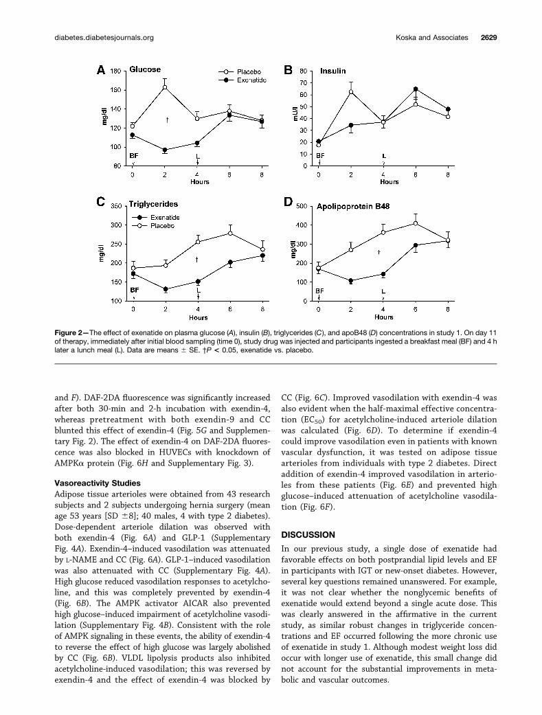
Study 1Thirty-six subjects completed both treatment arms, onewithdrew while on placebo, and five withdrew while onexenatide (Fig. 1A). Two patients had nausea with 10 mgexenatide and completed the study on 5 mg exenatide.Clinical characteristics did not differ between diabetesduration subgroups (Table 1). Exenatide treatment wasfollowed by modest reductions in body weight, systolicblood pressure, fasting glucose, and total and HDL choles-terol concentrations (Table 2). All participants consumedthe entire test meals. Postbreakfast glucose and insulinresponses were lower, and postlunch insulin responseswere higher after exenatide (Fig. 2A and B). Postprandialexcursions of triglycerides and apoB48 were markedly re-duced after exenatide (Fig. 2C and D). Acetaminophenconcentrations showed a significant interaction betweentreatment and time, trending lower 2 h postbreakfast after
2626 Effects of Exenatide on Endothelial Function Diabetes Volume 64, July 2015

exenatide (P = 0.01) whereas postlunch D-xylose concen-trations were not different between the treatments (Sup-plementary Fig. 1).
Exenatide increased overall RHI compared with pla-cebo (Fig. 3A), and this was similar in the two diabetes
duration subgroups (Fig. 3B). The increase in RHI afterexenatide remained significant after adjustment for base-line HbA1c, change in body weight over the study, orchanges in glucose, triglyceride, and insulin concentra-tions. It was modestly attenuated (P , 0.1) in a model
Figure 1—Participant flow for studies 1 (A) and 2 (B).
diabetes.diabetesjournals.org Koska and Associates 2627

including both glucose and triglyceride concentrations,explaining;36% of exenatide’s effect (Fig. 3C). The effectof exenatide did not show significant interactions withhistory of hypertension or use of lipid-lowering therapy(P . 0.5 both).
Study 2Thirty-two participants completed the study, one partic-ipant had a serious, study-unrelated adverse eventbetween the test sessions, and one withdrew from thestudy (Fig. 1B and characteristics in Table 1). In all threetest sessions, RHI was increased after the treatment (P ,0.0001 vs. baseline) (Fig. 4), consistent with rising EFduring the morning (35). The increment was greaterwith exenatide than with placebo, and this was completelyabolished with exendin-9 (Fig. 4). The increase in RHI withexenatide alone remained significant after adjustment for
glucose (P = 0.02) and insulin (P = 0.007), or both glucoseand insulin (P = 0.02) plasma concentrations. There wereno differences in responses to treatments between thosewith IGT and type 2 diabetes (P = 1.0).
Experiments in Endothelial CellsAMPKa phosphorylation was increased after 30 min ofincubation with exendin-4, PKA phosphorylation was un-changed after incubation with exendin-4, and Akt phos-phorylation was modestly reduced after 2-h exendin-4treatment (Fig. 5A and B). eNOS phosphorylation trendedhigher after 30 min (P = 0.09) and was significantly in-creased after 2-h exendin-4 treatment (Fig. 5A and B).The increase in AMPK phosphorylation with exendin-4was inhibited by pretreatment with exendin-9 (Fig. 5Cand D). Stimulation by exendin-4 of both AMPK andeNOS phosphorylation was abolished with CC (Fig. 5E
Table 1—Baseline clinical characteristics of the participants completing studies 1 and 2
Study 1 all Study 1 ,3 years diabetes Study 1 .5 years diabetes Study 2
n 36 16 20 32
Age (years) 63 6 6 63 6 6 62 6 6 60 6 6
BMI (kg/m2) 33 6 6 34 6 6 32 6 7 33 6 12
Race (% whites) 89% 100% 80% 85%
Sex (% males) 100% 100% 100% 97%
HbA1c (%) 6.4 6 0.8 6.3 6 0.5 6.5 6 1.0 6.1 6 0.5
HbA1c (mmol/mol) 47 6 9 46 6 6 48 6 11 43 6 5
Duration of diabetes (years) 5.5 (1–8) 1 (0.5–2) 7.5 (6–9.5) ,1
Systolic blood pressure (mmHg) 128 6 15 127 6 17 128 6 14 125 6 12
Diastolic blood pressure (mmHg) 78 6 9 78 6 9 78 6 9 80 6 8
History of hypertension (%) 81% 69% 90% 50%
Lipid-lowering therapy (%) 86% 81% 90% 59%
Data are means 6 SD or medians (25th–75th percentile).
Table 2—Clinical characteristics and fasting blood concentrations of several metabolic variables after 10 days of exenatide andplacebo treatment in 36 patients completing crossover study 1
Placebo Exenatide Change* P value
Body weight (kg) 101.4 6 22 100.7 6 21.5 20.95 (22 to 0.6) 0.01
Systolic blood pressure (mmHg) 131 6 11 126 6 13 25.5 (214 to 9) 0.03
Diastolic blood pressure (mmHg) 76 6 9 76 6 10 1 (25 to 6.5) 0.9
Heart rate (bpm) 69 6 12 71 6 11 1.5 (25.5 to 5.5) 0.2
Glucose (mg/dL) 122 6 25 113 6 23 27.5 (217.5 to 3.5) 0.01
Insulin (mU/L) 12.3 (8.6–24) 13.2 (8.8–25.6) 0.8 (21.5 to 4.4) 0.2
Triglycerides (mg/dL) 186 6 104 172 6 77 216 (251 to 34) 0.4
Total cholesterol (mg/dL) 163 6 36 150 6 33 210 (227 to 3) 0.002
LDL cholesterol (mg/dL) 80 6 10 81 6 28 7 (211 to 21) 0.8
HDL cholesterol (mg/dL) 38 6 8 36 6 7 21 (26 to 2) 0.048
Data are means6SD or medians (25th–75th percentile). P values indicate comparison between the two treatment arms by repeated-measuresANCOVA adjusted for treatment sequence and diabetes duration group. *Exenatide minus placebo.
2628 Effects of Exenatide on Endothelial Function Diabetes Volume 64, July 2015
and F). DAF-2DA fluorescence was significantly increasedafter both 30-min and 2-h incubation with exendin-4,whereas pretreatment with both exendin-9 and CCblunted this effect of exendin-4 (Fig. 5G and Supplemen-tary Fig. 2). The effect of exendin-4 on DAF-2DA fluores-cence was also blocked in HUVECs with knockdown ofAMPKa protein (Fig. 6H and Supplementary Fig. 3).
Vasoreactivity StudiesAdipose tissue arterioles were obtained from 43 researchsubjects and 2 subjects undergoing hernia surgery (meanage 53 years [SD 68]; 40 males, 4 with type 2 diabetes).Dose-dependent arteriole dilation was observed withboth exendin-4 (Fig. 6A) and GLP-1 (SupplementaryFig. 4A). Exendin-4–induced vasodilation was attenuatedby L-NAME and CC (Fig. 6A). GLP-1–induced vasodilationwas also attenuated with CC (Supplementary Fig. 4A).High glucose reduced vasodilation responses to acetylcho-line, and this was completely prevented by exendin-4(Fig. 6B). The AMPK activator AICAR also preventedhigh glucose–induced impairment of acetylcholine vasodi-lation (Supplementary Fig. 4B). Consistent with the roleof AMPK signaling in these events, the ability of exendin-4to reverse the effect of high glucose was largely abolishedby CC (Fig. 6B). VLDL lipolysis products also inhibitedacetylcholine-induced vasodilation; this was reversed byexendin-4 and the effect of exendin-4 was blocked by
CC (Fig. 6C). Improved vasodilation with exendin-4 wasalso evident when the half-maximal effective concentra-tion (EC50) for acetylcholine-induced arteriole dilationwas calculated (Fig. 6D). To determine if exendin-4could improve vasodilation even in patients with knownvascular dysfunction, it was tested on adipose tissuearterioles from individuals with type 2 diabetes. Directaddition of exendin-4 improved vasodilation in arterio-les from these patients (Fig. 6E) and prevented highglucose–induced attenuation of acetylcholine vasodila-tion (Fig. 6F).
DISCUSSION
In our previous study, a single dose of exenatide hadfavorable effects on both postprandial lipid levels and EFin participants with IGT or new-onset diabetes. However,several key questions remained unanswered. For example,it was not clear whether the nonglycemic benefits ofexenatide would extend beyond a single acute dose. Thiswas clearly answered in the affirmative in the currentstudy, as similar robust changes in triglyceride concen-trations and EF occurred following the more chronic useof exenatide in study 1. Although modest weight loss didoccur with longer use of exenatide, this small change didnot account for the substantial improvements in meta-bolic and vascular outcomes.
Figure 2—The effect of exenatide on plasma glucose (A), insulin (B), triglycerides (C), and apoB48 (D) concentrations in study 1. On day 11of therapy, immediately after initial blood sampling (time 0), study drug was injected and participants ingested a breakfast meal (BF) and 4 hlater a lunch meal (L). Data are means 6 SE. †P < 0.05, exenatide vs. placebo.
diabetes.diabetesjournals.org Koska and Associates 2629

GLP-1R agonists can reduce intestinal nutrient absorp-tion by slowing gastric emptying (12,36–38). Thus, it waspossible that by simply delaying nutrient absorption, exe-natide might reduce the postprandial rise of plasma lipidsand glucose concentrations, and postpone their peak untilafter subsequent meals. We therefore assessed gastricemptying by measuring absorbed acetaminophen andD-xylose with the breakfast and lunch meals, respectively.Consistent with previous studies showing slower gastricemptying with GLP-1R agonists (37,39), postbreakfastacetaminophen concentrations were reduced after exena-tide injection. However, this reduction was modest andwas observed only at the 2-h time point. The correspond-ing early delay in the postprandial glucose peak is inagreement with the involvement of gastric emptying inglucose-lowering effects of exenatide (37,39). In contrast,discordant postmeal rises between plasma apoB48 or tri-glycerides and acetaminophen or D-xylose indicate addi-tional mechanisms of triglyceride lowering by exenatide,such as inhibition of intestinal lipoprotein production andtriglyceride release as shown previously (40).
We considered the possibility that fat and carbohy-drates from the lunch meal, in addition to delayedabsorption of nutrients from the preceding breakfastmeal, might produce a late surge in blood glucose andtriglyceride concentrations that would overcome the bene-fits of exenatide treatment on EF in the postlunch period.This was supported by a previous report of equivalent risesin triglycerides after exenatide and placebo after a middaymeal in subjects with type 2 diabetes (41). In contrast,triglyceride concentrations in the current study were stillreduced 2 h postlunch with exenatide. EF was also stillimproved with exenatide at the 2-h postlunch period, con-sistent with the concept that GLP-1R agonists protect
Figure 3—The effect of exenatide on EF (RHI) in study 1. RHI wascalculated as the ratio of the average amplitude of the PAT signal overa 30 s time interval starting 90 s after blood pressure cuff deflationdivided by the average amplitude of the PAT signal of a 3.5-min timeperiod before cuff inflation. On day 11 of therapy, immediately afterinitial blood sampling (time 0), study drug was injected and partici-pants ingested a breakfast meal (BF) and 4 h later a lunch meal (L). A:RHI over the 8-h test period. B: RHI area under the curve (normalizedto 1 h, by trapezoid method) according to the duration of diabetes.Data are means 6 SE. †P < 0.05, exenatide vs. placebo. C: Multi-variate models of exenatide’s effect on RHI. The modeled effect isshown as b estimates and SE of exenatide’s effect on RHI before andafter adjustment for plasma glucose (Glc), insulin (Ins), and triglyceride(Trig) concentrations. Adjustments for individual or combinations ofthese variables did not significantly reduce the effect of exenatide.
Figure 4—Percent change in RHI after intravenous infusion of exe-natide, placebo, or exenatide + GLP-1R inhibitor exendin-9 instudy 2. RHI was calculated as the ratio of the average amplitudeof the PAT signal over a 30-s time interval starting 90 s after bloodpressure cuff deflation divided by the average amplitude of thePAT signal of a 3.5-min time period before cuff inflation. Dataare means 6 SE. †P < 0.05 between treatments.
2630 Effects of Exenatide on Endothelial Function Diabetes Volume 64, July 2015

Figure 5—The effects of exenatide in vitro in human endothelial cells. A and B: Phosphorylation of AMPKa (Thr172), PKA (Thr197), Aktkinase (Ser473), and eNOS (Ser1177) in HAECs after treatment with 10 nmol/L exendin-4 (EX) for 30 min and 2 h (A: representative Westernblot; B: densitometry analysis [means 6 SE]; n = 6). C and D: AMPKa phosphorylation in HAECs after EX (30 min and 2 h) with or withoutpretreatment for 30 min with 1 mmol/L GLP-1R inhibitor EX-9 (C: representative Western blot; D: densitometry analysis; n = 7–8). E and F:AMPKa and eNOS phosphorylation in HAECs after EX (2 h) with or without 1-h pretreatment with 5 mol/L of AMPKa inhibitor CC (E:representative Western blot; F: densitometry analysis; n = 8–9). The effect of EX on NO production (by DAF-2DA fluorescence) in HAECswith or without pretreatment with EX-9 or CC (n = 4–7) (G) and in HUVECs with knocked-down AMPKa gene expression (siRNA, n = 6) (H).Phosphorylated bands were normalized to total bands and a-tubulin. Control, untreated cells. Data are means 6 SE. *P < 0.05 vs. control.
diabetes.diabetesjournals.org Koska and Associates 2631

against high-fat meal–induced endothelial dysfunctionthrough reduction in postprandial triglycerides. The similartriglyceride concentrations and EF in both treatmentgroups at 4 h postlunch may reflect waning exenatide
concentrations. However, it is likely that the next ex-enatide dose in a typical 23/day regimen would reinstatethe mealtime benefit of exenatide, as indicated by signifi-cant reductions in postdinner glucose and triglyceride
Figure 6—The effects of exenatide (EX) ex vivo in isolated human adipose tissue arterioles. A: Vasodilation responses to increasing dosesof EX followed by papaverine before (control) and after treatment with vehicle, eNOS inhibitor L-NAME (5 mmol/L), or AMPK inhibitor CC(1 mmol/L). B: Vasodilation responses to acetylcholine before (control) and after exposure to high glucose for 2 h (HG, 33 mmol/L), HG withaddition of 10 nmol/L EX after 1 h (HG+EX), and HG+EX pretreated with 1 mmol/L CC (HG+EX+CC). C: Vasodilation responses toacetylcholine before (control) and after exposure to VLDL lipolysis products mixture for 2 h (VLDL, 150 mmol/L fatty acids), VLDL withaddition of 10 nmol/L EX after 1 h (VLDL+EX), and VLDL+EX pretreated with 1 mmol/L CC. D: EC50 of acetylcholine from experiments shownin panels B and C. EC50 was calculated by nonlinear regression and variable slope (four parameters) and least squares fit (GraphPad Prism5.0, San Diego CA). If the vessels dilated to <50% with maximum dose of acetylcholine, EC50 was set at 1024 mol/L. Acetylcholine inducedvasodilation in adipose tissue arterioles from subjects with type 2 diabetes before (control) and after 1-h exposure to 10 nmol/L EX (E) and HGand HG+EX (F). Data are means 6 SE. *P < 0.05 vs. control; †P < 0.05 vs. HG or VLDL; ‡P < 0.05 vs. EX 0 pmol/L (tested in control only).
2632 Effects of Exenatide on Endothelial Function Diabetes Volume 64, July 2015

concentrations reported by Schwartz et al. (41). Moreover,prolonged duration of action of newer GLP-1R agonistsmay further diminish the likelihood of any rebounds intriglyceride concentrations and EF impairment.
In our previous study, EF was only tested in individualswith IGT or diabetes of very short duration (,1 year)(14). Thus, it was not known whether exenatide wouldbe effective in patients with more established type 2 di-abetes and potentially greater vascular dysfunction. Asthe improvement of EF in study 1 was demonstrated inpatients with diabetes of both short and more prolongedduration, the beneficial effect of exenatide on EF appearsto occur across the full range of IGT to diabetes of at leastmoderate duration. The pattern of postmeal changes inEF was similar in both groups. Of note, the pattern ofexenatide improvement in patients with type 2 diabetesin the current study was slightly different than in ourprevious study. These minor differences may be explainedby a lower fat content of the breakfast meal (400 vs. 600kcal/m2) and/or higher systemic exenatide levels resultingfrom more chronic therapy (11 days vs. single dose) in thecurrent study.
More than one-third of exenatide’s effect on EF in ourprevious study remained unexplained by changes inplasma nutrients (14). Although it was possible that theunexplained portion simply reflected imprecise matchingof time points of EF measurement and blood draws forplasma glucose and lipid concentrations, we suspectedthat there may be a direct effect of exenatide on EF.Even with temporally linked EF and plasma measures inthe current study, improvement of EF after exenatide wasonly partly related to changes in plasma triglycerides andglucose concentrations, suggesting an additional vascularaction of exenatide. To test this point, exenatide was in-fused under fasting conditions in study 2 at a rate knownto achieve exenatide concentrations comparable to thoseobtained with standard dosing of subcutaneous exenatideinjection (30). EF was enhanced by almost 20% duringexenatide infusion compared with placebo. This is inagreement with a previous report showing improved EFafter subcutaneous exenatide injection (24) and earlierstudies demonstrating EF elevations after intravenousGLP-1 infusion (17,18).
We also hypothesized that GLP-1R agonists improveEF directly via endothelial GLP-1Rs (19). Previous in vitrostudies showed the GLP-1R inhibitor exendin-9 antago-nized exenatide-induced activation of eNOS in culturedendothelial cells (26). Consistent with this, exendin-9blunted exenatide-induced eNOS activation and NO pro-duction in HAECs. Moreover, exendin-9 completely abol-ished the effect of exenatide infusion on EF, providingimportant in vivo evidence for GLP-1R involvement inthe vasodilation effect of GLP-1R agonists.
GLP-1R agonists have been previously shown to improvevasodilation responses of rodent vessels ex vivo (15,16,22).In our study, human arterioles dilated in a concentration-dependent manner with increasing doses of exenatide or
GLP-1. Importantly, exenatide was already effective atconcentrations similar to those achieved with therapeuticdosing (42). The vasodilation was inhibited by eNOS in-hibitor L-NAME, indicating an NO-dependent mechanismof exenatide action. Exenatide also restored endothelium-mediated vasodilation that was attenuated by high glu-cose or VLDL lipolysis products. Exenatide’s protectionagainst high glucose–induced impairment of EF concurswith a previously reported effect of GLP-1 during in vivohyperglycemia (21). We now demonstrate the novel find-ing that exenatide directly prevents lipid-induced endo-thelial dysfunction.
Of the several GLP-1R downstream kinase pathwayspreviously postulated as relevant for eNOS activation(25–27), only AMPK was activated by exenatide inHAECs. AMPK activation appeared to precede phosphor-ylation of eNOS, consistent with reports of this pathwaysequence (43,44). Supporting a key role of the AMPK inendothelial vasodilation by GLP-1R agonists, AICARreproduced the effect and CC inhibited exenatide- andGLP-1–induced vasodilation in human arterioles. More-over, CC was nearly as effective as L-NAME in blockingexenatide vasodilation and also abolished exenatide’s pro-tection against high glucose– or lipid-induced impairmentof endothelium-mediated vasodilation ex vivo. In HAECs,CC also blocked exenatide-induced eNOS activation andNO production. To exclude the possibility that CC mighthave modulated exenatide’s action through inhibition ofother kinase pathways (45), we knocked down transcrip-tion of AMPKa in HUVECs and found reductions inexenatide-induced NO production in cells lacking AMPKa.
There are several potential limitations of the study.The majority of participants were male and we cannotconfidently extend our in vivo results to females. Al-though prior evidence (46) suggests it is unlikely thatexendin-9 has vascular consequences beyond its interfer-ence with exenatide action, this cannot be entirely ex-cluded. PAT was chosen for this study because of itsexcellent reproducibility during repeated measurements,good correlation with established gold standard methods(brachial flow-mediated dilation, direct measurement ofcoronary artery EF, and forearm plethysmography), andproven association with cardiovascular risk in large pop-ulations (47–49).
In conclusion, our in vivo and ex vivo studies showedthat 1) the GLP-1R agonist exenatide improved EF overtwo sequential meals in individuals with type 2 diabetesof short to moderate duration, 2) exenatide improved EFvia a direct vascular GLP-1R–mediated mechanism, 3)exenatide via direct vascular action ameliorated bothhigh glucose– and lipid-induced endothelial dysfunction,and 4) exenatide stimulated endothelial AMPK pathwayactivity, resulting in greater eNOS activation and NO pro-duction. Since endothelial dysfunction is associated withprogression of atherosclerosis (50) and predicts clinicalcardiovascular events (1–3), our findings provide furtherconceptual and mechanistic support for the potential use
diabetes.diabetesjournals.org Koska and Associates 2633

of GLP-1R agonists in cardiovascular protection in type 2diabetes.
Acknowledgments. The authors acknowledge the excellent project as-sistance provided by Linda McDonald RN, Irina Moroz, Keith Rasmussen, AshleyHaile, and Dewayne C. Thurmond (all at the Phoenix VA Health Care System).Funding. This work was supported in part by the American Diabetes Asso-ciation (1-10-CT-31 to J.K.), the Amyloidosis Foundation (to R.Q.M.), the NationalInstitutes of Health (R21-HL-092344-01 to R.Q.M.), VA Merit (BLRD I01BX007080to R.Q.M.), and the Office of Research and Development, Department of VeteransAffairs (1I01CX000598 to P.D.R.). Amylin Lilly provided study medications at nocosts.
The contents of this article do not represent the views of the Department ofVeterans Affairs or the U.S. Government.Duality of Interest. P.D.R. has received research grants from Bristol-Myers Squibb. No other potential conflicts of interest relevant to this articlewere reported.Author Contributions. J.K. designed the study, obtained funding, gen-erated research data, and prepared the manuscript. M.S. and C.B. researcheddata and reviewed and edited the manuscript. K.M.D. designed the experiments,generated research data, and edited the manuscript. K.R. collected the data andedited the manuscript. J.L. generated research data and edited the manuscript.S.T. and D.A.F. designed the experiments and edited the manuscript. E.A.S.helped design the experiments, collected the data, and edited the manuscript.D.C.S. helped design the study and edited the manuscript. D.D. consulted on thestudy design and reviewed the manuscript. R.Q.M. helped design the experi-ments, obtained funding, and reviewed and edited the manuscript. P.D.R. helpeddesign the study and experiments, obtained funding, and reviewed and edited themanuscript. J.K. is the guarantor of this work and, as such, had full access to allthe data in the study and takes responsibility for the integrity of the data and theaccuracy of the data analysis.Prior Presentation. Parts of this study were presented at the 73rd Sci-entific Sessions of the American Diabetes Association, Chicago, IL, 21–25 June2013, and the 74th Scientific Sessions of the American Diabetes Association, SanFrancisco, CA, 13–17 June 2014.
References1. Halcox JP, Schenke WH, Zalos G, et al. Prognostic value of coronary vas-cular endothelial dysfunction. Circulation 2002;106:653–6582. Gokce N, Keaney JF Jr, Hunter LM, Watkins MT, Menzoian JO, Vita JA. Riskstratification for postoperative cardiovascular events via noninvasive assessmentof endothelial function: a prospective study. Circulation 2002;105:1567–15723. Yeboah J, Crouse JR, Hsu FC, Burke GL, Herrington DM. Brachial flow-mediated dilation predicts incident cardiovascular events in older adults: theCardiovascular Health Study. Circulation 2007;115:2390–23974. McVeigh GE, Brennan GM, Johnston GD, et al. Impaired endothelium-dependent and independent vasodilation in patients with type 2 (non-insulin-dependent) diabetes mellitus. Diabetologia 1992;35:771–7765. Williams SB, Cusco JA, Roddy MA, Johnstone MT, Creager MA. Impairednitric oxide-mediated vasodilation in patients with non-insulin-dependent di-abetes mellitus. J Am Coll Cardiol 1996;27:567–5746. Ceriello A, Taboga C, Tonutti L, et al. Evidence for an independent andcumulative effect of postprandial hypertriglyceridemia and hyperglycemia onendothelial dysfunction and oxidative stress generation: effects of short- andlong-term simvastatin treatment. Circulation 2002;106:1211–12187. Bae JH, Bassenge E, Lee HJ, et al. Impact of postprandial hyper-triglyceridemia on vascular responses in patients with coronary artery disease:effects of ACE inhibitors and fibrates. Atherosclerosis 2001;158:165–1718. Kawano H, Motoyama T, Hirashima O, et al. Hyperglycemia rapidly sup-presses flow-mediated endothelium-dependent vasodilation of brachial artery.J Am Coll Cardiol 1999;34:146–154
9. Cervera A, Wajcberg E, Sriwijitkamol A, et al. Mechanism of action of ex-enatide to reduce postprandial hyperglycemia in type 2 diabetes. Am J PhysiolEndocrinol Metab 2008;294:E846–E85210. DeFronzo RA, Okerson T, Viswanathan P, Guan X, Holcombe JH, MacConellL. Effects of exenatide versus sitagliptin on postprandial glucose, insulin andglucagon secretion, gastric emptying, and caloric intake: a randomized, cross-over study. Curr Med Res Opin 2008;24:2943–295211. Edwards CM, Stanley SA, Davis R, et al. Exendin-4 reduces fasting andpostprandial glucose and decreases energy intake in healthy volunteers. AmJ Physiol Endocrinol Metab 2001;281:E155–E16112. Kolterman OG, Buse JB, Fineman MS, et al. Synthetic exendin-4 (exenatide)significantly reduces postprandial and fasting plasma glucose in subjects withtype 2 diabetes. J Clin Endocrinol Metab 2003;88:3082–308913. Meier JJ, Gethmann A, Götze O, et al. Glucagon-like peptide 1 abolishes thepostprandial rise in triglyceride concentrations and lowers levels of non-esterifiedfatty acids in humans. Diabetologia 2006;49:452–45814. Koska J, Schwartz EA, Mullin MP, Schwenke DC, Reaven PD. Improvementof postprandial endothelial function after a single dose of exenatide in individualswith impaired glucose tolerance and recent-onset type 2 diabetes. Diabetes Care2010;33:1028–103015. Richter G, Feddersen O, Wagner U, Barth P, Göke R, Göke B. GLP-1stimulates secretion of macromolecules from airways and relaxes pulmonaryartery. Am J Physiol 1993;265:L374–L38116. Yu M, Moreno C, Hoagland KM, et al. Antihypertensive effect of glucagon-like peptide 1 in Dahl salt-sensitive rats. J Hypertens 2003;21:1125–113517. Nyström T, Gutniak MK, Zhang Q, et al. Effects of glucagon-like peptide-1on endothelial function in type 2 diabetes patients with stable coronary arterydisease. Am J Physiol Endocrinol Metab 2004;287:E1209–E121518. Basu A, Charkoudian N, Schrage W, Rizza RA, Basu R, Joyner MJ. Ben-eficial effects of GLP-1 on endothelial function in humans: dampening byglyburide but not by glimepiride. Am J Physiol Endocrinol Metab 2007;293:E1289–E129519. Ban K, Noyan-Ashraf MH, Hoefer J, Bolz SS, Drucker DJ, Husain M. Car-dioprotective and vasodilatory actions of glucagon-like peptide 1 receptor are me-diated through both glucagon-like peptide 1 receptor-dependent and -independentpathways. Circulation 2008;117:2340–235020. Nathanson D, Erdogdu O, Pernow J, Zhang Q, Nyström T. Endothelialdysfunction induced by triglycerides is not restored by exenatide in rat conduitarteries ex vivo. Regul Pept 2009;157:8–1321. Ceriello A, Esposito K, Testa R, Bonfigli AR, Marra M, Giugliano D. Thepossible protective role of glucagon-like peptide 1 on endothelium during themeal and evidence for an “endothelial resistance” to glucagon-like peptide 1 indiabetes. Diabetes Care 2011;34:697–70222. Gaspari T, Liu H, Welungoda I, et al. A GLP-1 receptor agonist liraglutideinhibits endothelial cell dysfunction and vascular adhesion molecule expression inan ApoE-/- mouse model. Diab Vasc Dis Res 2011;8:117–12423. Chai W, Dong Z, Wang N, et al. Glucagon-like peptide 1 recruits micro-vasculature and increases glucose use in muscle via a nitric oxide-dependentmechanism. Diabetes 2012;61:888–89624. Ha SJ, Kim W, Woo JS, et al. Preventive effects of exenatide on endothelialdysfunction induced by ischemia-reperfusion injury via KATP channels. Arte-rioscler Thromb Vasc Biol 2012;32:474–48025. Dong Z, Chai W, Wang W, et al. Protein kinase A mediates glucagon-likepeptide 1-induced nitric oxide production and muscle microvascular recruitment.Am J Physiol Endocrinol Metab 2013;304:E222–E22826. Erdogdu O, Nathanson D, Sjöholm A, Nyström T, Zhang Q. Exendin-4stimulates proliferation of human coronary artery endothelial cells througheNOS-, PKA- and PI3K/Akt-dependent pathways and requires GLP-1 receptor.Mol Cell Endocrinol 2010;325:26–3527. Ben-Shlomo S, Zvibel I, Shnell M, et al. Glucagon-like peptide-1 reduceshepatic lipogenesis via activation of AMP-activated protein kinase. J Hepatol2011;54:1214–1223
2634 Effects of Exenatide on Endothelial Function Diabetes Volume 64, July 2015
28. Krasner NM, Ido Y, Ruderman NB, Cacicedo JM. Glucagon-like peptide-1(GLP-1) analog liraglutide inhibits endothelial cell inflammation through a calciumand AMPK dependent mechanism. PLoS ONE 2014;9:e9755429. Clements JA, Heading RC, Nimmo WS, Prescott LF. Kinetics of acetaminophenabsorption and gastric emptying in man. Clin Pharmacol Ther 1978;24:420–43130. Fehse F, Trautmann M, Holst JJ, et al. Exenatide augments first- andsecond-phase insulin secretion in response to intravenous glucose in subjectswith type 2 diabetes. J Clin Endocrinol Metab 2005;90:5991–599731. Goldfine AB, Conlin PR, Halperin F, et al. A randomised trial of salsalate forinsulin resistance and cardiovascular risk factors in persons with abnormalglucose tolerance. Diabetologia 2013;56:714–72332. Migrino RQ, Truran S, Gutterman DD, et al. Human microvascular dys-function and apoptotic injury induced by AL amyloidosis light chain proteins. AmJ Physiol Heart Circ Physiol 2011;301:H2305–H231233. Phillips SA, Hatoum OA, Gutterman DD. The mechanism of flow-induceddilation in human adipose arterioles involves hydrogen peroxide during CAD. AmJ Physiol Heart Circ Physiol 2007;292:H93–H10034. Eberts TJ, Sample RH, Glick MR, Ellis GH. A simplified, colorimetric mi-cromethod for xylose in serum or urine, with phloroglucinol. Clin Chem 1979;25:1440–144335. Gaenzer H, Sturm W, Neumayr G, et al. Pronounced postprandial lipemiaimpairs endothelium-dependent dilation of the brachial artery in men. CardiovascRes 2001;52:509–51636. Linnebjerg H, Park S, Kothare PA, et al. Effect of exenatide on gastricemptying and relationship to postprandial glycemia in type 2 diabetes. Regul Pept2008;151:123–12937. Salehi M, Vahl TP, D’Alessio DA. Regulation of islet hormone release andgastric emptying by endogenous glucagon-like peptide 1 after glucose ingestion.J Clin Endocrinol Metab 2008;93:4909–491638. Deane AM, Nguyen NQ, Stevens JE, et al. Endogenous glucagon-likepeptide-1 slows gastric emptying in healthy subjects, attenuating postprandialglycemia. J Clin Endocrinol Metab 2010;95:215–22139. Kolterman OG, Kim DD, Shen L, et al. Pharmacokinetics, pharmacody-namics, and safety of exenatide in patients with type 2 diabetes mellitus. AmJ Health Syst Pharm 2005;62:173–181
40. Xiao C, Bandsma RH, Dash S, Szeto L, Lewis GF. Exenatide, a glucagon-likepeptide-1 receptor agonist, acutely inhibits intestinal lipoprotein production inhealthy humans. Arterioscler Thromb Vasc Biol 2012;32:1513–151941. Schwartz SL, Ratner RE, Kim DD, et al. Effect of exenatide on 24-hour bloodglucose profile compared with placebo in patients with type 2 diabetes: a ran-domized, double-blind, two-arm, parallel-group, placebo-controlled, 2-weekstudy. Clin Ther 2008;30:858–86742. Fineman MS, Bicsak TA, Shen LZ, et al. Effect on glycemic control ofexenatide (synthetic exendin-4) additive to existing metformin and/or sulfo-nylurea treatment in patients with type 2 diabetes. Diabetes Care 2003;26:2370–237743. Levine YC, Li GK, Michel T. Agonist-modulated regulation of AMP-activatedprotein kinase (AMPK) in endothelial cells. Evidence for an AMPK -. Rac1 -.Akt -. endothelial nitric-oxide synthase pathway. J Biol Chem 2007;282:20351–2036444. Chen Z, Peng I-C, Sun W, et al. AMP-activated protein kinase functionallyphosphorylates endothelial nitric oxide synthase Ser633. Circ Res 2009;104:496–50545. Bain J, Plater L, Elliott M, et al. The selectivity of protein kinase inhibitors:a further update. Biochem J 2007;408:297–31546. Green BD, Hand KV, Dougan JE, McDonnell BM, Cassidy RS, Grieve DJ.GLP-1 and related peptides cause concentration-dependent relaxation of rat aortathrough a pathway involving KATP and cAMP. Arch Biochem Biophys 2008;478:136–14247. Bonetti PO, Pumper GM, Higano ST, Holmes DR Jr, Kuvin JT, Lerman A.Noninvasive identification of patients with early coronary atherosclerosis byassessment of digital reactive hyperemia. J Am Coll Cardiol 2004;44:2137–214148. Kuvin JT, Patel AR, Sliney KA, et al. Assessment of peripheral vascular endothelialfunction with finger arterial pulse wave amplitude. Am Heart J 2003;146:168–17449. Hamburg NM, Keyes MJ, Larson MG, et al. Cross-sectional relations ofdigital vascular function to cardiovascular risk factors in the Framingham HeartStudy. Circulation 2008;117:2467–247450. Halcox JP, Donald AE, Ellins E, et al. Endothelial function predicts pro-gression of carotid intima-media thickness. Circulation 2009;119:1005–1012
diabetes.diabetesjournals.org Koska and Associates 2635