EXAMPLES OF UTILISING 3D TECHNOLOGIES FOR...
23
EXAMPLES OF UTILISING 3D TECHNOLOGIES FOR CLINICAL USE 3D LifePrints UK Ltd 3D LIFEPRINTS
Transcript of EXAMPLES OF UTILISING 3D TECHNOLOGIES FOR...

EXAMPLES OF UTILISING 3D TECHNOLOGIES FOR CLINICAL USE
3D LifePrints UK Ltd
3D LIFEPRINTS

LOCATION: ALDER HEY CHILDRENS HOSPITAL, CARDIAC DEPARTMENT MODEL SOURCE: CONTRASTED TRI-PLANAR CT SCAN, ANGIOGRAM DESCRIPTION: �Two�3D�printed�models�of�a�patient’s�heart�were�created�as�
pre-surgical�assessment�tools�for�the�surgical�team�in�planning� an�operation�on�a�heart�with�a�tetralogy�of�fallot�including�a� narrow�pulmonary�artery�and�ventricular�septal�defect.
OUTCOME / BENEFITS: �The�pre-surgical�planning�process�was�enhanced�by�using�the�3D�
models�which�led�to�time�savings�in�the�operating�theatre�with�a�better�imaging�of�the�internal�part�of�the�heart�and�an�improved�patient�outcome.� Lead�surgeon�Mr�Rafael�Guerrero�said:�“This�is�a�great�use�of� 3D�printing�as�it�shows�me�exactly�what�we�needed�to�see�for�a�patient-specific�procedure.�The�initial�3D�model�was�used�to�plan� for�the�successful�repair�of�the�malformation�by�removing�the�obstruction,�closing�the�defect�(hole�in�the�heart)�and�enlarging� the�main�artery�to�the�lungs.”
3D LifePrints UK Ltd
CLINICAL USE 1A: PRE SURGICAL PLANNING SURGICAL PROCEDURE: TOTAL REPAIR OF TETRALOGY OF FALLOT

LOCATION: ALDER HEY CHILDRENS HOSPITAL, CARDIAC DEPARTMENT MODEL SOURCE: CONTRASTED TRI-PLANAR CT SCAN, ANGIOGRAM DESCRIPTION: ��This�model�was�printed�based�upon�the�volume�of�the�blood�within�
the�heart’s�chambers�rather�than�the�heart�itself�a�“Blood�Volume�Model”.�This�new�application�of�3D�printing�has�become�an�extremely�useful�technique�to�indirectly�image�malformations�of�the�heart�by�printing�the�blood�volume�within�the�cavities. Mr�Guerrero�said�“�this�is�a�very�impressive�and�imaginative�way�to�use�3D�printing�for�organ�imaging.�In�this�case,�we�were�able�to�complement�the�visualisation�of�the�cardiac�defects�and�make�the�decision�that�it�might�be�possible�to�perform�a�less�invasive�procedure.” This�model�is�a�significant�innovative�step�as�3D�LifePrints�and�the�surgeon�have�moved�away�from�simply�providing�a�copy�of�the�heart�to�create�something�that�is�specific�to�this�type�of�operation�and�is�of�greater�use�to�the�surgeon. In�this�case�Mr�Guerrero�and�his�team�assessed�the�images�of�the�heart�and�decided�that�it�was�likely�that�no�further�patching�of�the�perforations�was�necessary.�The�patient�directly�benefited�from�the�provision�of�the�model�in�that�the�second�surgery�was�not�as�long� or�complex.
3D LifePrints UK Ltd
CLINICAL USE 1B: POST SURGICAL ASSESSMENT MULTIPLE VENTRICULAR SEPTAL DEFECT : BLOOD VOLUME MODEL
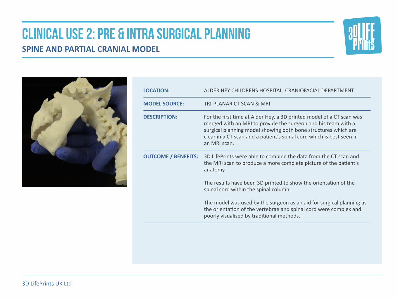
LOCATION: ALDER HEY CHILDRENS HOSPITAL, CRANIOFACIAL DEPARTMENT MODEL SOURCE: TRI-PLANAR CT SCAN & MRI DESCRIPTION: �For�the�first�time�at�Alder�Hey,�a�3D�printed�model�of�a�CT�scan�was�
merged�with�an�MRI�to�provide�the�surgeon�and�his�team�with�a�surgical�planning�model�showing�both�bone�structures�which�are� clear�in�a�CT�scan�and�a�patient’s�spinal�cord�which�is�best�seen�in� an�MRI�scan.
OUTCOME / BENEFITS: �3D�LifePrints�were�able�to�combine�the�data�from�the�CT�scan�and�
the�MRI�scan�to�produce�a�more�complete�picture�of�the�patient’s�anatomy.� The�results�have�been�3D�printed�to�show�the�orientation�of�the� spinal�cord�within�the�spinal�column. The�model�was�used�by�the�surgeon�as�an�aid�for�surgical�planning�as�the�orientation�of�the�vertebrae�and�spinal�cord�were�complex�and�poorly�visualised�by�traditional�methods.
3D LifePrints UK Ltd
CLINICAL USE 2: PRE & INTRA SURGICAL PLANNING SPINE AND PARTIAL CRANIAL MODEL

LOCATION: LIVERPOOL�CHEST�&�HEART�HOSPITAL,�LIVERPOOL,�ENGLAND MODEL SOURCE: CONTRASTED TRI-PLANAR CT SCAN, ANGIOGRAM DESCRIPTION: A�3D�printed�model�was�created�to�provide�surgeon�Robert�Cooper�
and�his��team�with�information�on�the�heart�wall�thickness�in�both� the�relaxed�and�the�contracted�stages�of�the�heartbeat�for�preparation�for�an�alcohol�septal�ablation.
OUTCOME / BENEFITS: A�study�consisting�of�nine�further�models�has�been�commissioned�
based�on�the�success�of�this�model.�Patients�are�currently�being�identified�to�participate�in�the�study. The�hospital�have�also�commissioned�a�study�into�the�use�the�3D�printed�anatomical�models�to�simulate�the�placement�of�a�device� to�plug�the�holes�in�a�septum The�simulation�of�the�placement�of�a�cardiac�device�in�a�3D�printed�model�will�allow�surgeons�to�determine�the�exact�size�and�type�of�device�to�use.
3D LifePrints UK Ltd
CLINICAL USE 3: PRE SURGICAL PLANNING 3D PRINT MODEL TYPE: HEART CARDIOMYOPATHY AND DEVICE SIZING

LOCATION: SOUTHAMPTON HOSPITAL, SOUTHAMPTON, ENGLAND MODEL SOURCE: TRI-PLANAR CT SCAN DESCRIPTION: A�pelivis�and�a�femoral�head�were�3D�printed�in�white�ABS�to�assist�with�
a�complex�bilateral�pelvic�procedure. OUTCOME / BENEFITS: Lead�surgeon�Alex�Aarvold�remarked:�“With�a�difficult�case�such�as�
this�one�that�needed�a�slightly�bespoke�pelvic�osteotomy,�the�benefit�was�more�in�the�surgical�planning�than�in�actual�operative�time�saved.�In�this�respect�it�was�fantastic,�and�we�could�see�what�we�needed�to�do�very�quickly�on�the�model,�which�was�not�nearly�so�clear�on�the�CT�images.�We�could�also�trial�the�planned�osteotomy�on�the�model�so�we�had�more�confidence�in�the�surgical�plan.”
3D LifePrints UK Ltd
CLINICAL USE 4: PRE SURGICAL PLANNING 3D PRINT MODEL TYPE: HIP OSTEOTOMY

LOCATION: ROYAL�LIVERPOOL�HOSPITAL MODEL SOURCE: TRI-PLANAR CT SCAN DESCRIPTION: �EVAR�is�a�type�of�endovascular�surgery�used�to�treat�aneurysmal�
pathology�of�the�aorta.��The�images�show�a�novel�technique�for�planing�and�simulating�the�repair�of�an�abdominal�aortic�aneurysm�(AAA). The�model�is�a�true�representation�of�the�lumenal�anatomy�which�allows�accurate�trialling�of�graft�size�and�positioning.�There�are�over�1000�EVAR�procedures�carried�out�in�the�UK�alone�each�year.
OUTCOME / BENEFITS: �This�is�a�new�study�into�the�optimal�type�of�model�to�use�while�
simulating�the�placement�of�the�graft.�The�vascular�surgeon�will�be�provided�with�a�range�of�models�in�different�materials,�orientation�and�transparencies.�It�is�believed�that�the�ability�to�simulate�the�surgery�will�not�only�improve�the�surgical�outcomes�but�also�allow�the�surgeon�to�more�easily�determine�the�type�of�graft�and�and�location�to�fit�it�within�the�aorta. Given�the�extra�information�and�ability�to�simulate�provided�by�the�model�it�may�be�possible�for�a�surgeon�to�utilise�a�off-the-shelf�rather�than�custom�made�graft�with�associated�cost�savings.
3D LifePrints UK Ltd
CLINICAL USE 5: Device Sizing and testing PLAN SURGERY AND SIMULATE THE PLACEMENT OF A GRAFT FOR ENDOVASCULAR ANEURYSM REPAIR (EVAR)

LOCATION: VIRTUAL�ENGINEERING�CENTRE,�LIVERPOOL�UNIVERSITY,�ENGLAND MODEL SOURCE: MRI SCAN DESCRIPTION: This�product�allows�a�surgeon�to�manipulate�a�handheld�and�patient�
specific�3D�print�of�an�organ,�such�as�a�heart,�to�virtually�navigate�its�internal�structure�and�external�surroundings�using�a�virtual�reality�headset.
OUTCOME / BENEFITS: The�Virtual�Engineering�Centre�commented:�“We�are�developing�an�
exciting�collaboration�with�3D�LifePrints,�bringing�together�the�specialist�expertise�of�both�organisations�in�3D�printing�and�immersive�virtual�reality�to�support�the�medical�sector.��We�are�looking�forward�to�forging�future�developments�with�such�an�innovative�organisation.”
3D LifePrints UK Ltd
CLINICAL USE 6: PRE SURGICAL PLANNING / TRAINING 3D PRINT MODEL TYPE: 3D VIRTUAL REALITY
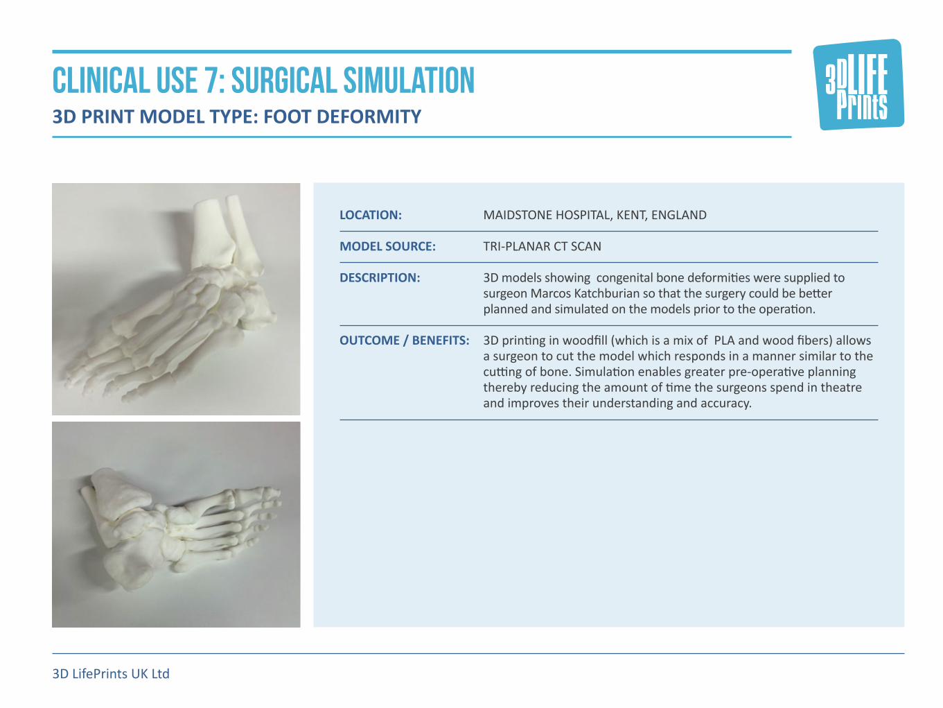
LOCATION: MAIDSTONE HOSPITAL, KENT, ENGLAND MODEL SOURCE: TRI-PLANAR CT SCAN DESCRIPTION: 3D�models�showing��congenital�bone�deformities�were�supplied�to�
surgeon�Marcos�Katchburian�so�that�the�surgery�could�be�better�planned�and�simulated�on�the�models�prior�to�the�operation.
OUTCOME / BENEFITS: 3D�printing�in�woodfill�(which�is�a�mix�of��PLA�and�wood�fibers)�allows�
a�surgeon�to�cut�the�model�which�responds�in�a�manner�similar�to�the�cutting�of�bone.�Simulation�enables�greater�pre-operative�planning�thereby�reducing�the�amount�of�time�the�surgeons�spend�in�theatre�and�improves�their�understanding�and�accuracy.�
3D LifePrints UK Ltd
CLINICAL USE 7: SURGICAL SIMULATION 3D PRINT MODEL TYPE: FOOT DEFORMITY

LOCATION: ALDER�HEY�HOSPITAL,�LIVERPOOL,�ENGLAND MODEL SOURCE: MRI SCAN DESCRIPTION: Created�in�collaboration�with�Lazarus�3D,�this�3D�model�of�part�of�an�
adult�brain�was�created�to�show�that�silicone�3D�printing�can�provide�a�realistic�replica�for�soft�tissue�and�organs.�The�silicone�is�printed�in�a�range�of�colours�and�densities�and�it�is�intended�to�be�used�in�medical�training�for�operations�such�as�Lobe�resection�and�Lesionectomy.
OUTCOME / BENEFITS: Feedback�from�the�surgeons�at�Alder�Hey�was�extremely�positive�
especially�concerning��the�realism�of�the�model�and�potential�future��use�of�medical�training�purposes. It�is�proposed�to�combine�the�silicone�brain�with�a�skull�printed�in�polyamide,�plaster�or�woodfill�to�provide�a�high�fidelity�model�for�surgical�simulation.
3D LifePrints UK Ltd
CLINICAL USE 8: SURGICAL TRAINING 3D PRINT MODEL TYPE: BRAIN

LOCATION: SURGICAL�SKILLS�CENTREL,�LIVERPOOL,�ENGLAND MODEL SOURCE: MRI SCAN DESCRIPTION: A�soft�stomach�3D�model�was�printed�in�flexible�silicone�in�collaberation�
with�Sandraw�Ltd�to�provide�surgeons�with�a�realistic��simulator�for�PEG�insertion�training.
OUTCOME / BENEFITS: This�training�model�will�be�used�by�theatre�staff�to�practice�stomach�
tube�insertions�prior�to�undertaking�the�procedure�on�patients.�Upwards�of�180�of�these�operations�are�carried�out�every�year�at�Alder�Hey�hospital�alone.�Feedback�from�the�surgeon�was�positive�in�that�the�3D�printed�training�model�was�superior�to�any�products�previously�available�to�the�surgical�team�particularly�in�texture�and�density�of�tissue.�The�training�model�can�also�be�easily�reprinted�to�include�specific�anatomical�features�for�particular�patients.
3D LifePrints UK Ltd
CLINICAL USE 9: SURGICAL TRAINING 3D PRINT MODEL TYPE: PERCUTANEOUS ENDOSCOPIC GASTROSTOMY

LOCATION: ALDER�HEY�HOSPITAL,�LIVERPOOL,�ENGLAND MODEL SOURCE: TRI-PLANAR CT SCAN DESCRIPTION: Created�in�collaboration�with�Materialise,�this��3D�printed�spinal�
model�was�used�to�assist��with�a�complex�procedure�to�rectify�a�congenital�spinal�kyphoscoliosis�on�an��eight-year-old�patient.�The�model�was�used�by�the�surgical�team��for�pre-operative�planning�purposes,�and�subsequently�sterilized��using��an�autoclave�container�and�brought�into�the�operating�theatre�to�be�used�for�guidance.
OUTCOME / BENEFITS: Lead�surgeon�Neil�Davidson�remarked:�“The�model�was�invaluable�for�
use�by�the�surgical�team�to�undertake�this�complex�procedure.�It�was�useful�both�preoperatively�and�intraoperatively�-�the�surgery�would�have�been�much�more�complicated�and�difficult�without�the�model� in�theatre.�Without�the�model,�the�surgical�team�would�have�had�a�higher�chance�of�needing�to�carry�out�an�anterior�approach�to�the�spine�which�would�have�increased�time�in�the�theatre�and�the�surgical�risks�of�complications�to�the�patient.”
3D LifePrints UK Ltd
CLINICAL USE 10: PRE & INTRA SURGICAL PLANNING 3D PRINT MODEL TYPE: SPINAL KYPHOSCOLIOSIS

LOCATION: ALDER�HEY�HOSPITAL,�LIVERPOOL,�ENGLAND MODEL SOURCE: MRI SCAN DESCRIPTION: A�kidney�with�a�tumor�was�dual-colour�printed�in�soft�silicone�with�
varying�densities�across�sections�of�the�model�for�surgical�training�use. The�models�are�created�in�collaboration�with�Lazarus�3D,�using�their�advanced�silicone�3D�printer.
OUTCOME / BENEFITS: A�simulated�partial�nephrectomy�was�carried�out�on�the�model,�
showing�potential�to�simulate�surgeries�and�thereby�improve�surgical�skills.�Feedback�from��the�surgeon�was�positive�in�that�the�3D�model�provided�an�accurate�representation�with�high�fidelity��for�simulating�the�removal�of�the�tumour.
3D LifePrints UK Ltd
CLINICAL USE 11: SURGICAL SIMULATION 3D PRINT MODEL TYPE: KIDNEYS FOR PARTIAL NEPHRECTOMY TRAINING

LOCATION: ALDER�HEY�HOSPITAL,�LIVERPOOL,�ENGLAND MODEL SOURCE: TRI-PLANAR CT SCAN DESCRIPTION: A�number�of�3D�models�were�created�for�a�variety�of�complex�tri-
planar�ankle�fractures.��These�models�were�used�by�the�surgeon�in�workshops,�where�the�model�was�separated�along�the�fracture�line�so�that�the�participating�clinicians�could�see�the�exact�topography�of�the�break�and�understand�how�it�could�be�treated.�
OUTCOME / BENEFITS: The�results�of�the�workshops�were�incredibly�positive�with�clinicians�
confirming�that�the�models�improved�their�understanding�the�issues,�as�well�as�greatly�improving�their�ability�to�describe�the�3D�configuration�of�the�break�for�teaching�purposes.�Surgeon�Roger�Walton�commented:�“I�believe�the�use�of�a�3D�model�could�improve�pre-operative�planning�and�produce�novel�operative�strategies�for� new�cases.”
3D LifePrints UK Ltd
CLINICAL USE 12: MEDICAL RESEARCH 3D PRINT MODEL TYPE: COMPLEX ANKLE FRACTURES

LOCATION: JOHN�MOORES�UNIVERSITY,�LIVERPOOL,�ENGLAND MODEL SOURCE: MICRO CT SCANNER DESCRIPTION: In�this�study�a�post-mortem�rabbit�heart�was�scanned�by�micro�CT�to�
identify�the�electrical�conduction�system�that�controls�the�heart�rhythm.�The�prints�were�commissioned�by�Professor�Jonathan�Jarvis�and�Dr�Robert�Stephenson�with�support�from�The�Alder�Hey�Children’s�charity,�and�working�with�their�clinical�colleagues�Dr�Caroline�Jones�and�Dr�Rafael�Guerrero.�The�models�were�printed�on�a�multi-jet�printer�which�allowed�several�colours�to�be�incorporated.
OUTCOME / BENEFITS: Feedback�from�Dr.�Robert�Stephenson:�“Working�with�3D�LifePrints�
we�have�produced�high�resolution�3D�prints�of�the�heart�of�unprecedented�quality�and�detail.�Generated�from�high�resolution�micro-CT�data�sets�such�prints�have�brought�our�virtual�data�to�life,�and�serve�to�improve�our�understanding�of�the�3D�micro-anatomy� of�the�heart.” A�further�series�of�3D�printed�models�based�upon�human�hearts�is�planned�which�will�increase�the�scope�of�this�project.
3D LifePrints UK Ltd
CLINICAL USE 13 : MEDICAL RESEARCH 3D PRINT MODEL TYPE: RABBIT HEART ATRIAL WALL

LOCATION: ALDER�HEY�HOSPITAL,�LIVERPOOL,�ENGLAND MODEL SOURCE: TRI-PLANAR CT SCAN DESCRIPTION: A�series�of�3D�printed�models�were�created�to�investigate�the�following�
“Can�3D�printing�replace�an�arthrogram�for�hip�imaging?”��Reconstructive�surgery�is�frequently�employed�to�improve�the�congruency�of�the�hip,�to�prolong�or�obviate�the�time-to-arthroplasty.�Surgical�decisions�regarding�reconstructive�surgery�can�be�challenging.�An�arthrogram�is�the�most�dynamic�investigation,�however�only�offers�two-dimensional�imaging�and�necessitates�a�general�anaesthetic�and�time�in�the�operating�theatre.
OUTCOME / BENEFITS: 3D�printing�offered�an�opportunity�to�produce�bespoke�dynamic�
models�of�diseased�hips,�which�enabled�the�surgeon�to�gain�a�greater�insight�into�the�surgery�required.�It�was�useful�in�the�process�of�obtaining�patient�consent,�enabling�surgeons�to�perform�‘surgery’� on�the�printed�model�preoperatively�as�a�‘trial-run’�or�when�training�trainee�surgeons.�By�testing�different�materials,�the�models�enabled�surgeons�to�test�optimal�osteotomy�positions�(cuts�in�the�bone),�and�enabled�them�to�reliably�template�(size)�the�materials�required�for� the�procedure.
3D LifePrints UK Ltd
CLINICAL USE 14: MEDICAL RESEARCH 3D PRINT MODEL TYPE: HIP DYSPLASIA

LOCATION: ALDER HEY CHILDRENS HOSPITAL, ORTHOPAEDIC DEPARTMENT MODEL SOURCE: TRI-PLANAR CT SCAN DESCRIPTION: �These�3D�printed�models�were�created�to�provide�surgeon�Harvey�
George�and�his�team�with�a�series�of�models�of�a�patient’s�elbow�to�allow�comprehensive�understanding�of�the�issues�prior�to�surgery.
OUTCOME / BENEFITS: ��The�models�have�been�supplied�to�the�consultant�and�we�await�
feedback�from�the�surgery.� The�first�model�showed�the�injured�left�elbow�socket,�modelled�from�an�initial�CT�scan�done�in�March.�The�second�in�the�series,�formed�from�a�scan�in�august�showed�the�elbow’s�deterioration�over�time.�The�third�model�showed�the�healthier�right�elbow�which�the�surgeons�need�to�mimic�in�their�corrective�surgery.�We�incorporated�artificial�bridges�between�some�of�the�3D�printed�bones�in�order�to�maintain�their�alignment�with�each�other.
3D LifePrints UK Ltd
CLINICAL USE 15 & 16: surgical assessment & Training 3D PRINT MODEL TYPE: MODELBOW REGION PRINT COMPARISON

LOCATION: ALDER�HEY�HOSPITAL,�LIVERPOOL,�ENGLAND MODEL SOURCE: 3D SCAN DESCRIPTION: The�3D�models�were�created�to�provide�an�exact�replica�of�the�plaster�
of�Paris�dental�moulds�currently�employed�by�the�hospital.�Once�the�existing�dental�moulds�have�been�scanned�the�data�can�be�stored�electronically�which�eliminates�the�need�to�keep�a�physical�mould� in�archives.
OUTCOME / BENEFITS: The�projected�outcome�would�remove�the�need�to�store�the�plaster�of�
Paris�moulds�as�the��virtual�mould�could�be�scanned�and�stored�on�a�cloud�based�platform�and�retrieved�and�3D�printed�when�necessary. A�further�project�would�be�to�eliminate�the�need�for�a�plaster�of� Paris�mould�by�directly�scanning�the�patient’s�teeth.
3D LifePrints UK Ltd
CLINICAL USE 17: MEDICAL ARCHIVING 3D PRINT MODEL TYPE: DENTAL ARCHIVING
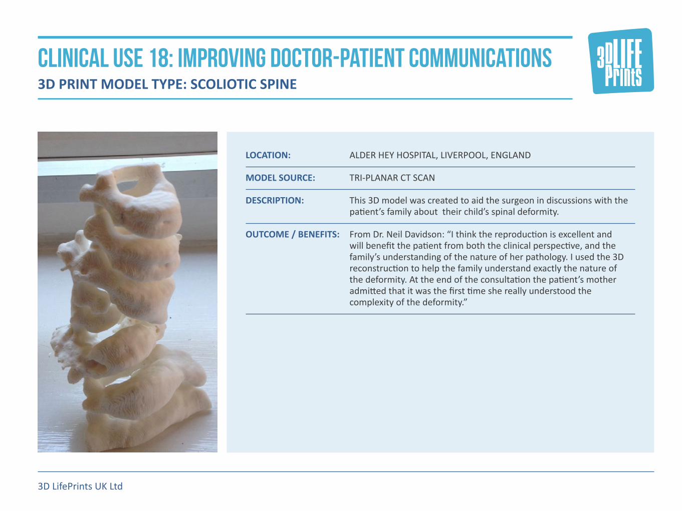
LOCATION: ALDER�HEY�HOSPITAL,�LIVERPOOL,�ENGLAND MODEL SOURCE: TRI-PLANAR CT SCAN DESCRIPTION: This�3D�model�was�created�to�aid�the�surgeon�in�discussions�with�the�
patient’s�family�about��their�child’s�spinal�deformity. OUTCOME / BENEFITS: From�Dr.�Neil�Davidson:�“I�think�the�reproduction�is�excellent�and�
will�benefit�the�patient�from�both�the�clinical�perspective,�and�the�family’s�understanding�of�the�nature�of�her�pathology.�I�used�the�3D�reconstruction�to�help�the�family�understand�exactly�the�nature�of� the�deformity.�At�the�end�of�the�consultation�the�patient’s�mother�admitted�that�it�was�the�first�time�she�really�understood�the�complexity�of�the�deformity.”
3D LifePrints UK Ltd
CLINICAL USE 18: IMPROVING DOCTOR-PATIENT COMMUNICATIONS 3D PRINT MODEL TYPE: SCOLIOTIC SPINE

LOCATION: ALDER�HEY�HOSPITAL,�LIVERPOOL,�ENGLAND MODEL SOURCE: EOS SCANNER DESCRIPTION: This�3D�printed�model�was�produced�from�a�stereo�radiographic�image�
(i.e�an�x-ray)�of�a�full�pelvis�and�scoliotic�spine.�The�EOS�scanner�creates�3D�images�from�x-ray�alone,�using�software�algorithms.
OUTCOME / BENEFITS: Due�to�the�nature�of�the�scan�the�model�was�not�used�for�surgical�
assessment,�rather�it�was�used�to�provide�reference�to�ascertain�the�load�bearing�capabilities�on�different�vertebrae.�Surgeons�found�the�tactile�model�to�be��helpful�in�planning�future�medical�treatment�for�the�patient.
3D LifePrints UK Ltd
CLINICAL USE 19: PATIENT CARE 3D PRINT MODEL TYPE: SCOLIOSIS SPINE
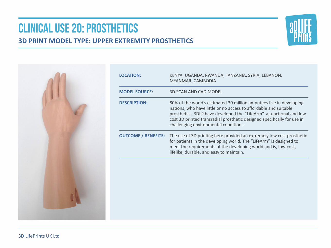
LOCATION: �KENYA,�UGANDA,�RWANDA,�TANZANIA,�SYRIA,�LEBANON,� MYANMAR,�CAMBODIA
MODEL SOURCE: 3D SCAN AND CAD MODEL DESCRIPTION: 80%�of�the�world’s�estimated�30�million�amputees�live�in�developing�
nations,�who�have�little�or�no�access�to�affordable�and�suitable�prosthetics.�3DLP�have�developed�the�“LifeArm”,�a�functional�and�low�cost�3D�printed�transradial�prosthetic�designed�specifically�for�use�in�challenging�environmental�conditions.
OUTCOME / BENEFITS: The�use�of�3D�printing�here�provided�an�extremely�low�cost�prosthetic�
for�patients�in�the�developing�world.�The�“LifeArm”�is�designed�to�meet�the�requirements�of�the�developing�world�and�is,�low-cost,�lifelike,�durable,�and�easy�to�maintain.
3D LifePrints UK Ltd
CLINICAL USE 20: PROSTHETICS 3D PRINT MODEL TYPE: UPPER EXTREMITY PROSTHETICS
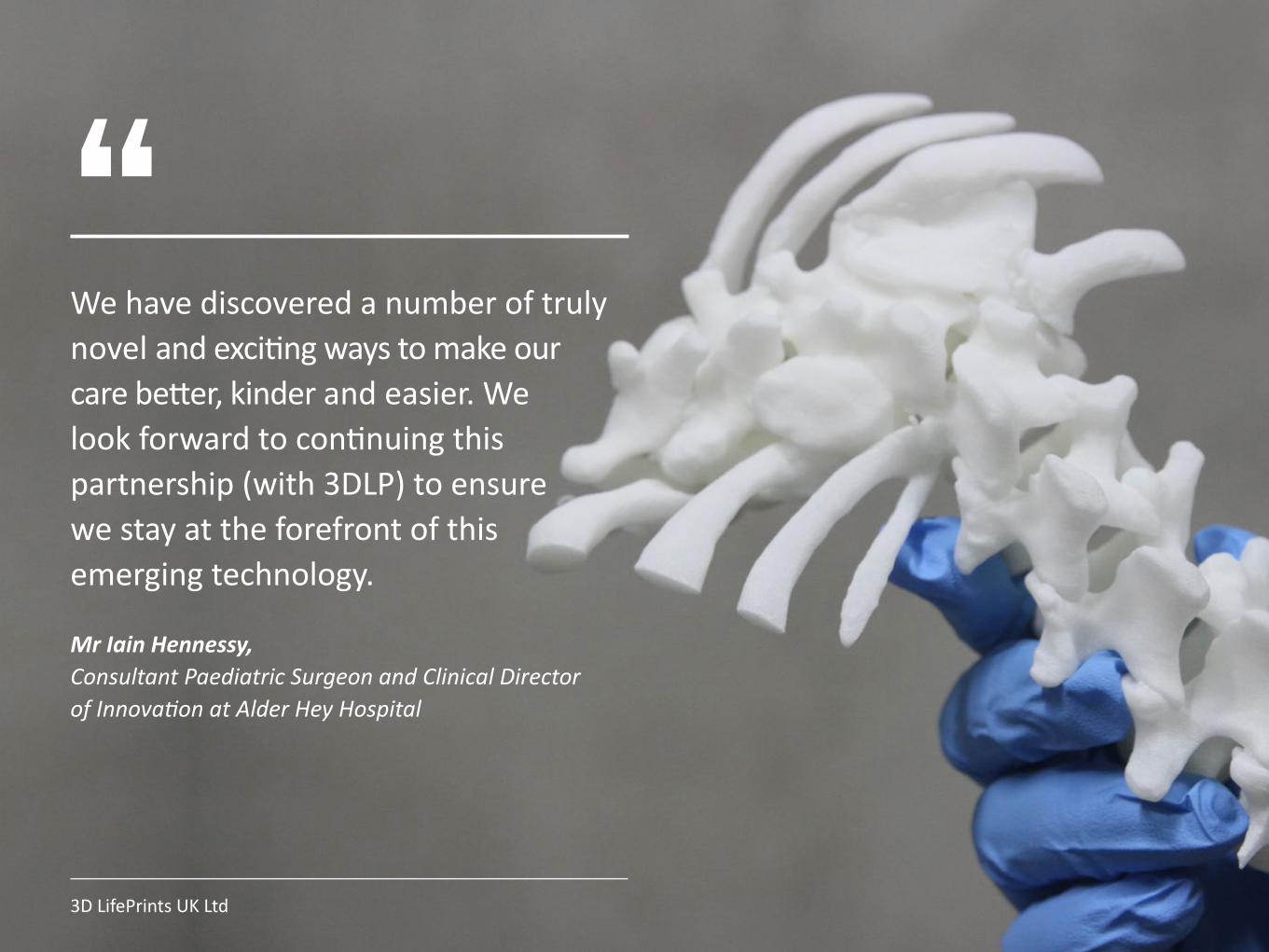
“We�have�discovered�a�number�of�truly�novel�and�exciting�ways�to�make�our� care�better,�kinder�and�easier.�We� look�forward�to�continuing�this�partnership�(with�3DLP)�to�ensure� we�stay�at�the�forefront�of�this� emerging�technology.
Mr Iain Hennessy, Consultant Paediatric Surgeon and Clinical Director of Innovation at Alder Hey Hospital
3D LifePrints UK Ltd
Please contact us below for any further information
Paul Fotheringham [email protected]
Henry Pinchbeck [email protected]
www.3dlifeprints.com
3D LifePrints UK Ltd