ew trends in scaffolds for tissue engineering
33
1 Recent progress in the fabrication techniques of 3D scaffolds for tissue engineering Mostafa Mabrouk 1,2,3 , Hanan H. Beherei 1 and Diganta B. Das 2 1 Refractories, Ceramics and Building materials Department, National Research Centre, 33El Bohouth st (former EL Tahrir st)-Dokki- Giza- Egypt P.O.12622 2 Department of Chemical Engineering, Loughborough University, Loughborough LE113TU, Leicestershire, UK 3 To whom correspondence should be addressed (e-mail: [email protected]; Tel/fax No.: 0201097302384) of interest: none Conflicts
Transcript of ew trends in scaffolds for tissue engineering

1
Recent progress in the fabrication techniques of 3D scaffolds for tissue
engineering
Mostafa Mabrouk1,2,3, Hanan H. Beherei1 and Diganta B. Das2
1Refractories, Ceramics and Building materials Department, National Research Centre, 33El Bohouth st (former EL Tahrir st)-Dokki- Giza- Egypt P.O.12622
2Department of Chemical Engineering, Loughborough University, Loughborough LE113TU, Leicestershire, UK
3To whom correspondence should be addressed (e-mail: [email protected]; Tel/fax No.: 0201097302384)
of interest: none Conflicts

2
Abstract
Significant advances have been made in the field of tissue engineering (TE), especially in the
synthesis of three-dimensional (3D) scaffolds for replacing damaged tissues and organs in
laboratory conditions. However, the gaps in knowledge in exploiting these techniques in
preclinical trials and beyond and, in particular, in practical scenarios (e.g., replacing real body
organs) have not been discussed well in the existing literature. Furthermore, it is observed in
the literature that while new techniques for the synthesis of 3D TE scaffold have been
developed, some of the earlier techniques are still being used. This implies that the
advantages offered by a more recent and advanced technique as compared to the earlier ones
are not obvious, and these should be discussed in detail. For example, one needs to be aware
of the reason, if any, behind the superiority of traditional electrospinning technique over
recent advances in 3D printing technique for the production of 3D scaffolds given the
popularity of the former over the latter, indicated by the number of publications in the
respective areas. Keeping these points in mind, this review aims to demonstrate the ongoing
trend in TE based on the scaffold fabrication techniques, focusing mostly, on the two most
widely used techniques, namely, electrospinning and 3D printing, with a special emphasis on
preclinical trials and beyond. In this context, the advantages, disadvantages, flexibilities and
limitations of the relevant techniques (electrospinner and 3D printer) are discussed. The paper
also critically analyzes the applicability, restrictions, and future demands of these techniques
in TE including their applications in generating whole body organs. It is concluded that
combining these knowledge gaps with the existing body of knowledge on the preparation of
laboratory scale 3D scaffolds, would deliver a much better understanding in the future for
scientists who are interested in these techniques.
Keywords: Tissue engineering; scaffold preparation techniques; electrospinning; 3D
printing.

3
1. Introduction
Tissue engineering (TE) approaches have demonstrated impressive results for the treatment
and substitution of damaged tissues and organs including skin, heart, and kidney tissues, in
addition to their potential to address some inherent bone defects [1-10]. When different
scientific fields, e.g., materials science, biology and engineering are combined together in an
interdisciplinary manner with a view to augment or regenerate malfunctioned human parts, it
promises to improve the success of the TE approaches [11-16]. For the TE systems to be
fruitful, the material utilized should generally be a mixture of scaffolds, growth factors, and
cells. They should also most certainly be able to replace the damaged tissue and have the
capacity to either work as the native tissue or mimic the native tissue [17-22].
Application of growth factors and exogenous materials with the sole aim of quickening and
enhancing the body's healing procedures could improve the tissue condition. Materials that
simulate the properties of extracellular matrix have been used for a long period time till now,
which accomplish more advantages other than supplying the physical structure [23–25].
Biomimetic materials can induce recovery of all, and they can be utilized for transport of
biomolecules, for example, growth factors that facilitate cells growth [18, 20, 24– 26]. At
first, it was thought that scaffolds are fundamental for cells’ physical support, the biomaterial
or scaffold can now be loaded with biological factors to facilitate tissue recovery [27–29].
Because of the diverse recovery limits of various tissues, some tissues do not demand cells
but rather simply the biomaterial and biological molecules. On the other hand, other tissues
have restricted recovery limits and demand the biomaterial, biomolecules, and cells for
recovery to happen. There are tissues and organs with constrained or no possibility for
recovery like ligament and cornea while others have great recovery capabilities such as the
liver and the lungs tissues [30, 31].
Based on most of the stated research articles dealing with TE, it is obvious that the scaffold
preparation techniques are important factors in this field. According to the data that have
been obtained from the Scopus (Fig. 1), it can be seen that up till now there are a number of
traditional scaffold fabrications techniques (e.g., freeze drying) which are still being utilized
(see, Fig. 1a). Also, it is obvious that there are an increasing number of publications recorded
for scaffolds that have been synthesized using electrospinning technique when compared with

4
those that have been synthesized using 3D printing technique (Fig.1b), even though both
techniques have originated at similar starting time.
It is also obvious that although there are different review articles that have discussed
scaffolds in TE, they have different focuses such as the nature of scaffold material and its
effect on the cell growth or healing of the damaged site [11]. Other review papers consider
issues on combining both inorganic and organic materials together and what is their effects
on the healing rate of hard tissue [15]. Reports have also been published that suggest that
these studies have handled the materials design and its effect on the regeneration of soft
tissues [21], while other reports the successful clinical trials for TE from a medicinal prospect
[22]. However, there is a lack of review papers that specifically highlights the effects of the
scaffold fabrication techniques and how they have progressed from the early beginning up till
now with the progress on TE as an engineering process, their limitations, challenges and
future aspects. Therefore, the main objective of the current review is to focus on the
implemented techniques in TE field for synthesis of scaffolds, stressing out the unclear points
in this specialty. In particular, this review focuses on discussing the two most widely used
techniques, namely, electrospinning and 3D printing. Finally, the paper discusses briefly the
applicability, restrictions, and future demands of these advanced techniques in the TE field
including their applications in generating whole body organs.
Fig.1. Demonstrates the data obtained from Scopus on scaffolds preparation method for a)
traditional techniques, b) advanced techniques and c) preclinical studies using biomaterial
scaffold.
2. Scaffold properties
Scaffolds support cells with the reasonable surviving conditions, ideal oxygen and nutrient
levels, successful supplement and waste transport in addition to providing adequate
mechanical support. The necessary environmental 3D conditions for cells in order to arrange

5
or to shape tissues are also attained using 3D scaffolds. The cells must produce their own
extracellular matrix (ECM) at the same time that is consumed for the scaffold biodegradation
to deliver identical 3D microstructures for the damaged sites. In fact, numerous parameters
influence the choice and properties of the scaffolds. Generally, the scaffolds should be
biocompatible, have suitable porosity, properties of surface and neutral pH, surface charge,
biodegradable, good physico-mechanical characteristics, and preferred for cells adhesion. A
few necessities have been distinguished as essential for the synthesis of scaffolds [32] as
summarized in Fig. 2. It is worth highlighting that the scaffold properties could be
manipulated through selection of the proper preparation technique. For example, if soft tissue
requires higher flexibility, the surface area and interconnected porosity of the scaffold are the
target parameters, the electro-spinner technique will be the perfect technique for this
application. On other hand, if hard tissue requires a load-bearing material with lesser porosity
and high mechanical properties as well as suitable bioactivity, 3D printing or freeze drying of
inorganic/organic composites will offer appropriate scaffolds.
Fig. 2. Schemes representing ideal porous scaffold properties
3. Scaffolds fabrication techniques
The scaffold fabrication techniques coupled with the choice of materials have a vital role in
the determining the scaffold properties for the target applications. There are two categories of
fabrication, namely, conventional and advanced methods of scaffolds fabrication. Table 1
represents the scaffold fabrication methods, discussed in this review.
6
Table 1 Scaffold preparation methods with examples (ex).
Preparation method Conventional Ref. Advanced Ref.
Solvent casting/ particulate leaching (SCPL) Ex: antibacterial bioactive glass/poly L-lactide composite scaffold.
Melt Molding
Ex: poly (lactic glycolic acid) scaffold.
Gas foaming
Ex: polylactic acid scaffold.
Thermally induced phase separation
Ex: Chitosan/bioactive glass scaffold.
Freeze drying
Ex: HEC/alginate/HA scaffolds.
[34]
[35]
[40]
[51]
[52]
[55]
Electrospinning -Classical electrospinning Ex: dexamethasone-loaded biphasic calcium phosphate nanoparticles/collagen scaffold. -Coaxial electrospinning Ex: PLLA/PCL/HA scaffold. Rapid prototyping - Stereolithography. - Selective laser sintering. - Inkjet 3D printer. - Multi-inkjet 3D printer Ex: calcium phosphate and collagen scaffolds. -3D bioplotter Ex: Si-hydroxyapatite / poly caprolactone /DMB.
[62]
[67]
[73]
[74]
[75]
[87]
[92]
3.1. Conventional scaffolds fabrication techniques
3.1.1. Solvent casting/particulate leaching (SCPL) The solvent casting/particulate leaching (SCPL) is the oldest technique used for scaffolds
fabrication. The concept of the method depends on the dispersion of porogens into a polymer
solution as shown in Fig. 3. The scaffolds obtained after solidification of the polymer and
dissolution of the porogens which left a high volume of pores resulting in a porous scaffold is
also known as the foam. Although the simplicity of this technique and its control over the

7
porosity of the fabricated scaffolds through the amount and the size of the porogens are
attractive, SCPL lacks the uniform arrangement of salt particles through the polymer solution.
This disadvantage is due to the dissimilar density of the salt and the polymer. Another
disadvantage is due to the way that polymer and the porogens are mixed where the porogens
are enveloped completely by the polymer solution that results in the difficulty of removing
salt particles with water. Therefore, the porosity of the scaffolds fabricated by this technique
is limited to 90% and the pore sizes range from 5 to 600 μm with irregular microstructures
[33, 34]. In addition, there are a few additional disadvantages which come typically from the
type of the polymer solvent used in which the scaffold material may be solubilized.
Typically, organic solvents are utilized, which are frequently lethal, which in turn would
prevent the cells seeding onto these scaffolds. Owing to these restrictions of this technique in
preparing scaffolds, only a few successful preclinical trials have been demonstrated in
mimicking tissues or organ including bone marrow and hard bone regeneration [35, 36].
Fig. 3. Demonstration of the solvent casting/particulate leaching (SCPL) technique, adapted
from [33].
3.1.2. Melt molding Melt molding technique is based on mixing of gelatin microspheres with a specific polymer
powder in a Teflon mold. The mixture is then heated above the polymer glass transition
temperature. The best polymer for this method is known to be poly (lactic glycolic acid)
(PLGA) due to its lower glass transition temperature. The heating allows the incorporation of
bioactive molecules through the gelatin resulted in a structural change which may affect its

8
aqueous solubility. The resulting composite is placed in water after heating in order to
dissolve the water-soluble microspheres resulting in a porous structure (Fig. 4.). This
technique possesses the same disadvantages of the SCPL technique, e.g., the porosity and the
pore size depend on the porogen concentration and diameter, respectively. In addition, the
variation in scaffold pore sizes is obtained by this technique. On the other hand, in the melt
molding technique the use of toxic solvents is avoided [37]. This technique if combined with
another one from traditional technique such as particle leaching could be with a potential for
tissue regeneration as earlier reported in a preclinical study [38].
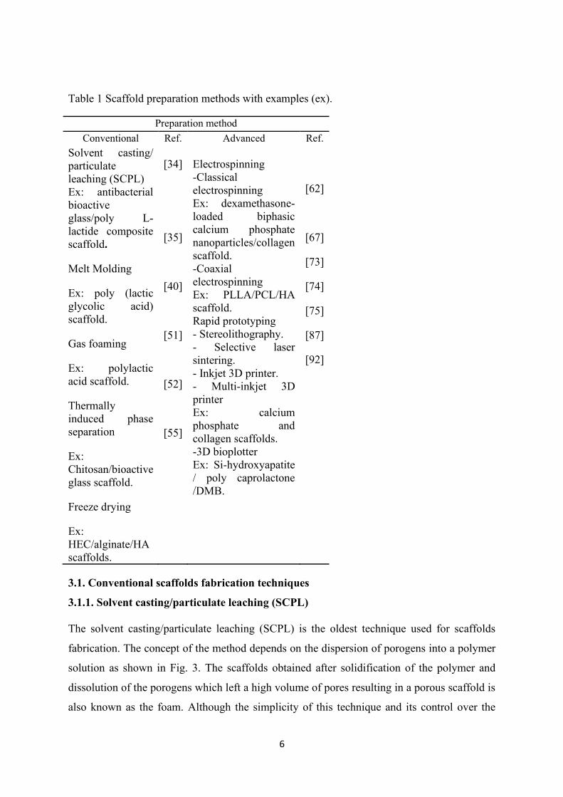
Fig.4. Schematic illustration showing the melt molding process utilized to fabricate PCL/BG-SA/Gel scaffold, adapted from [37]. 3.1.3. Gas foaming This technique is known as gas foaming technique because it uses a foaming agent (like
sodium bicarbonate) into the polymer. High-pressure gas is applied for disks of polymer in
order to prompt the nucleation and development of gas rise within the subjected substance
(Fig. 5). They are afterwards lyophilized to obtain scaffold characterized with pore diameters
of about 100 μm and percentage porosity up to 93% upon the gas liberation (like as N2 or
CO2) [39]. Gas is formed due to the action of the acidic aqueous solution with the foaming
agent and the liberation of gas producing a porous structure [39]. The disadvantage of this
method is that it increases the heterogeneity of the formed foam with a disordered porosity.
Here, the top of the foam has a highly porous structure, but the bottom layer exists with a
non-porous structure [40-41]. On the other hand, gas foaming procedure is reliable to create
9
solvents-free interconnected network polymeric scaffolds. Some of the prepared scaffolds by
this technique were introduced to preclinical stage with stem cells, which showed promising
results for bone tissue regeneration [42].
3.1.4. Thermally induced phase separation This procedure depends on a thermal energy difference that initiates a detachment of a
polymer solution with a homogeneous network into a multi-stage system domain using a
quench method. In the beginning, a high temperature is utilized to a solubilizied polymer
(either in phenol or naphthalene), trailed by scattering of naturally dynamic particle in these
solutions. Upon the temperature decrease the segregation is prompted, the resulted solution
isolates into polymer-free phase and solvent-free phase either by strong fluid de-blending or
fluid stage division component. This step is then followed by extraction, dissipation, and
sublimation in order to eliminate the used solvent, thus, resulting in 3D polymer matrix
impregnated with bioactive particles coordinated within this porous structure. The
microstructural characteristics of the achieved scaffolds are adjusted by controlling several
factors including the polymer characteristics, solvent, the centralization of the solution of
polymer and temperature at which phases are detached. Two kinds of this technique are
utilized as shown in Fig. 6, solid-liquid and liquid-liquid separations. The advantage of this
method is that it can function without much of the stretch consolidates with other
manufacture innovation as well as great mechanical properties obtained for the three-
dimensional structures with controlled pore morphology [43, 44].
Fig.5. Scaffold prepared by gas foaming technique, adapted from [41].
For example, mixing solid naphthalene and poly-lactic acid (PLLA) in a flask, and then
heating them with stirring provides a homogenous solution. Afterword, the solution is casted
into a cooled mold in order to form polymer-poor and polymer-rich phases. Then,

10
naphthalene is eliminated to produce the porous structure through a vacuum drying stage. The
kinetics of the phase separation controls the morphology and pore distribution of the foam. In
contrast with the previously mentioned techniques, this technique is characterized by the
relative uniformity of the distribution of the pore and the resulting pore diameters range from
50-100 μm. The polymer concentration affects the porosity percentage that can reach up to
87%. However, the limitation of this technique rises from the organic solvents usage that
might have a toxic effect on cells. There is some evidence of recent study which has used this
technique to prepare phase separated nanofibrous vascular scaffolds, modified with heparin
and SDF-1α confirming its applicability as a small diameter vascular graft in the preclinical
level [45].
Fig.6. Schematic diagrams of a) liquid-liquid phase and b) solid-liquid phase separation, adapted from [44].
11
3.1.5. Freeze drying technique All of the above-mentioned techniques have been used in a small number of publications (see
Fig. 1a) due to several disadvantages arising from a limited number of applicable materials to
those techniques using organic solvent and their lethal effects on cells and non-homogenous
microstructure which are not suitable for cells growth. This was the trend for all of the old
techniques except for freeze drying technique. In this technique the porosity of the scaffolds
is produced via the lyophilization, where the ice crystals of the solvent are formed after
leaving the composite in the freezer are supplemented by using the freeze-drying technique
(Fig.7) [46]. The ice crystals serve as porogens that control the pore sizes and this can be
achieved by controlling the freezing temperature and the polymer weight ratio in the solution.
This procedure till now has been very beneficial in the design of 3D-framworks in the room
to freeze temperatures and without using organic solvent. It is a lack of dehydration strategy
dependent on the evacuation, thus solidified structures are subjected to sublimation process,
in which, water particles are evaporated under vacuum, at low pressure, prompting 3D-
scaffold in the dried form [47-50]. The porous sponge structure of the produced scaffolds by
this technique is preserved even after the immersion in simulated body fluid. Recent trials
were conducted using lyophilization technique to prepare various 3D-structures from natural
or synthetic polymers and their composite with inorganic biomaterials such as hydroxy
apatite and bioactive glass [51-55]. These scaffolds showed promising results when they were
tested against normal cells and in vivo in rat models.
.
Fig.7. Scaffold prepared by lyophilization technique [45]. Bioceramics Development and Applications - Open Access Journals.
3.2. Advanced scaffolds fabrication techniques
The conventional strategies for creating 3D-permeable frameworks in the applications of TE
face some serious limitations including difficulties to adjust the microstructure, morphology,
connectivity and diameters of the pores as well as the total scaffold porosity. A three-
dimensional structure with pore sizes suitable for vascularization of cells become an

12
important need [56]. Therefore, new advanced techniques have arisen, such as
electrospinning [57] and rapid prototyping (RP) [58, 59]. In the following sections, these
advanced strategies will be discussed on several bases including applicability of these
systems for TE and the suitability of materials, scaffold development stages and, their
advantages and restrictions.
3.2.1. Electrospinning
Electrospinning strategy is a one of the delicate techniques where a 3D-structure is
synthesized utilizing electrostatic powers which create fibers from polymer solutions
characterized with small fiber diameters in the range of nanometer to micrometer with higher
surface area compared to those created by classical spinning forms. Over 200 natural
polymers and composites have been utilized in the electrospinning, for example, silk fibroin,
chitosan, gelatin, collagen, etc. and plenty of synthetic polymers including (PVA, PVP,
PLLA, PCL, etc.) are additionally utilized in this system [60]. To produce the electrospun
fibers a high DC voltage ranging between 10 to 40 kV is a must, and this could be done under
the ambient conditions.
Electrospinning setups are generally classified into two categories: horizontal and vertical
(Fig. 8a). Essentially, an electrospinning setup comprises of three noteworthy parts: a high
voltage control supply, a spinneret (e.g., a needle with metal tip) and a metallic collector for
the electrospun fibers. In order to prepare a homogeneous solution for electrospinning, the
polymers should be ideally dissolved in a carefully selected solvent. Afterwards, the obtained
solution is brought into the metal nozzle and subjected to a high voltage source; this in turn
accelerates the polymer solutions through the metal tip towards the metal collector of
opposite polarity at a steady stream rate [61]. Nevertheless, a few polymers may emanate an
undesirable or unsafe smell, so the procedures ought to be led inside closed enclosures with
ventilation aperture.
Owing to the use of high voltage in this procedure, the polymer solution is consistently
charged, and this causes a repulsive power inside the system [61]. The repulsive electrical
powers defeat the surface pressure powers only when the connected electric field reaches a
critical value, causing a charged stream of the solution to spine from the tip of the Taylor
cone and moves towards the collector charged cathode. Significant favorable circumstances

13
of this system are that it is adaptable, non-obtrusive and temperature independent fibers
production method.
It is obvious that this system has recorded an exceptional increment in research and business
considerations in various domains within the last ten years as it can provide very suitable
substrates for cell growth and consequent tissue development. With the extension of this
innovation, a few research teams have grown progressively advanced scaffolds that can
create increasingly complicated mats in a progressively controlled and productive way [62,
63]. Table 2 summarizes the electrospinning elements, factors controlling fibers diameter and
advantage of the electrospinning system for the preparation of scaffolds for TE.
Table 2 Electrospinning elements, fiber diameter controlling factors and advantages.
Electrospinning elements Fiber diameter controlling factors Advantages High voltage control supply. Polymer concentration and solution
viscosity An adaptable system for production 3D scaffolds.
Extrusion pump connected to syringe with metal tip.
Polymer surface tension Suitable for more than 200 natural and synthetic polymers
Metal collector (Stationary or movable)
Flow rate Temperature independent system and suitable for loading of active molecules
Chamber (enclosed system) Distance between needle tip and collector
High surface to volume ratio
These developments incorporate coaxial electrospinning, in which the spinner fibers are not
only one phase of polymer but also could be in a core shell form nanofiber with complicated
microstructures, mainly for the production of core shell staggered structures by using more
than one pump system through coaxial vessels. Thus, this expands the yield production of the
fibers and decreases the common electrostatic repulsion in this procedure (Fig. 8b) [64, 65].
Numerous parameters influence the fibers microstructural properties, such as, (i) polymer
type, the solution viscosity, the polymer solution surface tension, (ii) the pumping rate, the
distance between the collector and the needle tip, size of the needle tip, and (iii) the
encompassing environment (humidity and temperature) [66].
Impressively, great surface/volume values, adjusted porosity, flexibility to adjust to a wide
assortment of sizes and shapes and the capacity to control the nanofiber structure are some of
the benefits of the electro-spinning system. The high surface/volume ratios allow the loading
of active molecules as well as cell propagation followed by tissue regeneration for the
damaged site [67]. Impressive hydrophilicity and biocompatibility properties are obtained
14
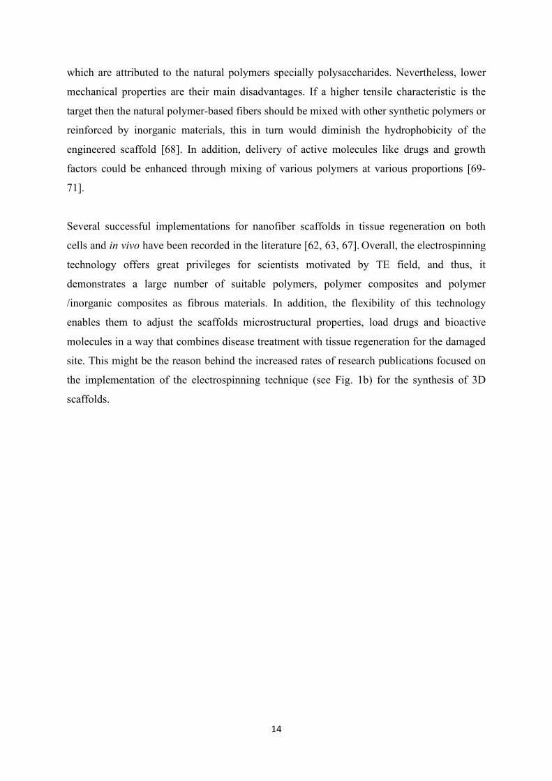
which are attributed to the natural polymers specially polysaccharides. Nevertheless, lower
mechanical properties are their main disadvantages. If a higher tensile characteristic is the
target then the natural polymer-based fibers should be mixed with other synthetic polymers or
reinforced by inorganic materials, this in turn would diminish the hydrophobicity of the
engineered scaffold [68]. In addition, delivery of active molecules like drugs and growth
factors could be enhanced through mixing of various polymers at various proportions [69-
71].
Several successful implementations for nanofiber scaffolds in tissue regeneration on both
cells and in vivo have been recorded in the literature [62, 63, 67]. Overall, the electrospinning
technology offers great privileges for scientists motivated by TE field, and thus, it
demonstrates a large number of suitable polymers, polymer composites and polymer
/inorganic composites as fibrous materials. In addition, the flexibility of this technology
enables them to adjust the scaffolds microstructural properties, load drugs and bioactive
molecules in a way that combines disease treatment with tissue regeneration for the damaged
site. This might be the reason behind the increased rates of research publications focused on
the implementation of the electrospinning technique (see Fig. 1b) for the synthesis of 3D
scaffolds.

15
Fig. 8. Different types of electro-spinning a) classical electrospinner reproduced from 6th
international conference Bucharest, Romania [70] and b) co-axial electro-spinneret
reproduced from PeerJ open access journal [71].
3.2.2. Rapid prototyping
Rapid prototyping and additive manufacturing, also known as 3D-printing, is a strategy that
was first portrayed in the year 1986. The classical name of rapid prototyping strategy is
stereolithography (SL) (Fig. 9a) in which thin layers of materials are deposited over each
other by hardening the layers utilizing ultraviolet waves [72-74]. Two-photon assimilation by
photopolymerization for planning of 3D-structures was initiated from this procedure where
blended layers of a photo-initiator agent and a monomer gel are cured and crosslinked by a
laser source. The photo-initiator contains two dynamic photons that are activated by a laser
light source; thus, the polymerization process takes place [74, 75]. Similarity, another
strategy was created in particular Selective Laser Sintering (SLS) (Fig. 9b) to make 3D-
structures. SLS utilizes a powerful laser to sinter polymer powders to create a 3D-structures
[72, 76].

16
In spite of the fact that rapid prototyping is effective in producing 3D-complexes, it possesses
some genuine impediments. Designers have found that the goals of the printing are course
reliant, for example, the 3D-structures delivered utilizing this strategy are observed to be
structurally stronger on one side and more fragile on the other side depending upon which
side is parallel to the heating from the laser. The second real inconvenience is the warm
warpage that arises from the utilization of high temperatures, thus, this would restrict the use
of biological contents such as proteins and cells owing to their thermo-sensitivity in the
manufacture of 3D-structures [77, 78]. Not many preclinical trials are recorded for scaffold
produced using this technique due to its limitations; however, cell attachment was confirmed
for 3D scaffolds from polylactide based polymer printed with the help of this technique [75].
In addition, fused deposition modeling (FDM) [79] was invented that possessed many
limitations due to the unavailability of different nozzles with different diameters which in turn
led to restricted design of the scaffold architecture. Another rapid prototyping technique that
was generally based on solvent-based strategies was developed; however, many limitations in
terms of longer fabrication time and weaker mechanical scaffold properties were observed for
this technique [80].
Upon the innovation of the first inkjet-based strategy in the year 2000, proteins and
endothelial cells were printed within the 3D-structures. For this purpose, the nozzles of a
commercial inkjet printer were modified so as to allow the researchers to change the nozzle
in the required axial (x-) and lateral (y-) positions. In medication conveyance, inkjet printers
are most usually implemented via this method. Like the inkjet printers, these printing
scaffolds store little beads of the medication on fluid film that exemplify the medication bead
to shape microparticles which can be utilized for medication conveyance. As this strategy
requires no high temperature, the loading of thermosensitive biomolecules as well as cells
was much applicable when compared to the SLS [81, 82]. In this technique a head of inkjet
printer persistently splashes the medication, cells or other bioactive particles, plus binder onto
a bed of polymer powder till a 3D-platform is shaped. In spite of the fact that this method
helped in the manufacture of 3D-structures with acceptable mechanical strength, it is not
applicable to design of complete 3D-tissue substitutes containing cells or proteins owing to
the utilization of binders that in turn diminish the final porosity of the obtained scaffolds [81-
84].

17
Numerous polymers, for example, dextrin, maltodextrin, saccharose and different saps are
utilized as binders. These fasteners normally break down in the dissolvable utilized for
bioprinting and after taking back to room temperature, it solidifies, thus resulting in higher
compressive bearing for the printed polymer and/or polymer composite scaffolds. These
binders are sugar derivative polymers that can be effectively degraded in the circulatory
systems to give glucose [84–86]. Polysaccharide polymers were pyrolyzed to carbon upon
applying of relative high temperatures [87, 88]. Polymers, for example, teflon
(polytetrafluoroethylene) and polypropylene may likewise be utilized as binders alongside
natural or semi-manufactured materials. For example, collagen as they give enhanced
compressive strength to the 3D-structures and furthermore assistance in avoiding corruption
and maintaining of the platform while utilizing acidic solvents. Presence of such binders in
the printed 3D-srtuctures results in semi-porosity-free. Furthermore, the pore diameters of the
platform cannot be adjusted through the printing process and this counts as a drawback for
the inkjet printing [89]. Low volume launch of ultrasound waves onto the deposition medium
followed by ejection of an acoustic droplet (Fig. 10) are initiated from inkjet printing [90–
93].
Fig.9. Schematic representation of a) stereolithography (SL) system and b) selective laser sintering (SLS) technique, adapted from [73].
Fig.10. Scheme demonstrates acoustic droplet ejection 3D printing process, adapted from
[73].

18
In vivo experiments were conducted with numerous 3D-printed polymeric and ceramic
structures in order to evaluate their ability for cytocompatibility and recovery. HAp/PCL and
demineralized bone scaffolds substituted with silicon were designed employing rapid
prototyping instrument. They possessed abilities for great osseointegration, osteoinductivity
and bone recovery when applied to rabbit models [94, 95]. Also, an improved bone recovery
in rat models was confirmed for 3D-structures made of resorbable dicalcium phosphate
(DCP) and prepared utilizing the SLS technique [95]. DCP 3D-structures obtained with
different techniques of additive manufacturing (e.g., tank polymerization, powder bed fusion,
material extrusion, and binder streaming additionally), which have demonstrated osteogenic
potential on the in vitro basis, however, they should be further approved utilizing in vivo
examinations [96]. Silicate and phosphate families 3D-printed scaffolds showed great success
in the recovery of bone defects, especially when they are complied with DNA [97].
For tissue recovery, the most preferred biomaterials are hydrogels owing to their
customizable hydrating capacity and mechanical characteristics, thus, enabling them to mimic
the natural tissue. PEG, gelatin and fibrin hydrogels as well as their derivatives were the most
common hydrogels utilized from natural and synthetic polymers in 3D printing loaded with
cells for bone recovery applications. Extrusion technique seems to be the most well-known
strategy utilized for cell-loaded hydrogel blends [98, 99]. In this case, a syringe pump loaded
with the solvent moves in the z-axis is utilized and the printing stage moves along the y-axis
for the cell-based printing [98]. Applying voltage on the inkjet head can assist in controlling
the speed of the solvent splashing and solvent droplet speed can be adjusted by applying
differential voltage as shown in Fig. 11 [99]. The correct choice and optimization of these
factors are very essential in the 3D printing procedure as they decide the pore size and
homogeneity of the 3D-structures [99, 100]. Engineered polymers and hydrogels originated
from PCL and PLGA are very common in producing of 3D-framworks utilizing rapid
prototyping, have demonstrated great recovery for rabbit tibias bone [99].
However, many restrictions are originated from the 3D printed techniques and this might be
the reason behind the decrease in the number of the publications recorded for scaffold
synthesis by 3D printing technique when compared to those produced by the electrospinning
technique. These restrictions are represented in limited precursor materials that are applicable
for the 3D printing technology, long time for optimization of the design parameters as well as
preserving the cell viability during and after the printing. All of these reasons were enough to
19
hold back the progress of the 3D printing technique; however, a lot of work is conducted
right now in this regard which might result in the change of this rate. Nowadays, researches
are conducted to advance materials to prepare platforms according to the patient-needs for
bone recovery utilizing the computerized tomography (CT) scan of patient to print big hard
tissue replacers. Utilizing open source coding programming, the CT scans are extrapolated,
and 3D printed to shape the bone substitutions that are anatomically like the scan with
comparable microstructural properties. The platform could then be covered or loaded up with
the undifferentiated cells from the patient and embedded again into the patient's body,
consequently enhancing bone recovery and disposing of any opportunity of dismissal [102–
108]. Presently, the 3D-printed polymer platforms are being evaluated and directed for use as
viable medication conveyance frameworks and tissue substitutes in the field of medical
procedure and medicine utilizing natural polymers, for example, chitosan and alginate.
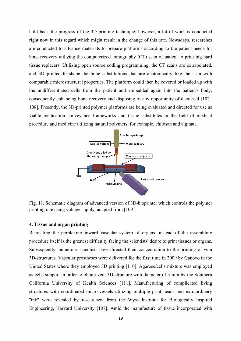
Fig. 11. Schematic diagram of advanced version of 3D-bioprinter which controls the polymer printing rate using voltage supply, adapted from [109].
4. Tissue and organ printing
Recreating the perplexing inward vascular system of organs, instead of the assembling
procedure itself is the greatest difficulty facing the scientists' desire to print tissues or organs.
Subsequently, numerous scientists have directed their concentration to the printing of vein
3D-structures. Vascular prostheses were delivered for the first time in 2009 by Ganovo in the
United States where they employed 3D printing [110]. Agarose/cells mixture was employed
as cells support in order to obtain vein 3D-structure with diameter of 3 mm by the Southern
California University of Health Sciences [111]. Manufacturing of complicated living
structures with coordinated micro-vessels utilizing multiple print heads and extraordinary
''ink" were revealed by researchers from the Wyss Institute for Biologically Inspired
Engineering, Harvard University [107]. Amid the manufacture of tissue incorporated with

20
veins, different kinds of cells, and extracellular framework, a 3D bioprinter with a few freely
controlled print heads was planned to develop these heterogeneous 3D-structures with
various materials. These scaffolds constituents should be printed exactly with the same
characteristics and at the same time. Gelatin methacrylate (GelMA) is utilized as a cell
support matrix, while poly (dimethyl siloxane) (PDMS) pigmented with various fluorophores
is utilized to label distinctive biomaterials [112]. In fact, there are effective instances of
printing tissue or organs by local and remote analysts.
Complete cellularized skin substitute loaded with fibroblasts and keratinocytes in a typical
3D spatial model was produced by using bioprinting associated with laser aid [113], where,
comprising of fibroblasts labeled in red and keratinocytes labeled in green on the surface of
MatriDerm®, that was used to treat skin wound in the mouse. Untreated part of the mouse
skin was used as a negative control for comparison. After testing the skin builds by utilizing
the dorsal skin overlay chamber in naked mice, the printed cells stayed dynamic, kept
multiplying, and generated the ECM. A bioprinting procedure for various layers of cells is an
essential for making complex tissue with bigger size. Mannoor et al. [114] utilized hydrogel
loaded with chondrocyte cells to print a bionic ear. In addition, in the anatomic geometry of a
human ear and cochlea-formed terminals silver nanoparticles were loaded (Fig. 12(a, b)). The
printed bionic ear is recommended to be utilized as a sound-related detecting device of radio
frequencies than the human ear. Working cardiovascular valve was introduced by a scientist
from Cornell University which used undeveloped cells and biopolymer materials (Fig. 12(c)),
and the loaded cells were transformed bit by bit into mature cells.
It is worthy to highlight that 3D printed organs for clinical applications are presently
available. For example, the world's first effective 3D printed transplantation of human organ
was done by Michigan University, where they had implanted a synthetic trachea into the
windpipe of a baby with a birth deformity to help develop baby’s breathing and speech (Fig.
12(d)). Other successful trials for the 3D bioprinting of tissue or organs were reported for
several damaged sites at the preclinical stage, among those trails the orthopedic tissue
regenerations for different sites such as cartilage [115], complete tooth [116], tooth root and
support [117]. Soft tissues printing trials were also reported for skin [118] and few other
internal organs, such as, nerve [119], tendon [120], and heart [122].

21
Fig.12. Illustrations for (a) bionic ear upon 3D printing; (b) bionic ear during in vitro culturing; (c) cardiac valve and (d) artificial trachea [114].
5. 3D pringitng restrictions
The utilization of 3D printing innovation for restorative implementations will unravel the
contributor deficiency issue for organ transplantations. In this way a rising and quickly
created interdisciplinary field will develop that firmly incorporates science of material, cell
biology, and clinical science. In spite of the fact that cell loading in 3D structure is possible,
considerable work still needs to be conducted to accomplish the perfect mimicry of the
original tissue [123]. It is hard to mimic the structure and organ capacity of the ECM in vitro,
as it is a complicated framework with different constituents. This issue is overcome by the
consolidation of conciliatory material during the development of a platform. This material
gives mechanical help to printing material by occupying the void spaces. These materials are
expelled by post preparing after the creation. Impediment initiated by the structure cause
brokenness of material due to the poor productivity of CAD plan into the machine [124].
Existing innovations that fundamentally utilize hydrogels loaded with cells cannot resolve the
issues of cell nourishment and supplying of oxygen. For bigger scaffolds, an adequate
number of cells cannot be supplied at present. Contrasted with cells that adhere to the
scaffold outer surface, preprophase cells do not get a satisfactory supply of supplements. This
leads to non-homogenous distribution to the cells within the 3D matrix [125]. Additionally,
different restrictions that incorporate cell survival, advancement, separation, and propagation,
should be illuminated for the further improvement of 3D structures printed, tissues, and
organs.

22
Materials confinements likewise occur based on the inherent properties of utilized materials.
A significant number of the metal materials that are commonly utilized for permanent
implants have a high flexible modulus, which frequently prompts a versatile mismatch
between the surrounding environments and implant. Printed biodegradable 3D structures are
commonly created from characteristic polymers with great biocompatibility, however, they
possess low mechanical strength, for example, sodium alginate, collagen, and different
hydrogels. Moreover, there are no global measure for picking biomaterials for 3D scaffolds.
Hence, just engineered assessments can be made depending on structure, work, clinical
impacts, and different features, instead of assessments dependent on solid markers and
adequate exploratory proof. Nowadays, scientists have developed autonomous research and
basic systems, which have mostly constrained the improvement of this innovation for medical
applications. In addition, ethical issues and cells stimulation are necessary to be considered.
In this manner, accomplishing the real usage of 3D printing for restorative applications will
require prolonged range endeavors [126–128].
Conclusions
After exploring the conducted research work related to the tissue regeneration applications, it
was confirmed that TE could be considered as one of the chosen ways to deal with the
ordinary tissue regeneration problems, based on both laboratory and preclinical studies but
not yet on the basis of clinical trials. The production of scaffold as the essential subunit that
supports mechanical properties, cell adhering, proliferation, and specialization, could be
achieved by manipulating the major key factors like material choice and 3D-structure
creation method that are suitable for specific organ damage or defect. Though the use of some
traditional techniques on both lab and preclinical bases (freeze drying) still apply for this
purpose when one compares it with advanced techniques, the demands for sophisticated TE
field hold great promises for the advanced techniques as could be noted from literature.
Electrospinning technique is of interest for the production of 3D scaffolds owing to its
tunable parameters and, thus, it has grown fast in both industrial and biomedical fields. On
the other hand, 3D printing possesses slow evolution rate when it is compared to the
electrospinning technique as it is a complicated process with multiple engineering factors
with no respect to the biological systems. Therefore, in the current days, much advancement
was considered to develop the 3D printer system in a compatible manner with tissue and
organ conditions. Accordingly, both electrospinning and 3D printing technologies are guiding
the ways for scientists to develop 3D-structures with cutting edge methods utilizing diverse

23
range of biomaterials which are now their focal point of research endeavors in the field of
TE.
Acknowledgment: The authors wish to express their sincere appreciation to the Academy of Medical Sciences, London, UK for supporting financially an academic visit of Dr Mostafa Mabrouk to Loughborough University through a Daniel Turnberg UK/Middle East Travel Fellowship. References
[1]. S. Giwa, J. K. Lewis, L. Alvarez et al., “The promise of organ and tissue preservation
to transform medicine,” Nature Biotechnology, 35 (6) (2017) 530–542.
[2]. B. Jones and M. Bes, “Keeping kidneys,” Bulletin of the World Health Organization,
90 (10) (2012) 718-719.
[3]. M. Colvin, J. M. Smith, M. A. Skeans et al., “OPTN/SRTR 2015 annual data report:
heart,” American Journal of Transplantation, 17 (1) (2017) 286–356.
[4]. A. Hart, J. M. Smith, M. A. Skeans et al., “OPTN/SRTR 2015 annual data report:
kidney,” American Journal of Transplantation, 17 (1) (2017) 21–116.
[5]. A. K. Israni, D. Zaun, C. Bolch et al., “OPTN/SRTR 2015 annual data report:
deceased organ donation,” American Journal of Transplantation, 17 (1) (2017) 503– 542.
[6]. B. L. Kasiske, S. K. Asrani, M. A. Dew et al., “The living donor collective: a
scientific registry for living donors,” American journal of transplantation, 17 (12) (2017)
3040–3048.
[7]. S. Nagral, M. Hussain, S. A. Nayeem, R. Dias, S. A. Enam, and S. Nundy, “Unmet
need for surgery in south asia,” BMJ, 357 (2017) article j1423.
[8]. A. M. Bailey, M. Mendicino, and P. Au, “An FDA perspective on preclinical
development of cell-based regenerative medicine products,” Nature Biotechnology, 32 (8)
(2014) 721–723.
[9]. P. S. Knoepfler, “From bench to FDA to bedside: us regulatory trends for new stem
cell therapies,” Advanced Drug Delivery Reviews, 82-83 (2015) 192–196.
[10]. X. L. Tang, Q. Li, G. Rokosh et al., “Long-term outcome of administration of c-
kitPOS cardiac progenitor cells after acute myocardial infarction: transplanted cells do not
become cardiomyocytes, but structural and functional improvement and proliferation of
endogenous cells persist for at least one year,” Circulation Research, 118 (7) (2016) 1091–
1105.
[11]. S. Bose, M. Roy, and A. Bandyopadhyay. Recent advances in bone tissue engineering
scaffolds. Trends in biotechnology, 30(10)(2012) 546-554.

24
[12]. L. L. Hench. The future of bioactive ceramics. Journal of Materials Science: Materials
in Medicine, 26(2) (2015) 86.
[13]. R. Detsch, S. Alles, J. Hum, P. Westenberger, F. Sieker, D. Heusinger, and A. R.
Boccaccini. Osteogenic differentiation of umbilical cord and adipose derived stem cells
onto highly porous 45S5 Bioglass®‐based scaffolds. Journal of Biomedical Materials
Research Part A, 103(3) (2015) 1029-1037.
[14]. R. El-Gendy, J. Kirkham, P. J. Newby, Y. Mohanram, A. R. Boccaccini, and X. B. Yang.
Investigating the vascularization of tissue-engineered bone constructs using dental pulp
cells and 45S5 Bioglass® scaffolds. Tissue Engineering Part A, 21(13-14) (2015) 2034-2043.
[15]. A. Philippart, A. R. Boccaccini, C. Fleck, D. W. Schubert, and J. A. Roether. Toughening
and functionalization of bioactive ceramic and glass bone scaffolds by biopolymer coatings
and infiltration: a review of the last 5 years. Expert review of medical devices, 12(1) (2015)
93-111.
[16]. T., Dvir, B. P. Timko, D. S. Kohane, and R. Langer. Nanotechnological strategies for
engineering complex tissues. Nature nanotechnology, 6(1) (2011) 13.
[17]. G. Gao and X. Cui, “Three-dimensional bioprinting in tissue engineering and
regenerative medicine,” Biotechnology Letters, 38 (2)(2016) 203–211.
[18]. X. Guan, M. Avci-Adali, E. Alarcin et al., “Development of hydrogels for
regenerative engineering,” Biotechnology Journal, 12 (5)(2017) 1-19.
[19]. M. J. Kraeutler, J. W. Belk, J. M. Purcell, and E. C. McCarty, “Microfracture versus
autologous chondrocyte implantation for articular cartilage lesions in the knee: a systematic
review of 5-year outcomes,” The American Journal of Sports Medicine, 46 (4) (2017) 995–
999.
[20]. H. Mistry, M. Connock, J. Pink et al., “Autologous chondrocyte implantation in the
knee: systematic review and economic evaluation,” Health Technology Assessment, 21(6)
(2017) 1–294.
[21]. A. G. Guex, F. M. Kocher, G. Fortunato et al., “Fine-tuning of substrate architecture
and surface chemistry promotes muscle tissue development,” Acta Biomaterialia, 8 (4)
(2012) 1481–1489.
[22]. A. S. Mao and D. J. Mooney, “Regenerative medicine: current therapies and future
directions,” Proceedings of the National Academy of Sciences of the United States of
America, 112 (47)(2015)14452–14459.

25
[23]. M. Alves da Silva, A. Martins, A. R. Costa-Pinto et al., “Electrospun nanofibrous
meshes cultured with Wharton’s jelly stem cell: an alternative for cartilage regeneration,
without the need of growth factors,” Biotechnology Journal, 12 (12) (2017) 1-9.
[24]. A. I. Goncalves, M. T. Rodrigues, and M. E. Gomes, “Tissue engineered magnetic
cell sheet patches for advanced strategies in tendon regeneration,” Acta Biomaterialia, 63
(2017) 110–122.
[25]. S. Pina, R. F. Canadas, G. Jimenez et al., “Biofunctional ionic doped calcium
phosphates: silk fibroin composites for bone tissue engineering scaffolding,” Cells, Tissues,
Organs, 204 (3-4) (2017) 150–163.
[26]. L. Drowley, C. Koonce, S. Peel et al., “Human induced pluripotent stem cell-derived
cardiac progenitor cells in phenotypic screening: a transforming growth factor-β type 1
receptor kinase inhibitor induces efficient cardiac differentiation,” Stem Cells Translational
Medicine, 5 (2) (2016) 164– 174.
[27]. K. Dzobo, T. Turnley, A. Wishart et al., “Fibroblast-derived extracellular matrix
induces chondrogenic differentiation in human adipose-derived mesenchymal stromal/stem
cells in vitro,” International Journal of Molecular Sciences, 17(8)(2016) 1-20.
[28]. N. D. Evans, E. Gentleman, X. Chen, C. J. Roberts, J. M. Polak, and M. M. Stevens,
“Extracellular matrix-mediated osteogenic differentiation of murine embryonic stem cells,”
Biomaterials, 31 (12) (2010) 3244–3252.
[29]. K. Sadtler, A. Singh, M. T. Wolf, X. Wang, D. M. Pardoll, and J. H. Elisseeff,
“Design, clinical translation and immunological response of biomaterials in regenerative
medicine,” Nature Reviews Materials, 1(7) 201616040.
[30]. A. Atala, “Regenerative medicine strategies,” Journal of PediatricSurgery, 47 (1)
(2012) 17–28.
[31]. D. N. Kotton and E. E. Morrisey, “Lung regeneration: mechanisms,applications and
emerging stem cell populations,”Nature Medicine, 20 (8) (2014) 822–832.
[32]. S.G. Kumbhar and S.H. Pawar, Self-Functionalized, Oppositely Charged Chitosan-
Alginate Scaffolds for Biomedical Applications, Biotechnol Ind J. 13(2) (2017)130.
[33]. D. Bartis, and J. Pongracz, Three dimensional tissue cultures and tissue engineering.
(2011) 10.13140/2.1.2793.5047.
[34]. A. M. El-Kady, R. A. Rizk, B. M. Abd El-Hady, M. W. Shafaa, M. M. Ahmed,
Characterization, and antibacterial properties of novel silver releasing nanocomposite
scaffolds fabricated by the gas foaming/salt-leaching technique. Journal of Genetic
Engineering and Biotechnology, 10 (2012) 229–238.

26
[35]. A. Sola, J. Bertacchini, D. D'Avella, et al., Development of solvent-casting particulate
leaching (SCPL) polymer scaffolds as improved three-dimensional supports to mimic the
bone marrow niche, Materials Science and Engineering: C, 96 (2019) 153-165.
[36]. Y. Xie, X.-R. Lan, R.-Y. Bao, et al., High-performance porous polylactide
stereocomplex crystallite scaffolds prepared by solution blending and salt leaching,
Materials Science and Engineering: C, 90 (2018) 602-609.
[37]. D. Mao, Q. Li, D. Li, Y. Tan, Q. Che, 3D porous poly(ε-caprolactone)/58S bioactive
glass– sodium alginate/ gelatin hybrid scaffolds prepared by a modified melt molding
method for bone tissue engineering, Materials and Design, 160 (2018) 1–8.
[38]. H.-M. Yin, J. Qian, J. Zhang, Engineering Porous Poly(lactic acid) Scaffolds with
High Mechanical Performance via a Solid State Extrusion/Porogen Leaching Approach,
Polymers 8 (2016) 213. doi:10.3390/polym8060213
[39]. S. P. Soundarya, A. H. Menon, S. V. Chandran, N. Selvamurugan, Bone tissue
engineering: Scaffold preparation using chitosan and other biomaterials with different
design and fabrication techniques, International Journal of Biological Macromolecules, 119
(2018) 1228–1239.
[40]. X. Liao, H. Zhang, T. He, Preparation of porous biodegradable polymer and its
nanocomposites by supercritical CO2 foaming for tissue engineering, J. Nanomater. 6
(2012) 1-12.
[41]. X. Wang, W. Li, V. Kumar, A method for solvent-free fabrication of porous polymer
using solid-state foaming and ultrasound for tissue engineering applications, Biomaterials,
27 (9) (2006) 1924–1929.
[42]. C. Ceccaldi, R. Bushkalova, D. Cussac, et al., Elaboration and evaluation of alginate
foam scaffolds for soft tissue engineering, International Journal of Pharmaceutics, 524 (1–
2)(2017) 433-442.
[43]. G. Tarun, S. Onkar, A. Saahil, R. Murthy, Scaffold: A Novel Carrier for Cell and
Drug Delivery. Critical reviews in therapeutic drug carrier systems. 29. 1-63.
10.1615/CritRevTherDrugCarrierSyst. 29. (2012)10.
[44]. U.G. Sampath, Y.C. Ching, C.H. Chuah, J.J. Sabariah, P.C. Lin, Fabrication of porous
materials from natural/synthetic biopolymers and their composites, Materials 9(12) (2016)
991.
[45]. W. Wang, D. Liu, D. Li, et al., Nanofibrous vascular scaffold prepared from miscible
polymer blend with heparin/stromal cell-derived factor-1 alpha for enhancing

27
anticoagulation and endothelialization, Colloids and Surfaces B: Biointerfaces, 181(2019)
963-972.
[46]. C.A. Martínez-Pérez, I. Olivas-Armendariz, J.S. Castro-Carmona, P.E. García-
Casillas, Scaffolds for tissue engineering via thermally induced phase separation, Advances
in Regenerative Medicine, InTech, (2011).
[47]. H. Liu and T. Webster,). Bioinspired Nanocomposites for Orthopedic Applications.
(2007)10.1142/9789812779656_0001.
[48]. A.S. Siva, M.N.M. Ansari, A review on bone scaffold fabrication methods, Int. Res. J.
Eng. Technol. 2 (6) (2015) 1232–1237.
[49]. M. Brotto, M.L. Johnson, Endocrine crosstalk between muscle and bone,
Curr.Osteoporos. Rep. 12 (2) (2014) 135–141.
[50]. K. Maji, S. Dasgupta Effect of β-Tricalcium Phosphate Nanoparticles Additions on
the Properties of Gelatin-Chitosan Scaffolds. Bioceram Dev Appl 7: 103 (2017) DOI:
10.4172/2090-5025.1000103.
[51]. S. Boulila, H. Oudadesse, R. Kallel, B. Lefeuvre, M. Mabrouk, K. Chaabouni, F. M.
Ayedi, T. Boudawara, A.Elfeki, H. Elfeki, In vivo study of hybrid biomaterial scaffold
bioactive glass–chitosan after incorporation of Ciprofloxacin. Polymer bulletin, 74(10)
(2017) 4153-4173, DOI 10.1007/s00289-017-1936-z.
[52]. R. V. Badhe, D. Bijukumar, D. R. Chejara, M. Mabrouk, Y. E. Choonara, P. Kumar,
L. C. du Toit, P. P.D. Kondiah, V. Pillay. A composite chitosan-gelatin bi-layered,
biomimetic macroporous scaffold for blood vessel tissue engineering. Carbohydrate
Polymers 157 (2017) 1215–1225.
[53]. K. M. Tohamy, M. Mabrouk, I. E. Soliman, H. H. Beherei, M. A. Aboelnasr, Novel
Alginate/Hydroxyethyl cellulose/ hydroxyapatite composite scaffold for bone regeneration:
in vitro cell viability and proliferation of human mesenchymal stem cells. International
Journal of Biological Macromolecules 112 (2018a) 448–460.
[54]. E. El-Meliegy, M. Mabrouk, S. A. M. El-Sayed, B. M. Abd El- Hady, M. R. Shehata
and Wafaa M. Hosny, Novel Fe2O3-doped glass /chitosan scaffolds for bone tissue
replacement. Ceramics International 44 (2018) 9140–9151.
[55]. K. M. Tohamy, I. E. Soliman, M. Mabrouk, S. ElShebiney, H. H. Beherei, M. A.
Aboelnasr and D. B. Das, Novel polysaccharide hybrid scaffold loaded with hydroxyapatite:
Fabrication, bioactivity, and in vivo study. Materials Science & Engineering C, 93 (2018b)
1–11.

28
[56]. J. Zhang, S. Zhao, M. Zhu, Y. Zhu, Y. Zhang, Z. Liu and C. Zhang, 3D-printed
magnetic Fe3O4/MBG/PCL composite scaffolds with multifunctionality of bone
regeneration, local anticancer drug delivery and hyperthermia, J. Mater. Chem. B, 2 (2014)
7583.
[57]. D.K. Srinivasan, C. Gandhimathi, J.R. Venugopal, S. Ramakrishna, S.S.W. Tay,
Osteogenic potency of electrosprayed silk fibroin/hydroxyapatite nanoparticles-an
innovative approach for bone tissue engineering, FASEB J. 30 (2016).
[58]. T.K. Merceron, M. Burt, Y-J. Seol, H-W. Kang, S. J. Lee, J. J. Yoo and A. Atala, A
3D bioprinted complex structure for engineering the muscle– tendon unit, Biofabrication 7
(2015), 035003.
[59]. A. Abarrategi, C. Moreno-Vicente, F.J. Martínez-Vázquez, A. Civantos, V. Ramos,
J.V. Sanz-Casado, R. Martinez-Corria, F.H. Perera, F. Mulero, P. Miranda, J.L. López-
Lacomba, Biological properties of solid free form designed ceramic scaffolds with BMP-2:
in vitro and in vivo evaluation, PLoS One 7 (3) (2012), e34117.
[60]. T.J. Sill, H.A. von Recum, Electrospinning: applications in drug delivery and tissue
engineering, Biomaterials 29 (13) (2008) 1989–2006. Sill TJ and von Recum HA,
Electrospinning: applications in drug delivery and tissue engineering, Biomaterials, 29 (13):
1989–2006, 2008.
[61]. Z.M. Huang, Y.Z. Zhang, M. Kotaki, S. Ramakrishna, A review on polymer
nanofibers by electrospinning and their applications in nanocomposites, Compos. Sci.
Technol. 63 (15) (2003) 2223–2253.
[62]. Y. Chen, N. Kawazoe, G. Chen, Preparation of dexamethasone-loaded biphasic
calciumphosphate nanoparticles/collagen porous composite scaffolds for bone tissue
engineering, Acta Biomater. 67 (2017) 341–353.
[63]. Y. Lei, Z. Xu, Q. Ke,W. Yin, Y. Chen, C. Zhang, Y. Guo, Strontium
hydroxyapatite/chitosannanohybrid scaffolds with enhanced osteoinductivity for bone tissue
engineering, Mater. Sci. Eng. C, 72 (2017) 134–142.
[64]. X. Qin, Coaxial electrospinning of nanofibers, Electrospun Nanofibers, 41–71, 2017.
[65]. M. Wojasiński, J. Goławski, T. Ciach, Blow-assisted multi-jet electrospinning of
poly-L-lactic acid nanofibers, J. Polym. Res. 24 (5) (2017) 76.
[66]. W. Cui, Y. Zhou, J. Chang, Electrospun nanofibrous materials for tissue engineering
and drug delivery, Sci. Technol. Adv. Mater. 11 (1) (2010) 014108.
29
[67]. H. Qi, Z. Ye, H. Ren, N. Chen, Q. Zeng, X. Wu, T. Lu, Bioactivity assessment of
PLLA/ PCL/HAP electrospun nanofibrous scaffolds for bone tissue engineering, Life Sci.
148 (2016) 139–144.
[68]. H. Liu, X. Ding, G. Zhou, P. Li, X.Wei, Y. Fan, Electrospinning of nanofibers for
tissue engineering applications, J. Nanomater. 3 (2013) (2013).
[69]. S. Bose, S. Tarafder, Calcium phosphate ceramic systems in growth factor and drug
delivery for bone tissue engineering: a review, Acta Biomater. 8 (4) (2012) 1401–1421.
[70]. A. Rabbi, K. Nasouri, S. A Mousavi, & A. Haji. fabrication of electrospun
polyacrylonitrile and polyurathane nanofibers for sound 6th international conference
Bucharest, Romania, October (2013) 17-18.
[71]. Hudecki et al. (2017), Structure and properties of slow-resorbing nanofibers obtained
by (co-axial) electrospinning as tissue scaffolds in regenerative medicine. (2017)
PeerJ5:e4125; DOI 10.7717/peerj.4125.
[72]. B.C. Gross, J.L. Erkal, S.Y. Lockwood, C. Chen, D.M. Spence, Evaluation of 3D
printing and its potential impact on biotechnology and the chemical sciences, Anal. Chem.
89 (7) (2014) 3240–3253.
[73]. S. M. Peltola, F. P. Melchels, D. W. Grijpma, and M. Kellomäki, A review of rapid
prototyping techniques for tissue engineering purposes. Annals of medicine, 40(4) (2008)
268-280.
[74]. S.V. Murphy, A. Atala, 3D bioprinting of tissues and organs, Nat. Biotechnol. 32 (8)
(2014) 773.
[75]. A. Selimis, V. Mironov, M. Farsari, Direct laser writing: principles and materials for
scaffold 3D printing, Microelectron. Eng. 132 (2015) 83–89.
[76]. B. Mueller, Additive manufacturing technologies–Rapid prototyping to direct digital
manufacturing, Assem. Autom. 32 (2) (2012).
[77]. S. Kumar, Selective laser sintering: a qualitative and objective approach, JOM 55 (10)
(2003) 43–47.
[78]. C.R. Deckard, J.J. Beaman, J.F. Darrah, U.S. Patent No. 5,155,324, U.S. Patent and
Trademark Office, Washington, DC, (1992).
[79]. I. Zein, et al., Fused deposition modeling of novel scaffold architectures for tissue
engineering applications. Biomaterials, 23(4) (2002) 1169-1185.
[80]. D. B. Kolesky, et al. ‘3D Bioprinting of Vascularized, Heterogeneous Cell-Laden
Tissue Constructs’, (2014) 3124–3130. doi: 10.1002/adma.201305506
30
[81]. R.R. Jose, M.J. Rodriguez, T.A. Dixon, F. Omenetto, D.L. Kaplan, Evolution of
bioinks and additive manufacturing technologies for 3D bioprinting, ACS Biomater. Sci.
Eng. 2 (10) (2016) 1662–1678.
[82]. W.C. Wilson, T. Boland, Cell and organ printing 1: protein and cell printers, Anat.
Rec. 272 (2) (2003) 491–496.
[83]. I.D. Ursan, L. Chiu, A. Pierce, Three-dimensional drug printing: a structured review,
J. Am. Pharm. Assoc. 53 (2) (2013) 136–144.
[84]. A. Butscher, M. Bohner, S. Hofmann, L. Gauckler, R. Müller, Structural and material
approaches to bone tissue engineering in powder-based three-dimensional printing, Acta
Biomater. 7 (3) (2011) 907–920.
[85]. S.F.S. Shirazi, S. Gharehkhani, M. Mehrali, H. Yarmand, H.S.C. Metselaar, N.A.
Kadri, N.A.A. Osman, A review on powder-based additive manufacturing for tissue
engineering: selective laser sintering and inkjet 3D printing, Sci. Technol. Adv. Mater. 16
(3) (2015) 033502.
[86]. P.Y.V. Leung, Sugar 3D printing: additive manufacturing with molten sugar for
investigating molten material fed printing, 3D Print. Addit. Manuf. 4 (1) (2017) 13–18.
[87]. S.H. Jariwala, G.S. Lewis, Z.J. Bushman, J.H. Adair, H.J. Donahue, 3d printing of
personalized artificial bone scaffolds, 3D Print. Addit. Manuf. 2 (2) (2015) 56–64.
[88]. B. Nan, X. Yin, L. Zhang, L. Cheng, Three-dimensional printing of Ti3SiC2-based
ceramics, J. Am. Ceram. Soc. 94 (4) (2011) 969–972.
[89]. J.A. Inzana, D. Olvera, S.M. Fuller, J.P. Kelly, O.A. Graeve, E.M. Schwarz, S.L.
Kates, H.A. Awad, 3D printing of composite calcium phosphate and collagen scaffolds for
bone regeneration, Biomaterials 35 (13) (2014) 4026–4034.
[90]. Z. Fu, L. Schlier, N. Travitzky, P. Greil, Three-dimensional printing of SiSiC lattice
truss structures, Mater. Sci. Eng. C, 560 (2013) 851–856.
[91]. S. Tasoglu, U. Demirci, Bioprinting for stem cell research, Trends Biotechnol. 31 (1)
(2013) 10–19.
[92]. F. Obregon, C. Vaquette, S. Ivanovski, D.W. Hutmacher, L.E. Bertassoni, Three
dimensional bioprinting for regenerative dentistry and craniofacial tissue engineering,J.
Dent. Res. 94 (9_suppl) (2015) 143S–152S.
[93]. R. Suntornnond, J. An, C.K. Chua, Bioprinting of thermoresponsive hydrogels for
next generation tissue engineering: a review, Macromol. Mater. Eng. 302 (1) (2017).
[94]. L. Meseguer-Olmo, V. Vicente-Ortega, M. Alcaraz-Baños, J.L. Calvo-Guirado, M.

31
Vallet-Regí, D. Arcos, A. Baeza, In-vivo behavior of Si-hydroxyapatite/
polycaprolactone/DMB scaffolds fabricated by 3D printing, J. Biomed. Mater. Res. A, 101
(7) (2013) 2038–2048.
[95]. U. Gbureck, T. Hölzel, U. Klammert, K. Würzler, F.A. Müller, J.E. Barralet,
Resorbable dicalcium phosphate bone substitutes prepared by 3D powder printing, Adv.
Funct. Mater. 17 (18) (2007) 3940–3945.
[96]. R. Trombetta, J.A. Inzana, E.M. Schwarz, S.L. Kates, H.A. Awad, 3D printing of
calcium phosphate ceramics for bone tissue engineering and drug delivery, Ann. Biomed.
Eng. 45 (1) (2017) 23–44.
[97]. A.V. Do, B. Khorsand, S.M. Geary, A.K. Salem, 3D printing of scaffolds for tissue
regeneration applications, Adv. Healthc. Mater. 4 (12) (2015) 1742–1762.
[98]. A.L. Rutz, K.E. Hyland, A.E. Jakus, W.R. Burghardt, R.N. Shah, A multi material
bioink method for 3D printing tunable, cell-compatible hydrogels, Adv. Mater. 27 (9)
(2015) 1607–1614.
[99]. N. Raja, H.S. Yun, A simultaneous 3D printing process for the fabrication of
bioceramic and cell-laden hydrogel core/shell scaffolds with potential application in bone
tissue regeneration, J. Mater. Chem. B 4 (27) (2016) 4707–4716.
[100]. S. Ling, Q. Zhang, D.L. Kaplan, F. Omenetto, M.J. Buehler, Z. Qin, Printing of
stretchable silk membranes for strain measurements, Lab Chip 16 (13) (2016)2459–2466.
[101]. Y. Han, C. Wei, J. Dong, Droplet formation and settlement of phase-change ink in
high resolution electrohydrodynamic (EHD) 3D printing, J. Manuf. Process. 20 (2015) 485–
491.
[102]. J. Wang, M. Yang, Y. Zhu, L. Wang, A.P. Tomsia, C. Mao, Phage nanofibers
inducevascularized osteogenesis in 3D printed bone scaffolds, Adv. Mater. 26 (29) (2014)
4961–4966, 2014.
[103]. J.P. Temple, D.L. Hutton, B.P. Hung, P.Y. Huri, C.A. Cook, R. Kondragunta, X. Jia,
W.L. Grayson, Engineering anatomically shaped vascularized bone grafts with hASCs and
3D-printed PCL scaffolds, J. Biomed. Mater. Res. A 102 (12) (2014) 4317–4325.
[104]. N. Bizzotto, A. Sandri, D. Regis, D. Romani, I. Tami, B. Magnan, Three-
dimensional printing of bone fractures: a new tangible realistic way for preoperative
planning and education, Surg. Innov. 22 (5) (2015) 548–551.
[105]. C.L. Ventola, Medical applications for 3D printing: current and projected uses,
Pharm. Ther. 39 (10) (2014)704.

32
[106]. M.M. Barak, M.A. Black, A novel use of 3D-printing demonstrates structural effects
of osteoporosis on cancellous bone stiffness and strength, J. Mech. Behav. Biomed. Mater.
78 (2018) 455–464.
[107]. J. Edwards, T. Rogers, The accuracy and applicability of 3D modeling and printing
blunt force cranial injuries, J. Forensic Sci. 63 (3) (2018) 683–691.
[108]. T. Peng, Analysis of energy utilization in 3d printing processes, Procedia CIRP 40 (2016) 62–67. [109]. M. Lee, H.Y. Kim, Toward nanoscale three-dimensional printing: nanowalls built of
electrospun nanofibers, Langmuir 30 (5) (2014) 1210–1214.
[110]. L. Qin, D.C. Li, C. Cheng, W.J. Zhang, Y.X. Liu, Tissue-engineered soft tissue
oriented manufacturing technologies and additive manufacturing. Chin J Tissue Eng Res,
18(8) (2014)1263–1269.
[111]. V. Mironov, V. Kasyanov, R.R. Markwald, Organ printing: from bioprinter to organ
biofabrication line. Curr Opin Biotechnol, 22(5) (2011)667–673.
[112]. D.B. Kolesky, R.L. Truby, A.S. Gladman, T.A. Busbee, K.A. Homan, J.A. Lewis,
3D bioprinting of vascularized, heterogeneous cell-laden tissue constructs. Adv Mater.,
26(19) (2014) 3124–3130.
[113]. S. Michael, H. Sorg, C.T. Peck, L. Koch, A. Deiwick, B. Chichkov, et al., Tissue
engineered skin substitutes created by laser-assisted bioprinting form skinlike structures in
the dorsal skin fold chamber in mice. PLoS One, 8(3) (2013) e57741.
[114]. M.S. Mannoor, Z. Jiang, T. James, Y.L. Kong, K.A. Malatesta, W.O. Soboyejo, et
al., 3D printed bionic ears. Nano Lett , 13(6) (2013) 2634–3629.
[115]. N.E. Fedorovich , W. Schuurman , H.M. Wijnberg , et al., Biofabrication of
osteochondral tissue equivalents by printing topologically defined, cell-laden hydrogel
scaffolds, Tissue Eng. Part C 18(1) ( 2012 ) 33-44.
[116]. K. Kim, C.H. Lee, B.K. Kim, et al., Anatomically shaped tooth and periodontal
regeneration by cell homing. J. Dent. Res. 89(8) (2010) 842-847
[117]. J.Y. Kim, X. Xin, E.K. Moioli, et al, Regeneration of dental-pulp-like tissue by chemotaxis-induced cell homing. Tissue Eng. Part A 16(10) (2010) 3023-31. [118]. C.H. Park , H.F. Rios , Q. Jin , et al., Biomimetic hybrid scaffolds for engineering human tooth-ligament interfaces. Biomaterials 31(23) (2010) 5945-52. [119]. A.D. Metcalfe, M.W. Ferguson, Tissue engineering of replacement skin: the crossroads of biomaterials, wound healing, embryonic development, stem cells and regeneration. J. R. Soc. Interface 4(14) (2007) 413-437. [120]. Y. Kitagawa, Y. Naganuma, Y. Yajima, et al., Patterned hydrogel microfibers prepared using multilayered microfluidic devices for guiding network formation of neural cells. Biofabrication 6(3) (2014) 035011. Doi: 10.1088/1758-5082/6/3/035011.

33
[121]. X. Li, J. He, W. Bian, et al., A novel silk-TCP-PEEK construct for anterior cruciate ligament reconstruction: an off-the shelf alternative to a bone-tendon-bone autograft. Biofabrication 6(1) (2014) 015010. Doi: 10.1088/1758-5082/6/1/015010. [122]. M. Vukicevic, B. Mosadegh , J.K. Min, et al., Cardiac 3D Printing and its Future Directions. JACC Cardiovasc Imaging. 10(2) (2017) 171-184. Doi: 10.1016/j.jcmg.2016.12.001. [123]. W. Quan 3D-printed alginate/hydroxyapatite hydrogel in combination of Atsttrin to repair bone defects [dissertation]. Hangzhou: Zhejiang University; (2015) Chinese. [124]. Y. Yan, S. Li, Z. Xiong, X.H. Wang, T. Zhang, R.J. Zhang, Fabrication technology of tissue engineering scaffold based on rapid prototyping. J Mech Eng, 46 (5) (2010) 93–98. [125]. K Tappa, U Jammalamadaka Novel Biomaterials Used in Medical 3D Printing Techniques. J Funct Biomater 9 (2018) E17. Link: https://bit.ly/2LEsNtp [126]. A.P. McGuigan and M.V. Sefton, Design criteria for a modular tissue-engineered construct. Tissue Eng, 13(5) (2007) 1079–1089. [127]. G. Chai, Y. Zhang, Q.H. Liu, S.X. Ma, Q.X. Hu, L. Cui, et al., A pilot study of three dimensional printing of human bone marrow stem cells (hBMSCs). Shanghai J Stomatol, 19(1) (2010) 77–80 Chinese. [128]. V. Mironov, C. Drake, X. Wen, Research project: Charleston bioengineered kidney project. Biotechnol J, 1(9) (2006) 903–905.