Evolutionarily conserved glycan signal to degrade aberrant brassinosteroid receptors ... · ·...
6
Evolutionarily conserved glycan signal to degrade aberrant brassinosteroid receptors in Arabidopsis Zhi Hong a,b,1 , Hiroyuki Kajiura c , Wei Su a,2 , Hua Jin a,3 , Akihisa Kimura a , Kazuhito Fujiyama c , and Jianming Li a,1 a Department of Molecular, Cellular, and Developmental Biology, University of Michigan, Ann Arbor, MI 48109; b State Key Laboratory of Pharmaceutical Biotechnology, School of Life Sciences, Nanjing University, Nanjing, 210093, China; and c International Center for Biotechnology, Osaka University, Osaka 565, Japan Edited* by Joanne Chory, The Salk Institute for Biological Studies and Howard Hughes Medical Institute, La Jolla, CA, and approved May 28, 2012 (received for review November 21, 2011) Asparagine-linked glycans (N-glycans) are crucial signals for protein folding, quality control, and endoplasmic reticulum (ER)-associated degradation (ERAD) in yeast and mammals. Although similar ERAD processes were reported in plants, little is known about their biochemical mechanisms, especially their relationships with N- glycans. Here, we show that a missense mutation in the Arabidop- sis EMS-mutagenized bri1 suppressor 3 (EBS3) gene suppresses a dwarf mutant, bri1-9, the phenotypes of which are caused by ER retention and ERAD of a brassinosteroid receptor, BRASSINO- STEROID-INSENSITIVE 1 (BR1). EBS3 encodes the Arabidopsis ortho- log of the yeast asparagine-linked glycosylation 9 (ALG9), which catalyzes the ER luminal addition of two terminal α1,2 mannose (Man) residues in assembling the three-branched N-glycan precur- sor [glucose(Glc)] 3 (Man) 9 [N-acetylglucosamine(GlcNAc)] 2 . Consis- tent with recent discoveries revealing the importance of the Glc 3 Man 9 GlcNAc 2 C-branch in generating an ERAD signal, the ebs3-1 mutation prevents the Glc 3 Man 9 GlcNAc 2 assembly and inhibits the ERAD of bri1-9. By contrast, overexpression of EBS4 in ebs3-1 bri1-9, which encodes the Arabidopsis ortholog of the yeast ALG12 catalyz- ing the ER luminal α1,6 Man addition, adds an α1,6 Man to the truncated N-glycan precursor accumulated in ebs3-1 bri1-9, pro- motes the bri1-9 ERAD, and neutralizes the ebs3-1 suppressor phe- notype. Furthermore, a transfer (T)-DNA insertional alg3-T2 mutation, which causes accumulation of an even smaller N-glycan precursor carrying a different exposed α1,6 Man, promotes the ERAD of bri1-9 and enhances its dwarfism. Taken together, our results strongly suggest that the glycan signal to mark an ERAD client in Arabidopsis is likely conserved to be an α1,6 Man-exposed N-glycan. A sparagine (Asn or N)-linked glycosylation is an important protein-modification process in all three domains of life (1). In animals, fungi, and plants, N-glycan is formed by transfer of a preassembled tetradecasaccharide Glc 3 Man 9 GlcNAc 2 (where Glc, Man, and GlcNAc represent glucose, mannose, and N- acetylglucosamine, respectively) from its lipid carrier, dolichyl- pyrophosphate (Dol-PP), to selective Asn residues on nascent polypeptides (2) (Fig. S1). The biosynthesis of the three-branched Glc 3 Man 9 GlcNAc 2 is a highly ordered assembly pathway at the endoplasmic reticulum (ER) membrane by sequential addition of sugars to the Dol-PP carrier involving highly specific glycosyl- transferases that include ALG3, ALG9, and ALG12 catalyzing the ER luminal addition of an α1,3 Man, two α1,2 Man, and an α1,6 Man, respectively (Fig. S1) (3). The N-linked Glc 3 Man 9 GlcNAc 2 glycan is subsequently processed in the ER and Golgi by extensive deglycosylation and sugar additions. Extensive studies in yeast and mammals revealed that ER- processed N-glycans serve as important signals for protein fold- ing, quality control (QC), degradation, and sorting (4). Rapid sequential trimming of two Glc residues on the A-branch by glucosidase I (GI) and GII generates Glc 1 Man 9 GlcNAc 2 , which interacts with calnexin (CNX) and its soluble homolog calreti- culin (CRT), which recruit additional chaperones to facilitate protein folding, whereas removal of the last Glc by GII liberates glycoproteins from CNX/CRT (5) (Fig. S1). An incompletely/ misfolded glycoprotein is recognized and reglucosylated by the ER- localized folding sensor UDP-Glc:glycoprotein glucosyltransferase (UGGT) for additional rounds of CNX/CRT-assisted folding, known as the CNX/CRT cycle (6). By contrast, slow trimming of the B-branch α1,2 Man by the ER α1,2 mannosidase I (ERManI) (7) (Fig. S1) was previously thought to create a critical N-glycan signal (Man 8 GlcNAc 2 ) that interrupts futile CNX/CRT cycles of terminally misfolded proteins to deliver them for ER-associated degradation (ERAD) that involves retrotranslocation and cyto- solic proteasomes (8). However, recent studies showed that whereas the B-branch α1,2 Man-trimming is a critical ERAD event, the actual N-glycan signal to mark an ERAD client is an exposed α1,6 Man generated by removing the terminal α1,2 Man from the C-branch (9, 10) (Fig. S1). Although many studies reported the existence of similar ERQC and ERAD processes in plants (11, 12), little is known about their biochemical mechanisms, especially their relationship with N- glycan biosynthesis. Two Arabidopsis leucine-rich-repeat receptor- like kinases, BRASSINOSTEROID-INSENSITIVE 1 (BRI1) and EF-Tu Receptor (EFR), have recently emerged as model pro- teins to study ERQC and ERAD in plants (13). BRI1 functions as a cell surface receptor for the plant steroid hormone brassinos- teroids (BRs) (14, 15), whereas EFR recognizes the bacterial translation elongation factor EF-Tu to initiate plant immunity responses (16). A Cys69Tyr mutation disrupting a highly con- served N-terminal disulfide bond and a Ser662Phe mutation in the BR-binding domain result in ER retention and ERAD of two structurally imperfect but biochemically competent BR receptors, bri1-5 and bri1-9, respectively, explaining their BR-insensitive dwarf phenotypes (17–19). A genetic screen looking for sup- pressors that restore the wild-type (WT) morphology to bri1-9 led to identification of EMS-mutagenized bri1 suppressor 1 (EBS1) and EBS2, encoding the Arabidopsis UGGT and a plant-specific CRT3, respectively (17, 18). To identify additional factors of the plant ERQC and ERAD systems, we isolated and studied additional ebs mutants. We recently discovered that EBS4 encodes the Arabidopsis ortholog of the yeast/human ALG12 that catalyzes the ER luminal addi- tion of an α1,6 Man residue of Glc 3 Man 9 GlcNAc 2 and con- cluded that transfer of a completely assembled N-glycan pre- cursor is required for successful ERAD of bri1-5 and bri1-9 (20). Author contributions: Z.H. and J.L. designed research; Z.H., H.K., W.S., H.J., A.K., and K.F. performed research; H.K. and K.F. contributed new reagents/analytic tools; Z.H., H.K., W.S., H.J., K.F., and J.L. analyzed data; and Z.H. and J.L. wrote the paper. The authors declare no conflict of interest. *This Direct Submission article had a prearranged editor. 1 To whom correspondence may be addressed. E-mail: [email protected] or jian@umich. edu. 2 Present address: State Key Laboratory of Genetic Engineering and Institute of Plant Biology, School of Life Sciences, Fudan University, Shanghai 200433, China. 3 Present address: Department of Biological Sciences, University of Illinois, Chicago, IL 60607. This article contains supporting information online at www.pnas.org/lookup/suppl/doi:10. 1073/pnas.1119173109/-/DCSupplemental. www.pnas.org/cgi/doi/10.1073/pnas.1119173109 PNAS | July 10, 2012 | vol. 109 | no. 28 | 11437–11442 PLANT BIOLOGY
Transcript of Evolutionarily conserved glycan signal to degrade aberrant brassinosteroid receptors ... · ·...

Evolutionarily conserved glycan signal to degradeaberrant brassinosteroid receptors in ArabidopsisZhi Honga,b,1, Hiroyuki Kajiurac, Wei Sua,2, Hua Jina,3, Akihisa Kimuraa, Kazuhito Fujiyamac, and Jianming Lia,1
aDepartment of Molecular, Cellular, and Developmental Biology, University of Michigan, Ann Arbor, MI 48109; bState Key Laboratory of PharmaceuticalBiotechnology, School of Life Sciences, Nanjing University, Nanjing, 210093, China; and cInternational Center for Biotechnology, Osaka University, Osaka 565,Japan
Edited* by Joanne Chory, The Salk Institute for Biological Studies and Howard Hughes Medical Institute, La Jolla, CA, and approved May 28, 2012 (received forreview November 21, 2011)
Asparagine-linked glycans (N-glycans) are crucial signals for proteinfolding, quality control, and endoplasmic reticulum (ER)-associateddegradation (ERAD) in yeast and mammals. Although similar ERADprocesses were reported in plants, little is known about theirbiochemical mechanisms, especially their relationships with N-glycans. Here, we show that a missense mutation in the Arabidop-sis EMS-mutagenized bri1 suppressor 3 (EBS3) gene suppressesa dwarf mutant, bri1-9, the phenotypes of which are caused byER retention and ERAD of a brassinosteroid receptor, BRASSINO-STEROID-INSENSITIVE 1 (BR1). EBS3 encodes the Arabidopsis ortho-log of the yeast asparagine-linked glycosylation 9 (ALG9), whichcatalyzes the ER luminal addition of two terminal α1,2 mannose(Man) residues in assembling the three-branched N-glycan precur-sor [glucose(Glc)]3(Man)9[N-acetylglucosamine(GlcNAc)]2. Consis-tent with recent discoveries revealing the importance of theGlc3Man9GlcNAc2 C-branch in generating an ERAD signal, the ebs3-1mutation prevents the Glc3Man9GlcNAc2 assembly and inhibits theERAD of bri1-9. By contrast, overexpression of EBS4 in ebs3-1 bri1-9,which encodes the Arabidopsis ortholog of the yeast ALG12 catalyz-ing the ER luminal α1,6 Man addition, adds an α1,6 Man to thetruncated N-glycan precursor accumulated in ebs3-1 bri1-9, pro-motes the bri1-9 ERAD, and neutralizes the ebs3-1 suppressor phe-notype. Furthermore, a transfer (T)-DNA insertional alg3-T2mutation,which causes accumulation of an even smaller N-glycan precursorcarrying a different exposed α1,6Man, promotes the ERADof bri1-9and enhances its dwarfism. Taken together, our results stronglysuggest that the glycan signal tomark an ERAD client inArabidopsisis likely conserved to be an α1,6 Man-exposed N-glycan.
Asparagine (Asn or N)-linked glycosylation is an importantprotein-modification process in all three domains of life (1).
In animals, fungi, and plants, N-glycan is formed by transfer ofa preassembled tetradecasaccharide Glc3Man9GlcNAc2 (whereGlc, Man, and GlcNAc represent glucose, mannose, and N-acetylglucosamine, respectively) from its lipid carrier, dolichyl-pyrophosphate (Dol-PP), to selective Asn residues on nascentpolypeptides (2) (Fig. S1). The biosynthesis of the three-branchedGlc3Man9GlcNAc2 is a highly ordered assembly pathway at theendoplasmic reticulum (ER) membrane by sequential addition ofsugars to the Dol-PP carrier involving highly specific glycosyl-transferases that includeALG3, ALG9, and ALG12 catalyzing theER luminal addition of an α1,3 Man, two α1,2 Man, and an α1,6Man, respectively (Fig. S1) (3). The N-linked Glc3Man9GlcNAc2glycan is subsequently processed in the ER and Golgi by extensivedeglycosylation and sugar additions.Extensive studies in yeast and mammals revealed that ER-
processed N-glycans serve as important signals for protein fold-ing, quality control (QC), degradation, and sorting (4). Rapidsequential trimming of two Glc residues on the A-branch byglucosidase I (GI) and GII generates Glc1Man9GlcNAc2, whichinteracts with calnexin (CNX) and its soluble homolog calreti-culin (CRT), which recruit additional chaperones to facilitateprotein folding, whereas removal of the last Glc by GII liberatesglycoproteins from CNX/CRT (5) (Fig. S1). An incompletely/misfolded glycoprotein is recognized and reglucosylated by the ER-
localized folding sensor UDP-Glc:glycoprotein glucosyltransferase(UGGT) for additional rounds of CNX/CRT-assisted folding,known as the CNX/CRT cycle (6). By contrast, slow trimming ofthe B-branch α1,2 Man by the ER α1,2 mannosidase I (ERManI)(7) (Fig. S1) was previously thought to create a critical N-glycansignal (Man8GlcNAc2) that interrupts futile CNX/CRT cycles ofterminally misfolded proteins to deliver them for ER-associateddegradation (ERAD) that involves retrotranslocation and cyto-solic proteasomes (8). However, recent studies showed thatwhereas the B-branch α1,2 Man-trimming is a critical ERADevent, the actual N-glycan signal to mark an ERAD client is anexposed α1,6 Man generated by removing the terminal α1,2 Manfrom the C-branch (9, 10) (Fig. S1).Although many studies reported the existence of similar ERQC
and ERAD processes in plants (11, 12), little is known about theirbiochemical mechanisms, especially their relationship with N-glycan biosynthesis. TwoArabidopsis leucine-rich-repeat receptor-like kinases, BRASSINOSTEROID-INSENSITIVE 1 (BRI1) andEF-Tu Receptor (EFR), have recently emerged as model pro-teins to study ERQC and ERAD in plants (13). BRI1 functions asa cell surface receptor for the plant steroid hormone brassinos-teroids (BRs) (14, 15), whereas EFR recognizes the bacterialtranslation elongation factor EF-Tu to initiate plant immunityresponses (16). A Cys69Tyr mutation disrupting a highly con-served N-terminal disulfide bond and a Ser662Phe mutation inthe BR-binding domain result in ER retention and ERAD of twostructurally imperfect but biochemically competent BR receptors,bri1-5 and bri1-9, respectively, explaining their BR-insensitivedwarf phenotypes (17–19). A genetic screen looking for sup-pressors that restore the wild-type (WT) morphology to bri1-9 ledto identification of EMS-mutagenized bri1 suppressor 1 (EBS1)and EBS2, encoding the Arabidopsis UGGT and a plant-specificCRT3, respectively (17, 18).To identify additional factors of the plant ERQC and ERAD
systems, we isolated and studied additional ebs mutants. Werecently discovered that EBS4 encodes the Arabidopsis orthologof the yeast/human ALG12 that catalyzes the ER luminal addi-tion of an α1,6 Man residue of Glc3Man9GlcNAc2 and con-cluded that transfer of a completely assembled N-glycan pre-cursor is required for successful ERAD of bri1-5 and bri1-9 (20).
Author contributions: Z.H. and J.L. designed research; Z.H., H.K., W.S., H.J., A.K., and K.F.performed research; H.K. and K.F. contributed new reagents/analytic tools; Z.H., H.K.,W.S., H.J., K.F., and J.L. analyzed data; and Z.H. and J.L. wrote the paper.
The authors declare no conflict of interest.
*This Direct Submission article had a prearranged editor.1To whom correspondence may be addressed. E-mail: [email protected] or [email protected].
2Present address: State Key Laboratory of Genetic Engineering and Institute of PlantBiology, School of Life Sciences, Fudan University, Shanghai 200433, China.
3Present address: Department of Biological Sciences, University of Illinois, Chicago, IL60607.
This article contains supporting information online at www.pnas.org/lookup/suppl/doi:10.1073/pnas.1119173109/-/DCSupplemental.
www.pnas.org/cgi/doi/10.1073/pnas.1119173109 PNAS | July 10, 2012 | vol. 109 | no. 28 | 11437–11442
PLANTBIOLO
GY

Here, we report that the Arabidopsis EBS3 gene encodes theArabidopsis ortholog of the yeast/human ALG9 catalyzing theluminal addition of two α1,2 Man residues in assemblingGlc3Man9GlcNAc2 (21), further confirming our previous con-clusion. Our studies using an Arabidopsis alg3-T2 mutant thataccumulates Man5GlcNAc2 (22, 23) and EBS4-overexpressingtransgenic ebs3-1 bri1-9 lines revealed that the glycan signal tomark ERAD clients in Arabidopsis is likely conserved to be anexposed α1,6 Man residue on N-glycans.
Resultsebs3-1 Mutant Is Defective in the ERAD of bri1-9. A previous geneticscreen for extragenic suppressors of bri1-9 isolated more than 80ebs mutants, including ebs1 and ebs2 mutants defective in re-taining bri1-9 in the ER (17, 18). A secondary screen looking forebs mutants with increased bri1-9 abundance identified severalpotential ERAD mutants including ebs3, ebs4 (20), and ebs5(24). As shown in Fig. 1A, both ebs3-1 and ebs4-1 accumulatemore bri1-9 than bri1-9. To test if the increased bri1-9 abundanceis caused by increased synthesis or reduced degradation, wetreated 3-wk-old seedlings with cycloheximide (CHX), a widelyused protein synthesis inhibitor, and analyzed the bri1-9 abun-dance by immunoblot. Fig. 1B shows that CHX caused a rapiddisappearance of the mutant BR receptor in bri1-9 but had amuch weaker effect on the bri1-9 abundance in ebs3-1 bri1-9. Asignificant amount of bri1-9 was still present in ebs3-1 bri1-9 36 hafter CHX treatment, whereas no bri1-9 was detectable after12 h of CHX treatment in bri1-9. We, thus, concluded that theebs3-1 mutation inhibits the bri1-9 ERAD.
We also treated protein extracts with Endo H, an endoglyco-sidase that cleaves high-mannose-type (H-type) N-glycans of ER-localized proteins but cannot remove Golgi-processed complex-type (C-type) N-glycans (25). Fig. 1A shows that the vast majorityof bri1-9 was Endo H-sensitive in bri1-9 and two ebs mutants,indicating its predominant ER localization in all three genotypes.However, loading more proteins of ebs3-1 bri1-9 detected a smallamount of bri1-9 carrying C-type N-glycans indicative of ER es-cape, whereas increased loading failed to detect the C-type N-glycan-containing BR receptor in bri1-9 (Fig. 1C). We suspectedthat the presence of a low level of the C-type N-glycan-containingbri1-9 in ebs3-1 bri1-9, likely attributable to saturation of the ERretention system by overaccumulated bri1-9 and its consequentleakage from the ER, is responsible for the suppressor phenotypebecause our earlier study showed that overexpression of bri1-9could also suppress the bri1-9 mutation (20). Consistently, ex-pression of a genomic EBS2 transgene, which encodes a rate-limiting factor for retaining bri1-9 in the ER (18, 24), neutralizedthe ebs3-1 suppressive effect on bri1-9 likely by preventing the ERleakage of a small amount of bri1-9 to the cell surface (Fig. S2).
ebs3-1 Mutation Weakly Suppresses bri1-9 with Partially Regained BRSensitivity. In line with accumulation of a low level of the C-typeN-glycan–carrying bri1-9, ebs3-1 is a weak suppressor of bri1-9.The rosette of ebs3-1 bri1-9 is larger than that of bri1-9 (Fig. 2A),and its etiolated hypocotyl and inflorescence stem are longerthan those of bri1-9 when grown in dark and soil, respectively(Fig. 2 B and C). The ebs3-1 also weakly suppresses the bri1-5mutant that produces another ER-retained mutant BR receptor(19) (Fig. S3 A and B) but has no detectable effect on plantgrowth in a BRI1+ background (Fig. S3C). As expected from themorphological and immunoblot analyses, ebs3-1 bri1-9 partiallyregained BR sensitivity. Similar to what was previously reported(26), increasing concentrations of brassinolide (BL) (the most
C
0 3 6 12 24 36
ebs3-1 bri1-9
bri1-9
RbcS
bri1-9
RbcS
bri1-9
B
bri1-9CT
bri1-9ER
bri1-9 ebs3-1 bri1-9Endo H
150
130
kDa - + +-+
A
BRI1/bri1-9
bri1-9ER
- Endo H + Endo H150130
kDa
RbcS
CHX (h)
Fig. 1. ebs3-1 mutation inhibits the ERAD of bri1-9. (A) Immunoblot anal-ysis of bri1-9 in ebs3-1 bri1-9. (B) Immunoblot analysis of bri1-9 stability inbri1-9 and ebs3-1 bri1-9. (C) Endo H analysis of bri1-9 in bri1-9 and ebs3-1bri1-9. For A and C, total proteins from 4-wk-old leaves were treated with orwithout Endo H, separated by SDS/PAGE, and analyzed by immunoblot withanti-BRI1 antibody. Equal amounts of total proteins were used in A, whereasfive times more proteins in bri1-9 than ebs3-1 bri1-9 were used in C, whichalso contains technical duplicates of Endo H-treated bri1-9 samples. For B,3-wk-old seedlings were transferred from 1/2 MS-agar medium into liquid 1/2 MS medium containing 180 μM CHX. Equal amounts of seedlings wereremoved at different time points to extract total proteins into 2× SDS samplebuffer, which were subsequently separated by SDS/PAGE and analyzed byimmunoblot with anti-BRI1 antibody. The numbers on the left in A and Cindicate molecular mass, bri1-9ER denotes Endo H-sensitive form, and bri1-9CT
represents bri1-9 carrying C-type N-glycans. Dashed lines in A and C show themobility difference between the bri1-9 band in bri1-9 and that of ebs3-1bri1-9. Coomassie blue staining of the small subunit of Rubisco (RbcS) servesas the loading control.
A
CWT bri1-9 ebs3-1 bri1-9
WT bri1-9 ebs3-1 bri1-9
B
WT
bri1-9
ebs3-1 bri1-9
Rel
ativ
e R
oo
t L
eng
th
BL Concentrations (µM)010-610-510-410-310-210-11
20
40
60
80
100
120D
E bri1-9ebs3-1bri1-9WT
- + - + - +pBES1
BES1
*
1 µM BLebs3-1 bri1-9
Fig. 2. ebs3-1 is a weak suppressor of the bri1-9 mutant. (A–C) Images of 3-wk-old soil-grown plants (A), 5-d-old etiolated seedlings (B), and 7-wk-oldsoil-grown mature plants (C) of WT, bri1-9, and ebs3-1 bri1-9. [Scale bars:1 cm (A and B) and 10 cm (C).] (D) Quantitative analysis of BR sensitivity. Rootlength of 10-d-old seedlings grown on BL-containing medium were mea-sured and presented as the relative value of average root length of BL-treated seedlings to that of mock-treated seedlings. Each data point repre-sents the average of ∼40 seedlings of duplicated experiments. Error barsdenote SE. (E) Immunoblot analysis of BL-induced BES1 dephosphorylation.Total proteins were extracted in 2× SDS buffer from 2-wk-old seedlingstreated with or without 1 μM BL for 1 h, separated by SDS/PAGE, and ana-lyzed by immunoblot using an anti-BES1 antibody (27). Star indicates anonspecific band for loading control.
11438 | www.pnas.org/cgi/doi/10.1073/pnas.1119173109 Hong et al.

active BR) had little effect on the root growth of bri1-9 butinhibited that of the WT, as well as the ebs3-1 bri1-9mutant, albeitto a lesser extent (Fig. 2D). The regained BR sensitivity was alsodetected by immunoblot analysis of the BL-induced dephos-phorylation of BRI1 EMS SUPPRESSOR1 (BES1), a veryrobust biochemical marker for BR signaling (27). Fig. 2E revealsthat BL had little effect on the BES1 phosphorylation status inbri1-9 but led to rapid dephosphorylation of BES1 in both theWTand ebs3-1 bri1-9.
ebs3-1 Mutation Affects Assembly of the N-Glycan Precursor. As in-dicated in Fig. 1 A and C, ebs3-1 increases not only the abundancebut also the electrophoretic mobility of bri1-9. Because the mo-bility of deglycosylated bri1-9 band of ebs3-1 bri1-9 is the same asthat of bri1-9 (Fig. 1 A and C), the mobility difference of bri1-9 isattributable to different glycoforms of bri1-9 in the two mutants.The bri1-9 is either hypoglycosylated at fewer sites or fully gly-cosylated with smaller glycans. Because hypoglycosylation oftenleads to multiple isoforms of an affected glycoprotein each car-rying different numbers of N-glycans (21), our detection of a sin-gle bri1-9 band in ebs3-1 bri1-9 on immunoblot suggested that theobserved mobility shift was caused by transfer of truncated N-glycan precursors to bri1-9. Further support for the presence offully glycosylated bri1-9 with truncated glycans in ebs3-1 bri1-9came from our observation that no obvious mobility difference ofthe WT BRI1 was detected between the WT and the two ebsmutants (Fig. 3A). A hypoglycosylated BRI1 should move fasterthan a fully glycosylated BRI1 on protein gels, whereas a fullyglycosylated BRI1 in WT, ebs3-1, and ebs4-1 should exhibit thesame mobility because BRI1 is not retained in the ER in all threegenotypes and the Arabidopsis ER/Golgi-mediated glycan pro-cessing can effectively convert truncated H-type N-glycans, whichare initially transferred in the ER to BRI1 in the two ebsmutants,to the same C-type N-glycans in the wild type (22, 23).Further confirmation of a glycan-assembly defect came from
size-fractionation (SF)-HPLC analysis of lipid-linked oligosaccha-rides (LLOs) of bri1-9 and ebs3-1 bri1-9, which were fluorescentlylabeled with 2-pyridylamino (PA) following acidic hydrolysis ofthe Dol-PP linker. As shown in Fig. 3B, whereas bri1-9 seedlings
clearly accumulated the mature Glc3Man9GlcNAc2 [with its PA-labeled derivative having an identical elution position with theGlc3Man9GlcNAc2 (G3M9)-PA standard], the correspondingpeak was absent in the ebs3-1 bri1-9 sample. Instead, two majorpeaks comigrating with M5 and M6 standards were detected inebs3-1 bri1-9. The identity of the M6 peak was determined byreverse-phase (RP)-HPLC as Man6GlcNAc2
ER, whereas the M5peak was found to be a mixture of a small amount of Man5-GlcNAc2
ER and several minor contaminants (Fig. S4 A and B).The effect of ebs3-1 on N-glycan assembly was also examined
for several other ER-localized proteins, including a protein di-sulfide isomerase (PDI) (28), an ER-localized heat shock protein70 (known as BiP) (29), and members of the CRT/CNX family(30). As shown in Fig. 3C, the mobility of PDI and CRT1 in ebs3-1 bri1-9 is slightly faster than that in bri1-9 and is similar to that inebs4-1 (20). Fig. 3C also shows that ebs3-1 slightly activates theunfolded protein response (UPR), a master ER-surveillancesystem that stimulates synthesis of many ER chaperones in re-sponse to ER stresses (12). The protein abundance of CRTs,PDI, and BiP was slightly increased in ebs3-1 compared with theWT and bri1-9 and was similar to that in ebs1-1 and ebs4-1.
Cloning of the EBS3 Gene. Based on the effects of ebs3-1 on thebri1-9 glycoforms and the LLO composition, we suspected thatEBS3 might encode another ER-localized mannosyltransferase.PCR-based molecular mapping (SI Materials and Methods) lo-cated the EBS3 locus to a 200-kb region on the top of chromo-some 1 that contains 59 annotated genes (Fig. S5 A and B). Oneof them, At1g16900, consisting of 10 exons and 9 introns (Fig.S5C), encodes a 570-aa protein highly similar to the yeast/humanALG9 (yALG9/hALG9) α1,2 mannosyltransferase and severalpredicted plant ALG9 homologs (Fig. S5D). The yALG9/hALG9catalyzes the ER luminal addition of two α1,2 Man residues toassemble Glc3Man9GlcNAc2 (Fig. S1), and mutations in yALG9result in accumulation of the Man6GlcNAc2
ER glycan and in-hibition of ERAD in yeast (21, 31–34). Sequencing analysis ofPCR-amplified At1g16900 DNA from ebs3-1 bri1-9 revealed asingle-nucleotide change that mutates Arg100 to Trp (Fig. S5D).This Arg residue is located near the end of the largest luminalloop between the first two predicted transmembrane segments(Fig. S5 D and E) and is absolutely conserved in all known ALG9sand two other α1,2 mannosyltransferases, phosphatidylinositolglycan anchor biosynthesis class B protein (PIG-B) and SMP3,involved in glycosylphosphatidylinositol synthesis (35). A furtherproof for At1g16900 being the EBS3 gene came from our rescueexperiment showing that a genomic At1g16900 transgene rescuedthe morphological phenotype of ebs3-1 (Fig. 4A and Fig. S6) andits N-glycan and ERAD defects of bri1-9 (Fig. 4B).
EBS3 Is the Arabidopsis Ortholog of yALG9. To directly test whetherEBS3 is an ortholog of yALG9, we took advantage of the existenceof a yeast Δalg9 wbp1-2 double mutant that exhibits a temperature-sensitive growth phenotype (21). We replaced the ORF of yALG9in the pYEp352-yALG9 expression plasmid with that of EBS3 togenerate pYEp352-EBS3, introduced the Arg100Trp mutation tomake pYEp352-ebs3-1, and individually transformed the threeplasmids and a vector control into the Δalg9 wbp1-2 strain. Asshown in Fig. 4C, transformation of the yALG9 or EBS3 plasmidbut not the ebs3-1 or vector plasmid suppressed the 33 °C growthdefect of the Δalg9 wbp1-2 mutant. We also transformed eachplasmid into the Δalg9 single mutant and analyzed the glycosyl-ation pattern of vacuolar carboxypeptidase Y (CPY) carrying 4glycosylation sites (36). As shown in Fig. 4D, both yALG9 andEBS3 but not the vector or ebs3-1 plasmid rescued the glycosylationdefect of CPY, converting three faster-moving hypoglycosylatedCPY* bands into a single mature CPY band (mCPY) with identicalmobility to that of WT cells. These results demonstrated that EBS3
CNXsCRT1CRT2
PDI
BiP
RbcS
60
50
75
60
50
kDa
C
WT ebs3-1 ebs4-1
BRI1
RbcS
150 kDaA B
Fig. 3. ebs3-1 likely affects assembly of the N-glycan precursor. (A) Immu-noblot analysis of BRI1 in WT and ebs3-1 BRI1+. (B) Analysis of LLOs in bri1-9,ebs3-1 bri1-9, and a transgenic gEBS4 ebs3-1 bri1-9 line. LLOs of matureplants were extracted, acid-hydrolyzed, fluorescently labeled with PA, andanalyzed using SF-HPLC by comparing their elution profiles with that of PA-sugar chain standards (Mn for MannGlcNAc2-PA and G3M9 forGlc3Man9GlcNAc2-PA). Asterisks indicate minor contaminants derived fromthe labeling process. (C) ebs3-1 affects the electrophoretic mobility of sev-eral ER-localized glycoproteins. For A and C, equal amounts of total proteinsfrom 4-wk-old leaves were separated by SDS/PAGE and analyzed by immu-noblot with antibodies against BRI1, maize CRT, PDI, or BiP. Numbers on theleft indicate molecular mass. Coomassie blue staining of RbcS served as aloading control.
Hong et al. PNAS | July 10, 2012 | vol. 109 | no. 28 | 11439
PLANTBIOLO
GY
is the Arabidopsis ortholog of yALG9 and that the Arg100Trpmutation destroys its α1,2 mannosyltransferase activity.
Exposed α1,6 Mannose Residue Is the Likely Glycan Signal for thebri1-9 ERAD. Recent studies revealed that the ERAD signal inyeast and mammals is an exposed α1,6 Man on N-glycans gen-erated by removing an α1,2 Man residue from the C-branch (9,10) (Fig. S1). This unique demannosylation was catalyzed by theyeast homologous to mannosidase I (Htm1) and its mammalianhomologs, ER degradation enhancing-mannosidase-like proteins(EDEMs) (37). We hypothesized that the most likely reason forthe defective bri1-9 ERAD in ebs3-1 bri1-9 is lack of a correctERAD glycan signal on bri1-9.To test our hypothesis, we used a previously described genetic
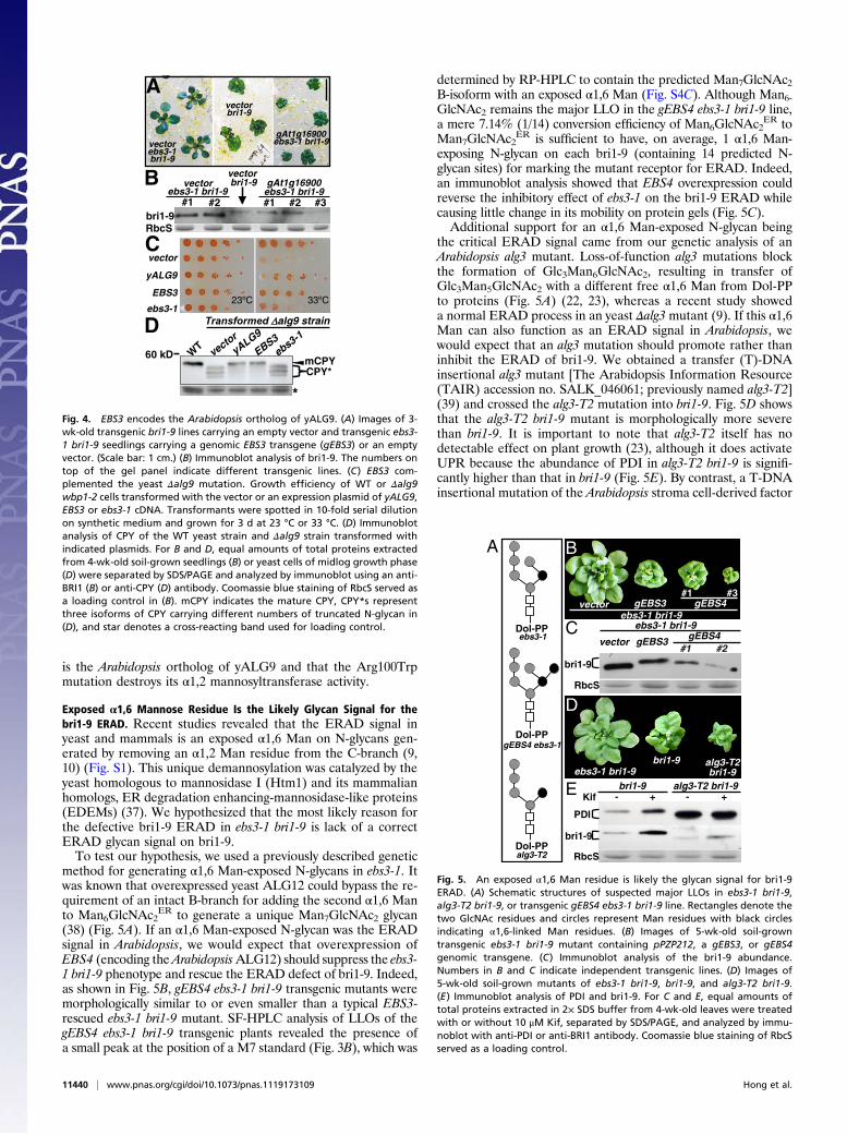
method for generating α1,6 Man-exposed N-glycans in ebs3-1. Itwas known that overexpressed yeast ALG12 could bypass the re-quirement of an intact B-branch for adding the second α1,6 Manto Man6GlcNAc2
ER to generate a unique Man7GlcNAc2 glycan(38) (Fig. 5A). If an α1,6 Man-exposed N-glycan was the ERADsignal in Arabidopsis, we would expect that overexpression ofEBS4 (encoding theArabidopsisALG12) should suppress the ebs3-1 bri1-9 phenotype and rescue the ERAD defect of bri1-9. Indeed,as shown in Fig. 5B, gEBS4 ebs3-1 bri1-9 transgenic mutants weremorphologically similar to or even smaller than a typical EBS3-rescued ebs3-1 bri1-9 mutant. SF-HPLC analysis of LLOs of thegEBS4 ebs3-1 bri1-9 transgenic plants revealed the presence ofa small peak at the position of a M7 standard (Fig. 3B), which was
determined by RP-HPLC to contain the predicted Man7GlcNAc2B-isoform with an exposed α1,6 Man (Fig. S4C). Although Man6-GlcNAc2 remains the major LLO in the gEBS4 ebs3-1 bri1-9 line,a mere 7.14% (1/14) conversion efficiency of Man6GlcNAc2
ER toMan7GlcNAc2
ER is sufficient to have, on average, 1 α1,6 Man-exposing N-glycan on each bri1-9 (containing 14 predicted N-glycan sites) for marking the mutant receptor for ERAD. Indeed,an immunoblot analysis showed that EBS4 overexpression couldreverse the inhibitory effect of ebs3-1 on the bri1-9 ERAD whilecausing little change in its mobility on protein gels (Fig. 5C).Additional support for an α1,6 Man-exposed N-glycan being
the critical ERAD signal came from our genetic analysis of anArabidopsis alg3 mutant. Loss-of-function alg3 mutations blockthe formation of Glc3Man6GlcNAc2, resulting in transfer ofGlc3Man5GlcNAc2 with a different free α1,6 Man from Dol-PPto proteins (Fig. 5A) (22, 23), whereas a recent study showeda normal ERAD process in an yeast Δalg3mutant (9). If this α1,6Man can also function as an ERAD signal in Arabidopsis, wewould expect that an alg3 mutation should promote rather thaninhibit the ERAD of bri1-9. We obtained a transfer (T)-DNAinsertional alg3 mutant [The Arabidopsis Information Resource(TAIR) accession no. SALK_046061; previously named alg3-T2](39) and crossed the alg3-T2 mutation into bri1-9. Fig. 5D showsthat the alg3-T2 bri1-9 mutant is morphologically more severethan bri1-9. It is important to note that alg3-T2 itself has nodetectable effect on plant growth (23), although it does activateUPR because the abundance of PDI in alg3-T2 bri1-9 is signifi-cantly higher than that in bri1-9 (Fig. 5E). By contrast, a T-DNAinsertional mutation of the Arabidopsis stroma cell-derived factor
Transformed alg9 strain
mCPYCPY*
60 kD
D
vectorebs3-1 bri1-9
vector bri1-9
bri1-9
B
RbcS
yALG9
vector
EBS3
ebs3-1
C
vectorbri1-9
gAt1g16900ebs3-1 bri1-9vector
ebs3-1 bri1-9
A
gAt1g16900ebs3-1 bri1-9
*
Fig. 4. EBS3 encodes the Arabidopsis ortholog of yALG9. (A) Images of 3-wk-old transgenic bri1-9 lines carrying an empty vector and transgenic ebs3-1 bri1-9 seedlings carrying a genomic EBS3 transgene (gEBS3) or an emptyvector. (Scale bar: 1 cm.) (B) Immunoblot analysis of bri1-9. The numbers ontop of the gel panel indicate different transgenic lines. (C) EBS3 com-plemented the yeast Δalg9 mutation. Growth efficiency of WT or Δalg9wbp1-2 cells transformed with the vector or an expression plasmid of yALG9,EBS3 or ebs3-1 cDNA. Transformants were spotted in 10-fold serial dilutionon synthetic medium and grown for 3 d at 23 °C or 33 °C. (D) Immunoblotanalysis of CPY of the WT yeast strain and Δalg9 strain transformed withindicated plasmids. For B and D, equal amounts of total proteins extractedfrom 4-wk-old soil-grown seedlings (B) or yeast cells of midlog growth phase(D) were separated by SDS/PAGE and analyzed by immunoblot using an anti-BRI1 (B) or anti-CPY (D) antibody. Coomassie blue staining of RbcS served asa loading control in (B). mCPY indicates the mature CPY, CPY*s representthree isoforms of CPY carrying different numbers of truncated N-glycan in(D), and star denotes a cross-reacting band used for loading control.
bri1-9
ebs3-1 bri1-9vector gEBS3 gEBS4
ebs3-1 bri1-9bri1-9 alg3-T2
bri1-9bri1-9 alg3-T2 bri1-9
Kif - -+
PDI
bri1-9
RbcS
+
B
C
D
E
Dol-PPalg3-T2
Dol-PPgEBS4 ebs3-1
Dol-PPebs3-1
A
RbcS
ebs3-1 bri1-9
vectorgEBS4gEBS3
Fig. 5. An exposed α1,6 Man residue is likely the glycan signal for bri1-9ERAD. (A) Schematic structures of suspected major LLOs in ebs3-1 bri1-9,alg3-T2 bri1-9, or transgenic gEBS4 ebs3-1 bri1-9 line. Rectangles denote thetwo GlcNAc residues and circles represent Man residues with black circlesindicating α1,6-linked Man residues. (B) Images of 5-wk-old soil-growntransgenic ebs3-1 bri1-9 mutant containing pPZP212, a gEBS3, or gEBS4genomic transgene. (C) Immunoblot analysis of the bri1-9 abundance.Numbers in B and C indicate independent transgenic lines. (D) Images of5-wk-old soil-grown mutants of ebs3-1 bri1-9, bri1-9, and alg3-T2 bri1-9.(E) Immunoblot analysis of PDI and bri1-9. For C and E, equal amounts oftotal proteins extracted in 2× SDS buffer from 4-wk-old leaves were treatedwith or without 10 μM Kif, separated by SDS/PAGE, and analyzed by immu-noblot with anti-PDI or anti-BRI1 antibody. Coomassie blue staining of RbcSserved as a loading control.
11440 | www.pnas.org/cgi/doi/10.1073/pnas.1119173109 Hong et al.

2-like protein (SDF2), required for the correct folding of EFR(40), failed to enhance the bri1-9 dwarfism (Fig. S7) even thoughthe sdf2-2 mutant was hypersensitive to ER stresses (41), sug-gesting that the phenotypic enhancement of bri1-9 by alg3-T2 isnot caused by an abnormal ER stress response but is likely at-tributable to a stimulatory effect of alg3-T2 on the bri1-9 ERAD.Indeed, immunoblotting with a BRI1 antibody revealed thatwhereas alg3-T2 increases the mobility of bri1-9 because ofsmaller N-glycans, it decreases the bri1-9 abundance, which is theexactly opposite of what was observed in ebs3-1 and ebs4-1mutants that accumulate larger N-glycan precursors (Fig. 5E).Interestingly, the ERAD of bri-9 in alg3-T2 bri1-9 could still beinhibited by treatment with kifunensine (Kif), a well-known in-hibitor of α1,2 mannosidase (42) that prevents ERAD of bothbri1-5 and bri1-9 carrying fully assembled N-glycans (19, 20)(Fig. 5E), although its inhibitory effect on the bri1-9 ERAD ismuch weaker in alg3-T2 bri1-9 than in bri1-9, suggesting that thebri1-9 ERAD might require additional Man trimming. Takentogether, these results strongly suggested that an exposed α1,6Man is likely the glycan signal for a plant ERAD process.
DiscussionIn this study, we demonstrated that EBS3 encodes the Arabidopsisortholog of yALG9 that catalyzes the ER luminal addition of twoα1,2 Man residues for assembling Glc3Man9GlcNAc2 (31). First,ebs3-1 contains a single-nucleotide change in At1g16900, and itsbri1-9 suppressor phenotype was rescued by expression of a ge-nomic At1g16900 transgene. Second, the predicted At1g16900protein exhibits the highest sequence homology among all anno-tatedArabidopsis proteins to the yeast ALG9 protein (Fig. S8). Theother two Arabidopsis proteins showing limited sequence homol-ogy are EBS4 and At5g14850 annotated to encode a putative ho-molog of the yeast PIG-B involved in glycosylphosphatidylinositolbiosynthesis (35). Third, the WT At1g16900 was able to com-plement the growth phenotype of the yeast Δalg9 wbp1-2 doublemutant and the N-glycosylation defect of CPY of the Δalg9 yeastmutant (Fig. 4 C and D). Consistent with sequence analysis sug-gesting a crucial catalytic role of the Arg100 residue for severalmannosyltransferases (35), our yeast complementation assayshowed that the R100W mutation destroys its α1,2 mannosyl-transferase activity in yeast cells, suggesting that ebs3-1 is likelya null mutant. Thus, EBS3 works together with the recently dis-covered AtALG3 (22, 23) and EBS4 (20) as the three ArabidopsisER-luminal mannosyltransferases to add four more Man residuesto Dol-PP-Man5GlcNAc2 to assemble Dol-PP-Man9GlcNAc2 thatwill be triglucosylated before the fully assembled Glc3Man9Glc-NAc2 can be transferred to nascent polypeptides (Fig. S1).In yeast, the α1,2 mannosyltransferase ALG9 catalyzes the ER
luminal addition of two terminal α1,2 Man residues to create theB- and C-dimannose branches (31), whereas the α1,6 man-nosyltransferase ALG12 exhibits high substrate specificity towardDol-PP-Man7GlcNAc2 but is very inefficient in adding the sec-ond α1,6 Man when the B-branch lacks the terminal α1,2 Man(43) (Fig. S1). Thus, null alg9 mutations in yeast result in accu-mulation of Dol-PP-Man6GlcNAc2 and protein hypoglycosyla-tion because of reduced transfer efficiency of immature glycansby the yeast oligosaccharide transferase (OST) (21, 43). Con-sistent with our findings that EBS3 is an Arabidopsis ortholog ofyALG9 and that the R100W mutation is either a null or verysevere mutation, our HPLC analyses of LLOs indicated thatebs3-1 bri1-9 accumulates Dol-PP-Man6GlcNAc2. Surprisingly,ebs3-1 bri1-9 also accumulates Dol-PP-Man5GlcNAc2, the knownsubstrate for ALG3 (22, 23). This is likely caused by feedbackinhibition of ALG3 by overaccumulation of its product, Dol-PP-Man6GlcNAc2, because Dol-PP-Man5GlcNAc2 was also detec-ted in a yeast alg9 mutant (21). Despite the glycan-assemblydefect, neither the WT BRI1 nor bri1-9 is hypoglycosylated,which is likely attributable to the fact that the Arabidopsis OSTs
can efficiently transfer truncated glycans from their Dol-PPlinker to bri1-9 and other glycoproteins (22, 23), explaining noobvious growth defect of an ebs3-1 BRI1+ mutant (Fig. S3C).A crucial decision in ERQC is when to stop futile refolding
attempts to divert a terminally misfolded protein from the CNX/CRT cycle to ERAD. A previously popular “mannosidase timer”model posited that the slow α1,2 Man trimming of the B-branch byERManI generates the ERAD glycan Man8GlcNAc2 that can berecognized by dedicated ERAD lectins (7). However, recentstudies showed that although trimming the B-branch is a necessaryERAD step, the true ERAD signal is an exposed α1,6 Man cre-ated by trimming the C-branch (9, 10). In this study, we carried outtwo genetic experiments that suggested that α1,6 Man-exposed N-glycans likely function as a conserved glycan signal for a plantERAD process. First, we showed that overexpression of EBS4 inebs3-1 bri1-9 neutralized the suppressive effect of ebs3-1 on bri1-9by promoting bri1-9 ERAD because overexpressed ALG12 inyeast could bypass the requirement of a complete B-branch foradding an α1,6 Man residue (Fig. 5A). SF-HPLC coupled with RP-HPLC analyses did detect the presence of a Dol-PP-Man7-GlcNAc2 lacking the B- and C-branch terminal α1,2 Man residuesin a gEBS4 ebs3-1 bri1-9 transgenic mutant (Fig. S4C). This uniqueα1,6 Man-exposing Man7GlcNAc2 glycan is likely transferred fromits Dol-PP linker to bri1-9 in the gEBS4 ebs3-1 bri1-9 transgenicline because its electromobility on immunoblot is slightly reducedcompared with that of bri1-9 in a transgenic ebs3-1 bri1-9 linecarrying an empty vector (Fig. 5C). Second, we crossed a T-DNAinsertional alg3-T2 mutation, which is known to create N-glycanscarrying a different free α1,6 Man residue (Fig. 5A) (22, 23), tobri1-9 and discovered that the bri1-9 level in alg3-T2 bri1-9 islower instead of higher than that in ebs3-1 bri1-9 and ebs4-1 bri1-9despite the N-glycans on bri1-9 carrying fewer Man residues inalg3-T2 than that in the two ebs mutants. Taken together, theseresults suggested that the glycan signal to mark an ERAD clientprotein is likely conserved in plants. Given the recent discoveriesshowing that EFR is misfolded and degraded in the absence ofUGGT, CRT3, and other ER proteins/chaperones (13), it will beinteresting to determine whether ERAD of incompletely foldedEFR also depends on such a conserved N-glycan signal.
Materials and MethodsPlant Materials and Growth Conditions. Arabidopsis ecotype Col-0 is the pa-rental line for mutants and transgenic plants except bri1-9 (Ws-2) for cloningEBS3 and bri1-5 (Ws-2) for genetic analysis. The T-DNAmutants of ALG3 (TAIRaccession no. SALK_046061) and SDF2 (TAIR accession no. SALK_141321) wereobtained from the Arabidopsis Biological Resource Center (ABRC) at OhioState University. Methods for seed sterilization and conditions for plantgrowth were described previously (44).
Plasmid Construction and Generation of Transgenic Plants. The p35S-EBS1 andthe genomic constructs of EBS2 and EBS4 were described previously (18, 20).A 5-kb genomic fragment of At1g16900, including 1.5-kb promoter and500-bp terminator sequences, was PCR-amplified from the BAC cloneF17F16, cloned into pPZP212 (45) to make pPZP212-gEBS3, and sequenced toensure no PCR error. Empty vectors and transgene constructs of EBS1, EBS2,EBS3, and EBS4 were individually transformed into bri1-9 or ebs3-1 bri1-9mutants by the vacuum infiltration method (46).
Yeast Growth and Complementation Assay. Standard growth medium andconditions were used to grow the wild-type yeast strain SS328, Δalg9 strainYG414, and Δalg9 wbp1-2 strain YG415 (21). The ORF of EBS3 was PCR-amplified from the At1g16900 cDNA clone U24181 to replace that of theyeast ALG9 from the pYEp352-yALG9 expression plasmid to create pYE-p352-EBS3 using the strategy described previously (20). The StratageneQuikChange XL Site-Directed Mutagenesis kit was used to generate pYEp352-ebs3-1 from pYEp352-EBS3 using the primers listed in Table S1. These plas-mids were fully sequenced to ensure no PCR error and were individuallytransformed into YG414 or YG415 via a rapid transformation protocol (47).
Hong et al. PNAS | July 10, 2012 | vol. 109 | no. 28 | 11441
PLANTBIOLO
GY

Protein Extraction and Immunoblot Analysis. Arabidopsis seedlings harvestedfrom agar, soil, or liquid 1/2 Murashige and Skoog (MS) medium supple-mented with or without BL (Chemiclones) or Kif (Toronto Research Chem-icals) were ground in liquid N2, dissolved (50 mg seedlings/100 μL) in 2× SDSbuffer [0.125 M Tris pH 6.8, 4% (wt/vol) SDS, 20% (vol/vol) glycerol, 0.2 MDTT, 0.02% (wt/vol) bromophenol blue] and boiled for 10 min. After cen-trifugation, supernatants were used for immunoblot analyses or incubatedwith or without 1,000 U of Endo Hf in 1× G5 buffer (New England Biolabs)for 1 h at 37 °C. To perform the CHX chase experiment, 3-wk-old seedlingswere transferred from 1/2 MS agar plates into 1/2 MS medium containing180 μM CHX (Sigma), and equal amounts of seedlings were removed atdifferent time points to extract total proteins into 2× SDS sample buffer.After 10 min of boiling, equal amounts of total proteins, equivalent of 5 mgof seedlings, were separated by 7.5% SDS/PAGE and analyzed by immuno-blot with anti-BRI1 antibody. To extract yeast proteins, cells of midlog phasegrown in a 28 °C shaking incubator were collected by centrifugation,resuspended in 1× extraction buffer [0.3 M sorbitol, 0.1 M NaCl, 5 mMMgCl2, and 10 mM Tris (pH7.4)], lysed by violent vortexing with glass beads,mixed with equal volume of 2× SDS buffer, boiled for 10 min, and centri-fuged to collect supernatants. Protein samples of plants or yeast cells were
separated on 7% or 10% SDS/PAGE gel, transferred onto Immobilon-Pmembrane (Millipore), and analyzed by immunoblot with antibodies madeagainst BRI1 (27), PDI (Rose Biotechnology), BiP (SPA-818; Stressgen), maize-CRTs (48), or a monoclonal anti-CPY antibody (10A5; Invitrogen).
Extraction and Analysis of LLOs. The LLOs from bri1-9, bri1-9 ebs3-1, and gEBS4ebs3-1 bri1-9 were extracted, hydrolyzed, pyridylaminated, and analyzed bySF-HPLC and RP-HPLC according to a previously reported protocol (23). Theelution positions of PA-labeled oligosaccharides were compared with thoseof PA-sugar standards purchased from Masuda Chemical Industries.
ACKNOWLEDGMENTS. We thank ABRC for supplying cDNA/BAC clones ofAt1g16900 and T-DNA insertional mutant of ALG3 (TAIR accession no.SALK_046061) and SDF2 (TAIR accession no. SALK_141321), F. Tax for seedsof bri1-9 (WS-2) and bri1-5, J. Chory for anti-BRI1 antibody, Y. Yin for anti-BES1 antiserum, R. Boston for anti-maize CRT antibody, A. Chang for anti-CPY antibody, M. Aebi for yeast strains and YEp352-yALG9 plasmid, andmembers of Li laboratory for stimulating discussion. This work was sup-ported, in part, by National Natural Science Foundation of China Grant31070246 (to Z.H.), National Institutes of Health Grant GM060519 (to J.L.),and Department of Energy Grant ER15672 (to J.L.).
1. Abu-Qarn M, Eichler J, Sharon N (2008) Not just for Eukarya anymore: Protein gly-cosylation in Bacteria and Archaea. Curr Opin Struct Biol 18:544–550.
2. Kelleher DJ, Gilmore R (2006) An evolving view of the eukaryotic oligosaccharyl-transferase. Glycobiology 16:47R–62R.
3. Helenius A, Aebi M (2004) Roles of N-linked glycans in the endoplasmic reticulum.Annu Rev Biochem 73:1019–1049.
4. Molinari M (2007) N-glycan structure dictates extension of protein folding or onset ofdisposal. Nat Chem Biol 3:313–320.
5. Caramelo JJ, Parodi AJ (2007) How sugars convey information on protein conforma-tion in the endoplasmic reticulum. Semin Cell Dev Biol 18:732–742.
6. Caramelo JJ, Parodi AJ (2008) Getting in and out from calnexin/calreticulin cycles.J Biol Chem 283:10221–10225.
7. Lederkremer GZ, Glickman MH (2005) A window of opportunity: Timing proteindegradation by trimming of sugars and ubiquitins. Trends Biochem Sci 30:297–303.
8. Vembar SS, Brodsky JL (2008) One step at a time: Endoplasmic reticulum-associateddegradation. Nat Rev Mol Cell Biol 9:944–957.
9. Clerc S, et al. (2009) Htm1 protein generates the N-glycan signal for glycoproteindegradation in the endoplasmic reticulum. J Cell Biol 184:159–172.
10. Quan EM, et al. (2008) Defining the glycan destruction signal for endoplasmicreticulum-associated degradation. Mol Cell 32:870–877.
11. Ceriotti A, Roberts LM (2006) Endoplasmic reticulum-associated protein degradationin plant cells. The Plant Endoplasmic Reticulum, ed Robinson DG (Springer, Heidel-berg), pp 75–98.
12. Vitale A, Boston RS (2008) Endoplasmic reticulum quality control and the unfoldedprotein response: Insights from plants. Traffic 9:1581–1588.
13. Saijo Y (2010) ER quality control of immune receptors and regulators in plants. CellMicrobiol 12:716–724.
14. Li J, Chory J (1997) A putative leucine-rich repeat receptor kinase involved in brassi-nosteroid signal transduction. Cell 90:929–938.
15. Kinoshita T, et al. (2005) Binding of brassinosteroids to the extracellular domain ofplant receptor kinase BRI1. Nature 433:167–171.
16. Zipfel C, et al. (2006) Perception of the bacterial PAMP EF-Tu by the receptor EFRrestricts Agrobacterium-mediated transformation. Cell 125:749–760.
17. Jin H, Yan Z, Nam KH, Li J (2007) Allele-specific suppression of a defective brassi-nosteroid receptor reveals a physiological role of UGGT in ER quality control. Mol Cell26:821–830.
18. Jin H, Hong Z, Su W, Li J (2009) A plant-specific calreticulin is a key retention factor fora defective brassinosteroid receptor in the endoplasmic reticulum. Proc Natl Acad SciUSA 106:13612–13617.
19. Hong Z, Jin H, Tzfira T, Li J (2008) Multiple mechanism-mediated retention of a de-fective brassinosteroid receptor in the endoplasmic reticulum of Arabidopsis. PlantCell 20:3418–3429.
20. Hong Z, et al. (2009) Mutations of an alpha1,6 mannosyltransferase inhibit endo-plasmic reticulum-associated degradation of defective brassinosteroid receptors inArabidopsis. Plant Cell 21:3792–3802.
21. Burda P, et al. (1996) Stepwise assembly of the lipid-linked oligosaccharide in theendoplasmic reticulum of Saccharomyces cerevisiae: Identification of the ALG9 geneencoding a putative mannosyl transferase. Proc Natl Acad Sci USA 93:7160–7165.
22. Henquet M, et al. (2008) Identification of the gene encoding the alpha1,3-man-nosyltransferase (ALG3) in Arabidopsis and characterization of downstream n-glycanprocessing. Plant Cell 20:1652–1664.
23. Kajiura H, Seki T, Fujiyama K (2010) Arabidopsis thaliana ALG3 mutant synthesizesimmature oligosaccharides in the ER and accumulates unique N-glycans. Glycobiology20:736–751.
24. Su W, Liu Y, Xia Y, Hong Z, Li J (2011) Conserved endoplasmic reticulum-associateddegradation system to eliminate mutated receptor-like kinases in Arabidopsis. ProcNatl Acad Sci USA 108:870–875.
25. Maley F, Trimble RB, Tarentino AL, Plummer TH, Jr. (1989) Characterization of gly-coproteins and their associated oligosaccharides through the use of endoglycosidases.Anal Biochem 180:195–204.
26. Clouse SD, Langford M, McMorris TC (1996) A brassinosteroid-insensitive mutant inArabidopsis thaliana exhibits multiple defects in growth and development. PlantPhysiol 111:671–678.
27. Mora-García S, et al. (2004) Nuclear protein phosphatases with Kelch-repeat domainsmodulate the response to brassinosteroids in Arabidopsis. Genes Dev 18:448–460.
28. Houston NL, et al. (2005) Phylogenetic analyses identify 10 classes of the protein di-sulfide isomerase family in plants, including single-domain protein disulfide isomer-ase-related proteins. Plant Physiol 137:762–778.
29. Sung DY, Vierling E, Guy CL (2001) Comprehensive expression profile analysis of theArabidopsis Hsp70 gene family. Plant Physiol 126:789–800.
30. Persson S, et al. (2003) Phylogenetic analyses and expression studies reveal two dis-tinct groups of calreticulin isoforms in higher plants. Plant Physiol 133:1385–1396.
31. Frank CG, Aebi M (2005) ALG9 mannosyltransferase is involved in two different stepsof lipid-linked oligosaccharide biosynthesis. Glycobiology 15:1156–1163.
32. Frank CG, et al. (2004) Identification and functional analysis of a defect in the humanALG9 gene: Definition of congenital disorder of glycosylation type IL. Am J HumGenet 75:146–150.
33. Jakob CA, Burda P, Roth J, Aebi M (1998) Degradation of misfolded endoplasmicreticulum glycoproteins in Saccharomyces cerevisiae is determined by a specific oli-gosaccharide structure. J Cell Biol 142:1223–1233.
34. Weinstein M, et al. (2005) CDG-IL: An infant with a novel mutation in the ALG9 geneand additional phenotypic features. Am J Med Genet A 136:194–197.
35. Oriol R, Martinez-Duncker I, Chantret I, Mollicone R, Codogno P (2002) Commonorigin and evolution of glycosyltransferases using Dol-P-monosaccharides as donorsubstrate. Mol Biol Evol 19:1451–1463.
36. Kostova Z, Wolf DH (2005) Importance of carbohydrate positioning in the recognitionof mutated CPY for ER-associated degradation. J Cell Sci 118:1485–1492.
37. Kanehara K, Kawaguchi S, Ng DT (2007) The EDEM and Yos9p families of lectin-likeERAD factors. Semin Cell Dev Biol 18:743–750.
38. Burda P, Jakob CA, Beinhauer J, Hegemann JH, Aebi M (1999) Ordered assembly ofthe asymmetrically branched lipid-linked oligosaccharide in the endoplasmic re-ticulum is ensured by the substrate specificity of the individual glycosyltransferases.Glycobiology 9:617–625.
39. Alonso JM, et al. (2003) Genome-wide insertional mutagenesis of Arabidopsis thali-ana. Science 301:653–657.
40. Nekrasov V, et al. (2009) Control of the pattern-recognition receptor EFR by an ERprotein complex in plant immunity. EMBO J 28:3428–3438.
41. Schott A, et al. (2010) Arabidopsis stromal-derived Factor2 (SDF2) is a crucial target of theunfolded protein response in the endoplasmic reticulum. J Biol Chem 285:18113–18121.
42. Tokunaga F, Brostrom C, Koide T, Arvan P (2000) Endoplasmic reticulum (ER)-associ-ated degradation of misfolded N-linked glycoproteins is suppressed upon inhibitionof ER mannosidase I. J Biol Chem 275:40757–40764.
43. Cipollo JF, Trimble RB (2000) The accumulation of Man(6)GlcNAc(2)-PP-dolichol in theSaccharomyces cerevisiae Deltaalg9 mutant reveals a regulatory role for the Alg3palpha1,3-Man middle-arm addition in downstream oligosaccharide-lipid and glyco-protein glycan processing. J Biol Chem 275:4267–4277.
44. Li J, Nam KH, Vafeados D, Chory J (2001) BIN2, a new brassinosteroid-insensitive locusin Arabidopsis. Plant Physiol 127:14–22.
45. Hajdukiewicz P, Svab Z, Maliga P (1994) The small, versatile pPZP family of Agro-bacterium binary vectors for plant transformation. Plant Mol Biol 25:989–994.
46. Bechtold N, Pelletier G (1998) In planta Agrobacterium-mediated transformation ofadult Arabidopsis thaliana plants by vacuum infiltration.Methods Mol Biol 82:259–266.
47. Gietz RD, Woods RA (2002) Transformation of yeast by lithium acetate/single-stranded carrier DNA/polyethylene glycol method. Methods Enzymol 350:87–96.
48. Pagny S, et al. (2000) Protein recycling from the Golgi apparatus to the endoplasmic re-ticulum in plants and its minor contribution to calreticulin retention. Plant Cell 12:739–756.
11442 | www.pnas.org/cgi/doi/10.1073/pnas.1119173109 Hong et al.


![Reactive Oxygen Species Are Involved in Brassinosteroid-Induced … · Reactive Oxygen Species Are Involved in Brassinosteroid-Induced Stress Tolerance in Cucumber1[W] ... Vert and](https://static.fdocuments.in/doc/165x107/60ebd8840c3a8322ad22a20e/reactive-oxygen-species-are-involved-in-brassinosteroid-induced-reactive-oxygen.jpg)