Evo-devo of non-bilaterian animals - · PDF fileEvo-devo of non-bilaterian animals ......
17
Evo-devo of non-bilaterian animals Emilio Lanna Departamento de Biologia Geral, Instituto de Biologia, Universidade Federal da Bahia, Salvador, BA, Brazil. Abstract The non-bilaterian animals comprise organisms in the phyla Porifera, Cnidaria, Ctenophora and Placozoa. These early-diverging phyla are pivotal to understanding the evolution of bilaterian animals. After the exponential increase in research in evolutionary development (evo-devo) in the last two decades, these organisms are again in the spot- light of evolutionary biology. In this work, I briefly review some aspects of the developmental biology of non- bilaterians that contribute to understanding the evolution of development and of the metazoans. The evolution of the developmental genetic toolkit, embryonic polarization, the origin of gastrulation and mesodermal cells, and the origin of neural cells are discussed. The possibility that germline and stem cell lineages have the same origin is also exam- ined. Although a considerable number of non-bilaterian species are already being investigated, the use of species belonging to different branches of non-bilaterian lineages and functional experimentation with gene manipulation in the majority of the non-bilaterian lineages will be necessary for further progress in this field. Keywords: early-diverging metazoans, BMP, FGF, Wnt, stem cells, evolution, nervous system. Received: January 11, 2015; Accepted: April 29, 2015. Introduction The field of evolutionary developmental biology (evo-devo) has gained momentum since its foundation in the 1980s, when HOX genes were discovered in both fly and mouse (Carroll et al., 2005). In its infancy, evo-devo studies were mainly carried out on classical developmental biology models, capitalizing on previously established techniques and a large body of knowledge on the develop- mental molecules and genetics of these organisms (Som- mer, 2009; Giles and Averof, 2014). As developmental biology is traditionally linked to medical sciences, the ani- mal models utilized in these former studies were mainly vertebrates and the fly. However, animal diversity greatly surpasses the variety represented by these organisms. If we are to understand the evolution of developmental pro- cesses, and how these processes are related to the evolution of the animals, more models are needed (Jenner and Wills, 2007; but see Sommer, 2009 for a different perspective). Thus, we are now experiencing an explosion of new model systems (Sommer, 2009), especially due to lower costs for sequencing genomes and transcriptomes and other techno- logical improvements (Giles and Averof, 2014). To understand how the diversity of forms and body plans of multicellular animals arose it is necessary to inves- tigate the five major lineages of metazoans living today. Vertebrates, Caenorhabditis elegans (a nematode) and the fly belong to the Bilateria lineage. The other four lineages (the phyla Porifera, Cnidaria, Ctenophora and Placozoa) consist of comparatively simple animals that generally lack bilateral symmetry in their body plans (however bilateral symmetry is present in the body plan of some cnidarians). These lineages are artificially grouped as non-bilaterian an- imals (also known as ‘early-branching’ or ‘basal’ metazo- ans) (Dohrmann and Wörheide, 2013). Non-bilaterians range from benthic sessile filter feeders to gelatinous planktonic carnivorous animals. One of these phyla, Pori- fera (sponges), has been traditionally described as organ- isms devoid of true epithelia and lacking the gastrulation stage during development (Leys, 2004; Leys and Hill, 2012; see below). Sponges lack a clear anterior-posterior (AP) polarity as adults (but the larvae do swim direction- ally), have no organs, are filter feeders, and have a body plan adapted to process as much water as needed for feed- ing and respiration (Leys and Hill, 2012). The other three non-bilaterian phyla are diploblastic (adult tissues are derivatives of two germ layers: ectoderm and endoderm [also known as endomesoderm]; Martindale, 2005). Placozoa lack AP polarity, nerve and muscle cells and a proper gut. The body plan of organisms in this phy- lum is very simple compared to other metazoans. They have a disc-shaped, double-layered body comprising only six cell types (Smith et al., 2014). Representatives of this phylum absorb nutrients through contact of the lower epi- Send correspondence to Emilio Lanna. Universidade Federal da Bahia, Instituto de Biologia, Departamento de Biologia Geral, Rua Barão de Jeremoabo s/n, Campus de Ondina, 40170-115 Salva- dor, BA, Brazil. E-mail: [email protected]. Genetics and Molecular Biology Online Ahead of Print Copyright © 2015, Sociedade Brasileira de Genética. Printed in Brazil DOI: http://dx.doi.org/10.1590/S1415-475738320150005
Transcript of Evo-devo of non-bilaterian animals - · PDF fileEvo-devo of non-bilaterian animals ......

Evo-devo of non-bilaterian animals
Emilio Lanna
Departamento de Biologia Geral, Instituto de Biologia, Universidade Federal da Bahia,
Salvador, BA, Brazil.
Abstract
The non-bilaterian animals comprise organisms in the phyla Porifera, Cnidaria, Ctenophora and Placozoa. Theseearly-diverging phyla are pivotal to understanding the evolution of bilaterian animals. After the exponential increasein research in evolutionary development (evo-devo) in the last two decades, these organisms are again in the spot-light of evolutionary biology. In this work, I briefly review some aspects of the developmental biology of non-bilaterians that contribute to understanding the evolution of development and of the metazoans. The evolution of thedevelopmental genetic toolkit, embryonic polarization, the origin of gastrulation and mesodermal cells, and the originof neural cells are discussed. The possibility that germline and stem cell lineages have the same origin is also exam-ined. Although a considerable number of non-bilaterian species are already being investigated, the use of speciesbelonging to different branches of non-bilaterian lineages and functional experimentation with gene manipulation inthe majority of the non-bilaterian lineages will be necessary for further progress in this field.
Keywords: early-diverging metazoans, BMP, FGF, Wnt, stem cells, evolution, nervous system.
Received: January 11, 2015; Accepted: April 29, 2015.
Introduction
The field of evolutionary developmental biology(evo-devo) has gained momentum since its foundation inthe 1980s, when HOX genes were discovered in both flyand mouse (Carroll et al., 2005). In its infancy, evo-devostudies were mainly carried out on classical developmentalbiology models, capitalizing on previously establishedtechniques and a large body of knowledge on the develop-mental molecules and genetics of these organisms (Som-mer, 2009; Giles and Averof, 2014). As developmentalbiology is traditionally linked to medical sciences, the ani-mal models utilized in these former studies were mainlyvertebrates and the fly. However, animal diversity greatlysurpasses the variety represented by these organisms. If weare to understand the evolution of developmental pro-cesses, and how these processes are related to the evolutionof the animals, more models are needed (Jenner and Wills,2007; but see Sommer, 2009 for a different perspective).Thus, we are now experiencing an explosion of new modelsystems (Sommer, 2009), especially due to lower costs forsequencing genomes and transcriptomes and other techno-logical improvements (Giles and Averof, 2014).
To understand how the diversity of forms and bodyplans of multicellular animals arose it is necessary to inves-
tigate the five major lineages of metazoans living today.Vertebrates, Caenorhabditis elegans (a nematode) and thefly belong to the Bilateria lineage. The other four lineages(the phyla Porifera, Cnidaria, Ctenophora and Placozoa)consist of comparatively simple animals that generally lackbilateral symmetry in their body plans (however bilateralsymmetry is present in the body plan of some cnidarians).These lineages are artificially grouped as non-bilaterian an-imals (also known as ‘early-branching’ or ‘basal’ metazo-ans) (Dohrmann and Wörheide, 2013). Non-bilateriansrange from benthic sessile filter feeders to gelatinousplanktonic carnivorous animals. One of these phyla, Pori-fera (sponges), has been traditionally described as organ-isms devoid of true epithelia and lacking the gastrulationstage during development (Leys, 2004; Leys and Hill,2012; see below). Sponges lack a clear anterior-posterior(AP) polarity as adults (but the larvae do swim direction-ally), have no organs, are filter feeders, and have a bodyplan adapted to process as much water as needed for feed-ing and respiration (Leys and Hill, 2012).
The other three non-bilaterian phyla are diploblastic(adult tissues are derivatives of two germ layers: ectodermand endoderm [also known as endomesoderm]; Martindale,2005). Placozoa lack AP polarity, nerve and muscle cellsand a proper gut. The body plan of organisms in this phy-lum is very simple compared to other metazoans. Theyhave a disc-shaped, double-layered body comprising onlysix cell types (Smith et al., 2014). Representatives of thisphylum absorb nutrients through contact of the lower epi-
Send correspondence to Emilio Lanna. Universidade Federal daBahia, Instituto de Biologia, Departamento de Biologia Geral, RuaBarão de Jeremoabo s/n, Campus de Ondina, 40170-115 Salva-dor, BA, Brazil. E-mail: [email protected].
Genetics and Molecular Biology Online Ahead of PrintCopyright © 2015, Sociedade Brasileira de Genética. Printed in BrazilDOI: http://dx.doi.org/10.1590/S1415-475738320150005

thelium with the substratum (Brusca and Brusca, 2003;Srivastava et al., 2008). Very little is known about their de-velopment (Eitel et al., 2008). Cnidaria (anemones, jelly-fish, and their kin) and Ctenophora (comb jellies) passthrough the gastrulation stage during embryogenesis, pro-ducing a gut that persists throughout the life cycle. The gutis blind ending in cnidarians, while ctenophores have ananal pore. Anal pores may serve as a primitive anus, assist-ing the mouth to egest undigested remains of the prey, andas an exit for metabolic wastes (Brusca and Brusca, 2003).Adults of both phyla have tentacles to capture prey andoral-aboral polarity is seen throughout their life cycle (Mar-tindale, 2005).
Although the phylogenetic relationships among non-bilaterians and also between non-bilaterians and Bilateriaare still contentious (see below), it has been assumed thatthe developmental biology of these four early-branchingphyla may play a pivotal role in interpreting the evolutionof metazoans. Although genomic and transcriptomic dataindicate that these phyla are similar to other metazoans,their body plans are quite different from those of Bilateriaand their development may be key to understanding manyquestions about animal evolution. As many of the events indevelopment may be obscured due to evolutionary diver-gence, use of molecular markers can facilitate our under-standing about deep homologies and the evolution ofanimals (Gold and Jacobs, 2003). Here, I provide a brief re-view of how studies on the development of non-bilateriansare aiding (or not) understanding of the evolution of ani-mals. I present the findings that contributed to this knowl-edge and the gaps that still require research. My objective isto compile information about different topics related tonon-bilaterian evo-devo in a single work. Consequently, Ido not provide a thorough review of the literature on thesetopics, as others have comprehensively discussed each is-sue, usually for each specific taxon (e.g. Technau andScholz, 2003; Lee et al., 2006; Burton, 2008; Galliot et al.,2009; Watanabe et al., 2009; Bosch et al., 2010; Technauand Steele, 2012; Ereskovsky et al., 2013; Funayama et al.,2013; Gold and Jacobs, 2013).
Phylogenetic Relationship of Non-BilaterianAnimals
One of the most disputed issues on the phylogeny ofanimals is the topology of the five major extant lineages ofthe metazoan tree (Porifera, Cnidaria, Ctenophora, Placo-zoa, and Bilateria) (Edgecomb et al., 2011; Dohrmann andWörheide, 2013). Understanding the relationships formingthe basis of the metazoan tree of life is important for an-swering questions about how metazoans traits (includingdevelopment) arose and evolved over the last 700 millionyears (Dohrmann and Wörheide, 2013). In the last two de-cades a forest of trees with very conflicting scenarios hasbeen published. From paraphyletic Porifera (the ‘we are all
derived from sponge larvae’ scenario; Nielsen, 2008), toctenophores branching out of the metazoan tree earlier thanany other phyla (the ‘Ctenophora-first’ scenario; Ryan et
al., 2013; Moroz et al., 2014;), a wide variety in the phylog-eny of basal metazoans has been proposed (reviewed inDohrmann and Wörheide, 2013). These inconsistenciesmay be due to insufficient molecular sampling and/or inad-equate sampling of taxa (Phillipe et al., 2009). However,addition of more sequences is not sufficient to provide amore reliable phylogeny of metazoans, and more integra-tive approaches combining morphology, development,cytology and genome architecture are fundamental to com-prehend animal phylogeny (Philippe et al., 2011;Dohrmann and Wörheide, 2013). Dohrmann and Wörheide(2013) suggest that it is better to rely on solid evidence(such as it is) instead of proposing new scenarios with everynewly published tree. I agree with this view and use the‘conservative’ evolutionary scenario proposed by these au-thors in this current work (Figure 1). Nonetheless, whennecessary I discuss different interpretations of findingsfrom the perspective of alternative phylogenetic scenarios.
Even though the relationship among phyla is still notagreed, the relationships of groups within phyla appear ro-bust (Figure 1), at least in sponges and cnidarians. Spon-ges have four monophyletic classes (Calcarea, Homoscle-romorpha, Demospongiae, and Hexactinellida) dividedinto two groups: Silicea (Demospongiae and Hexac-tinellida) and another group containing Homoscleromor-pha and Calcarea, for which no morphologicalsynapomorphy has been found (Philippe et al., 2009;Wörheide et al., 2012).
Placozoa is an enigmatic phylum that historicallybeen classified as monospecific, with Trichoplax
adhaerens the single species of the group. However, Voigtet al. (2004) showed that the high genetic divergenceamong placozoan lineages is similar to that observed, inother metazoans, for different families.
Cnidaria has five monophyletic classes: Anthozoa,Staurozoa, Cubozoa, Scyphozoa, and Hydrozoa; Anthozoais a sister group of Medusozoa, which contains the other thefour classes (Figure 1; Collins et al., 2006). Ctenophora isconsidered the most problematic branch among the non-bilaterians (Simion et al., 2015). The classical taxonomy ofthis group splitted the phylum in two classes Tentaculata(with six orders) and Nuda (one order). Even before the useof molecular markers for phylogenetic studies, Harbison(1985) suggested that these classes were not valid, aban-doning the use of this hierarchical group in ctenophores.More recently, Simion et al. (2015) have shown that manyof the orders, families and even genera are not mono-phyletic (Figure 1). Analysis of 18S RNA and ITS1 + 5.8S+ ITS2 sequences of several species of the phylum suggeststhat present-day ctenophores have passed through a recentradiation followed by a bottleneck (Podar et al., 2001;Simion et al., 2015). Even though there are still large gaps
Lanna
in understanding of the phylogeny of the non-bilaterians,current knowledge is already helping us to understand howthe development of these animals evolved and promotedthe diversification of the metazoans.
Brief Overview of the Embryogenesis ofNon-Bilaterian Animals
Porifera
It is very difficult to generalize the development ofsponges. There is large variation in developmental typeseven within a single class (Ereskovsky, 2010; Wörheide et
al., 2012). Gametogenesis starts from somatic cells(choanocytes or archaeocytes) that transdifferentiate eitherinto spermatogonia or oogonia. The gametogenic steps,however, are similar to other metazoans. There are fourmain types of cleavage: chaotic in Homoscleromorpha andsome oviparous Demospongiae; radial in oviparous Demo-spongiae and Hexactinellida; polyaxial in a few Demo-spongiae and one of the subclasses of Calcarea (Calcinea);and incurvational (palyntomic cleavage) in the other sub-
class of Calcarea (Calcaronea) (Ereskovsky et al., 2013).The boundaries of subsequent stages of development (blas-tulation, gastrulation, histogenesis and organogenesis) arefuzzy in all groups of sponges. Blastulation is hard to delin-eate because by the end of cleavage the cells of the embryohave already started to differentiate (Ereskovsky, 2010).Gastrulation in Porifera is also controversial, but there isextensive morphogenesis and cell differentiation followingcleavage. Finally, the embryo leads to a free-living larva(there are eight types of larvae; see Maldonado and Berg-quist, 2002; Ereskovsky, 2010) that swims unidirectionallyusing cilia until it settles and starts metamorphosis. Indemosponges and homoscleromorphs a series of cell rear-rangements and differentiation takes place to form the rha-gon, a juvenile with a single osculum and several choa-nocyte chambers, while in Calcarea, the olynthus, ajuvenile with a single, sac-like, choanocyte chamber thatopens in a apical osculum, is formed (Ereskovsky, 2010).
One of the main characteristics of sponges is theirhigh potential for asexual reproduction by fragmentation,budding or gemmulation (Funayama, 2010). Gemmules are
Evolution of early metazoans
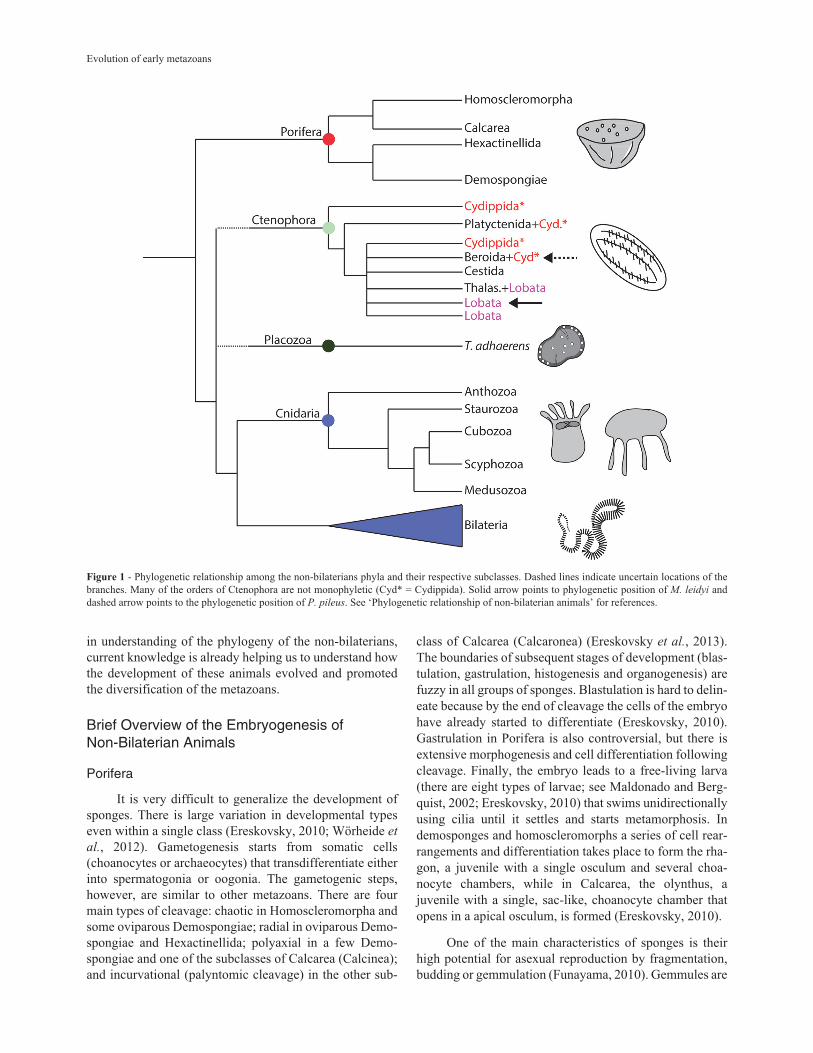
Figure 1 - Phylogenetic relationship among the non-bilaterians phyla and their respective subclasses. Dashed lines indicate uncertain locations of thebranches. Many of the orders of Ctenophora are not monophyletic (Cyd* = Cydippida). Solid arrow points to phylogenetic position of M. leidyi anddashed arrow points to the phylogenetic position of P. pileus. See ‘Phylogenetic relationship of non-bilaterian animals’ for references.

mainly found in freshwater sponges as bodies resistant tothe harsh environment. This characteristic allows them tobe kept in laboratory conditions and, subsequently, be usedas a model to study cell differentiation and aquiferous sys-tem development (e.g. Funayama, 2010; Windsor andLeys, 2010; Leys, 2015).
Some species of Porifera are currently used as evo-devo model species (Table 1). The main models are thedemosponge Amphimedon queenslandica and the calca-rean Sycon ciliatum, in which several steps of developmenthave been investigated, and genomes and transcriptomessequenced. In addition, many studies investigating the pat-tern of gene expression during development are available(e.g. Adamska et al., 2007; Srivastava et al., 2010a; Fortu-nato et al., 2014; Leininger et al., 2014). There are alsoother species that have been used in experimental manipu-lations. Borojevic and Levi (1964) and Borojevic (1966) in-vestigated cell differentiation during metamorphosis of thelarvae of the demosponge Mycale contarenii. In addition,in the last twenty years, studies have investigated the pat-tern of gene expression in the demosponges Ephydatia spp.and Suberites massa and in the homoscleromorphsOscarela spp. (Table 1) (Adell et al., 2003; Perovik-Ottstadt et al., 2004; Lapébie et al., 2009; Windsor andLeys, 2010; Okamoto et al., 2012; Funayama, 2013). Morerecently, gene expression has been manipulated using drugtreatments and RNA interference. Although drug treat-
ments to disturb gene function are not specific, there havebeen some attempts to manipulate development using dif-ferent protocols that have provided promising results(Lapébie et al., 2009; Windsor and Leys, 2010). Rivera et
al. (2011) demonstrated that, by soaking sponges with dou-ble-stranded RNA (RNAi) or feeding them with bacteriaexpressing RNAi, it was possible to efficiently affect targetgenes. However, little progress has been achieved since thispublication.
Placozoa
Placozoans present the simplest morphology amongall metazoans: they have a clear baso-apical polarity (uppervs. lower epithelia), no mesenchymal tissues and are for-med by only six somatic cell types (Smith et al., 2014). TheT. adhaerens genome was sequenced even before the basicbiology (e.g. life cycle and development) was unraveled,probably because of the difficulties in culturing placozoanembryos as they usually die around the 128-cell stage(Srivastava et al., 2008; Eitel et al., 2011). Thus, knowl-edge of the development of Placozoa is more complete forinvestigation of the molecular toolkit than for embryo-logical studies. For instance, in the last decade some studiesusing in situ hybridization (ISH) showed that developmen-tal molecules are not only present, but also are expressed inadults (Jakob et al., 2004; Hadrys et al., 2005; Schierwateret al., 2009). Morpholinos and RNAi have been used to in-
Lanna
Table 1 - Summary list of non-bilaterian models being used in evo-devo research. (Modified from Technau and Steele, 2012).
Phylum Class* Model Genome Transcriptome T/M/KDM* Font
Porifera Demospongiae Amphimedon queenslandica YES YES NO Srivastava et al., 2010a
Ephydatia muelleri NO NO NO Windsor and Leys, 2010
Ephydatia fluviatilis NO NO NO Funayama, 2010
Calcarea Sycon ciliatum YES YES NO Leininger et al., 2014
Sycon coactum NO YES NO Riesgo et al., 2014
Leucosolenia complicata YES YES NO Leininger et al., 2014
Homoscleromorpha Oscarella lobularis NO YES NO Ereskovsky et al., 2009
Oscarella carmela YES YES NO Nichols et al., 2006
Cnidaria Hydrozoa Hydra magnipapilata YES YES Yes, No, RNAi Chapman et al., 2010
Clytia hemispherica YES YES No, No, Mo** Houliston et al., 2011
Hydractinia echinata NO NO Yes, No, RNAi Kraus et al., 2014
Podocoryne carnea NO NO NO Spring et al., 2002
Scyphozoa Aurelia aurita NO YES NO Fuchs et al., 2014
Anthozoa Nematostella vectensis YES YES Yes, No, Mo/RNAi Putnam et al., 2007
Acropora digitifera YES YES NO Shinzato et al., 2011
Cubozoa Tripedalia cystophora NO NO NO Piatgorsky and Kozmik, 2004
Ctenophora Lobata Mnemiopsis leidyi YES YES NO Ryan et al., 2013
Pleurobrachiidae Pleurobrachia bachei YES YES NO Moroz et al.
Pleurobrachia pileus NO YES NO Alié et al., 2011
Placozoa - Trichoplax adhaerens YES YES Yes, No, No Schierwater et al., 2009
* - T/M/KDM = Transgenics, Mutants and knockdown methods. ** - Mo = morpholino.

vestigate gene function in these animals (Jakob et al.,2004).
Cnidaria
Numerous studies during the last two centuries haveinvestigated embryogenesis and the developmental biologyof cnidarians. Cnidarians are diploblastic, radially symmet-rical animals that include species of jellyfish, corals, andsea anemones (Brusca and Brusca, 2003). Cnidarians havea remarkably complex life cycle. To some extent, all groups(except Anthozoa) present metagenesis, i.e. an alternationbetween polyp and medusa forms (Brusca and Brusca,2003). Gametogenesis takes place in gonads growing incontact with the coelenteron (a cavity inside the organism).Eggs and sperm cells derive from mesenchymal cells(Extavour et al., 2005). Fertilization occurs either in thewater column or inside the parental organism bearing theeggs. The eggs usually lack a primary animal-vegetal axis,which is achieved with sperm entry, setting the body axisjust before the first cleavages (Byrun and Martindale, 2004;see Momose et al., 2008 for a different perspective in thehydrozoan Clytia). Cleavages in this phylum are usuallyrandom, but some species of direct-developing meduso-zoans present a more defined pattern during the first threedivisions (Freeman, 1983).
After cleavage, the embryo becomes a blastula (eithersolid or hollow) that gastrulates to form a ciliated planulalarva. Gastrulation in this phylum takes nine differentforms. There are two types of ingression (unipolar and mul-tipolar), three forms of delamination (blastula, morula, andsyncytial), invagination, epiboly, and at least two forms ofmixed gastrulation (involving more than one form of mor-phogenetic movements: (i) mixed delamination and (ii)forms combining ingression and invagination) (Byrun andMartindale, 2004). These different forms of gastrulation arenot exclusive or specific for the different evolutionary lin-eages of the group, although invagination seems to be theancestral form (Byrun and Martindale, 2004). Gastrulationleads to a diploblastic embryo, with an archenteron and ablastopore. The archenteron becomes the endoderm, fromwhich the gut develops, and the ectoderm becomes the epi-dermis (including cnidocytes) and the nervous cells (how-ever, in hydrozoans, the nervous cells are derived frominterstitial cells, which derive from endodermal cells)(Byrun and Martindale, 2004; Technau and Steele, 2012).During metamorphosis, cell proliferation starts at the budprimordia located at the apical region of the embryo, whicheventually form the tentacles.
Similar to sponges, cnidarians are well-known fortheir ability to regenerate lost parts. Hydra has been usedfor centuries to investigate the regeneration process inmetazoans. Regeneration in these organisms occurs by amechanism that does not require proliferation and growth(morphalaxis) (Cummings and Bode, 1984; Technau andSteele, 2012). It seems that the WNT pathway and transcrip-
tion factor Brachyury play an important role in patterningand differentiation of cells during this process in Hydra
(Technau and Bode, 1999; Technau and Steele, 2012). In-vestigation of the regeneration process of Nematostella
vectensis indicates that an alternate developmental trajec-tory is taken to reconstruct the body of the animal. Antho-zoan regeneration uses molecular mechanisms different toboth anthozoan embryonic development and regenerationin Hydra (Burton and Finnerty, 2009; Technau and Steele,2012).
The developmental biology of many species of Cni-daria is currently being investigated. The genomes of a fewspecies are already available (Table 1). In addition, thereare many other species that are routinely investigated inother lineages of the phylum (Table 1). There are manystudies dealing with gene expression and experimental em-bryology of Hydra, Clytia, Acropora, Nematostella andAurelia (Piatgorsky and Kozmik, 2004; Houliston et al.,2010; Fuchs et al., 2014; and other studies reviewed inTechnau and Steele, 2012). Transgenic lineages and mu-tants are available for Hydra, while gene knockdownthrough RNAi and morpholinos has already been used insome models (Wittlieb et al., 2006; Kunzel et al., 2010;Renfer et al., 2010; Technau and Steele, 2012).
Ctenophora
Ctenophore development has been studied for a longtime but in a limited number of species. Most ctenophoresare hermaphroditic. Gametogenesis takes place in gonadslocated in the walls of the meridional canals, with spermand eggs produced at the same time (Byrun and Martindale,2004; Alié et al., 2011). Gametes are shed into the waterthrough the mouth or, in some cases, through a gonadalduct (Brusca and Brusca, 2003). After spawning, the ga-metes meet in the water column and polyspermy is com-mon. The embryogenesis of ctenophores is unique andstereotyped. The first cleavage occurs in the sagittal plane,while the second occurs in the tentacular plane. The follow-ing cleavage generates E and M blastomeres that subse-quently divide to form a morula with large and smallblastomeres (see Martindale and Henry, 1997). Later,micromeres cover the macromeres through epiboly gener-ating the ectoderm (micromere-derived) and endoderm(macromere-derived). Mesodermal cells appear later in de-velopment, deriving from micromeres located in the oralregion (oral micromeres), with the fate of these cells estab-lished early in development. The oral micromeres inva-ginate to form the pharynx. Thereafter, the embryo reachesthe cydippid larval stage which shares the same symmetryand body plan of the adults, and grows until the adult forms(some groups do not pass through this stage). Therefore,ctenophores can be considered direct developers (Byrunand Martindale, 2004).
Comb jellies can regenerate virtually any lost part oftheir body, including the complex apical system (Brusca
Evolution of early metazoans

and Brusca, 2003). However, the molecular mechanisms ofregeneration and the repatterning of tissues and cells arestill poorly investigated (Martindale and Henry, 1996). Themain ctenophore models used in evo-devo are the invasivespecies Mnemiopsis leidyi, for which data on fate maps,gene expression, functional experimentation and genomicsare available (e.g. Martindale and Henry, 1997; Ryan et al.,2013), and Pleurobrachia pileus, which has attracted in-creased attention in recent years (e.g. Alié et al., 2011;Jager et al., 2011, 2013; Dayraud et al., 2012).
Investigations of the developmental biology of cni-darians have led to the establishment of new non-bilaterianmodels. As Technau and Steele (2012) highlighted, wheninvestigations on cnidarians started the “major goal was todetermine whether the genetic toolkit used to construct thebilaterian embryo [...] was in place in the ancestor of cni-darians and bilaterians.” Recently, other models have beenadded to answer other specific and more general questions.Below, I review some of the evolutionary questions that un-derstanding of the development of non-bilaterians is help-ing to address.
Was the Basic Developmental Genetic ToolkitPresent in the Animal Ancestor?
In their famous evo-devo book, Carroll et al. (2005)stated: “Basal animal lineages, including the diploblast
phyla (Cnidaria, Ctenophora) and the Porifera (sponges)
have much less developmental and morphological com-
plexity than do bilaterians.” At that time, little was knownabout the genome and developmental genetics of the non-bilaterians, leading them to ask: “Does their simple body
organization reflect a smaller complement of toolkit genes?
Or did the bilaterian toolkit predate the origin and radia-
tion of animals altogether?” (Carroll et al., 2005). Withmore than 10 genomes of non-bilaterians currently avail-able and many other transcriptomes, we are now closer toanswer these questions.
We now know that much of the developmental sig-naling and transcription factors repertoire was already pres-ent in the metazoan stem (Degnan et al., 2009; Srivastava et
al., 2010a; Riesgo et al., 2014). Consequently, the compar-atively morphological simplicity of non-bilaterian animalsand the great differences in morphology between spongesand arthropods or vertebrates cannot be due to the absenceof these protein-coding gene families. The increase in thecomplexity of the body plan observed in bilaterians mustinvolve the further expansion of transcription factor fami-lies, as well as differences in the temporal and spatial ex-pression of these genes and their regulation (Degnan et al.,2009; Srivastava et al., 2010a; Erwin et al., 2011). Srivas-tava et al. (2010a) suggested a correlation between theappearance or expansion of gene families and animal com-plexity. For instance, they argued that the enrichment ofhomeobox transcription factors and gap-junction proteins
were necessary for the increase in complexity observed inbilaterians (Srivastava et al., 2010a). The developmentaltoolkit genes are not only present in the genome of thenon-bilaterians, as are also expressed during their develop-ment. However, there are striking absences in the genomesof each lineage, such as the antagonists of some signalingpathways (such as WNT and TGF-BETA) in sponges andctenophores, some genes related to the deployment ofmesoderm in sponges, and some downstream genes of ge-netic developmental pathways (e.g. in placozoans, Notchand JAK/STAT signaling pathways are present, but mole-cules involved in the transduction of the signal, such as Ja-nus kinase in the JNK pathway, or Notch-like gene withtrue Notch domain are absent) (Srivastava et al., 2008,2010a; Ryan et al., 2013).
The Antennapedia (ANTP)-superclass of homeo-box-containing transcription factors counts for an interest-ing story. In bilaterians, these genes are grouped in threeclusters: Hox, Para-Hox and NKL (Holland, 2013). Multi-ple members of these classes were initially found in antho-zoans, indicating that they preceded the cnidarian-bilaterian divergence (Ryan et al., 2006). Later, membersof the ANTP-superclass were also found in the genome ofsponges, placozoans, ctenophores and other cnidarians(e.g. Monteiro et al., 2006; Derelle and Manuel, 2007;Larroux et al., 2007; Chapman et al., 2010; Fortunato et al.,2014). Larroux et al. (2007) found different genes belong-ing to the NKL cluster in Amphimedon, but no Hox orParaHox genes, which made them suggest that the NKLcluster pre-dated the appearance of the other ANTP clustersin metazoans. Furthermore, as non-bilaterians lack a dis-tinct mesoderm (see below) the maintenance of the NKLcluster in these organisms could be the result of fundamen-tal regulatory constraints inherent to the cluster’s organiza-tion (Derelle and Manuel, 2007; Larroux et al., 2007). Inanthozoans, Hox genes are present collinearly and spatiallyexpressed, as observed in modern bilaterians (Finnerty et
al., 2004; Putnam et al., 2007). Placozoans present a singlegene with sequence similar to a Hox-gene (TROX-2), whichis currently considered as a ParaHox gene (MendivilRamos et al., 2012). However, Hox and ParaHox geneswere not recovered in the genome of Amphimedon andMnemiopsis, raising the hypothesis that all animals exceptsponges and ctenophores form a group called ‘ParaHo-xozoa’ (Ryan et al., 2010; Fortunato et al., 2014). Cteno-phores appear to lack Hox and ParaHox genes, as thesegenes were not recovered in 10 species belonging to differ-ent evolutionary lineages of the phylum (Moroz et al.,2014). However, a recent analysis of transcriptomes andgenome assemblies of calcareous sponges Sycon andLeucosolenia indicated the presence of CDX ParaHoxgenes in Porifera, indicating that Amphimedon may havelost these genes (Fortunato et al., 2014). Fortunato et al.
(2014) further found that ParaHox genes were already pres-ent in pre-poriferan lineages corroborating the ‘ghost locus’
Lanna

hypothesis for the origin of Hox/ParaHox genes in animals(Mendivil Ramos et al., 2012). These findings highlight theimportance of sampling a larger pool of species belongingto different evolutionary lineages to understand the evolu-tion of the developmental genetic toolkit.
Could the Establishment of the Axial Propertiesbe Conserved Throughout Metazoan Evolution?
The body plans of animals can be defined in terms ofsymmetry and axes of polarity. Bilaterians have well de-fined anteroposterior and dorsoventral axes, while the ma-jority of sponges, cnidarians and ctenophores (at leastduring the larval stage) present a single axis: the apicobasal(or oral-aboral) (Manuel, 2009; Houliston et al., 2010).Hox genes define the anteroposterior axis in many bila-terians (Ryan and Baxevanis, 2007; Petersen and Reddien,2009). However, Hox genes are mostly absent in spongesand ctenophores (Adamska et al., 2011; Ryan et al., 2013;Fortunato et al., 2014). In cnidarians the evolutionary his-tory of Hox is complicated and these genes seem to play norole in the establishment of the primary axis of these ani-mals (Houliston et al., 2010). So, what sets the primary axisof non-bilaterians? And is the non-bilaterian embryonic po-larity homologous to that observed in bilaterians?
There is mounting evidence that the WNT pathwayestablishes the polarity of the main axis in sponges and cni-darians (Lee et al., 2006; Adamska et al., 2007; Leiningeret al., 2014). WNT ligands and downstream genes are ex-pressed in the posterior region of the larvae and the apicalregion of the juvenile in both Amphimedon and Sycon
(Adamska et al., 2007; Leininger et al., 2014). The samepattern can be observed in Hydra, Nematostella, Clytia andother cnidarians (reviewed in Lee et al., 2006; Momose et
al., 2008). Functional investigations with both repressionand over-expression of genes of this signaling pathway in-dicate that the WNT gradient (formed by WNT ligands andtheir repressors) is responsible for setting the oral-aboralaxis of cnidarians (Lee et al., 2006; Plickert et al., 2006;Momose et al., 2008; Petersen and Reddien, 2009; Marlowet al., 2013). Treatment with drugs that mimic WNT signal-ing (alsterpaullonne, lithium chloride, and a highly specificinhibitor called BIO) in both gemmule-derived juvenilesand adults also indicated that this pathway is involved in es-tablishing the polarity of the aquiferous system of sponges(Lapébie et al., 2009; Windsor and Leys, 2010) and growthmodules in arborescent demosponges (Wiens et al., 2008).In fact, WNT/Beta-catenin pathways are widely used in pat-terning the anteroposterior axis in metazoans. Besidessponges and cnidarians, WNT ligands are related to the pat-terning of the posterior region of deuterostomes, lopho-trocozoans (e.g. planarians, annelids, and mollusks) andecdysozoans (e.g. C. elegans and arthropods) (reviewed inPetersen and Reddien, 2009). These findings may indicatethat the ‘WNT code’ predated the ‘Hox code’ during animal
evolution and suggest that the primary axis of non-bila-terians and bilaterians could have a common origin (Ryanand Baxevanis, 2007; Adamska et al., 2011).
Surprisingly, however, WNT ligands and other com-ponents are expressed only after gastrulation in the devel-opment of comb jellies (Pang et al., 2010), suggesting thatthe patterning of the primary axis in Ctenophora is set byanother mechanism (Ryan and Baxevanis, 2007; Adamskaet al., 2011). The development of ctenophores is stereotypi-cal, with the oral-aboral axis established after the thirdcleavage, without expression of zygotic genes. These ob-servations suggest that the primary axis polarity is providedby maternal products (possibly proteins) located in the cy-toplasm of the fertilized egg (Pang et al., 2010). Nonethe-less, the WNT signaling pathway appears to be important inthe establishment of polarity later in the development ofctenophores, as indicated by the expression of many com-ponents around the mouth and other areas ofPleurobranchia pileus adults (Jager et al., 2013). Thesefindings led Jager et al. (2013) to suggest that WNTs are auniversal metazoan feature for axial polarization that is sec-ondarily lost during early development but retained in adultpolarization of ctenophores. Alternatively, if the ‘cteno-phora-first’ hypothesis is accepted, the use of WNTs in es-tablishing primary axis polarity could be a novelty in thelineage that led to all animals except ctenophores. New re-search on the expression of these genes during embryonicdevelopment of other comb jellies will help to understandthe early evolution of the establishment of the primary axis.
Bilaterians are also characterized by a secondary axis(dorsoventral) perpendicular to the primary axis. This axisis patterned by a complex interplay of BMP (a member ofthe TGF-BETA superfamily) signaling regulators (De Ro-bertis, 2008; Araújo et al., 2011; Leclère and Rentzsch,2014). The interaction of BMP (or DPP in insects) andChordin (short gastrulation in insects), in addition to otherBMP extracellular modulators, is used in the establishmentof the dorsoventral polarity of bilaterians (Araújo et al.,2011). In sponges, TGF-BETA genes are expressed alongthe main axis of the larvae, suggesting that, in conjunctionwith WNT, these signaling pathways were already settingthe radial symmetry of the ancestor of all metazoans(Adamska et al., 2007; Leininger et al., 2014). In situ hy-bridization of TGF-BETA signaling pathway genes in thectenophore Mnemiopsis embryos indicated that this path-way is probably not related to axial specification in cteno-phores (Pang et al., 2011). Both radial and bilateral formscan be found among cnidarians (Brusca and Brusca, 2003;Leclère and Rentzsch, 2014). The majority of medusozoansshow only radial symmetry, while anthozoans have a sec-ondary body axis perpendicular to the primary axis. Thesecondary axis is observed in adult anthozoans as anatomicdetail in the retractor muscles of the mesenteries (longitudi-nal ridge-like expansions in the gastrovascular cavity)(Brusca and Brusca, 2003; Leclère and Rentzsch, 2014). In
Evolution of early metazoans

addition, the expression pattern of BMPs and associatedgenes (e.g. CHD, RGM) is asymmetrical in relation to themain axis. Together, these findings support the existence ofa second body axis, the directive axis, in anthozoans (Sainaet al., 2009; reviewed in Technau and Steele, 2012; Leclèreand Rentzsch, 2014). As an asymmetric expression of theBMP signaling pathway components is not observed in Hy-
dra (Rentzsch et al., 2007), it was suggested that the breakin symmetry caused by BMP signaling was either lost dur-ing the evolution of cnidarians or reverted to a radial patternin the polyp stage of medusozoans, leading to a secondaryradialization of the body plan (Rentzsch et al., 2007;Technau and Steele, 2012). Although the same molecularmachinery seems to be used by anthozoans and bilateriansto break the symmetry of the secondary axis, there is moun-ting evidence that BMP/Chordin mechanisms are used in asignificantly different manner in cnidarians and bilaterians(Rentzsch et al., 2007; Saina et al., 2009).
An interesting issue related to body plan organizationis the presence of ‘organizers’. Organizers are localizedsignaling centers in the embryo that are able to generate po-larity in the surrounding tissues and induce differentiationin cell fate and behavior (Rentzsch et al., 2007). In amphib-ians, the transplantation of the dorsal blastopore lip to anectopic region induces the formation of a second body axisin the host embryo. Thus, the blastopore lip was labeled the‘organizer’ of amphibian embryos (Spemann and Mangold,1924). Later on, similar organizer fields were discovered inother vertebrates, such as the embryonic shield in fishes,Hensen’s node in birds and the node in mammals (Gilbert,2010). Interestingly, non-bilaterians also seem to have or-ganizers. Windsor and Leys (2010) demonstrated, in an ele-gant experiment, that the oscula of Ephydatia act asorganizers of the aquiferous system, probably through theaction of a diffusive molecule (WNT?). Further investiga-tions to identify this diffusive molecule, followed by loss-of-function studies, are necessary to confirm whether thesponge osculum could be considered a bona fide organizer.On the other hand, transplantation of the hypostome (whichexpresses Chordin and WNT) of Hydra polyps to lateral re-gions of hosts also induces the development of additionalbody axes in hydrozoans (Browne, 1909; Broun et al.,2005; Rentzsch et al., 2007). In anthozoans, BMP, Chordinand WNT are expressed around the blastopore of the em-bryos and the transplantation of part of the blastopore lip toother parts of the embryo induces the formation of a secondoral-aboral axis in the organisms (Kraus et al., 2007). Thesefindings support the proposition that cnidarian organizers(head organizer in hydrozoans and blastoporal lip in antho-zoans) are homologous to the organizers of vertebrates(Kraus et al., 2007; Technau and Steele, 2012). The estab-lishment of a proper phylogeny of non-bilaterians and fur-ther investigation of organizing fields in sponges andctenophores are necessary to understand whether organiz-ers are a characteristic of metazoan development.
The Origin of Gastrulation and the Origin ofGerm Layers Are Still Controversial
In an important review about the evolution of meta-zoan axial properties Martindale (2005) concluded that“gastrulation is perhaps the most important event in meta-
zoan evolution: it is the reason that animals are not all a
hollow ball of cells, and provides the opportunity for the in-
teraction of different tissue layers to give rise to complex
structures and organ systems.” There is no doubt that cni-darians and ctenophores gastrulate, but there is a long-standing debate whether sponges gastrulate or not. If weconsider sponges as the basal-most branch of the metazoantree, it is in these animals that we should focus to under-stand the origin of gastrulation.
Historically, there are three schools of thought aboutgastrulation in Porifera: (i) one that denies gastrulation en-tirely; (ii) one that affirms that sponges gastrulate duringembryogenesis; and (iii) one that affirms that sponges gas-trulate during metamorphosis (reviewed in Leys, 2004).Much of the debate took place before the advent of molecu-lar techniques now available and this is thoroughly summa-rized in Leys (2004) and Ereskovsky (2010). Morerecently, Nakanishi et al. (2014) and Leininger et al. (2014)provided conflicting results about the gastrulation ofsponges that increase the current debate. Cell labeling tech-niques indicated that the cell layers established duringembryogenesis were not restricted to juvenile cell layers inthe demosponge Amphimedon and that the epithelial cellsof the juvenile could transdifferentiate into other cell typesof the sponge (Nakanishi et al., 2014). These results sug-gest that the progressive cell differentiation expected forgerm cell layers is not present in this demosponge. In con-trast, the endomesodermal marker GATA was expressedonly (and constantly) in choanocytes. Nakanishi et al.
(2014) thus suggested that the conserved expression ofGATA in the internal cells of the body of animals, togetherwith the conserved differential expression of TGF-BETA
and WNT along the metazoan primary axes, were necessaryto provide positional information to cells in the metazoanancestor, not to specify germ layers.
Applying in situ hybridization techniques to the cal-careous sponge Sycon, Leininger et al. (2014) demonstra-ted that several genes related in cnidarians and bilateriansto endomesoderm specification (e.g. �-catenin, Brachyuryand GATA) were co-expressed in both embryo/larva cili-ated cells (micromeres, which are precursors of internalcells, choanocytes included) and adult choanocytes. Thesefindings suggest that the sponge choanoderm and bilaterianendoderm are homologous structures (Fortunato et al.,2014; Leininger et al., 2014) and ciliated cells/choanocytesare germ layers. The development of the heterocoelic aqui-ferous system of the juvenile of calcareous sponges relieson epithelial morphogenetic movements (E. Lanna, per-sonal observation), while in demosponges it is based on
Lanna

mesenchymal movements of the cells (Ereskovsky, 2010).Conservation of GATA expression in choanocytes of bothcalcareous sponges and demosponges (Leininger et al.,2014; Nakanishi et al., 2014) raises even more questions onwhether deployment of germ layers (gastrulation) occurs inporiferans or not. One may hypothesize that the mainte-nance of germ layers was lost in demosponges or that itsmaintenance in calcareous sponges is convergent inCalcarea and other metazoans. ‘Epithelial’ development isalso seen in homoscleromorphs, which could putativelyhave more stable germ layers than the calcareous sponges(Ereskovsky, 2010). The investigation of these and expres-sion of other endomesodermal genes in homoscleromorphswill be important to answer the conundrum about thehomology of gastrulation and germ layers in animals.
The Metazoan Ancestor Had anEndomesoderm That Later Gave Rise to theBilaterian Mesoderm
Non-bilaterians are diploblastic animals. Althoughhomology of germ layers is contested in sponges, the otherthree phyla present endoderm and ectoderm sandwichingan amorphous non-cellular layer. Some members of theMedusozoa present another layer that is linked neither toendoderm nor to ectoderm: the entocodon (a mesoderm-like structure that contributes to the formation of epidermalmuscles in some developmental stages of some hydrozo-ans; Spring et al., 2002; Seipel and Schmid, 2006). Thus, itis surprising that most of the genetic toolkit related to thedevelopment of mesoderm is found in all four phyla.
Cnidarians present an enriched set of genes involvedin mesoderm development in bilaterians that is expressedduring the development of the cnidarian embryo (Martin-dale et al., 2004; Putnam et al., 2007; Chapman et al., 2010;Houliston et al., 2010). These findings raised three hypoth-eses about the origin of mesoderm (Martindale et al., 2004;Houliston et al., 2010). The first hypothesis is that meso-derm may have had a more ancient origin during early ani-mal evolution, with subsequent reduction or loss in somecnidarian lineages. The second hypothesis is that thesegenes could have been engaged in regulation of cell move-ment during gastrulation and/or in the specification of anendodermal territory in the metazoan ancestor and werelater co-opted to specify the bilaterian mesoderm (Martin-dale et al., 2004; Houliston et al., 2010). Finally, the thirdhypothesis is that the ancestor of cnidarians and bilaterianswas a triploblastic animal, which employed the same genesinvolved in the mesoderm of current bilaterians in the de-velopment of its mesoderm. In this scenario, diploblastywould be a derived characteristic of cnidarians generatedby the fusion of the endo- and mesoderm (Martindale et al.,2004).
This last hypothesis can be considered in light of theidea that the entocodon of some hydrozoans and the meso-
derm of bilaterians as homologous structures (Seipel andSchmid, 2006; reviewed in Burton, 2008). The entocodonarises from the polyp ectoderm and gives rise to the striatedmuscles of medusae (Martindale et al., 2004; Burton,2008). If this homology is accepted, the hypothesis sug-gests that the third germ layer (entocodon/mesoderm) waspresent in the ancestor of the Cnidaria and Bilateria and waslost in all classes of Cnidaria except Hydrozoa. However,mesoderm ‘markers’, such as Twist, MEF2 and Brachyury,are not specifically expressed in the entocodon ofPodocoryne carnea (Spring et al., 2002; Martindale et al.,2004). Furthermore, since the mesoderm appears to be de-rived from endoderm (based on both developmental andmolecular data, reviewed in Martindale et al., 2004) and theentocodon is derived from ectoderm, it is most plausiblethat mesoderm and entocodon are not homologous germlayers (however, see Seipel and Schmid, 2006 for an alter-native view).
Therefore, the most plausible hypothesis for the evo-lution of the bilaterian mesoderm is the second presentedabove (Martindale et al., 2004). This hypothesis impliesthat mesoderm is derived from endoderm, followed by therestriction of the expression of the mesodermal markergenes in the new germ layer (Martindale et al., 2004). Inter-estingly, many of the endomesodermal markers are ex-pressed in the embryonic precursors of choanocytes and inthe choanoderm of Sycon (Leininger et al., 2014), implyingthat the origin of endomesoderm could have occurred in theancestor of all metazoans. Further investigation of expres-sion pattern through functional approaches in homosclero-morphs, ctenophores and different cnidarian classes arenecessary for a better comprehension of the evolution of thethird germ layer.
The Origin of Two Important Animal Traits:Muscle and Nerve Tissues
One of the most intriguing issues in the evolution ofanimals is the origin of complex cell types such as musclesand neurons (Arendt, 2008). Muscles and neurons are typi-cal cell types of bilaterians. Although some sponges havecontractile cells and others are capable of transmitting in-formation through electrical signaling (Leys and Hill,2012), they lack typical muscles and neurons. Yet cnidari-ans, ctenophores and bilaterians share smooth and striatedmuscles (Burton, 2008; Steinmetz et al., 2012). While themajority of bilaterians present cord-like neural tissue, cni-darians and ctenophores possess a neural net, althoughsome hydrozoan medusae and some ctenophores also pos-sess nerve cords and nerve plexuses (Koizumi, 2006; Jageret al., 2011).
In metazoans, there are two types of contractile cells:epitheliomuscular cells and muscle fiber cells. Both typesof cells have thick (myosin) and thin (actin) filaments, butmuscle fiber cells are connected to the body through con-
Evolution of early metazoans

nective tissue, while epitheliomuscular cells, as the namesuggests, are restricted to the epithelium (such as the epi-dermis and gastrodermis of cnidarians) (Burton, 2008). Theformer derive from endomesodermal precursors and the lat-ter from either endo- or ectoderm (Burton, 2008). It is inter-esting that the core of the molecular machinery necessaryfor muscle functioning was present earlier than the origin ofthe muscle cell sensu stricto, antedating the origin of ani-mals (Steinmetz et al., 2012). The myosin heavy chain(MyHC) seems to be the most ancient molecule related tothe regulation of muscle contraction. Phylogenetic ap-proaches indicated that the MyHC gene was duplicated inthe metazoan ancestor, giving rise to a non-muscle paralog(functioning in common cellular behavior, e.g. cell divisionand migration, and smooth muscle contraction) and a mus-cle paralog (functioning in vertebrate striated muscle con-traction) (Dayraud et al., 2012; Steinmetz et al., 2012). Allfour non-bilaterian phyla have both non-muscle and muscleMyHC paralogs and the expression pattern of these genes indemosponges suggests that the segregation of these para-logs occurred before the split of sponges and eumetazoans(Steinmetz et al., 2012). In cnidarians, the muscle paralogis expressed in both smooth and striated muscles, suggest-ing that the presence of this gene is not sufficient forstriation. However, genes coding other molecules related tothe striation of striated muscles in bilaterians are absent inthe cnidarian genome. Together, these findings indicatethat striated muscles of cnidarians and bilaterians (and mostprobably those of the ctenophore Euplokamis sp., as the ge-nome of ctenophores lacks the bilaterian machinery for stri-ated muscles; Ryan et al., 2013) are convergent structuresthat evolved from a common contractile cell (Dayraud et
al., 2012; Steinmetz et al., 2012).
The function of the nervous system is to sense andrapidly relay information about the environment (Wata-nabe et al., 2009). Nervous systems rely on neurons thattransmit rapid signals directly through long distances be-tween the sensory cells and also with other unities, such asmuscles. The appearance of neurons, then, was a crucialevent during animal evolution (Arendt, 2008; Anderson,2015; Leys, 2015). The success of metazoans can be attrib-uted, in large part, to the origin and evolution of nerve cells.Thus, it is not surprisingly that the development of nervecells is of great interest to the scientific community (Galliotet al., 2009; Moroz, 2009; Watanabe et al., 2009; Ryan,2014; see also Anderson, 2015 and the special edition ofThe Journal of Experimental Biology published in Febru-ary 2015).
Non-bilaterian animals are central to understandinghow nerve cells, and consequently nervous systems,evolved in Metazoa. Cnidarians and ctenophores have un-disputable nerve cells, organized in a myriad of patterns ofnerve net, although nerve cords and plexuses are found insome species (Koizumi, 2006; Jager et al., 2011). However,sponges and placozoans lack any recognizable cell type
(both at morphological and gene expression levels) exclu-sively for transmission and reception of electric signals(Schierwater et al., 2009; Leys and Hill, 2012).
The molecular machinery for a nervous system wasalready present in the last common animal ancestor.Sponges have orthologs of post-synaptic structural andproneural regulatory proteins in their genomes (Srivastavaet al., 2010a; Riesgo et al., 2014). In fact, most of genescoding for post-synaptic proteins were already present inthe genomes of the unicellular close relatives of metazoans(Alié and Manuel, 2010). Additionally, all the basic ma-chinery for the synthesis, release, and uptake of neuro-transmitters is also found in Trichoplax (Srivastava et al.,2008, in fact Smith et al., 2014 recently described a neuro-secretory cell in Trichoplax adhaerens). In fact, numerousgenes known to be involved in neurogenesis, such as mem-bers of the homeobox and basic helix-loop- helix (bHLH)transcription factor families, can be traced to ancient euka-ryotic genes with these signature domains, but many novel-ties appeared in the lineage of eumetazoans (Putnam et al.,2007; Richards et al., 2008).
Nevertheless, the current knowledge about neural de-velopment and specification in these organisms is still frag-mentary. It has already been shown that some neural bHLH
transcription factors are expressed in developing neurons inhydrozoans (Grens et al., 1995; Seipel et al., 2004; re-viewed in Watanabe et al., 2009). The identification of neu-ral inducing signals in cnidarians can also help us tounderstand the origin of neural induction and the evolutionof a centralized nervous system in bilaterians. Little isknown about this issue, but it seems that BMP signaling isprobably not related to neural induction in Nematostella
(Watanabe et al., 2009). This result was unexpected, as inthe large majority of bilaterians early patterning of theneurogenesis is based on the behavior of BMPs and theirantagonists (Watanabe et al., 2009). On the other hand, an-other widespread neural inducer, FGF signaling, was al-ready observed to be important for the development ofnervous cells of the apical tuft of Nematostella larvae(Rentzsch et al., 2008; Sinigaglia et al., 2015). Another im-portant neural inducer (LHX, belonging to the LIM homeo-box transcription factors) is not only present in sponge,placozoan, cnidarian and ctenophore genomes, but is alsoexpressed in neural progenitor cells in cnidarians andctenophores (Srivastava et al., 2010b; Simmons et al.,2012). In non-neural placozoans, the whole complement ofLHX is expressed randomly throughout the body ofTrichoplax, while in Amphimedon it is widely expressedduring embryogenesis, including in the cells associatedwith the larval photosensory ring (Srivastava et al., 2010b).As indicated by Simmons et al. (2012), LHX may have hadan ancient role in specifying sensory cells in the metazoanancestor and was later co-opted to pattern more diversifiedcomplements of neural and non-neural cell types of eume-tazoans.
Lanna

The contentious placement of Ctenophora on themetazoan tree is of great significance for understanding theevolution of the nervous system (Ryan et al., 2013; Morozet al., 2014; Ryan, 2014). If the ctenophore-first scenario isnot considered, the sponges are the first metazoans to havea sensory system to protect their filtering apparatus. Thenervous system of cnidarians, ctenophores and bilaterianswas then later constructed based on the co-option of thesponge system and other more sophisticated regulatory net-works (Leys, 2015). However, if the ‘ctenophore-first’ sce-nario is accepted, a different view of the evolution of thenervous system arises: either sponges and placozoans losttheir nervous systems (single-origin hypothesis), or the ner-vous system in ctenophores and cnidarians+bilateriansevolved in parallel (Ryan et al., 2013; Moroz et al., 2014).Considering the single-origin hypothesis (all the compo-nents of the nervous system were present in the animal an-cestor), ctenophores would have had to lose many of thegenes involved in neuronal and signaling toolkits and tosubstitute them with novel signaling molecules and recep-tors (Moroz et al., 2014). We will have better understand-ing of how the neural cells arose in ctenophores, cnidariansand bilaterians only after we achieve a robust phylogeny ofthe early metaozans that enables the recognition of ances-tral and derived traits and once functional experiments arecarried out in ctenophores (Ryan, 2014).
What Are the Boundaries Between Germ andSomatic Cell Lineages?
Germ cell segregation is an important event duringthe development of multicellular organisms, as these cellscarry the hereditary information for the next generation.This event is significant for evolutionary biology because ittakes into account how the differences between somaticcells and germ cells are initiated and maintained during de-velopment while subject to different selective pressures(Extavour and Akam, 2003). The fact that many classicmodel organisms (e.g. fly, frogs) segregate their germ celllineage early in development (i.e. preformation), led to theidea that there is a clear differentiation between germlineand soma within the animal body (Extavour and Akam,2003). However, there has been a history of disagreementabout how germ cells are specified. It is known that the lim-its between germline and soma are fluid (especially innon-bilaterians) and that the germ cells in the majority ofanimals arise by an inductive process (i.e. epigenesis)(Extavour and Akam, 2003). Non-bilaterians played andare still playing an important role in contributing to under-standing this important issue in the evolution of metazoans.
Sponges and placozoans lack gonads. In sponges, ga-metes are produced either from epithelial choanocytes orfrom mesenchymal archaeocytes (Maldonado and Riesgo,2008; Lanna and Klautau, 2010). Gametes in cnidariansand ctenophores are formed in specialized tissues (game-
togenic areas) located in the mesoglea in cnidarians, and themeridional canals in ctenophores (Eckelbarger and Larson,1992; Brusca and Brusca, 2003). In cnidarians and cteno-phores the gametes are formed from epithelial cells (endo-derm in anthozoans, scyphozoans and ctenophores, andectoderm in hydrozoans) that migrate to the mesoglea tostart their development (Brusca and Brusca, 2003; Amieland Houliston, 2009). Little is known about the gameto-genesis of placozoans. In fact, there are only few observa-tions of egg-laying under harsh conditions in the laboratory(Eitel et al., 2011). Most importantly, non-bilaterians donot seem to have a resident population of cells that is clearlysegregated during embryogenesis to generate their ga-metes. So, how can we recognize the cell populations thatgive rise to gametes in these lineages? In other words, howare the gametes of non-bilaterians specified?
There is a well-described germline molecular ma-chinery in metazoans (Ewen-Campen et al., 2010; Riesgoet al., 2014). Among these genes, the markers NANOS andVASA are universally expressed in germ cells and havebeen used to localize them in different animals (Extavourand Akam, 2003). These genes are found in the genomes ofall four non-bilaterian phyla (Putnam et al., 2007; Srivas-tava et al., 2008, 2010a; Ryan et al., 2013). These germlinemarkers are expressed in oocytes and somatic cells of cal-careous sponges (Leininger et al., 2014), in eggs and so-matic tissues of cnidarians (Extavour et al., 2005; Leclèreet al., 2012) and in the germline and somatic stem cells ofadult ctenophores (Alié et al., 2011). The conserved ex-pression pattern of these genes (in addition to the presenceof many other genes of the germline machinery, see Riesgoet al., 2014) suggests that primordial germ cells may be ho-mologous across all Metazoa (Extavour and Akam, 2003).Nonetheless, these genes are also observed in cells that arenot involved in gametogenesis. NANOS, VASA, PL10 andother germline genes are also expressed in somatic stemcells (see below). Thus, what appears to differ betweenstem cells and germline cells are the upstream signals regu-lating the expression of genes such as VASA and NANOS,and not the expression of these genes themselves (Extavouret al., 2005). Extavour and Akam (2003) suggest that thecomplex suite of molecular characters, including manygene expression profiles, subcellular architecture of germcells and possibly molecular mechanisms of regulatinggene activity, is likely to have evolved only once as a ho-mologous cell identity ‘program’. Nonetheless, this suitemay also be expressed in other cells from different germlayers at different times and locations during development.There is a wide overlap of this molecular machinery be-tween germ and somatic stem cell lineages, meaning thatneither the mechanisms that trigger germ cell formation,nor the cells in which the program is elicited, are homolo-gous (Extavour and Akam, 2003).
Evolution of early metazoans

Are Non-Bilaterians the Answer for SourcingStem Cells?
A rapid expansion of research on stem cells has oc-curred in the last decade related to their use in the search fora cure for cancer and degenerative diseases (Funayama,2013). Stem cells have been retrieved from the organs ofmost animals; however, we still know little about the evolu-tion of stem cells. Are stem cells conserved throughout theevolution of metazoans? Do all animals rely on the same setof genes to specify their stem cells? An interesting charac-teristic of non-bilaterians is their capacity for regenerationand asexual reproduction. This characteristic may be re-lated to the stem cells present throughout the life cycle. Canwe learn something about the use of stem cells for our ownregeneration?
Stem cells are usually defined by their capacity toself-renew and to generate other cell types. In metazoanadults, stem cells have a slow cycle and are undifferentiatedand multipotent cells located in special microenvironments(niches) (Bosch et al., 2010). In vertebrate models, a largeset of intrinsic and extrinsic cellular mechanisms balanceself-renewal and differentiation in all stem cells(Hemmrich and Bosch, 2008). The transcription factor trio- NANOG, OCT4 and SOX2 - has been established as thecore of the genetic machinery that governs multipotency. Inaddition, KLF-4 and MYC are also important to inducepluripotency in mammal stem cells. Are these genes foundin the genome of non-bilaterians? If so, are they expressedand functional in non-bilaterian stem cells? Among thenon-bilaterians, stem cells are most frequently studied inhydrozoans.
Hydra has three different stem cell lineages (the twoepithelial cell lineages and the interstitial cells, also knownas I-cells) that may be uni- or multipotent (Bosch et al.,2010; Plickert et al., 2012). I-cells are alleged to be themain stem cell source in hydrozoans, but their existence inother cnidarian lineages is improbable, as is the existence ofa distinct stem cell population (Gold and Jacobs, 2013).Three of the five genes associated with pluripotency in dif-ferentiated somatic cells of mammals (NANOG, KLF4 andOCT4) were clearly not present in the genome of either Hy-
dra or Nematostella (Chapman et al., 2010). However,MYC is expressed in I-cells of Hydra and is necessary forhomeostasis of populations of these cells (Ambrosone et
al., 2012). These findings indicate that some transcriptionfactors enrolled in the development of stem cells in verte-brates were already in use in cnidarians (Gold and Jacobs,2013). However, the analysis of other candidate genes andof the same genes in other cnidarians (including other hy-drozoans and species of other classes) do not support theidea that cnidarian I-cells (and similar) are homologous tovertebrate stem cells (reviewed in Gold and Jacobs, 2013).
Sponges are famous for their capacity to de-diffe-rentiate their cells to form new individuals (Wilson, 1907;
Funayama, 2013). Choanocytes and archaeocytes expresstwo different paralogs of PIWI, and a paralog of Musashi isexpressed in archaeocytes only (Funayama, 2010, 2013).PIWI proteins are known to maintain totipotency/ pluri-potency/ multipotency of cells in various organisms,mostly in a small RNA (piRNA)-mediated manner, via sev-eral different molecular mechanisms (Grimson et al., 2008;Funayama, 2013). Yet Musashi has a role in maintainingstem cell state through RNA binding (Okamoto et al., 2012;Funayama, 2013). Not surprisingly, choanocytes andarchaeocytes are capable of self-renewal and of generatingdifferent cell types in sponges (Funayama, 2013). Funaya-ma (2013) argues that choanocytes are probably the stemcell ‘ancestor’ of sponges and all other animals. In fact, incalcareous sponges, fully differentiated functional spongescan regrow from fractions containing mainly choanocytes(E. Lanna & M. Klautau, unpublished results) reinforcingthe hypothesis that these cells could be the former stem cellpopulation of sponges. As choanocytes are cells dealingwith many sources of DNA damage, it is possible thatsponges had to rely on a secondary source of stem cells,which may have led to archaeocytes working as stem cellsin sponges as well, mainly in demosponges (Funayama,2013). These secondary stem cells could now be occupiedsolely in protecting genetic information while they movedin the mesohyl (a gelatinous layer between the choanocyteepithelium and the pinacocyte epithelium) of the sponges.Finally, ctenophores also have PIWI, VASA, PL10 andBruno expressed not only in germ cells, but also in variouslocalized populations of adult stem cells confined to so-matic tissues and having nothing to do with the germline(Alié et al., 2011). Interestingly, while in cnidarians andsponges the stem cells are usually mobile and scatteredacross the organism body (suggesting the absence of a stemcell niche), in ctenophores stem cells are segregated in dif-ferent and stable groups in various regions of the adultbody, suggesting that in these animals a stem cell niche ispresent (Alié et al., 2011).
The recent interest in the evolution of the molecularmachinery regulating the development of stem cells has re-vealed that genes conventionally treated as germline mark-ers are also involved in stem cell regulation in non-bila-terians and basal bilaterians (Alié et al., 2011; Funayama,2013; Gold and Jacobs, 2013). As indicated by Alié et al.
(2011), there are two hypotheses for the occurrence of thesegenes in stem cells: (i) VASA, NANOS and other genes areexpressed in stem cell regions because the stem cells havethe potential to generate germ derivatives or (ii) these genesare part of the animal toolkit for the maintenance of stemcells, independent of the fate of their progeny. An investi-gation using the ctenophore Pleurobrachia pileus demon-strated the expression of germline genes in stem cellregions that never form gametes, favoring the second hypo-thesis (Alié et al., 2011). Therefore, it seems that thegermline toolkit was shared with stem cells early in the evo-
Lanna

lution of the animals, but was later restricted to regulate oneof the functions. A possible explanation for this hypothesisis that some of these genes (e.g. PIWI) work to protect thegenome through transposon silencing (Alié et al., 2011).
We are currently unable to answer whether the stemcell systems of animals are homologous or appeared sev-eral times during metazoan evolution. The current resultsindicate, however, that the genetic control of multipotencyhas a deep ancestry, but the ways in which stem cells areproduced and maintained are probably not homologousacross the Metazoa (Gold and Jacobs, 2013).
Final Remarks
Here, I have briefly reviewed some of the recent find-ings related to non-bilaterian development that are helpingus to understand animal evolution. I have shown that re-search on organisms belonging to different evolutionarylineages is usually complementary, and that sometimes itmay raise conflicting results. The divergence in some as-pects of development highlights the necessity to increasethe number of non-bilaterian models for evo-devo studies.Thus, paraphrasing Gold and Jacobs (2013): no single cladeof cnidarians, sponges or ctenophores (much less any singlespecies) can represent these phyla as a whole. Although thenumber of models is already significant (see Table 1), moremodels are still needed. We should begin investigating thenext wave of organisms, paying particular attention to thosethat will be of the greatest interest for comparative studies(either within or between classes of these organisms)(Steele, 2005).
Another important technological development to testthe hypotheses raised by comparative studies will be the es-tablishment of manipulative techniques to alter gene ex-pression (e.g. RNAi, morpholinos) in these lineages and thecultivation of mutant strains. With the exception of a fewcnidarians (Table 1), little advance in this area has yet beenachieved for sponges, placozoans and ctenophores. Someattempts have already been made to knockdown genes insponges. Soaking sponges in solutions containing RNAi orfeeding them with bacteria expressing RNAi proved to af-fect the phenotype of two different species (Rivera et al.,2011). Although it was a cost-effective technique to silencegenes in sponges, it has not yet been applied in new studies.The most anticipated technique for gene editing, known asTALENs and CRISPR/Cas, will certainly provide a solidframework for functional genetic studies in every group ofanimals (Giles and Averof, 2014). This advance may over-come the necessity of functional studies on a wide varietyof models, which is necessary if we are to understand moredeeply how the body plans of the animals evolved. Finally,a robust phylogeny is mandatory if we are to understand theevolution of development in metazoans.
Almost ten years have passed since Martindale(2005) wrote: “Unraveling the intricate details of the em-
bryonic regulation of axial patterning and gastrulation
events in embryos of basal metazoan taxa will yield a rich
source of information about the evolution of animal body
plans.” Many advances have been achieved, especially inthe discovery of genes related to the development of theseorganisms. Nevertheless, functional and experimental in-vestigations are still needed, and a thorough knowledge ofthe variation in embryogenesis and other aspects in differ-ent classes of these phyla is still needed to understand theevolution of animal body plans. The next years will be anexciting period for those interested in the early evolution ofanimals.
Acknowledgments
I thank the two anonymous reviewers who have sig-nificantly contributed to the improvement of the originalmanuscript. The Laboratório de Embriologia e BiologiaReprodutiva (LEBR) of the Instituto de Biologia of UFBAis financially supported by grants from CNPq, FAPESBand PROPCI-UFBA/PRODOC.
ReferencesAdamska M, Degnan SM, Green KM, Adamski M, Craigie A,
Larroux C and Degnan BM (2007) Wnt and TGF-� expres-sion in the sponge Amphimedon queenslandica and the ori-gin of metazoan embryonic patterning. PLoS ONE 2:e1031.
Adamska M, Degnan BM, Green K and Zwafink C (2011) Whatsponges can tell us about the evolution of developmentalprocesses. Zoology 114:1-10.
Adell T, Grebenjuk VA, Wiens M and Muller WEG (2003) Isola-tion and characterization of two T-box genes from sponges,the phylogenetically oldest metazoan taxon. Dev GenesEvol 213:421-434.
Alié A and Manuel M (2010) The backbone of the post-synapticdensity originated in a unicellular ancestor of choano-flagellates and metazoans. BMC Evol Biol 10:e34.
Alie A, Leclére L, Jager M, Dayraud C, Chang P, Le Guyader H,Queinnec E and Manuel M (2011) Somatic stem cells ex-press Piwi and Vasa genes in an adult ctenophore: Ancientassociation of “germline genes” with stemness. Dev Biol350:183-197.
Ambrosone A, Marchesano V, Tino A, Hobmayer B and Torti-glione C (2012) Hymyc1 downregulation promotes stem cellproliferation in Hydra vulgaris. PLoS One 7:e30660.
Amiel A and Houliston E (2009) Three distinct RNA localizationmechanisms contribute to oocyte polarity establishment inthe cnidarian Clytia hemisphærica. Dev Biol 327:191-203.
Anderson PA (2015) On the origins of that most transformative ofbiological systems-the nervous system. J Exp Biol218:504-505.
Araújo H, Fontenele MR and da Fonseca RN (2011) Position mat-ters: Variability in the spatial pattern of BMP modulatorsgenerates functional diversity. Genesis 49:698-718.
Arendt D (2008) The evolution of cell types in animals: Emergingprinciples from molecular studies. Nat Rev Genet 9:868-882.
Borojevic R (1966) Étude expérimentale de la différenciation descellules de l’éponge au cours de son développement. DevBiol 14:130-153.
Evolution of early metazoans

Borojevic R and Levi C (1964) Métamorphose artificielle delarves d’Éponges, aprés dissociation et réagrégation descellules larvaires. C R Acad Sci Paris 259:4364-4366.
Bosch TCG, Anton-Erxleben F, Hemmrich G and Khalturin K(2010) The Hydra polyp: Nothing but an active stem cellcommunity. Dev Growth Differ 52:15-25.
Broun M, Gee L, Reinhardt B and Bode HR (2005) Formation ofthe head organizer in hydra involves the canonical Wnt path-way. Development 132:2907-2916.
Browne EN (1909) The production of new hydranths in hydra bythe insertion of small grafts. J Exp Zool 7:1-23.
Brusca RC and Brusca GJ (2003) Invertebrates. 2nd edition.Sinauer Ass. Inc, Sunderland, 888 pp.
Burton PM (2008) Insights from diploblasts; the evolution ofmesoderm and muscle. J Exp Zool 310B:5-14.
Burton PM and Finnerty JR (2009) Conserved and novel gene ex-pression between regeneration and asexual fission inNematostella vectensis. Dev Genes Evol 219:79-87.
Byrun CA and Martindale MQ (2004) Gastrulation in the Cnidariaand Ctenophora. In: Stern C (ed) Gastrulation: From Cell toEmbryo. Cold Spring Harbor Laboratory Press, Cold SpringHarbor, pp 33-49.
Carroll SB, Grenier JK and Weatherbee SD (2005) From DNA toDiversity: Molecular Genetics and the Evolution of AnimalDesign. 2nd edition. Blackwell Publishing, Oxford, 258 pp.
Chapman JA, Kirkness EF, Simakov O, Hampson SE, Mitros T,Weinmaier T, Rattei T, Balasubramanian PG, Borman J,Busam D, et al. (2010) The dynamic genome of Hydra. Na-ture 464:592-596.
Collins AG, Chuchert PS, Marques AC, Jankowski T, Medina Mand Schierwater BS (2006) Medusozoan phylogeny andcharacter evolution clarified by new large and small subunitrDNA data and an assessment of the utility of phylogeneticmixture models. Syst Biol 55:97-115.
Cummings SG and Bode HR (1984) Head regeneration and polar-ity reversal in Hydra attenuata can occur in the absence ofDNA synthesis. Rouxs Arch Dev Biol 194:79-86.
Dayraud C, Alie A, Jager M, Chang P, Le Guyader H, Manuel Mand Queinnec E (2012) Independent specialisation of myo-sin II paralogues in muscle vs. non-muscle functions duringearly animal evolution: A ctenophore perspective. BMCEvol Biol 12:e107.
Degnan BM, Vervoort M, Larroux C and Richards G (2009) Earlyevolution of metazoan transcription factors. Curr OpinGenet Dev 19:591-599.
Derelle R and Manuel M (2007) Ancient connection betweenNKL genes and the mesoderm? Insights from Tlx expres-sion in a ctenophore. Dev Genes Evol 217:253-261.
De Robertis EM (2008) Evo-Devo: Variations on ancestralthemes. Cell 132:185-195.
Dohrmann M and Wörheide G (2013) Novel scenarios of early an-imal evolution - Is it time to rewrite textbooks? Integr CompBiol 53:503-511.
Eckelbarger KJ and Larson R (1992) Ultrastructure of the ovaryand oogenesis in the jellyfish Linuche unguiculata andStomolophus meleagris, with a review of ovarian structurein the Scyphozoa. Mar Biol 114:633-643.
Edgecombe GD, Giribet G, Dunn CW, Hejnol A, Kristensen RM,Neves RC, Rouse GW, Worsaae K and Sorensen MV (2011)Higher-level metazoan relationships: Recent progress andremaining questions. Org Divers Evol 11:151-172.
Eitel M, Guidi L, Hadrys H, Balsamo M and Schierwater B (2011)New insights into placozoan sexual reproduction and devel-opment. PLoS ONE 6:e19639.
Ereskovsky AV (2010) The Comparative Embryology of Spon-ges. Springer, Dordrecht, Heidelberg, London, New York,329 pp.
Ereskovsky AV, Borchiellini C, Gazave E, Ivanisevic J, LapebieP, Perez T, Renard E and Vacelet J (2009) The Homo-scleromorph sponge Oscarella lobularis, a promisingsponge model in evolutionary and developmental biology.Bioessays 31:89-97.
Ereskovsky AV, Renard E and Borchiellini C (2013) Cellular andmolecular processes leading to embryo formation in spon-ges: Evidences for high conservation of processes through-out animal evolution. Dev Genes Evol 223:5-22.
Erwin DH, Laflamme M, Tweedt SM, Sperling EA, Pisani D andPeterson KJ (2011) The Cambrian conundrum: Early diver-gence and later ecological success in the early history of ani-mals. Science 334:1091-1097.
Ewen-Campen B, Schwager EE and Extavour CGM (2010) Themolecular machinery of germ line specification. MolReprod Dev 77:3-18.
Extavour CG and Akam M (2003) Mechanisms of germ cell spec-ification across the metazoans: Epigenesis and preforma-tion. Development 130:5869-5884.
Extavour CG, Pang K, Matus DQ and Martindale MQ (2005) vasa
and nanos expression patterns in a sea anemone and the evo-lution of bilaterian germ cell specification mechanisms.Evol Dev 7:201-215.
Finnerty JR, Pang K, Burton P, Paulson D and Martindale MQ(2004) Origins of bilateral symmetry: Hox and dpp expres-sion in a sea anemone. Science 304:1335-1337.
Fortunato SA, Adamski M, Ramos OM, Leininger S, Liu J,Ferrier DE and Adamska M (2014) Calcisponges have aParaHox gene and dynamic expression of dispersed NKhomeobox genes. Nature 514:620-623.
Freeman G (1983) Experimental studies on embryogenesis in hy-drozoans (Trachylina and Siphonophora) with direct devel-opment. Biol Bull 165:591-618.
Fuchs B, Wang W, Graspeuntner S, Li Y, Insua S, Herbst EM,Dirksen P, Böhm A-M, Hemmrich G, Sommer F (2014).Regulation of polyp-to-jellyfish transition in Aurelia aurita.Curr Biol 24:263-273.
Funayama N (2010) The stem cell system in demosponges: In-sights into the origin of somatic stem cells. Dev Growth Dif-fer 52:1-14.
Funayama N (2013) The stem cell system in demosponges: Sug-gested involvement of two types of cells: Archeocytes (ac-tive stem cells) and choanocytes (food-entrapping flagel-lated cells). Dev Genes Evol 223:23-38.
Galliot B, Quiquand M, Ghila L, de Rosa R, Miljkovic-Licina Mand Chera S (2009) Origins of neurogenesis, a cnidarianview. Dev Biol 322:2-24.
Gilbert S (2010) Developmental Biology. 9th edition, Sinauer As-sociates, Inc., Sunderland, 711 pp.
Giles AF and Averof M (2014) Functional genetics for all: Engi-neered nucleases, CRISPR and the gene editing revolution.EvoDevo 5:e43.
Gold DA and Jacobs DK (2013) Stem cell dynamics in Cnidaria:Are there unifying principles? Dev Genes Evol 223:53-66.
Lanna

Grens A, Mason E, Marsh JL and Bode HR (1995) Evolutionaryconservation of a cell fate specification gene: The Hydraachaete-scute homolog has proneural activity in Drosophila.Development 121:4027-4035.
Grimson A, Srivastava M, Fahey B, Woodcroft BJ, Chiang HR,King N, Degnan BM, Rokhsar DS and Bartel, DP (2008)Early origins and evolution of microRNAs and Piwi-interacting RNAs in animals. Nature 455:1193-1197.
Hadrys T, DeSalle R, Sagasser S, Fischer N and Schierwater B(2005) The Trichoplax PaxB gene: A putative Proto-PaxA/B/C gene predating the origin of nerve and sensorycells. Mol Biol Evol 22:1569-1578.
Harbison GR (1985) On the classification and evolution of theCtenophora. In: Conway Morris S, George JD, Gibson R andPlatt HM (eds) The Origins and Relationships of Lower In-vertebrates. Clarendon, Oxford, 400 pp.
Hemmrich G and Bosch TC (2008) Compagen, a comparativegenomics platform for early branching metazoan animals,reveals early origins of genes regulating stem-cell differenti-ation. Bioessays 30:1010-1018.
Holland PWH (2013) Evolution of homeobox genes. WIREs DevBiol 2:31-45.
Houliston E, Momose T and Manuel M (2010) Clytia
hemisphaerica: A jellyfish cousin joins the laboratory.Trends Genet 26:159-167.
Jager M, Chiori R, Alié A, Dayraud C, Quéinnec E and Manuel M(2011) New insights on ctenophore neural anatomy:Immunofluorescence study in Pleurobrachia pileus
(Müller, 1776). J Exp Zool B Mol Dev Evol 316:171-187.Jager M, Dayraud C, Mialot A, Quéinnec E, Le Guyader H and
Manuel M (2013) Evidence for involvement of Wnt signal-ling in body polarities, cell proliferation, and the neuro-sensory system in an adult ctenophore. PLoS ONE8:e84363.
Jakob W, Sagasser S, Dellaporta S, Holland P, Kuhn K andSchierwater B (2004) The Trox-2 Hox/ParaHox gene ofTrichoplax (Placozoa) marks an epithelial boundary. DevGenes Evol 214:170-175.
Jenner RA and Wills MA (2007) The choice of model organismsin evo-devo. Nat Rev Genet 8:311-319.
Koizumi,O (2006) Nerve ring of the hypostome in Hydra: Is it anorigin of the central nervous system of bilaterian animals?Brain Behav Evol 69:151-159.
Kraus Y, Fritzenwanker JH, Genikhovich G and Technau U(2007) The blastoporal organiser of a sea anemone. CurrBiol 17:R874-R876.
Kraus Y, Flici H, Hensel K, Plickert G, Leitz T and Frank U(2014) The embryonic development of the cnidarianHydractinia echinata. Evol Dev 16:323-338.
Kunzel T, Heiermann R, Frank U, Muller W, Tilmann W, BauseM, Nonn A, Helling M, Schwarz RS and Plickert G (2010).Migration and differentiation potential of stem cells in thecnidarian Hydractinia analysed in eGFP-transgenic animalsand chimeras. Dev Biol 348:120-129.
Lanna E and Klautau M (2010) Oogenesis and spermatogenesis inParaleucilla magna (Porifera, Calcarea). Zoomorphology129:249-261.
Lapébie P, Gazave E, Ereskovsky A, Derelle R, Bézac C, RenardE, Houliston E and Borchiellini C (2009) WNT/�-Cateninsignalling and epithelial patterning in the homoscleromorphsponge Oscarella. PLoS ONE 4:e5823.
Larroux C, Fahey B, Degnan SM, Adamski M, Rokhsar DS andDegnan BM (2007) The NK homeobox gene cluster pre-dates the origin of Hox genes. Curr Biol17:706-710.
Leclère L, Jager M, Barreau C, Chang P, Le Guyader H, ManuelM and Houliston E (2012) Maternally localized germ plasmmRNAs and germ cell/stem cell formation in the cnidarianClytia. Dev Biol 364:236-248.
Leclère L and Rentzsch F (2014) RGM regulates BMP-mediatedsecondary axis formation in the sea anemone Nematostella
vectensis. Cell Rep 9:1921-1930.Lee PN, Pang K, Matus DQ and Martindale MQ (2006) A WNT of
things to come: Evolution of Wnt signaling and polarity incnidarians. Semin Cell Dev Biol 17:157-167.
Leininger S, Adamski M, Bergum B, Guder C, Liu J, Laplante M,Bråte J, Hoffmann F, Fortunato S, Jordal S, et al. (2014) De-velopmental gene expression provides clues to relationshipsbetween sponge and eumetazoan body plans. Nat Commun5:e3905.
Leys SP (2004) Gastrulation in sponges. In: Stern C (ed) Gastrula-tion: From Cell to Embryo. Cold Spring Harbor LaboratoryPress, Cold Spring Harbor, pp 23-31.
Leys SP (2015) Elements of a ‘nervous system’ in sponges. J ExpBiol 218:581-591.
Leys SP and Hill A (2012) The physiology and molecular biologyof sponge tissues. Adv Mar Biol 62:1-56.
Maldonado M and Bergquist PR (2002) Porifera. In: Young CM,Sewell MA and Rice ME (eds) Atlas of Marine InvertebrateLarvae. Academic Press, San Diego, pp. 21-50.
Maldonado M and Riesgo A (2008) Reproduction in Porifera: Asynoptic overview. Treb Soc Catalana Biol 59:29-49.
Manuel M (2009) Early evolution of symmetry and polarity inmetazoan body plans. C R Biol 332:184-209.
Marlow H, Matus DQ and Martindale MQ (2013) Ectopic activa-tion of the canonical wnt signaling pathway affects ecto-dermal patterning along the primary axis during larval de-velopment in the anthozoan Nematostella vectensis. DevBiol 380:324-334.
Martindale MQ (2005) The evolution of metazoan axial proper-ties. Nat Rev Genet 6:917-927.
Martindale MQ and Henry JQ (1996) Development and regenera-tion of comb plates in the ctenophore Mnemiopsis leidyi.Biol Bull 191:290-292.
Martindale MQ and Henry JQ (1997) Ctenophorans, the comb jel-lies. In: Gilbert S and Raunio AM (eds) Embryology: Con-structing the Organism. Sinauer, Sunderland, pp. 87-111.
Martindale MQ, Pang K and Finnerty JR (2004) Investigating theorigins of triploblasty: ‘Mesodermal’ gene expression in adiploblastic animal, the sea anemone, Nematostella
vectensis (phylum, Cnidaria; Class Anthozoa). Develop-ment 131:2463-2474.
Mendivil Ramos OM, Barker D and Ferrier DE (2012) Ghost lociimply Hox and ParaHox existence in the last common ances-tor of animals. Curr Biol 22:1951-1956.
Momose T, Derelle R and Houliston E (2008) A maternally local-ised Wnt ligand required for axial patterning in the cnidarianClytia hemisphaerica. Development 135:2105-2113.
Monteiro AS, Schierwater B, Dellaporta SL and Holland PWH(2006) A low diversity of ANTP class homeobox genes inPlacozoa. Evol Dev 8:174-182.
Moroz LL (2009) On the independent origins of complex brainsand neurons. Brain Behav Evol 74:177.
Evolution of early metazoans

Moroz LL, Kocot KM, Citarella MR, Dosung S, Norekian TP,Povolotskaya IS, Grigorenko AP, Dailey C, Berezikov E,Buckley KM, et al. (2014) The ctenophore genome and theevolutionary origins of neural systems. Nature 510:109-116.
Nakanishi N, Sogabe S and Degnan B (2014) Evolutionary originof gastrulation: Insights from sponge development. BMCBiol 12:e26.
Nichols SA, Dirks W, Pearse JS and King N (2006) Early evolu-tion of animal cell signaling and adhesion genes. Proc NatlAcad Sci USA 103:12451-12456.
Nielsen C (2008) Six major steps in animal evolution: Are we de-rived sponge larvae? Evol Dev 10:241-257.
Okamoto K, Nakatsukasa M, Alie A, Masuda Y, Agata K andFunayama N (2012) The active stem cell specific expressionof sponge Musashi homolog EflMsiA suggests its involve-ment in maintaining the stem cell state. Mech Dev 129:24-37.
Pang K, Ryan JF, NISC Comparative Sequencing Program, Mul-likin JC, Baxevanis AD and Martindale MQ (2010)Genomic insights into Wnt signaling in an early divergingmetazoan, the ctenophore Mnemiopsis leidyi. EvoDevo1:e10.
Pang K, Ryan JF, Baxevanis AD and Martindale MQ (2011) Evo-lution of the TGF-b signaling pathway and its potential rolein the ctenophore, Mnemiopsis leidyi. PLoS ONE 6:e24152.
Perovic-Ottstadt S, Cetkovic H, Gamulin V, Schröder HC, KropfK, Moss C, Korzhev M, Diehl-Seifert B, Müller IM andMüller WEG (2004) Molecular markers for germ cell differ-entiation in the demosponge Suberites domuncula. Int J DevBiol 48:293-305.
Petersen CP and Reddien PW (2009) Wnt signaling and the polar-ity of the primary body axis. Cell 139:1056-1068.
Philippe H, Derelle R, Lopez P, Pick K, Borchiellini C, Boury-Esnault N, Vacelet J, Renard E, Houliston E, Queinnec E, et
al. (2009) Phylogenomics revives traditional views on deepanimal relationships. Curr Biol 19:706-712.
Philippe H, Brinkmann H, Lavrov DV, Littlewood DTJ, ManuelM, Wörheide G and Baurain D (2011) Resolving difficultphylogenetic questions: Why more sequences are notenough. PLoS Biol 9:e1000602.
Piatgorsky J and Kozmik Z (2004). Cubozoan jellyfish: AnEvo/Devo model for eyes and other sensory systems. Int JDev Biol 48:719-729.
Plickert G, Jacoby V, Frank U, Müller WA and Mokady O (2006)Wnt signaling in hydroid development: Formation of theprimary body axis in embryogenesis and its subsequent pat-terning. Dev Biol 298:368-378.
Plickert G, Frank U and Müller WA (2012) Hydractinia, a pio-neering model for stem cell biology and reprogramming so-matic cells to pluripotency. Int J Dev Biol 56:519-534.
Podar M, Haddock SHD, Sogin ML and Harbison GR (2001) Amolecular phylogenetic framework for the phylum Cteno-phora using 18S rRNA genes. Mol Phylogenet Evol21:218-230.
Putnam NH, Srivastava M, Hellsten U, Dirks B, Chapman J,Salamov A, Terry A, Shapiro H, Lindquist E, KapitonovVV, et al. (2007) Sea anemone genome reveals ancestraleumetazoan gene repertoire and genomic organization. Sci-ence 317:86-94.
Renfer E, Amon-Hassenzahl A, Steinmetz PR and Technau U(2010) A muscle-specific transgenic reporter line of the sea
anemone, Nematostella vectensis. Proc Natl Acad Sci USA107:104-108.
Rentzsch F, Guder C, Vocke D, Hobmayer B and Holstein TW(2007) An ancient chordin-like gene in organizer formationof Hydra. Proc Natl Acad Sci 104:3249-3254.
Rentzsch F, Fritzenwanker JH, Scholz CB and Technau U (2008)FGF signalling controls formation of the apical sensory or-gan in the cnidarian Nematostella vectensis. Development135:1761-1769.
Richards GS, Simionato E, Perron M, Adamska M, Vervoort Mand Degnan BM (2008) Sponge genes provide new insightinto the evolutionary origin of the neurogenic circuit. CurrBiol 18:1156-1161.
Riesgo A, Farrar N, Windsor PJ, Giribet G and Leys SP (2014)The analysis of eight transcriptomes from all poriferan clas-ses reveals surprising genetic complexity in sponges. MolBiol Evol 31:1102-1120.
Rivera AS, Hammel JU, Haen KM, Danka ES, Cieniewicz B,Winters IP, Posfai D, Wörheide G, Lavrov D, Knight SW, et
al. (2011) RNA interference in marine and freshwater spon-ges: Actin knockdown in Tethya wilhelma and Ephydatia
muelleri by ingested dsRNA expressing bacteria. BMCBioltechnol 11:e67.
Ryan JF (2014) Did the ctenophore nervous system evolve inde-pendently? Zoology 117:225-226.
Ryan JF and Baxevanis AD (2007) Hox, Wnt, and the evolution ofthe primary body axis: Insights from the early-divergentphyla. Biol Direct 13:e37.
Ryan JF, Burton PM, Mazza ME, Kwong GK, Mullikin JC andFinnerty JR (2006) The cnidarian-bilaterian ancestor pos-sessed at least 56 homeoboxes: Evidence from the starlet seaanemone, Nematostella vectensis. Genome Biol:R64.
Ryan JF, Pang K, NISC Comparative Sequencing Program, Mul-likin JC, Martindale MQ and Baxevanis AD (2010) Thehomeodomain complement of the ctenophore Mnemiopsis
leidyi suggests that Ctenophora and Porifera diverged priorto the ParaHoxozoa. EvoDevo 1:e9.
Ryan JF, Pang K, Schnitzler CE, Nguyen A-D, Moreland RT,Simmons DK, Koch BJ, Francis WR, Havlak P, Smith SA,et al. (2013) The genome of the ctenophore Mnemiopsis
leidyi and its implications for cell type evolution. Science342:1242592-1-1242592-8.
Saina M, Genikhovich G, Renfer E and Technau U (2009) BMPsand chordin regulate patterning of the directive axis in a seaanemone. Proc Natl Acad Sci 106:18592-18597.
Schierwater B, de Jong D and DeSalle R (2009) Placozoa and theevolution of Metazoa and intrasomatic cell differentiation.Int J Biochem Cell Biol 41:370-379.
Seipel K and Schmid V (2006) Mesodermal anatomies in cni-darian polyps and medusae. Int J Dev Biol 50:589-599.
Seipel K, Yanze N and Schmid V (2004) Developmental and evo-lutionary aspects of the basic helix-loop-helix transcriptionfactors Atonal-like 1 and Achaete-scute homolog 2 in thejellyfish. Dev Biol 269:331-345.
Sinigaglia C, Busengdal H, Lerner A, Oliveri P and Rentzsch F(2015) Molecular characterization of the apical organ of theanthozoan Nematostella vectensis. Dev Biol 398:120-133.
Shinzato C, Shoguchi E, Kawashima T, Hamada M, Hisata K,Tanaka M, Fujie M, Fujiwara M, Koyanagi R, Ikuta T, et al.
(2011) Using the Acropora digitifera genome to understand
Lanna

coral responses to environmental change. Nature 476:320-324.
Simion P, Bekkouche N, Jager M, Quéinnec E, Manuel M (2015)Exploring the potential of small RNA subunit and ITS se-quences for resolving phylogenetic relationships within thephylum Ctenophora. Zoology 118:102-114.
Simmons DK, Pang K and Martindale MQ (2012) Lim homeoboxgenes in the ctenophore Mnemiopsis leidyi: The evolution ofneural cell type specification. EvoDevo 3:e2.
Smith CL, Varoqueaux F, Kittelmann M, Azzam RN, Cooper B,Winters CA, Eitel M, Fasshauer D and Reese TS (2014)Novel cell types, neurosecretory cells, and body plan of theearly-diverging metazoan Trichoplax adhaerens. Curr Biol24:1565-1572.
Sommer RJ (2009) The future of evo-devo: Model systems andevolutionary theory. Nat Rev Genet 10:416-422.
Spemann H and Mangold H (1924) Über Induktion von Embryo-nalanlagen durch Implantation artfremder Organisatoren. WRoux’s Arch Entwickl-Mech Org. Dev Genes Evol100:599-638.
Spring J, Yanze N, Josch C, Middel AM, Winninger B andSchmid V (2002) Conservation of Brachyury, Mef2, andSnail in the myogenic lineage of jellyfish: A connection tothe mesoderm of bilateria. Dev Biol 244:372-384.
Srivastava M, Begovic E, Chapman J, Putnam NH, Hellsten U,Kawashima T, Kuo A, Mitros T, Salamov A, Carpenter ML,et al. (2008) The Trichoplax genome and the nature ofplacozoans. Nature 454:955-960.
Srivastava M, Simakov O, Chapman J, Fahey B, Gauthier MEA,Mitros T, Richards GS, Conaco C, Dacre M, Hellsten U, et
al. (2010a) The Amphimedon queenslandica genome andthe evolution of animal complexity. Nature 466:720-726.
Srivastava M, Larroux C, Lu DR, Mohanty K, Chapman J,Degnan BM and Rokhsar DS (2010b) Early evolution of theLIM homeobox gene family. BMC Biol 8:e4.
Steele RE (2005) Genomics of basal metazoans. Integr Comp Biol45:639-648.
Steinmetz PRH, Kraus JEM, Larroux C, Hammel JU, Amon-Hassenzahl A, Houliston E, Worheide G, Nickel M, Degnan
BM and Technau U (2012). Independent evolution of stri-ated muscles in cnidarians and bilaterians. Nature 487:231-234.
Technau U and Bode HR (1999) HyBra1, a Brachyury homo-logue, acts during head formation in Hydra. Development126:999-1010.
Technau U and Scholz CB (2003) Origin and evolution of endo-derm and mesoderm. Int J Dev Biol 47:531-539.
Technau U and Steele RE (2012) Evolutionary crossroads in de-velopmental biology: Cnidaria. Development 139:1447-1458.
Voigt O, Collins AG, Pearse VB, Pearse JS, Ender A, Hadrys Hand Schierwater B (2004) Placozoa - no longer a phylum ofone. Curr Biol 14:R944-R945.
Watanabe H, Fujisawa T and Holstein TW (2009) Cnidarians andthe evolutionary origin of the nervous system. Dev GrowthDiffer 51:167-183.
Wiens M, Belikov SI, Kaluzhnaya OV, Adella T, Schroder HC,Perovic-Ottstadt S, Kaandorp JA and Muller WEG (2008)Regional and modular expression of morphogenetic factorsin the demosponge Lubomirskia baicalensis. Micron39:447-460.
Wilson HV (1907) On some phenomena of coalescence and re-generation in sponges. J Exp Zool 5:245-258.
Windsor PJ and Leys SP (2010) Wnt signaling and induction inthe sponge aquiferous system: Evidence for an ancient ori-gin of the organizer. Evol Dev 12:484-493.
Wittlieb J, Khalturin K, Lohmann JU, Anton-Erxleben F andBosch TC (2006) Transgenic Hydra allow in vivo tracking ofindividual stem cells during morphogenesis. Proc Natl AcadSci USA 103:6208-6211.
Wörheide G, Dohrmann M, Erpenbeck D, Larroux C, MaldonadoM, Voigt O, Borchiellini C and Lavrov DV (2012) Deepphylogeny and evolution of sponges (Phylum Porifera). AdvMar Biol 61:1-78.
Associate Editor: Igor Schneider
License information: This is an open-access article distributed under the terms of theCreative Commons Attribution License, which permits unrestricted use, distribution, andreproduction in any medium, provided the original work is properly cited.
Evolution of early metazoans