Evidence_based_paediatric_and_adolescent_diabetes
256
Evidence-based Paediatric and Adolescent Diabetes Edited by Jeremy Allgrove Barts and the London NHS Trust Royal London Hospital London, UK Peter G.F. Swift Leicester Royal Infirmary Children’s Hospital Leicester, UK Stephen Greene University of Dundee Department of Maternal and Child Health Sciences Ninewells Hospital Dundee, UK
-
Upload
mohammed-azharuddin -
Category
Health & Medicine
-
view
17.442 -
download
2
Transcript of Evidence_based_paediatric_and_adolescent_diabetes

Evidence-basedPaediatric andAdolescent DiabetesEdited by
Jeremy AllgroveBarts and the London NHS TrustRoyal London HospitalLondon, UK
Peter G.F. SwiftLeicester Royal InfirmaryChildren’s HospitalLeicester, UK
Stephen GreeneUniversity of DundeeDepartment of Maternal and Child Health SciencesNinewells HospitalDundee, UK
C© 2007 by Blackwell PublishingBMJ Books is an imprint of the BMJ Publishing Group Limited, used under licence
Blackwell Publishing, Inc., 350 Main Street, Malden, Massachusetts 02148-5020, USABlackwell Publishing Ltd, 9600 Garsington Road, Oxford OX4 2DQ, UKBlackwell Publishing Asia Pty Ltd, 550 Swanston Street, Carlton, Victoria 3053, Australia
The right of the Author to be identified as the Author of this Work has been asserted inaccordance with the Copyright, Designs and Patents Act 1988.
All rights reserved. No part of this publication may be reproduced, stored in a retrievalsystem, or transmitted, in any form or by any means, electronic, mechanical,photocopying, recording or otherwise, except as permitted by the UK Copyright, Designsand Patents Act 1988, without the prior permission of the publisher.
First published
1 2007
Library of Congress Cataloging-in-Publication Data
Evidence-based paediatric and adolescent diabetes / edited by Jeremy Allgrove, Peter G.F.Swift, Stephen Greene.
p. ; cm.“BMJ books.”Includes bibliographical references.ISBN 978-1-4051-5292-1 (hardback)
1. Diabetes in children. 2. Diabetes in adolescence. 3. Evidence-based medicine.I. Allgrove, Jeremy. II. Swift, Peter G. F. III. Greene, Stephen A.
[DNLM: 1. Diabetes Mellitus. 2. Adolescent. 3. Child. 4. Evidence-Based Medicine.WK 810 E93 2007]
RJ420.D5.E95 2007618.3′ 646–dc22
2007013626
ISBN: 978-1-4051-5292-1
A catalogue record for this title is available from the British Library
Set in 9.5/12 Minion by Aptara Inc., New Delhi, IndiaPrinted and bound in Singapore by Markono Print Media Pte Ltd
Commissioning Editor: Mary BanksEditorial Assistant: Victoria PittmanDevelopment Editor: Simone DudziakProduction Controller: Rachel Edwards
For further information on Blackwell Publishing, visit our website:http://www.blackwellpublishing.com
The publisher’s policy is to use permanent paper from mills that operate a sustainableforestry policy, and which has been manufactured from pulp processed using acid-free andelementary chlorine-free practices. Furthermore, the publisher ensures that the text paperand cover board used have met acceptable environmental accreditation standards.
Blackwell Publishing makes no representation, express or implied, that the drug dosages inthis book are correct. Readers must therefore always check that any product mentioned inthis publication is used in accordance with the prescribing information prepared by themanufacturers. The author and the publishers do not accept responsibility or legal liabilityfor any errors in the text or for the misuse or misapplication of material in this book.

Contents
List of contributors, v
Foreword, ix
Preface, xi
1 Methodology of evidence-based medicine, 1Jeremy Allgrove
2 Definition, epidemiology and classification of diabetes and structureof the diabetes team, 9Maria Craig, Sarah J. Glastras & Kim Donaghue
3 Aetiology of type 1 diabetes mellitus – genetics, autoimmunityand trigger factors, 26Loredana Marcovecchio, David B. Dunger,Mark Peakman & Keith W. Taylor
4 Type 1 diabetes mellitus – management, 42Joanne J. Spinks, Julie A. Edge, Krystyna Matyka & Shital Malik
5 Type 1 diabetes mellitus in the very young child, 63Stuart Brink
6 Adolescence and diabetes: clinical and social science perspectives, 76Alexandra Greene & Stephen Greene
7 Management of special situations in diabetes, 93Fergus J. Cameron & Jeremy Allgrove
8 Dietary management: optimising diabetes outcomes, 104Sheridan Waldron
9 Education in childhood diabetes, 123Peter G.F. Swift
10 Psychological interventions in childhood diabetes, 141John W. Gregory & Sue Channon
11 Screening for associated conditions and prevention of complications, 157Catherine Peters & Jeremy Allgrove
12 Type 2 diabetes mellitus – genetics, diagnosis and management.Polycystic ovarian syndrome, 175John Porter & Timothy G. Barrett
iii

Contents
13 Rare forms of diabetes, 197Julian Shield, Maciej T. Malecki, Nicola A. Bridges & Jeremy Allgrove
14 Diabetes and information technology, 221Kenneth J. Robertson
Abbreviations, 228
Index, 232
iv

Contributors
Jeremy Allgrove MB BChir, MA,MD, FRCP, FRCPCHConsultant in Paediatric Endocrinology and
DiabetesBarts and the London NHS TrustRoyal London HospitalLondon, UK
Timothy G. Barrett PhD, MB BS,MRCP, MRCPCH, DCHProfessor of PaediatricsInstitute of Child HealthBirmingham, UK
Nicola A. Bridges, DM, MRCP,FRCPCHConsultant Paediatric EndocrinologistChelsea and Westminster HospitalLondon, UK
Stuart Brink, MDSenior EndocrinologistNew England Diabetes and Endocrinology
Center (NEDEC)Associate Clinical Professor of PediatricsTufts University School of MedicineWaltham, USA
Fergus J. CameronAssociate ProfessorHead Diabetes ServicesDeputy DirectorDepartment of Endocrinology and DiabetesRoyal Children’s HospitalParkville, Australia
Sue Channon, BSc D Clin PsychConsultant Clinical PsychologistChild Psychology Department
Children’s CentreSt David’s Hospital CantonCardiff, UK
Maria Craig, MB BS, PhD, FRACP,MMed (ClinEpid)Paediatric EndocrinologistInstitute of Endocrinology and DiabetesChildren’s Hospital WestmeadWestmead, Australia
Kim Donaghue, MB BS, PhD,FRACPAssociate ProfessorHead of Diabetes ServicesThe Children’s Hospital at WestmeadUniversity of SydneyWestmead, Australia
David B. Dunger, MD, FRCPCHProfessor of PaediatricsDepartment of PaediatricsAddenbrooke’s NHS TrustCambridge, UK
Julie A. Edge, MD, FRCPCHConsultant in Paediatric Diabetes
and EndocrinologyDepartment of PaediatricsJohn Radcliffe HospitalOxford, UK
Sarah J. Glastras, MB BS(Hons),BSc Psychol(Hons)Junior Medical OfficerInstitute of Endocrinology and DiabetesThe Children’s Hospital at WestmeadWestmead, Australia
v

Contributors
Alexandra GreeneSenior Research FellowHealth Services Research CentreUniversity of AberdeenScotland, UK
Stephen Greene, MB BS, FRCP,FRCPCHReader in Child and Adolescent HealthMaternal and Child Health SciencesUniversity of DundeeNinewells HospitalDundee, UK
John W. Gregory, MB ChB, DCH,MD, FRCP, FRCPCHProfessor of Paediatric EndocrinologyDepartment of Child HealthWales College of MedicineCardiff UniversityCardiff, UK
Maciej T. Malecki, MD, PhDSenior LecturerDepartment of Metabolic DiseasesJagiellonian UniversityMedical CollegeKrakow, Poland
Shital Malik, MRCPCH, MD, DCH,DNBPaediatric Specialist RegistrarUniversity Hospital Coventry and
Warwickshire NHS TrustCoventry, UK
Loredana MarcovecchioResearch FellowUniversity of CambridgeDepartment of PaediatricsAddenbrooke’s HospitalCambridge, UK
Krystyna Matyka, MRCP, MDSenior Lecturer in PaediatricsClinical Sciences Research InstituteUniversity of WarwickCoventry, UK
Mark Peakman, BSc, MSc, PhD,MB BS, FRCPathProfessor of Clinical ImmunologyDepartment of ImmunologyKing’s College LondonSchool of Medicine at Guy’sKing’s College and St Thomas’ HospitalGuy’s HospitalLondon, UK
Catherine Peters, MD, MRCPCHSpR Paediatric EndocrinologyRoyal London HospitalLondon, UK
John Porter, BA (Hons), MB BSSpecialist RegistrarDepartment of EndocrinologyBirmingham Children’s HospitalBirmingham, UK
Kenneth J. Robertson, MB ChB,FRCP, FRCPCHConsultant PaediatricianRoyal Hospital for Sick ChildrenGlasgow, UK
J.P.H. Shield, MD, MRCP, FRCPCHReader in Diabetes and Metabolic
EndocrinologyUniversity of BristolBristol Royal Hospital for ChildrenBristol, UK
Joanne J. Spinks, BSc (Hons), BM,MRCPCHSpecialist Registrar Paediatric Diabetes and
EndocrinologyJohn Radcliffe HospitalOxford, UK
Peter G.F. Swift, MA, FRCPCH,DCHConsultant PaediatricianLeicester Royal InfirmaryChildren’s HospitalLeicester, UK
vi

Contributors
Keith W. Taylor, MB, PhD, FRCPEmeritus ProfessorBarts and the LondonQueen Mary’s School of Medicine and
DentistryLondon, UK
Sheridan Waldron, PhDDietetic ManagerLeicestershire Nutrition and DieteticServiceLeicester Royal InfirmaryLeicester, UK
vii


Foreword
There appear to be a number of irrefutable facts about diabetes in childhood: some todo with aetiology and others related to the management of this group of disorders [1].First, type 1 diabetes mellitus (T1DM) accounts for the vast majority of children andyouths with diabetes. T1DM is increasing in incidence worldwide at the rate of 2–5%per year, with immigrant populations relatively quickly assuming the higher incidence intheir new countries. Second, there has been a staggering increase in childhood obesityworldwide, bringing with it a significant increase in earlier onset of T2DM, probably notyet of the epidemic proportions in the youth that many have threatened. Third, molec-ular genetic technologies have helped unravel the mysteries of an increasing number ofmonogenic types of diabetes, both neonatal and childhood/young adult onset. Finally, thedata derived from two sentinel randomised control trials, namely the Diabetes Controland Complications Trial (DCCT) and its extension observation study Epidemiology ofDiabetes Interventions and Complications (EDIC) in T1DM, and the United KingdomProspective Diabetes Study (UKPDS) in T2DM inform the current approach to the controlof hyperglycaemia in order to prevent the onset or slow the progression of diabetes-relatedcomplications.
While certain ‘facts’ may seem irrefutable, what is less robust are the data needed to fillin the details about the why’s, when’s, what’s and how-to’s about the cause, course andcomplications of all types of diabetes. This is where a careful distillation of the availableinformation is required and decisions are made based on the most convincing evidence.The discipline of evidence-based medicine has arisen and rapidly evolved as a means ofaccomplishing this as accurately and reproducibly as possible in order to provide the state-of-the-art recommendations for diagnosis, treatment and prognosis of the condition underreview. There are several caveats that warrant attention here. First, the recommendationscan only be as strong as the data that underpin them. Second, there is in the field ofdiabetes in children and the youth a paucity of data on which to make the highest graderecommendations. This is a fact of life in most areas of paediatric medicine. Finally, theevidence changes, and it may do so quite rapidly with the emergence of new therapeuticagents (e.g. insulin analogues and oral hypoglycaemic agents). Hopefully, this means thatas steadily as the evidence accumulates and improves, so does the treatment and outcomeof the condition.
A couple of sobering thoughts are in order here. First, a study from the Centers forDisease Control in Atlanta, USA [2], in 2003 reported a loss of almost 20 life years for 10-year-old children diagnosed with diabetes in the year 2000. And Gale from Bristol [3] haspointed out that the majority of children with diabetes worldwide will not achieve levelsof control commensurate with reasonable protection from microvascular complications.Furthermore, ‘the individual and communal legacy of poor glucose control will remain withus for the next thirty years, even if an effective means of preventing new cases of the disease
ix
Foreword
were to be introduced tomorrow.’ Gale concluded that ‘the greatest need is for more effectiveimplementation of what is already known’ [3].
In this book, editor Jeremy Allgrove has marshalled the energies and expertise of ahighly qualified and accomplished international group of childhood diabetes specialiststo sift carefully through the evidence (‘what is already known’) and make the best possiblerecommendations for the care of children and the youth with diabetes. The result is anoutstanding addition to the literature in this field. This has been a gargantuan, but highlyworthwhile, task at a number of levels. First, it helps the reader understand just how strong(or not) the evidence is for recommending one approach over another. Then, it highlightsthe areas where the evidence is not based on the type of studies needed to provide high-grade recommendations, but in which there is general consensus as to a most sensibleapproach. In many of these instances, the gold-standard study, a randomised controlledtrial, is unlikely to be performed. Finally, it lays bare the issues that remain inadequatelyaddressed such that no definitive recommendations can be made.
Undoubtedly, both the editor and the chapter authors as well as the readers hope thatthe recommendations will soon be out of date with the emergence of ‘newer and better’approaches to diabetes prediction and prevention in both T1DM and T2DM, managementthat facilitates achievement and maintenance of normoglycaemia without the ever-presentthreat of hypoglycaemia and prevention or reversal of complications. Until such time asthese advances become reality, this volume will stand as a wonderful navigator for health-care professionals involved in the care of children with all types of diabetes. My heartiestcongratulations to Dr Allgrove and his contributors for their superb efforts.
Denis DanemanPast President, ISPAD
References
1 Daneman D. Type 1 diabetes. Lancet 2006; 367: 847–58.2 Narayan KM, Boyle JP, Thompson TJ et al. Lifetime risk for diabetes mellitus in the United States. JAMA
2003; 290: 1884–90.3 Gale EA. Type 1 diabetes in the young: the harvest of sorrow goes on. Diabetologia 2005; 48: 1435–8.
x

Preface
This book is intended to be part of a series of evidence based publications on a varietyof topics. It is particularly intended as a companion volume to ‘Evidence-Based Diabetes’which will deal in a similar manner with the field of adult diabetes. It is not intended tobe yet another guideline to the treatment of diabetes as several of these have already beenpublished, but rather to concentrate on the evidence that is available in the paediatric fieldto support the development of those guidelines. Whilst we have tried to be as compre-hensive as possible, there are certain topics that have not yet had a significant impact onpaediatric practice and are therefore not covered. These include inhaled insulins, the arti-ficial pancreas and pancreatic cell transplantation. Nevertheless, there are topics covered,not least the chapter on Type 2 Diabetes, which are unlikely to have been included in asimilar publication even five years ago but which are of increasing importance today.
It has been an enormous privilege to have been asked to edit this edition of ‘Evidence-Based Paediatric and Adolescent Diabetes’ and a great pleasure to be able to work withmy co-authors, Peter Swift and Stephen Greene, both of them long-standing colleaguesand good friends. I wish to thank them and all of our co-authors for their hard work andeffort in seeing this book through to its final stages. I also wish to thank the publishers,Blackwell’s, for their unstinting support and encouragement in making it possible.
Many thanks also to all of the authors who have contributed to the book and for theirefforts in getting manuscripts in on time so that publication can go ahead within thetime frame originally envisaged. Finally I wish to thank my wife, Natalie, for her patienceand understanding in tolerating my slaving over a hot computer when other attractionsbeckoned.
When one is responsible for editing a book that is dependent upon evidence, it is, ofcourse, necessary to ensure that the evidence presented is as was originally published,even if the conclusions reached in those papers were dubious. Martin Routh (1755–1854),British academic and President of Magdalen College, Oxford from 1791 until his deathin 1854, was once asked by an admiring student, towards the end of his life, to supply aprecept which might serve as a guiding principle in a young man’s life. ‘I think, sir,’ hereplied, after a moment’s thought, ‘since you come for the advice of an old man, you willfind it a very good practice always to verify your references!’ I hope that all of the referencesquoted here have been verified.
Jeremy Allgrove,Editor-in-Chief
xi


CHAPTER 1
Methodology of evidence-basedmedicine
Jeremy Allgrove
When one admits that nothing is certain one must, I think, also admit that some thingsare much more nearly certain than others.
—Bertrand Russell. ‘Am I an Atheist or an Agnostic?’ 1947British author, mathematician and philosopher (1872–1970)
Introduction
Over the past two decades evidence-based medicine has become increasingly importantin determining the way in which medicine is practised. The medical profession has alwayshad a reputation for questioning its own practices, as demonstrated by the number ofscientific publications that have appeared since medical journals were invented. As a result,considerable advances in health care have been achieved.
Nevertheless, it is not always the case that ideas that have developed are necessarilycorrect, and dogmatic statements or assumptions that have been made have sometimesturned out to be false when re-examined more rigorously. Although it has been suggestedthat ‘it is curious, even shocking, that the adjective “evidence-based” is needed’ [1], it isnevertheless the purpose of evidence-based medicine to limit these false assumptions andincorrect dogma so that patients may be treated in the best possible way with the toolsavailable.
What is evidence-based health care?
The Cochrane library [2] quotes three slightly different definitions of evidence-basedhealth care:� Evidence-based health care is the conscientious use of current best evidence in makingdecisions about the care of individual patients or the delivery of health services. Currentbest evidence is up-to-date information from relevant, valid research about the effects ofdifferent forms of health care, the potential for harm from exposure to particular agents,the accuracy of diagnostic tests and the predictive power of prognostic factors [3].� Evidence-based clinical practice is an approach to decision-making in which the clinicianuses the best evidence available, in consultation with the patient, to decide upon the optionwhich suits that patient best [4].� Evidence-based medicine is the conscientious, explicit and judicious use of currentbest evidence in making decisions about the care of individual patients. The practice
1

Chapter 1
of evidence-based medicine means integrating individual clinical expertise with the bestavailable external clinical evidence from systematic research [5].
All of these definitions are very similar but differ slightly in emphasis on such mattersas patient involvement and reliance on diagnostic tests.
What constitutes proof?
Scientific proof has always depended on probabilities rather than absolute proof and isdetermined by observation and perception. Both of these are open to misinterpretationand can be refuted by other observations that may be made under different circumstances.Statistical analysis is frequently used to ‘verify’ observations and it has become usualpractice to accept that a probability of something being true with 95% certainty ( p < 0.05)means that observation is ‘true’. By definition, it also means that there is a 5% chance thatit will not be true.
In contrast, there is a fundamental difference between a scientific proof and a mathe-matical proof [6, pp. 21–2]. In the latter, proof is absolute and remains so forever. If proofis not absolute, i.e. if a flaw can be found in the logic, then proof does not exist. A simpleexample of this is the proof of the well-known formula of Pythagoras:
a2 + b2 = c 2
where a , b and c are the values of the sides of a right-angled triangle, c being the hypotenuse.The proof of this theorem is straightforward [6, pp. 333–4] and it can be shown that therelationship is true under all circumstances. Thus, if the values of any two numbers areknown, the third can always be calculated.
However, this relationship can be rewritten as:
ax + bx = c x
where the value of x is any whole number greater than 2. The French mathematicianPierre de Fermat (1601–1665) postulated that there is no solution to this equation. Thishas become known as Fermat’s last theorem. He died having claimed that he had founda proof that there is no solution, but the proof was lost and the challenge to rediscover itbecame the most exciting in the field of mathematics for the next 329 years until finallysolved by Andrew Wiles in 1994.
Fermat’s last theorem is fiendishly difficult to prove. Initial attempts resulted in proofsthat the postulate is true for values of x = 4 and x = 3. The problem is that even if itis possible to show that for all values between, say, 3 and 1000 the postulate is also true,this does not prove the theorem, as there could still be values greater than 1000 that dosatisfy the equation. This is shown by another conjecture, that of the Swiss mathematicianLeonhard Euler, which states that there are also no solutions to the equation:
x4 + y4 + z4 = ω4
Initial attempts to solve it proved fruitless and the lack of a counter-example was takenas proof of its truth until a solution∗ was eventually found in 1988 some two centuries afterit was postulated [6]. Therefore, Euler’s postulate is absolutely not true in mathematical
∗2,682,4404 + 15,365,6394 + 18,796,7604 = 20,615,6734
2

Methodology of evidence-based medicine
terms, although in scientific terms it had been taken to be so. Thus, to obtain an absoluteproof, it is necessary to go back to first mathematical principles and demonstrate that theconditions apply to all numbers.
Scientific proof is not so rigorous and only demands that there is a sufficient bodyof evidence to suggest very strongly that a fact is ‘true’. Medicine is no different in thisrespect from other scientific disciplines and, particularly because one is dealing with abiological rather than a physical system, is particularly open to variations in response. Themost rigorous method available to scientists, in the realm of medicine, for determiningthe effectiveness of a treatment is the double-blind, placebo-controlled trial, properlyconducted under clearly defined conditions with sufficient numbers of patients and withremoval of bias. Some treatments have fulfilled these criteria, although others that areregularly used have never been tested under such circumstances. There has, for instance,never been such a trial of the use of insulin in type 1 diabetes mellitus (T1DM). It would,of course, be totally unethical to conduct such a trial now and yet there is little or no doubtthat insulin therapy is effective in treating T1DM. The statement ‘insulin is an effectivetreatment of T1DM’ is taken to be true. Evidence-based medicine depends upon scientificobservation rather than mathematical proof and is always open to some degree of doubt,however small. It is therefore necessary to have some means of gauging how reliable a pieceof evidence is in scientific terms.
Grading of evidence
Several methods of grading evidence have been used and different guideline developmentgroups (GDGs) have used different methods of classifying evidence. The classificationused by the Scottish Intercollegiate Guideline Network (SIGN) is the most detailed [7].The ‘levels of evidence’ are then converted into ‘grades of recommendation’ (A–D). Inaddition, they list ‘good practice points’ (GPPs).
The National Institute for Clinical Excellence (NICE), an independent body set up bythe UK Department of Health, uses a similar, though not quite so detailed, classification[8]. It gives grades A–D and GPPs, and also recommendations from NICE technologyappraisals.
The American Diabetes Association (ADA) has the simplest classification. This doesnot describe a level of evidence which is then converted into a grade but assigns a gradedirectly to a study [9]. The classification is shown in Table 1.1.
All of these grading methods are similar but, since this book is not designed to beanother guideline but rather to present the evidence, we have chosen to use the ADAclassification which does not include any GPPs, etc. The new International Society forPediatric and Adolescent Diabetes (ISPAD) guidelines also use the same gradings. Whererelevant, gradings have been assigned to references within the text.
Guidelines
Since the beginning of the 1990s there has been a move away from professional consensustowards more rigorous scientific methods, such as systematic reviews and meta-analyses[10]. This has usually been done in the context of creating guidelines, although the qualityof these guidelines has varied depending on how rigorously the methodology has beenapplied. In 2003, Burgers et al. published a study, on behalf of the Appraisal of Guidelines,
3

Chapter 1
Table 1.1 ADA evidence grading system for clinical practice recommendations
Level Description
A Clear evidence from well-conducted, generalisable, randomised controlled trials that areadequately powered, including:
� evidence from a well-conducted multicentre trial� evidence from a meta-analysis that incorporated quality ratings in the analysis� compelling non-experimental evidence, i.e. ‘all-or-none’ rule developed by Centre for
Evidence-Based Medicine at OxfordSupportive evidence from well-conducted randomised controlled trials that are adequatelypowered, including:
� evidence from a well-conducted trial at one or more institutions� evidence from a meta-analysis that incorporated quality ratings in the analysis
B Supportive evidence from well-conducted cohort studies:� evidence from a well-conducted prospective cohort study or registry� evidence from a well-conducted meta-analysis of cohort studies� supportive evidence from a well-conducted case-control study
C Supportive evidence from poorly controlled or uncontrolled studies:� evidence from randomised clinical trials with one or more major or three or more minor
methodological flaws that could invalidate the results� evidence from observational studies with high potential for bias (such as case series
with comparison to historical controls)� evidence from case series or case reports
Conflicting evidence with the weight of evidence supporting the recommendation
E Expert consensus or clinical experience
Note: There is no Grade D.
Research and Evaluation for Europe (AGREE) study group [11], in which they describedthe structures and working methods of 18 national GDGs from 13 different countriesworldwide. These did not include guideline development by NICE since this organisationwas formed only in 1999 and produced its first report in 2002. They concluded that‘principles of evidence-based medicine dominate current guideline programs’. As a result,it can be concluded that most of the current guidelines that have been developed arereasonably well evidence based and well referenced.
However, this is not always the case. For instance, the Consensus Guidelines for theManagement of Type 1 Diabetes in Children and Adolescents published by ISPAD in 2000contained no references. It raises the question of how truly evidence-based they were andhow much they depended on the views and opinions of the guideline development team.Having said that, they have proved invaluable as a resource. The situation is due to berectified with the publication of the new ISPAD Clinical Practice Consensus Guidelines2006/2007, which are heavily referenced. The first two chapters were published in 2006 (E)[12, 13], with the rest due to be published in 2007.
Bertrand Russell is quoted as saying [14], ‘The fact that an opinion has been widely held isno evidence whatever that it is not utterly absurd; indeed in view of the silliness of the majorityof mankind, a widespread belief is more likely to be foolish than sensible’. Although he wasreferring to marriage, he could as easily have been referring to clinical guidelines. That isnot to say that guidelines should not be followed, but it must be understood that, whilst
4

Methodology of evidence-based medicine
they are usually well researched, there are often aspects of the guidelines that are basedsolely on the personal opinions of those drawing them up with little or no hard evidenceto support them and there may be individual circumstances where they do not necessarilyapply.
There may also be a tendency, in some instances, for recommendations to be ‘transferred’from one guideline to another by default. Let us examine, as an example, the statementmade in all of the major national and international guidelines for the treatment of diabeticketoacidosis (DKA) in children that the dose of insulin should be ‘0.1 unit per kilogrambody weight per hour’ (E) [8, 15–18]. The British Society for Paediatric Endocrinology andDiabetes (BSPED) guidelines (E) [16] state that ‘Modifications (to their previous guideline)have been made in the light of the guidelines produced by the International Society for Pediatricand Adolescent Diabetes (2000) and the recent ESPE/LWPES consensus statement on diabeticketoacidosis in children and adolescents’, and the NICE guidelines (E) [8] say that ‘Thecurrent guidelines take account of recently published consensus statements developed by theEuropean Society for Paediatric Endocrinology and the Lawson Wilkins Pediatric EndocrineSociety. The guidelines highlight the need for further research to investigate the effectivenessof different concentrations of rehydration fluid, the rate of rehydration and the concentrationof insulin infusion in the management of diabetic ketoacidosis’. The implication of thesetwo statements is that they are merely following previous recommendations and have notre-examined the evidence.
Despite claims to the contrary [15], the evidence for the stated dose of insulin is weak. TheLawson Wilkins Pediatric Endocrine Society/British Society of Paediatric Endocrinologyand Diabetes (LWPES/BSPED) guidelines state that ‘Physiologic studies indicate that IVinsulin at a dose of 0.1 unit/kg per hour, which achieves steady state plasma insulin levels of∼100 to 200 μU/mL within 60 minutes, is effective’. However, as stated by Edge and Spinksin Chapter 4 of this book, ‘there is a body of opinion that a dose of 0.05 units/kg/hour issufficient to reverse the metabolic abnormalities and overcome any insulin resistance whilstreducing the blood glucose at a steadier rate’, and many units in the UK ignore the nationaland international guidelines and routinely use this lower dose.
The statement, which is given an A grading, is based on a study conducted in six adultswith established diabetes who were rendered ketotic by the administration of two doses ofdexamethasone and cessation of insulin in the 24 hours prior to the study [19]. They werethen given insulin infusions at varying rates (0.01, 0.1 and 1 U/kg/h) in random order.Steady-state levels of insulin were measured and the rates of fall of glucose and ketones, asmeasured by β-hydroxybutyric acid and acetoacetate, observed with the different doses.The principal conclusions were as follows:1 An infusion rate of 0.1 U/kg/h achieves a steady-state insulin concentration between100 and 200 μU/mL (an increase between 90 and 112 μU/mL over baseline).2 Logarithmic increases in infusion rates resulted in logarithmic increases in insulin con-centration.3 The effect of insulin on reducing ketones was maximal at 0.1 U/kg/h but the effect onreducing blood glucose had no such plateau effect; i.e. the rate of fall of blood glucosecontinues to increase with larger doses of insulin.
Unfortunately, an infusion rate of 0.05 U/kg/h was not tested but it can be deducedfrom the above that this lower rate of infusion would be likely to result in a steady-state concentration of insulin of ∼55 μU/mL, which may well be sufficient to switch offketogenesis (the principal aim of insulin therapy in the treatment of DKA) whilst reducing
5

Chapter 1
the rate of fall of blood glucose. This is supported by another study, also conducted inadults [20], and also quoted in the LWPES/BSPED guidelines, in which patients withnewly diagnosed diabetes were admitted with DKA and treated with insulin at a rate of1 mU/kg/min (≡0.06 U/kg/h). This resulted in a steady fall in blood glucose at an acceptablerate of 3.3 mmol/L/h and correction of the acidosis.
In some units it is considered important to control the rate of fall of blood glucosewith the use of systems that involve the use of solutions of different strengths of dextrose,used at different rates depending upon circumstances, a situation that arguably increasesthe risk of error. Even so, in one such study [21], which was conducted in children, therecommended dose of 0.1 U/kg/h was used and the blood glucose fell initially, when noglucose was being infused, by approximately 33 mmol/L in the first 5 hours (6.6 mmol/h),a rate which is now regarded as being too rapid. Although there is little evidence to supportit, a maximum of 5 mmol/L/h is recommended by the ISPAD guidelines (E) [17].
It is therefore clear that the evidence for the recommended dose of insulin is weakand has never been properly tested in children. It is possible that this dose is correct(although it may be different at different ages) but, as stated in the NICE guidelines, ‘furtherresearch to investigate the effectiveness of different concentrations of . . . insulin infusion in themanagement of diabetic ketoacidosis’ is required (see above). Evidence-based medicineshould ultimately be able to provide an answer.
Guidelines are widely quoted throughout this book and in many instances, the recom-mendations are clearly evidence based and have a high degree of validity. Nevertheless, inview of the fact that they are all consensus documents, they are always given an E grading.Whilst there is clearly a hierarchy of validity between A and C, an E grading does notnecessarily mean that this is the lowest level since consensus documents do often containsystematic reviews or meta-analyses, which, under other circumstances, might be rated A.Having said that, some C-graded articles, particularly those that are case reports, may stillcarry quite a lot of weight if they contain, for instance, convincing genetic data.
Sources of data
Electronic databases, such as MEDLINE, have proved enormously helpful in searching forrelevant studies. Not only do they make the searches much faster than previously, but theyare inevitably more thorough. We have made use of all the available databases including:� Allied & Complementary Medicine – 1985 to date� British Nursing Index – 1994 to date� CINAHL (R) – 1982 to date� DH-DATA – 1983 to date� EMBASE – 1974 to date� King’s Fund – 1979 to date� MEDLINE – 1950 to date� PsycINFO – 1806 to date.
These have all been available either via KA24, the National Health Service (NHS)portal available to NHS employees (accessible via http://www.hilo.nhs.uk/ to registeredpersonnel) [22], or via PUBMED, a service of the National Library of Medicine andthe National Institutes of Health (accessible via http://www.ncbi.nlm.nih.gov/entrez/query.fcgi?CMD=Pager&DB=pubmed).
6

Methodology of evidence-based medicine
In addition, the relevant Cochrane databases have been examined. These are a se-ries of systematic reviews based on available publications and are also available viahttp://www.hilo.nhs.uk/ [22]. (This requires no special permissions.) Cochrane describesa systematic review as follows:� To help identify which forms of health-care work, which do not and which are evenharmful, Results from similar randomised trials need to be brought together. Trials needto be assessed and those that are good enough can be combined to produce both a morestatistically reliable result and one that can be more easily applied in other settings. Thiscombination of trials needs to be done in as reliable a way as possible. It needs to besystematic. A systematic review uses a predefined, explicit methodology. The methodsused include steps to minimise bias in all parts of the process: identifying relevant studies,selecting them for inclusion and collecting and combining their data. Studies should besought regardless of their results.� A systematic review does not need to contain a statistical synthesis of the results from theincluded studies. This might be impossible if the designs of the studies are too differentfor an averaging of their results to be meaningful or if the outcomes measured are notsufficiently similar. If the results of the individual studies are combined to produce anoverall statistic, this is usually called a meta-analysis. A meta-analysis can also be donewithout a systematic review, simply by combining the results from more than one trial.However, although such a meta-analysis will have greater mathematical precision thanan analysis of any one of the component trials, it will be subject to any biases that arisefrom the study-selection process and may produce a mathematically precise, but clinicallymisleading, result.
The Cochrane databases deal mainly with adult practice and have little relevance topaediatrics. There is only one systematic review relating directly to children listed ontheir website [23]. Nevertheless, the principles of systematic reviews and meta-analysesare important and apply equally to children as to adults.
Summary and conclusions
Evidence-based medicine is becoming increasingly important in determining how bestpatients should be treated. There is an element of cost-effectiveness built into the systembut this is not the principal aim of the process. Unfortunately, in paediatric practice, thereis a certain paucity of studies in many areas and it has been necessary to rely on studies inadults which are then extrapolated into paediatrics. Whilst this is valid in some areas, itmay not be so in others and one has to retain a certain degree of scepticism in doing so.The aim of this book is to present the data that are available in the hope that they will shedsome light on why paediatricians treat their patients as they do and to highlight some ofthe areas where knowledge is lacking and which require further research.
References
1 Dickersin K, Straus SE, Bero LA. Increasing, not dictating, choice. BMJ 2007; 334(Supplement MedicalMilestones): s10.
2 The Cochrane Library. Evidence for healthcare decision-making. Available at: http://www3.interscience.wiley.com/cgi-bin/mrwhome/106568753/WhatAreSystematicReviews.html.
3 National Institute of Public Health. First Annual Nordic Workshop on how to critically appraise and useevidence in decisions about healthcare. Oslo, Norway, 1996.
7

Chapter 1
4 Muir-Gray JA. Evidence-Based Healthcare: How to Make Health Policy and Management Decisions.Churchill Livingstone, London, 1997.
5 Sackett DL, Rosenberg WM, Gray JA et al. Evidence based medicine: what it is and what it isn’t. BMJ1996; 312: 71–2.
6 Singh S. Fermat’s Last Theorem. Clays Ltd., St Ives, UK, 1997.7 Scottish Intercollegiate Guideline Network. SIGN 50: A Guideline Developers’ Handbook Sec-
tion 6: Forming Guideline Recommendations. Available at: http://www.sign.ac.uk/guidelines/fulltext/50/section6.html
8 NICE. Type 1 Diabetes in Children and Young People: Full Guideline 2004. Available at: http://www.nice.org.uk/page.aspx?o=CG015childfullguideline
9 American Diabetes Association. Summary of revisions for the 2007 clinical practice recommendations.Diabetes Care 2007; 30(suppl 1): S3.
10 Grimshaw J, Russell I. Achieving health gain through clinical guidelines. I: developing scientifically validguidelines. Qual Health Care 1993; 2: 243–8.
11 Burgers JS, Grol R, Klazinga NS et al. Towards evidence-based clinical practice: an international surveyof 18 clinical guideline programs. Int J Qual Health Care 2003; 15: 31–45.
12 Craig ME, Hattersley A, Donaghue K. ISPAD Clinical Practice Consensus Guidelines 2006–2007: defi-nition, epidemiology and classification. Pediatr Diabetes 2006; 7: 343–51.
13 Hattersley A, Bruining J, Shield J et al. ISPAD Clinical Practice Consensus Guidelines 2006–2007: thediagnosis and management of monogenic diabetes in children. Pediatr Diabetes 2006; 7: 352–60.
14 Russell B. Marriage and Morals. George Allen & Unwin, London, 1929.15 Dunger DB, Sperling MA, Acerini CL et al. European Society for Paediatric Endocrinology/Lawson
Wilkins Pediatric Endocrine Society consensus statement on diabetic ketoacidosis in children andadolescents. Pediatrics 2004; 113: e133–40.
16 Edge JA. BSPED Recommended DKA Guidelines. Available at: http://www.bsped.org.uk/professional/guidelines/docs/BSPEDDKAApr04.pdf
17 Wolfsdorf J, Craig ME, Daneman D et al. ISPAD Clinical Practice Consensus Guidelines 2006–2007:Diabetic Ketoacidosis. Pediatr Diabetes 2007; 8: 28–43.
18 NHMRC. The Australian Clinical Practice Guidelines on the Management of Type 1 Diabetes inChildren and Adolescents 2005. Available at: http://www.chw.edu.au/prof/services/endocrinology/apeg/apeg handbook final.pdf
19 Schade DS, Eaton RP. Dose response to insulin in man: differential effects on glucose and ketone bodyregulation. J Clin Endocrinol Metab 1977; 44: 1038–53.
20 Luzi L, Barrett EJ, Groop LC et al. Metabolic effects of low-dose insulin therapy on glucose metabolismin diabetic ketoacidosis. Diabetes 1988; 37: 1470–7.
21 Grimberg A, Cerri RW, Satin-Smith M et al. The ‘two bag system’ for variable intravenous dextrose andfluid administration: benefits in diabetic ketoacidosis management. J Pediatr 1999; 134: 376–8.
22 HILO (Health Information for London Online). KA24: Knowledge Access 24 hours. KA24. Availableat: http://www.hilo.nhs.uk/
23 Clar C, Waugh N, Thomas S. Routine hospital admission versus out-patient or home care in childrenat diagnosis of type 1 diabetes mellitus. Cochrane Database Syst Rev 2003; Issue 3: Art no CD004099.
8

CHAPTER 2
Definition, epidemiology andclassification of diabetes and structureof the diabetes team
Maria Craig, Sarah J Glastras & Kim Donaghue
Accurate knowledge is the basis of correct opinions; the want of it makes the opinions ofmost people of little value.
—Charles Simmons, American Writer (1924–)
Definition, epidemiology and classification
Diabetes mellitus is a group of metabolic diseases characterised by chronic hyperglycaemiaresulting from defects in insulin secretion, insulin action, or both. The abnormalities incarbohydrate, fat and protein metabolism that are found in diabetes are due to deficientaction of insulin on target tissues. If ketones are present in blood or urine, treatment isurgent, because ketoacidosis can evolve rapidly.
Diagnostic criteria for diabetes in childhood and adolescenceDiabetes in children usually presents with the characteristic symptoms of polyuria, poly-dipsia and weight loss, in association with glycosuria and ketonuria. In its most severeform ketoacidosis or, rarely, a non-ketotic hyperosmolar state may develop and lead tostupor, coma and, without treatment, death. The diagnosis is usually confirmed quicklyby measurement of a markedly elevated blood glucose level. If ketones are also present inblood or urine, treatment is urgent. Waiting another day to confirm the hyperglycaemiais dangerous as ketoacidosis can evolve rapidly (E).
In the presence of mild symptoms, the diagnosis of diabetes should never be madeon the basis of a single abnormal blood glucose value. Diagnosis may require continuedobservation with fasting and/or 2-hour postprandial blood glucose levels and/or an oralglucose tolerance test (OGTT) (E) [1, 2] (Table 2.1). In the absence of symptoms ofdiabetes, hyperglycaemia detected incidentally or under conditions of acute infection,trauma, circulation or other stress may be transitory and should not in itself be regardedas diagnostic of diabetes.
An OGTT should not be performed if diabetes can be diagnosed using fasting and ran-dom or postprandial criteria, as excessive hyperglycaemia can result. It is rarely indicatedin making the diagnosis of type 1 diabetes mellitus (T1DM) in childhood and adolescence(E) [1]. If doubt remains, periodic retesting should be undertaken until the diagnosis is
9

Chapter 2
Table 2.1 Criteria for the diagnosis of diabetes mellitus (E) [1, 2]
� Symptoms of diabetes plus casual plasma glucose concentration ≥11.1 mmol/L (200 mg/dL)*(Casual is defined as any time of day without regard to time since last meal.)or
� Fasting plasma glucose ≥7.0 mmol/L (≥126 mg/dL)(Fasting is defined as no caloric intake for at least 8 h.)or
� Two-hour post-load glucose ≥11.1 mmol/L (≥200 mg/dL) during an OGTT
The test should be performed as described by WHO [1], using a glucose load containing the equivalentof 75 g anhydrous glucose dissolved in water or 1.75 g/kg of body weight to a maximum of 75 g.
∗Corresponding values (mmol/L) are ≥10.0 for venous whole blood and ≥11.1 for capillary whole bloodand ≥6.1 for both venous and capillary whole blood.
established. In the absence of unequivocal hyperglycaemia with acute metabolic decom-pensation, these criteria should be confirmed by repeat testing on a different day.
Impaired glucose tolerance and impaired fasting glycaemiaImpaired glucose tolerance (IGT) and impaired fasting glycaemia (IFG) are intermedi-ate stages in the natural history of disordered carbohydrate metabolism between normalglucose homeostasis and diabetes (E) [1, 2]. IFG and IGT are not interchangeable andrepresent different abnormalities of glucose regulation; IFG is a measure of disturbedcarbohydrate metabolism in the basal state, whilst the IGT is a dynamic measure of car-bohydrate intolerance after a standardised glucose load.
Patients with IFG and/or IGT are now referred to as having ‘pre-diabetes’, indicating theirrelatively high risk for development of diabetes (A) [3, 4]. Pre-diabetes can be observed asan intermediate stage in any of the disease processes given in Table 2.2. IFG and IGT maybe associated with the metabolic syndrome, which includes obesity (especially abdom-inal or visceral obesity), dyslipidaemia of the high-triglyceride and/or low-high-densitylipoprotein type and hypertension (E) [5].
Individuals who meet criteria for IGT or IFG may be euglycaemic in their daily lives asshown by normal or near-normal glycated haemoglobin levels, and those with IGT maymanifest hyperglycaemia only when challenged with an OGTT. Recently, the EuropeanDiabetes Epidemiology Group has recommended revising the lower cut-off for IFG backto 6.1 mmol/L from the current value of 5.6 mmol/L due to the two- to fivefold increase inprevalence of IFG across the world (E) [6] but the American Diabetes Association (ADA)continues to recommend 5.6 mmol/L as the cut-off point for normal FPG [2].
Categories of fasting plasma glucose (FPG) are defined as follows [2]:� FPG <5.6 mmol/L (100 mg/dL) = normal fasting glucose� FPG 5.6–6.9 mmol/L (100–125 mg/dL) = IFG� FPG ≥7.0 mmol/L (126 mg/dL) = provisional diagnosis of diabetes (The diagnosis mustbe confirmed, as described above in the section Diagnostic criteria.)
The corresponding categories for stimulated plasma glucose when the OGTT is usedare as follows:� 2-hour post-load glucose <7.8 mmol/L (140 mg/dL) = normal glucose tolerance� 2-hour post-load glucose 7.8–11.1 mmol/L (140–199 mg/dL) = IGT� 2-hour post-load glucose ≥11.1 mmol/L (200 mg/dL) = provisional diagnosis of diabetes(The diagnosis must be confirmed, as described above.)
10

Diabetes and diabetes team
Table 2.2 Aetiological classification of disorders of glycaemia
I Type 1Beta-cell destruction, usually leading to absolute insulin deficiencyAutoimmuneIdiopathic
II Type 2It may range from predominantly insulin resistance with relative insulin deficiency to a
predominantly secretory defect with or without insulin resistance
III Other specific types(a) Genetic defects of beta-cell development or function
Chromosome 12, HNF-1α (MODY3)Chromosome 7, glucokinase (MODY2)Chromosome 20, HNF-4α (MODY1)Chromosome 13, insulin promoter factor-1 (IPF-1; MODY4)Chromosome 17, HNF-1ß (MODY5)Chromosome 2, NeuroD1 (MODY6)carboxyl exter lipase (CEL) gene (MODY7)Mitochondrial DNA mutation DIDMOAD (Wolfram)Chromosome 11 PNDMChromosome 11 PNDM/TNDMChromosome 6 TNDMChromosome 2 Wolcott—RallisonChromosome X IPEXChromosome 10 PNDM and cerebellar agenesisOthers
(b) Genetic defects in insulin actionType A insulin resistanceLeprechaunismRabson–Mendenhall syndromeLipoatrophic diabetesOthers
(c) Diseases of the exocrine pancreasPancreatitisTrauma/pancreatectomyNeoplasiaCystic fibrosisHaemochromatosisFibrocalculous pancreatopathyOthers
(d) EndocrinopathiesAcromegalyCushing syndromeGlucagonomaPhaeochromocytomaHyperthyroidismSomatostatinomaAldosteronomaOthers
(e) Drug or chemical inducedVacorPentamidineNicotinic acidGlucocorticoidsThyroid hormone
Continued
11

Chapter 2
Table 2.2 Continued
Diazoxideβ-adrenergic agonistsThiazidesDilantinInterferon alphaOthers
(f) InfectionsCongenital rubellaEnterovirusCytomegalovirusOthers
(g) Uncommon forms of immune-mediated diabetes‘Stif f-man’ syndromeAnti-insulin receptor antibodiesAutoimmune polyendocrine syndromes (APS) I and IIOthers
(h) Other genetic syndromes sometimes associated with diabetesDown syndromeKlinefelter syndromeTurner syndromeDIDMOAD (Wolfram) syndromeFriedreich ataxiaHuntington choreaLaurence–Moon–Biedl syndromeMyotonic dystrophyPorphyriaPrader–Willi syndromeOthers
IV Gestational diabetes
DIDMOAD, Diabetes insipidus, diabetes mellitus, optic atrophy and deafness; PNDM, permanentneonatal diabetes mellitus; TNDM, transient neonatal diabetes mellitus; IPEX, immune dysregulation,polyendocrinopathy, enteropathy X-linked syndrome.
Epidemiology of T1DMApproximately 50–60% of individuals with T1DM are diagnosed before the age of 15years (B) [7]. In most Western countries, T1DM accounts for over 90% of childhood andadolescent diabetes. However, T2DM is becoming more common and it accounts for asignificant proportion of youth-onset diabetes in certain at-risk populations (B) [8, 9].
T1DM incidence varies greatly between different countries, within countries, and be-tween different ethnic populations (B) [10]. Epidemiological incidence studies define the‘onset of T1DM’ by the date of the first insulin injection because of the variable timebetween the onset of symptoms and diagnosis (B) [10]. Annual incidence rates for child-hood T1DM (0–14 yr age group) comparing different countries of the world are shown inFigure 2.1 (0.1–43.9/100,000) [10–13]. Gender differences in incidence are found in some,but not all, populations (B) [10, 14–17].
Incidence rates show a close correlation with the frequency of human leucocyte antigen(HLA) susceptibility genes in the general population of white Caucasian ancestry; thislocus confers approximately 50% of the genetic susceptibility to T1DM (B) [18–20] (seeChapter 3 for a more detailed discussion of this). In countries where the incidence of T1DM
12

Diabetes and diabetes team
105
015
2025
3035
4045
Fiji
Chi
na (
Zun
yi)
Tha
iland
Col
umbi
aP
akis
tan
Par
agua
yC
hina
(H
.K.)
Hon
g K
ong
Kor
eaJa
pan
Cub
aM
aced
onia
Chi
leC
hina
(W
uhan
)R
oman
iaIs
rael
Arg
entin
aC
roat
iaR
ussi
aB
razi
lLa
tvia
Pol
and
Lith
uani
aS
udan
Italy
(P
enin
sula
r)F
ranc
eA
lger
iaS
love
nia
Liby
aP
ortu
gal
Hun
gary
Slo
vak
Rep
ublic
Cze
ch R
epub
licA
ustr
iaG
reec
eB
ulga
riaC
ypru
sLu
xem
bour
gU
SA
(H
ispa
nic)
Est
onia
Bel
gium
Italy
(Li
guria
)N
ethe
rland
sIc
elan
dG
erm
any
Mal
taS
pain
Den
mar
kP
uert
o R
ico
(US
A)
US
A (
Afr
ican
Am
eric
an)
US
A (
Whi
te)
UK
(O
xfor
d)C
anad
a (C
alga
ry)
Nor
way
UK
(N
th Ir
elan
d)A
ustr
alia
Kuw
ait
New
Zea
land
UK
(S
cotla
nd)
Sw
eden
Can
ada
(New
foun
dlan
d)S
ardi
nia
Fin
land
Fig
ure
2.1
An
nu
alin
cide
nce
rate
sfo
rT
1DM
(0–1
4yr
age
grou
p)co
mpa
rin
gdi
ffer
ent
cou
ntr
ies
inth
ew
orld
.(M
odifi
edfr
omID
FA
tlas
2003
[10,
11].
)
13

Chapter 2
is extremely low, HLA associations are different from those in white Caucasians [21]. Inaddition, a unique, slowly progressive form of T1DM is found in Japan (B) [22, 23].
Migrating populations may exhibit diabetes incidence rates closer to those of their newcountry compared with their country of origin [24, 25], although this is not universallythe case [26], suggesting that the interplay between genetics and environmental factors isvariable.
A well-documented rise in the incidence has been noted in many countries. From 1960to 1996, there was a significant increase in 65% of countries and an upward trend in32% (B) [27]. From 1995 to 1999, the average annual increase was 3.4% [10]. In Europethe average increase was 3.2% (95% CI 2.7–3.7) from 1989 to 1999, in keeping with theincreases reported in other parts of the world (B) [10, 28, 29]. In some reports there hasbeen a disproportionately greater increase in those under the age of 5 years (B) [10, 27,30, 31]. The rising incidence is unlikely to have a strong genetic basis, as it has occurredover too short a time period [32]. Possible explanations include increased exposure toinfections and wealth-related factors, such as lifestyle and nutrition [28, 33–35].
A seasonal variation in the presentation of new cases is well described, with the peakbeing in the autumn and winter months (B) [17, 30, 36]. Seasonality of birth has also beendescribed in some countries, suggesting a perinatal environmental trigger [37, 38]. Higherincidence rates have been found in countries with colder climates (B) [10] and in colderareas of large nations, such as China [39].
Despite familial aggregation, there is no recognisable pattern of Mendelian inheritance.The risk of diabetes to an identical twin of a patient with T1DM is about 36% (B) [40]and for a sibling the risk is approximately 4% by age 20 years (B) [41, 42] and 9.6% byage 60 years (B) [43], compared with 0.5% for the general population. The risk is higherin siblings of probands diagnosed at younger age (B) [42, 44]. T1DM is two to three timesmore common in the offspring of diabetic men (3.6–8.5%) compared with diabetic women(1.3–3.6%) (B) [42, 44–49].
The age of onset of T1DM has decreased in many countries (B) [31, 50]. This has beenaccompanied by increased weight and linear growth prior to the onset of diabetes and theobservation that children with T1DM are heavier and taller than their peers [34, 51]. Theseepidemiological findings suggest that insulin resistance is responsible for overloading thebeta cell (‘the accelerator hypothesis’) (B, C) [33, 34, 51–53], although this hypothesisremains to be proven and is not universally accepted.
When the clinical presentation is typical of T1DM (often associated with diabetic ke-toacidosis) but antibodies are absent, then the diabetes is classified as type 1B (idiopathic)[54]. This represents approximately 5% of T1DM in white populations but is more com-mon in other parts of the world, such as Japan [22].
ClassificationThe aetiological classification recommended by the ADA (E) [2] and the WHO expertcommittee on the classification and diagnosis of diabetes (E) [1] is shown in Table 2.2 withminor modification.
Classifying types of diabetesThe differentiation between T1DM, T2DM and monogenic diabetes has important impli-cations for both therapeutic decisions and educational approaches. Regardless of the typeof diabetes, however, the child who presents with severe fasting hyperglycaemia, metabolic
14
Diabetes and diabetes team
derangements and ketonaemia will require insulin therapy initially to reverse the metabolicabnormalities [55].
Measurement of diabetes-associated autoantibody markers, for example ICA, GAD,IA2, IAA and/or HbA1c, may be helpful in some situations. However, there is currentlyinsufficient evidence to support the routine use of HbA1c for the diagnosis of diabetes(E) [54].
Measurement of fasting insulin or C-peptide may be useful in the diagnosis of T2DM inchildren. Fasting insulin and C-peptide levels are usually normal or elevated, although notas elevated as might be expected for the degree of hyperglycaemia (E) [56]. If patients areinsulin treated, measuring C-peptide when the glucose is sufficiently high (>8 mmol/L)to stimulate C-peptide will detect if endogenous insulin secretion is still occurring. Thisis rare outside the honeymoon period (2–3 yr) in children with T1DM (E).
The possibility of other types of diabetes should be considered in:� the antibody-negative child who has a family history of diabetes consistent with autoso-mal dominant inheritance� associated conditions such as deafness, optic atrophy or syndromic features� marked insulin resistance� those requiring little or no insulin outside the partial remission phase� a history of exposure to drugs that are known to be toxic to beta cells or that cause insulinresistance.
The characteristic features of T1DM with T2DM and monogenic diabetes are comparedin Table 2.3.
Type 2 diabetes mellitusT2DM is rapidly increasing in children and adolescents (B, E) [8, 57]. There may be pre-dominantly insulin resistance with relative insulin deficiency or a predominantly secretorydefect (non-autoimmune mediated) with or without insulin resistance.
The possibility of T2DM should be considered in children and adolescents who areobese, have a strong family history of T2DM, come from an ethnic background at highrisk for diabetes, produce little or no ketonuria and manifest signs of insulin resistance(acanthosis nigricans or polycystic ovarian syndrome) (Table 2.3).
The rising incidence of T2DM is associated with the epidemic of obesity. Lifestyle factorssuch as little exercise and overeating leading to obesity have profound effects. Whilst manyindigenous ethnic groups are especially prone to T2DM (e.g. Pima Indians in Arizona,Cree–Ojibwe Indians in Canada, Pacific islanders, Australian Aborigines, Torres Straitislanders), the increased risk is now being seen in many populations (e.g. Indian, Pakistaniand Bangladeshi communities originating from South Asia, Chinese, Africans, African-Americans, African-Caribbeans, Arabs and Hispanic-Americans) (B) [8].
T2DM is often asymptomatic but may present with ketosis and even mild-to-moderateketoacidosis (B) [57]. It may be the underlying cause of hyperglycaemia associated withinfections and severe illness in some patients (see section Stress hyperglycaemia below).
The long-term microvascular complications of diabetes occur at least as frequently asin T1DM (B) [58] and the onset may be earlier (C) [59, 60]. The risk of nephropathyappears to be greater in patients with early onset of T2DM (<20 yr of age) comparedwith adult-onset T2DM. However, based on limited data, the risk of retinopathy does notappear to be increased in youth-onset T2DM [58]. Abnormalities of liver function arefound in up to half of youth with T2DM [61, 62]. Because T2DM may have a prolonged
15

Chapter 2
Table 2.3 Clinical characteristics of T1DM, T2DM and monogenic diabetes in children andadolescents
Characteristic Type 1 Type 2 Monogenic
Genetics Polygenic Polygenic Monogenic
Age Throughoutchildhood
Usually pubertal (or later) Often postpubertalexcept MODY2 andneonatal diabetes
Onset Most oftenacute, rapid
Variable; from slow, mild(often insidious) to severe
Variable
AssociationsAutoimmunity Yes No No
Ketosis Common Rare Rare in MODY, commonin neonatal diabetes
Obesity Reflects thebackgroundrisk
Very common Reflects the backgroundrisk
Acanthosisnigricans
No Yes No
Frequency(percentage ofall diabetes inyoung people)
Usually 90%+ Most countries < 10%(Japan 60–80%)
?1–3%
Parent withdiabetes
2–4% 80% 90%
asymptomatic phase, screening for complications should start at diagnosis or soon after(E) [63, 64]. The risks for macrovascular complications are also increased and reflect theunderlying metabolic syndrome in T2DM.
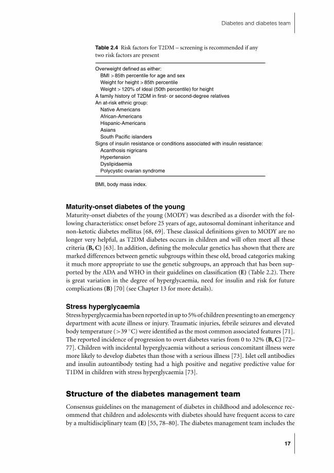
Screening for T2DMPopulation screening for T2DM in children and adolescents cannot be justified in somecountries because of the low prevalence. In countries with higher incidence, such as Japanand Taiwan, annual school glycosuria screening programmes are in place [9, 65]. Targetedscreening of youths is recommended at the point of medical contact if they are overweightand have any two risk factors listed below (E) [66, 67]. Testing may also be considered inother high-risk patients who have any of the risk factors (Table 2.4). Testing of high-risksubjects should be undertaken starting at age 10 years or at onset of puberty, whichever isearlier, and every 2 years thereafter (E) [56].
16
Diabetes and diabetes team
Table 2.4 Risk factors for T2DM – screening is recommended if anytwo risk factors are present
Overweight defined as either:BMI > 85th percentile for age and sexWeight for height > 85th percentileWeight > 120% of ideal (50th percentile) for height
A family history of T2DM in first- or second-degree relativesAn at-risk ethnic group:
Native AmericansAfrican-AmericansHispanic-AmericansAsiansSouth Pacific islanders
Signs of insulin resistance or conditions associated with insulin resistance:Acanthosis nigricansHypertensionDyslipidaemiaPolycystic ovarian syndrome
BMI, body mass index.
Maturity-onset diabetes of the youngMaturity-onset diabetes of the young (MODY) was described as a disorder with the fol-lowing characteristics: onset before 25 years of age, autosomal dominant inheritance andnon-ketotic diabetes mellitus [68, 69]. These classical definitions given to MODY are nolonger very helpful, as T2DM diabetes occurs in children and will often meet all thesecriteria (B, C) [63]. In addition, defining the molecular genetics has shown that there aremarked differences between genetic subgroups within these old, broad categories makingit much more appropriate to use the genetic subgroups, an approach that has been sup-ported by the ADA and WHO in their guidelines on classification (E) (Table 2.2). Thereis great variation in the degree of hyperglycaemia, need for insulin and risk for futurecomplications (B) [70] (see Chapter 13 for more details).
Stress hyperglycaemiaStress hyperglycaemia has been reported in up to 5% of children presenting to an emergencydepartment with acute illness or injury. Traumatic injuries, febrile seizures and elevatedbody temperature (>39 ◦C) were identified as the most common associated features [71].The reported incidence of progression to overt diabetes varies from 0 to 32% (B, C) [72–77]. Children with incidental hyperglycaemia without a serious concomitant illness weremore likely to develop diabetes than those with a serious illness [73]. Islet cell antibodiesand insulin autoantibody testing had a high positive and negative predictive value forT1DM in children with stress hyperglycaemia [73].
Structure of the diabetes management team
Consensus guidelines on the management of diabetes in childhood and adolescence rec-ommend that children and adolescents with diabetes should have frequent access to careby a multidisciplinary team (E) [55, 78–80]. The diabetes management team includes the
17
Chapter 2
child or adolescent, his or her family, paediatric endocrinologist or physician with spe-cialist training in childhood diabetes, diabetes educator, dietitian and social worker orpsychologist (Table 2.5). The aims of the diabetes management team are to provide edu-cation, initiate insulin replacement and blood glucose monitoring and provide nutritionalplanning and psychological support.
There have been no conclusive evidence-based studies validating the multidisciplinaryteam approach to diabetes management, as opposed to care provided by individual disci-plines. However, many studies have demonstrated that a team approach improves diabetesoutcomes. The largest study to date, the Diabetes Control and Complications Trial (DCCT),was a prospective, randomised multicentred clinical trial which convincingly showed thatstrict glycaemic control could be achieved by individualised treatment provided by a teamof highly trained diabetes doctors, nurses, educators and dietitians (A) [87]. Their strate-gies included frequent contact via outpatient visits as well as regular telephone contact.The investigators aimed to educate patients and parents and empower them in diabetesmanagement. The effectiveness of the diabetes management team in assisting patients toimprove glycaemic control and preventing or delaying onset of diabetes complications inthe DCCT has been the motivating factor in establishing the diabetes management teamas a necessary component of diabetes care (B, E) [55, 79, 80, 87]. In the paediatric popula-tion, a multidisciplinary team approach to intensive management of T1DM has also beenshown to improve glycaemic control (A, B) [88–91].
Involvement by the diabetes management team should begin at initial diagnosis ofdiabetes (E). This multidisciplinary approach has been shown to reduce hospital lengthof stay, as compared with management by an endocrinologist or general physician alone(B) [92]. Management by the multidisciplinary team at diagnosis may reduce the riskof microvascular disease at follow-up [84]. Many children and adolescents with newlydiagnosed diabetes can be managed from diagnosis at home rather than in hospital (A)[93, 94] (Table 2.6). Initial ambulatory management does not lead to any disadvantagesin terms of metabolic control, diabetes complications and subsequent hospitalisations orpsychosocial adjustments. However, children who present with diabetic ketoacidosis atdiagnosis must of necessity be managed in hospital until stabilisation (E) [55, 79, 80].
Ongoing review by the diabetes management team should be carried out regularly af-ter diagnosis. More frequent visits to a multidisciplinary diabetes clinic were associatedwith lower HbA1c levels (three to four visits compared with one to two visits) (B) [89,95]. Provision of a mobile diabetes education and care team to a rural area in Germanywas associated with lower HbA1c levels, fewer hospitalisations and better quality of life inchildren with T1DM (B) [96]. Current guidelines suggest that there should be at least onemajor annual review where growth, blood pressure, puberty, associated conditions, nutri-tion and complications are assessed and managed (E) [78, 79]. Availability of a patient’sHbA1c result at the time of the clinic visit improved glycaemic control (A) [97]. Diabetesmanagement via telemedicine may provide a more time-effective way of delivering care topatient with diabetes. Telemedicine may offer the advantage of more frequent contact andresult in improvement of glycaemic control, decreased insulin requirements and reducednumber of hypoglycaemic events (A, B) [98–102] (see Chapter 14 for a more detailed dis-cussion of this). However, regular phone contact with adolescents does not always improveglycaemic control (B) [103, 104].
Current guidelines recommend that the diabetes management team should be speciallytrained in the management of diabetes in children and adolescents [55, 78–80]. A child’s
18

Diabetes and diabetes team
Table 2.5 Evidence for the role of the diabetes management team
Team Level ofmember Role Evidence for effectiveness evidence
Diabetesspecialist
To providehigh-level medicalcare
(a) Specialist care was associated withhigher levels of participation in diabetesself-care practices and better glycaemiccontrol.(b) There was a higher incidence ofneuropathy, overt nephropathy andcoronary artery disease in those who didnot receive ongoing care from anendocrinologist, diabetologist ordiabetes clinic after diagnosis of T1DMin childhood.
(B) [81]
Diabeteseducator
To teach andsupport childrenand families tomanage thediabetes regimenand to becomeindependent
(a) Clinic attendance, hospital admissionand glycaemic control were improved in154 children after institution of adiabetes educator.(b) There was a 50% reduction in lengthof hospitalisation at diagnosis 2 yr afteremployment of an educator and A1clevels were lower 1 and 3 yr after usinga primary nurse managed in the team.
(A) [82, 83]
Dietitian To provide specificdiabetes-relateddietary advice andeducation
(a) Dietitians have special training inapplying and teaching about appropriatefood choices and exercise.(b) Their expertise is utilised by 86% ofconsultant paediatric services caring foryoung people with diabetes in the UK.
(C)
Socialworker
To support childand family andprovide financialand communitysupport as required
(a) Appropriate emotional supportshould be offered at diagnosis to allchildren with diabetes.(b) Intensive psychosocialeducation/support in the month followingdiagnosis led to better adherence totherapy, better family relations andbetter sociability.(c) Consultation with a social worker atdiagnosis reduced retinopathy 6 yr later.
(B, C) [80,84, 85]
Psychologist To providepsychologicalassessment andmanagement
(a) There was an improvement inglycaemic control from referral topsychologist and next clinic appointment
(C) [86]
Caseambassador
To encouragefamilies to seekmedical advice andremind orreschedule clinicappointments
(a) Their involvement increased visitsmade to the diabetes clinic.(b) When written material regardingspecific issues pertaining to diabetescare was provided, there was also asignificant reduction in hospital use andhypoglycaemic events.
(A) [82]
19

Chapter 2
Tab
le2.
6Ev
iden
cefo
rst
abili
sati
onin
ambu
lato
ryve
rsu
sin
-hos
pita
lcar
e
Evi
den
ceS
tud
yP
op
ula
tio
nIn
terv
enti
on
Co
mp
arat
or
Ou
tco
me
leve
l
Cla
r20
03S
ixst
udie
sin
volv
ing
child
ren
with
new
lydi
agno
sed
T1D
M.
Tota
lof2
37pa
tient
sin
the
outp
atie
nt/h
ome-
care
grou
p(m
ean
age
10–1
3yr
)
Hom
e-ba
sed
man
agem
ent
atdi
agno
sis
Inpa
tient
stab
ilisa
tion
atdi
agno
sis
No
sign
ifica
ntdi
ffere
nce
ingl
ycae
mic
cont
rol,
adm
issi
onto
hosp
itali
nfir
st2
yr,a
cute
com
plic
atio
nsan
dco
st
Coc
hran
esy
stem
atic
revi
ew(A
)[9
3]
Srin
ivas
an20
04A
llch
ildre
nne
wly
diag
nose
dw
ithT
1DM
(n=
61)
afte
rin
trod
uctio
nof
prog
ram
me
com
pare
dw
itha
pre-
inte
rven
tion
coho
rt(n
=49
)
Dia
bete
sda
y-ca
repr
ogra
mm
e
Inpa
tient
stab
ilisa
tion
atdi
agno
sis
Chi
ldre
nre
ceiv
ing
ambu
lato
ryst
abili
satio
nvi
aa
DC
CP
had
ash
orte
rin
itial
stay
inho
spita
land
suffe
red
noad
vers
eev
ents
(B)
[94]
McE
villy
2005
400
child
ren
with
diab
etes
Dia
bete
sho
me-
care
serv
ice
20-y
rco
mpa
rison
betw
een
outp
atie
ntca
reno
wan
din
patie
ntca
rebe
fore
(∼19
80)
Red
uced
read
mis
sion
rate
sto
hosp
ital,
HbA
1caf
ter
stab
ilisa
tion,
diab
etic
keto
acid
osis
and
cost
His
toric
alco
mpa
rison
(C)
[91]
20

Diabetes and diabetes team
age and stage of development affect management priorities such as age-specific glycaemicgoals as well as issues in self-management. Glycaemic goals in very young children shouldtake into account the unique risks of hypoglycaemia (see Chapter 5 for a more detaileddiscussion of this). The family unit is always an integral part of the management team.Whereas young children rely heavily on their families for diabetes care, middle- and high-school students should be expected to provide some of their own diabetes care whilemaintaining close supervision by their parents [55]. To date, there is a limited evidence baseto support these recommendations regarding age-specific management and education.
Conclusion
Childhood diabetes is predominantly due to T1DM that needs prompt treatment to pre-vent the development of ketoacidosis. Other forms of diabetes are also found and maybe more common than T1DM diabetes in certain populations. The incidence of T1DMvaries significantly worldwide and is increasing by approximately 3% per year globally.The rising incidence is likely to have a multifactorial basis, with recent evidence suggestingthat nutritional and lifestyle factors are important. Initial and ongoing care by a multidis-ciplinary diabetes team forms the standard for management of diabetes in children andadolescents.
References
1 Alberti K, Aschner P, Assal J-P et al. Definition, Diagnosis and Classification of Diabetes Mellitus and itsComplications. Report of a WHO Consultation Part 1: Diagnosis and Classification of Diabetes Mellitus.Geneva, 1999. Available at: http://www.staff.ncl.ac.uk/philip.home/who dmc.htm#Authors
2 American Diabetes Association. Diagnosis and classification of diabetes mellitus. Diabetes Care 2007;30(suppl 1): S42–7.
3 Harris R, Donahue K, Rathore SS et al. Screening adults for type 2 diabetes: a review of the evidencefor the U.S. Preventive Services Task Force. Ann Intern Med 2003; 138: 215–29.
4 Hoerger TJ, Harris R, Hicks KA et al. Screening for type 2 diabetes mellitus: a cost-effectiveness analysis.Ann Intern Med 2004; 140: 689–99.
5 Alberti KG, Zimmet P, Shaw J. The metabolic syndrome – a new worldwide definition. Lancet 2005;366: 1059–62.
6 Forouhi NG, Balkau B, Borch-Johnsen K et al. The threshold for diagnosing impaired fasting glucose:a position statement by the European Diabetes Epidemiology Group. Diabetologia 2006; 49: 822–7.
7 Vandewalle CL, Coeckelberghs MI, De Leeuw IH et al. Epidemiology, clinical aspects, and biology ofIDDM patients under age 40 years. Comparison of data from Antwerp with complete ascertainmentwith data from Belgium with 40% ascertainment. The Belgian Diabetes Registry. Diabetes Care 1997;20: 1556–61.
8 Pinhas-Hamiel O, Zeitler P. The global spread of type 2 diabetes mellitus in children and adolescents.J Pediatr 2005; 146: 693–700.
9 Wei JN, Sung FC, Lin CC et al. National surveillance for type 2 diabetes mellitus in Taiwanese children.JAMA 2003; 290: 1345–50.
10 The DIAMOND Project Group. Incidence and trends of childhood Type 1 diabetes worldwide 1990–1999. Diabet Med 2006; 23: 857–66.
11 International Diabetes Federation. Diabetes Atlas, 2nd edn. International Diabetes Federation, Brussels,2003.
12 Green A, Patterson CC. Trends in the incidence of childhood-onset diabetes in Europe 1989–1998.Diabetologia 2001; 44(suppl 3): B3–8.
13 Karvonen M, Viik-Kajander M, Moltchanova E et al. Incidence of childhood type 1 diabetes worldwide.Diabetes Mondiale (DiaMond) Project Group. Diabetes Care 2000; 23: 1516–26.
21

Chapter 2
14 Cucca F, Goy JV, Kawaguchi Y et al. A male-female bias in type 1 diabetes and linkage to chromosomeXp in MHC HLA-DR3-positive patients. Nat Genet 1998; 19: 301–2.
15 Gale EA, Gillespie KM. Diabetes and gender. Diabetologia 2001; 44: 3–15.16 Karvonen M, Pitkaniemi M, Pitkaniemi J et al. Sex difference in the incidence of insulin-dependent di-
abetes mellitus: an analysis of the recent epidemiological data. World Health Organization DIAMONDProject Group. Diabetes Metab Rev 1997; 13: 275–91.
17 Weets I, Kaufman L, Van der AB et al. Seasonality in clinical onset of type 1 diabetes in Belgian patientsabove the age of 10 is restricted to HLA-DQ2/DQ8-negative males, which explains the male to femaleexcess in incidence. Diabetologia 2004; 47: 614–21.
18 Barker JM. Clinical review: type 1 diabetes-associated autoimmunity: natural history, genetic associ-ations, and screening. J Clin Endocrinol Metab 2006; 91: 1210–17.
19 Gillespie KM, Bain SC, Barnett AH et al. The rising incidence of childhood type 1 diabetes and reducedcontribution of high-risk HLA haplotypes. Lancet 2004; 364: 1699–1700.
20 Kukko M, Virtanen SM, Toivonen A et al. Geographical variation in risk HLA-DQB1 genotypes fortype 1 diabetes and signs of beta-cell autoimmunity in a high-incidence country. Diabetes Care 2004;27: 676–81.
21 Sugihara S, Sakamaki T, Konda S et al. Association of HLA-DR, DQ genotype with different beta-cellfunctions at IDDM diagnosis in Japanese children. Diabetes 1997; 46: 1893–7.
22 Kawasaki E, Matsuura N, Eguchi K. Type 1 diabetes in Japan. Diabetologia 2006; 49: 828–36.23 Ohtsu S, Takubo N, Kazahari M et al. Slowly progressing form of type 1 diabetes mellitus in children:
genetic analysis compared with other forms of diabetes mellitus in Japanese children. Pediatr Diabetes2005; 6: 221–9.
24 Raymond NT, Jones JR, Swift PG et al. Comparative incidence of Type I diabetes in children agedunder 15 years from South Asian and White or Other ethnic backgrounds in Leicestershire, UK, 1989to 1998. Diabetologia 2001; 44(suppl 3): B32–6.
25 Shamis I, Gordon O, Albag Y et al. Ethnic differences in the incidence of childhood IDDM in Is-rael (1965–1993). Marked increase since 1985, especially in Yemenite Jews. Diabetes Care 1997; 20:504–8.
26 Neu A, Willasch A, Ehehalt S et al. Diabetes incidence in children of different nationalities: an epi-demiological approach to the pathogenesis of diabetes. Diabetologia 2001; 44(suppl 3): B21–6.
27 Onkamo P, Vaananen S, Karvonen M et al. Worldwide increase in incidence of Type I diabetes – theanalysis of the data on published incidence trends. Diabetologia 1999; 42: 1395–1403.
28 Haynes A, Bulsara MK, Bower C et al. Independent effects of socioeconomic status and place ofresidence on the incidence of childhood type 1 diabetes in Western Australia. Pediatr Diabetes 2006;7: 94–100.
29 Taplin CE, Craig ME, Lloyd M et al. The rising incidence of childhood type 1 diabetes in New SouthWales, 1990–2002. Med J Aust 2005; 183: 243–6.
30 EURODIABS ACE Study Group. Variation and trends in incidence of childhood diabetes in Europe.EURODIAB ACE Study Group. Lancet 2000; 355: 873–6.
31 Karvonen M, Pitkaniemi J, Tuomilehto J. The onset age of type 1 diabetes in Finnish children hasbecome younger. The Finnish Childhood Diabetes Registry Group. Diabetes Care 1999; 22: 1066–70.
32 Viskari HR, Koskela P, Lonnrot M et al. Can enterovirus infections explain the increasing incidence oftype 1 diabetes? Diabetes Care 2000; 23: 414–16.
33 Betts P, Mulligan J, Ward P et al. Increasing body weight predicts the earlier onset of insulin-dependantdiabetes in childhood: testing the ‘accelerator hypothesis’ (2). Diabet Med 2005; 22: 144–51.
34 EURODIABS Study Group. Rapid early growth is associated with increased risk of childhood type 1diabetes in various European populations. Diabetes Care 2002; 25: 1755–60.
35 Patterson CC, Dahlquist G, Soltesz G et al. Is childhood-onset type I diabetes a wealth-related disease?An ecological analysis of European incidence rates. Diabetologia 2001; 44(suppl 3): B9–16.
36 Levy-Marchal C, Patterson C, Green A. Variation by age group and seasonality at diagnosis of childhoodIDDM in Europe. The EURODIAB ACE Study Group. Diabetologia 1995; 38: 823–30.
37 Laron Z, Lewy H, Wilderman I et al. Seasonality of month of birth of children and adolescents withtype 1 diabetes mellitus in homogenous and heterogeneous populations. Isr Med Assoc J 2005; 7: 381–4.
38 Rothwell PM, Gutnikov SA, McKinney PA et al. Seasonality of birth in children with diabetes in Europe:multicentre cohort study. European Diabetes Study Group. BMJ 1999; 319: 887–8.
39 Yang Z, Long X, Shen J et al. Epidemics of type 1 diabetes in China. Pediatr Diabetes 2005; 6: 122–8.
22

Diabetes and diabetes team
40 Olmos P, A’Hern R, Heaton DA et al. The significance of the concordance rate for type 1 (insulin-dependent) diabetes in identical twins. Diabetologia 1988; 31: 747–50.
41 Harjutsalo V, Podar T, Tuomilehto J. Cumulative incidence of type 1 diabetes in 10,168 siblings ofFinnish young-onset type 1 diabetic patients. Diabetes 2005; 54: 563–9.
42 Steck AK, Barriga KJ, Emery LM et al. Secondary attack rate of type 1 diabetes in Colorado families.Diabetes Care 2005; 28: 296–300.
43 Lorenzen T, Pociot F, Hougaard P et al. Long-term risk of IDDM in first-degree relatives of patientswith IDDM. Diabetologia 1994; 37: 321–7.
44 Gillespie KM, Gale EA, Bingley PJ. High familial risk and genetic susceptibility in early onset childhooddiabetes. Diabetes 2002; 51: 210–14.
45 Dorman JS, Steenkiste AR, O’Leary LA et al. Type 1 diabetes in offspring of parents with type 1 diabetes:the tip of an autoimmune iceberg? Pediatr Diabetes 2000; 1: 17–22.
46 el Hashimy M, Angelico MC, Martin BC et al. Factors modifying the risk of IDDM in offspring of anIDDM parent. Diabetes 1995; 44: 295–9.
47 EURODIABS ACE Study Group. Familial risk of type I diabetes in European children. The EurodiabAce Study Group and The Eurodiab Ace Substudy 2 Study Group. Diabetologia 1998; 41: 1151–6.
48 Lorenzen T, Pociot F, Stilgren L et al. Predictors of IDDM recurrence risk in offspring of Danish IDDMpatients. Danish IDDM Epidemiology and Genetics Group. Diabetologia 1998; 41: 666–73.
49 Warram JH, Krolewski AS, Gottlieb MS et al. Differences in risk of insulin-dependent diabetes inoffspring of diabetic mothers and diabetic fathers. N Engl J Med 1984; 311: 149–52.
50 Dahlquist G, Mustonen L. Analysis of 20 years of prospective registration of childhood onset diabetestime trends and birth cohort effects. Swedish Childhood Diabetes Study Group. Acta Paediatr 2000;89: 1231–7.
51 Hypponen E, Virtanen SM, Kenward MG et al. Obesity, increased linear growth, and risk of type 1diabetes in children. Diabetes Care 2000; 23: 1755–60.
52 Clarke SL, Craig ME, Garnett SP et al. Increased adiposity at diagnosis in younger children with type1 diabetes does not persist. Diabetes Care 2006; 29: 1651–3.
53 Kordonouri O, Hartmann R. Higher body weight is associated with earlier onset of Type 1 diabetes inchildren: confirming the ‘Accelerator Hypothesis’. Diabet Med 2005; 22: 1783–4.
54 World Health Organization. Diagnosis and classification of diabetes mellitus. Diabetes Care 2006;29(suppl 1): S43–8.
55 Silverstein J, Klingensmith G, Copeland K et al. Care of children and adolescents with type 1 diabetes:a statement of the American Diabetes Association. Diabetes Care 2005; 28: 186–212.
56 American Diabetes Association. Type 2 diabetes in children and adolescents. American Diabetes As-sociation. Pediatrics 2000; 105: 671–80.
57 Pinhas-Hamiel O, Dolan LM, Zeitler PS. Diabetic ketoacidosis among obese African-American ado-lescents with NIDDM. Diabetes Care 1997; 20: 484–6.
58 Krakoff J, Lindsay RS, Looker HC et al. Incidence of retinopathy and nephropathy in youth-onsetcompared with adult-onset type 2 diabetes. Diabetes Care 2003; 26: 76–81.
59 Eppens MC, Craig ME, Cusumano J et al. Prevalence of diabetes complications in adolescents withtype 2 compared with type 1 diabetes. Diabetes Care 2006; 29: 1300–6.
60 Pavkov ME, Bennett PH, Knowler WC et al. Effect of youth-onset type 2 diabetes mellitus on incidenceof end-stage renal disease and mortality in young and middle-aged Pima Indians. JAMA 2006; 296:421–6.
61 Nadeau KJ, Klingensmith G, Zeitler P. Type 2 diabetes in children is frequently associated with elevatedalanine aminotransferase. J Pediatr Gastroenterol Nutr 2005; 41: 94–98.
62 Roberts EA. Non-alcoholic fatty liver disease (NAFLD) in children. Front Biosci 2005; 10: 2306–18.63 American Diabetes Association. Type 2 diabetes in children and adolescents. Diabetes Care 2000; 23:
381–9.64 American Diabetes Association. Screening for type 2 diabetes. Diabetes Care 2004; 27(suppl 1):
S11–14.65 Urakami T, Kubota S, Nitadori Y et al. Annual incidence and clinical characteristics of type 2 diabetes
in children as detected by urine glucose screening in the Tokyo metropolitan area. Diabetes Care 2005;28: 1876–81.
66 Alberti G, Zimmet P, Shaw J et al. Type 2 diabetes in the young: the evolving epidemic: the internationaldiabetes federation consensus workshop. Diabetes Care 2004; 27: 1798–1811.
23

Chapter 2
67 Pinhas-Hamiel O, Standiford D, Hamiel D et al. The type 2 family: a setting for development andtreatment of adolescent type 2 diabetes mellitus. Arch Pediatr Adolesc Med 1999; 153: 1063–7.
68 Fajans SS, Bell GI, Polonsky KS. Molecular mechanisms and clinical pathophysiology of maturity-onsetdiabetes of the young. N Engl J Med 2001; 345: 971–80.
69 Owen K, Hattersley AT. Maturity-onset diabetes of the young: from clinical description to moleculargenetic characterization. Best Pract Res Clin Endocrinol Metab 2001; 15: 309–23.
70 Ehtisham S, Hattersley AT, Dunger DB et al. First UK survey of paediatric type 2 diabetes and MODY.Arch Dis Child 2004; 89: 526–9.
71 Valerio G, Franzese A, Carlin E et al. High prevalence of stress hyperglycaemia in children with febrileseizures and traumatic injuries. Acta Paediatr 2001; 90: 618–22.
72 Bhisitkul DM, Vinik AI, Morrow AL et al. Prediabetic markers in children with stress hyperglycemia.Arch Pediatr Adolesc Med 1996; 150: 936–41.
73 Herskowitz-Dumont R, Wolfsdorf JI, Jackson RA et al. Distinction between transient hyperglycemiaand early insulin-dependent diabetes mellitus in childhood: a prospective study of incidence andprognostic factors. J Pediatr 1993; 123: 347–54.
74 Herskowitz RD, Wolfsdorf JI, Ricker AT et al. Transient hyperglycemia in childhood: identification ofa subgroup with imminent diabetes mellitus. Diabetes Res 1988; 9: 161–7.
75 Schatz DA, Kowa H, Winter WE et al. Natural history of incidental hyperglycemia and glycosuria ofchildhood. J Pediatr 1989; 115: 676–80.
76 Shehadeh N, On A, Kessel I et al. Stress hyperglycemia and the risk for the development of type 1diabetes. J Pediatr Endocrinol Metab 1997; 10: 283–6.
77 Vardi P, Shehade N, Etzioni A et al. Stress hyperglycemia in childhood: a very high risk group for thedevelopment of type I diabetes. J Pediatr 1990; 117: 75–7.
78 ISPAD. Consensus Guidelines for the Management of Type 1 Diabetes Mellitus in Children and Adolescents.Available at: http://www.diabetesguidelines.com/health/dkw/pro/guidelines/ispad/ispad.asp
79 NHMRC. The Australian Clinical Practice Guidelines on the Management of Type 1 Diabetes in Childrenand Adolescents 2005. Available at: http://www.chw.edu.au/prof/services/endocrinology/apeg/
80 NICE. Type 1 Diabetes in Children and Young People: Full Guideline 2004. Available at:http://www.nice.org.uk/page.aspx?o=CG015childfullguideline
81 Zgibor JC, Songer TJ, Kelsey SF et al. Influence of health care providers on the development of diabetescomplications: long-term follow-up from the Pittsburgh Epidemiology of Diabetes ComplicationsStudy. Diabetes Care 2002; 25: 1584–90.
82 Ahern JA, Ramchandani N, Cooper J et al. Using a primary nurse manager to implement DCCTrecommendations in a large pediatric program. Diabetes Educ 2000; 26: 990–4.
83 Cowan FJ, Warner JT, Lowes LM et al. Auditing paediatric diabetes care and the impact of a specialistnurse trained in paediatric diabetes. Arch Dis Child 1997; 77: 109–14.
84 Donaghue KC, Craig ME, Chan AK et al. Prevalence of diabetes complications 6 years after diagnosisin an incident cohort of childhood diabetes. Diabet Med 2005; 22: 711–18.
85 Galatzer A, Amir S, Gil R et al. Crisis intervention program in newly diagnosed diabetic children.Diabetes Care 1982; 5: 414–19.
86 Gelfand K, Geffken G, Lewin A et al. An initial evaluation of the design of pediatric psychologyconsultation service with children with diabetes. J Child Health Care 2004; 8: 113–23.
87 DCCT Research Group. The effect of intensive treatment of diabetes on the development and progres-sion of long-term complications in insulin-dependent diabetes mellitus. The Diabetes Control andComplications Trial Research Group. N Engl J Med 1993; 329: 977–86.
88 DCCT Research Group. Effect of intensive diabetes treatment on the development and progression oflong-term complications in adolescents with insulin-dependent diabetes mellitus: Diabetes Controland Complications Trial. Diabetes Control and Complications Trial Research Group. J Pediatr 1994;125: 177–88.
89 Kaufman FR, Halvorson M, Carpenter S. Association between diabetes control and visits to a multi-disciplinary pediatric diabetes clinic. Pediatrics 1999; 103: 948–51.
90 Lawson ML, Frank MR, Fry MK et al. Intensive diabetes management in adolescents withtype 1 diabetes: the importance of intensive follow-up. J Pediatr Endocrinol Metab 2000; 13:79–84.
91 McEvilly A, Kirk J. Twenty years of a multidisciplinary paediatric diabetes home care unit. Arch DisChild 2005; 90: 342–5.
24

Diabetes and diabetes team
92 Levetan CS, Salas JR, Wilets IF et al. Impact of endocrine and diabetes team consultation on hospitallength of stay for patients with diabetes. Am J Med 1995; 99: 22–8.
93 Clar C, Waugh N, Thomas S. Routine hospital admission versus out-patient or home care in childrenat diagnosis of type 1 diabetes mellitus. Cochrane Database Syst Rev 2003; Issue no 3: Art No CD004099
94 Srinivasan S, Craig ME, Beeney L et al. An ambulatory stabilisation program for children with newlydiagnosed type 1 diabetes. Med J Aust 2004; 180: 277–80.
95 Nordly S, Mortensen HB, Andreasen AH et al. Factors associated with glycaemic outcome of childhooddiabetes care in Denmark. Diabet Med 2005; 22: 1566–73.
96 von Sengbusch S, Muller-Godeffroy E, Hager S et al. Mobile diabetes education and care: interventionfor children and young people with Type 1 diabetes in rural areas of northern Germany. Diabet Med2006; 23: 122–7.
97 Cagliero E, Levina EV, Nathan DM. Immediate feedback of HbA1c levels improves glycemic controlin type 1 and insulin-treated type 2 diabetic patients. Diabetes Care 1999; 22: 1785–9.
98 d’Annunzio G, Bellazzi R, Larizza C et al. Telemedicine in the management of young patients withtype 1 diabetes mellitus: a follow-up study. Acta Biomed Ateneo Parmense 2003; 74(suppl 1): 49–55.
99 Farmer A, Gibson OJ, Tarassenko L et al. A systematic review of telemedicine interventions to supportblood glucose self-monitoring in diabetes. Diabet Med 2005; 22: 1372–8.
100 Gelfand K, Geffken G, Halsey-Lyda M et al. Intensive telehealth management of five at-risk adolescentswith diabetes. J Telemed Telecare 2003; 9: 117–21.
101 Izquierdo RE, Knudson PE, Meyer S et al. A comparison of diabetes education administered throughtelemedicine versus in person. Diabetes Care 2003; 26: 1002–7.
102 Liesenfeld B, Renner R, Neese M et al. Telemedical care reduces hypoglycemias and improves glycemiccontrol in children and adolescents with type 1 diabetes. Diabetes Technol Ther 2000; 2: 561–7.
103 Howells L, Wilson AC, Skinner TC et al. A randomized control trial of the effect of negotiated telephonesupport on glycaemic control in young people with Type 1 diabetes. Diabet Med 2002; 19: 643–8.
104 Lawson ML, Cohen N, Richardson C et al. A randomized trial of regular standardized telephonecontact by a diabetes nurse educator in adolescents with poor diabetes control. Pediatr Diabetes 2005;6: 32–40.
25

CHAPTER 3
Aetiology of type 1 diabetesmellitus – genetics, autoimmunityand trigger factors
Loredana Marcovecchio, David B. Dunger,Mark Peakman & Keith W. Taylor
A thing is not proved just because no one has ever questioned it. What has never beengone into impartially has never been properly gone into. Hence scepticism is the first steptoward truth. It must be applied generally, because it is the touchstone.
— Denis Diderot, French author, encyclopaedist and philosopher (1713–1784)
It is now well established that type 1 diabetes mellitus (T1DM) results from a combinationof a genetic predisposition and autoimmune processes that result in destruction of thebeta cells of the pancreas and cause absolute insulin deficiency. However, not everyonewho has this genetic predisposition develops T1DM and it is also thought that someadditional factor is required to trigger the autoimmune responses that cause the beta-cell destruction. This chapter presents the evidence that is currently available to definethat genetic predisposition, the immunological processes that occur to cause beta-celldestruction and the trigger factors that may provide the necessary spark.
Genetics of T1DMLoredana Marcovecchio and David B. Dunger
T1DM is an autoimmune disease with genetic and environmental factors implicated in itsaetiology. The role of genetic factors in the susceptibility to T1DM is strongly supportedby family and twin studies (B) [1, 2]. The familial clustering of T1DM is well known, with10–13% of newly diagnosed cases having a first-degree relative with T1DM and a 15-foldincreased risk of developing the disease for a sibling (5–6%) of a patient with T1DMwhen compared to the general population (0.4%) (B) [2]. The family recurrence of T1DMvaries depending on which relative is affected, with a higher risk for the siblings than forthe offspring or parents of a patient with T1DM (B) [2, 3]. Furthermore, the risk for anoffspring is higher when the father (6–9%) is affected than when the mother (1.3–4.0%)is the index case, and this difference might be related to imprinted genes or to a protectiverole of maternal environmental factors during pregnancy (B) [3].
In T1DM the concordance rate for monozygotic twins ranges between 21 and 70%, whilefor dizygotic twins it is 0–13% (B) [2]. The progression rate to T1DM for monozygotic
26

Aetiology of type 1 diabetes mellitus
Table 3.1 Examples of HLA haplotypes and genotypes associated with susceptibility for orprotection against T1DM
DR alleles DQ alleles OR-r (CI)
DR4: DRB1*0401 A1*0301, B1*0302 (DQ8) 8.3 (7.2–9.6)DR4: DRB1*0405 A1*0301, B1*0302 6.6 (5.3–8.3)High-riskDR4: DRB1*0402 A1*0301, B1*0302 4.0 (3.0–5.5)haplotypesDR4: DRB1*0404 A1*0301, B1*0302 3.5 (3.0–4.1)DR3: DRB1*0301 A1*0501, B1*0201 (DQ2) 4.1 (3.7–4.6)
DR2: DRB1*15 A1*01, B1*06 (DQ6) 0.0 (0.0–0.1)DR5: DRB1*11 A1*0501, B1*0301 0.1 (0.1–0.2)ProtectiveDR6: DRB*14 A1*01, B1*05 0.1 (0.1–0.2)haplotypesDR7: DRB*07 A1*0201, B1*0303 0.1 (0.1–0.2)DR4: DRB*0407 A1*0301, B1*0301 0.2 (0.1–0.4)
DR/DQ haplotype 1 DR/DQ haplotype 2 OR-r (CI)
DR4/DQ8 DR3/DQ2 47.2 (26.7–83.3)DR4/DQ8 DR4/DQ8 11.9 (6.1–23.1)GenotypesDR3/DQ2 DR3/DQ2 10.4 (6.0–18.0)DR2/DQ6 DR2/DQ6 0.1 (0.0–0.3)
OR-r (CI), odds ratios (confidence intervals), calculated relative to the DQB1*5-DRB1*01 referencehaplotype (OR = 1).Data from Koeleman BP et al., 2004 [7].
twins is greater than 50% if the index twin develops diabetes before the age of 5 years,while it is less than 10% if the diagnosis is made after the age of 25 years (A, B) [4, 5]. Theabsence of a 100% concordance between monozygotic twins, however, suggests that otherfactors, such as environmental triggers, are implicated (B) [2].
Multiple genetic loci contributing to T1DM risk have been identified but not all havebeen confirmed. They provide both susceptibility and protection (B) [6]. The humanleucocyte antigen (HLA) complex (IDDM1 locus) was the first susceptibility locus to beassociated with T1DM and it confers about 40–50% of the inherited risk (B) [6]. The HLAgenes, located within the major histocompatibility complex on the chromosome 6p21, arehighly polymorphic and have an important role in the modulation of the immune sys-tem (Figure 3.1). The risk for T1DM is mostly associated with class II genes, particularlywith HLA-DRB1, DQA1 and DQB1 (B) [1, 6–8] (Table 3.1). The high-risk haplotypesDR3/DQ2 (DRB1*0301-DQA1*0501-DQB1*0201) and DR4/DQ8 (DRB1*0401, *0402,*0405-DQA1*0301-DQB1*0201) are carried by more than 90% of white Caucasian individ-uals with T1DM and by fewer than 40% of normal controls. The highest risk is associatedwith DR3/DR4 heterozygosity, which is found in almost 50% of children who develop dia-betes before the age of 5 years, in 20–30% of adults presenting with diabetes and in only 2.4%of the general population (B) [2]. However, the HLA high-risk genotypes seem to be un-common in children with permanent neonatal diabetes and in those diagnosed before theage of 6 months, who are therefore more likely to have a non-autoimmune form of diabetes,often related to activating mutations in the Kir6.2 (KCNJ11) and SUR1 genes (A) [9–11].
The different HLA haplotypes are also associated with the pattern of autoantibodies, withinsulin autoantibodies (IAA) and IA-2 particularly frequent in patients with the DR4-DQ8haplotype and GAD-65 more common in patients with the DR3-DQ2 haplotype and the
27

Chapter 3
CD8+ T cell
Pancreatic beta -cell
beta -cell peptides e.g. insulin
HLA class II= IDDM1
HELP
T helper 1cell
TCR
CTLA-4 = IDDM12
Thymic insulinexpression
= IDDM2
Antigen-presenting
cell
Figure 3.1 A model for immune pathogenesis in T1DM: role of the susceptibility loci IDDM1,IDDM2 and IDDM12.
highest probability to be autoantibody negative for patients without DR3 and DR4 (B)[12]. Some HLA alleles can also confer protection from diabetes and among them the bestknown is HLA-DQ6 (HLA-DQB1*0602), carried by fewer than 1% of subjects with T1DMcompared to 20% of the general population (B) [2].
The second known susceptibility locus for T1DM is IDDM2 on chromosome 11p15.5,which includes the variable number of tandem repeats (VNTR) region located in thepromoter of the insulin gene and contributes about 10% to disease susceptibility (B) [2].There are three main classes of VNTR alleles, categorised on the basis of the number ofrepeats: class I (26–63 repeats), class II (approximately, 80 repeats) and class III (140–200repeats). Variation in the number of repeats affects the expression of the insulin gene inthe thymus and pancreas and the placental expression of the insulin-like growth factor2 gene [13]. The class III alleles determine the highest insulin levels within the thymusand confer a dominant protection against T1DM, while homozygosity for class I allelesis associated with the lowest insulin expression and the highest risk for the disease. Theassociation between insulin expression within the thymus and diabetes susceptibility isthought to be related to a dose-dependent effect of insulin in modifying the selection ofpotentially damaging autoreactive T cells and/or inducing insulin-specific regulatory T cells(i.e. high insulin levels in the thymus lead to deletion of the damaging cells and inductionof regulatory ones) [13] (Figure 3.1). The susceptibility related to VNTR polymorphismsshows race-dependent differences, with a greater effect in white Caucasians than in blackCaucasians and no effect in the Japanese population (A) [14]. Furthermore, in some studiesa parent-of-origin effect has been described, with protection deriving from the paternallytransmitted allele (A) [15].
The third susceptibility locus in T1DM (IDDM12) is located on chromosome 2q33 andencodes a co-stimulatory molecule, called cytotoxic T lymphocyte antigen 4 (CTLA-4),which is expressed on the surface of activated T cells (B) [16, 17] and plays a critical rolein the response to antigens through a negative regulation of T-cell activation. Differentpolymorphisms in the CTLA-4 gene have been associated with T1DM and they are locatedin the 3’ untranslated, promoter (−319C > T) and coding regions (+49A/G) (B) [17].
28
Aetiology of type 1 diabetes mellitus
Some of these polymorphisms are associated with reduced stability of the RNA or alter-ations in the splicing mechanism with a reduced expression of CTLA-4 on the cell surface(Figure 3.1). Recently, evidence for a role of CTLA-4 in other autoimmune en-docrinopathies, particularly Grave’s disease and hypothyroidism, has also been found(A) [18]. As for IDDM2, also for the IDDM12 locus there is an ethnic heterogeneity insusceptibility to T1DM (B) [19].
A fourth human susceptibility locus, protein tyrosine phosphatase non-receptor type 22(PTPN22), on chromosome 1p13, has recently been reported (B, A) [20, 21]. This locuscontains the gene encoding for a lymphocyte-specific phosphatase implicated in a negativeregulation of T-cell development and activation.
Recently, convincing statistical support has also been found for two other regions: theIL2RA/CD25 on chromosome 10p15.1, encoding the alpha chain of the IL-2 receptor,and the interferon-induced helicase 1 gene region (IFIH1/MDA5) on chromosome 2q24.3(A) [22, 23].
Other loci have been associated with T1DM, but for most of them the related genehas not been identified yet and different results have been often found in different pop-ulations (Table 3.2) [24–30]. Linkage and association analyses have been the two mainapproaches used to identify T1DM susceptibility genes but the main barrier in their map-ping results from small sample sizes and the heterogeneity of the populations analysedin different genome-wide scans (C). In order to facilitate the identification of genes im-plicated in T1DM with a good statistical power, a collection of DNA samples from 8000subjects with T1DM and the same number of controls has now been underway as partof the UK Genetic Resource Investigating Diabetes (GRID) study (A) [31]. Furthermore,a T1DM consortium has also been established with the aim of collecting and analysingthousands of DNA samples from affected sibling pairs (A) [24].
Immunology of T1DMMark Peakman
T1DM has all of the hallmarks of an autoimmune disorder; indeed, it is often referred toas the prototypic organ-specific autoimmune disease (Table 3.3). What exactly does thismean? In short, it implies that in a patient with the disease the immune system has becomesufficiently dysregulated to attack and destroy healthy islet beta cells. This process takesbetween a few months and several years and is the end result of a complex interplay of hostgenetic and environmental factors, which are becoming increasingly better understood.
Pathology of the islet in T1DMHealthy human islets of Langerhans are composed of a core of some 80% beta cells (makingthe glucose-regulating hormone insulin), with a mantle of other endocrine cell types,producing glucagon, somatostatin and pancreatic polypeptide (alpha, delta and PP cells,respectively) making up the remainder. Histologically, the islets of Langerhans at diagnosisof T1DM have a mixture of appearances:� Some islets are small, without beta cells and have no inflammatory cells.� Others have numerous beta cells surrounded by infiltrating activated T (CD4 and CD8)and B lymphocytes and various antigen-presenting cells (APCs) of the immune system(an appearance termed insulitis) [41].� Some islets appear normal.
29

Chapter 3
Table 3.2 T1DM susceptibility loci with LOD scores as observed in genome linkage scans
Locus Location Candidate gene LOD [reference]
IDDM1 6p21 HLA DR/DQ 116.4 [24]IDDM2 11p15 Insulin VNTR 1.87 [24]IDDM3 15q26 2.5 [25]IDDM4 11q13 ZFM1, LRP5, FADD 3.4 [26]IDDM5 6q25 SUMO4, MnSOD 1.96 [27]IDDM6 18q12-q21 JK, ZNF236, BCL-2 1.1 [26]IDDM7 2q33 NEUROD, IL1B, HOXD8, GAD1, GALNT3 3.34 [24]IDDM8 6q27 1.8 [27]IDDM9 3q22-25 3.4 [28]IDDM10 10p11-q11 3.21 [24]IDDM11 14q24-q31 ENSA, SEL1L 4.0 [29]IDDM12 2q31-q33 CTLA-4 3.34 [24]IDDM13 2q34-q35 IGFBP-2, IGFBP-5, IA-2, NRAMPI 2.6 [27]IDDM15 6q21 22.4 [24]IDDM16 14q32 IGHIDDM17 10q25 2.38 [30]IDDM18 5q33 IL12B
1p13 PTPN22 (LYP)1q42 2.2 [27]2q24.3 IFIH1 (MDA5)3p13-p14 1.52 [24]9q33-q34 2.20 [24]10p15 IL2RA (CD45)12q14-q12 1.66 [24]16p12-q11.1 1.88 [24]16q22-q24 2.64 [24]17q25 1.81 [27]19q11 1.80 [27]19p13.3-p13.2 1.92 [24]
LOD scores are mostly from the recent four genome-wide linkage scans in 1435 multiplex families (USA,UK and Scandinavia) [24]. As this analysis did not show significant linkage for some loci previouslyidentified, a LOD value from a different reference is reported, when available.LOD, logarithm of odds.
Table 3.3 Evidence that T1DM is an autoimmune disease
Criteria Reference
MajorPresence of circulating autoantibodies against targets in the beta cell [32]Presence of circulating, activated T cells directed against targets in the beta cell [33, 34]Clinical response to immune suppression [35–37]Coexistence of other autoimmune diseases (thyroiditis and the Addison disease) [38]Inclusion of T1DM in autoimmune polyendocrine syndromes (APS I, II) [38]
MinorExistence of relevant spontaneous animal model (NOD mouse) [39]Passive transfer of disease using T lymphocytes from diabetic NOD mouse tonaıve mouse
[39]
Association between T1DM and possession of HLA genes [40]
NOD, non-obese diabetic.
30

Aetiology of type 1 diabetes mellitus
It is probable that these appearances reflect stages of a process during which the immunecells travel between islets in a ‘seek, destroy, move on’ operation (‘flitting insulitis’). Thisimmune system’s ‘target-and-destroy’ operation is exquisitely specific – only the beta cellsare damaged, whilst the other endocrine cells remain intact and functional.
Islet cell autoantibodiesIslet cell autoantibodies are the defining immunological feature of T1DM. There are threeproteins that constitute the major target autoantigens for these autoantibodies:� insulin (IAA)� glutamic acid decarboxylse 65 (GAD-65 autoantibodies, GADA)� tyrosine phosphatase-like molecule called IA-2 (IA-2A)
Insulin is the only islet autoantigen specifically localised to the beta cells. GAD-65 andIA-2 may also be found in other islet cell types and in the central and peripheral nervoussystem. Although each individual autoantibody may only be present in a half to three-quarters of children at the time of development of T1DM, typically 90–95% of thesewill have at least one of the autoantibodies. Insulin autoantibodies are more prevalent inchildren than in adults, whilst the reverse is true of GADA (A) [32].
The technology to detect these three types of islet cell autoantibodies is now widelyavailable in clinical immunology laboratories, begging the question as to when their mea-surement might be useful clinically. In the vast majority of cases T1DM is diagnosedclinically in children because of the classical history and laboratory findings. Rarely inchildren, but more commonly in adults, the disease may evolve slowly or have an unusualpresentation and autoantibodies are useful as a confirmation. However, the detection ofislet cell autoantibodies is emerging as being especially important in disease prediction.From studies on individuals at risk of developing T1DM (e.g. first-degree relatives ofpatients), it has become clear that apparently healthy people can have islet cell autoanti-bodies – up to 10% of relatives are positive for IAA, GADA or IA-2A when tested. Throughthe prospective follow-up of such individuals for 5–10 years, a very clear pattern of dis-ease risk has emerged, in which some of these relatives do indeed progress to diabetes,the risk increasing with the number of autoantibodies present (B) [42]. Thus it wouldappear that the breadth and intensity of the autoimmune response, as reflected in thenumber of different autoantibodies produced, determines whether anti-islet immunitydevelops into clinical disease. These pioneering studies have now been translated intothe general population (A) [43]. (This is important for screening purposes, as 90% ofnew diabetic patients do not have relatives with the disease.) Although far fewer of thegeneral public are autoantibody positive, those who are have a disease risk determinedby the number of autoantibodies present. Combinations of autoantibodies and geneticmarkers (e.g. the high-risk HLA gene DQB1*0302) can be used to increase the specificityof prediction. Although disease prediction through islet cell autoantibody detection wasinitially an academic exercise, it has become the lynchpin of strategies for the developmentof new approaches to disease prevention (see below) and is being increasingly developedand refined.
The immune pathogenesis of T1DMAdvances in immunological understanding in the last 30 years, since the first descriptionsof HLA and autoantibody associations with T1DM, have led to a much clearer view of howbeta cells might undergo damage, although many questions remain. The fact that islet cell
31
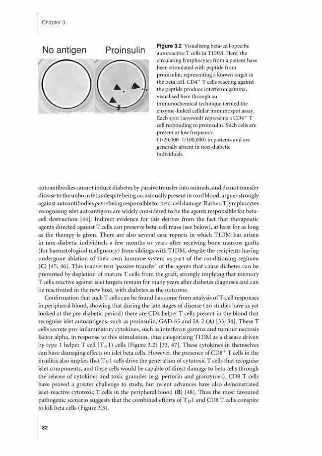
Chapter 3
No antigen ProinsulinFigure 3.2 Visualising beta-cell-specificautoreactive T cells in T1DM. Here, thecirculating lymphocytes from a patient havebeen stimulated with peptide fromproinsulin, representing a known target inthe beta cell. CD4+ T cells reacting againstthe peptide produce interferon gamma,visualised here through animmunochemical technique termed theenzyme-linked cellular immunospot assay.Each spot (arrowed) represents a CD4+ Tcell responding to proinsulin. Such cells arepresent at low frequency(1/20,000–1/100,000) in patients and aregenerally absent in non-diabeticindividuals.
autoantibodies cannot induce diabetes by passive transfer into animals, and do not transferdisease to the unborn fetus despite being occasionally present in cord blood, argues stronglyagainst autoantibodies per se being responsible for beta-cell damage. Rather, T lymphocytesrecognising islet autoantigens are widely considered to be the agents responsible for beta-cell destruction [44]. Indirect evidence for this derives from the fact that therapeuticagents directed against T cells can preserve beta-cell mass (see below), at least for as longas the therapy is given. There are also several case reports in which T1DM has arisenin non-diabetic individuals a few months or years after receiving bone marrow grafts(for haematological malignancy) from siblings with T1DM, despite the recipients havingundergone ablation of their own immune system as part of the conditioning regimen(C) [45, 46]. This inadvertent ‘passive transfer’ of the agents that cause diabetes can beprevented by depletion of mature T cells from the graft, strongly implying that memoryT cells reactive against islet targets remain for many years after diabetes diagnosis and canbe reactivated in the new host, with diabetes as the outcome.
Confirmation that such T cells can be found has come from analysis of T-cell responsesin peripheral blood, showing that during the late stages of disease (no studies have as yetlooked at the pre-diabetic period) there are CD4 helper T cells present in the blood thatrecognise islet autoantigens, such as proinsulin, GAD-65 and IA-2 (A) [33, 34]. These Tcells secrete pro-inflammatory cytokines, such as interferon gamma and tumour necrosisfactor alpha, in response to this stimulation, thus categorising T1DM as a disease drivenby type 1 helper T cell (TH 1) cells (Figure 3.2) [33, 47]. These cytokines in themselvescan have damaging effects on islet beta cells. However, the presence of CD8+ T cells in theinsulitis also implies that TH 1 cells drive the generation of cytotoxic T cells that recogniseislet components, and these cells would be capable of direct damage to beta cells throughthe release of cytokines and toxic granules (e.g. perforin and granzymes). CD8 T cellshave proved a greater challenge to study, but recent advances have also demonstratedislet-reactive cytotoxic T cells in the peripheral blood (B) [48]. Thus the most favouredpathogenic scenario suggests that the combined efforts of TH 1 and CD8 T cells conspireto kill beta cells (Figure 3.3).
32
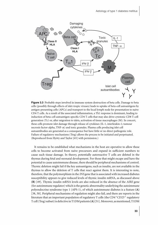
Aetiology of type 1 diabetes mellitus
Class IHLA
Treg
Plasma cell
IL-1
TNF- α
IFN-γM
IFN-γ
TH1
Class IIHLA
APCBeta cell
Islet cellautoantibodies
Damagingcytokines
NaïveT cell Tc
Toxicgranules
Figure 3.3 Probable steps involved in immune system destruction of beta cells. Damage to betacells (possibly through effects of islet-tropic viruses) leads to uptake of beta-cell autoantigens byantigen-presenting cells (APCs) and transport to the local lymph node for presentation to naıveCD4 T cells. As a result of the associated inflammation, a Th1 response is dominant, leading toinduction of beta-cell autoantigen-specific CD4 T cells that may also drive cytotoxic CD8 T-cellgeneration (Tc) or, after migration to islets, activation of tissue macrophages (M). In concert,these cells promote islet damage through release of cytokines (IL-1, interleukin-1; tumournecrosis factor-alpha, TNF-α) and toxic granules. Plasma cells producing islet cellautoantibodies are generated as a consequence but have little or no direct pathogenic role.Failure of regulatory mechanisms (Treg) allows the process to be initiated and perpetuated.(Reproduced from Hyoty and Taylor [65] with permission.)
It remains to be established what mechanisms in the host are operative to allow thesecells to become activated from naıve precursors and expand in sufficient numbers tocause such tissue damage. In theory, potentially autoreactive T cells are deleted in thethymus during fetal and neonatal development. For those that might escape and have thepotential to cause autoimmune disease, there should be peripheral mechanisms of control.Thymic deletion might fail if the key autoantigens, such as insulin, are not available in thethymus to allow the deletion of T cells that react against them. It is interesting to note,therefore, that the polymorphism in the INS gene that is associated with increased diabetessusceptibility appears to give reduced levels of thymic insulin mRNA, as discussed above(B) [49]. Thymic insulin mRNA levels are also reduced in the absence of the AIRE gene(for autoimmune regulator) which is the genetic abnormality underlying the autoimmunepolyendocrine syndrome type 1 (APS-1), of which autoimmune diabetes is a feature (A)[38, 50]. Peripheral mechanisms of regulation might also fail, and there are reports in theliterature that an important population of regulatory T cells (the CD4+CD25+ regulatoryT cell (Treg) subset) is defective in T1DM patients (A)[51]. Moreover, as mentioned, T1DM
33

Chapter 3
is associated with a polymorphism in the gene encoding CTLA-4, a negative regulator ofimmune responses that is expressed by some Tregs (B) [17].
Under unfavourable circumstances, therefore, such as incomplete thymic deletion ofinsulin-reactive T cells or impaired peripheral regulation, activation of autoreactive TH 1cells could ensue under at least two possible circumstances. The first of these would arise ifthere were mimicry between a pathogen and a beta-cell autoantigen, such that the immuneresponse to the microbe induced TH 1 cells with cross-reactivity with the beta cell. Althoughan attractive theory, there is very little evidence to support this pathogenic scenario inman. A second possibility is termed bystander activation. Here, an islet-tropic pathogeninduces an inflammatory response and limited islet damage that leads to presentation ofsome beta-cell autoantigens in the local lymph node. Poor islet-specific regulation, and/oravailability of T cells that have not been deleted in the thymus, could favour the initiation ofan autoimmune response, which leads to further beta-cell damage, and a slowly evolving,vicious cycle.
The piece of the jigsaw that is missing in these schemes is the role of HLA molecules inT1DM development. The genes encoding these confer by far the highest genetically deter-mined disease risk, yet the mechanism through which HLA molecules confer protectionor susceptibility is not known. Since HLA molecules present antigenic peptides to T cells,it is most likely that HLA exerts its effect during thymic selection or during the activationof autoreactive T cells in the periphery.
Diabetes prevention and cureAs discussed, advances in the use of genes and autoantibodies to predict diabetes makeit feasible to identify individuals who are pre-diabetic. Current protocols for this canpredict future diabetes in first-degree relatives of diabetic probands such that 40–50%will develop the disease within 5 years. This opens up the possibility of interfering inthe disease process at a relatively early stage. Two pioneering multicentre studies havedemonstrated the feasibility of this. In one (the US-based Diabetes Prevention Trials),either low-dose parenteral insulin (A) [52] or daily oral insulin (A) [53] was administeredto high-risk individuals, with screening of over 100,000 first-degree relatives required toachieve the requisite number of participants. The rationale for these studies was a series ofexperiments in animal models demonstrating that insulin administration by a number ofdifferent routes could prevent diabetes progression, possibly by restoration of tolerance tothis key autoantigen or by allowing beta-cell recovery [54]. The parenteral study showedno therapeutic effect. Likewise, at first analysis, the oral insulin study also showed no effectof therapy. However, a significant beneficial treatment effect was seen in a subgroup ofsubjects with high titre IAA that received oral insulin, suggesting that the approach worksbest in the presence of definite insulin-specific autoimmunity. In another study of similarscale, the European Nicotinamide Diabetes Intervention Trial (ENDIT), nicotinamide wasadministered as a beta-cell protectant, but without success (A) [55].
Although disappointing, these landmark studies have important sequelae. First, theoutcome of the oral insulin study is of great potential interest, and a repeat study is al-ready planned. Second, these studies have provided proof of concept that multicentreefforts, with sophisticated screening algorithms, can achieve appropriately powered re-cruitment targets and reach designated end points. As a direct result, an initiative calledDiabetes TrialNet (http://www.diabetestrialnet.org/) has been established. The aim is toachieve rapid throughput of clinical intervention strategies in T1DM, at both the preventive
34

Aetiology of type 1 diabetes mellitus
(pre-diabetic) and the intervention (at diagnosis) stages. Interventions that are effectiveat diagnosis in preserving residual beta-cell mass are also of potential importance, sincethey may reduce insulin requirements, enhance glycaemic control and delay initiation andprogression of complications. There is considerable interest in agents that might achievethis and monoclonal anti-CD3 antibody has emerged from two independent studies (A,B) [35, 36, 55] as a therapy that after a single administration preserves beta-cell functionfor 12–24 months (CD3 is a surface molecule present on all T cells). The anti-CD3 therapydoes not deplete the immune system of T cells, rather T cell function is transiently modu-lated, possibly leading to enhanced Treg function. Other immunosuppressive agents withsimilarly acceptable toxicity profiles are also in clinical trials.
It seems probable that immune suppression alone will not be the only approach requiredto cure or prevent T1DM. Like any complex human disease, combinations of therapeuticapproaches, each aimed at a different aspect of the disease process, will be needed to obtainany long-lasting impact on the disease. It is clear from the discussion above regarding theoutcome of bone marrow transplantation from diabetic donors, as well as the outcomeof twin–twin pancreas transplants (in which the recipient twin with T1DM destroyedthe new islets in the donor pancreas from their non-diabetic co-twin a short period afterengraftment (C)) [56], that autoreactive T cells capable of driving the T1DM process arelong-lived and capable of reactivation. One approach to achieving long-lasting therapeuticeffects is therefore to induce or promote the activity of beta-cell antigen-specific Tregs.This can be achieved in animal models through administration of beta-cell antigens in‘tolerogenic’ regimes, and this approach, termed antigen-specific immunotherapy, is anactive area of clinical research [57] (see also [37, 39, 40]).
In summary, through numerous avenues of clinical, animal and laboratory researchover the last four decades, the immune pathogenesis of T1DM has been unravelled. Ab-solute knowledge and certainty are lacking, but there is sufficient progress for empiricalintervention studies to be contemplated, designed and implemented.
Trigger factors for T1DMKeith W. Taylor
It is now generally accepted that a combination of genetic and environmental factorsis responsible for T1DM in children. The genetics of this form of diabetes is discussedelsewhere in this chapter. This short review will consider some of the more importantextraneous agents that are thought to be involved in inducing the disease especially ingenetically susceptible children. They are best classified under the headings:� toxic� nutritional� infective
Such factors are discussed in relation to specific effects on the B cells of pancreatic islets,which may lead to insulin deficiency.
Toxic factorsAlthough toxic factors may seem rather unlikely to be important triggers for initiatingdiabetes among children, they should not be entirely discounted. A host of toxic chemicalsis known that may damage the insulin-producing cells of the islets of Langerhans in
35
Chapter 3
animals and, clearly, this may be the case sometimes in man. The nitroso-compoundstreptozotocin used in treating tumours of the islets is in this category. Epidemiologicalstudies have however suggested that the onset of T1DM in children may be related to anexcess of nitrates in food or drinking water (B) [58, 59]. Nitrates could be a precursor ofnitroso-compounds. The present status of these agents is unclear in the induction of mostcases of childhood diabetes. Their effect may be additive to other factors.
Nutritional factors
Cow’s milk proteinsThe finding that breast-feeding as opposed to cow’s milk feeding in infancy appeared toprotect children from diabetes led to theories that breast milk proteins from a differentspecies might have diabetogenic properties (B) [60]. It was postulated that antibodies tocow’s milk proteins might cross-react with islet cell membrane proteins so as to inducedamage. The role of milk proteins in triggering diabetes is at present very controversial[61]. There are many other dietary factors that may influence the rate of development ofthe disease, without initiating it. They are reviewed elsewhere [61].
InfectionsEarly work on the seasonal incidence of diabetes has been interpreted as indicating aninfective aetiology for the disease [62]. Very recently, clustering analyses of the newlydiagnosed have reinforced this view (B) [63, 64].
Infections can act in two ways to promote the full clinical expression of childhooddiabetes. They can act non-specifically to reduce glucose tolerance, and thus to exacerbatea developing diabetes. Alternatively, they may act specifically on the B cells of islets at variousstages of a destructive process. This is the case with some viruses. Bacterial infections bycontrast may frequently diminish carbohydrate tolerance temporarily.
Virus infections and diabetesA large number of viruses have been reported to be diabetogenic in man and animals. Theyinclude both RNA and DNA viruses. In most instances effects on the islets of Langerhanshave been reported. They are discussed in detail in reviews elsewhere [65, 66].
However, only a few of these viruses seem relevant to childhood diabetes. These aremumps, rubella and certain members of the enterovirus group (coxsackie and echoviruses).Other viruses that have occasionally been associated with childhood diabetes includerotavirus, cytomegalovirus and retrovirus [65].
MumpsAn association between mumps infection and diabetes has been suggested for well overa hundred years, (C) [67] and there are many case reports in the literature that supportthis. More systematic analysis of mumps in large surveys has confirmed the association ofthe two diseases (C) [68]. Only a very small number of cases of newly diagnosed diabetesseemed directly attributable to mumps, in the period before MMR vaccines were in use [68].
RubellaThe link between the congenital rubella syndrome and the later development of diabeteswas first pointed out by Menser and her colleagues in Australia many years ago (B) [69].
36

Aetiology of type 1 diabetes mellitus
Follow-up on the children revealed that diabetes, sometimes of an insulin-requiring type,appeared in up to 25% of cases analysed and often after long intervals of time. This work hasbeen confirmed elsewhere [70]. Islet cell antibodies may be present. This form of diabetesshows that intrauterine infection may be a major risk factor for diabetes in children.
EnterovirusesMuch attention has been focused on enterovirus infection, which has now been shown tobe associated with the onset of diabetes in children frequently and under a wide variety ofcircumstances. The earliest suggestions for the involvement of enteroviruses in childhooddiabetes were derived from studies carried out by Gamble and his colleagues 40 years ago[71]. Despite methodological criticisms of earlier work, using more discriminatory tech-niques, the work has been recently confirmed for a particular enterovirus strain (B) [72].More modern methods using polymerase chain reaction technology have again under-lined the association between enterovirus infections and the onset of diabetes in childrenin Sweden, Germany, Australia and Japan, in many though not all cases. The very lownumbers of virus copies that may be present at diagnosis have contributed to the problem.
Several other lines of evidence underline the association of enterovirus infection with theonset of childhood diabetes. Thus, enterovirus isolated from the pancreas of child diabeticswas diabetogenic in animals on two separate occasions, thus fulfilling Koch’s postulates(C) [73, 74]. Typical insulin-deficiency diabetes may accompany acute enterovirus infec-tions. There are a number of case reports (C) [75]. In such cases, islet cell antibodies havebeen identified in several instances. Familial diabetes involving several members of thesame family and accompanying enterovirus infection has been encountered [76]. An en-terovirus (echovirus 6) was identified in identical infant twins, who had acquired diabetessimultaneously (C) [77]. Importantly, the enterovirus genome has been identified in thepancreas of diabetic children, post-mortem in some cases, but not in controls (B) [78].Much recent work has shown that human islets of Langerhans maintained in tissue cultureare susceptible to attack by appropriate strains of enteroviruses, where they may replicateand interfere with islet function, through cytokines (C) [79].
Long-term effects of enterovirus infection in relation to childhood diabetes. It has been sug-gested that enterovirus infection during pregnancy may be an important contributoryfactor for triggering childhood diabetes (B) [80]. This work needs confirmation.
Perhaps the most significant development in examining the role of enteroviruses in thegenesis of diabetes lies in recent prospective studies in groups of children with the geneticpropensity for acquiring diabetes (B) [81]. Such children in Finland have been followeduntil they acquire islet cell antibodies or until they become diabetic – a process continuingover months or years. These children exhibit enterovirus infections more than controls,and these infections seem associated with the development of islet cell antibodies (C) [82,83]. Following infections at intervals of time, the children become permanently diabetic.This is illustrated in Figure 3.4.
These important studies have very recently been confirmed in the United States [84].
Summary
Various factors, toxic, nutritional and infective, are considered as agents for the inductionof childhood diabetes. Of these, viruses appear to be the most plausible candidates. It is
37

Chapter 3
Viru
s an
tibod
y le
vel (
OD
492
)2.5
1.5
2
1
0.5
0
−6 Birth 3 6 9 12 15 18 21 24 30
Diabetes
Viral RNA in serum
Age (months)
ICAIAAGADAIA-2A
−−−−
−−−−
−−−−
− + + + + +++
+
+
+
++−−−
−−
− − − −−−
Increase in CD4 andechovirus 11 antibodies
Figure 3.4 Enterovirus infections and the appearance of autoantibodies and clinical diabetes ina child participating in the Finnish Diabetes Prediction and Prevention (DIPP) trial. Enterovirusantibody levels against different enterovirus antigens as measured by EIA from serial serumsamples are shown in the upper panel. Twofold or greater increases between the samples wereconsidered to be significant. The presence or absence of autoantibodies during the follow-up isindicated by (+/−) in the lower panel. Levels of coxsackie virus B4 IgG are shown by • and B4IgA by ©. Levels of echovirus 11 IgG are shown by � and IgA by ♦. (Reproduced from Hyotyand Taylor [65] with permission.)
now certain that enteroviruses are associated with the onset of diabetes in children in manyinstances. Growing evidence indicates that they may have a major role as trigger factorsin child diabetes. Final proof of their suspected clinical importance might have to awaitclinical trials of vaccines or antiviral agents, nevertheless.
References
1 Hirschhorn JN. Genetic epidemiology of type 1 diabetes. Pediatr Diabetes 2003; 4: 87–100.2 Redondo MJ, Fain PR, Eisenbarth GS. Genetics of type 1A diabetes. Recent Prog Horm Res 2001; 56:
69–89.3 Warram JH, Krolewski AS, Kahn CR. Determinants of IDDM and perinatal mortality in children of
diabetic mothers. Diabetes 1988; 37: 1328–34.4 Kumar D, Gemayel NS, Deapen D et al. North-American twins with IDDM. Genetic, etiological, and
clinical significance of disease concordance according to age, zygosity, and the interval after diagnosisin first twin. Diabetes 1993; 42: 1351–63.
5 Redondo MJ, Rewers M, Yu L et al. Genetic determination of islet cell autoimmunity in monozygotictwin, dizygotic twin, and non-twin siblings of patients with type 1 diabetes: prospective twin study.BMJ 1999; 318: 698–702.
6 Pociot F, McDermott MF. Genetics of type 1 diabetes mellitus. Genes Immun 2002; 3: 235–49.
38

Aetiology of type 1 diabetes mellitus
7 Koeleman BP, Lie BA, Undlien DE et al. Genotype effects and epistasis in type 1 diabetes and HLA-DQtrans dimer associations with disease. Genes Immun 2004; 5: 381–8.
8 Lambert AP, Gillespie KM, Thomson G et al. Absolute risk of childhood-onset type 1 diabetes definedby human leukocyte antigen class II genotype: a population-based study in the United Kingdom. J ClinEndocrinol Metab 2004; 89: 4037–43.
9 Babenko AP, Polak M, Cave H et al. Activating mutations in the ABCC8 gene in neonatal diabetesmellitus. N Engl J Med 2006; 355: 456–66.
10 Edghill EL, Dix RJ, Flanagan SE et al. HLA genotyping supports a nonautoimmune etiology in patientsdiagnosed with diabetes under the age of 6 months. Diabetes 2006; 55: 1895–8.
11 Gloyn AL, Pearson ER, Antcliff JF et al. Activating mutations in the gene encoding the ATP-sensitivepotassium-channel subunit Kir6.2 and permanent neonatal diabetes. N Engl J Med 2004; 350: 1838–49.
12 Kulmala P, Savola K, Reijonen H et al. Genetic markers, humoral autoimmunity, and prediction of type1 diabetes in siblings of affected children. Childhood Diabetes in Finland Study Group. Diabetes 2000;49: 48–58.
13 Kelly MA, Rayner ML, Mijovic CH et al. Molecular aspects of type 1 diabetes. Mol Pathol 2003; 56: 1–10.14 Undlien DE, Hamaguchi K, Kimura A et al. IDDM susceptibility associated with polymorphisms in the
insulin gene region: a study of blacks, Caucasians and orientals. Diabetologia 1994; 37: 745–9.15 Pugliese A, Awdeh ZL, Alper CA et al. The paternally inherited insulin gene B allele (1,428 FokI site)
confers protection from insulin-dependent diabetes in families. J Autoimmun 1994; 7: 687–94.16 Nistico L, Buzzetti R, Pritchard LE et al. The CTLA-4 gene region of chromosome 2q33 is linked to, and
associated with, type 1 diabetes. Belgian Diabetes Registry. Hum Mol Genet 1996; 5: 1075–80.17 Ueda H, Howson J, Esposito L et al. Association of the T-cell regulatory gene CTLA4 with susceptibility
to autoimmune disease. Nature 2003; 423: 506–11.18 Ban Y, Tozaki T, Taniyama M et al. Association of a CTLA-4 3’ untranslated region (CT60) single
nucleotide polymorphism with autoimmune thyroid disease in the Japanese population. Autoimmunity2005; 38: 151–3.
19 Marron MP, Raffel LJ, Garchon HJ et al. Insulin-dependent diabetes mellitus (IDDM) is associated withCTLA4 polymorphisms in multiple ethnic groups. Hum Mol Genet 1997; 6: 1275–82.
20 Bottini N, Musumeci L, Alonso A et al. A functional variant of lymphoid tyrosine phosphatase isassociated with type I diabetes. Nat Genet 2004; 36: 337–8.
21 Smyth D, Cooper JD, Collins JE et al. Replication of an association between the lymphoid tyrosine phos-phatase locus (LYP/PTPN22) with type 1 diabetes, and evidence for its role as a general autoimmunitylocus. Diabetes 2004; 53: 3020–3.
22 Smyth DJ, Cooper JD, Bailey R et al. A genome-wide association study of nonsynonymous SNPsidentifies a type 1 diabetes locus in the interferon-induced helicase (IFIH1) region. Nat Genet 2006; 38:617–19.
23 Vella A, Cooper JD, Lowe CE et al. Localization of a type 1 diabetes locus in the IL2RA/CD25 region byuse of tag single-nucleotide polymorphisms. Am J Hum Genet 2005; 76: 773–9.
24 Concannon P, Erlich HA, Julier C et al. Type 1 diabetes: evidence for susceptibility loci from fourgenome-wide linkage scans in 1,435 multiplex families. Diabetes 2005; 54: 2995–3001.
25 Field LL, Tobias R, Magnus T. A locus on chromosome 15q26 (IDDM3) produces susceptibility toinsulin-dependent diabetes mellitus. Nat Genet 1994; 8: 189–94.
26 Davies JL, Kawaguchi Y, Bennett ST et al. A genome-wide search for human type 1 diabetes susceptibilitygenes. Nature 1994; 371: 130–6.
27 Cox NJ, Wapelhorst B, Morrison VA et al. Seven regions of the genome show evidence of linkage to type1 diabetes in a consensus analysis of 767 multiplex families. Am J Hum Genet 2001; 69: 820–30.
28 Laine AP, Turpeinen H, Veijola R et al. Evidence for linkage to and association with type 1 diabetes atthe 3q21 region in the Finnish population. Genes Immun 2006; 7: 69–72.
29 Field LL, Tobias R, Thomson G et al. Susceptibility to insulin-dependent diabetes mellitus maps to alocus (IDDM11) on human chromosome 14q24.3-q31. Genomics 1996; 33: 1–8.
30 Babu SR, Conant GC, Eller E et al. A second-generation genome screen for linkage to type 1 diabetesin a Bedouin Arab family. Ann N Y Acad Sci 2004; 1037: 157–160.
31 UK GRID Project. UK GRID: Genetic Resource Investigating Diabetes. Available at: http://www-gene.cimr.cam.ac.uk/ucdr/grid.shtml
32 Bingley P, Bonifacio E, Mueller P. Diabetes Antibody Standardization Program: first assay proficiencyevaluation. Diabetes 2003; 52: 1128–36.
39

Chapter 3
33 Arif S, Tree TI, Astill T et al. Autoreactive T cell responses show proinflammatory polarization in diabetesbut a regulatory phenotype in health. J Clin Invest 2004; 113: 451–63.
34 Peakman M, Stevens EJ, Lohmann T et al. Naturally processed and presented epitopes of the islet cellautoantigen IA-2 eluted from HLA-DR4. J Clin Invest 1999; 104: 1449–57.
35 Herold K, Hagopian W, Auger J et al. Anti-CD3 monoclonal antibody in new-onset type 1 diabetesmellitus. N Engl J Med 2002; 346: 1692–8.
36 Keymeulen B, Vandemeulebroucke E, Ziegler A et al. Insulin needs after CD3-antibody therapy innew-onset type 1 diabetes. N Engl J Med 2005; 352: 2598–608.
37 Bougneres PF, Landais P, Boisson C et al. Limited duration of remission of insulin dependency in childrenwith recent overt type I diabetes treated with low-dose cyclosporin. Diabetes 1990; 39: 1264–72.
38 Anderson M. Autoimmune endocrine disease. Curr Opin Immunol 2002; 14: 760–4.39 Atkinson MA, Leiter EH. The NOD mouse model of type 1 diabetes: as good as it gets? Nat Med 1999;
5: 601–4.40 Todd JA, Wicker LS. Genetic protection from the inflammatory disease type 1 diabetes in humans and
animal models. Immunity 2001; 15: 387–95.41 Gepts W, Lecompte PM. The pancreatic islets in diabetes. Am J Med 1981; 70: 105–15.42 Bingley PJ, Williams AJ, Gale EA. Optimized autoantibody-based risk assessment in family members
Implications for future intervention trials. Diabetes Care 1999; 22: 1796–1801.43 Bingley PJ, Bonifacio E, Williams AJ et al. Prediction of IDDM in the general population: strategies
based on combinations of autoantibody markers. Diabetes 1997; 46: 1701–10.44 Tree T, I, Peakman M. Autoreactive T cells in human type 1 diabetes. Endocrinol Metab Clin North Am
2004; 33: 113–33.45 Lampeter EF, Homberg M, Quabeck K et al. Transfer of insulin-dependent diabetes between HLA-
identical siblings by bone marrow transplantation. Lancet 1993; 341: 1243–4.46 Lampeter EF, McCann SR, Kolb H. Transfer of diabetes type 1 by bone-marrow transplantation. Lancet
1998; 351: 568–9.47 Adorini L, Gregori S, Harrison LC. Understanding autoimmune diabetes: insights from mouse models.
Trends Mol Med 2002; 8: 31–8.48 Pinkse G, Tysma O, Bergen C et al. Autoreactive CD8 T cells associated with beta cell destruction in
type 1 diabetes. Proc Natl Acad Sci U S A 2005; 102: 18425-30.49 Pugliese A, Zeller M, Fernandez A, Jr, et al. The insulin gene is transcribed in the human thymus and
transcription levels correlated with allelic variation at the INS VNTR-IDDM2 susceptibility locus fortype 1 diabetes. Nat Genet 1997; 15: 293–7.
50 Anderson M, Venanzi E, Klein L et al. Projection of an immunological self shadow within the thymusby the aire protein. Science 2002; 298: 1395–1401.
51 Lindley S, Dayan C, Bishop A et al. Defective suppressor function in CD4+CD25+ T-cells from patientswith type 1 diabetes. Diabetes 2005; 54: 92–9.
52 Diabetes Prevention Trial – Type 1 Diabetes Study Group. Effects of insulin in relatives of patients withtype 1 diabetes mellitus. N Engl J Med 2002; 346: 1685–91.
53 Skyler J, Krischer J, Wolfsdorf J et al. Effects of oral insulin in relatives of patients with type 1 diabetes:the Diabetes Prevention Trial – type 1. Diabetes Care 2005; 28: 1068–76.
54 Shoda L, Young D, Ramanujan S et al. A comprehensive review of interventions in the NOD mouse andimplications for translation. Immunity 2005; 23: 115–26.
55 Gale EAM, Bingley PJ, Emmett CL et al. European Nicotinamide Diabetes Intervention Trial (ENDIT):a randomised controlled trial of intervention before the onset of type 1 diabetes. Lancet 2004; 363:925–31.
56 Sibley RK, Sutherland DE, Goetz F et al. Recurrent diabetes mellitus in the pancreas iso- and allograftA light and electron microscopic and immunohistochemical analysis of four cases. Lab Invest 1985; 53:132–44.
57 Peakman M, Dayan CM. Antigen-specific immunotherapy for autoimmune disease: fighting fire withfire? Immunology 2001; 104: 361–6.
58 Dahlquist GG, Blom LG, Persson LA et al. Dietary factors and the risk of developing insulin dependentdiabetes in childhood. BMJ 1990; 300: 1302–6.
59 Parslow RC, McKinney PA, Law GR et al. Incidence of childhood diabetes mellitus in Yorkshire, northernEngland, is associated with nitrate in drinking water: an ecological analysis. Diabetologia 1997; 40:550–6.
40
Aetiology of type 1 diabetes mellitus
60 Borch-Johnsen K, Joner G, Mandrup P et al. Relation between breast-feeding and incidence rates ofinsulin dependent diabetes mellitus: a hypothesis. Lancet 1984; 2: 1083–6.
61 Akerblom HK, Knip M. Putative environmental factors in Type 1 diabetes. Diabetes Metab Rev 1998;14: 31–67.
62 Gamble DR, Taylor KW. Seasonal incidence of diabetes mellitus. BMJ 1969; 3: 631–3.63 McNally RJQ, Feltbower RG, Parker L et al. Space-time clustering analyses of type 1 diabetes among 0-
to 29-year-olds in Yorkshire, UK. Diabetologia 2006; 49: 900–4.64 Zhao HX, Moyeed RA, Stenhouse EA et al. Space-time clustering of childhood Type 1 diabetes in Devon
and Cornwall, England. Diabet Med 2002; 19: 667–72.65 Hyoty H, Taylor KW. The role of viruses in human diabetes. Diabetologia 2002; 45: 1353–61.66 Jun HS, Yoon JW. The role of viruses in type I diabetes: two distinct cellular and molecular pathogenic
mechanisms of virus-induced diabetes in animals. Diabetologia 2001; 44: 271–85.67 Harris H. A case of diabetes mellitus quickly following mumps. Boston Med Surg J 1898; 40: 465–9.68 Gamble DR. Relation of antecedent illness to development of diabetes in children. BMJ 1980; 281:
99–101.69 Menser MA, Dods L, Harley JD. A twenty-five-year follow-up of congenital rubella. Lancet 1967; 2:
1347–50.70 Ginsberg-Fellner F. The inter-relationships of congenital rubella (CR) and insulin dependent diabetes
mellitus (IDDM). Pediatr Res 1980; 14: 572.71 Gamble DR, Kinsley ML, FitzGerald MG et al. Viral antibodies in diabetes mellitus. BMJ 1969; 3:
627–30.72 Frisk G, Tuvemo T. Enterovirus infections with beta-cell tropic strains are frequent in siblings of children
diagnosed with type 1 diabetes children and in association with elevated levels of GAD65 antibodies. JMed Virol 2004; 73: 450–9.
73 Champsaur H, Dussaix E, Samolyk D et al. Diabetes and Coxsackie virus B5 infection. Lancet 1980; 1:251.
74 Yoon JW, Austin M, Onodera T et al. Isolation of a virus from the pancreas of a child with diabeticketoacidosis. N Engl J Med 1979; 300: 1173–9.
75 Szopa TM, Titchener PA, Portwood ND et al. Diabetes mellitus due to viruses – some recent develop-ments. Diabetologia 1993; 36: 687–95.
76 Nelson PG, Arthur LJ, Gurling KJ et al. Familial juvenile-onset diabetes. BMJ 1977; 2: 1126.77 Smith CP, Clements GB, Riding MH et al. Simultaneous onset of type 1 diabetes mellitus in identical
infant twins with enterovirus infection. Diabet Med 1998; 15: 515–17.78 Ylipaasto P, Klingel K, Lindberg AM et al. Enterovirus infection in human pancreatic islet cells, islet
tropism in vivo and receptor involvement in cultured islet beta cells. Diabetologia 2004; 47: 225–39.79 Olsson A, Johansson U, Korsgren O et al. Inflammatory gene expression in Coxsackievirus B-4-infected
human islets of Langerhans. Biochem Biophys Res Commun 2005; 330: 571–6.80 Dahlquist GG, Ivarsson S, Lindberg B et al. Maternal enteroviral infection during pregnancy as a risk
factor for childhood IDDM A population-based case-control study. Diabetes 1995; 44: 408–13.81 Hyoty H, Hiltunen M, Knip M et al. A prospective study of the role of coxsackie B and other enterovirus
infections in the pathogenesis of IDDM Childhood Diabetes in Finland DiMe Study Group. Diabetes1995; 44: 652–7.
82 Hiltunen M, Hyoty H, Knip M et al. Islet cell antibody seroconversion in children is temporally associatedwith enterovirus infections Childhood Diabetes in Finland DiMe Study Group. J Infect Dis 1997; 175:554–60.
83 Sadeharju K, Lonnrot M, Kimpimaki T et al. Enterovirus antibody levels during the first two years oflife in prediabetic autoantibody-positive children. Diabetologia 2001; 44: 818–23.
84 Stene LC, Hyoty H, Olkarinen S. Enteroviral RNA detected in children positive for islet autoantibodiespredicts progression to Type 1 diabetes, DAISY study. Diabetes 2006; 55: A20.
41

CHAPTER 4
Type 1 diabetes mellitus – management
Joanne J. Spinks, Julie A. Edge, Krystyna Matyka & Shital Malik
A lie told often enough becomes the truth.—Vladimir I. Lenin, Russian Communist politician and revolutionary (1870–1924)
Repetition does not transform a lie into a truth.—Franklin D. Roosevelt, radio address, October 26, 1939
32nd president of US (1882–1945)
Diabetic ketoacidosisJoanne J. Spinks and Julie A. Edge
IntroductionDiabetic ketoacidosis (DKA) is the leading cause of morbidity and mortality in childrenwith type 1 diabetes mellitus (T1DM) (B) [1]. Most cases of DKA occur in children withestablished T1DM at a frequency of 1–10% per patient per year (A) [2–4] and are usuallyassociated with inadvertent or deliberate insulin omission (B) [5, 6]. DKA at onset ofT1DM is more common in younger children (A) [7, 8] and the frequency varies widelybetween 15 and 67% in Europe and North America (A) [9, 10]. It occurs in approximately25% at onset of T2DM [11].
PathophysiologyAbsolute or relative insulin deficiency combined with counter-regulatory hormone ex-cess causes glycogenolysis, gluconeogenesis and impaired peripheral glucose utilisation,with resultant hyperglycaemia [12]. Osmotic diuresis, electrolyte loss and dehydrationfollow, leading to poor tissue perfusion and lactic acidosis. Lipolysis with ketone body (β-hydroxybutyrate and acetoacetate) production causes ketonaemia and metabolic acidosis.Progressive dehydration and acidosis exaggerate stress hormone production and establisha self-perpetuating cycle of progressive metabolic decompensation.
The clinical manifestations are polyuria, polydipsia, dehydration, weight loss, vomiting,abdominal pain, Kussmaul respiration to buffer acidosis and progressive obtundationleading to coma. In younger children DKA may often be misdiagnosed as respiratory orabdominal disease.
The biochemical criteria for diagnosis include hyperglycaemia with ketonuria and ket-onaemia. Severity is defined by the degree of the acidosis, varying from mild (venous pH<7.30 and bicarbonate concentration 10–15 mmol/L) to moderate (pH <7.2 and bicar-bonate 5–10 mmol/L) to severe (pH <7.1 and bicarbonate <5 mmol/L) (E) [13]. Partially
42

Type 1 diabetes mellitus – management
Clinical History- polyuria- polydipsia- weight loss- abdominal pain- weakness- vomiting- confusion
Clinical signs- assess dehydration- deep sighing respiration (Kussmaul)- smell of ketones- lethargy and drowsiness
Biochemical signs- ketones in urine or blood- elevated blood glucose (>11 mmol/l)- acidaemia (pH < 7.3)- take blood also for electrolytes and
urea- perform other investigations if
indicated
Confirm diagnosisDiabetic ketoacidosis
Call Senior Staff
ShockReduced peripheral pulse volumeReduced conscious levelComa
Dehydration >5%Clinically acidoticVomiting
Dehydration <5%Clinically wellTolerating fluid orally
Resuscitation- Airway + N/G tube- Breathing (100% 02)- Circulation (10 mL/kgof 0.9% saline repeateduntil circulation restored,max 3 doses)
Intravenous therapy- calculate fluid requirements- correct over 48 h- 0.9% saline- add KCL 20 mmol every 500 mL- insulin 0.1 U/kg/h by infusion
Therapy- start with s.c insulin- give oral fluids
Observations- hourly blood glucose- neurological status at least hourly- hourly fluid input:output- electrolytes 2 h after start of IV-therapy andthen 4-hourly
No improvement
No improvement
Blood glucose<15 mmol\L
Neurological deteriorationWarning signs:headache, irritability, slowingheart rate, reduced consciouslevel and specific signs raisedintra-cranial pressure
Intravenous therapy- change to 0.45% saline + dextrose 5% if
had >6 h fluid- otherwise add glucose to 0.9% saline- continue monitoring as above
- consider reducing insulin 0.05/kg/h, butonly when pH > 7.3
Excludehypoglycaemia
Is itcerebral oedema?
Re-evaluate- fluid balance + IV-therapy- if continued acidosis, mayrequire further resuscitationfluid- check insulin dose correct- consider sepsis
Improvement-clinically well, drinking well, tolerating food-urine ketones may still be positive
Insulinstart subcutaneous insulin thenstop intravenous insulin 1 hlater
Management-give mannitol 0.5 g/kg-call senior staff-restrict I.V. fluids by 2/3-move to ITU-CT Scan when stabilised
Figure 4.1 Algorithm for the management of DKA in children and adolescents. (From BSPEDConsensus statement for the management of DKA [14].)
treated children may have only modestly elevated blood glucose levels, sometimes less than11 mmol/L.
ManagementManagement is described according to the guidelines produced by the British Societyof Paediatric Endocrinology and Diabetes (Figure 4.1) (E) [14]. Further evidence-basedguidelines produced by the International Society of Pediatric and Adolescent Diabetes
43

Chapter 4
have been published in 2007 [14a]. These are general guidelines for management and eachchild requires frequent reassessment with modification of treatment as appropriate to suitthe individual. The algorithm should be used together with the following essential notes(Figure 4.1).
General issuesThe child with DKA should be cared for in a unit that has:� a specialist/consultant paediatrician with training and expertise in the management ofDKA� experienced nursing staff trained in monitoring and management� a clear protocol� access to laboratories for frequent evaluation of biochemical variables (E) [13].
Children with signs of severe DKA or those with signs of, or at risk of, cerebral oedema(including <5 yr of age, new onset) should be considered for treatment in a high depen-dency or intensive care unit (C) [15].
ResuscitationDKA is a medical emergency and, in common with other emergencies, resuscitation shouldfollow the ‘ABC’ pattern. The airway should be secured and a nasogastric tube should beinserted and left on free drainage if the child is semiconscious or vomiting. One hundredper cent oxygen should be given and intravenous access established. If the child is in shock,10 mL/kg 0.9% saline should be given as a bolus and repeated to a maximum of 30 mL/kg.
Clinical assessmentA clinical evaluation to confirm the diagnosis should be made. The child should be weighedand the severity of dehydration assessed. The extent of dehydration is often overestimatedand there is rarely a fluid deficit of over 10%. Conscious level should be assessed alongwith any signs or symptoms of cerebral oedema.
Fluid managementDKA is characterised by severe depletion of water and electrolytes from both the intra-cellular and the extracellular fluid compartments. The objectives of fluid and electrolytetherapy are to restore the circulating volume in order to enhance clearance of glucose andketones by the kidneys and to replace sodium and water deficit cautiously to reduce therisk of cerebral oedema.
Fluid rateOnce the circulating volume has been restored, ongoing fluid requirement can be calculatedas follows:
Fluid requirement = Maintenance + fluid deficit − resuscitation fluid given
Deficit should be replaced evenly over 48 hours [16] and should never exceed 10% bodyweight (E). Urinary losses should not be added to the calculation of replacement fluidsunless these are excessive and preventing rehydration.
Fluid typeThere are theoretical advantages to the use of colloids over crystalloids in resuscitation,but no data comparing the two in DKA. Solutions used should never be more dilute than
44

Type 1 diabetes mellitus – management
0.45% saline, since such fluids may lead to a rapid osmolar change and movement of fluidinto the intracellular space. After resuscitation, fluid replacement should be with 0.9%saline for at least 4–6 hours (C, E) [16–19]. Thereafter, a solution that has a tonicity equalto or greater than 0.45% saline with added potassium chloride should be used (C) [16–18,20]. It has been suggested that a low serum sodium level or failure of serum sodium to riseafter glucose levels fall may be a sign of impending cerebral oedema [18, 21, 22], and in suchcases it may be prudent to increase the sodium content of the fluid and reduce the infusionrate, although this has not been shown to affect outcome [18]. The use of large amountsof 0.9% saline has been associated with the development of a hyperchloraemic acidosis.
Insulin therapyThe primary aim of insulin therapy is to reverse metabolic acidosis by inhibiting ketogenesis[23]. Intravenous insulin should be commenced 1–2 hours after starting fluid-replacementtherapy, as there is some evidence that insulin therapy within the first hour of treatmentincreases the risk of cerebral oedema (B) [24]. An intravenous bolus should not be used (C)[24]. The dose of insulin recommended in all international guidelines is 0.1 U/kg/h, whichachieves steady-state plasma insulin levels of around 100–200 μU/mL within 60 minutes,and is effective (B) [25]. However, there is a body of opinion that a dose of 0.05 U/kg/h issufficient to reverse the metabolic abnormalities and overcome any insulin resistance whilstreducing the blood glucose at a steadier rate (see Chapter 1 for a more detailed discussionof this). Some DKA management guidelines do modify their insulin recommendations tothe effect that lower doses may be appropriate in younger children, although there is noresearch evidence to support this, and the ISPAD guidelines recommend 0.1 U/kg/h. Thisdose of insulin should continue until resolution of acidosis. Once the blood glucose fallsto 14 mmol/L, glucose should be added to rehydration fluid to prevent hypoglycaemia.
Potassium replacementTotal body potassium deficits of 3–6 mmol/kg have been demonstrated in adults withDKA [26, 27], but at presentation plasma potassium levels may be normal, increased ordecreased [28]. The plasma potassium concentration may decrease abruptly on admin-istration of insulin and correction of the acidosis, which may predispose the patient tocardiac arrhythmias [29]. Potassium should be added to the intravenous fluids at a con-centration of 40 mmol/L after initial volume expansion, with subsequent replacementbased on 2–4 hourly plasma potassium measurements (E). If the patient is hyperkalaemic,potassium replacement can be deferred until urine output is documented, although thismay not be necessary in children.
Phosphate replacementAlthough total body phosphate depletion occurs in DKA [26, 27], prospective studies haveshown no clinical benefit from phosphate replacement (A) [30–32].
Bicarbonate therapyInsulin and fluid therapy reverse acidosis by prevention of ketoacid production and im-provement in tissue perfusion and renal function (A). Controlled trials have shown noclinical benefit from bicarbonate administration (B) [33–35]. Bicarbonate therapy maycause paradoxical central nervous system acidosis [36, 37] and hypokalaemia [36, 38,39] and has been implicated in the development of cerebral oedema [21]. Only patients
45
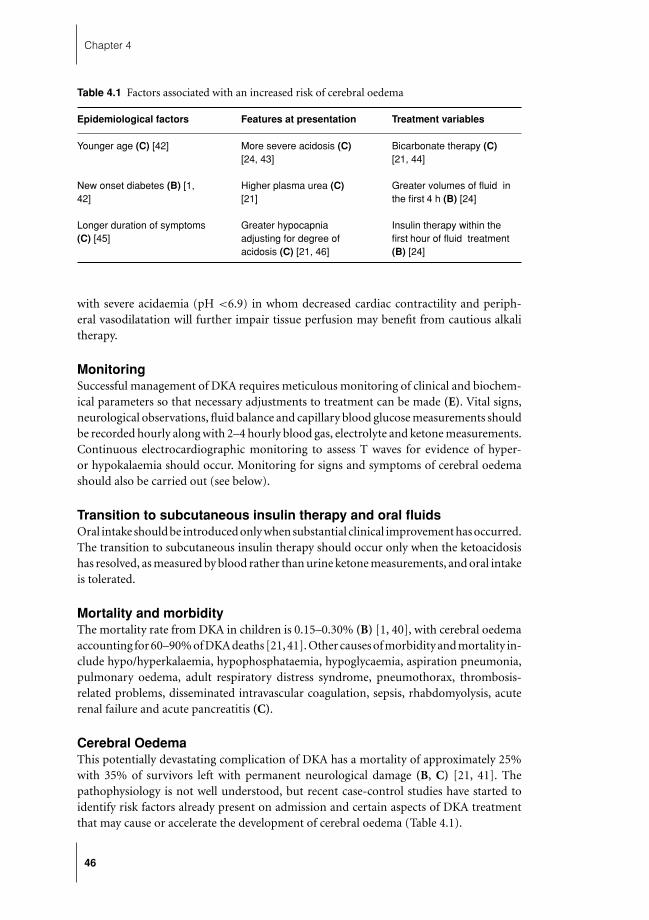
Chapter 4
Table 4.1 Factors associated with an increased risk of cerebral oedema
Epidemiological factors Features at presentation Treatment variables
Younger age (C) [42] More severe acidosis (C)[24, 43]
Bicarbonate therapy (C)[21, 44]
New onset diabetes (B) [1,42]
Higher plasma urea (C)[21]
Greater volumes of fluid inthe first 4 h (B) [24]
Longer duration of symptoms(C) [45]
Greater hypocapniaadjusting for degree ofacidosis (C) [21, 46]
Insulin therapy within thefirst hour of fluid treatment(B) [24]
with severe acidaemia (pH <6.9) in whom decreased cardiac contractility and periph-eral vasodilatation will further impair tissue perfusion may benefit from cautious alkalitherapy.
MonitoringSuccessful management of DKA requires meticulous monitoring of clinical and biochem-ical parameters so that necessary adjustments to treatment can be made (E). Vital signs,neurological observations, fluid balance and capillary blood glucose measurements shouldbe recorded hourly along with 2–4 hourly blood gas, electrolyte and ketone measurements.Continuous electrocardiographic monitoring to assess T waves for evidence of hyper-or hypokalaemia should occur. Monitoring for signs and symptoms of cerebral oedemashould also be carried out (see below).
Transition to subcutaneous insulin therapy and oral fluidsOral intake should be introduced only when substantial clinical improvement has occurred.The transition to subcutaneous insulin therapy should occur only when the ketoacidosishas resolved, as measured by blood rather than urine ketone measurements, and oral intakeis tolerated.
Mortality and morbidityThe mortality rate from DKA in children is 0.15–0.30% (B) [1, 40], with cerebral oedemaaccounting for 60–90% of DKA deaths [21, 41]. Other causes of morbidity and mortality in-clude hypo/hyperkalaemia, hypophosphataemia, hypoglycaemia, aspiration pneumonia,pulmonary oedema, adult respiratory distress syndrome, pneumothorax, thrombosis-related problems, disseminated intravascular coagulation, sepsis, rhabdomyolysis, acuterenal failure and acute pancreatitis (C).
Cerebral OedemaThis potentially devastating complication of DKA has a mortality of approximately 25%with 35% of survivors left with permanent neurological damage (B, C) [21, 41]. Thepathophysiology is not well understood, but recent case-control studies have started toidentify risk factors already present on admission and certain aspects of DKA treatmentthat may cause or accelerate the development of cerebral oedema (Table 4.1).
46

Type 1 diabetes mellitus – management
Table 4.2 Proposed criteria for the diagnosis of cerebral oedema∗
Diagnostic criteria Abnormal motor or verbal response to painDecorticate or decerebrate postureCranial nerve palsyAbnormal neurogenic respiratory pattern
Major criteria Altered mentation/fluctuating level of consciousnessSustained heart rate deceleration (decrease more than 20 beats/min) notattributable to improved intravascular volume or sleep stateAge-inappropriate incontinence
Minor criteria VomitingHeadacheLethargy or not easily arousableDiastolic blood pressure > 90 mmHgAge < 5yr
∗From [51].
It has also been suggested that an attenuated rise in plasma sodium may be a risk factorfor the development of cerebral oedema (C) [18, 21, 22] but it is unclear if this is actuallya consequence of cerebral oedema rather than a causative factor and may merely reflectthe effect of cerebral oedema on renal salt handling.
PathophysiologyA number of mechanisms have been proposed and recent studies with newer imagingtechniques are beginning to unravel the pathways of events leading to brain swelling[47, 48]. Previous hypotheses postulating osmotically mediated fluid shifts driven by theretention of intracellular osmolytes in the brain during rehydration form the basis forthe current fluid management standards in DKA. However, recent findings suggest thatthe predominant mechanism of cerebral oedema may be cerebral ischaemia with resultantactivation of ion transporters in the endothelial cells of the blood–brain barrier leading toreperfusion injury [48].
ManagementCerebral oedema usually develops 4–12 hours after treatment has started but can occurbefore any treatment (B, C) [21, 49, 50]. It is possible that quick intervention after earlysymptoms may reduce mortality and morbidity, so early recognition may be critical. Amethod of clinical diagnosis for the early detection of cerebral oedema has been proposed(Table 4.2). One diagnostic criterion, two major criteria or one major and two minorcriteria had a sensitivity of 92% and a false-positive rate of only 4% in predicting cerebraloedema when used retrospectively (C) [51]. Furthermore, a large UK prospective studyidentified headache and bradycardia as features which should prompt early review [52].
As soon as cerebral oedema is suspected, mannitol 0.5 g/kg or 3% saline 5–10 mL/kgover 30 minutes should be given (C) [45, 53]. Intubation and ventilation may be necessaryfor the patient with impending respiratory failure but aggressive hyperventilation hasbeen associated with a poorer outcome and is not recommended (C) [54, 55]. A cranialcomputed tomography scan is useful to exclude other possible intracerebral causes of
47

Chapter 4
neurological deterioration especially thrombosis or haemorrhage that may benefit fromspecific therapy.
There are no intervention studies with a large enough power to detect a real reductionin the incidence of cerebral oedema and so currently the only definitive method to preventcerebral oedema is to prevent DKA itself.
Prevention of DKAIncreased public awareness of signs and symptoms of diabetes can lead to earlier diagno-sis before progression to DKA in those of new-onset diabetes (B) [56]. A cause for theepisode of DKA should be identified in those with established diabetes particularly thosewith recurrent DKA. Insulin omission can be prevented by schemes that provide educa-tion, psychosocial evaluation and treatment combined with adult supervision of insulinadministration (B) [5, 6].
Ongoing insulin managementKrystyna Matyka and Shital Malik
Overall IntroductionThe optimum management of a child with diabetes is a complicated issue. Children candevelop diabetes from 0 to 18 years and come from many different backgrounds in cul-tural, sociodemographic and developmental terms. Management therefore needs to beindividually tailored and will likely change, as the child grows in both physiological andemotional terms. This will require a good deal of clinical experience from the clinicianas well as a good multidisciplinary team that will nurture the family and get to know itsstrengths and weaknesses.
As a result of this immense ‘natural’ variability, the study of management interventionsis fraught with problems as it becomes difficult to standardise interventions which can thenbe workable in a clinical situation. Children need to be studied within circumscribed agegroups that will define both physiological and emotional parameters and social and culturalissues will need to be considered. Adequate numbers of children need to be investigatedto allow the study to have enough power to be interpreted and this is difficult unlessmulticentre studies are performed which will then introduce inter-centre confounders.As a result, a review of the data available for the day-to-day management of diabetes inchildhood highlights significant deficiencies and there are few good quality, randomisedcontrolled trials to guide clinical practice.
Insulin regimensThe optimal insulin regimen for paediatric patients with T1DM remains controversial. In1995, the Hvidøre study group, an international multicentre group comprising 21 centresfrom 17 countries in Europe, Japan and North America, performed a survey on the qualityof care in its respective institutions and a follow-up survey was performed in 1998 [57, 58].Of 2873 children (age 0–18 yr) surveyed in 1995, 872 adolescents (11–18 yr) were restudiedin 1998. Although the use of multiple injection regimens increased from 42 to 71%, thisdid not lead to an improvement in glycaemic control as judged by glycated haemoglobinconcentrations (B). Although there was a tendency towards an increase in the rate of severehypoglycaemia in the group of children/adolescents on intensive insulin regimens, this didnot reach statistical significance, perhaps due to the low number of events recorded [57].
48

Type 1 diabetes mellitus – management
Very few randomised clinical trials of insulin regimens are available for children andadolescents. Even observational studies in this age group are few in number and are usuallyfrom single centres or countries. We found 19 studies on insulin regimens (excludingcontinuous subcutaneous insulin infusion, CSII) over a 10-year period from 1996 to 2006.Of these only 8 were true comparative studies, while the rest were observational studiesand the majority involved the use of insulin analogues (Table 4.3). These studies will beexamined with respect to age group studied.
Preschool childrenThere are very few studies based on preschool children, reflecting the ethical problemsof research in this age group of children. One observational study has examined theeffectiveness of postprandial insulin lispro in five toddlers with diabetes (C)[67]. This studyshowed that postprandial rapid-acting insulin administration results in blood glucosevalues as good as, or better than, those obtained with the same dose of soluble insulin givenbefore the meal and no different from those seen with preprandial rapid-acting insulin.The possibility of reducing or omitting the short-acting insulin when the child does not eatoffered an important potential advantage. Another observational study of flexible insulintherapy, adjusting doses of rapid-acting insulin based on carbohydrate content of food,found that although glycaemic control improved, there was a significant reduction infrequency of severe hypoglycaemia, although interestingly this benefit occurred only inthe 57% of patients who were normal weight as opposed to overweight (C) [68] (for amore detailed discussion of the particular problems associated with the very young, seeChapter 8).
Prepubertal childrenThe majority of studies in school-aged children have been open randomised crossoverstudies and again although there have been some benefits of using analogue insulins com-pared to human insulins, there has been little demonstrable improvement in glycatedhaemoglobin [59–62, 65]. One study found that there were some improvements in post-meal glucose excursions when comparing rapid-acting analogues and soluble insulin (B)[60]. Other studies examined the potential consequences of giving rapid-acting analogueafter food compared with either regular human insulin or rapid-acting insulin beforemeals (B) [59, 65]. Neither study showed a worsening in glycaemic control with postmealdosing, and treatment satisfaction was equally high for both patients and carers. However,neither of these studies was of long duration and one lasted only 6 weeks [65].
Schober [62] compared insulin glargine and neutral protamine Hagedorn (NPH) insulinin 349 children and adolescents, aged 5–16 years, with T1DM. Although fasting bloodglucose levels decreased significantly more in the insulin glargine group than in the NPHinsulin group ( p = 0.02), there were no significant differences in HbA1c (B).
Studies in adolescentsThere has been a small number of studies of insulin regimens in adolescents and mosthave examined the benefits of analogue therapy. Studies of rapid-acting insulin suggestthat there may be improvements in both postprandial glucose excursions and a reductionof nocturnal hypoglycaemia. A large (n = 463) crossover, open-label study comparingmealtime insulin lispro for 4 months with soluble insulin for 4 months concluded thatinsulin lispro significantly improved postprandial glycaemic control and reduced episodes
49
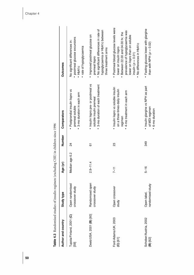
Chapter 4
Tab
le4.
3R
ando
mis
edst
udi
esof
insu
linre
gim
en(e
xclu
din
gC
SII)
inch
ildre
nsi
nce
1996
Au
tho
ran
dco
un
try
Stu
dyty
pe
Ag
e(y
r)N
um
ber
Co
mp
arat
ors
Ou
tco
mes
Tupo
la/F
inla
nd,2
001
(C)
[59]
Ope
nra
ndom
ised
cros
sove
rst
udy
Med
ian
age
6.2
24�
Pos
tpra
ndia
lins
ulin
lispr
ovs
prem
eals
olub
le�
3-m
odu
ratio
nin
each
arm
No
sign
ifica
ntdi
ffere
nces
in:
�
post
mea
lglu
cose
excu
rsio
ns�
HbA
1c�
rate
ofhy
pogl
ycae
mia
Dee
b/U
SA
,200
1(B
)[6
0]R
ando
mis
edop
encr
osso
ver
stud
y2.
9–11
.461
�
Insu
linlis
pro
pre-
orpo
stm
ealv
sso
lubl
ein
sulin
prem
eal
�
3-m
odu
ratio
nof
each
trea
tmen
t
�
Impr
oved
post
mea
lglu
cose
onpr
emea
llis
pro
�
No
sign
ifica
ntdi
ffere
nces
inra
teof
hypo
glyc
aem
iaor
HbA
1cbe
twee
nth
ree
trea
tmen
tarm
s
For
d-A
dam
s/U
K,2
003
(C)
[61]
Ope
ncr
osso
ver
stud
y7–
1123
�
Insu
linlis
pro
vsso
lubl
ein
sulin
ona
thre
e-tim
es-d
aily
insu
linre
gim
en�
4-m
otr
eatm
enti
nea
char
m
�
Pos
tmea
lblo
odgl
ucos
ele
vels
wer
elo
wer
onin
sulin
lispr
o�
Bet
wee
n22
.00
and
04.0
0hr
,the
prev
alen
ceof
hypo
glyc
aem
iaw
aslo
wer
onlis
pro
than
onso
lubl
ein
sulin
(p=
0.01
)�
No
diffe
renc
esin
HbA
1c
Sch
ober
/Aus
tria
,200
2(B
)[6
2]O
pen-
labe
l,ra
ndom
ised
stud
y5–
1634
9�
Insu
lingl
argi
nevs
NP
Has
part
ofM
DIr
egim
en�
6-m
odu
ratio
n
�
Fas
ting
gluc
ose
low
erw
ithgl
argi
neth
anw
ithN
PH
(p=
0.02
)
50

Type 1 diabetes mellitus – management
Hol
com
be/U
SA
,200
2(B
)[6
3]C
ross
over
open
-labe
lstu
dy9–
1846
3�
Insu
linlis
pro
vsre
gula
rin
sulin
aspa
rtof
MD
Ireg
imen
�
4m
oin
each
trea
tmen
tarm
�
Lisp
robe
fore
brea
kfas
tand
even
ing
mea
lled
tosi
gnifi
cant
lylo
wer
mea
n2-
hpo
stpr
andi
albl
ood
gluc
ose
leve
lsco
mpa
red
with
solu
ble
�
No
diffe
renc
esin
HbA
1c�
Low
erin
cide
nce
ofhy
pogl
ycae
mia
epis
odes
per
patie
ntpe
r30
dw
ithin
sulin
lispr
o(p
=0.
023)
Mur
phy/
UK
,200
3(A
)[64
]R
ando
mis
edcr
osso
ver
tria
l12
–18
28�
Insu
lingl
argi
ne+
lispr
ovs
NP
Hin
sulin
+so
lubl
eas
part
ofM
DI
regi
men
�
16w
kin
each
arm
�
Noc
turn
alhy
pogl
ycae
mia
was
43%
low
eron
glar
gine
+lis
pro
�
No
diffe
renc
ein
HbA
1cor
self-
repo
rted
hypo
glyc
aem
ia
Dan
ne/G
erm
any,
2003
(B)
[65]
Ope
nra
ndom
ised
cros
sove
rtr
ial
6–12
13–1
742 34
�
Pre
pran
dial
vspo
stpr
andi
alin
ject
ions
ofin
sulin
aspa
rtas
part
ofM
DIr
egim
en�
6w
kin
each
arm
�
No
diffe
renc
ein
HbA
1cor
rate
ofhy
pogl
ycae
mia
�
Trea
tmen
tsat
isfa
ctio
nw
aseq
ually
high
Mor
tens
en/D
enm
ark,
2006
(B)
[66]
Ran
dom
ised
,op
en-la
bel,
para
llel-g
roup
stud
y
Ado
lesc
ents
167
�
Insu
linas
part
with
mea
lspl
usN
PH
insu
linat
bedt
ime
vspr
emix
edhu
man
insu
linat
brea
kfas
t,so
lubl
ein
sulin
atlu
nch
and
dinn
eran
dN
PH
atbe
dtim
e�
4-m
odu
ratio
n
�
No
diffe
renc
ein
HbA
1cor
rate
ofhy
pogl
ycae
mia
51

Chapter 4
Tab
le4.
4R
ando
mis
edst
udi
esof
CSI
Iin
child
ren
sin
ce19
96
Au
tho
ran
dco
un
try
Stu
dy
typ
eA
ge
(yr)
Nu
mb
erC
om
par
ato
rsO
utc
om
es
Kau
fman
/US
A,2
000
(C)
[71]
Ran
dom
ised
cros
sove
r–
4-w
kst
udy
7–10
10�
Noc
turn
alC
SII
vsm
orni
ngN
PH
and
mea
ltim
elis
pro
�
4-w
kdu
ratio
n
�
CS
IIle
dto
decr
ease
inav
erag
e(p
<0.
001)
,bre
akfa
st(p
<0.
0001
)an
d3
a.m
.(p
<0.
003)
gluc
ose
�
Dec
reas
edfe
arof
hypo
glyc
aem
ia
Wei
ntro
b/Is
rael
,20
03–2
004
(C)
[72,
73]
Ran
dom
ised
open
cros
sove
rst
udy
9.4–
13.9
23�
CS
IIus
ing
lispr
ovs
MD
I(us
ing
NP
Han
dso
lubl
ein
sulin
)�
3.5
mo
�
HbA
1can
dfr
eque
ncy
ofse
vere
hypo
glyc
aem
iaeq
uiva
lent
betw
een
the
two
grou
ps�
Hig
her
trea
tmen
tsat
isfa
ctio
nw
ithC
SII
�
Usi
ngC
GM
Sth
ere
was
alo
nger
dura
tion
ofw
ithin
targ
etgl
ucos
eon
CS
II,sm
alle
rar
eaun
der
curv
efo
rno
ctur
nal
hypo
glyc
aem
iaan
dhi
gher
rate
ofhy
perg
lyca
emic
epis
odes
DiM
eglio
/US
A,2
004
(C)
[74]
Ran
dom
ised
,op
en-la
bel,
pros
pect
ive
stud
y
1.8–
4.7
42�
21ch
ildre
nra
ndom
ised
toC
SII
�
21ch
ildre
nre
mai
ned
onpr
ior
regi
men
(ver
yva
ried)
�
6-m
ost
udy
�
Red
uctio
nin
HbA
1cin
both
grou
psdu
ring
stud
y�
No
sign
ifica
ntbe
twee
ngr
oup
diffe
renc
ein
HbA
1cat
6m
o�
No
diffe
renc
ein
rate
ofse
vere
hypo
glyc
aem
iabu
tsig
nific
ant
incr
ease
inm
eter
-det
ecte
dhy
pogl
ycae
mia
onC
SII
(p<
0.05
)
52
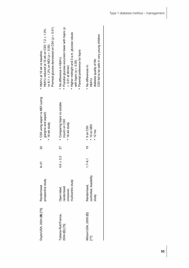
Type 1 diabetes mellitus – management
Doy
le/U
SA
,200
4(B
)[7
5]R
ando
mis
edpr
ospe
ctiv
est
udy
8–21
32�
CS
IIus
ing
aspa
rtvs
MD
I(us
ing
glar
gine
and
aspa
rt)
�
16-w
kst
udy
�
HbA
1cat
16w
kvs
base
line:
HbA
1cre
duce
dat
16w
kon
CS
II:7.
2±
1.0%
vs8.
1±
1.2%
onM
DI(
p<
0.05
)P
rem
ealg
luco
sede
crea
sed
onC
SII
(p<
0.01
)
Tubi
ana-
Rufi
/Fra
nce,
2004
(C)
[76]
Ope
n-la
bel,
rand
omis
edcr
osso
ver,
mul
ticen
tre
stud
y
4.6
±2.
227
�
Com
parin
glis
pro
vsso
lubl
ein
sulin
onC
SII
�
16-w
kst
udy
�
No
diffe
renc
ein
HbA
1c�
Pra
ndia
lglu
cose
excu
rsio
nlo
wer
with
lispr
o(p
<0.
01at
dinn
er)
�
Hig
her
mid
nigh
tand
3a.
m.g
luco
seva
lues
with
lispr
o(p
<0.
05)
�
Par
enta
lpre
fere
nce
for
lispr
o
Wils
on/U
SA
,200
5(C
)[7
7]R
ando
mis
ed,
cont
rolle
d,fe
asib
ility
stud
y
1.7–
6.1
19�
9on
CS
II�
10on
MD
I�
12m
o
�
No
diffe
renc
esin
:H
bA1c
diab
etes
qual
ityof
life
CS
IIfe
ltto
besa
fein
very
youn
gch
ildre
n
53

Chapter 4
of nocturnal hypoglycaemia (B) [63]. However, no differences in HbA1c were observed.Another study examined insulin aspart injected at mealtimes with NPH insulin at bedtimeversus a human insulin regimen: premixed human insulin at breakfast, soluble insulinat lunch and evening meal and NPH insulin at bedtime. One hundred and sixty-sevenadolescents took part in the study that used a parallel group design (B) [66]. After 4months there were no significant differences in HbA1c between the two groups.
Only one RCT of the use of glargine in adolescents was found (A) [64]. Murphy com-pared insulin glargine plus insulin lispro versus NPH insulin plus soluble insulin as partof a multiple injection regimen. The results showed that nocturnal hypoglycaemia wassignificantly reduced (43%) with insulin glargine. Postprandial glucose excursions werealso lower after breakfast and lunch, but again there were no significant differences inHbA1c [64].
Free mixingWith the advent of intensive insulin regimens the need to mix insulin in syringes ratherthan using insulin pens has decreased remarkably. However there remains a number ofchildren and adolescents for whom an increase in the frequency of insulin injections isnot suitable. Recent studies have looked at the possibility of mixing rapid-acting insulinanalogues with insulin glargine in the same syringe (C) [69, 70]. These studies suggest thatthese insulins can be mixed with no detrimental effect on insulin action, provided theyare used immediately.
Continuous subcutaneous insulin infusionWe found 18 studies on CSII over a 10-year period. Of these only 6 were randomisedinterventional studies (Table 4.4) and all of them studied only small numbers of children[71, 72, 74–77]. One study reported data from the same cohort in two individual papers:one of overall control and one discussing data from continuous glucose monitoring system(CGMS) recordings (B) [73]. Only Doyle (B) [75] found an improvement in HbA1c onCSII compared to an analogue multiple daily injection (MDI) regimen. The other studiesshowed little difference in either glycaemic control or rate of hypoglycaemia. Interestingly,in contrast to the studies of insulin regimen, the majority of these studies were performedin very young children. Preschool children appear to be especially concerning in terms ofoverall management and some of these studies looked to see if CSII could be safely usedin this age group (B) [74].
A number of observational ‘before-and-after’ studies have examined HbA1c and rateof hypoglycaemia, often looking retrospectively through case records, and have foundthat improved glycaemic control can be achieved without a concomitant rise in rate ofhypoglycaemia (C) [78, 79]. However, observational studies are difficult to interpret asother variables that may impact on diabetic control will not have been standardised.
Hypoglycaemia and insulin regimenThe impact on rates of hypoglycaemia using analogue insulins has been examined in onlya few studies. A large study of 463 children aged 9–18 years showed a lower prevalenceof hypoglycaemic episodes per patient per 30 days seen with insulin lispro (4.02 ± 4.5vs 4.37 ± 4.5, respectively; p = 0.023) and significantly fewer hypoglycaemic episodesbetween midnight and 6 a.m. (1.0 ± 1.9 vs 1.7 ± 2.6; p < 0.001) (B) [63]. Two studiesusing rapid-acting analogues as part of a basal bolus regimen have found no differences
54

Type 1 diabetes mellitus – management
in the rate of hypoglycaemia between analogues and regular human insulin (B, C) [59,60]. Another study compared insulin lispro and soluble insulin as part of a three-times-daily insulin regimen, with isophane insulin as background. The prevalence of low bloodglucose concentrations in the early part of the night was lower with analogue insulins(C) [61].
Glucose monitoringRegular home blood glucose monitoring (HBGM) is felt to be an essential part of day-to-day diabetes management. The American Diabetes Association has recommended thatpatients with T1DM test their blood glucose a minimum of two times each day and needto be taught how to make adjustments to their diabetes management regimen in light ofthe results (E).
Glucose monitoring and glycaemic controlThere are few robust data on how useful HBGM may be. A few recent studies have examinedfactors, including frequency of HBGM, which may predict glycaemic control in childrenattending paediatric diabetes services [3, 80, 81]. One of these studies, of 300 children, hasfound that frequency of HBGM was the only modifiable predictor of HbA1c. Childrenperforming one or fewer blood tests per day had an average HbA1c of 9.1 ± 0.34%compared to a value of 8.0 ± 0.31% in those performing five or more blood tests (C) [81].From these data it is not possible to decide whether HBGM improves glycaemic controlor whether those children who were more compliant with their diabetes management,and hence are more likely to have better glycaemic control, may have been doing moreHBGM. Another study of 415 subjects aged 0–34 years found that frequency of HBGMalso predicted severe hypoglycaemia: a finding that was independent of intensive insulintherapy and glycaemic control (C) [80].
There appear to be fewer studies of how well patients respond to blood glucose re-sults performed at home. One study has examined responses to hypoglycaemia (glucose<3.3 mmol/L) and hyperglycaemia (>13.2 mmol/L) using a telephone 24-hour recall in-terview questionnaire, with both a group of 125 adolescents and their parents (C) [82].The authors found that adolescents failed to respond appropriately in 38% episodes ofhypoglycaemia and 29% episodes of hyperglycaemia. Episodes of hypoglycaemia weremore appropriately managed when the adolescents were supervised by their parents but,interestingly, episodes of hyperglycaemia were more likely to be inappropriately managedwhen adolescents were supervised by parents. The authors suggested that health-care pro-fessionals may underestimate adolescents’ ability to respond to HBGM results. Parents mayhave more of a problem as more diabetes education is aimed at adolescents rather thanparents at a time of life when adolescents are taking more responsibility for their diabetescare.
A number of patients feel that HBGM is unnecessary, as they can ‘feel’ what their bloodglucose may be. This was examined in a small (n = 6) group of well-controlled childrenwith diabetes aged from 6 to 15 years. Subjects were asked to estimate their blood glucoseprior to performing HBGM over a period of a month. Blood glucose varied from 1.7 to16.6 mmol/L. Children were accurate in 42% of readings and only slightly inaccurate in37% (C) [83]. However, there was a dangerous misestimation in 19% of readings thatwould have had implications for management and safety.
55
Chapter 4
Continuous glucose monitoringIn recent years continuous glucose monitoring has become available as an importantclinical tool.
Continuous glucose monitoring systemThe CGMS system includes a glucose-oxidase-based platinum electrode that is insertedinto the subcutaneous tissue. Glucose oxidase catalyses the oxidation of glucose in theinterstitial fluid generating an electrical current. This is carried by a cable to a pager-sizedmonitor that provides a glucose reading every 5 minutes for a maximum of 72 hours.Glucose data are stored in the memory and are subsequently downloaded – no real-timedisplay is provided.
Studies of CGMS have highlighted the limitations of conventional HBGM [84, 85]. Astudy of 56 children aged 2–18 years compared 3 days of CGMS monitoring with 10–15values from HBGM over the same time period. Interestingly, examining average dailyblood glucose concentrations using CGMS and HBGM values, it was demonstrated thatstandard premeal HBGM provides a reasonably accurate reflection of control. Yet standardmeasures missed significant postmeal hyperglycaemia with 50% of values going above 16.5mmol/L, even in young people on rapid-acting analogues. In addition, 68% of subjectshad at least one episode of asymptomatic nocturnal hypoglycaemia and 12 children (21%)had asymptomatic hypoglycaemia on all three nights (C) [84]. Unfortunately, there are fewdata examining the benefit of regular CGMS monitoring on long-term glycaemic control.One small study of 11 subjects randomised five children to CGMS and had six controls(C) [86]. Children in the CGMS group used six 3-day sensors over a 30-day period andboth groups used HBGM four times a day. HbA1c was measured at baseline, 1 month and3 months. Glycaemic control improved in both groups but was significant only betweenthe groups at 1 month and lost significance again at 3 months. This is too small a study tobe clinically informative and larger studies are needed.
More recently, real-time monitors, some of which transmit directly to the pumps, havebeen developed but there are no studies in children to show what advantages, if any,these have over the CGMS. Studies are also being conducted in children to investigate thepossibility of linking glucose sensors directly to pumps in such a way as to enable them toact as an ‘artificial pancreas’ but the results of these studies will not be available for somewhile yet.
Glucowatch biographerAlthough the CGMS may be useful as a clinic tool, patients would welcome a non-invasiveglucose monitor that provides a real-time display as well as having alarms to alert theindividual to both hyper- and hypoglycaemia. The Glucowatch biographer (GWB2) isshaped like a watch and uses iontophoresis to sample interstitial fluid from which theglucose is measured and displayed every 10 minutes for up to 13 hours. Although notas accurate as HBGM, it has been found to be useful and acceptable to patients andtheir families in early small studies (C) [87, 88]. However a recent multicentre parallelgroup study of 200 subjects aged 7–18 years found less satisfactory results (B) [89]. Thoserandomised to the GWB2 group were asked to use the watch at least twice a week, but asoften as desired, over the 6-month period of the study. Those in the control group weregiven a GWB2 to use after the intervention period of the study. Over the course of theintervention period 16% of subjects averaged two uses of more than 8 hours per week and
56
Type 1 diabetes mellitus – management
by 6 months 27% of subjects had stopped using the monitor completely. There was nochange in HbA1c over time or between the two groups. Some of the reasons for not usingthe GWB2 included local skin reactions in 76%, the monitor alarmed too often in 47%and did not provide accurate glucose readings in 33%.
HypoglycaemiaHypoglycaemia is a common and much feared complication of the management of T1DMin childhood.
PrevalenceA large number of studies have examined the prevalence of episodes of severe hypogly-caemia [2, 3, 90–92]. These have taken place across the world in North America, Europeand Australia. A variety of definitions of severe hypoglycaemia has been used and preva-lence rates have varied from 3.1 to 85.7 episodes per 100 patient-years. It is likely that thereare numerous factors that have influenced the wide variation in results presented. Some ofthese variables have been highlighted by the epidemiological studies themselves. Howeverit is important to note that these studies have not been scientifically designed to examinerisk factors for hypoglycaemia – they have merely looked at statistical correlations and donot study important clinical variables known to influence risk of hypoglycaemia, such asphysical activity and diet.
Risk factorsYoung age has been consistently found to be a risk factor for hypoglycaemia. Insulinregimen may be a confounder, as a number of these studies, even the most recent, willpresent relatively old data. Many younger children will not have been on more intensiveinsulin regimens, as these are relatively new approaches to management of diabetes inpreschool children. More recent data on intensive insulin regimens such as CSII do suggestthat glycaemic control can be tightened without a concomitant rise in the risk of severehypoglycaemia (see section ‘continuous subcutaneous insulin infusion’). A recent largestudy of paediatric diabetes centres in Germany suggests that centre experience, based onthe number of children attending a clinic service, also influences risk of hypoglycaemia (B)[92]. This suggests that good education and support of patients is important in minimisingthe risk of hypoglycaemia.
Nocturnal hypoglycaemia may be a separate entity. Studies suggest that up to 50% ofepisodes of severe hypoglycaemia occur during sleep. Other studies have shown that manychildren sleep through episodes of hypoglycaemia, which can be profound and prolonged(B) [93–95]. Studies of overnight counter-regulation have found that this is blunted duringsleep in both healthy children and those with diabetes, a fact that may influence symptomrecognition overnight (B) [96]. Recent studies have shown that daytime exercise may bea risk factor for overnight hypoglycaemia, but episodes of nocturnal hypoglycaemia mayalso occur after sedentary days (B) [97, 98].
Treatment of hypoglycaemiaTreatment of hypoglycaemia will depend on the severity. ISPAD consensus guidelines [99]suggest that mild hypoglycaemia, during which the individual is cooperative, can be treatedwith 5–15 g of oral fast-acting carbohydrate (E). More severe hypoglycaemia where thesubject is unable to cooperate but is still conscious with an intact gag reflex should be
57

Chapter 4
treated with oral glucose gel. If the subject is unconscious, intramuscular glucagon shouldbe given in the first instance (0.5 mg if less than 5 yr and 1.0 mg if older than 5 yr). If thisis unsuccessful then intravenous 10% dextrose should be given at a dose of 1–2 mL/kguntil a response is achieved. It is not apparent that there have been any studies that haveinformed these decisions.
Many episodes of hypoglycaemia are dealt with at home by parents and/or other adultcarers. Recent studies have shown that low-dose glucagon given using an insulin syringerather than the more frightening glucagon syringe can be effective in managing hypogly-caemia when a child is unwilling to take oral carbohydrate (C) [100, 101].
Avoidance of hypoglycaemiaThere are very few studies that have examined interventions designed to prevent hypogly-caemia. A review of studies examining insulin regimens has already been presented (seesection ‘insulin regimen’).
Other approaches which have been used to prevent hypoglycaemia have included fam-ily education. In a study of Scandinavian subjects, 332 children with T1DM were ran-domised, in a 1:1:1 controlled study, to receive either an educational package (videotapesand brochures) aimed at informing subjects about hypoglycaemia or a more generic pack-age about diabetes, or traditional treatment only. In families that were given the hypo-glycaemia package, there was a significant reduction in the yearly prevalence of severehypoglycaemia from 42 to 27%, with no change in either control group [102].
References
1 Edge JA, Ford-Adams ME, Dunger DB. Causes of death in children with insulin dependent diabetes1990–96. Arch Dis Child 1999; 81: 318–23.
2 Rewers A, Chase HP, Mackenzie T et al. Predictors of acute complications in children with type 1diabetes. JAMA 2002; 287: 2511–18.
3 Rosilio M, Cotton JB, Wieliczko MC et al. Factors associated with glycemic control. A cross-sectionalnationwide study in 2,579 French children with type 1 diabetes. The French Pediatric Diabetes Group.Diabetes Care 1998; 21: 1146–53.
4 Smith CP, Firth D, Bennett S et al. Ketoacidosis occurring in newly diagnosed and established diabeticchildren. Acta Paediatr 1998; 87: 537–41.
5 Golden MP, Herrold AJ, Orr DP. An approach to prevention of recurrent diabetic ketoacidosis in thepediatric population. J Pediatr 1985; 107: 195–200.
6 Flood RG, Chiang VW. Rate and prediction of infection in children with diabetic ketoacidosis. Am JEmerg Med 2001; 19: 270–3.
7 Komulainen J, Kulmala P, Savola K et al. Clinical, autoimmune, and genetic characteristics of veryyoung children with type 1 diabetes. Childhood Diabetes in Finland (DiMe) Study Group. DiabetesCare 1999; 22: 1950–5.
8 Pinkey JH, Bingley PJ, Sawtell PA et al. Presentation and progress of childhood diabetes mellitus: aprospective population-based study. The Bart’s-Oxford Study Group. Diabetologia 1994; 37: 70–4.
9 Komulainen J, Lounamaa R, Knip M et al. Ketoacidosis at the diagnosis of type 1 (insulin dependent)diabetes mellitus is related to poor residual beta cell function. Childhood Diabetes in Finland StudyGroup. Arch Dis Child 1996; 75: 410–15.
10 Levy-Marchal C, Papoz L, de Beaufort C et al. Clinical and laboratory features of type 1 diabeticchildren at the time of diagnosis. Diabet Med 1992; 9: 279–84.
11 American Diabetes Association. Type 2 diabetes in children and adolescents. American Diabetes As-sociation. Diabetes Care 2000; 23: 381–9.
12 Foster DW, McGarry JD. The metabolic derangements and treatment of diabetic ketoacidosis. N EnglJ Med 1983; 309: 159–69.
58

Type 1 diabetes mellitus – management
13 Dunger DB, Sperling MA, Acerini CL et al. ESPE/LWPES consensus statement on diabetic ketoacidosisin children and adolescents. Arch Dis Child 2004; 89: 188–94.
14 BSPED. BSPED Consensus statement for the management of DKA. Available at: www.bsped.org.uk/professional/guidelines/docs/BSPDDKAApr04.pdf
14a Wolfsdorf J, Craig ME, Daneman D et al. Pediatr Diabetes 2007; 8: 28–43.15 Monroe KW, King W, Atchison JA. Use of PRISM scores in triage of pediatric patients with diabetic
ketoacidosis. Am J Manag Care 1997; 3: 253–8.16 Harris GD, Fiordalisi I. Physiologic management of diabetic ketoacidemia: a 5-year prospective pedi-
atric experience in 231 episodes. Arch Pediatr Adolesc Med 1994; 148: 1046–52.17 Adrogue HJ, Barrero J, Eknoyan G. Salutary effects of modest fluid replacement in the treatment of
adults with diabetic ketoacidosis: use in patients without extreme volume deficit. JAMA 1989; 262:2108–113.
18 Harris GD, Fiordalisi I, Harris WL et al. Minimizing the risk of brain herniation during treat-ment of diabetic ketoacidemia: a retrospective and prospective study. J Pediatr 1990; 117: 22–31.
19 Mel JM, Werther GA. Incidence and outcome of diabetic cerebral oedema in childhood: are therepredictors? J Paediatr Child Health 1995; 31: 17–20.
20 Rother KI, Schwenk WF. Effect of rehydration fluid with 75 mmol/L of sodium on serum sodiumconcentration and serum osmolality in young patients with diabetic ketoacidosis. Mayo Clin Proc1994; 69: 1149–53.
21 Glaser N, Barnett P, McCaslin I et al. Risk factors for cerebral edema in children with diabetic ke-toacidosis. The Pediatric Emergency Medicine Collaborative Research Committee of the AmericanAcademy of Pediatrics. N Engl J Med 2001; 344: 264–9.
22 Hale PM, Rezvani I, Braunstein AW et al. Factors predicting cerebral edema in young children withdiabetic ketoacidosis and new onset type I diabetes. Acta Paediatr 1997; 86: 626–31.
23 Luzi L, Barrett EJ, Groop LC et al. Metabolic effects of low-dose insulin therapy on glucose metabolismin diabetic ketoacidosis. Diabetes 1988; 37: 1470–7.
24 Edge JA, Jakes RW, Roy Y et al. The UK case-control study of cerebral oedema complicating diabeticketoacidosis in children. Diabetologia 2006; 49: 2002–9.
25 Kitabchi AE. Low-dose insulin therapy in diabetic ketoacidosis: fact or fiction? Diabetes Metab Rev1989; 5: 337–63.
26 Atchley DW, Loeb RF, Richards DW et al. On diabetic ketoacidosis: a detailed study of electrolytebalances following the withdrawal and reestablishment of insulin therapy. J Clin Invest 1933; 12:297–326.
27 Nabarro JD, Spencer AG, Stowers JM. Metabolic studies in severe diabetic ketosis. Q J Med 1952; 21:225–48.
28 Adrogue HJ, Lederer ED, Suki WN et al. Determinants of plasma potassium levels in diabetic ketoaci-dosis. Medicine (Baltimore) 1986; 65: 163–72.
29 DeFronzo RA, Felig P, Ferrannini E et al. Effect of graded doses of insulin on splanchnic and peripheralpotassium metabolism in man. Am J Physiol 1980; 238: E421–7.
30 Becker DJ, Brown DR, Steranka BH et al. Phosphate replacement during treatment of diabetic ketosis.Effects on calcium and phosphorus homeostasis. Am J Dis Child 1983; 137: 241–6.
31 Fisher JN, Kitabchi AE. A randomized study of phosphate therapy in the treatment of diabetic ke-toacidosis. J Clin Endocrinol Metab 1983; 57: 177–80.
32 Keller U, Berger W. Prevention of hypophosphatemia by phosphate infusion during treatment ofdiabetic ketoacidosis and hyperosmolar coma. Diabetes 1980; 29: 87–95.
33 Green SM, Rothrock SG, Ho JD et al. Failure of adjunctive bicarbonate to improve outcome in severepediatric diabetic ketoacidosis. Ann Emerg Med 1998; 31: 41–8.
34 Hale PJ, Crase J, Nattrass M. Metabolic effects of bicarbonate in the treatment of diabetic ketoacidosis.BMJ (Clin Res Ed) 1984; 289: 1035–8.
35 Okuda Y, Adrogue HJ, Field JB et al. Counterproductive effects of sodium bicarbonate in diabeticketoacidosis. J Clin Endocrinol Metab 1996; 81: 314–20.
36 Assal JP, Aoki TT, Manzano FM et al. Metabolic effects of sodium bicarbonate in management ofdiabetic ketoacidosis. Diabetes 1974; 23: 405–11.
37 Ohman JL, Jr, Marliss EB, Aoki TT et al. The cerebrospinal fluid in diabetic ketoacidosis. N Engl J Med1971; 284: 283–90.
59

Chapter 4
38 Lever E, Jaspan JB. Sodium bicarbonate therapy in severe diabetic ketoacidosis. Am J Med 1983; 75:263–8.
39 Soler NG, Bennett MA, Dixon K et al. Potassium balance during treatment of diabetic ketoacidosiswith special reference to the use of bicarbonate. Lancet 1972; 2: 665–7.
40 Curtis JR, To T, Muirhead S et al. Recent trends in hospitalization for diabetic ketoacidosis in Ontariochildren. Diabetes Care 2002; 25: 1591–6.
41 Edge JA, Hawkins MM, Winter DL et al. The risk and outcome of cerebral oedema developing duringdiabetic ketoacidosis. Arch Dis Child 2001; 85: 16–22.
42 Rosenbloom AL. Intracerebral crises during treatment of diabetic ketoacidosis. Diabetes Care 1990;13: 22–33.
43 Durr JA, Hoffman WH, Sklar AH et al. Correlates of brain edema in uncontrolled IDDM. Diabetes1992; 41: 627–32.
44 Bureau MA, Begin R, Berthiaume Y et al. Cerebral hypoxia from bicarbonate infusion in diabeticacidosis. J Pediatr 1980; 96: 968–73.
45 Bello FA, Sotos JF. Cerebral oedema in diabetic ketoacidosis in children. Lancet 1990; 336: 64.46 Mahoney CP, Vlcek BW, DelAguila M. Risk factors for developing brain herniation during diabetic
ketoacidosis. Pediatr Neurol 1999; 21: 721–7.47 Cameron FJ, Kean MJ, Wellard RM et al. Insights into the acute cerebral metabolic changes associated
with childhood diabetes. Diabet Med 2005; 22: 648–53.48 Glaser NS, Wootton-Gorges SL, Marcin JP et al. Mechanism of cerebral edema in children with diabetic
ketoacidosis. J Pediatr 2004; 145: 164–171.49 Fiordalisi I, Harris GD, Gilliland MG. Prehospital cardiac arrest in diabetic ketoacidemia: why brain
swelling may lead to death before treatment. J Diabetes Complications 2002; 16: 214–19.50 Lawrence SE, Cummings EA, Gaboury I et al. Population-based study of incidence and risk factors for
cerebral edema in pediatric diabetic ketoacidosis. J Pediatr 2005; 146: 688–92.51 Muir AB, Quisling RG, Yang MC et al. Cerebral edema in childhood diabetic ketoacidosis: natural
history, radiographic findings, and early identification. Diabetes Care 2004; 27: 1541–6.52 Edge JA, Flint J, Roy Y et al. Can cerebral oedema be identified before the reduction in conscious level?
Pediatr Diabetes 2004; 5(suppl 1): 48.53 Curtis JR, Bohn D, Daneman D. Use of hypertonic saline in the treatment of cerebral edema in diabetic
ketoacidosis (DKA). Pediatr Diabetes 2001; 2: 191–4.54 Marcin JP, Glaser N, Barnett P et al. Factors associated with adverse outcomes in children with diabetic
ketoacidosis-related cerebral edema. J Pediatr 2002; 141: 793–7.55 Tasker RC, Lutman D, Peters MJ. Hyperventilation in severe diabetic ketoacidosis. Pediatr Crit Care
Med 2005; 6: 405–11.56 Vanelli M, Chiari G, Ghizzoni L et al. Effectiveness of a prevention program for diabetic ketoacidosis
in children: an 8-year study in schools and private practices. Diabetes Care 1999; 22: 7–9.57 Holl RW, Swift PG, Mortensen HB et al. Insulin injection regimens and metabolic control in an
international survey of adolescents with type 1 diabetes over 3 years: results from the Hvidore studygroup. Eur J Pediatr 2003; 162: 22–9.
58 Mortensen HB, Hougaard P. Comparison of metabolic control in a cross-sectional study of 2,873children and adolescents with IDDM from 18 countries. The Hvidore Study Group on ChildhoodDiabetes. Diabetes Care 1997; 20: 714–20.
59 Tupola S, Komulainen J, Jaaskelainen J et al. Post-prandial insulin lispro vs. human regular insulin inprepubertal children with Type 1 diabetes mellitus. Diabet Med 2001; 18: 654–8.
60 Deeb LC, Holcombe JH, Brunelle R et al. Insulin lispro lowers postprandial glucose in prepubertalchildren with diabetes. Pediatrics 2001; 108: 1175–9.
61 Ford-Adams ME, Murphy NP, Moore EJ et al. Insulin lispro: a potential role in preventing nocturnalhypoglycaemia in young children with diabetes mellitus. Diabet Med 2003; 20: 656–60.
62 Schober E, Schoenle E, Van Dyk J et al. Comparative trial between insulin glargine and NPH insulin inchildren and adolescents with type 1 diabetes mellitus. J Pediatr Endocrinol Metab 2002; 15: 369–76.
63 Holcombe JH, Zalani S, Arora VK et al. Comparison of insulin lispro with regular human insulin forthe treatment of type 1 diabetes in adolescents. Clin Ther 2002; 24: 629–38.
64 Murphy NP, Keane SM, Ong KK et al. Randomized cross-over trial of insulin glargine plus lisproor NPH insulin plus regular human insulin in adolescents with type 1 diabetes on intensive insulinregimens. Diabetes Care 2003; 26: 799–804.
60

Type 1 diabetes mellitus – management
65 Danne T, Aman J, Schober E et al. A comparison of postprandial and preprandial administration ofinsulin aspart in children and adolescents with type 1 diabetes. Diabetes Care 2003; 26: 2359–64.
66 Mortensen H, Kocova M, Teng LY et al. Biphasic insulin aspart vs. human insulin in adolescents withtype 1 diabetes on multiple daily insulin injections. Pediatr Diabetes 2006; 7: 4–10.
67 Rutledge KS, Chase HP, Klingensmith GJ et al. Effectiveness of postprandial Humalog in toddlers withdiabetes. Pediatrics 1997; 100: 968–72.
68 Alemzadeh R, Berhe T, Wyatt DT. Flexible insulin therapy with glargine insulin improved glycemiccontrol and reduced severe hypoglycemia among preschool-aged children with type 1 diabetes mellitus.Pediatrics 2005; 115: 1320–4.
69 Fiallo-Scharer R, Horner B, McFann K et al. Mixing rapid-acting insulin analogues with insulin glarginein children with type 1 diabetes mellitus. J Pediatr 2006; 148: 481–4.
70 Kaplan W, Rodriguez LM, Smith OE et al. Effects of mixing glargine and short-acting insulin analogson glucose control. Diabetes Care 2004; 27: 2739–40.
71 Kaufman FR, Halvorson M, Kim C et al. Use of insulin pump therapy at nighttime only for children7–10 years of age with type 1 diabetes. Diabetes Care 2000; 23: 579–82.
72 Weintrob N, Benzaquen H, Galatzer A et al. Comparison of continuous subcutaneous insulin infusionand multiple daily injection regimens in children with type 1 diabetes: a randomized open crossovertrial. Pediatrics 2003; 112: 559–64.
73 Weintrob N, Schechter A, Benzaquen H et al. Glycemic patterns detected by continuous subcuta-neous glucose sensing in children and adolescents with type 1 diabetes mellitus treated by multipledaily injections vs continuous subcutaneous insulin infusion. Arch Pediatr Adolesc Med 2004; 158:677–84.
74 DiMeglio LA, Pottorff TM, Boyd SR et al. A randomized, controlled study of insulin pump therapy indiabetic preschoolers. J Pediatr 2004; 145: 380–4.
75 Doyle EA, Weinzimer SA, Steffen AT et al. A randomized, prospective trial comparing the efficacyof continuous subcutaneous insulin infusion with multiple daily injections using insulin glargine.Diabetes Care 2004; 27: 1554–8.
76 Tubiana-Rufi N, Coutant R, Bloch J et al. Special management of insulin lispro in continuous subcu-taneous insulin infusion in young diabetic children: a randomized cross-over study. Horm Res 2004;62: 265–71.
77 Wilson DM, Buckingham BA, Kunselman EL et al. A two-center randomized controlled feasibilitytrial of insulin pump therapy in young children with diabetes. Diabetes Care 2005; 28: 15–19.
78 McMahon SK, Airey FL, Marangou DA et al. Insulin pump therapy in children and adolescents:improvements in key parameters of diabetes management including quality of life. Diabet Med 2005;22: 92–6.
79 Plotnick LP, Clark LM, Brancati FL et al. Safety and effectiveness of insulin pump therapy in childrenand adolescents with type 1 diabetes. Diabetes Care 2003; 26: 1142–6.
80 Allen C, LeCaire T, Palta M et al. Risk factors for frequent and severe hypoglycemia in type 1 diabetes.Diabetes Care 2001; 24: 1878–81.
81 Levine BS, Anderson BJ, Butler DA et al. Predictors of glycemic control and short-term adverseoutcomes in youth with type 1 diabetes. J Pediatr 2001; 139: 197–203.
82 Johnson SB, Perwien AR, Silverstein JH. Response to hypo- and hyperglycemia in adolescents withtype I diabetes. J Pediatr Psychol 2000; 25: 171–8.
83 Uchigata Y, Kawatahara M, Ohsawa M et al. Characteristics and learning effects of the predictability ofthe self-monitored blood glucose level in children with type 1 diabetes. Diabetes Res Clin Pract 2004;65: 79–83.
84 Boland E, Monsod T, Delucia M et al. Limitations of conventional methods of self-monitoring ofblood glucose: lessons learned from 3 days of continuous glucose sensing in pediatric patients withtype 1 diabetes. Diabetes Care 2001; 24: 1858–62.
85 Kaufman FR, Gibson LC, Halvorson M et al. A pilot study of the continuous glucose monitoring system:clinical decisions and glycemic control after its use in pediatric type 1 diabetic subjects. Diabetes Care2001; 24: 2030–4.
86 Chase HP, Kim LM, Owen SL et al. Continuous subcutaneous glucose monitoring in children withtype 1 diabetes. Pediatrics 2001; 107: 222–6.
87 Chase HP, Roberts MD, Wightman C et al. Use of the GlucoWatch biographer in children with type 1diabetes. Pediatrics 2003; 111: 790–4.
61

Chapter 4
88 Hathout E, Patel N, Southern C et al. Home use of the GlucoWatch G2 biographer in children withdiabetes. Pediatrics 2005; 115: 662–6.
89 Chase HP, Beck R, Tamborlane W et al. A randomized multicenter trial comparing the GlucoWatchBiographer with standard glucose monitoring in children with type 1 diabetes. Diabetes Care 2005;28: 1101–6.
90 Craig ME, Handelsman P, Donaghue KC et al. Predictors of glycaemic control and hypoglycaemia inchildren and adolescents with type 1 diabetes from NSW and the ACT. Med J Aust 2002; 177: 235–8.
91 Vanelli M, Cerutti F, Chiarelli F et al. Nationwide cross-sectional survey of 3560 children and adolescentswith diabetes in Italy. J Endocrinol Invest 2005; 28: 692–9.
92 Wagner VM, Grabert M, Holl RW. Severe hypoglycaemia, metabolic control and diabetes managementin children with type 1 diabetes in the decade after the Diabetes Control and Complications Trial – alarge-scale multicentre study. Eur J Pediatr 2005; 164: 73–9.
93 Beregszaszi M, Tubiana-Rufi N, Benali K et al. Nocturnal hypoglycemia in children and adolescentswith insulin-dependent diabetes mellitus: prevalence and risk factors. J Pediatr 1997; 131: 27–33.
94 Matyka KA, Wigg L, Pramming S et al. Cognitive function and mood after profound nocturnalhypoglycaemia in prepubertal children with conventional insulin treatment for diabetes. Arch DisChild 1999; 81: 138–42.
95 Porter PA, Keating B, Byrne G et al. Incidence and predictive criteria of nocturnal hypoglycemia inyoung children with insulin-dependent diabetes mellitus. J Pediatr 1997; 130: 366–72.
96 Jones TW, Porter P, Sherwin RS et al. Decreased epinephrine responses to hypoglycemia during sleep.N Engl J Med 1998; 338: 1657–62.
97 Admon G, Weinstein Y, Falk B et al. Exercise with and without an insulin pump among children andadolescents with type 1 diabetes mellitus. Pediatrics 2005; 116: e348–55.
98 Tsalikian E, Mauras N, Beck RW et al. Impact of exercise on overnight glycemic control in childrenwith type 1 diabetes mellitus. J Pediatr 2005; 147: 528–34.
99 ISPAD. ISPAD Clinical Practice Consensus Guidelines 2006–2007: Diabetic ketoacidosis. Available at:http://www.ispad.org/FileCenter/10-Wolfsdorf Ped Diab 2007,8.28-43.pdf
100 Hartley M, Thomsett MJ, Cotterill AM. Mini-dose glucagon rescue for mild hypoglycaemia in childrenwith type 1 diabetes: the Brisbane experience. J Paediatr Child Health 2006; 42: 108–11.
101 Haymond MW, Schreiner B. Mini-dose glucagon rescue for hypoglycemia in children with type 1diabetes. Diabetes Care 2001; 24: 643–5.
102 Nordfeldt S, Johansson C, Carlsson E et al. Prevention of severe hypoglycaemia in type I diabetes: arandomised controlled population study. Arch Dis Child 2003; 88: 240–5.
62

CHAPTER 5
Type 1 diabetes mellitus inthe very young child
Stuart Brink
I think we ought always to entertain our opinions with some measure of doubt. I shouldn’twish people dogmatically to believe any philosophy, not even mine.
—Bertrand Russell, British author, mathematician and philosopher (1872–1970)
Diagnosis
The diagnosis of type 1 diabetes mellitus (T1DM) in very young children under theage of 6 is relatively rare (E) [1] and often not considered by health-care providers orparents (B) [2]. Such youngsters frequently present after days or weeks of symptomsthat were missed, in varying stages of diabetic ketoacidosis (DKA) with an increasedrisk of morbidity and mortality (B) [2]. Symptoms are similar to other age groups withinsulin deficiency but more difficult to recognise: polyuria (often manifest as saturatednappies), excessive thirst and unexplained weight loss. Vomiting, when present, is oftenmisinterpreted as gastroenteritis or other viral illness. Suspecting the diagnosis of T1DMin young children is critical in avoiding severe dehydration and ketoacidosis. In any illchild, polydipsia or weight loss should trigger a question about volume of urination nomatter what the suspected cause and especially in the midst of apparent viral epidemics. Ifurinary volume appears to be increased, urinalysis for glycosuria (and ketonuria) shouldbe performed and consideration given to blood glucose testing (E) [3–5]. However, thereis no evidence that teaching efforts aimed specifically at health-care providers who care forsuch youngsters have been able to encourage such routine considerations of diabetes in veryyoung children (B) [6] but there are some studies which suggest that this possibility shouldbe reconsidered and could be successful (B) [7]. New-onset T1DM occurs as a randomevent in the vast majority of families (see Chapter 2). Therefore, a negative family historyof diabetes does not exclude the diagnosis. Neonatal diabetes (E) [8] (strictly defined asdiabetes occurring during the first month of life and lasting more than 2 wk) is even morerare but infants developing diabetes before the age of 6 months should have appropriategenetic testing for possible sulphonylurea and potassium-channel genetic mutations, as thismay allow treatment with sulphonylurea oral agents instead of multiple insulin injections(see Chapter 13).
New diagnosis of symptomatic diabetes mellitus is one of the true paediatric endocrineemergencies especially if dehydration and DKA occur. The principles of the treatment ofDKA are similar to those of older children and are reviewed in Chapter 4. Once ketoaci-dosis is brought under control, balancing of insulin and food with frequent blood glucose
63

Chapter 5
monitoring should take place for the next several days in preparation for ongoing out-patient/ambulatory treatment. In the very young where the diagnosis of T1DM is madesufficiently early and there is no significant clinical dehydration, subcutaneous insulinmust be started on the day of diagnosis to avoid a rapid deterioration towards DKA. Aslong as the young child is able to eat and drink and caretakers are able to be taught aboutmonitoring and feeding, in many parts of the world, expensive inpatient hospitalisationmay be avoided (B) [9]. Telephone access and daily education and support are necessary forsuch outpatient management. There are several studies documenting that this is possibleto accomplish safely. This provides benefits not only in saving health-care costs but alsoin avoiding the inevitable psychological trauma of hospitalisation for the child as well asthe family (B) [9].
Treatment goals
There are no prospective, controlled or randomised clinical trials treating very young chil-dren. Major problems with making the diagnosis in very young children are compoundedby the imprecision of insulin therapy, impossibility of exact control of nutrient intakeand irregularities of daily life. Good glucose control often remains elusive (C, E) [10–12]especially when compared to older children, adolescents and adults. Despite these obviousclinical problems, the principles of T1DM management in young children remain similarto other age groups (E) [1, 13] – to obtain the best possible blood glucose control whilstminimising hypoglycaemia (E) [14, 15].
These general goals of therapy must take into account:� age and size of the child� insulin requirements:
– C-peptide reserve– absorption and insulin kinetics – peak effect and duration of effect of the insulin being
used– brittleness – determined by frequent blood glucose testing
� nutritional requirements for growth and well-being� ethnic and family cultural traditions about feeding� likes and dislikes� developmental inconsistencies of a pre-school-age child’s eating patterns� unpredictable activity and sleep patterns� likely need for more frequent blood glucose monitoring needs� the types of insulin available� psychosocial concerns including appropriate limit setting, how other members of thefamily can be helpful, babysitting and day-care issues and who assumes responsibility fordiabetes care of the very young child.
Insulin therapy
There are no evidence-based clinical prospective studies comparing twice-a-day, short-acting and isophane (Neutral pH Hagedorn, NPH) insulins (so-called conventional ther-apy) with multi-dose, rapid-acting (lispro or aspart) and basal isophane or basal long-acting (glargine or detemir) insulins (so-called basal-bolus therapy) that show superioroutcomes. However, there are several studies that document fewer hypoglycaemic episodes
64

Type 1 diabetes mellitus in the young child
when utilising insulin analogues versus either animal-based or recombinant human short-acting insulin (B) [16], as well as glargine or detemir instead of isophane (NPH) asthe basal insulin. Dogmatic use of one or another type of insulin regimen should beavoided.
Twice-daily insulin regimens are still used in many centres but, despite the lack ofevidence, increasingly popular regimens use rapid-acting insulin analogues (lispro oraspart) for preprandial and presnack bolus coverage coupled with bedtime long-actinginsulin analogues (glargine or detemir) as a multi-dose regimen designed to mimic insulinphysiology and minimise hypoglycaemia. There is almost no paediatric experience withglulisine, the newest rapid-acting insulin analogue. Experience with the long-acting insulinanalogues suggests that they may not last a full 24 hours in pre-school-age children andeither a morning or lunchtime dose of isophane can be added or a second dose of (morning)long-acting analogue added to the bedtime dose. There is evidence to show that in olderpeople HbA1c does not differ if glargine is given at bedtime, breakfast or lunchtimeand therefore the timing should be based on detailed blood glucose patterns. Differentintermediate- or long-acting insulin, such as lente or ultralente, may be of value, althoughthese are becoming increasingly unavailable in many countries.
For toddlers, the benefits of aspart or lispro are:� the feasibility of giving the insulin immediately before the child eats because of rapidinsulin absorption (B) [17]� a documented decrease in hypoglycaemia compared to short-acting human insulin(A) [18]� ability to administer these analogues immediately after, instead of before, meals thusallowing parents to know how much food is ingested, thereby avoiding food conflicts inchildren with unpredictable eating habits (B) [17].
Insulin-pump therapy has been used successfully in many parts of the world (France(C) [19], the United States (B, E) [20–22] and the Netherlands (B) [23]) in very youngchildren but is expensive and requires team expertise as well as commitment on the part ofthe family to be successful (E) [24]. Such insulin-pump therapy, even for the pre-school-agechild with diabetes, is increasing, with a goal of providing more flexibility than basal-bolusinsulin programmes and more importantly, less hypoglycaemia (B) [25–27].
Food advice
As with all other aspects of this topic, there are no prospective, controlled clinical trialsaddressing the issue of meal planning or specific dietary recommendations for youngchildren with diabetes. The principles of dietary advice are similar to those of older childrenand are dealt with in detail in Chapter 8. There is increasing consensus, although noscientific evidence to support it in paediatrics, to recommend carbohydrate counting toenable increased flexibility and more accurate balancing of insulin-to-carbohydrate ratios.The few studies that have been published in adults usually refute earlier (more restrictive)dietary regimens as having no specific proof of benefit.
General recommendations (E) [3, 5, 14, 28, 29] are much more based upon common-sense approaches since scientific studies in the preschool child are virtually non-existent.Complex high-fibre carbohydrates, such as pasta, bran, whole-wheat breads and cereals,vegetables and fruit (rather than juice), are encouraged with a general carbohydrate intakeof approximately 50–55% of total calories coming from such complex carbohydrates.
65
Chapter 5
Protein generally makes up 10–15% of total calories with fat comprising the remaining30–35%.
Little children often eat in spurts; for weeks their appetite may be good and then theyrevert to being fussy. There is some behavioural paediatric literature about feeding choicesand limit setting that applies to children with diabetes just as much as to those withoutdiabetes but few controlled studies of what works or does not work except for the emphasison individualised advice rather than dogma (B) [30]. While a variety of foods are importantfor proper provision of vitamins and minerals, battling over food should be avoidable withreasonable parental limit setting. With the newer rapid-acting analogues, some battlescan be avoided since insulin may be given immediately after meals. As with most otherchildhood behavioural issues, positive reinforcement is superior to negative (E) [31].Grandparents frequently need special encouragement to understand these principles ofdietary management because of the tendency for them to overindulge.
Special events such as holiday or religious feast meals and birthdays as well as day-careor nursery-school parties require special organisation with adjustments to activity and/orinsulin worked into the plans. Parents can be taught how to make such adjustmentsthemselves by using blood glucose monitoring to help learn and get feedback on theirchoices. Mealtime interactions relate directly to dietary adherence as well as to glycaemiccontrol, and specific behavioural interventions are useful in improving glycaemic outcome(B) [32].
Ice cream, long considered a forbidden food for people with diabetes, is a part of manycultures, especially in the warm summer months, and is popular with young children.Studies (C) [33] report that the glycaemic effect of potatoes is higher than that of icecream and so there is no reason to eliminate ice cream from the meal plan of youngsterswith diabetes. Because of the high-fat content of most ice creams, the glycaemic effectsmay be more prolonged than other types of foods and therefore may be an ideal food tocounterbalance prolonged activity, such as a day at the beach or following several hoursof sustained activities. Similarly, when offered as a bedtime snack, ice cream’s glycaemiceffect, delayed because of the fat content and slower gastrointestinal absorption, may alsobe an ideal food to help decrease nocturnal hypoglycaemia (E) [28].
Activity
Activity holds the same importance for toddlers as it does for older children but is evenmore difficult to quantify. Planning a certain amount, duration or intensity of activity ona day-to-day basis is an exercise in futility since little children are too unpredictable. Itis therefore almost impossible prospectively to increase food and/or decrease insulin forsuch activity bursts but experience with blood glucose monitoring may help carers to learnhow best to approach such changes (E) [34].
Health-care providers for the very young
An interdisciplinary team approach (E) [35, 36] is often recommended not only to assistteaching at diagnosis but also to follow up education and treatment adjustments with littleevidence-based data to support such recommendations. The coordinated efforts of paedi-atric diabetes specialists, nurses and educators, dietitians, social workers and psychologists
66

Type 1 diabetes mellitus in the young child
working together with family physicians and paediatricians remains an impossible idealin many parts of the world. Such teams obviously increase health-care costs but must bebalanced against the hoped-for improvement in outcome. However, there is abundantanecdotal evidence indicating that highly motivated, experienced, single-handed profes-sionals can provide successful care even without the benefit of interdisciplinary teams.
Parent and carer education
As with other aspects of the care of the very young, the principles of treatment are similarto those of older children but are heavily influenced by the greater dependence of thisgroup on their caregivers.
The principles of ‘survival’ followed by ‘continuing’ education are discussed in detailin Chapter 9. Special attention should be drawn to involving not just mothers but alsofathers and other family members. Sometimes mothers assume all the care of the childwith diabetes and fathers are inadvertently excluded from such care. Not only does thiscause additional caretaker burdens for mothers but it may set up a pattern so that fathersnever fully participate in the children’s diabetes care because of their lack of full trainingand experience. This may also contribute to an attitude that the diabetes treatment itselfis not so important, otherwise fathers would also participate (E) [37].
Helping to understand these new and often frightening tasks is a critical part of whatthe diabetes team must provide in the initial weeks after diagnosis. The child is no longerperceived as healthy and the psychological loss of this healthy child may be an importantemotional factor to consider in how parents deal with the diagnosis (C, B, E) [38–40].Initial-stage education for parents of young children, as with anyone else newly diagnosedwith diabetes, is based on the concept that parents are in a state of shock or bereavementand must come to grips with the diagnosis before substantial learning retention is possible(B) [41].
Parents must be the central focus of educational and supervisory efforts when childrenunder 5 years of age are diagnosed since they provide nearly all the needed care (C, E) [29,42–45]. Awareness of specific ethnic and cultural patterns, which have an influence onparental roles and care provision, must also be acknowledged, as must the needs of childcare and nursery staff. While most of the education is directed at parents, the young childwith diabetes must not be forgotten. Pre-school-age children think concretely (E) [46].They do not have future concepts on which to rely and a great deal of their informationand behaviours are centred on the moment at hand. Explanations should be positive,short, unambiguous and specific to the immediate situation. Little children also believetheir parents are omnipresent and omniscient. They may not understand why grown-ups, supposed to care and protect them, are now holding them down, inflicting painwith needles and lancets and saying ‘no’ to so many previously acceptable things. Unlesscarefully managed, they are likely to believe that they are being punished for being a ‘badchild’. Body integrity is important developmentally in 3–5-year old children so that bloodtesting and insulin injections may easily become major battlegrounds. The very size of thelancet holders and syringes may be intimidating and out of proportion to the pain anddiscomfort of modern lancets and syringe needles.
Play therapy, utilising dolls or soft toys, can be helpful so that children can learn to ‘care’for their dolls, ‘inject’ them with insulin, ‘check’ their glucose levels and help with other
67

Chapter 5
diabetes-related tasks. Colouring books, children’s books incorporating diabetes into thestory line and videos as well as CDs may help with this educational process. There is norational way to explain this so that all young children will understand. The best approachis usually to be honest, provide information appropriate to the level of questioning andavoid unnecessary procrastination, with common sense as one’s best guide coupled withopen observation of the child’s actual behaviour and comments.
Limit setting is important and should be carried out in a loving, caring, concernedfashion and consistent manner. Helping parents of young children to understand this helpsthem to understand why they must give injections and say ‘no’ to certain foods. Hugs, kissesand praise (E) [47] work positively when used liberally. Giving even very young childrensome choices, such as which fruit would they like, is a helpful strategy, particularly as adistraction from unpleasant experiences (B) [48]. Even little children can be educated tounderstand certain parts of their own diabetes care, such as explaining that sugar-freedrinks are acceptable whereas sugar-containing ones are not. Praise is a powerful tool inencouraging children to wear medic alert bracelets and to say ‘no’ to offers of candy andsugar-containing foods (from well-meaning but uninformed adults). These requirementsneed to be explained to siblings at home, in neighbourhood playgroups and at nurseryschool.
Continuing education (E) [46, 49] follows the initial survival education. It should focusnot only on technical aspects of diabetes care, updating meal plans and continued util-isation of blood glucose data but more on self-sufficiency with sick-day guidelines andwith day-to-day insulin and food adjustments. In addition, paying particular attentionto psychosocial needs and providing assistance with (parental) stress reduction may bevaluable (C) [50]. Regularly scheduled outpatient follow-up is an accepted essential venuefor such ongoing education and common sense (but no evidence base) suggests that thesefamilies benefit from an interdisciplinary team approach, if available. If not, then the di-abetes specialist must assume the role of educator as well as doctor (E) [12]. The goal ofcontinuing education includes reviewing basic concepts of diabetes treatment to be surethat when new information is available it is transmitted (e.g. new insulins) and to reviewpreviously taught concepts that may have been forgotten, mixed up or somehow lost intranslation. Family weekend retreats or camping experiences are now being used in manyparts of the world to bring such parents and families together with their children (with andwithout diabetes) so that experiences can be shared, ideas exchanged and sense of loneli-ness diminished (see Chapter 10). Group learning makes the learning process more variedand more interesting, and it potentially fosters positive behavioural changes designed tohelp in the overall management of diabetes (E) [51].
Over subsequent weeks and months, specific teaching can occur that focuses on indi-vidual needs, often supplemented by home learning using printed and media tools, suchas Ragnar Hanas’ Type 1 Diabetes Mellitus (E) [40] teaching guide, Peter Chase’s ‘PinkPanther’ Type 1 Diabetes manual (E) [52] and Jean Betschart’s Diabetes Care for Babies,Toddlers and Preschoolers (E) [47]. Several of these manuals have been translated into dif-ferent languages and are available in many countries or via Internet booksellers. Locallywritten manuals are also frequently available and provided by health-care providers. Localsupport groups, diabetes organisations and other Internet sources such as the JuvenileDiabetes Research Foundation (www.jdrf.org) and Children with Diabetes (accessible viawww.childrenwithdiabetes.com) can also provide support and may help to minimise theisolation and fears reported by many parents and caregivers.
68

Type 1 diabetes mellitus in the young child
Monitoring of diabetes in the very young
A key rationale for frequent blood glucose monitoring is the provision of sufficient dataupon which to make food and/or insulin adjustments. When pre- and postprandial bloodglucose information is available, it can be analysed in logbooks or with computer down-loading of memory meters to identify patterns. Coupled with carbohydrate counting toallow estimation of insulin needs, such monitoring can help to determine if the chosenbasal insulin regimen is working, if there is nocturnal hypoglycaemia – even if such hy-poglycaemia is asymptomatic – and whether or not sufficient insulin is being providedto cover prandial glycaemic excursions. The exact number of insulin injections is not acritical treatment factor but rather the decisions about age-specific glucose target goalspreprandially, postprandially and overnight so that there can be optimal glucose control,growth and development, as well as minimising and avoiding hypoglycaemia (E) [53]. Theconcept of clear target setting and goals would seem to be very important for both parents(and caretakers) and health-care providers.
Blood glucose and ketone monitoring
The ISPAD Declaration of Kos (E) [54] and the 1995 ISPAD, IDF, ALAD and WHOEurope Consensus Guidelines (E) [55] reiterate the importance of monitoring equipmentthat should routinely be available around the world. Minimum monitoring of either urineor blood glucose might be a twice-a-day schedule before breakfast and the evening meal –the two times when most parents are more likely to make insulin and/or carbohydrateadjustments in relation to their next few hours of activity, food, etc. More sophisticatedmonitoring would involve testing at each meal and snack and learning how to use suchdata for more frequent food and insulin adjustments (B, E) [56, 57].
Urine ketone testing with appropriate testing strips is commonly taught for sick-daymonitoring, whenever blood glucose levels are elevated above 13–14 mmol/L (approxi-mately 250 mg/dL). More recent availability of blood ketone test strips using small dropsof capillary blood with a meter allow for accurate determination of ß-hydroxybutyric acidanalysis. An added benefit of blood ß-hydroxybutyric acid testing is the potential for iden-tifying impending ketosis sooner than when urine specimens are utilised (A) [58] coupledwith no longer having the need to obtain urine on demand.
Hypoglycaemia
Hypoglycaemia is extremely common and problematic in all children with T1DM (B)[59, 60]. Because young children do not always communicate in a manner that facilitatesunderstanding how they are feeling, observation coupled with blood glucose confirma-tion helps to identify episodes of hypoglycaemia with a goal of minimising or preventingsevere hypoglycaemia. It is unlikely that any injectable insulin regimen, even with insulinpumps, will totally avoid hypoglycaemia. Recent studies of continuous glucose monitoringdocument how frequently this occurs and how often such hypoglycaemia is unrecognisedor extremely subtle (C) [61]. Particularly in pre-school-age children, it is a priority toavoid moderate hypoglycaemia (where assistance of others is required) and severe hypo-glycaemia (where there are convulsions and/or unconsciousness) (E, B) [62, 63]. Fear of
69

Chapter 5
hypoglycaemia, for either psychological reasons or legitimate safety reasons, is often thelimiting step in achieving more optimal control (E, C) [62, 64] and sometimes leads tooverfeeding or underinsulinisation (B) [62].
Safety and clinical risks of hypoglycaemia have been evaluated in several retrospectivestudies from Pittsburgh (E, C) [65] and Toronto (C) [66] and have suggested that thedeveloping brain may be particularly susceptible not only to recurring and severe episodesbut also to more subtle or asymptomatic hypoglycaemia because of how commonly itreally occurs. Studies from Zurich, however, have documented worse long-term cognitionproblems related to hyperglycaemia rather than hypoglycaemia (A) [67] even in thosewith a history of convulsive hypoglycaemia. Studies from Yale (B) [25] suggest one of themost important benefits of insulin-pump use in very young children is the ability to titrateinsulin needs more effectively with fewer episodes of hypoglycaemia and a decrease inseverity of hypoglycaemia, even without a deterioration in HbA1c.
Treatment for hypoglycaemia follows the same general principles as for anyone elsewith diabetes-associated hypoglycaemia. For the most severe episodes, glucagon is givenintramuscularly or subcutaneously and can be given anywhere in the body, in a doseappropriate for age and size (see Chapter 4). If given intramuscularly, this may speedup absorption time. Parents and anybody else responsible for supervising young childrenwith diabetes should be taught how to use glucagon for such emergency situations. Becauseglucagon is not often used, it is wise to have an annual re-education to help relearn whenand how it is used.
Monitoring goals
Normal premeal, postprandial and night-time values are impossible to obtain even inhospital or research facilities except during the honeymoon period (B) [68] particularly invery young children. Health-care providers must adapt targets to individual contexts andexpectations within family circumstances. There is no evidence-based research to supportany specific glucose target and, as with most aspects of diabetes management, individualteam philosophy and individual parental philosophy are often the key factors. Researchsuggests that the average glucose value (or HbA1c as its surrogate) is more important thanthe daily variability of blood glucose levels in predicting, and preventing, complications(A) [69].
If moderate or severe hypoglycaemia occurs as a result of attempting to intensify insulintherapy, either the goals should be redefined or therapy modified (C) [66]. This is especiallytrue if such hypoglycaemia is a recurring problem and seizures continue.
Glycated haemoglobin
There is no evidence-based research in children about optimum frequency of measurementof long-term glycaemia. There are consensus recommendations for HbA1c determinationsoffered by such organisations as the ADA (E) [5] and ISPAD (E) [3], suggesting that HbA1cmeasurements should be obtained a minimum of every 3–4 months. The Diabetes Controland Complications Trial (DCCT) [70] confirmed the importance of HbA1c and severalpaediatric studies (B) [71–73] confirm its relevance. In the DCCT not only were HbA1ctests done monthly but results were provided to patients and providers and utilised toadjust treatment goals and strategies. Capillary HbA1c measurements should be available
70

Type 1 diabetes mellitus in the young child
Table 5.1 Pre-school-age children and uncontrolled diabetes†
1. Intercurrent infections – most frequent cause of DKA2. Leaking at insulin injections/technique problems3. Lipohypertrophy interfering with insulin absorption4. Failure to increase insulin for growth spurts/lack of medical follow-up with inadequate
professional supervision/monitoring (poverty or parental guilt)5. ∗Abnormal counter-regulation6. Surgery or severe trauma7. ∗Cortisone-like medications (e.g. for asthma or urticaria)
8. *Major emotional turmoil (e.g. parental divorce, child abuse or neglect, parental alcoholor drug abuse)
9. Hypoglycaemia fears (and thus inadequate insulin provision)10. Insulin being bound and released sporadically by the body
†Adapted from Brink (E) [34].∗Most commonly associated with ketoacidosis if not properly treated.
for very young children so that venepuncture can be avoided. More recently, some diabetesspecialists (E) [16] have recommended seeing very young patients every 4 weeks withHbA1c measurements every 4–8 weeks in an effort to be more proactive, identify problems,make more adjustments and focus on the psychosocial aspects of living with T1DM usingthe DCCT care model.
Poor glycaemic control
Uncontrolled diabetes can be caused by unavailability of insulin or test equipment, anykind of ongoing non-compliance (omitted insulin, dietary misinformation or errors), bybeing uneducated about goals of diabetes care or by illness, major emotional stress, surgeryor trauma (C) [74]. Table 5.1 lists some specific clinical causes of uncontrolled diabetes inchildren less than 6 years with asterisks next to the ones that are commonly associated withketoacidosis if not properly treated. While many cases of poorly controlled diabetes in olderage groups can be corrected with intensive re-education and/or insulin adjustment, thedifficulties of managing very young children often cannot be approached from as logicaland rational a perspective. However, psychological or supervisory problems of parentsor other adults may be identified and remediable steps be taken to help the situation.Psychosocial intervention often focuses on parenting issues, appropriate and consistentlimit setting and ways to improve discipline, reinforce positive behaviour and reducenegative behaviour. Such problems are not diabetes specific but are often exaggeratedbecause of the interplay between the behaviour and the diabetes management at issue.
Sick days caused by intercurrent illnesses in young children are handled in much thesame way as in adolescents and school-age youngsters. Protocols for sick-day managementusually suggest extra fluids and extra insulin, but vomiting and diarrhoea in young childrenmay result in significant hypoglycaemia (see Chapter 7). Under such circumstances verysmall doses of glucagon can be used temporarily to boost glucose levels when nutrientintake is inadequate (C) [75]. Recurrent ketoacidosis is rare in this age group unlessparental incompetence is the issue. Since parents are responsible for insulin administration,any episodes of recurring ketoacidosis reflect parental misunderstanding or neglect anddemand immediate remediation.
71
Chapter 5
Summary
In young children with diabetes, the availability of more frequent blood glucose mon-itoring, better analysis of such blood glucose results, more physiologic insulin deliverywith either multi-dose insulin treatment algorithms or insulin pumps are increasinglyutilised to achieve improved glucose control. More satisfactory HbA1c levels will helpto prevent future glucose-associated long-term micro- and macrovascular complications.Interdisciplinary team efforts are utilised to assist not only with the complex educationalneeds for modern treatment of young children, but also with psychosocial issues that ofteninfluence outcomes significantly in this age group. Efforts to recognise and treat hypogly-caemia and especially to prevent or minimise such hypoglycaemia are equally important.Parents must be the focus for these medical, nursing, dietary, psychosocial and educationefforts since they are the primary-care providers for young children. Emphasis on positivebehaviour reinforcement, appropriate limit setting, stress reduction, conflict avoidanceand common-sense flexibility should allow more youngsters to live better and happierlives without interference from severe hypoglycaemia, growth failure or the complicationsassociated with long-term hyperglycaemia until better treatment options become avail-able. If financial circumstances do not allow optimum management, efforts by health-careproviders and government policy makers should ensure that minimum care recommen-dations are met initially but continue to improve standards of care since the long-termfinancial costs to the individual and to society far exceed the short-term costs of providingimproved monitoring and treatment.
References
1 Brink S. Natural history and associated problems of type 1 diabetes in children less than 5 years old.Pediatric and Adolescent Endocrinology. S Karger AG, Basel, Switzerland, 1985.
2 Neu A, Willasch A, Ehehalt S et al. Ketoacidosis at onset of type 1 diabetes mellitus in children –frequency and clinical presentation. Pediatr Diabetes 2003; 4: 77–81.
3 ISPAD. ISPAD Clinical Practice Consensus Guidelines 2006–2007: Phases of Diabetes. Available at:http://www.ispad.org/FileCenter/2-Couper Ped Diab 2007,8.44-47.pdf
4 NHMRC. The Australian Clinical Practice Guidelines on the Management of Type 1Diabetes in Chil-dren and Adolescents, 2005. Available at: www.chw.edu.au/prof/services/endocrinology/apeg/apeghandbook final.pdf
5 Silverstein J, Klingensmith G, Copeland K et al. Care of children and adolescents with type 1 diabetes:a statement of the American Diabetes Association. Diabetes Care 2005; 28: 186–212.
6 Japan and Pittsburg Childhood Diabetes Research Groups. Coma at the onset of young insulin-dependent diabetes in Japan. Diabetes 1985; 34: 1241–6.
7 Alexander V, on behalf of DiabNet Scotland. Reducing DKA: a practical approach. J Pedtr EndocrinolMetab 2002; 15(suppl 2).
8 von Muhlendahl KE, Herkenhoff H. Long-term course of neonatal diabetes. N Engl J Med 1995; 333:704–8.
9 Siminerio LM, Charron-Prochownik D, Banion C et al. Comparing outpatient and inpatient diabeteseducation for newly diagnosed pediatric patients. Diabetes Educ 1999; 25: 895–906.
10 Golden MP, Russell BP, Ingersoll GM et al. Management of diabetes mellitus in children younger than5 years of age. Am J Dis Child 1985; 139: 448–52.
11 Greene S. Diabetes in the pre-school child. Diabetes Nutr Metab 1999; 12: 96–101.12 Kushion W, Salisbury PJ, Seitz KW et al. Issues in the care of infants and toddlers with insulin-dependent
diabetes mellitus. Diabetes Educ 1991; 17: 107–110.13 Kelnar CJDH. Childhood and Adolescent Diabetes. Chapman & Hall Medical, London, 1995.
72

Type 1 diabetes mellitus in the young child
14 Kaufman FR. Diabetes mellitus. Pediatr Rev 1997; 18: 383–92.15 Sperling M. Diabetes mellitus. In: Sperling MA, ed. Pediatric Endocrinology. WB Saunders, Philadelphia,
1997: 241–6.16 Schober E, Schoenle E, Van Dyk J et al. Comparative trial between insulin glargine and NPH insulin in
children and adolescents with type 1 diabetes mellitus. J Pediatr Endocrinol Metab 2002; 15: 369–76.17 Deeb LC, Holcombe JH, Brunelle R et al. Insulin lispro lowers postprandial glucose in prepubertal
children with diabetes. Pediatrics 2001; 108: 1175–9.18 Brunelle BL, Llewelyn J, Anderson JH, Jr, et al. Meta-analysis of the effect of insulin lispro on severe
hypoglycemia in patients with type 1 diabetes. Diabetes Care 1998; 21: 1726–31.19 Boselli E, Bougneres PF, Couprie C et al. Treatment of diabetes in children under 3 years of age:
indications, methods and results. Arch Fr Pediatr 1987; 44: 759–64.20 DiMeglio LA, Pottorff TM, Boyd SR et al. A randomized, controlled study of insulin pump therapy in
diabetic preschoolers. J Pediatr 2004; 145: 380–4.21 Kaufman FR, Halvorsan M, Miller D et al. Insulin pump therapy in the type 1 pediatric patient:
indications and outcomes. New perspectives in childhood diabetes and childhood obesity. 7th InternationalISPAD Course, Cosenza, Italy. Editoriale Bios s.a.s., Pisa, Italy, 1999: 111–25.
22 Litton J, Rice A, Friedman N et al. Insulin pump therapy in toddlers and preschool children with type1 diabetes mellitus. J Pediatr 2002; 141: 490–5.
23 de Beaufort CE, Bruining GJ. Continuous subcutaneous insulin infusion in children. Diabet Med 1987;4: 103–8.
24 Fisher LK, Halvorson M. Future developments in insulin pump therapy: progression from continuoussubcutaneous insulin infusion to a sensor-pump system. Diabetes Educ 2006; 32: 47S–52S.
25 Dixon B, Peter CH, Burdick J et al. Use of insulin glargine in children under age 6 with type 1 diabetes.Pediatr Diabetes 2005; 6: 150–4.
26 Hathout EH, Fujishige L, Geach J et al. Effect of therapy with insulin glargine (lantus) on glycemiccontrol in toddlers, children, and adolescents with diabetes. Diabetes Technol Ther 2003; 5: 801–6.
27 Weinzimer SA, Ahern JH, Doyle EA et al. Persistence of benefits of continuous subcutaneous insulininfusion in very young children with type 1 diabetes: a follow-up report. Pediatrics 2004; 114: 1601–5.
28 Brink S. Pediatric and Adolescent Type 1 Diabetes Mellitus Meal Planning 1992: our best advice toprevent, postpone and/or minimize angiopathy. In: Weber B, Burger W, Danne T, eds. Structural andFunctional Abnormalities in Subclinical Diabetic Angiopathy. S Karger AG, Basel, Switzerland, 1992:156–69.
29 Etzwiler DD. Education and participation of patients and their families. Pediatr Ann 1983; 12: 638–40,-42.
30 Powers SW, Byars KC, Mitchell MJ et al. Parent report of mealtime behavior and parenting stress inyoung children with type 1 diabetes and in healthy control subjects. Diabetes Care 2002; 25: 313–18.
31 Connell JE. Pizazz in the pediatric population. Diabetes Educ 1991; 17: 251–2, -4, -6.32 Patton SR, Dolan LM, Powers SW. Mealtime interactions relate to dietary adherence and glycemic
control in young children with type 1 diabetes. Diabetes Care 2006; 29: 1002–6.33 Nathan DM, Godine JE, Gauthier-Kelley C et al. Ice cream in the diet of insulin-dependent diabetic
patients. JAMA 1984; 251: 2825–7.34 Brink SJ. The very young child with diabetes. In: Brink SJ, Serban V, eds. Pediatric and Adolescent
Diabetes. Brumar, Timisoara, Romania, 2003: 189–232.35 Brink SJ, Miller M, Moltz KC. Education and multidisciplinary team care concepts for pediatric and
adolescent diabetes mellitus. J Pediatr Endocrinol Metab 2002; 15: 1113–30.36 Laffel LM, Brackett J, Ho J et al. Changing the process of diabetes care improves metabolic outcomes
and reduces hospitalizations. Qual Manag Health Care 1998; 6: 53–62.37 Brink S. Preschool-age diabetes. In: Brink S, ed. Pediatric and Adolescent Diabetes Mellitus. Year Book
Medical Publisher, Chicago, 1984: 3–31.38 Galatzer A, Amir S, Gil R et al. Crisis intervention program in newly diagnosed diabetic children.
Diabetes Care 1982; 5: 414–19.39 Hamman RF, Cook M, Keefer S et al. Medical care patterns at the onset of insulin-dependent diabetes
mellitus: association with severity and subsequent complications. Diabetes Care 1985; 8(suppl 1): 94–100.
40 Hanas R. Type 1 Diabetes: A Guide for Children, Adolescents, Young Adults and Their Caregivers. Marlow& Co., New York, 2005.
73

Chapter 5
41 Delamater AM, Bubb J, Davis SG et al. Randomized prospective study of self-management trainingwith newly diagnosed diabetic children. Diabetes Care 1990; 13: 492–8.
42 Banion CR, Miles MS, Carter MC. Problems of mothers in management of children with diabetes.Diabetes Care 1983; 6: 548–51.
43 Hatton DL, Canam C, Thorne S et al. Parents’ perceptions of caring for an infant or toddler withdiabetes. J Adv Nurs 1995; 22: 569–77.
44 Laron Z. The role of education in the treatment of diabetic children. In: Laron Z, Pinelli L, eds. Theoreticaland Practical Aspects of the Treatment of Diabetic Children: 5th International ISPAD Course. EditorialeBios s.a.s., Cosenza, Italy, 1985: 115–18.
45 Lee PD. An outpatient-focused program for childhood diabetes: design, implementation, and effective-ness. Tex Med 1992; 88: 64–8.
46 Piaget J. The Child’s Conception of the World. Littlefield, New Jersey, 1965.47 Betschart J. Diabetes Care for Babies, Toddlers and Preschoolers: A Reassuring Guide. John Wiley & Sons,
New York, 1999.48 Wysocki T, Huxtable K, Linscheid TR et al. Adjustment to diabetes mellitus in preschoolers and their
mothers. Diabetes Care 1989; 12: 524–9.49 Karp MM. Education of diabetic children and adolescents. Diabetes Rev 1989; 2: 10–14.50 Streisand R, Swift E, Wickmark T et al. Pediatric parenting stress among parents of children with type
1 diabetes: the role of self-efficacy, responsibility, and fear. J Pediatr Psychol 2005; 30: 513–21.51 Brink SJ. Camping and psychosocial group programs for children and adolescents with IDDM. In:
Lifshitz F, ed. Pediatric Endocrinology: A Clinical Guide, 3rd edn. Marcal Dekker, Inc., New York, 1996:671–6.
52 Chase HP. A First Book for Understanding Diabetes. Chidren’s Diabetes Foundation, Denver, Colorado,2004.
53 Dorchy H. Insulin regimens and insulin adjustments in diabetic children, adolescents and young adults:personal experience. Diabetes Metab 2000; 26: 500–7.
54 Weber B, Brink S, Bartsocas C et al. ISPAD declaration of Kos. International Study Group of Diabetesin Children and Adolescents. J Paediatr Child Health 1995; 31: 156.
55 ISPAD, IDF, and WHO. Consensus Guidelines for the Treatment of Insulin Dependent Diabetes Mellitusin Children and Adolescents, Europe, 1995.
56 Bloomgarden ZT, Karmally W, Metzger MJ et al. Randomized, controlled trial of diabetic patienteducation: improved knowledge without improved metabolic status. Diabetes Care 1987; 10: 263–72.
57 Brink SJ, Moltz KC. The message of the DCCT for children and adolescents. Diabetes Spectrum 1997;10: 259–67.
58 Laffel LM, Wentzell K, Loughlin C et al. Sick day management using blood 3-hydroxybutyrate (3-OHB) compared with urine ketone monitoring reduces hospital visits in young people with T1DM: arandomized clinical trial. Diabet Med 2006; 23: 278–84.
59 Cox DJ, Kovatchev BP, Julian DM et al. Frequency of severe hypoglycemia in insulin-dependent diabetesmellitus can be predicted from self-monitoring blood glucose data. J Clin Endocrinol Metab 1994; 79:1659–62.
60 Daneman D, Frank M, Perlman K et al. Severe hypoglycemia in children with insulin-dependent diabetesmellitus: frequency and predisposing factors. J Pediatr 1989; 115: 681–5.
61 Kaufman FR, Gibson LC, Halvorson M et al. A pilot study of the continuous glucose monitoring system:clinical decisions and glycemic control after its use in pediatric type 1 diabetic subjects. Diabetes Care2001; 24: 2030–4.
62 Cryer PE. Banting Lecture. Hypoglycemia: the limiting factor in the management of IDDM. Diabetes1994; 43: 1378–89.
63 White NH, Cleary PA, Dahms W et al. Beneficial effects of intensive therapy of diabetes during ado-lescence: outcomes after the conclusion of the Diabetes Control and Complications Trial (DCCT). JPediatr 2001; 139: 804–12.
64 Sullivan-Bolyai S, Deatrick J, Gruppuso P et al. Mothers’ experiences raising young children with type1 diabetes. J Spec Pediatr Nurs 2002; 7: 93–103.
65 Becker DJ, Ryan CM. Hypoglycemia: a complication of diabetes therapy in children. Trends EndocrinolMetab 2000; 11: 198–202.
66 Rovet JF, Ehrlich RM, Hoppe M. Intellectual deficits associated with early onset of insulin-dependentdiabetes mellitus in children. Diabetes Care 1987; 10: 510–15.
74

Type 1 diabetes mellitus in the young child
67 Schoenle EJ, Schoenle D, Molinari L et al. Impaired intellectual development in children with type Idiabetes: association with HbA(1c), age at diagnosis and sex. Diabetologia 2002; 45: 108–114.
68 Kiess W, Kapellen T, Siebler T et al. Practical aspects of managing preschool children with type 1 diabetes.Acta Paediatr Suppl 1998; 425: 67–71.
69 Kilpatrick ES, Rigby AS, Atkin SL. The effect of glucose variability on the risk of microvascular com-plications in type 1 diabetes. Diabetes Care 2006; 29: 1486–90.
70 Writing Team for the Diabetes Control and Complications Trial/Epidemiology of Diabetes Interventionsand Complications Research Group. Effect of intensive therapy on the microvascular complications oftype 1 diabetes mellitus. JAMA 2002; 287: 2563–69.
71 Donaghue KC, Fung AT, Hing S et al. The effect of prepubertal diabetes duration on diabetes: microvas-cular complications in early and late adolescence. Diabetes Care 1997; 20: 77–80.
72 Donaghue KC, Fairchild JM, Craig ME et al. Do all prepubertal years of diabetes duration contributeequally to diabetes complications? Diabetes Care 2003; 26: 1224–9.
73 Rudberg S, Dahlquist G. Determinants of progression of microalbuminuria in adolescents with IDDM.Diabetes Care 1996; 19: 369–71.
74 Golden MP, Herrold AJ, Orr DP. An approach to prevention of recurrent diabetic ketoacidosis in thepediatric population. J Pediatr 1985; 107: 195–200.
75 Haymond MW, Schreiner B. Mini-dose glucagon rescue for hypoglycemia in children with type 1diabetes. Diabetes Care 2001; 24: 643–5.
75

CHAPTER 6
Adolescence and diabetes: clinicaland social science perspectives
Alexandra Greene & Stephen Greene
Boys will be boys. And even that wouldn’t matter if only we could prevent girls from beinggirls.
—Anthony Hope Hawkins, British author (1863–1933)
What a cunning mixture of sentiment, pity, tenderness, irony surrounds adolescence, whatknowing watchfulness! Young birds on their first flight are hardly so hovered around.
—Georges BernanosFrench novelist and political writer (1888–1948)
Introduction
Adolescence is a critical period for the young person with diabetes. While the age of pre-sentation of type 1 diabetes mellitus (T1DM) is decreasing in many countries, worldwidethe adolescent period is still the commonest age for the newly diagnosed patient (B) [1].It is also a time that frequently leads to deterioration in glycaemic control and, with it,the potential for disease complications. Many epidemiological studies have consistentlydocumented this period of poor glycaemic control (A, B) [2–5]. The DIABAUD survey in1998 across Scotland (B) [1] shows worsening glycaemic control with increasing age. Arecent review of the outcome of young people with T1DM shows persisting poor control(B) [6] and personal information presented to the Scottish Study Group for the Care ofthe Young with Diabetes (SSGCYD) (Waugh et al., November 2006) shows that glycaemiccontrol deteriorates through older adolescence into early adult years, improving only inthe third decade of age.
Clinical perspectives: why does glycaemic controldeteriorate during adolescence?
Several factors appear to combine in preventing young people achieving tight glycaemiccontrol (A) [2].
Physiological changes of puberty and diabetesPhysiological changes of glucose metabolism are seen in non-diabetic adolescents. Theredoes appear to be increasing insulin ‘resistance’ and this can be reflected in changes in
76

Adolescence and diabetes
fasting insulin levels when moving from childhood into puberty (B) [7]. There are alsomajor changes in the insulin-like growth factor 1 (IGF-1)-binding protein and growthhormone concentration in association with the pubertal growth spurt (B) [8]. We haveshown marked insulin resistance in non-diabetic young people in their response to a stan-dard meal [9], raising the concern over emerging T2DM phenotype in a large percentageof the young population. This, taken together with the emerging ‘epidemic’ of obesity inthe young (B) [10] and true clinical T2DM in adolescence (E) [11], is of concern to theT1DM population where in theory the two insulin problems may coexist. Body compo-sition changes, with an increase in the percentage of body fat reflecting both the insulinresistance and the hormonal changes of puberty, may also relate to a change in glucosemetabolism. These physiological changes in glucose metabolism appear to be heightenedin adolescents with T1DM. There appears to be an apparent need for an increase in insulindosage to maintain a similar level of glycaemia moving from childhood to adolescence.IGF-1 decreases significantly in the puberty years and there are major increases in IGF-binding protein and growth hormone concentration (B) [8]. Body composition changescan be quite marked in the young with diabetes, particularly in females (B) [12].
Associated diseasesThe development of other diseases associated with diabetes can occur during adolescence,and undoubtedly a sudden change in the stability of the diabetes requires attention. Specificdiseases are associated with diabetes and these may lead to erratic and brittle control. Otherautoimmune diseases are the most likely: antibodies to thyroid tissue have been reported infrom 10% to as much as 50% of people with diabetes under the age of 25 years (E) [13] (seeChapter 11 for more details). Hypothyroidism or thyrotoxicosis are rarer (<5%), but theseshould be considered in any case of deteriorating control as should asymptomatic coeliacand Addison’s diseases. Chronic infection (e.g. tuberculosis) is no longer associated withdiabetes in many countries. However, all children with diabetes are open to other generalchildhood illnesses that should be considered, such as infection, anaemia and leukaemia.In addition, there are major behavioural changes and social and cultural influences inadolescence that also appear to affect the ability of young people to manage their diabetessuccessfully over time, leading to persistent high glycaemic levels. It is believed that theseare the dominant factors in producing deterioration in glycaemia.
Psychological issues of adolescence and diabetesMany studies show that, following on from well-established clinical education pro-grammes, the understanding of diabetes information in children, young people and theirparents is high (see Chapter 10). This suggests that the ability to translate the educationaladvice given by health professionals into their home management is not, for the vast ma-jority, a lack of knowledge. Rather, evidence suggests that it is the desire and the want totake this information forward, which is the key to concordance with the diabetes regimens(A) [14]. It is not surprising, therefore, that many learn that insulin omission is a usefulmethod of keeping weight down.
Insulin omission
Insulin deficiency producing erratic control with the possible scenario of, and subsequentbrittle diabetes with, frequent episodes of diabetic ketocidosis (DKA) is most likely to
77

Chapter 6
be due to insulin omission. Manipulation and factitious insulin delivery have been seenin the most severe, psychologically disturbed patients. Several case reports have beenpublished of hidden insulin, diluted insulin preparations, tampering with intravenous andintraperitoneal catheters. As mentioned, most of these have come to light with extensivehospital investigation (E) [13].
More recently, insulin omission has emerged as a common phenomenon, probablyaccounting for the vast majority of episodes of DKA in the older adolescents known to havediabetes. While clinicians have always suspected this, since the initial treatment of T1DM,direct evidence has only recently become available. Capillary blood glucose testing andglycated haemoglobin estimation in the 1980s firmly established the suboptimal control ofmany young people with diabetes (see above). It was appreciated that a lack of concordancewith the prescribed therapy regimens (insulin and diet) may be the basis for such poorcontrol and agreed with the developing literature on the psychological impact of thischronic disease (see below). As Steel so insightfully stated, ‘There is a reluctance to believethat patients would deliberately cheat their doctor and, after all “she is such a nice girl shewould never do anything like that” ’ (E) [15].
In our unit, Thompson described the differences between young people with DKA andthe older, more established diabetes (B) [16]. No specific cause was established in themajority and they responded dramatically to simple fluid and insulin therapy. The often-quoted reason of infection as the cause of the DKA was not established. More specifically,we were able to show that in young people the reason for DKA was insulin omission. TheDARTS/MEMO study allows for a direct measure of insulin prescription in the Taysideregion. Using the community health index number, a six-figure unique identifier, weare able to measure individual encashment of prescriptions. We examined in 90 youngpeople with T1DM (aged <25 yr) the insulin prescription rate over 1 year and comparedthe encashment rate with the prescribed insulin dose through the clinics (B) [17]. Inthe older subjects we found a failure to collect all of their prescribed insulin over 1 yearin over 50% with on average 28% collecting less than one-third of their insulin. A lowencashment of insulin was associated with a high admission rate for DKA and overall poorcontrol (Figure 6.1). DKA and persisting poor control (HbA1c >11%) in our clinic issynonymous with a lack of concordance with prescribed insulin therapy (see below).
Insulin, weight and eating disorders
Diabetes, particularly for girls, is a battleground for the powerful effect that insulin hason fat metabolism and the culturally unacceptable body images of excessive weight gainin modern society. ‘High doses’ of insulin are frequently used to achieve as low bloodglucose levels as possible. The effect on the glucose levels may be for many at the expenseof extra carbohydrate intake and alteration in body fat distribution and mass. Data fromthe Hvidøre International Study Group (B) [4] confirm the ‘excessive weight’ of older girlsand many of us have a group in our clinic with marked weight gain. For these girls this isa major problem.
At the same time it has been accepted that there is a relatively higher incidence of eatingdisorders (anorexia nervosa, bulimia and binge eating) in, particularly, girls with diabetes(A) [18]. With the realisation that eating disorders are common in young people, it isnot surprising that this is increased in diabetes, with its emphasis on diet as a treatmentmodality. The combination of insulin omission and erratic eating habits is a powerful tool
78

Adolescence and diabetes
200
300
400
500
600
700
800
<7% 7.1–8.2% 8.3–9.9% >10%
Quartiles of HbA1c
Day
s in
sulin
co
vera
ge
Figure 6.1 Lack of concordance in insulin encashment and influence on glycaemic control.Insulin omission in the middle teenage years is associated with poor HbA1c levels. (From Morriset al. [17] with permission.)
to reduce weight or keep weight steady at the expense of glycaemic control. For many thisis acceptable but for some the erratic control of diabetes leads to a more brittle picture,with the risk of an episode of DKA.
Long-term consequences of diabetes
Concomitant with the poor glycaemic control is the concern for the development ofthe early signs of microvascular disease and changes in pathophysiology indicating anincreased risk of future macrovascular disease. While the vast majority of older childrenand adolescents have no symptoms of vascular disease, most guidelines suggest that ascreening programme should be introduced during this age range: guidelines producedby the National Institute for Clinical Excellence (NICE) (E) [19] and the InternationalSociety for Pediatric and Adolescent Diabetes (ISPAD) (E) [20] on balance suggest thatscreening for early biophysical markers of diabetic retinopathy and nephropathy, togetherwith blood pressure screening, should start to be introduced systematically from the ageof 12 years and be well established on an annual basis definitely by the age of 18 years (seeChapter 11).
Emotional and behavioural problems
The diagnosis of T1DM may be accompanied by a period of denial followed by gradualacceptance during which feelings of grief, stress and difficulty in coping are usually experi-enced. Whether the initial emotional response to diagnosis disappears, whether beneficialfamily dynamics exist (e.g. family cohesion), and how age at diagnosis affects childrenand young people and their families remains an issue and various elements of familyadaptation to chronic illness (e.g. family system, adaptation to a stressful event, familialknowledge, skills and resources and use of coping strategies) determine the emotional andbehavioural response in individuals (E) [21]. Children and young people with the leastopen and expressive families (as reported by children and young people and their mothers)demonstrated a greater deterioration in glycaemic control (as reported by mothers and
79
Chapter 6
children and young people). Males from less cohesive families and those with greater con-flict showed a greater deterioration in HbA1c levels over 4 years compared with females(B) [22]. A 10-year follow-up study measuring the effect of diabetes on self-esteem in 57children and young people with diabetes and 54 children and young people with acuteillnesses (B) [23] showed that, when controlled for sex and socioeconomic status, therewas no difference in self-esteem scores between the children and the young people withdiabetes and the children and the young people with acute illnesses after 10 years. How-ever, significant differences were reported in perceived competence, global self-worth andsociability. A Swiss study of 38 children and young people revealed that 24% of mothersand 22% of fathers had features of posttraumatic stress syndrome within 6 weeks of theirchildren’s diagnosis (B) [24].
The SIGN guideline reported that the following factors contributed to an increased riskof children and young people with T1DM developing psychological problems (E) [25]:� avoidance of coping strategies� increased responsibility given to the child� family dysfunction� non-effective communication between the family and the health professionals� low socioeconomic status� single-parent families� maternal morbidity (particularly psychological morbidity).
Anxiety and depression
In 2000, the Office for National Statistics surveyed the prevalence of mental health problemsin children and young people aged 5–15 years living in Great Britain: 5% had clinicallysignificant conduct disorders and 4% suffered from emotional disorders (anxiety anddepression) (E) [26]. In comparison, the prevalence of depression among adolescents withT1DM ranged from two to three times that of young people without diabetes (E) [27].Correlates of depression in this population may include age, duration of diabetes and sex.
Psychological interventions
In 2004 Hampson et al. (A) [28] reported on the effects of educational and psychosocialinterventions for adolescents with diabetes mellitus in a systematic review. Their findingsare listed in Table 6.1. NICE recommended a series of approaches to behavioural support,listed in Table 6.2.
Social and cultural issues of adolescence and diabetes
Our group has systematically explored the wider issues of social and cultural factors thatinfluence the delivery and response to health-care strategies for the young person withT1DM (E) [29]. Our observations have led us to the view that we should widen thedebate about normative interpretations of adolescence by using the example of a ‘culture-bound syndrome’, as described by Hill and Fortenberry (E) [30], and to frame this debatewithin the context of diabetes care. We suggest that the concept of ‘medicalisation ofadolescence’ brings into question the tendency in the NHS to talk about adolescence as ifit were an age-based disorder that requires controlling alongside chronic illness and in this
80

Adolescence and diabetes
Table 6.1 Effects of educational and psychosocial interventions for adolescents with T1DM
1 Educational and psychosocial interventions have small-to-medium beneficial effects on variousdiabetes management outcomes.
2 Well-designed trials of such interventions are needed in the UK. (No completed randomisedcontrolled trials (RCTs) of educational or psychosocial interventions for adolescents with T1DMconducted in the UK were found.)
3 The evidence, arising primarily from studies in the USA, provides a starting point for the design ofinterventions in the UK.
4 Quantitative and narrative analysis of the evidence suggested that interventions are more likely tobe effective if they demonstrate the inter-relatedness of the various aspects of diabetesmanagement. The effectiveness of interventions should be evaluated by assessing outcomes thatthe intervention explicitly targets for change and at the appropriate point in time post-intervention toreflect the impact of the intervention.
5 Interventions need to be evaluated by well-designed studies, such as RCTs, including adequatelypowered patient-preference trials reporting results in such a way as to enable effect sizes to becalculated.
6 An important gap in the evidence is that there is no systematic understanding of whetherinterventions should be targeted (e.g. modified for different disease stages, different types ofdiabetes management problems or the different age groups subsumed by adolescence).
7 To reap economic returns, interventions need to show durable, favourable effects on behaviour andmetabolic control, but there is a lack of cost-effective studies that fully address the resourceimplications of educational interventions for adolescents and long-term consequences.
From [28]. http://www.hta.nhsweb.nhs.uk/execsumm/summ510.htm
Table 6.2 Psychological support for the adolescent with diabetes recommendations from NICE
1 Diabetes care teams should be aware that poor psychosocial support has a negative impact on avariety of outcomes of T1DM in children and young people, including glycaemic control andself-esteem.
2 Children and young people with T1DM, especially young people using multiple-daily-injectionregimens, should be offered structured behavioural intervention strategies because these mayimprove psychological well-being and glycaemic control.
3 Young people with T1DM should be offered specific support strategies, such as mentoring andself-monitoring of blood glucose levels supported by problem-solving, to improve their self-esteemand glycaemic control.
4 Families of children and young people with T1DM should be offered specific support strategies(such as behavioural family systems therapy) to reduce diabetes-related conflict between familymembers.
5 Children and young people with T1DM and their families should be offered timely and ongoingaccess to mental health professionals because they may experience psychological disturbances(such as anxiety, depression, behavioural and conduct disorders and family conflict) that canimpact on the management of diabetes and well-being.
6 Diabetes care teams should have appropriate access to mental health professionals to supportthem in the assessment of psychological dysfunction and the delivery of psychosocial support.
From [19]. http://www.hta.nhsweb.nhs.uk/fullmono/mon510.pdf
way, also reveals the importance of seeing young people’s lives in a broad context ratherthan purely through the filter of adolescence or diabetes. Relating to this is the idea thatsuccessful management of diabetes appears to relate not only to young people’s strengthof individuality, but also to the strength of their social support networks.
81
Chapter 6
Conceptualisations of adolescence and diabetes
While puberty is viewed in many societies as a universal occurrence, the social positioningof adolescents, that is cultural beliefs about adolescents, is less easily defined (E, B) [30–32].A number of societies choose to mark the transition from childhood to adulthood as acelebration and a public event involving formalised rites of passage (E) [33]. In the UKadolescence is more likely to be seen as an individual ‘betwixt-and-between’ affair, whereyoung people must learn to work out, on a rather ad hoc and trial-and-error basis, the socialroles of adulthood. Accordingly, this phase in the life cycle is typically defined in socialand health policy terms as a problematic stage of adjustment, where normally developingyoung people start to confront parental boundaries and strive to become independentand autonomous decision-makers. In the diabetes arena this view understandably setsup the expectation that normal adolescents will behave badly by having ‘poor control’ oftheir diabetes and that ‘good controllers’ are somehow deficient in the normal qualities ofsocial development and is illustrated in the following statement by a health professional,‘Adolescents with good control are abnormal; I would say, even a little bit worrying’ [34].
Yet, we also know that decisions about lifestyle made during adolescence may have animportant bearing on the long-term health of the young person. The concept of patient-centred care, involvement and expertise (E) [35–38], as echoed in the Scottish DiabetesFramework (E) [39], involves both informing adult patients about the consequences oftheir risk-taking behaviour to their future health and relies on individuals playing anactive role in safeguarding their own health. In the case of young people with T1DM,the concept of patient education, responsibility and expertise, however, seems infinitelymore complex. This is because, on the one hand, health professionals are expected toacknowledge dominant western discourse about the normality of adolescent individualismand challenge to adult authority and on the other hand, they are increasingly pressurisedto improve diabetes outcome in a group where greater independence appears to lead toa risk-taking lifestyle, such as the poor control of diabetes. Understandably, this leadsmany clinicians to strive merely to ‘weather the storm of the teenage years’ in the hopethat as their patients get older they will develop a perception of their own vulnerability tocomplications, and thus improve their management of diabetes.
Adolescence as a culture-bound syndrome
As early as the 1920s, adolescence was perceived as a difficult phase of life in the West.The basis of this ideology is evident in the ‘storm-and-stress’ conceptualisations of thepsychologist Hall in the early twentieth century (E) [40], along with other fashionableterms of the time, such as ‘identity crisis’, as explored in the work of Erikson (E) [41].Despite protests in the 1950s about this style of framing youth culture by researcherssuch as Elkin and Westely, adolescence continued to be characterised as a phase of sexualfrustration, social restrictions, difficulties in emancipation from the family and otherauthority relationships, in the form of conflict between the generations, and general risktaking (E) [42]. In fact, this view led Margaret Mead to Polynesia to discover whetheradolescent Samoan girls were beset with the same developmental difficulties as Americans.Her conclusions were to begin to lead the way to rethinking the content of adolescence asbeing highly culture specific, rather than the result of characteristics that were universalto this age group (E) [43]. Nevertheless, adolescence today remains a twenty-first-century
82
Adolescence and diabetes
phenomenon associated with a disturbing and difficult development phase beleagueredwith the problems of substance abuse, crime and sexually transmitted disease. As Hilland Fortenberry state, ‘Adolescence per se is seen as the inevitable “risk” factor for thesewidespread problems as if the origins of these problems were innate to adolescents ratherthan a product of complex interactions of individual biology, personality, cultural preference,political expediency and social dysfunction. By creating adolescence as a development perioddefined by its problems, “adolescent health” becomes an oxymoron.’ (E) [30].
In the light of this view, adolescence appears to have been ‘medicalised’ into a conditionthat is inherently pathological and, in the case of adolescents with T1DM, adding to theimpressions of health professionals that they are looking after patients with two conditions,diabetes and adolescence, which need controlling.
The concept of a culture-bound syndrome first appeared in medical literature in the1950s (E) [42, 44] and continues to be drawn on (E) [45]. Our use of the term is based onRittenbaugh’s definition, which is described as ‘a constellation of symptoms which has beencategorised as a disease’ and which cannot be understood apart from a specific culture.Another example of a culturally specific syndrome might be obesity, where what is con-sidered as a sign of beauty or health in one society may be seen in another as an epidemic(E) [46].
Action research
In diabetes literature, as described above, adolescence is described as a critical time foryoung people with diabetes as the result of puberty and the challenges of engaging withthem to take responsibility for the management of their illness (E, B) [13, 47–51]. Co-currently, policy documents, in both diabetes and the care of the young (E) [39, 52],describe a new orientation in care delivery and a move in attention beyond developmentalmodels that focus purely on how young people will turn out when they reach adulthood tothose that validate the immediacy of their experiences of coping with a long-term illness.In social science literature, this reveals a change in ideology that shifts young people’sposition from ‘social objects’ of enquiry into active research agents with legitimate voicesof their own (E) [53]. This, likewise, challenges beliefs about the commonality of youthculture and blanket prescriptions about what is needed in their best interests (A, E) [28,51]. Our action research study drew on the theoretical and methodological frameworksof anthropology and psychology and generated a high-quality data set that allowed us toexplore the above objectives in some detail. Our methods involved observing young peopleand health professionals in the clinic consultation and interviewing them separately in aplace of their own choice to document issues that were of importance to both. The aimof this approach was to examine whether the rhetoric of patient-centred policy had been‘translated’ into practice in the care of young people and, if not, to raise important questionsabout the lack of mechanisms in place to help health professionals ‘action’ patient-centredcare for young people with diabetes (E) [29].
Results
A key issue about the delivery of care was the importance for young people of seeing theirlives in a broad context rather than purely through the filter of diabetes, i.e. de-centringdiabetes. While the design of our study enabled young people to talk about matters that
83

Chapter 6
were significant to them, the extent to which they were preoccupied by other issues and thecentrality of these issues in their lives became increasingly apparent as fieldwork progressed.Many were facing a range of personal and familial challenges that were either prior orsubsequent to having diabetes. These included issues around:� sexuality� schooling� higher education (particularly the emphasis placed on assessments and exams)� economic hardship and redundancy� parental divorce� peer friendships� relationships with adults and the management of diabetes.
The inappropriateness of health professionals seeing diabetes as the central issue in theirlives was something many spoke openly about and felt it was imposed upon them in anumber of the clinics. In fact, many told us they had been unsure about taking part in theproject for if it was merely about diabetes, then it was not seen as wholly relevant and wouldhold little meaning for them. This was particularly so for those who had had diabetes for anumber of years and felt they had ‘normalised’ their situations by incorporating the illnessinto the normal complexities of lives.
The need to de-centre diabetes in a way that does not define it as the key life experiencein a young person’s life, which determines his or her subsequent life chances, is a themepertained to in the Scottish Diabetes Framework (E) [39], but which appeared less wellattended to in the narratives of some health professionals who felt they were struggling tocope with the rise in patient numbers and the constant changes in the NHS brought aboutby escalating performance assessments.
Issues relating to being an adolescent
Similarly, of importance to young people was the need for health professionals to see theirlives in a broad context rather than purely through the ‘filter’ of adolescence, particularly,as it was associated with such negative connotations. As with diabetes, seeing adolescenceas the central issue in their life and the cause of ‘poor control’ was a concept that few couldidentify with. Control was much more about issues related to coping with puberty and thepressure placed on them (by adults) to be independent and ‘stand on their own two feet’.As such, notions of adolescence as an age-based disorder unintentionally drew attentionaway from other significant challenges in their lives. In relation to our data, there appearedto be a mismatch in understanding: health professionals, it seemed, were more inclined tosee non-adherence as an inherent quality of adolescence and something they would growout of, while young people were more likely to see themselves as master negotiators dealingwith the challenges of a difficult illness in a life few felt they had any control over. Notionsof independence, and the importance of it during adolescence, are related to a powerfulethos of individualism that seems to permeate Western discourse and influence medicalattitudes towards young people (E) [54, 55]. An example of this is evident in a publicationby the Royal College of Physicians and the British Paediatric Association that calls forhealth professionals to promote young people’s accountability for disease managementand not to ‘mollycoddle’ them (E) [56]. This raises important questions about the power
84

Adolescence and diabetes
of such discourse when evidence shows that early responsibility for diabetes managementhas been shown to increase poor glycaemic control and DKA (A, E) [57–59].
Social support networks
In fact, sharing diabetes management with supportive others, such as family, peers, teachersand employers, seemed of major importance to young people (E) [29]. Likewise, relation-ships with health professionals who related management issues to their specific needs werehighly valued. However, many said that they were acutely aware of how preoccupied staffwere with the lack of resources and the pressures to improve outcomes. Also, of impor-tance, and in spite of having diabetes over many years, was how anxious many felt aboutattending clinic appointments. In particular, most feared learning about ‘bad news’ or‘feeling guilty’ about a condition they felt they had little control over. Most, therefore,spoke about their preference for seeing staff that appreciated the efforts they made tocome to clinic, reassuring them in the face of good or bad news, and ‘warming them up’by framing the management of diabetes within the context of their daily routines and ex-periences. Continuity with the staff allowed them to problem-solve and build on their lifeexperiences with diabetes and, in this way, increase their sense of control over a difficult con-dition. Solidarity with health professionals helped to promote a sense of claiming health-care relationships as a reciprocal process, suggestive of intimacy and familiarity betweenpeople.
Clinic non-attendance
Where the above features are missing, or inconsistently applied, such as being referredto inexperienced staff at clinic, the regimen of management and clinic attendance maybecome a trial for young people. Non-attendance is therefore often a sign of a one-wayprocess, expressing low self-esteem, anger, disillusionment and resolve. This suggests thatthe task of sustaining diabetes management over many years is challenging for youngpeople and that disadvantaged relationships with staff may begin to outweigh the effortsthat young people make towards rigorous management of diabetes. Of importance thenis how health professionals manage the inherent power relations embedded in medicalencounters; moreover, how, despite the best intentions, adult knowledge may be prioritisedover the young persons. It is clear that young people are receptive to supportive relationshipsduring adolescence and appear to be major agents in thinking about these relationshipsand how their meanings and practices are shaped.
It therefore appears that the outcome of Western views of adolescence, in relation toa chronic disorder such as diabetes, is the stereotyping of adolescence as an age-baseddisorder. This, as Hill and Fortenberry argue and our study suggests, draws attentionaway from the important bases of youth morbidity and mortality in the form of racism,poverty, unemployment and inadequate education. The conceptualisation of adolescenceas a disease or an epidemic that needs control and prevention merely contributes to amisconception about the essence of adolescence, and with it their experiences and needs.It is our hope that consideration of these issues (Table 6.3) may help to reshape carestrategies for this age group and lead to its necessary part in care delivery reform and theresearch agenda setting.
85

Chapter 6
Table 6.3 Young people’s priorities for care delivery
1 The need to de-centre diabetes and see their lives in a broad context rather than purely throughthe filter of diabetes, i.e. by acknowledging the range of personal and familial challenges they face.
2 The need to de-centre diabetes in a way that does not define it as the key life experience thatdefines them and determines their subsequent life chances.
3 The need to de-centre adolescence and see their lives in a broad context rather than purelythrough the filter of adolescence; i.e. notions of adolescence as an age-based disorder drawattention away from other significant challenges in their lives.
4 The need to de-centre adolescence and reconsider the pressure on this age group to managediabetes independently, which may increases its sense of isolation, low self-esteem, anger,disillusionment and resolve.
5 The need to recognise the significance of young people’s social support networks in managingdiabetes; i.e. inclusion of family, friends, mentors, teachers and employers may help to reduce asense of isolation.
6 The need to recognise the importance of continuity with supportive health professionals thatrecognise young people’s specific needs, i.e. framing diabetes management in the context of theother significance challenges in their lives.
7 The need to recognise that reciprocity between young people and health professionals helps topromote diabetes ownership, suggestive of intimacy and familiarity between people.
Transition of care from the paediatric to the adult service
The commonest age of diagnosis of T1DM is during childhood, with 10 years beingthe current average in the UK (E) [19]. Consequently, as they move through pubertyand adolescence, they have to cope not only with the issues of their society and culturesurrounding their position as adolescents, but also with the practical issues of managingtheir diabetes (see above). As they move from the children’s health service into the adulthealth-care system, it is accepted that young people with T1DM have specific health needsrelating to these physical and sociocultural changes of adolescence.
Offering a special service for adolescents with T1DM
NICE, through the National Collaborating Centre for Women’s and Children’s Health,systematically reviewed and published the evidence of effective transition of care as partof the guidelines for the UK on the management of T1DM in children and young (E) [19].
Effectiveness of young adult clinics
In the UK a number of centres across the country transfer young people into special transi-tion clinics rather than transfer them directly from paediatric to the general adult diabetesclinics (E) [60]. NICE found no studies that examined the clinical or cost-effectivenessof transition clinics. However, there are some studies that have compared the differencesbetween various outcomes between children’s and adult clinics. A survey investigating thetransfer of young people from children’s to adult clinics in the Oxford region showed thatage of transfer ranged from 13.3 to 22.4 years (mean age 17.9 yr) (B) [61]. A similar rangeoccurs currently in Scotland (SSGCYD, personal data). The rate at which clinic attendanceoccurred at least every 6 months dropped from 98% at 2 years before transfer to 61%at 2 years after transfer. A letter of transfer was identified in the clinical records for 86%
86

Adolescence and diabetes
of the young people, and the attendance rate at the first appointment in the new clinicwas 79%.
Another study from the UK National Children’s Bureau examined young people’s knowl-edge of adult clinics before transfer, preparation for transfer and how young people feltabout the move (E) [62]. Young people who were attending an adolescent or transitionclinic seemed to have little knowledge about the clinic they would be going to in the future.Of the young people attending adult clinics, 35% had discussed the change beforehand,16% reported having had a choice about the move, 84% felt they were ready to move and40% felt they were well prepared by staff for the move. However, 79% were not pleased tomove.
A Canadian survey examined the experience of young people with T1DM during theperiod of transfer from paediatric to adult care (B) [63]. The mean age at transfer was18.5 years, and this was lower than the age of transfer suggested by the patients (18.8 yr):21% of patients felt that they should have been transferred earlier, whereas 65% felt thatthey should have been transferred later. After transfer, 13% had no regular contact withadult-care services, 3% had contact with a family physician and the remainder had contactwith an endocrinologist or a diabetes clinic. Thirty-three per cent of patients felt theyhad a problem with the transition from paediatric to adult care. Twenty-seven per centexperienced a delay of more than 6 months between their last visit to the paediatric clinicand their first visit to the adult clinic. (In 17% of patients this delay was greater than 1 yr.)
A Finnish study examined glycaemic control in young people 1 year before and 1 yearafter they were transferred from a paediatric clinic to an adult clinic (B) [64], with themean age of transfer 17.5 years and the mean HbA1 level improving from 1 year beforetransfer to 1 year after transfer (11.2% vs 9.9%).
Where to and when to transfer?
An Australian survey of young people with T1DM found that patients wished to be treatedin a range of care places: 72% public hospital, 43% private specialist and 14% generalpractitioner only (B) [65]. Also, they had differing views on the age of transfer: 6% feltthat transfer should occur before the age of 17 years, 49% felt that transfer should occurbetween the ages of 17 and 20 years and 45% felt that transfer should occur at any age upto 25 years.
A UK survey of young people in Exeter showed that the average age of transfer was15.9 years (range 12–20 yr), and 27% offered some reason for transfer of care (B) [66].The patients thought that it would be more helpful to visit the young adults’ clinic beforetransfer than for a nurse or physician from the young adults’ clinic to visit the paediatricclinic. The young people thought that the staff in the paediatric clinic assigned moreimportance to school progress and family relations than did staff in the young adults’clinic but less importance to exercise, avoidance of complications and blood glucose. Thepaediatric and young adults’ clinic staff did not differ in their assignment of importanceto diet, insulin management or privacy.
In preparation of the guideline, NICE organised a young people’s consultation day,where it was found that some parents suggested that age of transfer of young people withT1DM from paediatric to adult services should be standardised and that clinics should bejointly run by paediatric and adult services to provide continuity of care, whereas otherparents thought that young people with T1DM should be involved in the decision about
87

Chapter 6
Table 6.4 Recommendations for the transition of care in adolescents with T1DM
1 Young people with T1DM should be encouraged to attend clinics on a regular basis (three or fourtimes per year) because regular attendance is associated with good glycaemic control.
2 Young people with T1DM should be allowed sufficient time to familiarise themselves with thepracticalities of the transition from paediatric to adult services because this has been shown toimprove clinic attendance.
3 Specific local protocols should be agreed for transferring young people with T1DM from paediatricto adult services.
4 The age of transfer to the adult service should depend on the individual’s physical developmentand emotional maturity, and local circumstances.
5 Transition from the paediatric service should occur at a time of relative stability in the individual’shealth and should be coordinated with other life transitions.
6 Paediatric diabetes care teams should organise age-banded clinics for young people and youngadults jointly with their adult specialty colleagues.
7 Young people with T1DM who are preparing for transition to adult services should be informed thatsome aspects of diabetes care will change at transition. The main changes relate to targets forshort-term glycaemic control and screening for complications.
From [19]. http://www.hta.nhsweb.nhs.uk/fullmono/mon510.pdf
when transfer should occur. Young people with T1DM appear to like age-banded clinics,in both the paediatric and adult services.
How to transfer
The UK National Service Framework for Diabetes states that transfer of young peoplewith diabetes from paediatric services to adult services often occurs at a sensitive timein relation to the young person’s diabetes and personal life (E) [67]. The cultural changethat occurs at transition is found to be unacceptable by many young people, and youngpeople’s attendance rates at adult clinics are often low. Sensitive and skilled care at tran-sition can assist in achieving good diabetes management, with a consequent avoidance ofcomplications. A multidisciplinary approach is particularly effective for young people attransition. Young people with T1DM who are preparing for transition to adult servicesshould be informed that some aspects of diabetes management will change at transition.
NICE drew attention to some points that had been recommended in the adult guidelinesthat had a different emphasis in the children’s and young person’s guideline. Of particularnote was:� the increasing emphasis on screening for not only microvascular but also macrovasculardisease� the emphasis on tight glycaemic control with pre- and postprandial blood glucose targetsof 4–7 mmol/L and less than 9 mmol/L, respectively.
Recommendations for transition
NICE summed up its recommendations for transition of care in seven points (see Table 6.4).The recommendations are broad and assume that each individual young adult will receivea personalised-care package from a multidisciplinary team, experienced in the issues ofteenagers in their respective cultural setting.
88
Adolescence and diabetes
A recent review (B) [68] of provision of services for children and adolescents withdiabetes in the UK did not specifically assess the transition process, but did show con-tinuing improvements in organisational structure of services for older children. NICErecommended that paediatric diabetes care teams should organise age-banded clinics foradolescents and young adults jointly with their adult colleagues: 71% of hospitals clinicssurveyed in 2005 (n = 119) have age-stratified clinics of which 63% have an adolescentclinic and 48% of the centres have joint clinics with adult colleagues. This is a markedchange since the previous surveys in the previous 15 years.
From the limited evidence base the recommendations were weighted based on theNICE criteria. NICE strongly suggested that further research is needed to investigateyoung people’s experiences of transition from paediatric to adult services for people withT1DM.
Forming a transition service
Undoubtedly, teenagers are a special group that requires a different clinical service com-pared with young children. Several issues, however, should be considered in establishinga transition service:� Perhaps, most importantly, the children’s team needs to communicate with and be com-fortable working closely with the adult team.� Both must share a philosophy, as well as an understanding of the social positioning intheir culture of adolescence.� The transition clinic should be serviced by both teams, to their mutual convenience.� Agreement should be made on the culturally designed and influenced:
– venue– age range– position of parents and peers– clinical standards and care strategies.
� The service should have easy access to psychological support.� Education programmes should include advice and direction on the management ofdiabetes and various lifestyle issues:
– alcohol– smoking cessation– avoidance of recreational drugs– pre-pregnancy planning and contraception– work and diabetes– extreme sports and diabetes.The limited evidence for the UK suggests that transition clinics should start around
18 years of age (following on from age-banded clinics in the paediatric service), facilitatethe participation of family and peers and encourage an intensive approach to insulintherapy, accompanied by formalised and frequent screening for micro- and macrovascularcomplications.
Post transition
A dilemma is arising with the management of patients who were managed as childrenwith T1DM in the paediatric service and who were then transferred through a successful
89
Chapter 6
transition service. Most groups recommend the end of transition to be around 22–25 yearsof age, usually coinciding with the finishing of tertiary education. This raises the questionof who provides the service for older people with T1DM. Should they be retained into aspecial T1DM service or be managed by their general practitioner or family physician? Ifthe latter, should these health professionals be designated as diabetes specialists?
In reality, certainly in the UK, a considerable number of these young adults ‘disappear’from the health service. This ‘lost tribe of T1DM’ then re-emerges with the clinical dec-laration of micro- and macrovascular disease. The adult diabetes service is often unableto cope with additional workload against the background explosion of T2DM. A way for-ward could be to develop specialised family practitioners, trained in T1DM management,and/or to consider a dedicated specialist service for T1DM, which could offer continuityacross the age range.
Summary
A matching of priorities and goals between young people with T1DM and health profes-sionals appears to be a significant factor in producing good clinical outcome.
This chapter suggests several themes that interfere with the process of matching prioritiesand goals.
Despite differences in organisation and resources in the diabetes centres studied, thesethemes appeared constant.
References
1 Greene SA. Factors influencing glycemic control in young people with type 1 diabetes in Scotland: apopulation-based study (DIABAUD2). Diabetes Care 2001; 24: 239–44.
2 Diabetes Control and Complications Trial Research Group. The effect of intensive treatment of diabeteson the development and progression of long-term complications in insulin-dependent diabetes mellitus.The Diabetes Control and Complications Trial Research Group. N Engl J Med 1993; 329: 977–86.
3 Mortensen HB, Marinelli K, Norgaard K et al. A nation-wide cross-sectional study of urinary albuminexcretion rate, arterial blood pressure and blood glucose control in Danish children with type 1 diabetesmellitus. Danish Study Group of Diabetes in Childhood. Diabet Med 1990; 7: 887–97.
4 Mortensen HB, Hougaard P. Comparison of metabolic control in a cross-sectional study of 2,873children and adolescents with IDDM from 18 countries. The Hvidore Study Group on ChildhoodDiabetes. Diabetes Care 1997; 20: 714–20.
5 Rosilio M, Cotton JB, Wieliczko MC et al. Factors associated with glycemic control: a cross-sectionalnationwide study in 2,579 French children with type 1 diabetes. The French Pediatric Diabetes Group.Diabetes Care 1998; 21: 1146–53.
6 Scottish Study Group for the Care of the Young with Diabetes. A longitudinal observational study ofinsulin therapy and glycaemic control in Scottish children with type 1 diabetes: DIABAUD 3. DiabetMed 2006; 23: 1216–21.
7 Hindmarsh PC, Matthews DR, Di Silvio L et al. Relation between height velocity and fasting insulinconcentrations. Arch Dis Child 1988; 63: 665–66.
8 Dunger DB, Cheetham TD, Holly JM et al. Does recombinant insulin-like growth factor I have a rolein the treatment of insulin-dependent diabetes mellitus during adolescence? Acta Paediatr Suppl 1993;388: 49–52.
9 Green F, Khan F, Kennedy G et al. Syndrome X and endothelial function in children. Diabet Med 1999;16(suppl 1): 49–52.
10 White EM, Wilson AC, Greene SA et al. Body mass index centile charts to assess fatness of Britishchildren. Arch Dis Child 1995; 72: 38–41.
11 Pinhas-Hamiel O, Zeitler P. Type 2 diabetes in adolescents, no longer rare. Pediatr Rev 1998; 19: 434–5.
90

Adolescence and diabetes
12 Gregory JW, Wilson AC, Greene SA. Body fat and overweight among children and adolescents withdiabetes mellitus. Diabet Med 1992; 9: 344–8.
13 Greene SA. Diabetes in childhood and adolescence. In: Pickup J, Williams G, eds. Textbook of Diabetes.Blackwell, Oxford, 1997.
14 Franklin VL, Waller A, Pagliari C et al. A randomized controlled trial of Sweet Talk, a text-messagingsystem to support young people with diabetes. Diabet Med 2006; 23: 1332–8.
15 Steel JM. ‘Such a nice girl’. Lancet 1994; 344: 765–6.16 Thompson CJ, Cummings F, Chalmers J et al. Abnormal insulin treatment behaviour: a major cause of
ketoacidosis in the young adult. Diabet Med 1995; 12: 429–32.17 Morris AD, Boyle DI, McMahon AD et al. Adherence to insulin treatment, glycaemic control, and
ketoacidosis in insulin-dependent diabetes mellitus. The DARTS/MEMO Collaboration. Diabetes Auditand Research in Tayside Scotland. Medicines Monitoring Unit. Lancet 1997; 350: 1505–10.
18 Jones JM, Lawson ML, Daneman D et al. Eating disorders in adolescent females with and without type1 diabetes: cross sectional study. BMJ 2000; 320: 1563–6.
19 NICE Type 1 Diabetes in Children and Young People: Full Guideline, 2004. Available at:http://www.nice.org.uk/page.aspx?o=CG015childfullguideline
20 ISPAD. ISPAD Consensus Guidelines for the Management of Type 1 Diabetes Mellitus in Children andAdolescents. Medical Forum International, Zeist, Netherlands, 2000.
21 Clawson JA. A child with chronic illness and the process of family adaptation. J Pediatr Nurs 1996; 11:52–61.
22 Jacobson AM, Hauser ST, Lavori P et al. Family environment and glycemic control: a four-year prospec-tive study of children and adolescents with insulin-dependent diabetes mellitus. Psychosom Med 1994;56: 401–9.
23 Jacobson AM, Hauser ST, Willett JB et al. Psychological adjustment to IDDM: 10-year follow-up of anonset cohort of child and adolescent patients. Diabetes Care 1997; 20: 811–18.
24 Landolt MA, Ribi K, Laimbacher J et al. Posttraumatic stress disorder in parents of children with newlydiagnosed type 1 diabetes. J Pediatr Psychol 2002; 27: 647–52.
25 Scottish Intercollegiate Guidelines Network. SIGN 50: A Guideline Developer’s Handbook. Scottish In-tercollegiate Guidelines Network, Edinburgh, 2001.
26 Meltzer H, Gatward R, Goodman R et al. The Mental Health of Children and Adolescents in Great Britain.Summary Report. Office of National Statistics, London, 2000.
27 Grey M, Whittemore R, Tamborlane W. Depression in type 1 diabetes in children: natural history andcorrelates. J Psychosom Res 2002; 53: 907–11.
28 Hampson SE, Skinner TC, Hart J et al. Effects of educational and psychosocial interventions for ado-lescents with diabetes mellitus: a systematic review. Health Technol Assess 2001; 5: 1–79.
29 Greene A, Skinner C, Greene S. Quality Care in Young People with Type 1 Diabetes. Diabetes UK, London,2003.
30 Hill RF, Fortenberry JD. Adolescence as a culture-bound syndrome. Soc Sci Med 1992; 35: 73–80.31 Mayall B. Children, Health and the Social Order. Buckingham: Open University Press, 1996.32 Young B, Dixon-Woods M, Windridge KC et al. Managing communication with young people who
have a potentially life threatening chronic illness: qualitative study of patients and parents. BMJ 2003;326: 305.
33 van Gennep A. The Rites of Passage: A Classic Study of Cultural Celebrations. Chicago: University ofChicago Press, 1961.
34 Greene A. Health carers’ and young peoples’ conceptualisations of chronic illness: an anthropologicalinterpretation of diabetes mellitus. Unpublished PhD thesis, 2000.
35 Department of Health. The Expert Patient: A New Approach to Chronic Disease Management for the 21stCentury. Department of Health, London, 2001.
36 Department of Health Patient and Public Involvement. Available at: http://www.dh.gov.uk/PolicyAndGuidance/OrganisationPolicy/PatientAndPublicInvolvement/fs/en
37 Detmer DE, Singleton PD, MacLeod A et al. The Informed Patient: Study Report. Cambridge UniversityPress, Cambridge, 2003.
38 Patient Information Forum. Patient Information Forum: Better Communication, Better Health. Avail-able at: http://www.pifonline.org.uk/mod sys/site search.asp
39 Scottish Executive. Scottish Diabetes Framework: Action Plan. Available at: http://www.scotland.gov.uk/Publications/2006/06/12111211/0
91

Chapter 6
40 Hall GS. Adolescence: Its Psychology and Its Relations to Psychology, Anthropology, Sociology, Sex, Crime,Religion and Education. New York: Appleton, 1904.
41 Erikson E. Childhood and Society. New York: Norton, 1950.42 Yap PM. Mental diseases peculiar to certain culture: a survey of comparative psychiatry. J Ment Sci 1951;
97: 313–27.43 Mead M. Coming of Age in Samoa: A Psychological Study of Primitive Youth for Western Civilisation. New
York: Dell, 1968.44 Yap PM. Koro – a culture bound depersonalization syndrome. Br J Psychiatry 1965; 111: 43–50.45 Helman CG. Heart disease and the cultural construction of time: the type A behaviour pattern as a
Western culture-bound syndrome. Soc Sci Med 1987; 25: 969–79.46 Ritenbaugh C. Obesity as a culture-bound syndrome. Cult Med Psychiatry 1982; 6: 347–64.47 Grey M, Boland EA, Davidson M et al. Short-term effects of coping skills training as adjunct to intensive
therapy in adolescents. Diabetes Care 1998; 21: 902–8.48 Ingersoll GM, Orr DP, Herrold AJ et al. Cognitive maturity and self-management among adolescents
with insulin-dependent diabetes mellitus. J Pediatr 1986; 108: 620–3.49 Meldman LS. Diabetes as experienced by adolescents. Adolescence 1987; 22: 433–4.50 Olsen R, Sutton J. More hassle, more alone: adolescents with diabetes and the role of formal and informal
support. Child Care Health Dev 1998; 24: 31–9.51 Skinner C, Petzing J, Johnston C. Peer support and metabolic control in adolescence. J Diab Nurs 1999;
3: 140–4.52 Department of Health. National Service Framework for Children, Young People and Maternity Services:
Core Standards. Department of Health, London, 2004.53 Greene A. The caring relationship: chronic adolescent illness in Scotland. Proceedings of the British
Scientific Association Glasgow, 2001.54 Brannen J, Dodd K, Oakley A, Storey P. Young People, Health and Family Life. Buckingham: Open
University Press, 1995.55 Williams C. Alert assistants in managing chronic disease: the case of mothers and teenage sons. Sociol
Health Illn 2000; 22: 254–72.56 Royal College of Physicians. Alcohol and the young. Report of a joint working party of the Royal College
of Physicians and the British Paediatric Association. Royal College of Physicians, London, 1995.57 Burroughs TE, Harris MA, Pontious SL et al. Research on social support in adolescents with IDDM: a
critical review. Diabetes Educ 1997; 23: 438–48.58 Drotar D. Relating parent and family functioning to the psychological adjustment of children with
chronic health conditions: what have we learned? What do we need to know? J Pediatr Psychol 1997; 22:149–65.
59 Snoek F, Skinner TC. Psychology in Diabetes Care. Chichester: John Wiley & Sons, 2000.60 Rawlins MD. NICE work – providing guidance to the British National Health Service. N Engl J Med
2004; 351: 1383–5.61 Jefferson IG, Swift PG, Skinner TC et al. Diabetes services in the UK: third national survey confirms
continuing deficiencies. Arch Dis Child 2003; 88: 53–6.62 Datta J. Moving up with Diabetes: The Transition from Paediatric to Adult Care. National Children’s
Bureau, London, 2003.63 Pacaud D, McConnell B, Huot C et al. Transition from pediatric care to adult care for insulin-dependent
diabetes patients. Can J Diabet Care 1996; 20: 14–20.64 Salmi J, Huupponen T, Oksa H et al. Metabolic control in adolescent insulin-dependent diabetics
referred from pediatric to adult clinic. Ann Clin Res 1986; 18: 84–7.65 Court JM. Issues of transition to adult care. J Paediatr Child Health 1993; 29(suppl 1): S53–5.66 Eiser C, Flynn M, Green E et al. Coming of age with diabetes: patients’ views of a clinic for under-25
year olds. Diabet Med 1993; 10: 285–9.67 Diabetes NSF Implementation Group. National Service Framework for Diabetes: Delivery Strategy.
Available at: http://www.dh.gov.uk/prod consum dh/groups/dh digitalassets/@dh/@en/documents/digitalasset/dh 4032823.pdf
68 Edge JA, Swift PG, Anderson W et al. Diabetes services in the UK: fourth national survey: are we meetingNSF standards and NICE guidelines? Arch Dis Child 2005; 90: 1005–9.
92

CHAPTER 7
Management of special situationsin diabetes
Fergus J. Cameron & Jeremy Allgrove
The first precept was never to accept a thing as true until I knew it as such without asingle doubt.
—Rene Descartes, ‘Le Discours de la Methode’, 1637French mathematician and philosopher (1596–1650)
Good control of diabetes depends on a balance between insulin given in suitable doses, themaintenance of a healthy diet and participation in adequate amounts of exercise. However,even excellent control can be disrupted by a variety of intercurrent problems, such as illness,the need for surgery, travel and holidays. This chapter aims to present the evidence, suchas it is, for how to address these issues as and when they arise.
IllnessFergus J. Cameron
Despite some in vitro evidence of altered immune function in diabetes (B) [1–3], childrenand adolescents with well-controlled diabetes do not appear to be at greater risk of infec-tious disease than their peers (C) [4]. However, they are not immune to infection eitherand intercurrent illnesses are largely unavoidable.
Intercurrent illnesses can be divided into three categories according to their impactupon glycaemia:� those illnesses that elevate blood glucose levels� those illnesses that lower blood glucose levels� those illnesses that have no impact upon blood glucose levels.
Examples of the first category include viral and bacterial infections associated withsignificant pyrexia and malaise, and examples of the second category include vomitingand diarrhoeal illnesses. Illnesses that have no impact upon glycaemia include trivialor mild viral infections. Ketogenesis is associated with impaired insulin action due toeither low circulating insulin levels (e.g. following a dose reduction in order to deal withhypoglycaemia) or impaired insulin bioactivity (e.g. with increased counter-regulatoryhormone secretion during significant pyrexia). Frequently, elevations in blood glucoseand ketones may precede the overt signs and symptoms of the underlying illness (E) [5].
Generic principles of management include treating the underlying illness and ensuringadequate rest and hydration. The main aims of diabetes-specific therapy are to avoid
93

Chapter 7
Table 7.1 Extra insulin doses required during sick-day management
Blood glucose Blood ketones Extra rapid-acting(mmol/L) Urinary ketones (mmol/L) insulin requirement
< 15 Nil < 1.0 Nil> 15 Nil < 1.0 Increase scheduled dose by 5%> 15 Trace/small 1.0–1.4 Give extra insulin every 2–4 h
(5–10% of the total daily dose)> 15 Moderate/large > 1.5 Give extra insulin every 2–4 h
(10–20% of the total daily dose)
Adapted from [7].
Table 7.2 Minidose glucagon rescue doses
Glucagon dose Glucagon dose if drawn upAge (yr) (μμg) in an insulin syringe (units)
< 2 20 22–15 10 per year of age 1 per year of age> 15 150 15
diabetic ketoacidosis and hypoglycaemia. Glycaemia and ketosis are usually unstable, andfrequent (1–2 hourly) monitoring of blood glucose levels and ketones is therefore critical.Measurement of bloodβ-hydroxybutyrate levels has significant advantages over measuringurinary acetoacetate levels (B) [6].
Illnesses that result in hyperglycaemia and ketosis require increased insulin doses. Theways in which this can be done will depend upon the underlying insulin-replacementregimen and the degree of hyperglycaemia/ketosis. Glycaemia and ketosis thresholds fordose changes will vary from centre to centre. The Australasian guidelines for patientsreceiving intermittent subcutaneous insulin injections are summarised in Table 7.1 (E) [7].
Patients who are receiving continuous subcutaneous insulin infusion (or insulin-pump)therapy require a different regimen. In the absence of ketones a correctional bolus shouldbe given (e.g. calculated using the ‘100 rule’ (E) [8]). If this fails to correct the blood glucoselevels and/or ketones develop then pump failure should be presumed and the pump shouldbe disconnected and a subcutaneous injection of insulin given. In this context there maybe no active insulin ‘on board’ and an increased insulin dose may need to be given.
Illnesses that result in hypoglycaemia can be dealt with in various ways. Initially, pa-tients should be given ‘hypo’ foods or fluids if possible. If vomiting renders this optionimpracticable, then glucagon can be given either in the standard way if the patient is un-conscious or alternatively using the ‘Minidose glucagon rescue’ protocol (Table 7.2) (B)[9]. If hypoglycaemia persists then insulin doses may need to be reduced by 20–50%. It iscritical that insulin not be ceased altogether, as this will trigger lipolysis and ketogenesis.It is also important to remind patients to check for ketones even if their blood glucose isnot elevated in this situation.
Critical to the success of any of these above-mentioned strategies is the requirement offrequent blood glucose and ketone testing and ongoing contact with a suitable memberof the diabetes care team. When these ambulatory strategies are unsuccessful, prompthospital admission is usually required.
94

Management of special situations
Travel
Young people and adolescents travel frequently for recreational, sporting and educationalreasons. Fear of diabetes-related problems, however, can deter many young diabetic peoplefrom pursuing such activities (B) [10]. Travel can present challenges for diabetes manage-ment with the following aspects of a planned trip needing to be considered:� the length of the planned trip� the medical resources of the planned destination country (countries)� the climate of the planned destination countries� planned activities� foods available in destination countries� change in time zones between the place of origin and the place of destination� likelihood of infectious illness� health/travel insurance needs.
Generally speaking, travel that involves either changing time zones (east–west travel byplane), physical activity or travel to countries of differing health resources for extendedperiods of time (e.g. a 2–3-week, high-altitude trekking holiday in a third-world coun-try) is more likely to have an impact upon diabetes control than that of a more simpleand less adventurous trip (E, C) [11, 12]. Several weeks prior to departure a plan for in-sulin adjustment during the trip should be negotiated with the diabetes care team. Thiswill vary according to what the usual insulin regime is (twice daily, multiple daily injec-tions or pump therapy), time zone change, duration of flight and any stopovers. Plansshould include contingencies for unscheduled events, such as missed flight connections,etc.
The young persons or their families should inform themselves as to the medical systemof the country of destination and idiosyncratic needs for immunisations, travel insurance,etc. Letters of introduction and explanation of the need to carry diabetes equipment areusually required for security services, customs and overseas health providers. Plans shouldbe discussed for how to deal with unexpected illnesses, etc.
Medical supplies (insulin, glucagon, test strips, batteries, needles, etc.) should be carriedas hand luggage in an insulated container during travel. Adequate supplies should be takenfor the duration of the trip, with some extra in case of loss or destruction. Allowances shouldbe made for extremes in climate, which may affect insulin storage, electronic function ofblood glucose meters/pumps, etc. (B) [13], and how needles/lancets will be disposedof appropriately. Young people with diabetes should not rely on meal services duringflights/cruises/bus trips and should carry their own supply of ‘hypo’ foods to treat anyhypoglycaemia. Blood glucose monitoring will need to be more frequent during travel,particularly in the first 24 hours after arrival if active or if eating unfamiliar foods. Theusual guidelines for insulin adjustment with activities or sick days should apply. Non-urgent advice can sometimes be provided by the diabetes care team in the country oforigin, using either e-mail or phone contact.
Exercise
There is conflicting evidence as to underlying fitness levels in children and adolescentswith type 1 diabetes mellitus (T1DM) (B) [14, 15] and the frequency with which they
95

Chapter 7
undertake exercise. Different studies have indicated that children and adolescents withT1DM undertake more, less and the same amounts of exercise as their non-diabetic peers(B) [16–18].
Most diabetes care centres encourage regular exercise amongst their patients. Whilstthe cardiovascular and psychosocial benefits of exercise are recognised (C) [19, 20], thereappear to be inconsistent data as to the metabolic benefits of exercise in the context ofT1DM and the risks of marked fluctuations in glycaemia. In general terms, the glycaemicimpact of exercise will depend upon the type and frequency of the exercise (C) [21], aswell as the mode and type of insulin delivery.
The impact of exercise upon overall glycaemic control is unclear. The FinnDiane Studyof 1030 adults with T1DM showed that leisure-based exercise had no impact upon HbA1clevels in men but was associated with increased HbA1c levels in women (A) [22]. In anotherstudy of 91 adolescents with T1DM, regular exercise was associated with better glycaemiccontrol, with the improvement correlating with duration of exercise (measured in minutesper week) (B) [16].
Repeated episodes of prolonged exercise of both low and moderate intensities appearto cause some degree of hypoglycaemia unawareness through blunted counter-regulatoryhormone responses (C) [23]. Whilst intercurrent/short-term hypoglycaemia during exer-cise is avoidable using supplemental carbohydrate ingestion (C, B) [24–26], it appears thatlonger (>60 min duration) exercise is best managed by a 20–30% reduction in insulin dose(B) [27]. A study of 50 adolescents undergoing exercise on a treadmill in the afternoonshowed that prolonged, moderate, aerobic exercise resulted in a mean fall of blood glucoseof 2.6 mmol/L (47 mg/dL) by 45 minutes after completion of the exercise (C) [28]. Eighty-three per cent of subjects experienced a fall in blood glucose of ≥25% of their baselinevalue with hypoglycaemia occurring frequently if the baseline blood glucose value was<6.6 mmol/L (120 mg/dL) (C) [28]. These changes occurred despite significant increasesin both growth hormone and norepinephrine during the exercise protocol. The same groupreported that later-onset nocturnal hypoglycaemia occurred significantly more frequentlywhen compared with a non-exercise afternoon (C) [29]. A study of 10 exercising childrenand adolescents who were receiving insulin-pump therapy had no greater intercurrenthypoglycaemia incidence whether pumps were continued at 50% of the usual basal rate orsuspended. However, later-onset hypoglycaemia was more likely in those children who didnot totally suspend their insulin-pump therapy leading the investigators to recommendthat this practice be adopted (C) [30]. There is some evidence that subcutaneous absorp-tion of the long-acting insulin analogue glargine may not be affected by exercise (C) [30,31]. It should also be recognised that perturbations in electrolytes such as potassium canalso occur after intense exercise if hypoinsulinaemia is present (B) [32].
Consensus guidelines (E) [7, 33, 34] have suggested that an extra 15 grams of carbohy-drate be ingested for every 30 minutes of planned moderate-to-intense physical activity.In addition, if blood glucose levels are <7 mmol/L then ‘hypo’ foods/fluids should betaken. The DIRECNET (Diabetes Research in Children Network) Study Group’s find-ings; however; indicate that 15 grams of carbohydrate was not enough reliably to treathypoglycaemia during four sequential 15-minute moderate exercise sessions (C) [28].Guidelines recommend that exercise should be avoided if significant hyperglycaemia orketones are present. However, mild hyperglycaemia (12.4 ± 2.1 mmol/L) under clampconditions does not appear to influence exercise capacity or intracellular glucose oxidation(C) [35].
96
Management of special situations
Increasing body mass index throughout puberty and adolescence is a problem in T1DM,particularly for females (B) [36]. Significant obesity leading to metabolic syndrome in theFinnDiane Study was present in 33% of 18–30-year olds with T1DM (B) [37]. Metabolicsyndrome in this cohort was found to be independently associated with a 3.75-fold riskof developing diabetic nephropathy (B) [37]. Therefore, regardless of any potential ben-efits upon glycaemic control, physical fitness and associated weight control are desirableamongst diabetic adolescents. Managing diabetes with exercise and sporting activities doespresent some challenges but these can be largely overcome through an understanding ofglucose/exercise physiology, planning, ‘trial-and-error’ approaches and by being flexiblein choice of insulin regimen (E) [38].
Surgery
Diabetes management during elective and emergency surgery requires both medical andnursing expertise. Most guideline statements therefore recommend that such surgeryshould be undertaken in hospitals with dedicated paediatric and diabetes facilities (E)[7, 33, 34].
Ideally, patients should be as metabolically stable as possible prior to any surgery andif ketoacidosis is present then surgery should be delayed if at all possible whilst this iscorrected. If the underlying diabetes control is poor and the surgery is elective, then pre-operative optimisation of glycaemic control should be attempted, sometimes requiringinpatient diabetes care. Most guidelines recommend that patients undergoing electivesurgery be placed at the beginning of the surgical list, preferably in the morning. In-travenous dextrose and hourly testing of blood glucose are common to most protocols.Perioperative insulin regimens vary between centres and between patients according totheir underlying insulin-replacement strategy and time of surgery. Strategies have beenpublished, which rely either on intermittent subcutaneous insulin or alternatively on intra-venous insulin (E) [7, 39]. There has been one published trial comparing these two optionsin children and adolescents undergoing surgery (C) [40]. This study found that insulinrequirements increased by up to 66.6% in the perioperative period and that glycaemia wasmost stable using intravenous insulin delivery. It is noteworthy that this trial used regularand intermediate-acting insulin and pre-dated the newer analogue insulins.
Perioperative glycaemic management for patients using insulin pumps is relativelystraightforward for minor surgical procedures. Basal infusion rates should allow for main-tenance of euglycaemia in the context of fasting and hence these usually do not requireadjustment. Given that patients are fasting, meal boluses are omitted until oral intake isre-established. Patients need to be frequently monitored however (blood glucose/ketonesevery 1–2 h, whilst conscious and blood glucose every 20–30 min whilst anaesthetised).Major surgical procedures will require the use of intravenous insulin and dextrose asmentioned above.
Alcohol consumption
It is a fact of life that, despite prohibition of alcohol consumption under various agelimits, alcohol is readily accessible and is consumed by adolescents (C) [41, 42]. Thisis concerning not only because of the unknown effects of alcohol on still-developingneurocognition, but also because of the patterns and context of alcohol consumption in
97

Chapter 7
this age group. Adolescents are more likely to engage in binge drinking (E) [43], mixalcohol consumption with other recreational drugs [44] (see below) and associate alcoholconsumption with risk-taking behaviours, such as unprotected sexual activity, violence anddistinctly adolescent activities including ‘train surfing’, ‘car joyriding’ or graffiti writing inhazardous environments (E, C) [45–47]. There is some evidence that patients with T1DMconsume alcohol to a similar extent to the general population (B) [48].
In the context of T1DM there is a theoretical risk of hypoglycaemia associated withalcohol consumption due to reduced hepatic gluconeogenesis and depletion of hepaticglycogen stores (E) [49]. In practice though, alcohol can have a variety of effects most ofwhich are context dependent. There have been several small studies in adults with T1DMthat have shown late-onset hypoglycaemia or no glycaemic effect at all (C, B) [50–52].In a study of 26 adult patients with either T1DM or insulin-requiring T2DM, 1g/kg ofalcohol with a meal caused no hypoglycaemia (C) [50]. Two recent studies have used con-tinuous glucose-sensing technology to study this issue further. In a controlled ambulantsetting in adults, 0.85 g/kg of vodka resulted in more reported symptomatic hypoglycaemiaand a mean 1.2 mmol/L lower interstitial tissue glucose than placebo (C) [53]. In a non-controlled ambulant setting in adolescents, however, social consumption of alcohol wasassociated with increased glycaemic lability but no increase in hypoglycaemia (B) [54].These differences have been postulated to be due to alcohol type, associated beverages(particularly, premixed sweetened alcoholic drinks), intercurrent activity and food inges-tion [54]. In an analysis of 141 adult patients presenting with severe hypoglycaemia andinsulin-dependent diabetes (including T1DM, T2DM and secondary diabetes), alcoholwas present in blood samples of 17% of the patients (E) [55]. Other studies have reportedcomparable rates of 6 and 19% (C, E) [56, 57]. It should be stressed that literature in thisfield deals overwhelmingly with adults, the study by Ismail et al. (B) [54] being the onlystudy investigating adolescents.
In adults there appear to be interdependent and additive effects of alcohol consumptionand hypoglycaemia upon cognitive function (B) [58, 59]. In addition, alcohol consumptionappears to be associated with reduced adherence to insulin in adolescents (E) [60] and inadults (B) [61]. However, there is some limited evidence that alcohol consumption lessensover time in adult patients with either T1DM or T2DM [62].
Given that alcohol consumption is inevitable for most young patients with T1DM, itappears that the most pragmatic approach to adopt is that of harm minimisation. Patientsneed to be informed of the risks of alcohol consumption in a non-judgemental fashionand taught techniques (such as eating a substantial meal with or before alcohol, avoidbinge drinking, using ‘diet’ or non-sugar mixers and checking blood glucose levels afterdrinking) to decrease the risk of marked glycaemic fluctuations.
Recreational drug use
Whilst recreational drug use is common amongst adolescents, generally (E) [63], there hasbeen little research into recreational drug usage amongst adolescents with T1DM. In onestudy of diabetic British adolescents and young adults, 29% admitted to using recreationaldrugs, with 68% of these using them habitually more often than once per month (E) [64].Worryingly, 72% of respondents were unaware of what impact such drug usage had uponglycaemic control. The adverse effects of recreational drug use in adolescents with diabetes
98

Management of special situations
are in some respects the same as those for their non-diabetic peers. In the context of T1DM,however, there are additional risks summarised as:� potential interactions between drug effects and body metabolism� confusion between hypoglycaemia and an altered sensorium for other reasons� longer term effects of drugs upon motivation to maintain diabetes control.
Multiple recreational drugs are often taken simultaneously, further confusing the overallglycaemic outcome (E) [65]. Ketamine and alcohol, either individually or in combination,can trigger and exacerbate ketoacidosis (E) [65]. In a prospective study of diabetic Danishadult patients experiencing severe hypoglycaemia, marijuana and amphetamines wereidentified in 5% and amphetamines in 1% of patients, respectively, with 4% of patientshaving both alcohol and an illicit drug present on screening (E) [55].
The context of recreational drug taking will also have a significant impact upon glycaemia(e.g. increased caloric intake with cannabis, associated rave dancing/frenzied activity with‘Ecstasy’ or methamphetamine and vomiting with alcohol bingeing). Thus the short-termeffects upon glycaemia are not only drug dependent but also context dependent, and itis hard to be dogmatic when counselling adolescents about generic harm-minimisationstrategies. One overarching strategy is to recommend having a decent meal before going outfor a night’s social activity and checking blood glucose levels as often as possible during thenight, particularly before going to bed. The credibility of any advice from medical expertswill depend upon an up-to-date knowledge of recreational drug-taking mores/activitiesand a non-judgemental, realistic approach.
Immunisation advice
It is recommended that children and adolescents with T1DM be routinely immunisedaccording to their regional immunisation schedules (E) [7]. The value of optional, addi-tional immunisations, such as influenza and pneumococcal vaccines, in adolescents is stillunclear. However, T1DM should not be seen as a contraindication in this group. Similarly,immunisations for travel should be undertaken as for the non-diabetic population.
Fasting and feastingJeremy Allgrove
Many religions include, as part of their beliefs, periods of fasting that are often ended byfeasting. These have been well reviewed by Green (E) [66]. Thus, in the Christian faith, Lentis a time of relative abstinence, although it is not usually accompanied by any prolongedfasting. The Jewish faith includes two periods during the year, Yom Kippur and TishaB’Av, when adolescents and adults are required to fast for a full 24-hour period. Children,pregnant women and those who are unwell are exempt.
In the Muslim faith, Ramadan lasts for a full 30-day period. During this ‘holy month ofIslam’, it is an obligatory duty for all healthy adolescents and adult Muslims to fast duringdaylight hours. Prepubertal children and sick patients are exempted by ‘the holy Q’uran’from fasting during Ramadan. Strict fasting involves eating and drinking nothing betweendawn and dusk, a period that can vary from 11 to 19 hours depending on latitude and thetime of the year. Therefore, diabetic patients are theoretically at high risk of developing acutecomplications, particularly hypoglycaemia and/or ketoacidosis, during fasting. Despite
99
Chapter 7
this, many diabetic patients, including some children and many adolescents, insist onfasting regardless of medical advice.
There has been one small observational study in children and adolescents using ‘conven-tional’ twice daily soluble insulin with isophane insulins (C) [67]. Most other studies havebeen in adults, mainly in T2DM, although one study (B) [68] in patients with T1DM, in-cluding a few adolescents, showed that the incidence and frequency of hypoglycaemia werelower in those patients taking insulin lispro instead of soluble insulin as the short-actingcomponent of a basal-bolus insulin regimen.
However, since the introduction of basal-bolus insulin regimens, it has been suggestedthat fasting during Ramadan might be less likely to result in hypoglycaemia than withmore conventional twice-daily-insulin regimens and it has been recommended (E) [69]that patients wishing to fast should be on a multiple-daily-dose regimen using either oncedaily insulin glargine or twice daily insulin detemir as the basal insulin. One study in adultswith T1DM showed that the use of insulin glargine was safe and maintained reasonableinsulin levels in plasma over the period of the fast (C) [70]. No such studies have beenconducted in children.
Fasting during the day influences the control of diabetes because of changes in mealtimes, the type of food eaten and daily lifestyle (E) [69]. Several previous studies havemade recommendations for treatment of adults with diabetes (both T1DM and T2DM)wishing to fast during Ramadan (E) [71, 72] but no similar recommendations for childrenor adolescents have been made and no observations made on children using basal-bolusinsulin regimens. Our own preliminary unpublished studies have suggested that it ispossible for adolescents with diabetes to fast successfully during Ramadan if they are ona basal-bolus regimen but that they should consider reducing the dose of basal insulin toprevent hypoglycaemia in the late afternoon but may need to use larger doses of rapid-acting insulin to compensate for the greater rise in blood glucose that occurs at times ofbreaking the fast during the hours of darkness (E).
The episodes of feasting that follow periods of fasting may need to be treated withincreased doses of short- or rapid-acting insulin to compensate for the increased calo-rie intake at these times, particularly as these calories are often largely obtained fromcarbohydrate. In this respect, those patients who are on a basal-bolus or insulin-pumpregimen are likely to find it easier to deal with than those who are taking twice dailyinsulin.
It is important to be sensitive to strongly held religious views and, rather than beingdogmatic about not fasting, to try to adapt the insulin regimen to the person’s way of liferather than vice versa (E) [73].
References
1 Delamaire M, Maugendre D, Moreno M et al. Impaired leucocyte functions in diabetic patients. DiabetMed 1997; 14: 29–34.
2 Gallacher SJ, Thomson G, Fraser WD et al. Neutrophil bactericidal function in diabetes mellitus:evidence for association with blood glucose control. Diabet Med 1995; 12: 916–20.
3 Mowat A, Baum J. Chemotaxis of polymorphonuclear leukocytes from patients with diabetes mellitus.N Engl J Med 1971; 284: 621–7.
4 Liberatore RdRJ, Barbosa S, Alkimin M et al. Is immunity in diabetic patients influencing the susceptibil-ity to infections? Immunoglobulins, complement and phagocytic function in children and adolescentswith type 1 diabetes mellitus. Pediatr Diabetes 2005; 6: 206–12.
100

Management of special situations
5 Schade DS, Eaton RP. Metabolic and clinical significance of ketosis. Spec Top Endocrinol Metab 1982; 4:1–27.
6 Laffel LMB, Wentzell K, Loughlin C et al. Sick day management using blood 3-hydroxybutyrate 3-OHB compared with urine ketone monitoring reduces hospital visits in young people with T1DM: arandomized clinical trial. Diabet Med 2006; 23: 278–84.
7 NHMRC. The Australian Clinical Practice Guidelines on the Management of Type 1 Diabetes in Childrenand Adolescents 2005. Available at: http://www.nhmrc.gov.au/publications/synopses/cp102syn.htm
8 Walsh J, Roberts R. Pumping Insulin: Everything You Need for Success with an Insulin Pump. Torrey Pines,San Diego, CA, 2000.
9 Haymond MW, Schreiner B. Mini-dose glucagon rescue for hypoglycemia in children with type 1diabetes. Diabetes Care 2001; 24: 643–5.
10 Nordfeldt S, Ludvigsson J. Fear and other disturbances of severe hypoglycaemia in children and ado-lescents with type 1 diabetes mellitus. J Pediatr Endocrinol Metab 2005; 18: 83–91.
11 Brubaker P. Adventure travel and type 1 diabetes: the complicating effects of high altitude. DiabetesCare 2005; 28: 2563–72.
12 Driessen SO, Cobelens FG, Ligthelm RJ. Travel-related morbidity in travelers with insulin-dependentdiabetes mellitus. J Travel Med 1999; 6: 12–15.
13 Fink K, Christensen D, Ellsworth A. Effect of high altitude on blood glucose meter performance. DiabetesTechnol Ther 2002; 4: 627–35.
14 Heyman E, Briard D, Gratas D et al. Normal physical working capacity in prepubertal children withtype 1 diabetes compared with healthy controls. Acta Paediatr 2005; 94: 1389–94.
15 Komatsu W, Gabbay M, Castro M et al. Aerobic exercise capacity in normal adolescents and those withtype 1 diabetes mellitus. Pediatr Diabetes 2005; 6: 145–9.
16 Bernardini A, Vanelli M, Chiari G et al. Adherence to physical activity in young people with type 1diabetes. Acta Biomed Ateneo Parmense 2004; 75: 153–7.
17 Massin MM, Lebrethon MC, Rocour D et al. Patterns of physical activity determined by heart ratemonitoring among diabetic children. Arch Dis Child 2005; 90: 1223–6.
18 Sarnblad S, Ekelund U, Aman J. Physical activity and energy intake in adolescent girls with type 1diabetes. Diabet Med 2005; 22: 893–9.
19 Mosher PE, Nash MS, Perry AC et al. Aerobic circuit exercise training: effect on adolescents withwell-controlled insulin-dependent diabetes mellitus. Arch Phys Med Rehabil 1998; 79: 652–7.
20 Roberts L, Jones T, Fournier P. Exercise training and glycemic control in adolescents with poorlycontrolled type 1 diabetes mellitus. J Pediatr Endocrinol Metab 2002; 15: 621–7.
21 Guelfi K, Jones T, Fournier P. The decline in blood glucose levels is less with intermittent high-intensity compared with moderate exercise in individuals with type 1 diabetes. Diabetes Care 2005; 28:1289–94.
22 Waden J, Tikkanen H, Forsblom C et al. Leisure time physical activity is associated with poor glycemiccontrol in type 1 diabetic women: the FinnDiane study. Diabetes Care 2005; 28: 777–82.
23 Sandoval D, Guy D, Richardson M et al. Effects of low and moderate antecedent exercise on counter-regulatory responses to subsequent hypoglycemia in type 1 diabetes. Diabetes 2004; 53: 1798–1806.
24 Dube M, Weisnagel SJ, Prud’homme D et al. Exercise and newer insulins: how much glucose supplementto avoid hypoglycemia? Med Sci Sports Exerc 2005; 37: 1276–82.
25 Francescato M, Geat M, Fusi S et al. Carbohydrate requirement and insulin concentration duringmoderate exercise in type 1 diabetic patients. Metabolism 2004; 53: 1126–30.
26 Perrone C, Laitano O, Meyer F. Effect of carbohydrate ingestion on the glycemic response of type 1diabetic adolescents during exercise. Diabetes Care 2005; 28: 2537–8.
27 Grimm JJ, Ybarra J, Berne C et al. A new table for prevention of hypoglycaemia during physical activityin type 1 diabetic patients. Diabetes Metab 2004; 30: 465–70.
28 Tansey M, Tsalikian E, Beck R et al. The effects of aerobic exercise on glucose and counterregulatoryhormone concentrations in children with type 1 diabetes. Diabetes Care 2006; 29: 20–5.
29 Tsalikian E, Mauras N, Beck R et al. Impact of exercise on overnight glycemic control in children withtype 1 diabetes mellitus. J Pediatr 2005; 147: 528–34.
30 Admon G, Weinstein Y, Falk B et al. Exercise with and without an insulin pump among children andadolescents with type 1 diabetes mellitus. Pediatrics 2005; 116: e348.
31 Peter R, Luzio S, Dunseath G et al. Effects of exercise on the absorption of insulin glargine in patientswith type 1 diabetes. Diabetes Care 2005; 28: 560–5.
101

Chapter 7
32 Harmer A, Ruell P, McKenna M et al. Effects of sprint training on extrarenal potassium regulation withintense exercise in type 1 diabetes. J Appl Physiol 2006; 100: 26–34.
33 NICE. Type 1 Diabetes in Children and Young People: Full Guideline, 2004. Available at: http://www.nice.org.uk/page.aspx?o=CG015childfullguideline
34 ADA. Standards of medical care in diabetes. Diabetes Care 2006; 29: S4–42.35 Stettler C, Jenni S, Allemann S et al. Exercise capacity in subjects with type 1 diabetes mellitus in eu-
and hyperglycaemia. Diabetes Metab Res Rev 2006; 22: 300–6.36 Dabadghao P, Vidmar S, Cameron FJ. Deteriorating diabetic control through adolescence – do the
origins lie in childhood? Diabet Med 2001; 18: 889–94.37 Thorn L, Forsblom C, Fagerudd J et al. Metabolic syndrome in type 1 diabetes: association with diabetic
nephropathy and glycemic control the FinnDiane study. Diabetes Care 2005; 28: 2019–24.38 Lisle D, Trojian T. Managing the athlete with type 1 diabetes. Curr Sports Med Rep 2006; 5: 93–8.39 ISPAD. Consensus Guidelines for the Management of Type 1 Diabetes Mellitus in Children and Adolescents.
Medical Forum International, Zeist, Netherlands, 2000.40 Kaufman FR, Devgan S, Roe TF et al. Perioperative management with prolonged intravenous insulin
infusion versus subcutaneous insulin in children with type I diabetes mellitus. J Diabetes Complications1996; 10: 6–11.
41 Carballo J, Oquendo M, Giner L et al. Prevalence of alcohol misuse among adolescents and young adultsevaluated in a primary care setting. Int J Adolesc Med Health 2006; 18: 197–202.
42 Chisholm D, Kelleher K. Admission to acute care hospitals for adolescent substance abuse: a nationaldescriptive analysis. Subst Abuse Treat Prev Policy 2006; 1: 17.
43 Bonomo Y. Adolescent alcohol problems: whose responsibility is it anyway? Med J Aust 2005; 183: 430–2.44 Baumann M, Spitz E, Predine R et al. Do male and female adolescents differ in the effect of individual
and family characteristics on their use of psychotropic drugs? Eur J Pediatr 2007; 166(1): 29–35.45 Eaton D, Kann L, Kinchen S et al. Youth risk behavior surveillance – United States 2005. MMWR CDC
Surveill Summ 2006; 55: 1–108.46 Marshall C, Boyd KT, Moran CG. Injuries related to car crime: the joy-riding epidemic. Injury 1996;
27: 79–80.47 Strauch H, Wirth I, Geserick G. Fatal accidents due to train surfing in Berlin. Forensic Sci Int 1998; 94:
119–27.48 Ingberg CM, Palmer M, Aman J et al. Social consequences of insulin-dependent diabetes mellitus are
limited: a population-based comparison of young adult patients vs healthy controls. Diabet Med 1996;13: 729–33.
49 Plougmann S, Hejlesen O, Turner B et al. The effect of alcohol on blood glucose in type 1 diabetes:metabolic modelling and integration in a decision support system. Int J Med Inform 2003; 70: 337–44.
50 Koivisto VA, Tulokas S, Toivonen M et al. Alcohol with a meal has no adverse effects on postprandialglucose homeostasis in diabetic patients. Diabetes Care 1993; 16: 1612–14.
51 Moriarty KT, Maggs DG, Macdonald IA et al. Does ethanol cause hypoglycaemia in overnight fastedpatients with type 1 diabetes? Diabet Med 1993; 10: 61–5.
52 Turner BC, Jenkins E, Kerr D et al. The effect of evening alcohol consumption on next-morning glucosecontrol in type 1 diabetes. Diabetes Care 2001; 24: 1888–93.
53 Richardson T, Weiss M, Thomas P et al. Day after the night before: influence of evening alcohol on riskof hypoglycemia in patients with type 1 diabetes. Diabetes Care 2005; 28: 1801–2.
54 Ismail D, Gebert R, Vuillermin P et al. Social consumption of alcohol in adolescents with type 1 diabetesis associated with increased glucose lability, but not hypoglycaemia. Diabet Med 2006; 23: 830–33.
55 Pedersen-Bjergaard U, Reubsaet J, Nielsen S et al. Psychoactive drugs, alcohol, and severe hypoglycemiain insulin-treated diabetes: analysis of 141 cases. Am J Med 2005; 118: 307–10.
56 Hart SP, Frier BM. Causes, management and morbidity of acute hypoglycaemia in adults requiringhospital admission. Q J Med 1998; 91: 505–10.
57 Potter J, Clarke P, Gale EA et al. Insulin-induced hypoglycaemia in an accident and emergency depart-ment: the tip of an iceberg? BMJ 1982; 285: 1180–2.
58 Cheyne EH, Sherwin RS, Lunt MJ et al. Influence of alcohol on cognitive performance during mildhypoglycaemia; implications for type 1 diabetes. Diabet Med 2004; 21: 230–7.
59 Kerr D, Macdonald IA, Heller SR et al. Alcohol causes hypoglycaemic unawareness in healthy volunteersand patients with type 1 insulin-dependent diabetes. Diabetologia 1990; 33: 216–21.
102

Management of special situations
60 Kyngas HA, Kroll T, Duffy ME. Compliance in adolescents with chronic diseases: a review. J AdolescHealth 2000; 26: 379–88.
61 Ahmed AT, Karter AJ, Liu J. Alcohol consumption is inversely associated with adherence to diabetesself-care behaviours. Diabet Med 2006; 23: 795–802.
62 Gallagher A, Connolly V, Kelly WF. Alcohol consumption in patients with diabetes mellitus. DiabetMed 2001; 18: 72–3.
63 Greene J, Ahrendt D, Stafford E. Adolescent abuse of other drugs. Adolesc Med Clin 2006; 17: 283–318.64 Ng RSH, Darko DA, Hillson RM. Street drug use among young patients with type 1 diabetes in the UK.
Diabet Med 2004; 21: 295–6.65 Lee P, Nicoll AJ, McDonough M et al. Substance abuse in young patients with type 1 diabetes: easily
neglected in complex medical management. Intern Med J 2005; 35: 359–61.66 Green V. Understanding different religions when caring for diabetes patients. Br J Nurs 2004; 13: 658–62.67 Salman H, Abdallah MA, Abanamy MA et al. Ramadan fasting in diabetic children in Riyadh. Diabet
Med 1992; 9: 583–4.68 Kadiri A, Al-Nakhi A, El-Ghazali S et al. Treatment of type 1 diabetes with insulin lispro during Ramadan.
Diabetes Metab 2001; 27: 482–6.69 Al-Arouj M, Bouguerra R, Buse J et al. Recommendations for management of diabetes during Ramadan.
Diabetes Care 2005; 28: 2305–11.70 Mucha GT, Merkel S, Thomas W et al. Fasting and insulin glargine in individuals with type 1 diabetes.
Diabetes Care 2004; 27: 1209–10.71 Omar MA, Motala AA. Fasting in Ramadan and the diabetic patient. Diabetes Care 1997; 20: 1925–6.72 Pinar R. Management of people with diabetes during Ramadan. Br J Nurs 2002; 11: 1300–3.73 Burden M. Culturally sensitive care: managing diabetes during Ramadan. Br J Community Nurs 2001;
6: 581–5.
103

CHAPTER 8
Dietary management: optimisingdiabetes outcomes
Sheridan Waldron
He that takes medicine and neglects diet, wastes the skill of the physician.—Chinese proverb
Introduction
Diet is integral to successful diabetes care for young people with type 1 diabetes mellitus(T1DM), and yet dietary education methods remain controversial and poorly evaluated.Dietary education is primarily concerned with glycaemic control with limited evidenceregarding the effect of diet on serum lipids, cardiovascular outcomes, the incidence ofhypoglycaemia, weight management, education models, adherence to dietary recommen-dations and psychosocial interventions.
Successful self-management education for young people with diabetes can result inoptimum glycaemic control, prevention of hypoglycaemia and a reduction in micro-and macrovascular complications. When used in combination with other componentsof diabetes care, effective dietary intervention can contribute to improving clinical andmetabolic outcomes through attention to:� motivation� behaviour-management approaches� tailoring information to the individual� individualised diet history� altering insulin dosages for varying food intake� intensive insulin therapy� regular dietary advice� education (A, B, E) [1–3].
Effective methods of education need to be found to optimise diabetes outcomes forchildren and young people (E) [4–6] whilst maintaining quality of life (E, B) [4, 7].
Aims of dietary management
Expert consensus exists over the aims of dietary management in childhood (E) [3, 6, 8–11]to:� provide sufficient and appropriate energy intake and nutrients for optimal growth, de-velopment and good health
104
Dietary management
� encourage healthy, lifelong eating habits whilst preserving and maintaining social, cul-tural and psychological well-being and respecting the individual’s wishes and willingnessto change� achieve a balance between food intake, metabolic requirements, energy expenditure andinsulin profiles/levels to attain optimum glycaemic control without excessive hypogly-caemia� achieve and maintain an appropriate body mass index (BMI) and waist circumference.This includes the strong recommendation for children and young people to undertakeregular physical activity� prevent and treat acute complications of diabetes, such as hypoglycaemia, hyperglycaemiccrises, illness and exercise-related problems� reduce the risk of microvascular complications through optimising glycaemiccontrol� modify nutrient intake and lifestyle as appropriate for the prevention and treatmentof the chronic complications of diabetes, obesity, dyslipidaemia, cardiovascular disease,hypertension and nephropathy.
Growth, development, nutrients and energy balance
Dietary habits and food preferences form early in childhood (E) [5] and therefore goodhabits need to be established from the start of diabetes. Dietary recommendations are basedon achieving optimal growth, development and health for all children and adults (E) [12].In addition, young people with diabetes are at a higher risk of diabetic complications thatcan be delayed or prevented by dietary intervention. Therefore, to optimise outcomes, anindividualised approach for each child is essential to combine:� the eating pattern� appetite� food preferences� lifestyle� personal wishes of the child/young person as to the most appropriate insulin regimen.Care should be in the context of the families’ culture, ethnicity and traditions.
Children’s growth rate is continually changing but the phases of particularly rapidgrowth are in infancy and puberty. Growth rapidly increases the nutritional requirementsof children. However, after these rapid phases of growth, or cessation of growth, failure toreduce energy intake will lead to obesity. Adjusting dietary intake during these continuallychanging metabolic demands is central to the dietary management of childhood diabetesand requires ongoing regular review.
Growth potential may not be fulfilled when glycaemic control is poor, as glycosuria cancause significant urinary energy loss, while insufficient insulin treatment can cause inad-equate anabolism. Energy requirements, carbohydrate intake and insulin doses increasethroughout childhood and rise markedly during puberty. Adolescents and parents needto be reassured that increasing carbohydrate intake is both normal and essential at thistime and will not jeopardise overall metabolic control. Parents often compensate for theincreased appetite of puberty by increasing inappropriate non-carbohydrate foods that arehigh in fat and/or protein.
105

Chapter 8
Total daily energy intake should be distributed as follows:� protein 10–15% decreasing with age� fat 30–35%:
– <10% saturated fat+trans fatty acids– <10% polyunsaturated fat– >10% monounsaturated fat (MUFA; up to 20% total energy)– n − 3 fatty acids (cis configuration): 0.15 g/d
� carbohydrate 50–55%:– based on non-starch polysaccharide (fibre) containing foods such as whole-graincereals, fruits and vegetables– moderate sucrose intake (up to 10% total energy)
Protein (10–15% of total daily energy)Animal sources of protein are associated with higher fat intake, especially saturated fat, andtherefore should not be consumed in large amounts. Intake decreases during childhoodfrom approximately 2 g/kg/d in early infancy to 1 g/kg/d for a 10-year old and from 0.8 to0.9 g/kg/d in later adolescence (E) [12, 13]. Protein can be used for growth only if sufficienttotal energy is available.
When persistent microalbuminuria or established nephropathy occurs, excessive proteinintake may be detrimental; protein intake at the lower end of the recommended rangeshould be considered (E) [14]. Modifications to protein intake in adolescence should notbe allowed to interfere with normal growth.
Lean sources of animal protein, including lean cuts of meat and fat-reduced dairyproducts, are also recommended. Sources of vegetable protein such as beans, legumes andlentils should be encouraged.
Dietary fat (30–35% total daily energy)Fat is necessary in children’s diets to provide energy, fat-soluble vitamins and essential fattyacids for normal growth and development. Some countries, e.g. Canada, recommend thatfrom the age of 2 years to the end of linear growth there should be a transition from the high-fat diet of infancy (approximately 50% of total energy) to adult recommendations (<30%of energy as fat and<10% of energy as saturated fat) (E) [15]. During this transition, energyand nutrient intake should be sufficient to achieve normal growth and development. TheCanadian Paediatric Society and Health, Canada, believe there is insufficient evidence tosupport lowering fat intake to adult levels during childhood to prevent chronic diseases ofadulthood.
Other countries recommend that children should follow adult guidelines from >5 years(E) [6, 8–12]. Up to the age of 5 years it is expected that the proportion of energy derivedfrom dietary fats will fall from about 50%, as supplied by breast-feeding or infant formula,to those recommended for adults. This change should not occur before 2 years of age.In practice, this means that the change from whole-fat milk to semi-skimmed or evenskimmed milk should be delayed until the age of 2. Below this age, a high energy densityof foods is important and, in addition, if low-fat foods are given to toddlers, there can beassociated rapid gastric emptying and diarrhoea.
A key goal for all people is to decrease total fat intake, with particular attention given todecreasing saturated and trans fatty acids. Daily fat intake should be ≤30% of total energy
106

Dietary management
requirements, comprised of ≤10% saturated fat due to its effect on cholesterol and low-density (LDL) levels, ≤10% polyunsaturated fat, with the remainder coming from MUFAdue to its effect on cholesterol and low-density lipoprotein (LDL) levels. High MUFAintakes have several potential metabolic advantages, including improving insulin sensitivityand glycaemic control and possibly reducing atheroma (E) [16]. A major benefit of a higherMUFA diet is palatability and aiding compliance to an otherwise low-fat diet.
Unsaturated fatty acids of the n − 3 variety found in oily fish and certain vegetable oilsmay be particularly beneficial (A) [17, 18]. Advice for children is to eat oily fish once totwice weekly in amounts of 80–120 g (E) [19]. The use of plant sterol and stanol esters (inmargarine and dairy products) may be considered for children of 5 years and older if totaland/or LDL cholesterol is elevated (B) [20, 21].
Carbohydrate (50–55% total daily energy)There is international agreement that carbohydrate should not be restricted, as it may havedeleterious effects on growth. The energy distribution of 50–55% is based on requirementsfor healthy children/adolescents (E) [11]. If carbohydrate intake provides only 45% energythen carbohydrate and MUFA together should provide 60–70% energy (E) [9].
Evidence suggests that children with diabetes achieve between 44 and 55% of their totalenergy intake from carbohydrate (A, C, E) [22–25]. Practical suggestions may be useful toencourage larger amounts of carbohydrate to prevent excess protein and fat intakes and toencourage healthy sources of carbohydrate foods, such as whole-grain breads and cereals,fruits, vegetables and low-fat dairy products.
Sucrose (<10% total daily energy)Sucrose does not increase glycaemia more than isocaloric amounts of starch (A) [26]. Therecommendation of 10% total energy is the same as for the general population (E) [3, 11].Sucrose can be substituted for other carbohydrate sources in moderation without causinghyperglycaemia or, if added, should be appropriately covered with insulin (E) [8]. Sucrose-sweetened drinks may cause hyperglycaemia and large amounts should be avoided. Also,sucrose-rich foods are often energy dense with poor nutritional value, so care must betaken to ensure a balanced diet.
Non-starch polysaccharide (fibre)Non-starch polysaccharides may be classified into two broad categories – soluble (includinggums, gels and pectin) and insoluble (including cellulose and lignin). Soluble fibre canbenefit both glycaemic control and lipid metabolism, reducing fasting and postprandialglucose values. An improvement in insulin sensitivity is postulated as the mechanism bywhich soluble fibre can improve fasting hyperglycaemia. Studies support the benefit ofincreasing soluble fibre (A, B, E) [27–29].
Intakes are recommended to the level suggested for the general population. A rangeof fibre intakes have been reported in children with diabetes (A, B) [23, 30–32]. Somechildren find it more difficult than others to achieve desired targets. Emphasis needs tobe placed on increasing fibre intake along with particular emphasis on increasing fruitand vegetable intake [33]. Fibre intake should be increased slowly to prevent abdominaldiscomfort and should be accompanied by an increase in fluid intake.
107
Chapter 8
Vitamins and antioxidant nutrientsFoods naturally rich in dietary antioxidants (tocopherols, carotenoids, vitamin C and pos-sibly, flavanoids) should be strongly encouraged. Highly reactive oxygen-free radicals areincreasingly implicated in the pathogenesis of atherosclerosis. Foods rich in antioxidants,such as fresh fruit and vegetables, should be encouraged. Five portions of fruit and vegeta-bles are recommended [33], and this may provide a means of protecting against long-termcardiovascular disease in populations at increased risk.
Scientific evidence on the benefits of vitamins and dietary antioxidants is still evolving,and further research is required in children before firm recommendations can be made.In the meantime, it is appropriate to achieve at least the recommended values for dietaryvitamins (E) [12] and promote foods that naturally contain significant quantities of di-etary antioxidants (E) [34]. Current evidence does not support the routine use of dietarysupplementation with vitamins or minerals.
SaltSalt intake is in general too high and in Western countries it is difficult to decrease, as it isadded to many processed foods. Only 20% of intake is added at the table and in cooking.Reducing processed and manufactured foods is essential and practical advice is to developcooking skills with a lower salt intake. Reduction is recommended to the general adultpopulation. In most European countries this constitutes a reduction of 50%, to less than6 g of salt daily (E) [14].
AlcoholAlcohol has no place in the normal nutrition of young people with or without diabetes, andin many countries alcohol ingestion in children and young people is either illegal or agerestricted and culturally unacceptable. However, adolescents do experience and experimentwith alcoholic drinks, and the effect of alcohol on their diabetes requires discussion. It isimportant to explain the risks of alcohol-induced hypoglycaemia and stress the dangersof nocturnal hypoglycaemia induced by inhibition of gluconeogenesis and glycogenolysis.The benefits of taking complex carbohydrates before, during and after drinking alcoholto reduce the risk of hypoglycaemia need to be explained, and it may also be necessaryto adjust the insulin dose particularly if rigorous exercise is performed before or afterdrinking. Practical education is needed on the alcohol content of different drinks (seeChapter 7 for a fuller discussion of this).
Weight management
At diagnosis, appetite and energy intake are often high to restore the preceding catabolicweight loss. Energy intake should be reduced when appropriate weight is restored (E) [11].
Children and young people with diabetes are heavier at all ages when compared withhealthy controls (E) [35]. The reason for this is not clear but may result from intensiveinsulin therapy (A, B, E) [35–38]. In addition, female adolescents with diabetes are partic-ularly at risk of being overweight (B, E) [35, 39, 40] and lower physical activity levels mayalso contribute (B) [41].
Achieving ideal body weight in T1DM is essential, as being overweight or obese predis-poses individuals to insulin resistance and dyslipidaemia, which are linked to increased riskof cardiovascular disease. Weight should be regularly monitored in the child with diabetes
108

Dietary management
and appropriate weight-management advice should be provided. Targeting females wouldalso seem appropriate, not only because of their higher risk of being overweight but alsobecause of the higher relative mortality rate from cardiovascular disease compared withmales with diabetes (A) [42]. However, it is important to approach weight managementwith sensitivity and care, as females with diabetes appear to be at a higher risk of dys-functional eating behaviour and eating disorders than their peers without diabetes (A, E)[43, 44].
Promoting a healthy weight should include eating habits, lifestyle and physical activityadvice from diagnosis and should be part of the holistic care to promote cardiovascularhealth and prevent obesity. Successful weight management is difficult in this group ofchildren, and carefully organised trials of lifestyle management and interventions arerequired.
Practical management� In puberty, energy intake and nutritional demands increase substantially along withsignificant increases in insulin dosage� Careful attention given to monitoring total energy and lifestyle activity is essential toprevent weight increase in the developing child: plot the growth curve, BMI (SDS/Z score)(B) [45, 46] and possibly waist circumference (B) [47] every 3 months (B) [46]. Currently,there are no international reference ranges for waist circumference in children youngerthan 16 years. Target reference values for young people aged 16 years and older are <80 cmfor women and <94 cm for men (E) [48]� The insulin (amount and type) should be adapted where possible to the child’s eatingpattern and appetite. Having a child eat consistently without an appetite or withholdingfood in an effort to control blood glucose should be discouraged, as this may impactadversely on weight gain and development (E) [9]� Consistent advice should be given in the prevention and treatment of hypoglycaemia byall team members� Review of the insulin regimen should be done to minimise hypoglycaemia and the needfor large snacks� Emphasis on food quantity, portion size and fat and sugar intake is essential (E) [11]� Psychological counselling should be considered for severely obese and young people withdisordered eating/eating disorders (E) [50].
The influence of diet on glycaemic control
Effective dietary management plays a key role in optimising glycaemic control (A) [36,51]. The Diabetes Control and Complications Trial (DCCT) also highlighted the expandedrole of the dietitian in achieving these targets (A) [1, 2].
The DCCT (A) [1] used many methods to achieve target HbA1c levels, including mealplanning, exchange systems, carbohydrate counting and weighing and measuring foods.In the trial it appears that the particular method of dietary education was less importantthan diet behaviours, factors that were associated with a statistically significant reductionin HbA1c levels were:
109
Chapter 8
� Adherence to the agreed meal plan� Adjusting food and insulin in response to hyperglycaemia� Appropriate treatment of hypoglycaemia� Consumption of agreed snacks within the meal plan.
The DCCT also showed that intensive nutrition education, not necessarily carbohydrateassessment, with frequent blood glucose monitoring in conjunction with adjustment ofinsulin doses on a meal-to-meal and day-to-day basis, improved glycaemic control. Thisrequires motivation, recording blood glucose results and altering insulin doses accordingto experience and often using an insulin-adjustment algorithm.
There are still persistent differences amongst international centres in glycaemic controland hypoglycaemia in children and adolescents with T1DM (B) [52], and the influenceof diet behaviours between these centres still requires further research. Initial findings ofinternational comparisons between centres have shown some diet behaviours and lifestyleactivities to be associated with poorer glycaemic control (B) [53]. These include:� consumption of unhealthy food choices� skipping breakfast� bingeing� increased television watching.
Also, Danne et al. (B) [52] found lower severe hypoglycaemic events in centres withHbA1c values below the grand mean of the study. The authors suggested that the reasonmay be better education programmes and management from the onset of the disease. Thusdiffering attitudes of the diabetes team and/or differing degrees of patient empowermentmay represent a major factor underlying differences between centres (B) [52, 54].
Education
Education provides the child, parent and carer with the knowledge and skills needed toperform diabetes self-care, manage critical events and make lifestyle changes to managethe condition successfully [10]. Major factors that will affect the content of an educationalpackage include age, maturity, sex, ethnic/cultural background, the psychosocial infras-tructure and support available and the level of educational attainment of the child andfamily. Consequently, education should be varied, with a range of methods and deliverystyles that adhere to child learning principles [55]. Education opportunities include in-dividual consultations, play sessions, structured patient education, activity weekends andholidays. These should be appropriate to the needs of the family and staged at a pacewith which the family is comfortable (E) [4, 56]. As families become more confident withmanaging diabetes, education may become more complex, and as children grow and takemore responsibility, regular re-education is essential. Education is most effective whenintegrated into routine care, parents are involved and self-efficacy is promoted (E) [10].
Structured patient educationThere is a distinct lack of well-designed education programmes in children’s diabetes (E)[4, 5]. Although many centres have educational programmes for young people, few if anyare structured, assess outcomes, are based on theoretical principles, use formal curriculaor provide training for educators (E) [4, 5]. Therefore, the value of these programmesis uncertain. Guidance from the Diabetes National Service Framework (E) [57], and theNational Institute for Clinical Excellence (E) [6], Department of Health and Diabetes UK,
110

Dietary management
(E) [5] support the delivery of structured education that is designed to meet the needs andlearning styles of the population and is delivered by appropriately trained staff.
Recent structured patient-education programmes for children have been developed (B,E) [56, 58, 59], drawing on the long-established, successful, adult education programmes(E) [60, 61] and the more recent dose adjustment for normal eating (DAFNE) programme(B) [62]. These programmes are intensive, skill-based education with estimation of car-bohydrate content of food (using 10–15-g carbohydrate portions), adjusting insulin andintensive blood glucose monitoring. Pilot studies have been performed in young people inthe UK and Germany respectively (B, E) [56, 58], with promising results in improvementsin quality of life. These approaches have many benefits, although they require extensivenutrition education. Young people enjoy the freedom of eating a wider variety of foodsand the flexibility of a less rigid meal pattern, sensitive to the varying daily energy expen-diture of childhood and addressing postprandial glycaemic excursions, all of which areinadequately managed by conventional therapy.
Education toolsDietary education tools need to be selected carefully for each child and family to achievemaximum understanding and compliance. Food pyramid (Figure 8.1) and food plate(Figure 8.2) models are useful in providing basic nutritional information and healthy eatingconcepts. They also visually illustrate carbohydrate in relation to other food componentsand are attractive visual aids for children.
Carbohydrate assessmentExpert consensus agrees that the total carbohydrate intake from a meal or snack is arelatively reliable predictor of postprandial blood glucose (A, E) [9, 65]. Many methods ofcounting or estimating carbohydrate intake are used in paediatric practice, e.g. qualitativeapproach, portions/servings, carbohydrate exchanges, intensive nutritional managementwith estimation of carbohydrate effects and low glycaemic index (GI) (B) [66, 67]. Thereis no consensus in favour of one particular method and some methods are better suited toparticular children and families. The necessity to achieve optimum glycaemic control andthe introduction of insulin analogues into the clinical care of children have increased theuse of carbohydrate quantification, particularly for children and adolescents on intensiveinsulin therapy (E) [3, 8, 11].
The impact of the type of carbohydrate on postprandial glucose levels has continued tobe an area of debate. However, a recent analysis (B) [68] of the randomised controlled trialsthat have examined the efficacy of the GI on overall blood glucose control indicates thatthe use of this technique can provide an additional benefit over that observed when totalcarbohydrate is considered alone. The American Diabetes Association accepts that boththe amount and the type of carbohydrate are important in the management of diabetes(A, E) [9, 69].
Carbohydrate countingHistorically, carbohydrate was counted in order to achieve regular carbohydrate intake,with great attention given to precise quantities at meals and snacks (E) [70]. This methodwas inaccurate and was likely to underestimate the true carbohydrate requirement. It isalso likely that this method caused a restriction of carbohydrate and an attendant increasein fat to meet energy requirements (B) [63]. Changing one nutrient (e.g. carbohydrate)
111

Chapter 8
Key
:F
at (
natu
rally
occ
urin
g an
d ad
ded)
S
ugar
s (a
dded
)T
hese
sym
bols
sho
w fa
ts a
nd a
dded
sug
ars
in fo
ods.
Mea
t, po
ultr
y, fi
sh, d
ry b
eans
,eg
gs a
nd n
uts
grou
p2–
3 se
rvin
gs
Fru
it gr
oup
2–4
serv
ing
s Bre
ad, c
erea
l,ric
e an
dpa
sta
grou
p6–
11se
rvin
gs
Fat
s, o
ils a
nd s
wee
tsU
se s
par
ing
ly
Milk
, yog
urt a
ndch
eess
e gr
oup
2–3
serv
ing
s
Veg
etab
le g
roup
3–5
serv
ing
s
Fig
ure
8.1
Illu
stra
tion
ofth
e‘fo
odpy
ram
id’d
emon
stra
tin
gth
epr
inci
palf
ood
type
sth
atco
nst
itu
tea
nor
mal
bala
nce
ddi
et.
(Rep
rodu
ced
from
htt
p://
team
nu
trit
ion
.usd
a.go
v/re
sou
rces
/mpk
clos
e.pd
f,w
ith
perm
issi
on.)
112

Dietary management
T H E A U S T R A L I A N G U I D E T O H E A L T H Y E A T I N G
Vegatables legumes’
Fruit
Milk,yogurt and
cheeseBread,cereals,
rice, pastas and noodles
Lean meat, fish, poultry, eggs, nuts and
legumes
Drink plenty of water
Enjoy a varietyof foods every day
Choose these somet imes or in small amounts
Figure 8.2 Illustration of the ‘food plate’ demonstrating the five principal food types thatconstitute a normal balanced diet [64]. ( C© Commonwealth of Australia, reproduced withpermission.)
113

Chapter 8
is likely to have a detrimental effect upon another nutrient (e.g. fat), which may in turnaffect other diabetes outcomes (i.e. cardiovascular risk factors). Restricting carbohydratealso makes no allowances for diversity of energy expenditure [71] and growth. There is adanger that such dietary prescriptions can lead to carbohydrate constraint as the child isgrowing, restricting growth and creating abnormal eating practices that are detrimentalto normal family functioning [72]. Children and families find it difficult to follow a re-strictive carbohydrate plan, and rigid approaches to diabetes management may contributeto disordered eating behaviour (E, C) [44, 73]. These findings illustrate the importance ofevaluating macronutrient intake following dietary education.
Modern carbohydrate countingCarbohydrate counting received renewed interest when it was used as one of the four meth-ods in the DCCT (A) [36]. Modern carbohydrate counting is a meal-planning approachthat focuses on improving glycaemic control while allowing maximum flexibility of foodchoices. This system is used in conjunction with self-management education for insulindose adjustments.
A stepwise learning approach to carbohydrate counting has been identified by the Amer-ican Dietetic Association and the American Diabetes Association [74].
Level 1: consistent carbohydrate intake. This level introduces the concept of carbohydrateas the food component that raises blood glucose. A consistent intake of carbohydrate isencouraged, using exchange or portion lists of measured quantities of food that containall types of sugars and starches. This level allows a greater variety of carbohydrate foods(based on knowledge of the GI) than was previously accepted. With regular carbohydrateintake and blood glucose monitoring results, it is possible for a dietitian (if permitted byregulatory body and within scope of practice), nurse or doctor to advise on appropriatebaseline insulin doses.
Level 2: pattern management principles. This level is an intermediate step in which patientscontinue to eat regular carbohydrate, use a consistent baseline insulin dose and frequentlymonitor blood glucose levels. They learn to recognise patterns of blood glucose responseto carbohydrate intake modified by insulin and exercise. With this understanding, theylearn to make their own adjustments to insulin doses or alter carbohydrate intake or timingof exercise to achieve blood glucose goals. Alterations to baseline insulin doses should bemade in response to a pattern of blood glucose results over a few days, not in response toa single high or low level.
Levels 1 and 2 may assist in achieving better adherence to recommendations and im-proved blood glucose control, even for those young people not on intensive insulin regi-mens (B) [54]. Twice-daily-insulin regimes of short- and long-acting insulin require day-to-day consistency in carbohydrate intake (often as ‘snacks’) to prevent hypoglycaemiaduring periods of hyperinsulinaemia (E) [75]. Particular attention should be paid to theenergy profile of snacks to prevent excessive weight gain, and low-fat choices should beencouraged.
Level 3: carbohydrate to insulin ratios. This level of self-management education for car-bohydrate counting is appropriate for people on multiple-dose-insulin or insulin-pumptherapy. It requires a solid understanding of the first two levels and motivation in orderto be able to monitor blood glucose levels closely. Once the appropriate baseline insulindoses have been established on a regular intake of carbohydrate, an insulin-to-carbohydrate
114

Dietary management
ratio can be calculated (e.g. 1 unit rapid-acting insulin to 10 g carbohydrate). With thedetermined insulin-to-carbohydrate ratio, it allows the adjustment of the premeal in-sulin according to the estimated carbohydrate content of the meal or snack. This providesgreater dietary flexibility than a traditional exchange diet and helps reduce the frequencyof hypoglycaemia as well as postprandial hyperglycaemia after large meals.
Extensive patient education materials are available in most countries to help peopleestimate the carbohydrate content of foods. Considerable time is spent educating pa-tients on how to read and interpret food labels, assess the carbohydrate content of thesnack/meal and understand the nutrient content of foods in order to make healthy choices.Most national diabetes associations also produce useful literature on how to read foodlabels.
Use of the full carbohydrate-counting system at Level 3 requires intensive self-management education and an ongoing support from an experienced multidisciplinarydiabetes team and motivated patients. This type of structured patient education pro-gramme has been shown to improve dietary freedom and quality of life in adults with T1DM(E) [62]. It is at present being evaluated in young people by several centres (E, B) [56, 58].
Further advantages of multiple-daily-injection regimes include more flexible meal timesand the decreased need for between meal snacks. It is also possible to give the rapid-actinginsulin after the meal if the quantity of the carbohydrate is not known. This is especiallyuseful in younger children.
Glycaemic index and glycaemic load
GI is the measure of the change in blood glucose following ingestion of carbohydrate-containing foods. The type of carbohydrate is best described by its GI. Foods are rankedaccording to their immediate effect on blood glucose levels; the higher the GI number, thegreater the glycaemic impact. Extensive GI tables are available [76].� Low-GI carbohydrate foods (GI <55) may assist with postprandial hyperglycaemia whenthey are chosen to replace higher GI foods (GI >70)� Examples of low-GI food sources include whole-grain breads, pasta, temperate fruits anddairy products.
The GI ranking of a food does not take into account the effect of portion size of the food.The glycaemic load (GL) of a food is the product of the GI of the food and the amount ofcarbohydrate in the serving. The addition of the GL of individual foods will give the GLof the whole meal.� GL is another method to predict the postprandial blood glucose response, which alsotakes into account the portion size and the GI of the food (A, E) [68, 77].
Further evaluation of the practical application of the use of GI and GL diets in children isrequired. However, a recent study in children showed a statistically significant reduction inHbA1c levels after they were taught to follow a low-GI diet (A) [78]. Other advantages of thismethod are that it is easy for children and families to implement by including low-GI foodsat every meal and snack without having to adhere to defined quantities of carbohydrate.
Cardiovascular considerations
Evidence confirms the increased incidence of cardiovascular disease in people with earlyonset T1DM compared with the general population (A) [79]. This establishes high HbA1c
115

Chapter 8
as an independent risk factor in the development of premature atherosclerosis and in-creased cardiovascular events.
High serum cholesterol and LDL-C are important predictors of cardiovascular diseasein people with T1DM, and higher levels have been documented in children with T1DM (E,B) [80, 81]. The association between dietary intake, glycaemic control and cardiovascularrisk in young people with T1DM still needs further investigation in prospective studies.Virtanen et al. (B) [81] found lower total cholesterol and triglycerides values in youngT1DM subjects who had better glycaemic control. The authors concluded that serum totalcholesterol and low-density cholesterol could be lowered by improving their metaboliccontrol and decreasing their saturated fatty acid intake. Other authors have shown (B)[82] that even a modest increase in MUFA in adolescents with T1DM seemed to improveinsulin sensitivity. Forsander et al. [22] found that children with poor glycaemic controlconsumed more fat, and an association was found between saturated fat and insulin dose.Similar findings of better glycaemic control and healthier food habits were found byLodefalk and Aman (B) [30].
However, there have been no longitudinal studies in children and young people withdiabetes to prove that a reduction in dietary fat, saturated fat and trans fatty acids willreduce either serum cholesterol, LDL-C or cardiovascular disease. However, it would seemprudent to promote all dietary factors that are cardioprotective, recommended in theISPAD consensus guidelines to reduce CV risk (E) [10], e.g. optimum weight, reduction intotal fat, promote the ideal fatty acid profile, aim for vitamin and antioxidant daily intake,increase soluble fibre.
Do children achieve dietary guidelines?
The dietary habits of children across the world are changing rapidly; in general thesechanges include poor-quality, energy-dense, low-nutrient foods [83, 84], contributingto the increase in childhood obesity. It is against this background that young peoplewith diabetes strive to make dietary change. International dietary comparisons [23] showvariation in adherence to dietary recommendations (E) [10]. Some authors report goodadherence (E) [23, 31], whilst other authors report poorer adherence, i.e. a higher intake offat (C, B) [24, 32, 54, 85] and lower intake of fibre (E, B) [31, 54]. This indicates that certaindietary recommendations may be more difficult to achieve than others, with the likelihoodthat the indigenous diet has a major impact on food choices. Evidence is inconclusive inrelation to dietary compliance during adolescence. Some authors report a deterioration indietary quality as the child moves into adolescence, seeing rises in saturated fat (B) [86, 87]and general diabetes mismanagement, e.g. missed meals and snacks (B) [88]. However,some authors [30, 89–91] report that the food habits of young people with T1DM weremainly healthier than those of control subjects.
Psychological difficulties, dysfunctional eating behaviourand eating disorders
Care and education should include acknowledging developmental and psychosocial factors[92, 93] for, although diabetes is not inevitably associated with psychological difficulties, itmay result in additional psychosocial vulnerabilities, e.g. eating problems and depression[43, 94]. It is known that psychological issues such as behaviour disorders and depression
116

Dietary management
are greater in young children with diabetes, and this in turn is associated with poormetabolic control (B) [95].
Disordered eating behaviour has long been recognised in young people with T1DM [44,73]. It has become increasingly recognised that adolescents may manipulate their insulindose and/or diet because of weight and shape concerns in ways that may not be immediatelyor easily identified as symptoms of an eating disorder (B) [96]. Recent evidence shows thatthe incidence of eating disorders in adolescent girls with diabetes is higher than that in thenon-diabetic population, and its incidence is increasing (A) [43].
In association with eating disorders, the omission of insulin is a well-described tacticin attempts at weight loss in overweight, insulin-treated patients (B) [39, 97]. Individualswith eating disorders have higher HbA1c levels and an earlier age of onset of diabeticcomplications; one study reported that eating disorders were associated with a threefoldincrease in the risk of diabetic retinopathy (A) [43]. This early onset of diabetic compli-cations is more of a concern when accompanied by ineffective treatment for the eatingdisorder (B) [98]. The disordered eating patterns are also an indication of mental healthproblems, requiring psychological support (B) [99].
It is not only adolescent girls that are vulnerable but there are some indications thatbinge eating and misuse of insulin is also common in adolescent boys. Evidence from theYoung Diabetes Conference in 1987 indicated that 71% of young people with T1DM admitto ‘binge’ eating, which is often associated with feelings of extreme guilt [100].
Classical approaches to eating-disorder diagnosis and management need to be modifiedto incorporate the specific demands of diabetes regimens (B) [99]. Clinicians need totake into account potential insulin omission, glycaemic control, dietary dissatisfaction ormanipulation, body dissatisfaction and family functioning, as well as hospital attendancehistory.
There are clear, evidence-based guidelines for the management of eating disorders (E)[50]. These recommend a range of psychotherapeutic approaches for the treatment ofanorexia and bulimia. Motivational enhancement therapy has recently been suggestedas a clinically effective approach in the treatment of adolescents with abnormal eatingbehaviours [101]. Solution-focused therapy has also been used to encourage young peopleto focus on positive skills in order to take control of the eating disorder and diabetes; italso empowers families to participate in the day-to-day management of diabetes.
The treatment of severe malnutrition related to an eating disorder associated withdiabetes should be substantially the same as for eating disorder unrelated to diabetes.Nasogastric tube feeding should be instituted for significant malnutrition where the youngperson is unable or refuses to take the required calorie intake.
Behavioural approaches in diabetes education
The management of diabetes in young children is recognised as requiring a multidisci-plinary team approach, and parents are in need of understanding and non-judgementalsupport from all health-care professionals [102]. Paediatric dietitians should be trained incommunication to promote listening and the ability to respond and manage developingpsychopathology in the child and necessary early referrals to mental health specialists. Theinclusion of behavioural approaches, such as empowerment, cognitive behavioural ther-apy and motivational interviewing to patient education, provides an alternative modelof patient care (E) [103]. It has also been shown that exploring problem-solving skills
117

Chapter 8
through experiential learning leads to greater self-efficacy and improved dietary outcomes(E) [104]. There is some evidence that these behavioural interventions are effective for ado-lescents with T1DM (A, E) [105, 106]. Dietary goal-setting is also a very useful techniqueto measure dietary interventions [89]. Education and/or counselling should be regularlyreviewed to meet the constantly changing needs and requirements of the developing child,parents and carers.
Summary
The nutritional care of children with diabetes is complex. It involves not only the child butalso the family and multiple carers. It requires a deep understanding of the relationshipbetween treatment regimens and constantly changing physiological requirements, includ-ing growth, fluctuations in appetite associated with changes in growth velocity, varyingnutritional requirements and sporadic episodes of physical activity. In addition, diabetesmanagement is set within the current context of frequently dysfunctional family dynam-ics, deteriorating national dietary characteristics, issues of non-compliance, peer pressure,emerging independence and the ultimate aim of maintaining quality of life. However,evidence suggests that it is possible to improve diabetes outcomes through meticulous at-tention to nutritional management and an individualised approach. This requires a clearfocus on the dietetic targets in relation to glycaemic control and reduction in cardiovas-cular risk. The fundamental premise of successful dietary outcomes is the developmentof a trusting relationship between the health professional, the child and the family/carer,which will facilitate behaviour change during the challenges and turbulence of childhoodand adolescent development.
References
1 Delahanty LM, Halford BN. The role of diet behaviors in achieving improved glycemic control inintensively treated patients in the Diabetes Control and Complications Trial. Diabetes Care 1993; 16:1453–8.
2 Delahanty LM. Clinical significance of medical nutrition therapy in achieving diabetes outcomes andthe importance of the process. J Am Diet Assoc 1998; 98: 28–30.
3 Wolever T, Gougeon R, Freeze C et al. Clinical practice guidelines for the prevention and managementof diabetes in Canada. Chapter 9: Nutrition Therapy. Can J Diabet 2003; 27(suppl 2): S27–31.
4 Making Every Young Person with Diabetes Matter: Report of the Children and Young Peoplewith Diabetes Working Group. Department of Health, 2007. Available at: http://www.dh.gov.uk/en/Publicationsandstatistics/Publications/PublicationsPolicyAndGuidance/DH 073674
5 Department of Health. Structured Patient Education in Diabetes: Report from the Patient EducationWorking Group. Available at: http://www.diabetes.nhs.uk/downloads/structured patient educationdiabetes report.pdf
6 NICE. Type 1 Diabetes in Children and Young People: Full Guideline, 2004. Available at: http://www.nice.org.uk/page.aspx?o=CG015childfullguideline
7 Hoey H, Aanstoot HJ, Chiarelli F et al. Good metabolic control is associated with better quality of lifein 2,101 adolescents with type 1 diabetes. Diabetes Care 2001; 24: 1923–8.
8 American Diabetes Association. Nutrition principles and recommendations in diabetes. Diabetes Care2004; 27(suppl 1): S36–46.
9 Franz MJ, Bantle JP, Beebe CA et al. Evidence-based nutrition principles and recommendations forthe treatment and prevention of diabetes and related complications. Diabetes Care 2003; 26(suppl 1):S51–61.
118
Dietary management
10 Swift PGF. ISPAD Clinical Practice Consensus Guidelines 2006–2007. Diabetes Education. PediatricDiabetes 2007; 8: 103–109.
11 NHMRC. The Australian Clinical Practice Guidelines on the Management of Type 1 Diabetes in Childrenand Adolescents, 2005. Available at: http://www.nhmrc.gov.au/publications/synopses/cp102syn.htm
12 Department of Health. Dietary reference values for food energy and nutrients for the United Kingdom.Report of the Panel on Dietary Reference Values of the Committee on Medical Aspects of Food Policy.Rep Health Soc Subj (Lond) 1991; 41: 1–210.
13 Dewey KG, Beaton G, Fjeld C et al. Protein requirements of infants and children. Eur J Clin Nutr 1996;50(suppl 1): S119–147.
14 Mann JI, De L, I, Hermansen K et al. Evidence-based nutritional approaches to the treatment andprevention of diabetes mellitus. Nutr Metab Cardiovasc Dis 2004; 14: 373–94.
15 Canadian Paediatric Society (Joint Working Group). Nutrition recommendations update: dietary fatand children. Report of the Joint Working Group of the Canadian Paediatric Society (CPS) and Health,Canada, 2004. Available at: http://www.cps.ca/english/statements/n/n94-01.htm.
16 Ha TK, Lean ME. Recommendations for the nutritional management of patients with diabetes mellitus.Eur J Clin Nutr 1998; 52: 467–81.
17 Friedberg CE, Janssen MJ, Heine RJ et al. Fish oil and glycemic control in diabetes: a meta-analysis.Diabetes Care 1998; 21: 494–500.
18 Hooper L, Thompson RL, Harrison RA et al. Risks and benefits of omega 3 fats for mortality, cardio-vascular disease, and cancer: systematic review. BMJ 2006; 332: 752–60.
19 de Deckere EA, Korver O, Verschuren PM et al. Health aspects of fish and n − 3 polyunsaturated fattyacids from plant and marine origin. Eur J Clin Nutr 1998; 52: 749–53.
20 Amundsen AL, Ntanios F, Put N et al. Long-term compliance and changes in plasma lipids, plantsterols and carotenoids in children and parents with FH consuming plant sterol ester-enriched spread.Eur J Clin Nutr 2004; 58: 1612–20.
21 Ketomaki AM, Gylling H, Antikainen M et al. Red cell and plasma plant sterols are related duringconsumption of plant stanol and sterol ester spreads in children with hypercholesterolemia. J Pediatr2003; 142: 524–31.
22 Forsander G, Malmodin B, Eklind C et al. Relationship between dietary intake in children with diabetesmellitus type 1, their management at diagnosis, social factors, anthropometry and glycaemic control.Scand J Nutr 2003; 47: 75–84.
23 Virtanen SM, Virta-Autio P, Rasanen L et al. Changes in food habits in families with a newly diagnosedchild with type 1 diabetes mellitus. J Pediatr Endocrinol Metab 2001; 14(suppl 1): 627–36.
24 Waldron S, Swift PG. Can children in the UK achieve the ISPAD nutritional guidelines? J PediatrEndocrinol Metab 2000; 13(suppl 4): 53.
25 Waldron S. PhD Thesis The Evaluation of a Low Fat Dietary Intervention in Children with Diabetes.Leicester University, Leicester, UK, 2004.
26 Rickard KA, Loghmani ES, Cleveland JL et al. Lower glycemic response to sucrose in the diets ofchildren with type 1 diabetes. J Pediatr 1998; 133: 429–34.
27 Magrath G, Hartland BV. Dietary recommendations for children and adolescents with diabetes: animplementation paper. British Diabetic Association’s Professional Advisory Committee. Diabet Med1993; 10: 874–85.
28 Pereira MA, O’Reilly E, Augustsson K et al. Dietary fiber and risk of coronary heart disease: a pooledanalysis of cohort studies. Arch Intern Med 2004; 164: 370–6.
29 Toeller M, Buyken AE, Heitkamp G et al. Fiber intake, serum cholesterol levels, and cardiovasculardisease in European individuals with type 1 diabetes. EURODIAB IDDM Complications Study Group.Diabetes Care 1999; 22(suppl 2): B21–8.
30 Lodefalk M, Aman J. Food habits, energy and nutrient intake in adolescents with type 1 diabetesmellitus. Diabet Med 2006; 23: 1225–32.
31 Pinelli L, Mormile R, Gonfiantini E et al. Recommended dietary allowances (RDA) in the dietary man-agement of children and adolescents with IDDM: an unfeasible target or an achievable cornerstone? JPediatr Endocrinol Metab 1998; 11(suppl 2): 335–46.
32 Schober E, Langergraber B, Rupprecht G et al. Dietary intake of Austrian diabetic children 10 to 14years of age. J Pediatr Gastroenterol Nutr 1999; 29: 144–7.
33 Eating 5 a day is easy. Available at: http://www.5aday.nhs.uk
119

Chapter 8
34 World Health Organization. Diet, Nutrition and the Prevention of Chronic Diseases. Technical ReportSeries 916. Geneva, Switzerland, 2003. Available at: http://whqlibdoc.who.int/trs/WHO TRS 916.pdf
35 Mortensen HB, Robertson KJ, Aanstoot HJ et al. Insulin management and metabolic control of type 1diabetes mellitus in childhood and adolescence in 18 countries. Hvidore Study Group on ChildhoodDiabetes. Diabet Med 1998; 15: 752–9.
36 The effect of intensive treatment of diabetes on the development and progression of long-term compli-cations in insulin-dependent diabetes mellitus. The Diabetes Control and Complications Trial ResearchGroup. N Engl J Med 1993; 329: 977–86.
37 Danne T, Kordonouri O, Enders I et al. Factors influencing height and weight development in childrenwith diabetes. Results of the Berlin Retinopathy Study. Diabetes Care 1997; 20: 281–5.
38 Holl RW, Grabert M, Sorgo W et al. Contributions of age, gender and insulin administration to weightgain in subjects with IDDM. Diabetologia 1998; 41: 542–7.
39 Bryden KS, Neil A, Mayou RA et al. Eating habits, body weight, and insulin misuse: a longitudinalstudy of teenagers and young adults with type 1 diabetes. Diabetes Care 1999; 22: 1956–60.
40 Domargard A, Sarnblad S, Kroon M et al. Increased prevalence of overweight in adolescent girls withtype 1 diabetes mellitus. Acta Paediatr 1999; 88: 1223–8.
41 Sarnblad S, Ekelund U, Aman J. Physical activity and energy intake in adolescent girls with type 1diabetes. Diabet Med 2005; 22: 893–9.
42 Laing SP, Swerdlow AJ, Slater SD et al. Mortality from heart disease in a cohort of 23,000 patients withinsulin-treated diabetes. Diabetologia 2003; 46: 760–5.
43 Jones JM, Lawson ML, Daneman D et al. Eating disorders in adolescent females with and withouttype 1 diabetes: cross sectional study. BMJ 2000; 320: 1563–6.
44 Rodin GM, Daneman D. Eating disorders and IDDM: a problematic association. Diabetes Care 1992;15: 1402–12.
45 Cole TJ, Freeman JV, Preece MA. Body mass index reference curves for the UK, 1990. Arch Dis Child1995; 73: 25–9.
46 Cole TJ, Bellizzi MC, Flegal KM et al. Establishing a standard definition for child overweight andobesity worldwide: international survey. BMJ 2000; 320: 1240–3.
47 Fernandez JR, Redden DT, Pietrobelli A et al. Waist circumference percentiles in nationally repre-sentative samples of African-American, European-American, and Mexican-American children andadolescents. J Pediatr 2004; 145: 439–44.
48 World Health Organization. Obesity: Preventing and Managing the Global Epidemic. Report on a WHOConsultation Technical Report Series, No 894. Available at: http://www.who.int/bookorders/anglais/detart1.jsp?sesslan=1&codlan=1&codcol=10&codcch=894
49 Chanoine JP, Hampl S, Jensen C et al. Effect of orlistat on weight and body composition in obeseadolescents: a randomized controlled trial. JAMA 2005; 293: 2873–83.
50 National Collaborating Centre for Mental Health. Eating Disorders Guidelines: Core Interventions inthe Treatment and Management of Anorexia Nervosa, Bulimia Nervosa and Related Eating Disorders.Available at: http://www.bps.org.uk/publications/core/eating/eating home.cfm?&redirectCount=0
51 Effect of intensive diabetes treatment on the development and progression of long-term complicationsin adolescents with insulin-dependent diabetes mellitus: Diabetes Control and Complications Trial.Diabetes Control and Complications Trial Research Group. J Pediatr 1994; 125: 177–88.
52 Danne T, Mortensen HB, Hougaard P et al. Persistent differences among centers over 3 years in glycemiccontrol and hypoglycemia in a study of 3,805 children and adolescents with type 1 diabetes from theHvidore Study Group. Diabetes Care 2001; 24: 1342–7.
53 Daneman D, Swift PGF, De Beaufort C et al. Do differences in nutritional intake, eating habits andlifestyle in adolescents influence glycaemic outcome and explain the differences between internationalcenters? Pediatr Diabetes 2006; 7(suppl 5): 65.
54 Dorchy H, Bourguet K. Nutritional intake of Belgian diabetic children. Diabetes Care 1997; 20:1046–7.
55 Kyriacou C. Essential Teaching Skills, 2nd edn. Nelson Thomas Ltd., Cheltenham, UK, 1998.56 Knowles J, Waller H, Eiser C et al. The development of an innovative education curriculum for 11–16 yr
old children with type 1 diabetes mellitus (T1DM). Pediatr Diabetes 2006; 7: 322–8.57 Department of Health. National Service Framework for Diabetes: Delivery Strategy. Available at:
http://www.dh.gov.uk/en/Publicationsandstatistics/Publications/PublicationsPolicyAndGuidance/DH 4003246
120

Dietary management
58 von Sengbusch S, Muller-Godeffroy E, Hager S et al. Mobile diabetes education and care: interventionfor children and young people with type 1 diabetes in rural areas of northern Germany. Diabet Med2006; 23: 122–7.
59 Waller H, Eiser C, Heller S et al. Implementing a new paediatric structured education programme. JDiabet Nurs 2005; 9: 332–8.
60 Berger M, Muhlhauser I. Implementation of intensified insulin therapy: a European perspective. DiabetMed 1995; 12: 201–8.
61 Pieber TR, Brunner GA, Schnedl WJ et al. Evaluation of a structured outpatient group educationprogram for intensive insulin therapy. Diabetes Care 1995; 18: 625–30.
62 DAFNE Study Group. Training in flexible, intensive insulin management to enable dietary freedomin people with type 1 diabetes: dose adjustment for normal eating (DAFNE) randomised controlledtrial. BMJ 2002; 325: 746.
63 Price KJ, Lang JD, Eiser C et al. Prescribed versus unrestricted carbohydrate diets in children with type1 diabetes. Diabet Med 1993; 10: 962–7.
64 Australian Government Department of Health and Ageing. Australian Guide to Healthy Eating –What is the National Food Selection Guide? 2001. Available at: www.health.gov.au/internet/wcms/Publishing.nsf/ Content/health-pubhlth-strateg-food-guide-index.htm
65 Wolever TM, Bolognesi C. Source and amount of carbohydrate affect postprandial glucose and insulinin normal subjects. J Nutr 1996; 126: 2798–806.
66 Hanas R. Type 1 Diabetes in Children, Adolescents and Young Adults: How to Become an Expert in YourOwn Diabetes, 2nd edn. London: Class, 2004.
67 Waldron S, Swift PG, Raymond NT et al. A survey of the dietary management of children’s diabetes.Diabet Med 1997; 14: 698–702.
68 Brand-Miller J, Hayne S, Petocz P et al. Low-glycemic index diets in the management of diabetes: ameta-analysis of randomized controlled trials. Diabetes Care 2003; 26: 2261–7.
69 Sheard NF, Clark NG, Brand-Miller JC et al. Dietary carbohydrate (amount and type) in the preventionand management of diabetes: a statement by the American Diabetes Association. Diabetes Care 2004;27: 2266–71.
70 Lawrence RD. The Diabetic Life: Its Control by Diet and Insulin. A Concise Practical Manual for Practi-tioners and Patients, 16th edn. Churchill, London, 1927.
71 Swift PG. Flexible carbohydrate. Diabet Med 1997; 14: 187–8.72 Swift PGF, Waldron S, Glass C. A child with diabetes: distress, discrepancies and dietetic debate. Pract
Pediatr 1995; 12: 59–62.73 Steele JM, Young RJ, Lloyd GG et al. Abnormal eating attitudes in young insulin-dependent diabetics.
Br J Psychiatr 1989; 155: 515–21.74 Gillespie SJ, Kulkarni KD, Daly AE. Using carbohydrate counting in diabetes clinical practice. J Am
Diet Assoc 1998; 98: 897–905.75 Wolever TM, Hamad S, Chiasson JL et al. Day-to-day consistency in amount and source of carbo-
hydrate associated with improved blood glucose control in type 1 diabetes. J Am Coll Nutr 1999; 18:242–7.
76 Foster-Powell K, Holt SH, Brand-Miller JC. International table of glycemic index and glycemic loadvalues: 2002. Am J Clin Nutr 2002; 76: 5–56.
77 Colombani PC. Glycemic index and load-dynamic dietary guidelines in the context of diseases. PhysiolBehav 2004; 83: 603–10.
78 Gilbertson HR, Brand-Miller JC, Thorburn AW et al. The effect of flexible low glycemic index dietaryadvice versus measured carbohydrate exchange diets on glycemic control in children with type 1diabetes. Diabetes Care 2001; 24: 1137–43.
79 Nathan DM, Lachin J, Cleary P et al. Intensive diabetes therapy and carotid intima-media thicknessin type 1 diabetes mellitus. N Engl J Med 2003; 348: 2294–303.
80 Dorchy H. Dietary management for children and adolescents with diabetes mellitus: personal experi-ence and recommendations. J Pediatr Endocrinol Metab 2003; 16: 131–48.
81 Virtanen SM, Rasanen L, Virtanen M et al. Associations of serum lipids with metabolic control anddiet in young subjects with insulin-dependent diabetes mellitus in Finland. Eur J Clin Nutr 1993; 47:141–9.
82 Donaghue KC, Pena MM, Chan AK et al. Beneficial effects of increasing monounsaturated fat intakein adolescents with type 1 diabetes. Diabetes Res Clin Pract 2000; 48: 193–9.
121
Chapter 8
83 EU Platform on diet, Physical Activity and Health. Synopsis. Commitments 2006. Available at: http://ec.europa.eu/health/ph determinants/life style/nutrition/platform/docs/synopsis commitments2006en.pdf
84 Rolls BJ. The supersizing of America: portion size and the obesity epidemic. Nutr Today 2003; 38:42–53.
85 Kalk WJ, Kruger M, Slabbert A et al. Fat, protein and carbohydrate content of diets of white insulin-dependent diabetic adolescents and young adults. S Afr Med J 1992; 81: 399–402.
86 Pietilainen KH, Virtanen SM, Rissanen A et al. Diet, obesity, and metabolic control in girls with insulindependent diabetes mellitus. Arch Dis Child 1995; 73: 398–402.
87 Virtanen SM, Rasanen L, Tumme R et al. A follow-up study of the diet of Finnish diabetic adolescents.Acta Paediatr 1992; 81: 153–7.
88 Weissberg-Benchell J, Glasgow AM, Tynan WD et al. Adolescent diabetes management and misman-agement. Diabetes Care 1995; 18: 77–82.
89 Waldron S, Swift PGF, Kurinczuk J et al. Goal setting as a tool for focused dietary management of UKchildren with type 1 diabetes. Pediatr Diabetes 2004; 5(suppl I): 26.
90 Waldron S, Swift PGF. Positive food choices in children and adolescents with type 1 diabetes. J PediatrEndocrinol Metab 2003; 16(suppl 4): 941.
91 Waldron S, Morgan G, Swift P; Trent Paediatric Diabetes Interest Group. Prospective audit of the foodintake of children and adolescents. International Society for Pediatric and Adolescent Diabetes AnnualMeeting, Cambridge, UK., Pedaitr Diab 2006; 7(suppl 5): 41–2.
92 Datta J. Moving up with Diabetes: The Transition from Paediatric to Adult Care. National Children’sBureau, London, 2003.
93 Eiser C. Growing up with a Chronic Disease: The Impact on Children and Their Families. Jessica KingsleyPublishers, London, 1993.
94 Doherty Y, Dovey-Pearce G. Understanding the developmental and psychological needs of youngpeople with diabetes. Pract Diabetes Int 2005; 22: 59–64.
95 Northam EA, Matthews LK, Anderson PJ et al. Psychiatric morbidity and health outcome in type 1diabetes – perspectives from a prospective longitudinal study. Diabet Med 2005; 22: 152–7.
96 Fairburn CG, Peveler RC, Davies B et al. Eating disorders in young adults with insulin dependentdiabetes mellitus: a controlled study. BMJ 1991; 303: 17–20.
97 Takii M, Komaki G, Uchigata Y et al. Differences between bulimia nervosa and binge-eating disorderin females with type 1 diabetes: the important role of insulin omission. J Psychosom Res 1999; 47:221–31.
98 Peveler RC, Bryden KS, Neil HA et al. The relationship of disordered eating habits and attitudes toclinical outcomes in young adult females with type 1 diabetes. Diabetes Care 2005; 28: 84–8.
99 Olmsted MP, Daneman D, Rydall AC et al. The effects of psychoeducation on disturbed eating attitudesand behavior in young women with type 1 diabetes mellitus. Int J Eat Disord 2002; 32: 230–9.
100 Newton RW, Connacher A, Morris AD et al. Dilemmas and directions in the care of the diabeticteenager: the Arnold Bloom Lecture 1999. Pract Diabetes Int 2000; 17: 15–20.
101 Nielsen S, Molbak AG. Eating disorder and type 1 diabetes: overview and summing-up. Eur Eat DisordRev 1998; 6: 4–26.
102 Anderson B, Bracket J. Diabetes during childhood. In: Snoek FJ, Skinner TC, eds. Psychology in DiabetesCare. John Wiley & Sons, Winchester, 2000.
103 Anderson RM, Funnell MM, Hernandez CA. Choosing and using theories in diabetes educationresearch. Diabetes Educ 2005; 31: 513–18, -20.
104 Turan B, Osar Z, Molzan TJ et al. The role of coping with disease in adherence to treatment regimenand disease control in type 1 and insulin treated type 2 diabetes mellitus. Diabetes Metab 2002; 28:186–93.
105 Hampson SE, Skinner TC, Hart J et al. Behavioral interventions for adolescents with type 1 diabetes:how effective are they? Diabetes Care 2000; 23: 1416–22.
106 Meetoo D. Clinical skills: empowering people with diabetes to minimize complications. Br J Nurs 2004;13: 644–51.
122

CHAPTER 9
Education in childhood diabetes
Peter G.F. Swift
The important thing is not to stop questioning. Curiosity has its own reason for existing.One cannot help but be in awe when one contemplates the mysteries of eternity, of life, ofthe marvellous structure of reality. It is enough if one tries merely to comprehend a littleof this mystery every day.
—Albert Einstein, US (German-born) physicist (1879–1955)
Education is what survives when what has been learned has been forgotten.—BF Skinner (after Einstein), American psychologist (1904–1990)
Introduction
Historically, treatment of disease has been prescribed by doctors. Although this prescriptiveprocess of treatment may have applied to diabetes when insulin was first discovered, it wassoon realised that type 1 diabetes mellitus (T1DM) required far more than just insulin (E)[1]. Numerous enlightened physicians, paediatricians and other health-care professionals(HCPs) realised that managing T1DM demands a much more holistic, patient-centred,non-didactic management approach (E) [2–4].
In 1922 insulin was, as it is now, a miraculous life saver but, particularly in childrenand young adults, it changed diabetes from a rapidly fatal disease into a chronic disorderwith serious long-term complications. As time has progressed it has become more apparentthat diabetes requires constant, lifelong attention to complex details of treatment to keep itunder consistently good control. It also intrudes into the normal lives of affected individualsand their carers. Not surprisingly, therefore, diabetes is now seen as a condition requiringserious lifestyle management to deal with it successfully. This conclusion applies to T1DMin children and probably even more to the emerging problem in paediatric practice ofT2DM most commonly associated with obesity and a family history of T2DM (E) [5].
There is a philosophical debate as to the most effective methods of patient educationbased on the strong evidence that a large proportion of patients do not comply or adhere totreatment schedules (E) [6]. This debate has been mainly conducted around adult diabetesand particularly with regard to T2DM but, of course, in the adolescent age group, non-adherence is well recognised (B) [7]. The debate then focuses upon the most effectivemethods of preventing or managing such non-adherence.
Education is always quoted as being the key to successful management and outcomesbut what diabetes education is required and what style of education is most effective? Thetraditional medical model as described above is often criticised for its lack of empathy, itsdidactic nature, its judgemental approach fostering conflict and tension and, of course,
123

Chapter 9
engendering a feeling of frustration in the physician because of the lack of effectiveness(E) [2, 6, 8]. Do these criticisms apply to paediatric care; and is the alternative of em-powerment education, based on counselling psychology, the way forward for childhooddiabetes?
What is diabetes education?
A suggested definition is:
‘The process of providing the person with the knowledge and skills needed to performdiabetes self-care, manage crises and to make lifestyle changes to successfully manage thedisease’ [8].
This definition incorporates several important features (E):� Education should involve not only acquisition of knowledge but also development ofpractical skills.� Practical skills that enable the performance of self-care procedures.� Self-care involves change in lifestyle (essential for diabetes management).
Until relatively recent times, education in both adults and children has utilised generalcommon-sense guidelines without an evidence base, often in an unstructured ad hocmanner, generally without much thought as to whether or not the educational process islikely to be successful. A lack of, or inadequate, education may result in poor outcomesboth for glycaemic control (A) [9] and in levels of patient satisfaction, quality of life anda higher risk of complications (A) [10, 11]. Inadequate education may be in part alsoresponsible for the fact that alterations in insulin regimens per se, even using newer insulinregimens, have not resulted in better glycaemic outcomes (B) [12].
We should ask whether the two landmark studies the Diabetes Control and Compli-cations Trial (DCCT) (A) [10, 11] in T1DM and the UK Prospective Diabetes Study(UKPDS) [13] in T2DM provide evidence for education in improving diabetes manage-ment and outcomes. Both of these studies were constructed to compare the relationshipof different levels of glycaemia with the development and progression of vascular andneurological complications. The DCCT unequivocally showed that sustained and lowerlevels of glycaemia (as reflected in lower HbA1c) were associated with significantly lowerrisks of vascular complications. These results have been emphasised more recently bythe follow-up EDIC (Epidemiology of Diabetes Interventions and Complications) stud-ies showing the longer term benefits of earlier better glycaemic control (A) [14, 15]. TheUKPDS showed that better blood glucose control reduced the risk of vascular eye andkidney disease by around a quarter and better blood pressure control also significantlyreduced diabetes-related deaths, strokes and visual deterioration [16]. But the questionremains as to whether the better outcomes were the result of treatment schedules or al-tered behaviour or a combination of both and whether increased education was the criticalfactor.
There is no doubt from the DCCT experience that the participants, especially in theadolescent age group (A) [11], were a very special group of people who were difficult torecruit, and the intensively treated group received an enormous amount of (very expensive)support, over a long period of time [17]. After the DCCT was completed and the levelof education and support diminished, the participants demonstrated a deterioration inglycaemic control [14]. Although there are no publications from the UKPDS on the role
124
Education in childhood diabetes
of education, summaries of its findings highlight the fact that more educational staff (forbetter blood pressure control) and ‘ensuring a good understanding of targets for bloodglucose and blood pressure’ were important, using ‘culturally appropriate education’ (E)[18]. The DCCT illustrates very clearly the value of more frequent contact with patients,continuing education and unconditional consistent psychosocial support and advice. But,as neither trial was designed to have directly comparable control groups, it is impossibleto dissect out the effect size of different parts of the intervention.
This chapter will further explore the evidence base for using education to improve theclinical practice of diabetes care in children, adolescents and young adults. Evidence-basedpractice in medicine is usually expected to involve controlled trials and interventions, butin diabetes care of young people, appropriately conducted qualitative research may beequally instructive in demonstrating the effectiveness of different educational approaches.Have they been performed and are they available for analysis?
What does the literature say about different modes ofeducation and educational interventions?
Diabetes outcomes in young people have frequently been disappointing but variable be-tween countries, and so guidelines to improve the quality of management have prolifer-ated. In childhood diabetes there are numerous national guidelines, all of them identi-fying education as an essential component of management (E) [19–22]. The only globalguidelines for children and adolescents put education ‘at the centre of clinical manage-ment . . . providing not only a knowledge base but also, when delivered in a style which ispatient-centred and appropriate for the age and maturity of the young person and the cultureof the family, it becomes the vehicle for optimal self-management, the key to success’ [23]. Thispublication has no scientific references and no evidence gradings but its advice is based onworldwide consensus by experts in the field (E). It has been widely quoted and translatedinto 10 different languages. In contrast to other guidelines it has a separate chapter ondiabetes education.
The four major-referenced and evidence-graded national guidelines for childhood di-abetes also strongly recommend the theoretical importance of education but only one ofthem devotes much space to an analysis of the evidence (E) [21].
In a broader context, the Diabetes Education Study Group (DESG), founded in 1979by JP Assal, was established with the aim of ‘realising the importance of patient educationas a therapeutic measure’. The group has published many practical documents to assist inthe development of successful educational techniques, including its Basic Curriculum forHealth Professionals on diabetes therapeutic education (E) [24]. This publication containsmany useful referenced recommendations.
The term ‘therapeutic patient education’, the words often used by DESG, has beenusurped by other titles, such as structured patient education (SPE) and diabetes self-management education (DSME) (E) [25]. Diabetes UK has recently published, jointlywith the UK Department of Health, a guidance booklet on SPE and notes that a structurededucation programme should be based on four clear criteria (E) [26]. It should:� have a structured, agreed, written curriculum� use trained educators� be quality assured� be audited.
125
Chapter 9
These criteria are also highlighted in the DSME document (E) [25] and a guidancedocument from National Institute for Clinical Excellence UK (E) [27], and putting theminto practice it has been recommended that:� structured education should be available to all people with diabetes at the time of initialdiagnosis, or when it is appropriate for them, and then as required on an ongoing basis,based on a formal, regular individual assessment of need (E)� education should be provided by an appropriately trained interdisciplinary team –the team should have a sound understanding of the principles governing teaching andlearning (E)� interdisciplinary teams providing education should include, as a minimum, a diabetesspecialist nurse and a dietitian (E)� sessions should be held in a location accessible to individuals and families, whether inthe community or the inpatient centre (E)� educational programmes should use a variety of teaching techniques, adapted whereverpossible to meet the different needs, personal choices, learning styles of both the youngpeople with diabetes and the parents and local models of care (E).
What is the evidence that SPE is effective in T1DM? Most of the evidence comes fromadult work (see [27] which unfortunately does not include references). However, the doc-ument quotes studies that are small in number and the only randomised controlled trial(RCT) of an education intervention alone did not show a significant difference in gly-caemic control (C) [28]. Other controlled trials in both adults and young people havetested the effects of intensified insulin therapy or self-monitoring of blood glucose butincluded strong educational elements (B, C) [29–32] with variable results. Not surpris-ingly, the educational element itself did not always have a significant effect on glycaemiabut had an additional impact when used alongside a new technology or skill. Anotherwidely quoted study, and the only UK intervention at that time, the Dose Adjustment forNormal Eating (DAFNE) study in adults, did not meet the inclusion criteria, as there wasa concurrent ‘control’ group for only 6 months after which this group attended a delayedDAFNE training course. The study reported significant differences in glycaemic controlbetween the intervention group and the delayed intervention group at 6 months, a sig-nificant but less impressive lowering of HbA1c at 12 months and improved quality of life(B) [33].
A systematic review of educational and psychosocial interventions in adolescents waspublished by Hampson et al. in 2001 (A) [34]. This was the first time such a databasehad been constructed. The studies analysed revealed numerous methodological weak-nesses, including inadequate descriptions of the skills and reasoning behind interventions,and outcome measures were often unclear and seemed to change during analysis. Nodata had been published about the longer term effectiveness of the interventions. Of 62studies, 25 were RCTs, none performed in the UK. Conclusions drawn from this reviewincluded (A):� educational and psychosocial interventions having small-to-medium beneficial effectson:
– psychosocial outcomes (effect size 0.37)– glycated haemoglobin (effect size 0.33–0.08 excluding outliers)
� interventions most likely to be effective if they demonstrate the interrelatedness of variousaspects of diabetes management
126

Education in childhood diabetes
� an important gap in the evidence where the interventions should be targeted, e.g. age ofpatient, duration of diabetes and type of management problem� the need of more carefully constructed research, which is also able to show longer termeffects and provide estimates of cost-effectiveness.
Many of these conclusions were also reached in a further review of the interventionliterature in paediatrics (B) [35]. This review investigated the results of predominantlybehavioural or psychosocial interventions (1985–2003) that might put it outside the scopeof this chapter on education but it highlights the overlap between the two areas of in-tervention that are inextricably connected (see also Chapter 10). Most of the more effec-tive interventions discussed at length were the same ones discussed by Hampson et al.(A) [34].
Six research groups were highlighted that initiated diabetes-related interventions, usingcontrol groups. One group taught communication and problem-solving skills around dia-betes (C) [36], while another group, working with recently diagnosed children, compared:� supportive counselling focusing on psychosocial adjustment and family cooperationaround diabetes care� advanced teaching on self-blood-glucose monitoring to problem-solve and adjustinsulins� standard care (A) [37].
The group from the Joslin clinic evolved a series of studies based on trained teamworkers,using clear written protocols and psychoeducational materials to deliver sessions designedto enhance not only diabetes skills but also family cooperation and conflict reduction (B, A)[38–40]. Others have used group coping skills training used specifically around intensifiedinsulin regimens utilising cognitive behavioural principles (C) [41]. Some have focusedon problem-solving around diabetes self-care and stress reduction (B) [42] or otherson stress-management training (C) [43]. The outcomes of these studies were of variableeffectiveness but, in general, show improvements in psychosocial measures, with little effecton glycaemic control. All the aforementioned studies focused attention on modificationsof diabetes management, whereas Wysocki et al. have tested the efficacy of a generic (notdiabetes-related) intervention in families and adolescents with diabetes (A) [44, 45]. Thisstructured behavioural family systems therapy is used to enhance communication withinfamilies, improve problem-solving and aid conflict resolution. In controlled studies theintervention groups showed improvements in parent–adolescent relations and reductionin family conflicts and diabetes-related conflicts, sustained to 12 months. There was noimpact on glycaemic control. The authors of this review (A) [35] make some importantcomments that echo some of those made by Hampson et al. [34]:� These highlighted controlled interventions appear to provide us with evidence that in-terventions which were directed by good theoretically based methodologies had greatestimpact. They tend to be driven by cognitive behavioural techniques to enhance education,family cohesion, problem-solving, coping, communication, stress reduction and conflictresolution.� The quality of many other studies is questionable with low participation rates, poorlydefined theoretical constructs to interventions, selection bias and many studies have nocontrol or comparison groups.� Wysocki’s group perhaps gives a clue that the interventions do not necessarily have tobe diabetes specific [44, 45]. This must, however, be a guarded conclusion, as the positive
127
Chapter 9
results of most of these interventions appear to apply mainly to psychosocial outcomesrather than improvements in glycaemic control.
Recently, the systematic review by Hampson et al. has been updated by analysing a further168 papers (1999–2005) of which 24 were psychoeducational interventions and 13 wereRCTs (A) [46]. The review noted that the quality of studies had improved, demonstratinggreater clarity in the methodology of intervention used. This, as reported by Northam et al.(B) [35], was most often of a behavioural therapeutic nature, sometimes using cognitivebehaviour therapy (C) [41, 47] or motivational interviewing (C) [48]. Studies that en-hance both psychosocial support and education utilising modern technology (continuousglucose monitoring (C) [49], text messaging (C) [50], telephone contacts, telemedicine(C) [51] were discussed, including the first UK-based RCT (A) [52] involving negoti-ated telephone support that showed a significant improvement in self-efficacy but not inHbA1c. The interesting conclusion from that study was that the frequency and durationof telephone contact seemed to be of more importance than the (educational) content ofthe conversation.
Accepting that the outcomes of routine or conventional clinical care remain suboptimal,the overall summary and conclusions of this further systematic review (A) [46] are ofconsiderable interest, indicating that:� education appears to be most effective when integrated into routine care, e.g. addedcontacts, teamwork intervention, behavioural therapy, involvement of parents and whenadolescent self-efficacy is promoted� education alone has only a modest effect on outcomes unless linked to intensive insulinmanagement or behavioural interventions (although studies comparing similar levels ofintervention in more conventional therapy have not been tested in an RCT)� it is likely that several modalities of interventions, by different motivated intervention-ists in a variety of settings, might show positive effects with certain targeted groups ofindividuals.
The review concludes by stating that ‘there are no adequately powered, randomised,multicentre trials examining the feasibility, effectiveness and consistency of delivering psycho-educational interventions in paediatric populations’.
Of particular interest to paediatricians should be:� the teamwork approach of Anderson et al. to make more effective use of staff to increasethe frequency and duration of contacts (C) [38–40]� the use of new technologies to improve self-efficacy, interest and motivation (C, A)[50–52]� the establishment of clear goals and targets� the involvement and maintenance of parental support (A) [44, 45].
Winkley et al. (A) [53] have recently looked at psychological interventions in T1DM,comparing adult studies with those in children and adolescents. Many of the studiesanalysed were again those reviewed by Hampson et al. [34] and Northam et al. [35].Not surprisingly, therefore, this further review provides evidence that psychological in-terventions can modestly improve glycaemic control in children and adolescents, butnot in adults. The psychological effect size (−0.46) is greater than the impact on gly-caemic control (effect size –0.35, equivalent to reduction in HbA1c of nearly 0.5%). Theauthors comment that previous reviewers have not always distinguished between edu-cational and psychological interventions, between T1DM and T2DM, nor even betweenchildren and adults. They state that ‘educational interventions use didactic and enhanced
128
Education in childhood diabetes
learning methods to improve self management by reducing identifiable gaps in knowledge,whereas psychological therapies use the alliance between patient and therapist, in which thepatient’s problems are understood in terms of emotions, cognitions and behaviours’. Whetherthe reasoning behind these comments might have influenced the identification, grad-ing or even the analysis of the review is open to debate. Educationists might contestwords such as didactic, although it has been one of the criticisms levelled at medicalteaching.
All the interventions summarised so far have been in T1DM and in young people. Deakinet al. (A) [54] have concluded an important study in adults with T2DM that involved acarefully planned, structured educational package for the intervention group that wascompared with a control group. The study deserves analysis because it illustrates thedifficulties of carrying out purely educational interventions. At 14 months the interventiongroup showed improvements in HbA1c (−0.6%), body mass index, waist circumference,total cholesterol, need for medication, nutritional intake, knowledge, self-managementskills and satisfaction with treatment. In an accompanying editorial, Skinner (E) [55]argues that this was a well-conducted study of an educational intervention with blinding atrandomisation and at final analysis with strictly specified outcome measures and a controlgroup which also had increased contact time with HCPs. He contrasts these excellentfeatures with the previously cited DAFNE intervention in adult T1DM (B) [33] that didnot have a true control group and whose results showed that the advantages of the primaryoutcome group at 6 months regressed by 12 months. He also questioned the effect ofgroup work as compared with individual therapy. ‘Without a psychologically equivalentplacebo, we have no means of knowing that the effects of these group programmes is no morethan the impact of intensive contact with highly motivated, charismatic and enthusiasticeducators, and the well documented therapeutic effects of groups through increased affiliation,social support and social comparison’. Skinner concludes that the study ‘provides furthercompelling evidence that when enthusiastic and motivated healthcare professionals investmore time with patients in a structured way to help them manage their diabetes, we seeimproved biomedical outcomes’. Clearly, even though it is extraordinarily difficult, we haveto apply the same critical scientific principles to self-management education as we do topharmacological research.
Can primarily educational interventions in 61-year-old adults with T2DM be applied tochildren and adolescents with either type of diabetes? Although a major problem with pae-diatric research in diabetes is the underpowering of studies due to the lack of large numbers,the answer is probably affirmative in view of the carefully structured controlled studies de-scribed previously (C) [37–40, 44, 45]. Moreover if research interventions are targeted [35]or large multicentre studies are carefully designed, requiring substantial resourcing [56],there may be a possibility of carrying out an adequately powered, randomised, multicentretrial, examining ‘the feasibility, effectiveness and consistency of delivering psycho-educationalinterventions in paediatric populations’ (A) [46].
In summary, psychoeducational interventions that have been used to improve the clinicalcare of young people have been very thoroughly analysed in recent years. Virtually all thecontrolled studies have been performed in the USA and it may be that this biases theselection of individuals, the style of intervention and the very need for interventionsthat have a stronger psychological/behavioural element than more purely educational.Reviewing evidence in the literature seems to allow some general conclusions that mightperhaps help to shape future research and clinical practice guidelines:
129

Chapter 9
1 Purely educational interventions produce modest benefits to young people with diabetes(A) [34, 46].2 Interventions are most often a combination of educational and psychosocial elements(C) [41], (A) [34, 46, 53].3 Psychoeducational interventions produce greater benefits in psychological domains thanglycaemic control (A) [53].4 Psychoeducational interventions should be based on clear theoretical principles (A)[34], (B) [35].5 Interventions are best adapted to or added onto routine clinical care especially as anadjunct to intensive insulin management (C) [29, 31, 32, 41, 49].6 The success of interventions might not be a result of the educational package itself butin part be accounted for by greater frequency and/or duration of contact (B) [33, 46], (A)[52].7 Highly motivated, enthusiastic, charismatic interventionists may strongly influence re-sults (C) [29, 31, 33, 41, 49], (B) [46, 54], (E) [55].8 Parental or family involvement is important because most successful interventions in-volve behavioural elements (A, B) [37–40, 44, 45].9 The educational and behavioural domains that appear to be most frequently used arerelated to:� problem-solving (A, C) [36, 37, 44, 45]� communication skills (C) [36]� conflict resolution (B, A) [37–40, 44, 45]� coping skills training including stress management (C, B) [41–43, 47]� motivational interviewing (C) [48]� enhancement of self-efficacy (A) [52]� empowerment (E) [2].
How should education for young people be implemented?
Authors who have studied educational methods and have understood the unsatisfactoryoutcomes and lack of compliance/adherence, which is so common in medicine generally,and diabetes in particular, are very critical of the medical model (E) [2, 3, 57, 58]. Despiteattempts by medical schools to teach students, the principles of holistic patient-centredmedicine, personal observations and those of many others indicate that many medicalstudents and junior doctors maintain the didactic, prescriptive practices of their forebears.The usual excuse is that there is no time to invest in ‘proper’ education or guidance. Recentfocus groups with adolescents in the UK confirm that doctors often give out the wrongsignals (E) [21] and are the least convincing teachers in the health-care team (personalcommunication and (E) [59]). So, what can be done to enable medical (and other healthcare) professionals to improve their success in diabetes education?
First and foremost, HCPs need to understand that, although meta-analyses and reviewsconsistently demonstrate that knowledge is predictive of better self-care and glycaemiccontrol (E) [60], the association is weak. The acquisition of knowledge is unlikely by itselfto alter behaviour significantly to improve diabetes control, especially in those who finddiabetes difficult to manage. There seems, therefore, a need to train HCPs in the principlesof education and teaching sufficient to effect and maintain behaviour change (E) [57–59].
130

Education in childhood diabetes
Table 9.1 Principles and practice of education in children
1 MotivationThe learner needs to and/or have a desire to learn
2 ContextWhere is the learner now?Where does the leaner want to be later?
3 EnvironmentLearner centred, comfortable and trustingEnjoyable/entertaining/interesting/‘open’
4 SignificanceMeaningful, important, links or joins upReward or gain
5 ConceptsSimple to complex in gentle steps (short attention span)
6 ActivityConstantly interactivePractical (fitting into real life)
7 ReinforcementRepetition, review and summarise
8 Reassess, evaluate and audit9 Move forward, continuing education
Table 9.2 Qualities looked for by OFSTED (E)
1 Lessons should be purposeful with high expectations conveyed2 Learners should be given some opportunities to organise their own work
[guard against overdirection by teachers]3 Lessons should elicit and sustain learner’s interest and be perceived by
pupils to be relevant and challenging4 The work should be well matched to learner’s abilities and learning
needs5 Learner’s language should be developed and extended
[teachers’ questioning skills play a part here]6 A variety of learning activities should be employed7 Good order and control should be largely based on skilful management
of learner’s involvement in the lesson and mutual respect
Adapted from [59].
HCPs should consider what children need when they are being taught. Table 9.1 isadapted from texts on teaching practice and discussions with teachers and young people.The evidence base for this is then the experience of the educational establishment (E).
Similarly, the HCPs who are teaching need to understand how lessons are to be formu-lated and in the UK the schools inspectorate (Office for Standards in Education, OFSTED;see [59]) has introduced qualities that they expect in the planning of lessons. These areshown in Table 9.2 and may be adapted to improve standards in teaching in the medicalscene.
Modern teachers are also advised to develop some sort of agreement or contract with thelearners in order to emphasise the two-way flow of responsibilities required in successfuleducation.
131

Chapter 9
Of necessity, diabetes education is very different from school education in that it mustinvolve as many of the relatives and carers who will be involved in helping the child tomanage the disease as confidently as possible. This added responsibility for carers as wellas patient puts a different and sometimes troublesome perspective on paediatric diabeteseducation.
How should these principles be put into practice in diabetes care? The ISPAD ConsensusGuidelines, 2000 (E) [23], introduced the following universal principles for childhooddiabetes (quoted also in [20–22]):� Children and adolescents, their parents and other care providers should all have easyaccess to and be included in the educational process.� Diabetes education should be delivered by HCPs with a clear understanding of the specialand changing needs of young people and their families as they grow through the differentstages of life.� Diabetes education needs to be adaptable and personalised so that it is appropriate toeach individual’s age, stage of diabetes, maturity and lifestyle, culturally sensitive and at apace to suit individual and family needs.� The priorities for HCPs in diabetes education may not match those of the child andfamily. Thus diabetes education should be based on a thorough assessment of the person’sattitudes, beliefs, learning style, ability and readiness to learn existing knowledge and goals.� Educators (doctors, nurses, dietitians and other health care providers) shouldhave access to continuing specialised training in diabetes education and educationalmethods.� Diabetes education needs to be a continuous process and repeated for it to be effective.
Levels of diabetes education
Level 1 – at diagnosisThe education programme for young people necessarily divides itself into two time scales.The first is at the time of diagnosis, a time when there may be enormous anxiety, fear,anger, guilt and misunderstandings about the new diagnosis. The child may have be-come exceedingly ill in diabetic ketoacidosis or in contrast shown few symptoms andappeared well despite the devastating diagnosis. There is of course no documented ev-idence that missing out various parts of the following curriculum has particular con-sequences, but common sense suggests that education at this time needs to follow acarefully planned but flexible schedule, be empathetic but informative and, over a pe-riod of time, either inside or outside the hospital environment, needs to impart both‘survival skills’ and most of the list below (Table 9.3) (E). The content of this teaching pro-gramme is extensive and, in contrast to most other diseases, is a uniquely complex com-bination of emergency knowledge base, immediately applicable practical skills, lifestylechanges and short- to long-term educational principles of self-care, monitoring andreadjustments.
Level 2 – continuing educationSoon after the family has adjusted to the new diagnosis, there is a need to follow a continuingand continuous process of further education (see Table 9.4), without which the youngperson and family may drift away from satisfactory glycaemic control (B) [61, 62]. This is
132

Education in childhood diabetes
Table 9.3 Primary (Level 1) education: at diagnosis – survival skills
1 Explanation of how the diagnosis has been made and reasons for symptoms2 Simple explanation of the uncertain cause of diabetes. No cause for blame3 The need for immediate insulin and how it will work4 What is glucose?
Normal blood glucose levels and glucose targets5 Practical skills
Insulin injectionsBlood and/or urine testing and reasons for monitoring
6 Basic dietetic advice7 Simple explanation of hypoglycaemia8 Diabetes during illnesses. Advice not to omit insulin9 Diabetes at home or at school including the effects of exercise10 Identity cards, necklets, bracelets and other equipment11 Membership of a diabetes association and other available support services12 Psychological adjustment to the diagnosis13 Details of emergency telephone contacts
Adapted from [23].
Table 9.4 Secondary (Level 2) continuing educational curriculum
1 Pathophysiology, epidemiology, classification and metabolism2 Insulin secretion, action and physiology3 Insulin injections, types, absorption, action profiles, variability and adjustments4 Nutrition:
Food plansQualitative and quantitative advice on intake of carbohydrate, fat, proteins and fibreCoping with special events and eating outGrowth and weight gain, ‘diabetic foods’, sweeteners and drinks (see Chapter 8)
5 Monitoring, including glycated haemoglobin and clear (agreed) targets of control6 Hypoglycaemia and its prevention, recognition and management including glucagon7 Intercurrent illness, hyperglycaemia, ketosis and prevention of ketoacidosis8 Problem-solving and adjustments to treatment9 Micro- and macrovascular complications and their prevention; the need for regular
assessment10 Exercise, holiday planning and travel, including educational holidays and camps11 Smoking, alcohol and drugs12 School, college, employment and driving vehicles13 Sexuality, contraception, pregnancy and childbirth
Adapted from [23].
also the educational phase when, without frequent and positive support, diabetes has thepotential to be devastatingly destructive both physically and psychologically.
Once again it can be seen that this list of contents is very complex and, for HCP educatorsto be successful, there needs to be a high level of commitment, motivation, expertise andexperience. Such a resource is not always available as surveys have shown, with pooreroutcomes, where expertise is unavailable (E) [63].
Earlier in the chapter, attention was drawn to interventions to improve clinical careand outcomes. Little mention was made about the fundamental differences between theeducation of adults, adolescents and children. This differentiation between age groups, the
133
Chapter 9
levels of maturity and understanding, differing cultures, beliefs and attitudes are all partof the challenge of paediatric diabetes. Moreover, the essential ingredient of childhood,its ever-changing circumstances associated with growth, maturity and position in society,means that diabetes educators at both the clinical and the researcher level have to adapt tothese changing perspectives. It is also important to know that the impact and burden ofdiabetes may be viewed from different perspectives by adolescents, parents and HCPs (B)[64]. Although parents might be satisfied with the attention given to them, focus groupsof adolescents have criticised HCPs for talking to parents and not to the patient (E) [21].
Other texts describe the educational requirements of different age groups (E) [20, 22,23]. Practical experience makes it obvious that continuing or specific re-education coursesare essential for older children who have been diagnosed as young children.
Barriers to education and their consequences
The education of children is very different from adults and it should be helpful that schoolchildren are already within an educational system, with teachers and learning being part oftheir usual daily lives. Most of the children being taught the practicalities of diabetes careassimilate the information with extraordinary speed. Unfortunately, however, a proportionof young people do not enjoy school or education, and some have adverse experiences.Diabetes teaching usually takes place alongside parents (and perhaps siblings) which forsome people enhances the experience but for others it may seriously interfere and inhibitsuccessful learning. When the family is under significant stress, or parents exhibit littlemotivation in learning or are in conflict, this is likely to have a negative effect on diabeteseducation and care in the young person (E, B, C) [65–67]. Educators will see here thechallenging opportunities of involving both child and parent equally and synergistically.The enormous variability of educational ability and maturity has also to be taken intoconsideration such that the HCP has to assess the learning capabilities and concentrationspan of individuals (see Table 9.1). There have been no studies which have compared theuse of individual education as against group education (A) [34]. Both modes of educationcan be successful in the correct settings.
Most young people are most attracted to education if it is fun. It is also much moreengaging if the learning process is grounded in practical activity and perhaps role-play.The Chinese proverb of ‘give a man a fish and he will not go hungry today; teach a man tofish and he will not go hungry for the rest of his life’ (E) [65] can be considered, in moreacademic terms, like the Miller pyramid of competence that starts with ‘knowing’ but endswith ‘doing’ [68]. In childhood education, doing something successfully is very importantin breeding confidence and self-efficacy.
It is commonly argued, and there is abundant evidence to prove it, that non-complianceor non-adherence is common in childhood and adolescence (B) [7]. However, it hasbeen suggested that these terms are counter-productive, especially as non-compliancemay result from behavioural or communication difficulties and insufficient or poorlydelivered education. The relationships developed between HCPs, children and parentsmust therefore involve an educational process that empowers the young person towardsself-care and autonomy (E) [2, 60] with the unconditional support of parents.
Total ‘compliance’ may be construed as a pathological state. HCPs should not be per-fectionistic but should expect all patients to stray from recommended paths during somephases of diabetes care (E) [2].
134
Education in childhood diabetes
So, is there evidence that changes in educational input (or other interventions) canpromote positive patterns of behaviour to help in the successful management of diabetes?The research on cognitive behavioural approaches to education and training in conflictresolution would seem to add support to the notion of preventing further deteriorationin behaviour (A) [37, 44, 45]. Perhaps we should screen our clinic population to pick outthose at greatest risk or showing early signs of adverse behaviour (B) [35] and interveneearly with evidence-based interventions (A, B) [36–40, 44, 45]. The alternative strategyis that clinic staff are trained to use mental-health skills (B, A) [38–40] in identifyingbehavioural problems and either utilise those skills in managing the problems or enlist thehelp of mental-health experts (A) [44, 45]. It would seem clear from these approaches thatservices for children with diabetes benefit from the assistance of mental-health experts,either to initiate behaviourally based interventions or to educate or train other HCPs inbehavioural techniques or to provide clinical management for young people and familieswhen required. Unfortunately, such expertise is not readily available in most clinics (E) [63].
Is there evidence on how education shouldbe implemented?
It has been argued that the three overriding aims of diabetes management are the bestpossible glycaemic control, without excessive hypoglycaemia and with optimal quality oflife. There is evidence from many countries that the quality of glycaemic control in youngpeople falls far short of the optimum expected by HCPs (A) [69]. There is also no doubtthat some centres achieve far better levels of glycaemic control than others, with lowerlevels of hypoglycaemia and also with some evidence that quality of life is at least equaland may be better (A, B) [64, 70].
The question is how do these centres achieve such superior results and is this due tohigher and more successful levels of education? If education is the key element in thismechanism, what are the secrets of success, and can they be reproduced elsewhere?
This author, having had first-hand knowledge of the Hvidøre Study Group (HSG)since its inception in 1994, has developed bias as to reasons for the success of certaingroups, but is there evidence emerging from these and other successful groups whichmight be transferable to help other centres? There are numerous non-randomised studiesreported of improvements in glycaemic control with fewer hypoglycaemic episodes andimprovements in quality of life after changes to treatment regimens, such as multiple-daily-injection therapy or pump therapy (A, B, C, E) [29, 49, 71, 72]. Having acceptedthat there is a great deal of evidence to suggest that the insulin regimen itself is not amajor factor influencing glycaemic control (A) [12], we must assume that these resultsare associated with an intervention effect (E) [73], albeit usually associated with increasededucation from enthusiastic educators (E, A) [71, 72].
Two major centres in Europe have reported on their successful management of diabetesin terms of excellent glycaemic control and low rates of hypoglycaemia (C, B) [49, 74].These two centres have very different approaches to the insulin regimens but their reports,and those of others, once again emphasise the point that it is not the regime per se thatmakes a difference but the ways in which these regimens are put into practice. Indeed, oneof the centres compared those children on twice daily insulin with those on basal bolus andfound no differences in outcome. One centre reports seeing patients in clinic on averagesix to nine times per year with 70-minute consultations, the other four times per year
135

Chapter 9
but with other contacts by nurse educators and other points of contact outside the clinic.Both centres have a lead clinician who is a full-time paediatric diabetologist and both havefrequent diabetic clinics. After every clinic all the patients are discussed with all the team.There is emphasis in their reports of the overriding importance of education, psychosocialsupport, attending to family dynamics, problem-based education, self-management withnutritional and insulin adjustments. Both departments have the support of psychologistand social worker to discuss and manage the difficulties experienced by a proportion oftheir patients.
Some other centres in the HSC also have consistently excellent results. One of thesecentres has been the subject of comparison with a centre achieving significantly poorerglycaemic control (HbA1c 7.6 vs 9.1%). A qualitative study was carried out by medicalanthropologists looking at differences between the centres which might have influencedtheir discordant results (C) [74]. The results suggested that the centre with the lower HbA1cwas working in a cultural environment which was more egalitarian, family centred, moreconstantly supportive and empowering (especially to adolescents), had a more consistentfamily dietary education and the service offered was diabetes dedicated. In contrast, theculture of the other centre was individualistic with less family support and it medicalisedthe adolescent, assuming the adolescent to be non-adherent but expected to take over man-agement at a younger age. The service was not dedicated to diabetes (there were multipleother paediatric responsibilities), had fewer clinics and was less patient centred and em-powering. The researchers suggested that cultural factors, independent of clinical practice,appear to determine a variety of social behaviours resulting in a low level of motivation andhigher degrees of non-adherence. It is of interest that the author from one of the successfulEuropean centres analysed above also suggests that perhaps his results are ‘not exportablewithout adjustment to the local way of life’ (E) [75]. This view would seem to be validatedby comments from other centres in the HSG that have consistently excellent results and yethave an almost entirely hospital (non-community)-based treatment and virtually all theeducation is performed by physicians and not nurse educators (personal communication).This seems to bear out the commentary referred to above about the success of interventionsbeing dependent not so much on the intervention but the interventionist – the personalcharisma and style of the educator who implements the education or the intervention.
In 2005, the HSG completed a further study to examine the factors that might be respon-sible for the differences in glycaemic outcomes between centres. The results have not beencompletely analysed but it is already apparent that education and service provision factorsthat influence glycaemic control are target setting insulin and nutritional adjustments,24-hour hotlines and also numerous lifestyle and family factors. The strongest effect isthat of target setting by the team and the perceived targets by the individual patient. Thesefactors must be influenced by the content and quality of diabetes education provided tothe patients.
Summary and guidance
There is universal agreement that education is of crucial importance in developing andmaintaining standards of care. Clinical experience from centres of excellence around theworld and the results of scientifically validated research provide evidence that purelyeducational interventions have a modest effect on glycaemic control but they have a greatereffect on psychosocial functioning.
136
Education in childhood diabetes
Educational and psychosocial interventions are usually inextricably interwoven andwhen used together in certain studies have been shown to have positive effects.
Evidence has been described which indicates that improvements in diabetes care forchildren and adolescents, particularly in the psychoeducational domains, are more likelyif the following features exist or are available:� HCPs who are enthusiastic, motivating, charismatic educators (C) [29, 31, 33, 41, 49,75], (B) [46, 54], (E) [55]� Carefully designed structured education (E) [26]� Cultural environments that are more conducive than others to assist in the process oftransmitting positive educational messages (C) [74]� Interdisciplinary teams that are experienced and specialise in managing diabetes withdedicated time and resources for more frequent (E) [52, 75], longer duration contacts (A)[52], (E) [22]� The educational style is patient centred, empowering patients and family to take chargeof their own diabetes self-management (E) [2, 3, 20–24]� Diabetes education that:
– teaches adjustment of insulin in daily management (C) [11, 75, 76]– sets clear targets for good glycaemic control (HbA1c) (C) [76]
� Psychoeducational interventions should be based on clear theoretical principles(A) [34]� Interventions appear to be most effective when integrated into routine clinical careespecially as an adjunct to intensive insulin management (C) [29, 31, 32, 41, 49]� Interventions should maintain the support of and involve parents, carers and familybecause most successful interventions involve elements of behavioural counselling (A, B)[37–40, 44, 45]� The educational and behavioural approaches that appear to be most frequently used arerelated to:
– problem-solving (A, B) [36, 37, 44, 45]– communication skills (C) [36]– family counselling including conflict resolution (A, B) [37–40, 44, 45]– coping skills training including stress management (B) [41–43, 47]– motivational interviewing (C) [48]– enhancement of self-efficacy (A) [52]– empowerment (E) [2]
� Consideration should be given to training, education and support in the use of newtechnologies in providing educational variety and new motivation to young people withdiabetes.
References
1 Bliss M. A continuing epilogue. The Discovery of Insulin. Paul Harris Publishing, Edinburgh, 1983: 242.2 Anderson RM, Funnell MM. Patient empowerment: reflections on the challenge of fostering the adop-
tion of a new paradigm. Patient Educ Couns 2005; 57: 153–7.3 Assal JP. Revisiting the approach to treatment of long-term illness: from the acute to the chronic state.
A need for educational and managerial skills for long-term follow-up. Patient Educ Couns 1999; 37:99–111.
4 Lestradet H, Besse J, Grenet P. Problems medico-sociaux chez le jeune diabetique. Le Diabete de l’enfantet de l’adolescent. Publ Librairie Maloine, Paris, 1968: 289–335.
137

Chapter 9
5 ADA Consensus Statement. Type 2 diabetes in children and adolescents. American Diabetes Association.Diabetes Care 2000; 23: 381–9.
6 Doherty Y, James P, Roberts S. Stage of change counselling. In: Snoek FJ, Skinner TC, eds. Psychologyin Diabetes Care. John Wiley, Chichester, England, 2002: 99–139.
7 Morris AD, Boyle DI, McMahon AD et al. Adherence to insulin treatment, glycaemic control, andketoacidosis in insulin-dependent diabetes mellitus. The DARTS/MEMO Collaboration. Diabetes Auditand Research in Tayside Scotland. Medicines Monitoring Unit. Lancet 1997; 350: 1505–10.
8 Clement S. Diabetes self-management education. Diabetes Care 1995; 18: 1204–14.9 Nicolucci A, Cavaliere D, Scorpiglione N et al. A comprehensive assessment of the avoidability of
long-term complications of diabetes. A case-control study. SID-AMD Italian Study Group for theImplementation of the St. Vincent Declaration. Diabetes Care 1996; 19: 927–33.
10 DCCT Research Group. The effect of intensive treatment of diabetes on the development and pro-gression of long-term complications in insulin-dependent diabetes mellitus. The Diabetes Control andComplications Trial Research Group. N Engl J Med 1993; 329: 977–86.
11 DCCT Research Group. Effect of intensive diabetes treatment on the development and progressionof long-term complications in adolescents with insulin-dependent diabetes mellitus: Diabetes Controland Complications Trial. Diabetes Control and Complications Trial Research Group. J Pediatr 1994;125: 177–88.
12 Holl RW, Swift PG, Mortensen HB et al. Insulin injection regimens and metabolic control in an inter-national survey of adolescents with type 1 diabetes over 3 years: results from the Hvidøre Study Group.Eur J Pediatr 2003; 162: 22–9.
13 Diabetes Trials Unit. UK Prospective Diabetes Study. Available at: http://www.dtu.ox.ac.uk/index.php?maindoc=/ukpds/
14 DCCT Trials Unit. Retinopathy and nephropathy in patients with type 1 diabetes four years after atrial of intensive therapy. The Diabetes Control and Complications Trial/Epidemiology of DiabetesInterventions and Complications Research Group. N Engl J Med 2000; 342: 381–9.
15 Nathan DM, Lachin J, Cleary P et al. Intensive diabetes therapy and carotid intima-media thickness intype 1 diabetes mellitus. N Engl J Med 2003; 348: 2294–303.
16 Mogensen CE. Combined high blood pressure and glucose in type 2 diabetes: double jeopardy. Britishtrial shows clear effects of treatment, especially blood pressure reduction. BMJ 1998; 317: 693–4.
17 Lorenz RA, Bubb J, Davis D et al. Changing behavior: practical lessons from the diabetes control andcomplications trial. Diabetes Care 1996; 19: 648–52.
18 Diabetes UK. UKPDS – Implications for the Care of People with Type 2 Diabetes. Available at: http://www.diabetes.org.uk/About us/Our Views/Position statements/UKPDS Implications for the care ofpeople with Type 2 diabetes /
19 Canadian Diabetes Association. Clinical Practice Guidelines e-Guidelines. Available at: http://www. dia-betes.ca/cpg2003/chapters.aspx
20 NHMRC. The Australian Clinical Practice Guidelines on the Management of Type 1 Diabetes in Childrenand Adolescents 2005. Available at: http://www.chw.edu.au/prof/services/endocrinology/apeg/
21 NICE. Type 1 Diabetes in Children and Young People: Full Guideline 2004. Available at: http://www.nice.org.uk/page.aspx?o=CG015childfullguideline
22 Silverstein J, Klingensmith G, Copeland K et al. Care of children and adolescents with type 1 diabetes:a statement of the American Diabetes Association. Diabetes Care 2005; 28: 186–212.
23 ISPAD. Consensus Guidelines for the Management of Type 1 Diabetes Mellitus in Children and Adolescents.Medical Forum International, Zeist, Netherlands, 2000.
24 Diabetes Education Study Group (DESG). Basic Curriculum for Health Professionals on Diabetes Ther-apeutic Education. Available at: http://www.desg.org/article/articlestatic/50/1/10/
25 Mensing C, Boucher J, Cypress M et al. National Standards for Diabetes Self-Management Education.Diabetes Care 2005; 28(suppl 1): S72–9.
26 Diabetes UK DoH. Structured Patient Education: Report from the Patient Education Working Group.Available at: http://www.diabetes.org.uk/Document/Reports/StructuredPatientEd.pdf
27 National Institute for Clinical Excellence (NICE). Guidance on the Use of Patient-Education Models forDiabetes. Available at: http://www.nice.org.uk/page.aspx?o=TA060guidance
28 Terent A, Hagfall O, Cederholm U. The effect of education and self-monitoring of blood glucose onglycosylated hemoglobin in type I diabetes: a controlled 18-month trial in a representative population.Acta Med Scand 1985; 217: 47–53.
138

Education in childhood diabetes
29 Boland EA, Grey M, Oesterle A et al. Continuous subcutaneous insulin infusion: a new way to lowerrisk of severe hypoglycemia, improve metabolic control, and enhance coping in adolescents with type 1diabetes. Diabetes Care 1999; 22: 1779–84.
30 Daneman D, Siminerio L, Transue D et al. The role of self-monitoring of blood glucose in the routinemanagement of children with insulin-dependent diabetes mellitus. Diabetes Care 1985; 8: 1–4.
31 Muhlhauser I, Bruckner I, Berger M et al. Evaluation of an intensified insulin treatment and teachingprogramme as routine management of type 1 (insulin-dependent) diabetes. The Bucharest-DusseldorfStudy. Diabetologia 1987; 30: 681–90.
32 Reichard P, Britz A, Cars I et al. The Stockholm Diabetes Intervention Study (SDIS): 18 months’ results.Acta Med Scand 1988; 224: 115–22.
33 DAFNE Study Group. Training in flexible, intensive insulin management to enable dietary freedom inpeople with type 1 diabetes: dose adjustment for normal eating (DAFNE) randomised controlled trial.BMJ 2002; 325: 746.
34 Hampson SE, Skinner TC, Hart J et al. Effects of educational and psychosocial interventions for ado-lescents with diabetes mellitus: a systematic review. Health Technol Assess 2001; 5: 1–79.
35 Northam EA, Todd S, Cameron FJ. Interventions to promote optimal health outcomes in children withtype 1 diabetes – are they effective? Diabet Med 2006; 23: 113–21.
36 Satin W, La Greca AM, Zigo MA et al. Diabetes in adolescence: effects of multifamily group interventionand parent simulation of diabetes. J Pediatr Psychol 1989; 14: 259–75.
37 Delamater AM, Bubb J, Davis SG et al. Randomized prospective study of self-management trainingwith newly diagnosed diabetic children. Diabetes Care 1990; 13: 492–8.
38 Anderson BJ, Brackett J, Ho J et al. An office-based intervention to maintain parent-adolescent teamworkin diabetes management: impact on parent involvement, family conflict, and subsequent glycemiccontrol. Diabetes Care 1999; 22: 713–21.
39 Laffel LM, Vangsness L, Connell A et al. Impact of ambulatory, family-focused teamwork interventionon glycemic control in youth with type 1 diabetes. J Pediatr 2003; 142: 409–16.
40 Svoren BM, Butler D, Levine BS et al. Reducing acute adverse outcomes in youths with type 1 diabetes:a randomized, controlled trial. Pediatrics 2003; 112: 914–22.
41 Grey M, Boland EA, Davidson M et al. Coping skills training for youth with diabetes mellitus haslong-lasting effects on metabolic control and quality of life. J Pediatr 2000; 137: 107–13.
42 Mendez FJ, Belendez M. Effects of a behavioral intervention on treatment adherence and stress man-agement in adolescents with IDDM. Diabetes Care 1997; 20: 1370–5.
43 Boardway RH, Delamater AM, Tomakowsky J et al. Stress management training for adolescents withdiabetes. J Pediatr Psychol 1993; 18: 29–45.
44 Wysocki T, Harris MA, Greco P et al. Randomized, controlled trial of behavior therapy for families ofadolescents with insulin-dependent diabetes mellitus. J Pediatr Psychol 2000; 25: 23–33.
45 Wysocki T, Greco P, Harris MA et al. Behavior therapy for families of adolescents with diabetes: main-tenance of treatment effects. Diabetes Care 2001; 24: 441–6.
46 Murphy HR, Rayman G, Skinner TC. Psycho-educational interventions for children and young peoplewith type 1 diabetes. Diabet Med 2006; 23: 935–43.
47 Hains AA, Davies WH, Parton E et al. A stress management intervention for adolescents with type 1diabetes. Diabetes Educ 2000; 26: 417–24.
48 Channon S, Smith VJ, Gregory JW. A pilot study of motivational interviewing in adolescents withdiabetes. Arch Dis Child 2003; 88: 680–3.
49 Nordfeldt S, Johansson C, Carlsson E et al. Prevention of severe hypoglycaemia in type I diabetes: arandomised controlled population study. Arch Dis Child 2003; 88: 240–5.
50 Franklin V, Waller A, Pagliari C et al. ‘Sweet Talk’: text messaging support for intensive insulin therapyfor young people with diabetes. Diabetes Technol Ther 2003; 5: 991–6.
51 Liesenfeld B, Renner R, Neese M et al. Telemedical care reduces hypoglycemias and improves glycemiccontrol in children and adolescents with type 1 diabetes. Diabetes Technol Ther 2000; 2: 561–7.
52 Howells L, Wilson AC, Skinner TC et al. A randomized control trial of the effect of negotiated tele-phone support on glycaemic control in young people with type 1 diabetes. Diabet Med 2002; 19:643–8.
53 Winkley K, Ismail K, Landau S et al. Psychological interventions to improve glycaemic control in patientswith type 1 diabetes: systematic review and meta-analysis of randomised controlled trials. BMJ 2006;333: 65–8.
139

Chapter 9
54 Deakin TA, Cade JE, Williams R et al. Structured patient education: the diabetes X-PERT Programmemakes a difference. Diabet Med 2006; 23: 944–54.
55 Skinner TC. What does make the difference? Diabet Med 2006; 23: 933–4.56 The NHS Health Technology Assessment Programme. Development and Evaluation of a Psychosocial
Intervention for Children and Teenagers Experiencing Diabetes (DEPICTED). Available at: http://www.ncchta.org/projectdata/1 project record not published.asp?PjtId=1450&SearchText=depicted
57 Anderson RM, Funnell M, Carlson A et al. Facilitating self-care through empowerment. In: Snoek FJ,Skinner TC, eds. Psychology in Diabetes Care. John Wiley, Chichester, England, 2000: 69–97.
58 Coles C. Diabetes education: letting the patient into the picture. Pract Diabetes 1990; 7: 110–12.59 Knowles J, Waller H, Eiser C et al. The development of an innovative education curriculum for 11–16 yr
old children with type 1 diabetes mellitus (T1DM). Pediatr Diabetes 2006; 7: 322–8.60 Skinner TC, Channon S, Howells L et al. Diabetes during adolescence. In: Snoek FJ, Skinner TC, eds.
Psychology in Diabetes Care. John Wiley & Sons, Chichester, England, 2000: 27.61 Jacobson AM, Hauser ST, Willett J et al. Consequences of irregular versus continuous medical follow-up
in children and adolescents with insulin-dependent diabetes mellitus. J Pediatr 1997; 131: 727–33.62 Kaufman FR, Halvorson M, Carpenter S. Association between diabetes control and visits to a multidis-
ciplinary pediatric diabetes clinic. Pediatrics 1999; 103: 948–51.63 Edge JA, Swift PG, Anderson W et al. Diabetes services in the UK: fourth national survey; are we meeting
NSF standards and NICE guidelines? Arch Dis Child 2005; 90: 1005–9.64 Hoey H, Aanstoot HJ, Chiarelli F et al. Good metabolic control is associated with better quality of life
in 2,101 adolescents with type 1 diabetes. Diabetes Care 2001; 24: 1923–8.65 Hanas R. Getting to grips with diabetes. Type 1Diabetes in Children, Adolescents and Young Adults, 3rd
edn. Class Publishing, London, 2007: 5.66 Hanson CL, De Guire MJ, Schinkel AM et al. Empirical validation for a family-centered model of care.
Diabetes Care 1995; 18: 1347–56.67 White K, Kolman ML, Wexler P et al. Unstable diabetes and unstable families: a psychosocial evaluation
of diabetic children with recurrent ketoacidosis. Pediatrics 1984; 73: 749–55.68 Miller GE. The assessment of clinical skills/competence/performance. Acad Med 1990; 65: S63–7.69 Mortensen HB, Hougaard P. Comparison of metabolic control in a cross-sectional study of 2,873
children and adolescents with IDDM from 18 countries. The Hvidøre Study Group on ChildhoodDiabetes. Diabetes Care 1997; 20: 714–20.
70 Danne T, Mortensen HB, Hougaard P et al. Persistent differences among centers over 3 years in glycemiccontrol and hypoglycemia in a study of 3,805 children and adolescents with type 1 diabetes from theHvidøre Study Group. Diabetes Care 2001; 24: 1342–7.
71 Battelino T. Risk and benefits of continuous subcutaneous insulin infusion (CSII) treatment in schoolchildren and adolescents. Pediatr Diabetes 2006; 7(suppl 4): 20–4.
72 Danne T, von Schutz W, Lange K et al. Current practice of insulin pump therapy in children andadolescents – the Hannover recipe. Pediatr Diabetes 2006; 7(suppl 4): 25–31.
73 Adair JG. The Hawthorne effect: a reconsideration of the methodological artefact. J Appl Psychol 1984;69: 334–45.
74 Greene AC, Tripaldi M, Chiarelli F et al. Cross-cultural differences in the management of children andadolescents with diabetes. Horm Res 2002; 57(suppl 1): 75–7.
75 Dorchy H. Insulin regimens and insulin adjustments in diabetic children, adolescents and young adults:personal experience. Diabetes Metab 2000; 26: 500–7.
76 Swift PGF, de Beaufort CE, Skinner TC on behalf of the Hvidøre Study Group. Services provided by thediabetes team: do they affect glycemic outcome? Pediatr Diabetes 2006; 7(suppl 5): OP6 19.
140
CHAPTER 10
Psychological interventions inchildhood diabetes
John W. Gregory & Sue Channon
We should not pretend to understand the world only by the intellect; we apprehend itjust as much by feeling. Therefore, the judgment of the intellect is, at best, only the halfof truth, as must, if it be honest, also come an understanding of its inadequacy.
—Carl Jung, Swiss psychiatrist and founder ofanalytical psychology (1875–1961)
Introduction
The management of diabetes imposes considerable practical and psychological burdenson young people with diabetes. Variations in blood glucose concentrations may impactdirectly on behaviour and mood (B) [1]. Furthermore, the demands of complying with acomplex regimen may lead to either overdependence of children on their adult carers (B) [2]or adverse effects on behaviour including an increase in suicidal thoughts (B) [3]. Copingwith diabetes also places particular pressures on the child’s family, ranging from grief atdiagnosis (B) [4] to changes in patterns of communication and increased family conflictwith parents experiencing frustration and guilt at failure to achieve optimal outcomes (E)[5]. It is therefore unsurprising that childhood diabetes is associated with increased risksof psychological morbidity (A, B) [6, 7]. This chapter will therefore review the evidencethat psychoeducational interventions impact on metabolic control and measures of mentalhealth in young people with diabetes, in particular highlighting some of the key randomisedcontrolled trials (RCTs) in the field.
By contrast with other aspects of the management of type 1 diabetes mellitus (T1DM)in childhood, psychoeducational interventions have been poorly evaluated until relativelyrecently. A systematic review in 2001 identified 62, mostly North American, studies ofpsychosocial and educational interventions of which only 42% were RCTs and none wasfrom the UK (A) [8]. The mean (median) effect size was 0.36 (0.37) for psychosocialoutcomes and 0.33 (0.18) for glycated haemoglobin. An effect size for HbA1c of 0.18 isequivalent to a reduction in HbA1c of 0.31%, suggesting modest beneficial effects fromsuch interventions. However, the practical implications of these studies were limited bytheir piecemeal development and lack of theoretical basis, their short-term nature withfollow-up rarely beyond a year, lack of targeting and scant economic evaluation. A morerecent review of studies published from 1999 to 2005 (A) [9] identified a further 24interventions of which 13 were RCTs. Although characterised by improved quality, effectsizes for psychoeducational and HbA1c outcomes were similar.
141

Chapter 10
Diagnosis
Severity of presentationIn Italy, a non-randomised prevention programme largely focused around posters provid-ing information about the symptoms of diabetes displayed in schools has suggested thatit is possible to reduce the risk of presentation in diabetic ketoacidosis (DKA) at diagnosis(B) [10].
Treatment at home or in hospital
Once diagnosed with diabetes, there are a few studies that have evaluated the relativebenefits of care from diagnosis in those sufficiently well to be treated at home as opposedto hospital. Those that have were of a retrospective nature or uncontrolled and difficult tointerpret (B) [11, 12] and so there is insufficient evidence to recommend either model ofcare from the psychoeducational perspective. However, an RCT by Simell et al. (A) [13]suggests no benefit on metabolic control of long-term (1 mo) over short-term (1 wk)hospitalisation at diagnosis.
Psychoeducational interventions
With respect to the effectiveness of specific psychoeducational interventions at diagnosis,an RCT has shown that whether the family live together within the hospital during thatperiod seems to have no effect on outcome (A) [14]. By contrast, another such trialsuggested that the deterioration in glycaemic control frequently seen 6–24 months afterdiagnosis at the end of the ‘honeymoon period’ may be ameliorated by effective self-management training shortly after diagnosis (B) [15]. RCTs at diagnosis have shown abenefit of a family psychosocial intervention delivered out of hospital on family functioningbut not glycaemic control (A) [16] and that a parent–mentor programme may reduce theimpact of diabetes on family life (A) [17].
Problems with ongoing management
It is clear that in most clinics, suboptimal outcomes are achieved with the majority ofyoung people with diabetes demonstrating less than ideal blood glucose control, resultingin HbA1c concentrations above the ideal target of 7.5%. A few individuals experience moresignificant problems with compliance, including poor adherence to recommendationsabout diet and exercise, failure of blood glucose monitoring, omission of insulin injections,poor clinic attendance and recurrent hospital admissions with either severe hypoglycaemiaor DKA.
Comparison of studies that have evaluated psychoeducational interventions to improveoutcomes is difficult due to the range of psychological outcome measures evaluated, thoughmost have evaluated effects on glycaemic control. A recent systematic review (A) [9] hasconcluded that with respect to optimising glycaemic control, no particular theoretical basis,or individual intervention, appears to be superior to others, with the possible exceptionof coping skills training in those who already have good glycaemic control and supportivefamily backgrounds (C) [18]. The range of interventions evaluated by an RCT and the
142

Psychological interventions
Table 10.1 Effect sizes for glycated haemoglobin and psychosocial outcomes for eachintervention. Effect sizes were averaged across different outcomes within each category withineach study
Study Glycated haemoglobin Psychosocial
Anderson (A) [19] 0.47 N/AAnderson (B) [20] −0.48 0.72Boardway (A) [21] N/A 0.11Brown (B) [22] −0.11 0.36Cook (B) [23] 0.19 0.23Daley (B) [24] N/A 0.28Delamater – intervention 1 (B) [15] 0.18 N/ADelamater – intervention 2 (B) [15] 0.18 N/AForsander (A) [16] −0.11 N/AGilbertson (A) [25] 0.23 N/AGrey (A, B) [26, 27] 0.34 0.48Hains (C) [28] −0.31 0.41Hansson [29] N/A 0.62Howells (A) [30] −0.20 0.42Laffel (A) [31] 0.50 −0.35Ludvigsson (A) [32] 0.45 N/AMarrero (A) [33] −0.17 N/AMcNabb (C) [34] 0.15 N/AOlmsted (B) [35] 0.14 0.34Satin – intervention 1 (A) [36] 1.18 N/ASatin – intervention 2 (A) [36] 2.03 N/ASimell (A) [13] 0.23 N/ASullivan-Bolyai (A) [17] N/A 1.08Sundelin (A) [14] N/A 0.00Wysocki (A) [37] −0.03 0.37
All studiesMean 0.21 0.36Median 0.18 0.36
N/A, not available.Derived from Hampson et al. [8] and Murphy et al. [9].
effect sizes of these interventions on HbA1c and psychosocial outcomes identified by twosystematic reviews (A) [8, 9] are shown in Table 10.1.
Improving HbA1c
Significant heterogeneity in effect sizes of psychoeducational interventions on HbA1c hasbeen reported (A) [8]. Typically, RCTs of the effectiveness of psychoeducational interven-tions on HbA1c have produced modest effect sizes ranging from −0.48 to 0.50 for HbA1c.However, one study that has shown much more dramatic results was that reported bySatin et al. (A) [36], in which parental simulation of diabetes was compared with usualcare following group therapeutic sessions of three to five families at a time during which thefamilies’ feelings about diabetes and influence on treatment adherence had been explored.For 1 week in the intervention group, parents were trained by their adolescent offspring
143

Chapter 10
to self-administer injections of saline, monitor urine samples and follow appropriate di-etary and educational advice. This resulted in effect sizes of 1.18 and 2.03 in two studies,equivalent to an improvement in HbA1c of 2.5–3.0%. These dramatic benefits by com-parison with other studies suggest that unique influences of this intervention producedconsequences through mechanisms that have not been clarified.
Targeting poor glycaemic control
A few studies have now been published that focus interventions on subgroups of patients,usually adolescents with poor blood glucose control. The only RCT published evaluates acontinuous glucose-monitoring system in a small group of individuals with a mean HbA1cof 8.0% using intensive insulin or pump therapy and demonstrates a significant reductionin HbA1c (A) [32].
A pilot study has evaluated the effectiveness of an intervention based upon motivationaland solution-focused techniques in addition to elements of proven psychotherapeutictreatments including systemic and narrative approaches and cognitive behavioural therapyin a subgroup of adolescents with a mean HbA1c of 10.2% and demonstrated a significantreduction to 8.7% after 4–6 months, the effect being partially maintained for up to a year(B) [38]. The wider potential of motivational interviewing is discussed below. Two otherrecent studies have targeted individuals with poor glycaemic control but with disappointingoutcomes. A non-randomised trial of individual home visits and weekly phone calls froma diabetes educator transiently improved diabetes knowledge and HbA1c but had nosustained benefit once this extra support was withdrawn (A) [39]. A stress-managementintervention in adolescents (C) [28] showed small behavioural improvements but no effecton HbA1c by comparison with a waiting-list control group.
In summary, little effort has been invested thus far in targeting interventions in childrenwith poor glycaemic control and those that have demonstrated generally disappointingresults. This area requires further research especially in the vulnerable adolescent age group.
Behavioural and other psychosocial interventions
Approximately half the published studies of psychoeducational interventions have an ex-plicit theoretical basis to the intervention. These may be subdivided into those based onfamily therapy, behavioural principles, social learning theory or other principles such asanchored instruction or social support.
Family interventions
A recent systematic review identified 13 RCT studies that have investigated the potentialbenefits of family interventions to promote better outcomes in children with T1DM, ofmean age approximately 10 years (A) [40]. Twelve studies measured effects on metaboliccontrol, of which seven showed an improvement in HbA1c. The overall effect on HbA1cin those studies from which data could be pooled showed a reduction of 0.6% (95%confidence interval −1.2 to −0.1; Figure 10.1) (A, B, C) [14, 20, 36, 41–46] whereasthe overall effect size on parental-diabetes-knowledge-related outcomes was large (0.94),suggesting a net beneficial effect of such interventions (see section Family conflict below).The work of Anderson et al. (B, A) [20, 31] is of note in that the basis of the intervention
144

Psychological interventions
And
erso
n
Hac
kett
Sat
in
Sun
delin
Win
g*
Wys
ocki
Poo
led
effe
ct =
Indi
vidu
al s
tudy
=
Blo
omfie
ld
Dou
gher
ty
Men
dez
(Fav
ours
trea
tmen
t)(F
avou
rs c
ontr
ol)
Net
ch
ang
e G
Hb
(%
)
−6−5
−4−3
−2−1
01
23
45
67
8
Fig
ure
10.1
Net
chan
ge(9
5%co
nfi
den
cein
terv
als)
ingl
ycat
edh
aem
oglo
bin
(GH
b)at
dist
alfo
llow
-up
for
stu
dies
invo
lvin
gfa
mily
inte
rven
tion
s.T
he
stu
dyof
Win
get
al.r
epor
tsG
Hb
for
adu
lts
and
isn
otin
clu
ded
inth
epo
oled
effe
ct.(
From
Arm
our
etal
.[40
],w
ith
perm
issi
on.)
145

Chapter 10
has developed over more than one trial. These interventions that focus on parent–teenagerresponsibility for sharing diabetes tasks and ways to avoid conflicts produced a large effectsize on psychosocial outcomes (0.72) but initially, a disappointing outcome for HbA1c(effect size −0.48) (B) [20]. However, a further study that developed this family-focusedteamwork intervention has now shown an improvement in HbA1c (A) [31]. These findingssuggest that family involvement in diabetes management may prevent the deteriorationin glycaemic control seen in adolescence but without increased conflict or deterioratingquality of life.
Unfortunately, the heterogeneity of most study designs evaluating family interventionshas made it difficult to determine which key characteristics of such interventions may beresponsible for beneficial outcomes. Interventions that target families’ abilities to cope withstress or diabetes-related conflict and those which increase diabetes-related knowledgeseem to have potential with respect to their psychosocial aspects even if influences onglycaemic control seem less consistent. Longer term outcome studies are required in thisarea.
Behavioural interventions
Behavioural interventions based on the principles of cognitive behavioural therapy (CBT),either alone (C, A) [26, 28, 30, 47, 48] or in combination with behavioural therapy (B)[23] or solution-focused theory (B) [38], have been evaluated by a number of groups.Whilst these studies have shown mostly moderate benefits on psychosocial outcomes,effects on HbA1c have been more variable. An RCT by Grey et al. (B, A) [26, 27, 49, 50] hasevaluated coping skills training in the context of intensive management based around socialproblem-solving, social skills, cognitive behavioural modification and conflict resolution.This resulted in moderate improvements in HbA1c and psychosocial outcomes (effect sizesof 0.43 and 0.48, respectively) that were maintained 1 year later. By contrast, two recentstudies, one based on negotiated telephone support from a paediatric dietitian (A) [30]for an unselected population of young people with diabetes and the other involving stressmanagement for a subgroup with HbA1c values greater than 9.0% whilst showing smallpsychological benefits, showed no beneficial effects on HbA1c (C) [28].
Motivational interviewing has recently been evaluated given that the principles of thistechnique, which was developed in the addiction field, seem particularly appropriate tothe management of teenagers. A pilot study (C) [51] has confirmed the value of thisbehavioural technique with respect to influencing HbA1c concentrations in a group ofindividuals demonstrating ‘readiness-to-change’ behaviour with a mean HbA1c of 10.8%.Motivational interviewing has now been more stringently evaluated by the same groupusing an RCT design (C) [47] that compared motivational interviewing with non-specificcounselling to allow for contact time with a therapist in a relatively unselected group ofteenagers. This study has demonstrated a significant reduction in HbA1c over the 1 yearof the intervention, with the benefit being maintained for a further year thereafter.
A number of studies have been published that have evaluated a range of other genericinterventions. Svoren et al. (A) [52] have extended the observations made by the group atthe Joslin Diabetes Center (B, A) [20, 31] to evaluate the effectiveness of a case-manager andpsychoeducational modules to monitor and encourage routine diabetes care. This RCTdemonstrated significant reduction in rates of hypoglycaemia and admission to hospital,albeit at the expense of more routine visits to the diabetes centre. An improvement in
146
Psychological interventions
HbA1c was seen only in those with higher HbA1c concentrations at the time of entry intothe study. Another RCT has shown that videos and brochures designed for self-study toreview skills aimed at preventing severe hypoglycaemia may also reduce the risk of severehypoglycaemic episodes (A) [53]. A small RCT has evaluated the effectiveness of stressmanagement and coping skills (A) [21]. Although diabetes-specific stress was reduced,adherence, coping style, self-efficacy and glycaemic control were unaffected.
Problems of everyday life
Very little work has been done on interventions targeting the mental health needs of youngpeople with diabetes. Most research has reviewed mental health needs and prevalencerather than the effectiveness of interventions to improve mental health outcomes. The restof this chapter therefore looks at the evidence and, where possible, extrapolates from twoother areas: the research on mental health interventions in the general population andinterventions that have targeted diabetes-related outcomes but that have had an impacton indices of mental health.
Emergence of independence
The move from dependence on the parent for diabetes care to full independence in self-care is a gradual process that needs to be negotiated over time and matched to the child’scognitive and emotional development. If it is too early and the young person is not ready,the research evidence consistently shows that glycaemic control will deteriorate (B) [54].This negotiation between the family members can be facilitated by the diabetes service witha positive impact on levels of conflict in the family and HbA1c (see section Family conflict)(B) [20]. This approach has been taken forward in the UK by the FACTS (Families Ado-lescents and Children’s Teamwork Study) group (E) [55]. The intervention provides foursessions per year, integrated into routine clinical care. Families are seen in small groupswith separate sessions for children and young people in a structured programme thatincorporates sessions on dietetics, blood sugar monitoring, communication and indepen-dence. Preliminary findings suggest that there is increased agreement between parents andadolescents, more involvement of parents and adolescents in diabetes care and short-termimprovements in glycaemic control. An RCT is currently under way (E) [56, 57].
Family conflict
Family factors are significantly associated with metabolic control in children: low levelsof conflict, good communication and the appropriate involvement of parent and child indiabetes management are associated with higher regimen adherence (C) [58] and bettermetabolic control (B) [20].
Whilst recognising that independence in diabetes care too early can have a negativeimpact on the child’s glycaemic control, inclusion of the parents in the care may elicit con-flict for some families, either because diabetes is an area of particular difficulty or becauseof general patterns of interaction existing within the family. A relatively low-intensityintervention (20–30 min following routine clinic appointments every 3–4 months) toimprove parent–adolescent teamwork in diabetes management has been shown to re-duce family conflict (B) [20]. In an RCT of behavioural family systems therapy (A) [46],
147

Chapter 10
10 sessions over 3 months yielded improvements in parent–adolescent relationships andreduced diabetes-specific conflicts, improvements that were sustained at 12-month follow-up. A recent systematic review of family interventions in people with diabetes concludedthat interventions targeting diabetes-related conflict or stress can reduce the number ofconflicts in families (A) [40].
Peer relationships
Diabetes management occurs across contexts so, whilst familial support is necessary, peerrelationships may also play an important part in helping or hindering children with theirdiabetes care. This could be particularly true in the teenage years, as peer influence steadilyincreases through childhood and peaks during early adolescence (B) [59]. Work on peerrelationships and diabetes has shown evidence of both positive and negative effects: whenpresented with hypothetical social situations, in adolescence there is an increase in choiceconsistent with peer desires (compared to children) and decrease in regimen adherence (C)[60]. More positively, friends have been found to provide more emotional support thanfamilies in relation to diabetes (C) [61]. In one study, the inclusion of peers in treatmenthas been associated with reduced diabetes-related conflict in the families and parentsreporting better adherence by the teenager (C) [48]. However, perceived peer supportdoes not correlate with metabolic control or self-report of adherence (C) [62].
Psychological difficulties and psychiatric disorders
It is recognised that living with a chronic condition in childhood increases the likelihood ofpsychological difficulties. Diabetes is no exception to this and studies have shown a greaterprevalence of psychiatric disorder in young people with diabetes compared to the generalpopulation (E) [63]. There is also a relationship between psychiatric disorder and diabetesoutcomes including higher HbA1c, hospitalisations, DKA and severe hypoglycaemia (B)[64, 65]. There are several potential mechanisms for this association (described in moredetail in Garrison et al. (B) [64]), including non-adherence leading to psychological diffi-culties, mental health issues leading to intentional self-harm (e.g. excessive or underuse ofinsulin), apathy or disordered beliefs leading to poor self-care or an external factor suchas family environment underlying both the psychological and the diabetes-managementdifficulties. The relationship between the two is likely to be bi-directional with difficultiesin each area exacerbating the other.
Depression and anxiety
Young people with diabetes have greater rates of depression (B) [65], with reports ofprevalence two- to threefold higher than in those without diabetes [66] and those withdepression have poorer glycaemic control [67]. In a retrospective cohort study using datafrom 37 children’s hospitals in the USA (B) [64], it was found that internalising disorderssuch as anxiety and depression are associated with increases in repeat hospitalisationsfor diabetes, suggesting that screening for comorbid internalising disorders would helptarget resources at those most vulnerable to readmission. Despite these clear areas ofconcern, there is an absence of clinical trials of any therapeutic approach in children andadolescents who have both depression and diabetes. As described in a recent review of the
148

Psychological interventions
area (E) [68], few adolescents meet the criteria for the diagnosis of depression at any onetime. However, there is some evidence that subthreshold manifestations in young peoplemay be just as disabling as syndromatic depression (C) [69], with difficulties includingirritability, sadness, sleep disturbance and pessimism. The symptoms may not be at a levelto reach criteria for diagnosis but early intervention with high-risk subgroups could beparticularly helpful to prevent deterioration, which will then have a significant impact ontheir diabetes. One intervention that was designed to reduce risk for mental health problemsin children with a range of chronic illnesses was the family-to-family network (A) [70]. Ina randomised, controlled clinical trial, this study demonstrated modest positive effect inpromoting adjustment in 7–11-year-old children through a combination of professionalintervention and support contact from mothers whose children had the same condition.
In the general population, both individual and group CBT have been shown in RCTsto be effective in working with depression (A) [71, 72]. In a series of RCTs (A) [73]comparing CBT, family therapy and non-directive support for adolescents, those in theCBT arm showed the greatest improvement, particularly those with comorbid anxiety.However this positive finding for CBT was reduced by adverse family factors, in particularwhere the mother was also clinically depressed and where there were high levels of familyconflict. In the non-directive support arm of the study, there was a higher proportionof both rapid responders and non-responders to treatment. For a diabetes service whereaccess to psychiatric services may be limited, this would suggest that, if possible, providingsupportive therapy might be worthwhile whilst waiting for them to be seen or as part ofthe assessment to decide if a referral is appropriate.
Anxiety disorders are the most common psychiatric disorder in childhood, with preva-lence studies showing 8–12% of 4–20-year olds experiencing one or more diagnosableanxiety disorders (A) [74]. Fears and worries are a part of the normal process of devel-opment and anxiety disorders (including phobias and obsessive-compulsive disorder) arediagnosed only when the difficulties are disabling and persistent (A) [75]. Individual andgroup CBT have been demonstrated to be the most effective approach for phobias andgeneralised anxiety (B) [76]. For obsessive-compulsive disorder this needs to be combinedwith medication for the best outcomes (E) [77].
In studies where improvements in low mood or levels of anxiety have been reported,these outcomes have not been the specific target of the intervention but rather a positivesubsidiary outcome of an intervention that targeted a diabetes-related outcome. For ex-ample, Grey et al. (B) [50] used a positive coping skills training programme in relationto an intensive insulin regimen. As well as demonstrating positive changes in relation todisease control there were also improvements in psychosocial outcomes. Programmes suchas these, which have the potential to impact on a range of outcomes, could be modifiedto focus on improving coping abilities to reduce stress and depression in the context ofdiabetes.
Eating disturbance and eating disorders
Disturbed eating behaviour and eating disorders are more common in pre-teen andteenager girls with T1DM (A) [78, 79]. In a multicentre study, eating disorders were foundto be more than twice as common among girls with diabetes as in their peers without di-abetes (A) [80]. Even mild eating disturbances significantly increase the risk of long-termdiabetes-related medical complications (B) [81] and are very common. Binge eating is
149

Chapter 10
reported by 60–80% of young women with T1DM (C) [82] and up to 38% report insulinomission to assist weight loss (E) [83]. Longitudinal studies indicate that early, relativelymild eating disturbances are likely to persist and worsen (B) [84]. There is little evidenceavailable of effective interventions with this group of young people. In a psychoeducationalintervention over six sessions for adolescent girls with disturbed eating patterns, adaptedfrom a standardised intervention for eating disorders (B) [35], there were improvementsin dieting and body dissatisfaction compared to the usual treatment control group butthere were no improvements in insulin omission or HbA1c levels.
In the general population there is little clinical consensus on the most effective approachto treating eating disorders. Early identification and intervention is important, and anytherapeutic input needs to be multifaceted, including family and individual work.
Transition from paediatric to adult services
Transition from adolescence to adulthood is a developmental phase in which lifelongpatterns of self-care can become established (E) [85]. It is critical for long-term healthoutcomes that the transition from paediatric to adult services is a ‘purposeful and plannedmovement’ as defined by the position statement for the Society for Adolescent Health (E)[86]. The importance of this process is also underlined in the National Service Frameworkfor Diabetes Standard 6, which states, ‘All young people with diabetes will experience asmooth transition of care from paediatric diabetes services to adult services . . . ’ (E) [87].However it is unclear what components are necessary for this to be achieved and thereare concerns that a significant proportion of young people become dislocated from thehealth-care system as a result (B) [88].
A multimethod review of transition for young people with chronic illness or disability(E) [89] concluded that there is a paucity of high-quality research in this area, with mostof the work failing to take account of the multidimensional nature of transition. A reviewand analysis of 43 transition studies in young people with special health-care needs (E)[90] similarly concludes that the research is at a very early stage with most studies beingexploratory, using a descriptive or qualitative design.
From the work that has been done, there are some central themes beginning to emergeas the basis for good practice and further research. These fall into two broad, interrelatedareas: psychosocial aspects of transition and the organisation of transition.
Psychosocial aspects of transitionThe work of Anderson and Wolpert (E) [85] takes a developmental perspective and outlinessome development-based principles for clinical practice in the young adult period:1 The practitioner–patient relationship. Retrospective studies have shown that irregularclinic attendance and loss to follow-up are important predictors for the ultimate devel-opment of complications of diabetes (C) [91]. In a comparison between four districts inthe Oxford region that employed different transfer methods, prior contact with the adultphysician seemed to be an important determinant of future clinic attendance (B) [85].Patients respond more positively to providers who facilitate a problem-solving approachand who use a collaborative model of care provision rather than a traditional authoritarianmodel that can lead to patient disengagement (E) [92]. The focus of the consultation needsto be personally salient for the patient, reflecting their goals rather than the more abstractfocus of improving control that may be perceived as too difficult to achieve (E) [93].
150

Psychological interventions
2 Recognition of the developmental phase of the patient. Many services already recognisethe impact of pubertal and educational changes on the patient’s capacity to engage withself-care. However the developmental picture is more complex than this, encompassingchanges in all domains including cognitive, social, emotional, financial and familial acrossa broad time frame, with individuals often only taking on a fully adult role in their latetwenties (E) [94]. These competing priorities will detract from a commitment to self-care and so it may be unrealistic to initiate significant changes in early adulthood whenreceptiveness to change is limited and it is experienced as a time to assert independencethat often means a rejection of external control (E) [94].3 Understanding experience and expectations. The patient’s individual experience withinpaediatric services will influence the appropriate timing of transfer for them. When therelationship with paediatric care providers is a positive, constructive relationship, it canhelp individuals weather the impact on their diabetes of changes in other areas of the lives(E) [95]. However, if it is a more complex relationship, particularly where there have beendifficulties in achieving collaborative working, it may be better for the young person tomake a fresh start with the adult provider, particularly when that service has experiencewith adolescents and young adults (B) [96]. At the point of transition it may be valuableto assess the patients’ expectations of the change: do they regard it as a pragmatic changewith no implications for their behaviour or is it seen as an opportunity for a new start withpossibilities of change?
The organisation of transitionIn a review of practice, Viner (E) [97] outlines six key principles of a good transitionprogramme:1 Policy on timing of transfer. Whilst a flexible approach is important to respond to thedevelopmental readiness of the individual and the particular circumstances of the adult ser-vices, a target transfer age is useful, preferably after many of the developmental milestonesare passed, e.g. growth is complete, they have left school, etc.2 Preparation period and education programme. The young people should only be trans-ferred once they are able to manage their diabetes largely independently. Young peopleshould be able to contribute to the planning of the process and receive appropriate edu-cation to equip them for independence.3 Coordinated transfer process. In the year leading up to transfer, the young person shouldhave a timetable of the process and should attend at least one clinic in the adult service.4 Interested and capable adult service. The transition programme can only be successful ifthe staff from the adult service are fully engaged with it.5 Administrative support. Resources are required to ensure that the organisational aspectsof transition are efficiently managed, e.g. appointments sent, medical records available,etc.6 Primary care involvement. Transition planning needs to include the primary care teamthat can provide continuity during the transition.
Conclusions
In summary, despite some recent methodological improvements, the literature regardingpsychoeducational interventions in childhood T1DM remains difficult to interpret dueto low participation rates, small, unrepresentative samples, unstandardised measures of
151
Chapter 10
poorly defined constructs, selection bias, varied methods and instruments and multiplecomponents to the intervention, making it difficult in those studies that have producedbeneficial outcomes to identify the key elements responsible for these changes (B) [98].Despite the well-recognised interrelationship between psychological health and diabetesoutcomes, there is a dearth of intervention research targeting specific mental health diffi-culties, such as anxiety and depression. There is also very little work with the pre-teenagegroup in any domain. There needs to be more work on adapting research in other areas,targeting these difficulties, taking note of the Hampson review’s conclusion that theoret-ically driven intervention-generated larger effect sizes than atheoretical ones (A) [8]. It isalso clear that routine mental health screening as instituted in Royal Children’s Hospital,Melbourne (B) [99], could help facilitate early identification of high-risk subgroups withinthe clinic.
References
1 Matyka KA, Wigg L, Pramming S et al. Cognitive function and mood after profound nocturnal hy-poglycaemia in prepubertal children with conventional insulin treatment for diabetes. Arch Dis Child1999; 81: 138–42.
2 Evans CL, Hughes IA. The relationship between diabetic control and individual and family character-istics. J Psychosom Res 1987; 31: 367–74.
3 Goldston DB, Kelley AE, Reboussin DM et al. Suicidal ideation and behavior and noncompliance withthe medical regimen among diabetic adolescents. J Am Acad Child Adolesc Psychiatry 1997; 36: 1528–36.
4 Lowes L, Lyne P, Gregory JW. Childhood diabetes: parents’ experience of home management and thefirst year following diagnosis. Diabet Med 2004; 21: 531–8.
5 Jacobson AM. The psychological care of patients with insulin-dependent diabetes mellitus. N Engl JMed 1996; 334: 1249–53.
6 Blanz BJ, Rensch-Riemann BS, Fritz-Sigmund DI et al. IDDM is a risk factor for adolescent psychiatricdisorders. Diabetes Care 1993; 16: 1579–87.
7 Kovacs M, Goldston D, Obrosky DS et al. Psychiatric disorders in youths with IDDM: rates and riskfactors. Diabetes Care 1997; 20: 36–44.
8 Hampson SE, Skinner TC, Hart J et al. Effects of educational and psychosocial interventions for ado-lescents with diabetes mellitus: a systematic review. Health Technol Assess 2001; 5: 1–79.
9 Murphy HR, Rayman G, Skinner TC. Psycho-educational interventions for children and young peoplewith type 1 diabetes. Diabet Med 2006; 23: 935–43.
10 Vanelli M, Chiari G, Ghizzoni L et al. Effectiveness of a prevention program for diabetic ketoacidosisin children: an 8-year study in schools and private practices. Diabetes Care 1999; 22: 7–9.
11 Siminerio LM, Charron-Prochownik D, Banion C et al. Comparing outpatient and inpatient diabeteseducation for newly diagnosed pediatric patients. Diabetes Educ 1999; 25: 895–906.
12 Swift PG, Hearnshaw JR, Botha JL et al. A decade of diabetes: keeping children out of hospital. BMJ1993; 307: 96–8.
13 Simell T, Kaprio EA, Maenpaa J et al. Randomised prospective study of short-term and long-term initialstay in hospital by children with diabetes mellitus. Lancet 1991; 337: 656–60.
14 Sundelin J, Forsander G, Mattson SE. Family-oriented support at the onset of diabetes mellitus: acomparison of two group conditions during 2 years following diagnosis. Acta Paediatr 1996; 85: 49–55.
15 Delamater AM, Bubb J, Davis SG et al. Randomized prospective study of self-management trainingwith newly diagnosed diabetic children. Diabetes Care 1990; 13: 492–8.
16 Forsander GA, Sundelin J, Persson B. Influence of the initial management regimen and family socialsituation on glycemic control and medical care in children with type I diabetes mellitus. Acta Paediatr2000; 89: 1462–8.
17 Sullivan-Bolyai S, Grey M, Deatrick J et al. Helping other mothers effectively work at raising youngchildren with type 1 diabetes. Diabetes Educ 2004; 30: 476–84.
18 Anderson BJ, Auslander WF, Jung KC et al. Assessing family sharing of diabetes responsibilities. J PediatrPsychol 1990; 15: 477–92.
152

Psychological interventions
19 Anderson BJ, Wolf FM, Burkhart MT et al. Effects of peer-group intervention on metabolic control ofadolescents with IDDM: randomized outpatient study. Diabetes Care 1989; 12: 179–83.
20 Anderson BJ, Brackett J, Ho J et al. An office-based intervention to maintain parent-adolescent teamworkin diabetes management: impact on parent involvement, family conflict, and subsequent glycemiccontrol. Diabetes Care 1999; 22: 713–21.
21 Boardway RH, Delamater AM, Tomakowsky J et al. Stress management training for adolescents withdiabetes. J Pediatr Psychol 1993; 18: 29–45.
22 Brown SJ, Lieberman DA, Germeny BA et al. Educational video game for juvenile diabetes: results of acontrolled trial. Med Inform (Lond) 1997; 22: 77–89.
23 Cook S, Herold K, Edidin DV et al. Increasing problem solving in adolescents with type 1 diabetes: thechoices diabetes program. Diabetes Educ 2002; 28: 115–24.
24 Daley BJ. Sponsorship for adolescents with diabetes. Health Soc Work 1992; 17: 173–82.25 Gilbertson HR, Brand-Miller JC, Thorburn AW et al. The effect of flexible low glycemic index dietary
advice versus measured carbohydrate exchange diets on glycemic control in children with type 1 diabetes.Diabetes Care 2001; 24: 1137–43.
26 Grey M, Boland EA, Davidson M et al. Coping skills training for youth with diabetes mellitus haslong-lasting effects on metabolic control and quality of life. J Pediatr 2000; 137: 107–13.
27 Grey M, Boland EA, Davidson M et al. Short-term effects of coping skills training as adjunct to intensivetherapy in adolescents. Diabetes Care 1998; 21: 902–8.
28 Hains AA, Davies WH, Parton E et al. A stress management intervention for adolescents with type 1diabetes. Diabetes Educ 2000; 26: 417–24.
29 Hansson K, Ryden O, Johnsson P. Parent-rated family climate: a concomitant to metabolic control injuvenile IDDM. Fam Syst Med 2006; 12: 405–13.
30 Howells L, Wilson AC, Skinner TC et al. A randomized control trial of the effect of negotiated telephonesupport on glycaemic control in young people with type 1 diabetes. Diabet Med 2002; 19: 643–8.
31 Laffel LM, Vangsness L, Connell A et al. Impact of ambulatory, family-focused teamwork interventionon glycemic control in youth with type 1 diabetes. J Pediatr 2003; 142: 409–16.
32 Ludvigsson J, Hanas R. Continuous subcutaneous glucose monitoring improved metabolic control inpediatric patients with type 1 diabetes: a controlled crossover study. Pediatrics 2003; 111: 933–8.
33 Marrero DG, Vandagriff JL, Kronz K et al. Using telecommunication technology to manage chil-dren with diabetes: the Computer-Linked Outpatient Clinic (CLOC) Study. Diabetes Educ 1995; 21:313–19.
34 McNabb WL, Quinn MT, Murphy DM et al. Increasing children’s responsibility for diabetes self-care:the in control study. Diabetes Educ 1994; 20: 121–4.
35 Olmsted MP, Daneman D, Rydall AC et al. The effects of psychoeducation on disturbed eating attitudesand behavior in young women with type 1 diabetes mellitus. Int J Eat Disord 2002; 32: 230–9.
36 Satin W, La Greca AM, Zigo MA et al. Diabetes in adolescence: effects of multifamily group interventionand parent simulation of diabetes. J Pediatr Psychol 1989; 14: 259–75.
37 Wysocki T, Harris MA, Greco P et al. Randomized, controlled trial of behavior therapy for families ofadolescents with insulin-dependent diabetes mellitus. J Pediatr Psychol 2000; 25: 23–33.
38 Viner RM, Christie D, Taylor V et al. Motivational/solution-focused intervention improves HbA1c inadolescents with type 1 diabetes: a pilot study. Diabet Med 2003; 20: 739–42.
39 Couper JJ, Taylor J, Fotheringham MJ et al. Failure to maintain the benefits of home-based interventionin adolescents with poorly controlled type 1 diabetes. Diabetes Care 1999; 22: 1933–7.
40 Armour TA, Norris SL, Jack L, Jr, et al. The effectiveness of family interventions in people with diabetesmellitus: a systematic review. Diabet Med 2005; 22: 1295–1305.
41 Bloomfield S, Calder JE, Chisholm V et al. A project in diabetes education for children. Diabet Med1990; 7: 137–42.
42 Dougherty G, Schiffrin A, White D et al. Home-based management can achieve intensification cost-effectively in type I diabetes. Pediatrics 1999; 103: 122–8.
43 Hackett AF, Court S, Matthews JN et al. Do education groups help diabetics and their parents? Arch DisChild 1989; 64: 997–1003.
44 Mendez FX, Olivares J, Ros MC et al. Applicability of the Parent’s stress reduction strategies on thechildren with insulin-dependent diabetes mellitus. Analisis y Modificacion Conducta 1997; 23: 669.
45 Wing RR, Marcus MD, Epstein LH et al. A ‘family-based’ approach to the treatment of obese type IIdiabetic patients. J Consult Clin Psychol 1991; 59: 156–62.
153

Chapter 10
46 Wysocki T, Greco P, Harris MA et al. Behavior therapy for families of adolescents with diabetes: main-tenance of treatment effects. Diabetes Care 2001; 24: 441–6.
47 Channon S, Huws-Thomas MV, Rollnick S et al. Beneficial long-term effects of motivational interviewing(MI) in adolescents with insulin dependent diabetes mellitus: a multicentre randomised controlled trial.Pediatr Diabetes 2005; 6(suppl 3): 29.
48 Greco P, Pendley JS, McDonell K et al. A peer group intervention for adolescents with type 1 diabetesand their best friends. J Pediatr Psychol 2001; 26: 485–90.
49 Grey M, Boland EA, Davidson M et al. Coping skills training for youths with diabetes on intensivetherapy. Appl Nurs Res 1999; 12: 3–12.
50 Grey M, Davidson M, Boland EA et al. Clinical and psychosocial factors associated with achievementof treatment goals in adolescents with diabetes mellitus. J Adolesc Health 2001; 28: 377–85.
51 Channon S, Smith VJ, Gregory JW. A pilot study of motivational interviewing in adolescents withdiabetes. Arch Dis Child 2003; 88: 680–3.
52 Svoren BM, Butler D, Levine BS et al. Reducing acute adverse outcomes in youths with type 1 diabetes:a randomized, controlled trial. Pediatrics 2003; 112: 914–22.
53 Nordfeldt S, Johansson C, Carlsson E et al. Prevention of severe hypoglycaemia in type I diabetes: arandomised controlled population study. Arch Dis Child 2003; 88: 240–5.
54 Resnick MD, Bearman PS, Blum RW et al. Protecting adolescents from harm. Findings from the NationalLongitudinal Study on Adolescent Health. JAMA 1997; 278: 823–32.
55 Skinner TC, Murphy H, Hews-Thomas MV. Diabetes in adolescents. In: Snoek FJ, Skinner TC, eds.Psychology in Diabetes Care, 2nd edn. J Wiley and Sons, London, 2005: 25–59.
56 Byard AJ, Murphy HR, Skinner TC et al. The development evaluation and evolution of a paediatriccarbohydrate counting programme from the Family and Teamwork Study (FACTS). Diabet Med 2004;21(suppl 2): A86.
57 Murphy HR, Skinner TC, Wadham C et al. The Families and Children Teamwork Study (FACTS). DiabetMed 2004; 21(suppl 2): A84.
58 Schafer LC, Glasgow RE, McCaul KD et al. Adherence to IDDM regimens: relationship to psychosocialvariables and metabolic control. Diabetes Care 1983; 6: 493–8.
59 Fuligni AJ, Eccles JS, Barber BL et al. Early adolescent peer orientation and adjustment during highschool. Dev Psychol 2001; 37: 28–36.
60 Thomas AM, Peterson L, Goldstein D. Problem solving and diabetes regimen adherence by childrenand adolescents with IDDM in social pressure situations: a reflection of normal development. J PediatrPsychol 1997; 22: 541–61.
61 La Greca AM, Auslander WF, Greco P et al. I get by with a little help from my family and friends:adolescents’ support for diabetes care. J Pediatr Psychol 1995; 20: 449–76.
62 Schroff-Pendley J, Kasmen LJ, Miller DL et al. Peer and family support in children and adolescents withtype 1 diabetes. J Pediatr Psychol 2002; 27: 429–38.
63 Dantzer C, Swendsen J, Maurice-Tison S et al. Anxiety and depression in juvenile diabetes: a criticalreview. Clin Psychol Rev 2003; 23: 787–800.
64 Garrison MM, Katon WJ, Richardson LP. The impact of psychiatric comorbidities on readmissions fordiabetes in youth. Diabetes Care 2005; 28: 2150–4.
65 Mayou R, Peveler R, Davies B et al. Psychiatric morbidity in young adults with insulin-dependentdiabetes mellitus. Psychol Med 1991; 21: 639–45.
66 Kokkonen J, Kokkonen ER. Mental health and social adaptation in young adults with juvenile-onsetdiabetes. Nord J Psychiatry 1995; 49: 175–81.
67 La Greca AM, Swales T, Klemp S et al. Adolescents with diabetes: gender differences in psychosocialfunctioning and glycaemic control. Child Health Care 1995; 24: 61–78.
68 Stewart SM, Rao U, White P. Depression and diabetes in children and adolescents. Curr Opin Pediatr2005; 17: 626–31.
69 Lewinsohn PM, Solomon A, Seeley JR et al. Clinical implications of ‘subthreshold’ depressive symptoms.J Abnorm Psychol 2000; 109: 345–51.
70 Chernoff RG, Ireys HT, DeVet KA et al. A randomized, controlled trial of a community-based supportprogram for families of children with chronic illness: pediatric outcomes. Arch Pediatr Adolesc Med2002; 156: 533–9.
71 Harrington R, Whittaker J, Shoebridge P. Psychological treatment of depression in children and ado-lescents: a review of treatment research. Br J Psychiatry 1998; 173: 291–8.
154
Psychological interventions
72 Reinecke MA, Ryan NE, DuBois DL. Cognitive-behavioral therapy of depression and depressive symp-toms during adolescence: a review and meta-analysis. J Am Acad Child Adolesc Psychiatry 1998; 37:26–34.
73 Brent DA, Holder D, Kolko D et al. A clinical psychotherapy trial for adolescent depression comparingcognitive, family, and supportive therapy. Arch Gen Psychiatry 1997; 54: 877–85.
74 Bernstein GA, Borchardt CM, Perwien AR. Anxiety disorders in children and adolescents: a review ofthe past 10 years. J Am Acad Child Adolesc Psychiatry 1996; 35: 1110–19.
75 Dadds MR, Spence SH, Holland DE et al. Prevention and early intervention for anxiety disorders: acontrolled trial. J Consult Clin Psychol 1997; 65: 627–35.
76 Flannery-Schroeder EC, Kendall PC. group and individual cognitive-behavioral treatments for youthwith anxiety disorders: a randomized clinical trial. Cognit Ther Res 2000; 24: 251–78.
77 March JS, Franklin M, Nelson A et al. Cognitive-behavioral psychotherapy for pediatric obsessive-compulsive disorder. J Clin Child Psychol 2001; 30: 8–18.
78 Colton P, Olmsted M, Daneman D et al. Disturbed eating behavior and eating disorders in preteen andearly teenage girls with type 1 diabetes: a case-controlled study. Diabetes Care 2004; 27: 1654–9.
79 Nielsen S. Eating disorders in females with type 1 diabetes: an update of a meta-analysis. Eur Eat DisordRev 2002; 10: 241–54.
80 Jones JM, Lawson ML, Daneman D et al. Eating disorders in adolescent females with and without type 1diabetes: cross sectional study. BMJ 2000; 320: 1563–1566.
81 Rydall AC, Rodin GM, Olmsted MP et al. Disordered eating behavior and microvascular complicationsin young women with insulin-dependent diabetes mellitus. N Engl J Med 1997; 336: 1849–54.
82 Stancin T, Link DL, Reuter JM. Binge eating and purging in young women with IDDM. Diabetes Care1989; 12: 601–603.
83 Crow SJ, Keel PK, Kendall D. Eating disorders and insulin-dependent diabetes mellitus. Psychosomatics1998; 39: 233–43.
84 Bryden KS, Neil A, Mayou RA et al. Eating habits, body weight, and insulin misuse: a longitudinal studyof teenagers and young adults with type 1 diabetes. Diabetes Care 1999; 22: 1956–60.
85 Anderson BJ, Wolpert HA. A developmental perspective on the challenges of diabetes education andcare during the young adult period. Patient Educ Couns 2004; 53: 347–52.
86 Blum RW, Garell D, Hodgman CH et al. Transition from child-centered to adult health-care systems foradolescents with chronic conditions. A position paper of the Society for Adolescent Medicine. J AdolescHealth 1993; 14: 570–6.
87 Diabetes NSF Implementation Group. National Service Framework for Diabetes: Delivery Strategy, 2002.Available at: http://www.dh.gov.uk/prod consum dh/groups/dh digitalassets/@dh/@en/documents/digitalasset/dh 4032823.pdf
88 Kipps S, Bahu T, Ong K et al. Current methods of transfer of young people with type 1 diabetes to adultservices. Diabet Med 2002; 19: 649–54.
89 Forbes A, While A, Ullman R, Lewis S, Mathes L, Griffiths P. A multi-method review to identifycomponents of practice which may promote continuity in the transition from child to adult care foryoung people with chronic illness or disability. Report for the National Co-ordinating Centre for NHSDelivery and Organisation R&D (NDDSCO), 2001.
90 Betz CL. Transition of adolescents with special health care needs: review and analysis of the literature.Issues Compr Pediatr Nurs 2004; 27: 179–241.
91 Jacobson AM, Adler AG, Derby L et al. Clinic attendance and glycemic control: study of contrastinggroups of patients with IDDM. Diabetes Care 1991; 14: 599–601.
92 Wolpert HA. Working with young adults who have type 1 diabetes. In: Anderson B, Rubin R, eds.Practical Psychology for Diabetes Clinicians, 2nd edn. American Diabetes Association, Alexandria, VA,2002: 161–70.
93 Rollnick S, Mason P, Butler C. Health Behavior Change: A guide for Practitioners. Churchill Livingstone,Edinburgh, 1999.
94 Arnett JJ. Emerging adulthood: a theory of development from the late teens through the twenties. AmPsychol 2000; 55: 469–80.
95 Betts PR, Jefferson IG, Swift PG. Diabetes care in childhood and adolescence. Diabet Med 2002; 19(suppl4): 61–5.
96 Orr DP, Fineberg NS, Gray DL. Glycemic control and transfer of health care among adolescents withinsulin dependent diabetes mellitus. J Adolesc Health 1996; 18: 44–7.
155

Chapter 10
97 Viner R. Effective transition from paediatric to adult services. Hosp Med 2000; 61: 341–3.98 Northam EA, Todd S, Cameron FJ. Interventions to promote optimal health outcomes in children with
type 1 diabetes – are they effective? Diabet Med 2006; 23: 113–21.99 Cameron FJ, Smidts D, Hesketh K et al. Early detection of emotional and behavioural problems in
children with diabetes: the validity of the Child Health Questionnaire as a screening instrument. DiabetMed 2003; 20: 646–50.
156

CHAPTER 11
Screening for associated conditionsand prevention of complications
Catherine Peters & Jeremy Allgrove
Be not astonished at new ideas; for it is well known to you that a thing does not thereforecease to be true because it is not accepted by many.
—Baruch Spinoza, Dutch philosopher (1632–1677)
Introduction
Since the discovery of insulin and its first use in 1922, it has become apparent that treatingdiabetes with insulin, whilst life saving, is not the only requirement for a healthy life.Since type 1 diabetes mellitus (T1DM) is principally an autoimmune condition, it isnot surprising that other autoimmune diseases may be associated with it. This is nowrecognised to be the case. However, a balance has to be struck between screening for theseand identifying additional problems when they arise. This balance is dependent on howoften the condition readily manifests itself (i.e. whether or not it may remain ‘silent’), howfrequently it occurs and what therapy, if any, is available to treat it (i.e. how cost-effectivescreening is).
Furthermore, it is now well recognised that complications directly related to the presenceof the diabetes itself are common and that good control can prevent or delay the onset ofthese.
Nevertheless, some patients, however well controlled, still develop complications or haveassociated problems, such as hypertension, which increase the likelihood of complicationsdeveloping and it is now well established that these should be screened for in adult patients.The situation is not so clear in children and adolescents. This chapter reviews the dataavailable concerning screening for associated conditions and complications specifically inchildren and adolescents.
Screening for associated conditions
Thyroid diseaseIt has long been recognised that there is an increased incidence of autoimmune thyroiddisease, particularly hypothyroidism, in T1DM (B) [1–5]. Studies have shown that an-tithyroid antibodies, either thyroid peroxidase (TPO) (B) [6–10], most commonly, orthyroglobulin (TgA) (B) [11], are present in 8–50% of patients. Only a proportion ofpatients with positive thyroid antibodies develop biochemical or clinical abnormalities(B) [12] and most studies have found that about 4–10% of children and adolescents with
157

Chapter 11
T1DM have associated thyroid disease, as defined by raised thyroid-stimulating hormone(TSH) with or without low free thyroxine (fT4) (B) [13].
TPO antibodies have proved to be the most predictive of thyroid autoimmunity (B)[13]. Titres rise with age, particularly after the age of 10 years, and are more common ingirls than boys (A, B) [11, 14–18]. In some studies (B) [19, 20], though not others (A, B)[16, 21], the presence of antiglutamic acid dehydrogenase (GAD-65) antibodies increasesthe likelihood of TgA and particularly TPO antibodies. All racial groups are affected (A,B) [16, 22–27], although susceptibility varies between them. Screening for thyroid diseasein T1DM was first recommended in 1981 (B) [28] and has since been endorsed by manyauthors. It is now recommended that screening for thyroid disease in T1DM should beundertaken annually (E) [29–31].
Screening for thyroid diseaseThe method of screening is somewhat controversial. Some authors (B) [12, 32] recommendthat this is done by testing for thyroid antibodies with follow-up of those who are antibodypositive. Others have suggested that TPO antibodies and TSH are the most cost-effectivemethods (B) [13]. The measurement of TPO antibodies has been questioned as not havinga very high specificity, and some authors suggest that TSH is the most effective method ofscreening. Some clinics prefer to screen for raised TSH using capillary blood spot sampling,as this does not require venepuncture and is particularly useful in very young patients. IfTSH is raised (or undetectable), thyroid antibodies can be done to confirm the diagnosisof autoimmune thyroid disease, even if the fT4 is normal.
There is no evidence, at least in children with the Hashimoto thyroiditis, to supportthe treatment of antibody-positive patients who have a normal TSH (B) [33], so the iden-tification of positive antithyroid antibodies has no clinical relevance. It therefore followsthat the most logical approach is to screen patients annually with TSH (±fT4) and to con-firm the diagnosis with TPO antibodies if necessary. This was the conclusion of the WestMidlands Health Technology Assessment Collaboration (A) [34] and is the policy pursuedin our clinic. It enables patients whose thyroid function is becoming compromised tobe treated during the development of the autoimmune process, often before they becomesymptomatic. Both the National Institute for Clinical Excellence (NICE) (E) [31] and theInternational Society for Pediatric and Adolescent Diabetes (ISPAD) (E) [29] guidelinesrecommend using thyroid function tests as the best method of annual screening and torepeat these at annual intervals.
The ISPAD guidelines suggest that thyroid antibodies should be done at the time ofdiagnosis and other authors state that thyroid screening should be undertaken at thistime. Ketoacidosis is associated with preferential deiodination of thyroxine to reverse T3
(A, B) [35–37] and if present, it may be associated with a ‘sick euthyroid’ syndrome thatmay confuse the picture (A, B) [38–40]. Screening for thyroid disease should therefore bedelayed until after initial stabilisation and treatment of diabetic ketoacidosis.
HyperthyroidismThyrotoxicosis develops much less frequently than does hypothyroidism in associationwith T1DM (B, C) [18, 41–44]. Occasionally, its occurrence will be detected on TSHscreening but it is usually diagnosed on overt clinical grounds.
158
Associated conditions and complications
Coeliac disease
Background and definitionCoeliac disease is caused by an inflammatory reaction to dietary gluten, leading to smallintestinal villous atrophy and varying degrees of malabsorption. The European Society ofPaediatric Gastroenterology, Hepatology and Nutrition (ESPGHAN) criteria recommendthat the diagnosis is made on the basis of small intestinal biopsy related to a gluten challenge(E) [45]. The intestinal changes should resolve after elimination of dietary gluten. Silentcoeliac disease is characterised by the typical gut changes seen in coeliac disease, i.e. villousatrophy and crypt hyperplasia, but without symptoms (E) [46].
PrevalenceCoeliac disease was first reported in association with diabetes in 1969 (C) [47] and it is nowrecognised to be the second most common autoimmune condition associated with diabetesafter hypothyroidism. There appear to be wide geographical differences in prevalence withan average prevalence of 4.5% calculated from worldwide screening studies of diabeticchildren (C) [48 and references 8–33 therein]. The prevalence of coeliac disease appearsto be particularly high in regions of Saharan Africa (B) [49], with an estimated prevalenceof 16.4% in diabetic children screened in Algeria (B) [50]. Most prevalence data are basedon autoantibody screening tests.
The differences in prevalence may be partly explained by genetic susceptibility andthe presence of common HLA markers DR3 and DQ2. In European countries, the riskof coeliac disease in diabetic children is increased by the presence of HLA-DQB1*02-DQA1*05 and by TNF-308A (B) [51]. However, at this time, genetic testing and subsequentclinical predictions for the timing of coeliac disease screening in diabetic children are notpossible.
Presentation and diagnosisIn patients with diabetes and coeliac disease, up to 88% are diagnosed with diabetes first(C) [52]. Coeliac disease can present at any age in childhood or adulthood, and theremay be a period of subclinical or latent coeliac disease with minimal symptoms (E) [46].However, after the diagnosis of coeliac disease is made, there may have retrospectivelybeen subtle complaints in those thought to be asymptomatic. The risk of the subclinicalstate is unclear, but children are at risk of developing gluten intolerance at a later date(E) [46].
The classical presentation of a young child with coeliac disease includes failure to thrive,diarrhoea with pale stools, abdominal bloating, buttock wasting and miserable affect, withthe onset occurring shortly after the introduction of dietary gluten. The presentation ofolder children may be more variable with short stature, poor weight gain, diarrhoea and/orconstipation, anaemia and delayed puberty (E) [53].
Complications of coeliac diseaseMalabsorption can lead to vitamin D deficiency and rickets. Impaired bone mineralisationmay also lead to later osteoporosis or osteopaenia (A) [54], and there are reports of anincrease in bone fractures in adults with untreated coeliac disease (B) [55].
Untreated coeliac disease may also lead to other complications in adulthood, such asreproductive disorders including infertility (B) [56, 57], and there is an increased risk of
159
Chapter 11
non-Hodgkin’s lymphoma of the small bowel and other gastrointestinal cancers (B, A)[49, 58, 59].
Treatment of coeliac diseaseAfter a diagnosis of coeliac disease is confirmed by small bowel biopsy, patients are advisedto commence a gluten-free diet (GFD). This dietary restriction may be particularly difficultfor the child with diabetes, but in Europe many food stores have targeted this section ofthe market with better labelling of products and more availability of specific gluten-freeproducts. In the UK, it is possible to prescribe certain gluten-free products.
Treatment with a GFD in symptomatic patients has been shown to improve the symp-toms, signs and complications of coeliac disease. However, the effects of a GFD on diabeticcontrol are less well established. Initial reports of improved hypoglycaemic control werebased on children who were diagnosed with coeliac disease associated with malabsorption(A) [60], but there have subsequently been reports of improvement in diabetic patients withsubclinical coeliac disease (C) [61]. There are other studies reporting no effect, improvedcontrol and improvement of number of hypoglycaemic episodes (C) [48].
ScreeningThe case for (and against) screening diabetic children for coeliac disease is controver-sial, and there is no clear consensus. Asymptomatic patients with subclinical disease maybe difficult to detect, but some clinicians claim that accurate and astute questioning ofthe patient should reveal symptoms and signs indicating the possibility of coeliac disease(E) [62]. However, some symptoms, such as growth failure or delayed puberty, may beattributed to poor glycaemic control rather than coeliac disease itself (C) [61].
The ISPAD guidelines recommend that immunological screening for diabetes should beconsidered close to the time of diagnosis of diabetes and repeated if clinical circumstancessuggest the possibility of coeliac disease (E) [29]. They recommend the use of endomysealantibodies (EMA) as the most specific test but caution that this should be combined withtotal IgA to exclude false-negative results.
An extensive systematic review studying the role of autoantibody testing for coeliacdisease in newly diagnosed children with diabetes was carried out in 2004 on behalf ofthe National Health Service Health Technology Assessment programme in the UK (A)[34]. The panel concluded that IgA EMA appeared to be the most accurate test with thehighest pooled positive likelihood ratios and lowest negative likelihood ratio. However, ifan enzyme-linked immunosorbent assay was required, tissue transglutaminase (TTG) isthe most likely to be accurate. Relatively few (20.9%) of the previous studies have usedTTG as a screening test (A) [34]. However, it is becoming more commonplace, as it canbe automated. Our own studies suggest that some patients may have positive TTG butnegative EMA antibodies. These were found to be biopsy negative in all cases, in contrastto those with positive antibodies to both (C) [63]. This is supported by other studies whichsuggest that the presence of antibodies to both antigens is associated with an abnormalbiopsy in most cases (A, B) [64, 65].
The health economic model used for the health technology assessment report found thatscreening is cost-effective but was unable to conclude when or how frequently screeningshould be undertaken. Given that patients with diabetes have a lifelong risk of developingcoeliac disease, the optimum interval between screens is yet to be determined. In a 6-yearprospective study of 273 diabetic children from diagnosis by Barera et al. (B) [66], coeliac
160

Associated conditions and complications
disease was found in 3.3% of children at diagnosis and a further 2.9% over the next 4 years.No child was found to have coeliac disease after 4 years from diagnosis. This suggests thatthe greatest detection rate is likely to be in the first few years after diagnosis but lateroccurrence cannot be excluded.
Screening for other autoimmune conditionsMany of the studies that have looked at the prevalence of thyroid and/or coeliac disease havealso considered the presence of antibodies to other organs, particularly gastric parietal cellsand adrenal glands. Some of these have demonstrated these antibodies in a high proportionof patients, though less frequently than thyroid or coeliac antibodies (B, E) [3, 7, 15, 43,67, 68].
In practice, the association of the Addison disease with T1DM is rare and, even ifadrenal antibodies are present, adrenal function usually remains normal (B) [69]. Currentguidelines make no recommendations for screening for the Addison disease.
Screening for complications of diabetesIt has long been recognised that diabetes is associated with a variety of complications.They occur in both T1DM and T2DM. The most clinically significant of these are microal-buminuria, which may lead to frank proteinuria and ultimately to chronic renal failure(CRF), retinopathy, which may result in blindness, and macrovascular disease, associatedwith coronary heart, cerebrovascular and peripheral vascular disease. Although there hasbeen some improvement in the prevalence of these complications in the past few years,diabetes remains the leading cause of dialysis-dependent CRF, blindness and coronaryheart disease in Western societies. In addition, peripheral neuropathy may lead to loss ofall modalities of sensation that, together with micro- and macrovascular abnormalities,increases the likelihood of ulceration of the leg and contributes to arthropathy and alteredmicrovascular responses to normal stimuli. Necrobiosis lipoidica (see below) may also de-velop and may even be present at diagnosis. Autonomic neuropathy may be associated withabsence of the normal nocturnal reduction in blood pressure, loss of normal heart ratevariation and abnormal pupillary responses. It may lead to impotence in males. Changesin the skin and limitation of movement of the small joints of the hand have also beendescribed and may be the first indication that microvascular changes are occurring evenbefore these become clinically relevant. Alterations of growth and abnormalities of tootheruption and propensity to tooth decay may also be associated with diabetes.
The Diabetes Control and Complications Trial (DCCT) (A) [70] showed that, in ado-lescents and young adults, the time taken for clinically significant complications to occurcould be increased, and early complications could be reversed, by maintaining good dia-betic control or improving poor control. However, the prevalence of complications duringthe prepubertal years is low. Furthermore, there has been debate as to how much influencethe presence of diabetes during the prepubertal years affects the likelihood of developingcomplications later.
As with any screening programme, the costs and inconvenience must be weighed againstthe potential benefits, particularly where identifying and treating early complications mayprevent the more serious late ones. In adults, the cost benefits of screening for nephropa-thy, neuropathy, hypertension, retinopathy and hyperlipidaemia are well established andcurrent guidelines recommend a programme of screening for all of these (E) [31]. Thebenefits of screening in children and adolescents, particularly the former, are less clear-cut
161

Chapter 11
and, in some cases, there is a lack of agreement. Nevertheless, it is incumbent upon paedi-atric and adolescent physicians to be aware of the possibility of complications and, whererelevant, to screen for them.
Complications for which screening procedures are usually performed
GrowthThere are conflicting data about growth parameters at diagnosis. Some authors claim thatheight is above average (B) [71, 72], whilst others claim that this is not the case (A) [73].It may depend on the time over which the diabetes has developed. Several studies to datesuggest a trend towards an increase in body mass index (BMI) in children with diabetesat diagnosis (B, A) [74–77]. It is suggested that this increase in BMI is one reason forthe increasing incidence of diabetes and supports the ‘accelerator hypothesis’ (B) [78].This effect of increased BMI is particularly noted in children of a younger age (A) [77],although there is evidence that in children under the age of 5, there is a decrease in growthparameters after 5 years of diabetes duration (C) [79].
Growth and puberty usually progress normally during adolescence even when diabeticcontrol is only ‘adequate’ (B) [80, 81]. However, in older children, particularly pubertalfemales, there is a trend of weight gain, which may represent poor metabolic control andincreased insulin resistance (B) [79, 82–84]. This pubertal weight gain seems to stabiliseafter adolescence with improving metabolic control (B) [83].
There are few data on final height in children with diabetes and these studies are conflict-ing. Some reports suggest that diabetic children can be expected to reach their target finalheight (B) [72], with others suggesting a possible reduction in final height (A, B) [84–86].There is some evidence that blunting of the pubertal growth spurt may occur in diabeticadolescents and this may contribute to a reduced final height (B, A) [84, 85]. The reportedreduction in final height may also relate to poorer glycaemic control in adolescence (B) [86].
Insulin sensitivity varies during adolescence but is negatively related to BMI. Insulinclearance rates are highest during midpuberty when insulin resistance is at its greatest (B)[87]. IGF-1 levels do not rise during puberty as much as in normal non-diabetic controlsdespite higher than normal growth hormone levels and this is related to poor diabeticcontrol (B, A) [88, 89] and growth hormone binding protein concentrations are lowerthan in normal controls. This may reflect a degree of growth hormone resistance duringpuberty.
Poor diabetes control is associated with slowing of growth after diagnosis and somepubertal delay. In its most extreme form, the Mauriac syndrome, a combination of shortstature, delayed puberty and hepatomegaly may rarely develop (A) [90].
All of the published guidelines recommend that children should be weighed and mea-sured at each clinic visit, BMI calculated and the results plotted, as part of the surveillanceof diabetic care. One of the goals of good care is to ensure that growth and developmentare as normal as possible, and abnormalities in height of weight may indicate that opti-mum control is not being achieved or, in some cases, that other associated conditions havesupervened (e.g. hypothyroidism or coeliac disease).
NephropathyDiabetes may affect both renal glomerular and tubular function (B) [91]. The most com-monly used method for detecting abnormal function is to measure some modality of
162

Associated conditions and complications
microalbuminuria. This can be done either on a random or early morning sample mea-suring albumin-to-creatinine ratio (ACR) or on a timed sample, taken overnight (B, E) [92,93] or over 24 hours, to measure albumin excretion rate (AER) or total albumin excretion(B) [94]. Significant microalbuminuria is usually defined as either a rate >15 μg/min or>30 mg/d or ACR of >2.5 mg/mmol in females or >3.5 mg/mmol in males.
Using one or other of these methods, microalbuminuria has been found to be presentin up to 35% of patients and increases with duration of diabetes at least up to 10 yearsafter diagnosis (B) [95]. It is rarely identified in children who have had their diabetes forfewer than 5 years (B) [96] or those who are prepubertal (B) [97]. In many instances themicroalbuminuria is intermittent (B) [98] and may depend on the time of day at whichthe sample is taken (A) [99]. The presence of persistent microalbuminuria, defined asraised AER on two out of three consecutive occasions within a 3–6-month period, andthe risk of progressing to renal failure is increased by the coexistence of hypertension (A)[100, 101] and poor diabetic control. Nephropathy may be associated with markers ofpolyneuropathy (A) [102, 103].
Most paediatric guidelines recommend starting to screen annually for microalbumin-uria from the age of 11 or 12 years.
HypertensionA review of the literature by a US working group has led to the development of definitionsfor hypertension in childhood (E) [104]. Blood pressure centiles for sex, age and height havebeen calculated and hypertension has been defined as an average systolic blood pressureand/or diastolic blood pressure that is ≥95th percentile for age, gender and height on threeor more occasions.
It has been recommended that children with pre-hypertension (blood pressure between90th and 95th centile for age, gender and height) should be encouraged to adopt lifestylechanges, such as diet, weight control and increased exercise, to prevent development ofhypertension (E) [104]. If a child is proven to be hypertensive, pharmacological treatmentis recommended, and the treatment threshold for a child with T1DM should be lower thanthat for children without diabetes (E) [104].
If these recommendations for hypertensive children were implemented in all diabeticpatients, then 5–10% of all children with diabetes would fulfil the criteria for pharmaco-logical treatment (E) [105]. In a German study of atherogenic risk factors (B) [106], onlya quarter (2.1%) of the 8.1% patients with hypertension were given antihypertensives.
In order to identify patients with hypertension, recommendations have been made thatscreening should be undertaken. Current consensus guidelines recommend starting eitherat diagnosis (E) [30] or at the age of 12 years (E) [31], although there is little evidencebase to support this. It is suggested that the screening should be done annually or morefrequently if abnormalities are detected. One study advocates measuring blood pressureevery 3–6 months but did not specify a starting time (E) [107].
The US working party on childhood hypertension (E) [104] recommends that thefirst-line antihypertensive treatment should be an angiotensin converting enzyme in-hibitor. There is evidence that ACE inhibitors may improve microalbuminuria and de-lay the progression to nephropathy in adults (E) [108], and they may therefore beparticularly beneficial in the diabetic child because of the greater time of exposure todiabetes.
163

Chapter 11
Ophthalmic complications
CataractsDiabetic cataracts occur rarely in childhood but may be present in up to 1% of children(B, C) [109, 110]. They may be present at diagnosis (C) [111] or appear soon afterwardsand are more likely to be associated with:� a prolonged duration of symptoms before diagnosis� a very high HbA1c at diagnosis (a reflection of the above)� female gender� adolescence� poor diabetic control (C) [112].
The aetiology is not clear but it has been suggested that excessive induction of aldosereductase within the lens of the eye may be responsible (C) [112].
Although none of the current clinical guidelines specifically recommends screening forcataracts, the NICE guidelines suggest that they should ‘be considered’ at clinic visits. Theyshould be easy to detect by routine ophthalmoscopy and should at least be looked for atdiagnosis (C) [112].
RetinopathyDiabetic retinopathy is a leading cause of acquired blindness in adults and may initially beasymptomatic. The onset of retinopathy is related to metabolic control (A) [113], durationof diabetes and age of onset (B) [114, 115]. Hypertension and hyperlipidaemia are alsomodifiable risk factors (A, B) [70, 115, 116]. Children under the age of 10 years are atminimal risk of retinopathy (B) [115], but the prevalence rate increases after 5 years fromdiagnosis in postpubertal patients (B) [115]. However, the DCCT showed that improvingglycaemic control in those with early retinopathy may cause an initial worsening of theretinopathy but with significant benefits in the long run (A) [70, 113].
Current consensus guidelines from the American Academy of Pediatrics (AAP) (E) [117]and ISPAD (E) [29] recommend annual ophthalmological screening, although the age ofonset of screening varies. The AAP suggests an initial ophthalmology examination 3–5years after diagnosis if older than 9 years and annually thereafter (E) [117]. The ISPADguidelines (E) [29] suggest that clinical examination of the eyes and ophthalmoscopyshould be performed soon after diagnosis to exclude cataract formation or other disorders,and initial education should be provided. The age of onset of screening is dependent onwhether the child is pubertal. The recommendation for prepubertal children is to startscreening 5 years after onset or at age 11 years, or at puberty (whichever is earlier). Pubertalchildren should commence screening 2 years after diagnosis.
A recent study advocated biannual screening in developed countries, arguing that theprevalence of retinopathy has decreased in populations of children with more intensiveinsulin therapy and that severe retinopathy would not be missed (B) [118]. However,some studies suggest that young diabetic patients are still missing out on ophthalmologicalassessments [119, 120] and, as pointed out by the AAP, the most important point in thedevelopment of screening programme is to ensure that the guidelines (whether annuallyor biannually) are implemented. The most effective method of undertaking screening is touse seven-field fundal photography (E) [30], although this may not be available in many
164

Associated conditions and complications
clinics for whom indirect ophthalmoscopy or monochromatic single-field photographymay have to suffice.
LipidsDyslipidaemia is a further risk factor for atherosclerosis and has been demonstrated to bepresent in up to 28.6% of children and young adults with T1DM (B) [106]. Glycaemiccontrol appears to be the main risk factor for abnormal lipid profiles (B) [121, 122], andthis may be related to the increased glycation of low-density lipoprotein (LDL) particles,which reduces their receptor affinity and limits the clearance of LDL cholesterol from thecirculation (B) [106].
There is controversy over lipid screening in children. Some guidelines suggest thatchildren should be screened for dyslipidaemia within 6–12 months of diagnosis of diabetesand should be performed every 2–5 years or more frequently if abnormal (E) [29]. Otherssuggest that there is no evidence for such screening (E) [31] based on studies of globalchildhood screening for hypercholesterolaemia (E) [123]. However, these studies do nottake into consideration the increased risk of dyslipidaemia and macrovascular diseasein a diabetic population and there is an increasing consensus that screening for lipidabnormalities is appropriate in children with diabetes. Checking for a family history ofvascular disease may also be an important aspect of the screening process in the diabeticclinic.
The ISPAD guidelines recommend treating with lipid-lowering drugs if there is anabnormal lipid profile and another risk factor is present. HMG-CoA reductase inhibitors(statins) are the treatment of choice in adults with hypercholesterolemia and appear safein children (E) [124]. Other studies in children with familial hypercholesterolaemia haveshown that this group of drugs is safe and effective (A) [125, 126] but there are no studiesusing these agents in diabetic subjects. However, there is no reason to suppose that they areany less safe. Other lipid-lowering agents have more side-effects and are not recommendedin children (E) [124].
Foot careThe NICE guidelines (E) [31] recommend that all children and young people should beoffered an annual ‘foot care review’. In practice this is not specifically to identify peripheralneuropathy or peripheral vascular disease (see below) but offers an opportunity to educatepatients about good foot care, cutting toenails, properly fitting shoes, etc., and to identifythe presence of any skin or foot abnormalities.
Complications for which screening procedures are notregularly performed
Macrovascular diseaseMacrovascular complications are leading causes of morbidity in individuals who developdiabetes in childhood, and cardiovascular disease is now the major cause of prematuremortality in young adults with diabetes. Compared with the general population, T1DMpatients have a 2.7–5.7 increased risk of dying from cardiovascular events, depending onage and sex (A) [127, 128].
165

Chapter 11
There is evidence that atherosclerosis develops earlier and is more prevalent in childrenwith diabetes compared with healthy, aged-matched control groups (A) [105]. Risk factorsfor atherosclerosis include obesity, hypertension, dyslipidaemia, poor glycaemic controland smoking. A recent large cross-sectional study of these atherogenic risk factors indiabetic children and young adults found that over 50% of patients in all age groups had atleast one of these risk factors (B) [106]. In addition there was an increase in the prevalenceof all these risk factors with age, suggesting that early intervention is desirable for thelong-term health of the diabetic child and adolescent. Cardiovascular risk factor screeningin adults with diabetes is commonplace [129] but, as yet, there is no evidence to show thatsuch screening in the paediatric age range is of benefit.
SmokingIt is recommended in guidelines that education is important for the child with diabetes thatsmoking tobacco will increase the risk of cardiovascular complications (E) [29, 31]. Studiesin the US indicate that the prevalence of smoking in diabetic and non-diabetic adults issimilar and unchanged over the 11 years between 1990 and 2001 (A) [130]. In adolescentsthe rate of smoking in those over 15 years of age is at least 20% in most countries (A) [131].Of more concern is the fact that the prevalence of smoking in adolescents with chronicdisease, including T1DM, is as high as that in their non-diabetic peers (E) [132]. Althoughthis emphasises the need for intensive education about the risks of smoking to teenagerswith diabetes and support to those that wish to stop, the success of such programmes islow (A) [131].
Skin changes and limited joint mobilityThirty years ago, limited joint mobility of the hands was described as being one of theearliest and commonest complications of diabetes (B) [133]. It was described in 28% of 7–18-year-old children at a diabetic camp and was unusual below the age of 10 years (B) [134].This complication has diminished in frequency, attributed to better metabolic control (B)[135]. Limited joint mobility is not usually particularly troublesome but its importanceis as a marker of microvascular disease (B) [136], neuropathy (B) [137], retinopathy andnephropathy (B) [138].
Although there are no specific recommendations to look for limited joint mobility inchildren, if it does occur, it should raise suspicions of other complications being present.
NeuropathyDiabetic neuropathy is a major complication of T1DM and can be classified as autonomic,focal or polyneuropathic. The prevalence of diabetic neuropathy in children is unclear,with few large epidemiological studies and no clear diagnostic criteria. Studies using elec-trophysiological nerve conduction studies estimate a prevalence of subclinical neuropathyin 57% of children (B) [139, 140].
Clinical examination for signs of neuropathy may not be as sensitive or specific as nerveconduction studies (B) [140], but remain practical for screening. In the DCCT, clinicalexamination identified subclinical polyneuropathy in 39% of asymptomatic adolescentsand young adults (A) [70, 113]. The DCCT also suggested that intensive insulin treatmentand good metabolic control decreased the prevalence of neuropathy by 38–59%. It isunclear if optimising glucose control reverses early neuropathy. Other factors contributingto neuropathy appear to be older age, puberty and longer duration of diabetes (B, A) [141,
166

Associated conditions and complications
142]. Autoimmunity, vascular insufficiency and growth factor deficiencies may also havea role (E) [143].
Autonomic neuropathy has been documented in diabetic children and includes abnor-mal pupillary responses (A) [103] and cardiac dysfunction (A, E) [144, 145]. However, inmost paediatric centres, current screening for neuropathy is limited to clinical assessmentfor evidence of peripheral neuropathy. The ISPAD recommendations suggest that screen-ing for neuropathy should include careful history for evidence of paraesthesia or pain andexamination of skin sensation, vibration and ankle reflexes (E) [29].
Necrobiosis lipoidicaThe prevalence of necrobiosis lipoidica in diabetic children is low (<1%) [146]. There is asuggestion that children with this condition are at an increased risk for nephropathy andretinopathy (B) [147], and it should raise suspicions of these complications being present.
Dental abnormalitiesThere is evidence that salivary glucose levels are raised in children with diabetes with poormetabolic control, particularly during the first year after diagnosis (B) [148]. Older age andpoor metabolic control increase the risk of periodontal disease (A) [149] and caries (A)[150]. Some studies also suggest an increase in dental pathogens in the saliva of diabeticchildren (A) [151]. An association between periodontal disease with duration of diabetesand the presence of diabetic complications has been reported (B) [152], probably reflectingpoor metabolic control in these patients.
Although there are no studies to determine how frequently diabetic children shouldhave their teeth examined, it is advisable that children with diabetes are encouraged tomaintain good oral hygiene, inform their dentist of their diabetes and attend regular dentalfollow-ups (E) [29] and 6-monthly follow-up is recommended (E) [153].
Summary
Diabetes is associated with both a number of other conditions and with complications.Associated conditions are usually those that are also autoimmune mediated. Of these,hypothyroidism is sufficiently serious to demand regular screening and coeliac disease issufficiently common to consider screening, despite some disagreement as to the efficacyof treatment in the child with diabetes. Longer term studies of coeliac disease in childrenwith diabetes are required. Other autoimmune conditions occur but there is no evidencethat screening for them is of value.
Overt vascular complications of diabetes are rare in prepubertal children but becomemore common during later adolescence. The strength of influence of the duration ofprepubertal diabetes on the development of later complications remains the subject ofresearch. Studies have suggested that some complications remain subclinical during ado-lescence and routine screening for them is not recommended. However, there is increasingconsensus on the need for screening for serious complications such as retinopathy andnephropathy because there is strong evidence that good diabetic control can delay theonset of these problems and that, if signs are detected, improving glycaemic control mayreverse them.
Finally, although no scientific evidence exists, it would seem good practice to introducethe process of screening for complications in childhood, so that it might help in educating
167

Chapter 11
Table 11.1 Recommended programme of screening for complicationsand associated conditions in children with diabetes
1 Three-monthly or at each clinic visitMeasure height and weightCalculate BMI and plot on chartHbA1c (result should be DCCT aligned and available in clinic)Assess state of injection sites
2 AnnuallyPhysical examination:
Blood pressure (from age 12 yr)Retinal examination(from age 12 yr)Foot examination (principally for education about foot care)
Take blood to screen for:Thyroid disease (at or shortly after diagnosis and annually thereafter)Coeliac disease (at diagnosis and at least every 2–3 yr thereafter)Lipids (from age 12 yr)
Take urine sample for assessment of microalbuminuria either as:Albumin-to-creatinine ratio orAlbumin excretion rate (overnight or 24 h) orTotal albumin excretion (overnight or 24 h)
3 Other complications to be considered:Juvenile cataractsNecrobiosis lipoidicaAddison diseaseLimited joint mobilityPeripheral or autonomic neuropathySmoking status
patients and their carers that careful surveillance will become more essential as the durationof diabetes progresses (Table 11. 1).
References
1 Court S, Parkin JM. Hypothyroidism and growth failure in diabetes mellitus. Arch Dis Child 1982; 57:622–4.
2 Custro N, Scafidi V, Costanzo G et al. Thyroid hormone anomalies in patients with insulin-dependentdiabetes mellitus and circulating antithyroid microsomal antibodies. Minerva Med 1989; 80: 427–30.
3 Goldstein DE, Drash A, Gibbs J et al. Diabetes mellitus: the incidence of circulating antibodies againstthyroid, gastric, and adrenal tissue. J Pediatr 1970; 77: 304–6.
4 Hecht A, Gershberg H. Diabetes mellitus and primary hypothyroidism. Metabolism 1968; 17: 108–13.5 Papalia D, Vigneri R, Casale P et al. Thyroid function in diabetes mellitus. Folia Endocrinol 1967; 20:
81–93.6 Abrams P, De Leeuw IH, Vertommen J. In new-onset insulin-dependent diabetic patients the presence
of anti-thyroid peroxidase antibodies is associated with islet cell autoimmunity and the high riskhaplotype HLA DQA1*0301-DQB1*0302 Belgian Diabetes Registry. Diabet Med 1996; 13: 415–19.
7 Barker J, Yu J, Yu L et al. Autoantibody subspecificity in type 1 diabetes: risk for organ-specificautoimmunity clusters in distinct groups. Diabetes Care 2005; 28: 850–5.
8 Korpal S, Dorant B, Birkholz D et al. Thyroid autoantibodies in children with recently diagnosedinsulin-dependent diabetes mellitus. Endokrynol Diabetol Chor Przemiany MateriiWieku Rozw 2002;8: 73–6.
9 Lindberg B, Ericsson UB, Ljung R et al. High prevalence of thyroid autoantibodies at diagnosis ofinsulin-dependent diabetes mellitus in Swedish children. J Lab Clin Med 1997; 130: 585–9.
168

Associated conditions and complications
10 Lopez Medina JA, Lopez-Jurado Romero de la Cruz R, Delgado Garcıa A et al. Beta-cell, thyroid andceliac autoimmunity in children with type 1 diabetes. An Pediatr (Barc) 2004; 61: 320–5.
11 Smorawinska A, Walczak M, Korman E et al. Anti-thyroid autoantibodies in children with insulin-dependent diabetes mellitus. Endokrynol Pol 1989; 40: 163–70.
12 Kordonouri O, Deiss D, Danne T et al. Predictivity of thyroid autoantibodies for the develop-ment of thyroid disorders in children and adolescents with type 1 diabetes. Diabet Med 2002; 19:518–21.
13 Bilimoria K, Pescovitz O, DiMeglio L. Autoimmune thyroid dysfunction in children with type 1diabetes mellitus: screening guidelines based on a retrospective analysis. J Pediatr Endocrinol Metab2003; 16: 1111–17.
14 Holl RW, Bohm B, Loos U et al. Thyroid autoimmunity in children and adolescents with type 1 diabetesmellitus: effect of age, gender and HLA type. Horm Res 1999; 52: 113–18.
15 De Block CE, De Leeuw IH, Vertommen JJ et al. Beta-cell, thyroid, gastric, adrenal and coeliac au-toimmunity and HLA-DQ types in type 1 diabetes. Clin Exp Immunol 2001; 126: 236–41.
16 Chang CC, Huang CN, Chuang LM. Autoantibodies to thyroid peroxidase in patients with type 1diabetes in Taiwan. Eur J Endocrinol 1998; 139: 44–8.
17 Kordonouri O, Klinghammer A, Lang E et al. Thyroid autoimmunity in children and adolescents withtype 1 diabetes: a multicenter survey. Diabetes Care 2002; 25: 1346–50.
18 Lorini R, d’Annunzio G, Vitali L et al. IDDM and autoimmune thyroid disease in the pediatric agegroup. J Pediatr Endocrinol Metab 1996; 9: 89–94.
19 Barova H, Perusicova J, Hill M et al. Anti-GAD-positive patients with type 1 diabetes mellitus havehigher prevalence of autoimmune thyroiditis than anti-GAD-negative patients with type 1 and type 2diabetes mellitus. Physiol Res 2004; 53: 279–86.
20 De Block CE. Diabetes mellitus type 1 and associated organ-specific autoimmunity. Verh K AcadGeneeskd Belg 2000; 62: 285–328.
21 Chen BH, Chung SB, Chiang W et al. GAD65 antibody prevalence and association with thyroidantibodies, HLA-DR in Chinese children with type 1 diabetes mellitus. Diabetes Res Clin Pract 2001;54: 27–32.
22 Abdullah MA, Salman H, Bahakim H et al. Antithyroid and other organ-specific antibodies in SaudiArab diabetic and normal children. Diabet Med 1990; 7: 50–2.
23 Abe K, Fukui S, Shigemasa C et al. The incidences of thyroid autoimmunity in diabetics and in patientswith insulin autoimmunity. Yonago Acta Med 1976; 20: 1–6.
24 Burek CL, Rose NR, Guire KE et al. Thyroid autoantibodies in black and in white children andadolescents with type 1 diabetes mellitus and their first degree relatives. Autoimmunity 1990; 7: 157–67.
25 Czerniawska E, Szalecki M, Piatkowska E et al. Prevalence of thyroid antibodies TPO and ATG at theonset of type 1 diabetes mellitus in children treated in two diabetes centres in Lodz and Kielce. MedWieku Rozwoj 2003; 7: 223–8.
26 Frasier SD, Penny R, Snyder R et al. Antithyroid antibodies in Hispanic patients with type I diabetesmellitus: prevalence and significance. Am J Dis Child 1986; 140: 1278–80.
27 Menon PS, Vaidyanathan B, Kaur M. Autoimmune thyroid disease in Indian children with type 1diabetes mellitus. J Pediatr Endocrinol Metab 2001; 14: 279–86.
28 Riley WJ, Maclaren NK, Lezotte DC et al. Thyroid autoimmunity in insulin-dependent diabetes mel-litus: the case for routine screening. J Pediatr 1981; 99: 350–4.
29 ISPAD. Consensus Guidelines for the Management of Type 1 Diabetes Mellitus in Children and Adolescents.Medical Forum International, Zeist, Netherlands, 2000.
30 NHMRC. The Australian Clinical Practice Guidelines on the Management of Type 1 Diabetes in Childrenand Adolescents, 2005. Available at: http://www.nhmrc.gov.au/publications/synopses/cp102syn.htm
31 NICE. Type 1 Diabetes in Children and Young People: Full Guideline 2004. Available at:http://www.nice.org.uk/page.aspx?o=CG015childfullguideline
32 McKenna MJ, Herskowitz R, Wolfsdorf JI. Screening for thyroid disease in children with IDDM.Diabetes Care 1990; 13: 801–3.
33 Rother KI, Zimmerman D, Schwenk WF. Effect of thyroid hormone treatment on thyromegaly inchildren and adolescents with Hashimoto disease. J Pediatr 1994; 124: 599–601.
34 Dretzke J, Cummins C, Sandercock J et al. Autoantibody testing in children with newly diagnosed type1 diabetes mellitus. Health Technol Assess 2004; 8: 7–14.
169
Chapter 11
35 Bernasconi S, Vanelli M, Nori G et al. Serum TSH, T4, T3, FT4, FT3, rT3, and TBG in youngsters withnon-ketotic insulin-dependent diabetes mellitus. Horm Res 1984; 20: 213–17.
36 Lee PD. Thyroid dysfunction in insulin-dependent diabetes mellitus. Am J Dis Child 1987; 141: 604–5.37 Radetti G, Paganini C, Gentili L et al. Altered adrenal and thyroid function in children with insulin-
dependent diabetes mellitus. Acta Diabetol 1994; 31: 138–40.38 Chiarelli F, Tumini S, Verrotti A et al. Effects of ketoacidosis and puberty on basal and TRH-stimulated
thyroid hormones and TSH in children with diabetes mellitus. Horm Metab Res 1989; 21: 494–7.39 Gilani BB, MacGillivray MH, Voorhess ML et al. Thyroid hormone abnormalities at diagnosis of
insulin-dependent diabetes mellitus in children. J Pediatr 1984; 105: 218–22.40 Tan SH, Lee BW, Low PS et al. Assessment of complications in children with insulin-dependent diabetes
mellitus. Ann Acad Med Singapore 1985; 14: 266–71.41 Chambers TL. Coexistent coeliac disease, diabetes mellitus, and hyperthyroidism. Arch Dis Child 1975;
50: 162–4.42 Kalicka K, Dziatkowiak H, Nazim J et al. Thyroid peroxidase antibodies and thyroid diseases in children
and adolescents with type 1 diabetes mellitus from Southeast Poland. Przegl Lek 2003; 60: 403–6.43 Kontiainen S, Schlenzka A, Koskimies S et al. Autoantibodies and autoimmune diseases in young
diabetics. Diabetes Res 1990; 13: 151–6.44 Roldan MB, Alonso M, Barrio R. Thyroid autoimmunity in children and adolescents with type 1
diabetes mellitus. Diabetes Nutr Metab 1999; 12: 27–31.45 Troncone R, Bhatnagar S, Butzner D et al. Celiac disease and other immunologically mediated disor-
ders of the gastrointestinal tract. Working Group report of the Second World Congress of PediatricGastroenterology, Hepatology and Nutrition. J Pediatr Gastroenterol Nutr 2004; 39(suppl 2): 601–10.
46 Ferguson A, Arranz E, O’Mahony S. Clinical and pathological spectrum of coeliac disease: active,silent, latent, potential. Gut 1993; 34: 150–1.
47 Walker-Smith JA, Grigor W. Coeliac disease in a diabetic child. Lancet 1969; 1: 1021.48 Holmes GKT. Screening for coeliac disease in type 1 diabetes. Arch Dis Child 2002; 87: 495–8.49 Catassi C, Ratsch IM, Gandolfi L et al. Why is coeliac disease endemic in the people of the Sahara?
Lancet 1999; 354: 647–8.50 Boudraa G, Hachelaf W, Benbouabdellah M et al. Prevalence of coeliac disease in diabetic children
and their first-degree relatives in west Algeria: screening with serological markers. Acta Paediatr Suppl1996; 412: 58–60.
51 Sumnik Z, Cinek O, Bratanic N et al. Risk of celiac disease in children with type 1 diabetes is modifiedby positivity for HLA-DQB1*02-DQA1*05 and TNF-308A. Diabetes Care 2006; 29: 858–63.
52 Pocecco M, Ventura A. Coeliac disease and insulin-dependent diabetes mellitus: a causal association?Acta Paediatr 1995; 84: 1432–3.
53 British Society for Gastroenterology. Guidelines for the Management of Patients with Coeliac Disease.Available at: http://www.bsg.org.uk/pdf word docs/coeliac.doc
54 Mora S, Barera G, Beccio S et al. A prospective, longitudinal study of the long-term effect of treatmenton bone density in children with celiac disease. J Pediatr 2001; 139: 516–21.
55 Vasquez H, Mazure R, Gonzalez D et al. Risk of fractures in celiac disease patients: a cross-sectional,case-control study. Am J Gastroenterol 2000; 95: 183–9.
56 Collin P, Vilska S, Heinonen PK et al. Infertility and coeliac disease. Gut 1996; 39: 382–4.57 Meloni GF, Dessole S, Vargiu N et al. The prevalence of coeliac disease in infertility. Hum Reprod 1999;
14: 2759–61.58 Askling J, Linet M, Gridley G et al. Cancer incidence in a population-based cohort of individuals
hospitalized with celiac disease or dermatitis herpetiformis. Gastroenterology 2002; 123: 1428–35.59 Holmes GK, Prior P, Lane MR et al. Malignancy in coeliac disease: effect of a gluten free diet. Gut 1989;
30: 333–8.60 Mohn A, Cerruto M, Lafusco D et al. Celiac disease in children and adolescents with type I diabetes:
importance of hypoglycemia. J Pediatr Gastroenterol Nutr 2001; 32: 37–40.61 Thain ME, Hamilton JR, Ehrlich RM. Coexistence of diabetes mellitus and celiac disease. J Pediatr
1974; 85: 527–9.62 Freemark M, Levitsky L. Screening for celiac disease in children with type 1 diabetes: two views of the
controversy. Diabetes Care 2003; 26: 1932–9.63 Peters C, Allgrove J. Low incidence of biopsy positive coeliac disease following screening in type 1
diabetes. Pediatr Diabetes 2006; 7(suppl 5): 52–3.
170
Associated conditions and complications
64 Hansen D, Bennedbaek FN, Hansen LK et al. Thyroid function, morphology and autoimmunity inyoung patients with insulin-dependent diabetes mellitus. Eur J Endocrinol 1999; 140: 512–18.
65 Hansen D, Bennedbaek FN, Høier M et al. A prospective study of thyroid function, morphology andautoimmunity in young patients with type 1 diabetes. Eur J Endocrinol 2003; 148: 245–51.
66 Barera G, Bonfanti R, Viscardi M et al. Occurrence of celiac disease after onset of type 1 diabetes: a6-year prospective longitudinal study. Pediatrics 2002; 109: 833–8.
67 Betterle C, Coco G, Zanchetta R. Adrenal cortex autoantibodies in subjects with normal adrenalfunction. Best Pract Res Clin Endocrinol Metab 2005; 19: 85–99.
68 Marks S, Girgis R, Couch R. Screening for adrenal antibodies in children with type 1 diabetes andautoimmune thyroid disease. Diabetes Care 2003; 26: 3187–8.
69 Silva RC, Sallorenzo C, Kater CE et al. Autoantibodies against glutamic acid decarboxylase and 21-hydroxylase in Brazilian patients with type 1 diabetes or autoimmune thyroid diseases. Diabetes NutrMetab 2003; 16: 160–8.
70 DCCT Research Group. The effect of intensive treatment of diabetes on the development and progres-sion of long-term complications in insulin-dependent diabetes mellitus. The Diabetes Control andComplications Trial Research Group. N Engl J Med 1993; 329: 977–86.
71 Bognetti E, Riva MC, Bonfanti R et al. Growth changes in children and adolescents with short-termdiabetes. Diabetes Care 1998; 21: 1226–9.
72 Lebl J, Schober E, Zidek T et al. Growth data in large series of 587 children and adolescents with type1 diabetes mellitus. Endocr Regul 2003; 37: 153–61.
73 Hoskins PJ, Leslie RD, Pyke DA. Height at diagnosis of diabetes in children: a study in identical twins.BMJ 1985; 290: 278–80.
74 Betts P, Mulligan J, Ward P et al. Increasing body weight predicts the earlier onset of insulin-dependentdiabetes in childhood: testing the ‘accelerator hypothesis’ (2). Diabet Med 2005; 22: 144–151.
75 Clarke SL, Craig ME, Garnett SP et al. Increased adiposity at diagnosis in younger children with type1 diabetes does not persist. Diabetes Care 2006; 29: 1651–3.
76 Kibirige M, Metcalf B, Renuka R et al. Testing the accelerator hypothesis: the relationship betweenbody mass and age at diagnosis of type 1 diabetes. Diabetes Care 2003; 26: 2865–70.
77 Knerr I, Wolf J, Reinehr T et al. The ‘accelerator hypothesis’: relationship between weight, height, bodymass index and age at diagnosis in a large cohort of 9,248 German and Austrian children with type 1diabetes mellitus. Diabetologia 2005; 48: 2501–4.
78 Kordonouri O, Hartmann R. Higher body weight is associated with earlier onset of type 1 diabetes inchildren: confirming the ‘accelerator hypothesis’. Diabet Med 2005; 22: 1783–4.
79 Clarke WL, Vance ML, Rogol AD. Growth and the child with diabetes mellitus. Diabetes Care 1993;16(suppl 3): 101–6.
80 Clarson C, Daneman D, Ehrlich RM. The relationship of metabolic control to growth and pubertaldevelopment in children with insulin-dependent diabetes. Diabetes Res 1985; 2: 237–41.
81 Jackson RL, Holland E, Chatman ID et al. Growth and maturation of children with insulin-dependentdiabetes mellitus. Diabetes Care 1978; 1: 96–107.
82 Choudhury S, Stutchfield P. Linear growth and weight gain in diabetic children: a cross-sectional andlongitudinal evaluation. J Pediatr Endocrinol Metab 2000; 13: 537–44.
83 Domargard A, Sarnblad S, Kroon M et al. Increased prevalence of overweight in adolescent girls withtype 1 diabetes mellitus. Acta Paediatr 1999; 88: 1223–8.
84 Elamin A, Hussein O, Tuvemo T. Growth, puberty, and final height in children with type 1 diabetes. JDiabetes Complicat 2006; 20: 252–6.
85 Brown M, Ahmed ML, Clayton KL et al. Growth during childhood and final height in type 1 diabetes.Diabet Med 1994; 11: 182–7.
86 Penfold J, Chase HP, Marshall G et al. Final adult height and its relationship to blood glucose controland microvascular complications in IDDM. Diabet Med 1995; 12: 129–33.
87 Acerini CL, Cheetham TD, Edge JA et al. Both insulin sensitivity and insulin clearance in children andyoung adults with type I (insulin-dependent) diabetes vary with growth hormone concentrations andwith age. Diabetologia 2000; 43: 61–8.
88 Danne T, Kordonouri O, Enders I et al. Factors influencing height and weight development in childrenwith diabetes. Results of the Berlin Retinopathy Study. Diabetes Care 1997; 20: 281–5.
89 Knip M, Tapanainen P, Pekonen F et al. Insulin-like growth factor binding proteins in prepubertalchildren with insulin-dependent diabetes mellitus. Eur J Endocrinol 1995; 133: 440–4.
171
Chapter 11
90 Mauras N, Merimee T, Rogol AD. Function of the growth hormone-insulin-like growth factor I axisin the profoundly growth-retarded diabetic child: evidence for defective target organ responsivenessin the Mauriac syndrome. Metabolism 1991; 40: 1106–11.
91 Abdel-Shakour S, el-Hefnawy H, el-Yamani MY et al. Urinary N-acetyl-beta-D-glucosaminidase inchildren with diabetes as an early marker of diabetic nephropathy. East Mediterr Health J 2002; 8:24–30.
92 Al-Hermi BE, Al-Abbasi AM, Rajab M et al. Diabetic nephropathy in children with type 1 diabetesmellitus in Bahrain. Saudi Med J 2005; 26: 294–7.
93 Chiari G, Daneman D, Vanelli M. Practical considerations on screening for microalbuminuria inchildren and adolescents with type 1 diabetes. Acta Biomed Ateneo Parmense 2000; 71: 97–104.
94 Baak MA, Odink RJ, Delemarre van de Waal HA. Microalbuminuria as risk factor for nephropathy inchildren with insulin-dependent diabetes mellitus. Ned Tijdschr Geneeskd 1993; 137: 1349–52.
95 Schultz CJ, Konopelska B, Dalton RN et al. Microalbuminuria prevalence varies with age, sex, andpuberty in children with type 1 diabetes followed from diagnosis in a longitudinal study OxfordRegional Prospective Study Group. Diabetes Care 1999; 22: 495–502.
96 Rudberg S, Ullman E, Dahlquist G. Relationship between early metabolic control and the developmentof microalbuminuria: a longitudinal study in children with type 1 insulin-dependent diabetes mellitus.Diabetologia 1993; 36: 1309–14.
97 Levy M, Sahler C, Cahane M et al. Risk factors for microalbuminuria in children and adolescents withtype 1 diabetes. J Pediatr Endocrinol Metab 2000; 13: 613–20.
98 Mullis P, Kochli HP, Zuppinger K et al. Intermittent microalbuminuria in children with type 1 diabetesmellitus without clinical evidence of nephropathy. Eur J Pediatr 1988; 147: 385–8.
99 Koch HC, Burmeister W, Liappis N et al. Microalbuminuria in children and adolescents with andwithout type-I diabetes mellitus IDDM. Klin Padiatr 1991; 203: 167–72.
100 Kordonouri O, Danne T, Hopfenmuller W et al. Lipid profiles and blood pressure: are they risk factorsfor the development of early background retinopathy and incipient nephropathy in children withinsulin-dependent diabetes mellitus? Acta Paediatr 1996; 85: 43–8.
101 Kowalewski M, Peczynska J, Glowinska B et al. The assessment of 24-hour ambulatory blood pres-sure monitoring ABPM, microalbuminuria and diabetic autonomous neuropathy in children withtype 1 diabetes and hypertension. Endokrynol Diabetol Chor Przemiany MateriiWieku Rozw 2006; 12:103–6.
102 dos Santos LHC, Bruck I, Antoniuk S et al. Evaluation of sensorimotor polyneuropathy in childrenand adolescents with type I diabetes: associations with microalbuminuria and retinopathy. PediatrDiabetes 2002; 3: 101–8.
103 Karavanaki K, Baum JD. Coexistence of impaired indices of autonomic neuropathy and diabeticnephropathy in a cohort of children with type 1 diabetes mellitus. J Pediatr Endocrinol Metab 2003;16: 79–90.
104 National High Blood Pressure Education Program Working Group on High Blood Pressure in Childrenand Adolescents. The fourth report on the diagnosis, evaluation, and treatment of high blood pressurein children and adolescents. Pediatrics 2004; 114: 555–76.
105 Dahl-Jorgensen K, Larsen JR, Hanssen KF. Atherosclerosis in childhood and adolescent type 1 diabetes:early disease, early treatment? Diabetologia 2005; 48: 1445–53.
106 Schwab K, Doerfer J, Hecker W et al. Spectrum and prevalence of atherogenic risk factors in27,358 children, adolescents, and young adults with type 1 diabetes: cross- sectional data fromthe German diabetes documentation and quality management system DPV. Diabetes Care 2006; 29:218–25.
107 Sochett E, Daneman D. Early diabetes-related complications in children and adolescents with type1 diabetes: implications for screening and intervention. Endocrinol Metab Clin North Am 1999; 28:865–82.
108 Navaneethan S, Querques M, Bonifati C et al. Antihypertensive agents in patients with diabetes:trade-off between renal and cardiovascular protection. Diabetes Educ 2006; 32: 596–602.
109 Falck A, Laatikainen L. Diabetic cataract in children. Acta Ophthalmol Scand 1998; 76: 238–40.110 Montgomery EL, Batch JA. Cataracts in insulin-dependent diabetes mellitus: sixteen years’ experience
in children and adolescents. J Paediatr Child Health 1998; 34: 179–82.111 Lang-Muritano M, La Roche GR, Stevens JL et al. Acute cataracts in newly diagnosed IDDM in five
children and adolescents. Diabetes Care 1995; 18: 1395–6.
172
Associated conditions and complications
112 Datta V, Swift PG, Woodruff GH et al. Metabolic cataracts in newly diagnosed diabetes. Arch Dis Child1997; 76: 118–20.
113 DCCT Research Group. Effect of intensive diabetes treatment on the development and progression oflong-term complications in adolescents with insulin-dependent diabetes mellitus: Diabetes Controland Complications Trial. Diabetes Control and Complications Trial Research Group. J Pediatr 1994;125: 177–88.
114 Holl RW, Lang GE, Grabert M et al. Diabetic retinopathy in pediatric patients with type-1 diabetes:effect of diabetes duration, prepubertal and pubertal onset of diabetes, and metabolic control. J Pediatr1998; 132: 790–4.
115 Klein R, Klein BE, Moss SE et al. The Wisconsin Epidemiologic Study of Diabetic Retinopathy. IX:four-year incidence and progression of diabetic retinopathy when age at diagnosis is less than 30 years.Arch Ophthalmol 1989; 107: 237–43.
116 Tight blood pressure control and risk of macrovascular and microvascular complications in type 2diabetes: UKPDS 38. UK Prospective Diabetes Study Group. BMJ 1998; 317: 703–13.
117 Lueder GT, Silverstein J. Screening for retinopathy in the pediatric patient with type 1 diabetes mellitus.Pediatrics 2005; 116: 270–3.
118 Maguire A, Chan A, Cusumano J et al. The case for biennial retinopathy screening in children andadolescents. Diabetes Care 2005; 28: 509–13.
119 McCarty CA, Taylor KI, McKay R et al. Diabetic retinopathy: effects of national guidelines on thereferral, examination and treatment practices of ophthalmologists and optometrists. Clin ExperimentOphthalmol 2001; 29: 52–8.
120 Witkin SR, Klein R. Ophthalmologic care for persons with diabetes. JAMA 1984; 251: 2534–7.121 Erciyas F, Taneli F, Arslan B et al. Glycemic control, oxidative stress, and lipid profile in children with
type 1 diabetes mellitus. Arch Med Res 2004; 35: 134–40.122 Maahs D, Maniatis A, Nadeau K et al. Total cholesterol and high-density lipoprotein levels in pediatric
subjects with type 1 diabetes mellitus. J Pediatr 2005; 147: 544–6.123 Newman TB, Browner WS, Hulley SB. The case against childhood cholesterol screening. JAMA 1990;
264: 3039–43.124 Rodenburg J, Vissers M, Daniels S et al. Lipid-lowering medications. Pediatr Endocrinol Rev 2004; 2:
171–80.125 de Jongh S, Ose L, Szamosi T et al. Efficacy and safety of statin therapy in children with familial hyper-
cholesterolemia: a randomized, double-blind, placebo-controlled trial with simvastatin. Circulation2002; 106: 2231–7.
126 Stein EA, Illingworth DR, Kwiterovich PO, Jr, et al. Efficacy and safety of lovastatin in adolescentmales with heterozygous familial hypercholesterolemia: a randomized controlled trial. JAMA 1999;281: 137–44.
127 Laing SP, Swerdlow AJ, Slater SD et al. The British Diabetic Association Cohort Study, II: cause-specificmortality in patients with insulin-treated diabetes mellitus. Diabet Med 1999; 16: 466–71.
128 Laing SP, Swerdlow AJ, Slater SD et al. The British Diabetic Association Cohort Study, I: all-causemortality in patients with insulin-treated diabetes mellitus. Diabet Med 1999; 16: 459–65.
129 NICE. Type 1 Diabetes in Adults: Quick Reference Guide. Available at: http://www.nice.org.uk/guidance/CG15/quickreference/pdf/English
130 Ford E, Mokdad A, Gregg E. Trends in cigarette smoking among US adults with diabetes: findingsfrom the Behavioral Risk Factor Surveillance System. Prev Med 2004; 39: 1238–42.
131 Thomas R, Perera R. School-based programmes for preventing smoking. Cochrane Database Syst Rev2006; Issue 3: Art no CD001293.
132 Tyc VL, Throckmorton-Belzer L. Smoking rates and the state of smoking interventions for childrenand adolescents with chronic illness. Pediatrics 2006; 118: e471–87.
133 Grgic A, Rosenbloom AL, Weber FT et al. Joint contracture: common manifestation of childhooddiabetes mellitus. J Pediatr 1976; 88: 584–8.
134 Rosenbloom AL, Silverstein JH, Lezotte DC et al. Limited joint mobility in diabetes mellitus of child-hood: natural history and relationship to growth impairment. J Pediatr 1982; 101: 874–8.
135 Lindsay JR, Kennedy L, Atkinson AB et al. Reduced prevalence of limited joint mobility in type 1diabetes in a U.K. clinic population over a 20-year period. Diabetes Care 2005; 28: 658–61.
136 Rosenbloom AL, Silverstein JH, Lezotte DC et al. Limited joint mobility in childhood diabetes mellitusindicates increased risk for microvascular disease. N Engl J Med 1981; 305: 191–4.
173

Chapter 11
137 Starkman HS, Gleason RE, Rand LI et al. Limited joint mobility (LJM) of the hand in patients withdiabetes mellitus: relation to chronic complications. Ann Rheum Dis 1986; 45: 130–5.
138 Garg SK, Chase HP, Marshall G et al. Limited joint mobility in subjects with insulin dependent diabetesmellitus: relationship with eye and kidney complications. Arch Dis Child 1992; 67: 96–9.
139 Hyllienmark L, Brismar T, Ludvigsson J. Subclinical nerve dysfunction in children and adolescentswith IDDM. Diabetologia 1995; 38: 685–92.
140 Nelson D, Mah JK, Adams C et al. Comparison of conventional and non-invasive techniques for theearly identification of diabetic neuropathy in children and adolescents with type 1 diabetes. PediatrDiabetes 2006; 7: 305–10.
141 Abad F, Dıaz G, Rodrıguez I et al. Subclinical pain and thermal sensory dysfunction in children andadolescents with type 1 diabetes mellitus. Diabet Med 2002; 19: 827–31.
142 Riihimaa PH, Suominen K, Tolonen U et al. Peripheral nerve function is increasingly impaired duringpuberty in adolescents with type 1 diabetes. Diabetes Care 2001; 24: 1087–92.
143 Trotta D, Verrotti A, Salladini C et al. Diabetic neuropathy in children and adolescents. Pediatr Diabetes2004; 5: 44–57.
144 Suys BE, Huybrechts SJ, De Wolf D et al. QTc interval prolongation and QTc dispersion in childrenand adolescents with type 1 diabetes. J Pediatr 2002; 141: 59–63.
145 Verrotti A, Giuva PT, Morgese G et al. New trends in the etiopathogenesis of diabetic peripheralneuropathy. J Child Neurol 2001; 16: 389–94.
146 De Silva BD, Schofield OM, Walker JD. The prevalence of necrobiosis lipoidica diabeticorum in childrenwith type 1 diabetes. Br J Dermatol 1999; 141: 593–4.
147 Verrotti A, Chiarelli F, Amerio P et al. Necrobiosis lipoidica diabeticorum in children and adolescents:a clue for underlying renal and retinal disease. Pediatr Dermatol 1995; 12: 220–3.
148 Twetman S, Nederfors T, Stahl B et al. Two-year longitudinal observations of salivary status and dentalcaries in children with insulin-dependent diabetes mellitus. Pediatr Dent 1992; 14: 184–8.
149 de Pommereau V, Dargent-Pare C, Robert JJ et al. Periodontal status in insulin-dependent diabeticadolescents. J Clin Periodontol 1992; 19: 628–32.
150 Siudikiene J, Machiulskiene V, Nedzelskiene I. Dietary and oral hygiene habits in children with type Idiabetes mellitus related to dental caries. Stomatologija 2005; 7: 58–62.
151 Siudikiene J, Machiulskiene V, Nyvad B et al. Dental caries and salivary status in children with type 1diabetes mellitus, related to the metabolic control of the disease. Eur J Oral Sci 2006; 114: 8–14.
152 Al Shammari KF, Al Ansari JM, Moussa NM et al. Association of periodontal disease severity withdiabetes duration and diabetic complications in patients with type 1 diabetes mellitus. J Int AcadPeriodontol 2006; 8: 109–14.
153 Iughetti L, Marino R, Bertolani MF et al. Oral health in children and adolescents with IDDM: a review.J Pediatr Endocrinol Metab 1999; 12: 603–10.
174
CHAPTER 12
Type 2 diabetes mellitus – genetics,diagnosis and management.Polycystic ovarian syndrome
John Porter & Timothy G. Barrett
In all things it is a good idea to hang a question mark now and then on the things wehave taken for granted.
—Bertrand Russell, British philosopher, logician and mathematician (1872–1970)
Genetics of type 2 diabetes mellitusJohn Porter
The World Health Organization defines type 2 diabetes mellitus (T2DM) as ‘characterisedby disorders of insulin action and insulin secretion, either of which may be the predom-inant feature’ [1]. Under the World Health Organization definition, other specific typesof diabetes (which used to be considered as subtypes of T2DM) are distinct from T2DM.These rare forms of diabetes will not be further considered in this chapter (see Chapter 13for a detailed discussion of these).
The importance of genetic and environmental factors in T2DMWhilst the evidence of the importance of genetics in the development of T1DM is strong[2], the situation with T2DM is rather less clear. There is an increased risk of T2DMin siblings of patients than in the general population (∼10% vs 3%) [3], and initial twinstudies seemed to show strong concordance between monozygotic but not dizygotic twins,suggesting a major effect of genetics on developing T2DM. This suggestion was furtherstrengthened by the finding that some ethnic groups, such as the Pima Native Americans,are particularly at risk for T2DM [3a]. However, twin studies have been flawed in manycases by the use of a clinic-based, rather than population (i.e. twin)-based, approach thattends to recruit patients with more severe diabetes and by the use of different definitionsof diabetes in the initial twin versus the second twin. The situation is further complicatedby the rising prevalence of T2DM in the background population, which increases the riskthat concordance in twins may be a chance finding. Finally, there is evidence that theintrauterine environment may have an effect on the development of T2DM, and this maybe influenced by twin-to-twin transfusion, which is specific to monozygotic twins. It isperhaps not surprising therefore that recent studies in twins have been less convincing ofmonozygotic/dizygotic concordance [4, 5].
175
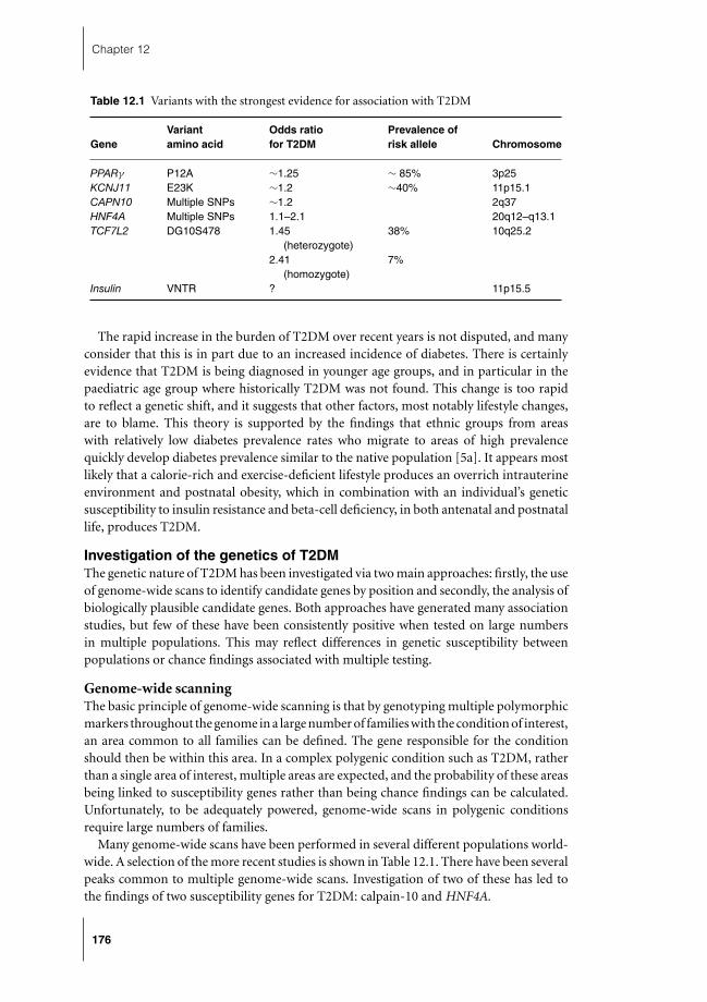
Chapter 12
Table 12.1 Variants with the strongest evidence for association with T2DM
Variant Odds ratio Prevalence ofGene amino acid for T2DM risk allele Chromosome
PPARγ P12A ∼1.25 ∼ 85% 3p25KCNJ11 E23K ∼1.2 ∼40% 11p15.1CAPN10 Multiple SNPs ∼1.2 2q37HNF4A Multiple SNPs 1.1–2.1 20q12–q13.1TCF7L2 DG10S478 1.45 38% 10q25.2
(heterozygote)2.41 7%
(homozygote)Insulin VNTR ? 11p15.5
The rapid increase in the burden of T2DM over recent years is not disputed, and manyconsider that this is in part due to an increased incidence of diabetes. There is certainlyevidence that T2DM is being diagnosed in younger age groups, and in particular in thepaediatric age group where historically T2DM was not found. This change is too rapidto reflect a genetic shift, and it suggests that other factors, most notably lifestyle changes,are to blame. This theory is supported by the findings that ethnic groups from areaswith relatively low diabetes prevalence rates who migrate to areas of high prevalencequickly develop diabetes prevalence similar to the native population [5a]. It appears mostlikely that a calorie-rich and exercise-deficient lifestyle produces an overrich intrauterineenvironment and postnatal obesity, which in combination with an individual’s geneticsusceptibility to insulin resistance and beta-cell deficiency, in both antenatal and postnatallife, produces T2DM.
Investigation of the genetics of T2DMThe genetic nature of T2DM has been investigated via two main approaches: firstly, the useof genome-wide scans to identify candidate genes by position and secondly, the analysis ofbiologically plausible candidate genes. Both approaches have generated many associationstudies, but few of these have been consistently positive when tested on large numbersin multiple populations. This may reflect differences in genetic susceptibility betweenpopulations or chance findings associated with multiple testing.
Genome-wide scanningThe basic principle of genome-wide scanning is that by genotyping multiple polymorphicmarkers throughout the genome in a large number of families with the condition of interest,an area common to all families can be defined. The gene responsible for the conditionshould then be within this area. In a complex polygenic condition such as T2DM, ratherthan a single area of interest, multiple areas are expected, and the probability of these areasbeing linked to susceptibility genes rather than being chance findings can be calculated.Unfortunately, to be adequately powered, genome-wide scans in polygenic conditionsrequire large numbers of families.
Many genome-wide scans have been performed in several different populations world-wide. A selection of the more recent studies is shown in Table 12.1. There have been severalpeaks common to multiple genome-wide scans. Investigation of two of these has led tothe findings of two susceptibility genes for T2DM: calpain-10 and HNF4A.
176

Type 2 diabetes mellitus
Calpain-10Calpain-10 is a calcium-activated protease involved in a number of cellular functions, par-ticularly intracellular signalling. A genome-wide scan in Mexican-Americans with T2DMidentified a locus on chromosome 2q. Linkage-disequilibrium mapping was used to nar-row the area of interest down and calpain-10 emerged as the candidate gene able bestto explain the linkage to 2q [6]. This was interesting, as calpain-10 did not at the timeseem a good biological candidate as a T2DM causative gene. In Mexican-Americans thestrongest susceptibility was caused by a haplotype of three intronic polymorphisms, butthis association was not confirmed in several other populations studied. A study in theUK found a separate single-nucleotide polymorphism (SNP-44) in calpain-10 in linkagedisequilibrium with a non-synonymous coding polymorphism (T504A) that would alsoaccount for the diabetes susceptibility, and which was more likely to have functional signif-icance. Meta-analysis of all published SNP-44 association studies suggested an associationwith T2DM, with an overall odds ratio of 1.2 [7]. These associations between T2DM andcalpain-10 have prompted research into the role of calpain-10 in glucose homeostasis.Although the function of calpain-10 is still only partially understood, it appears that itmay be important in the mitochondrial response to glycolysis, the transport of insulin tothe plasma membrane and insulin secretion [8].
HNF4A promoter variantsThe HNF4A gene encodes a transcription factor, HNF4α, which is expressed in liver andpancreas. Mutations in HNF4A were found to be causative for maturity-onset diabetesof the young (MODY-1) in 1996 [9]. As MODY has some similarities to T2DM, theMODY genes were investigated as biological candidates for T2DM susceptibility genes.However this approach did not produce the expected yield and initial investigation ofHNF4A did not show a clear association with T2DM [10, 11], despite 20q (the location forHNF4A) being noted as a peak in several genome scans. The finding of a distant upstreamalternative promoter in HNF4A [12] provided a further candidate for linkage to T2DM,and indeed when linkage to 20q was explored further with a series of SNPs across the area,polymorphisms near the alternative promoter were found to associate with T2DM risk intwo independent studies with different populations [13, 14].
TCF7L2A foretaste of the possible future of genetics research was the finding of a new gene associatedwith diabetes by the biopharmaceutical company deCode in 2006 [15]. The company hadperformed a genome-wide scan in the Icelandic population that reported evidence oflinkage to10q [16], a region already highlighted by a study in Mexican-Americans [17]. Bya comprehensive single-marker-association analysis, the company found a single markerDG10S478 that was associated with T2DM in Icelandic, Danish and American populations.This marker is in an intron of TCF7L2, a gene coding for a transcription factor in the Wntintracellular signalling pathway. TCF7L2 may be having an effect on T2DM or may be inlinkage disequilibrium to another effector gene. TCF7L2 regulates proglucagon expressionin enteroendocrine tissues. An important product of proglucagon is glucagon-like peptide1 (GLP-1), one of the executors of the incretin effect [18]. The effect of carrying any ofthe susceptibility alleles at DG10S478 is a ∼1.5-fold increase in the risk of T2DM, andcarrying two increases this to ∼2.4 times the risk. This suggests a significant health risk,although this association needs to be explored more fully.
177

Chapter 12
Investigation of candidate genesThe second method of investigation of the genetic basis of T2DM that has been used is theinvestigation of biologically plausible candidate genes. The plethora of genes involved inthe processes of intracellular signalling in the beta cell, insulin production and secretion,and end-organ response to insulin, provide numerous candidate genes for T2DM. Manyof these candidates have been investigated, but to date few have proven to be associatedwith T2DM.
HNF1AThe genes for MODY were obvious candidates to investigate as susceptibility genes forT2DM. Mutations in HNF1A are the commonest cause of MODY in the UK. Investigationof HNF1A in European populations did not, however, yield a major T2DM susceptibilitygene. By contrast, in the Oji-Cree people of Canada, who have the third highest prevalenceof diabetes in the world, a mutation in HNF1A (G319S) was found to associate with earlyonset of T2DM. Possession of the heterozygous mutation increases the risk of diabetes inthe Oji-Cree by 2 and the homozygous mutation by 4. However, even in the Oji-Cree, 60%of those with T2DM do not possess the S319 allele, so this mutation has only a minorimpact.
NEUROD1NEUROD1 is a helix-loop-helix transcription factor involved in pancreatic embryogenesisand insulin secretion. NEUROD1 binds to the insulin gene promoter in conjunction withE47, another helix-loop-helix protein, and increases insulin gene expression. NEUROD1knockout mice exhibit pancreatic dysgenesis and diabetes. Investigation of NEUROD1in 94 families with T2DM and apparent autosomal dominant inheritance revealed twofamilies in which mutations in NEUROD1 segregated with diabetes [19]. Further studieshave shown that NEUROD1 mutations are a minor cause of T2DM and MODY, and donot have a major impact on T2DM.
Insulin geneThe insulin gene was clearly a good candidate for conferring T2DM susceptibility. Investi-gation of the insulin gene has shown association between variation in the gene and T1DM,and there were initial reports of association between the area in the insulin gene witha variable number of tandem repeats (VNTR) and insulin resistance and T2DM, whichappeared to be confirmed by a meta-analysis of six previous studies [20]. Further inves-tigation of this association using the Warren resource of parent–offspring trios showeda parent of origin effect, with diabetic offspring more likely to have inherited a type IIIVNTR allele from his or her father than predicted (69% vs 50% predicted) [21]. This resultsuggested that the insulin VNTR effect is mediated through imprinting of the insulin gene.Disconcertingly, a study of 1462 Danes with T2DM failed to show any increase in the typeIII allele of the insulin VNTR [22]. However a more recent study from the FraminghamHeart Study seems to confirm the importance of the VNTR as a susceptibility locus butdid not show any parent of origin effect [23].
178

Type 2 diabetes mellitus
PPARγ
PPARγ is a transcription factor predominantly expressed in adipose tissue but has also beenshown to be expressed in small quantities in liver, muscle and pancreas [24]. It has becomeincreasingly clear that PPARγ has a vital role in the regulation of body fat distributionand insulin sensitivity, acting to promote energy storage as a thrifty response. Interest inPPARγ as a T2DM candidate gene stemmed from the fact that the thiazolidinedione classof drugs act through PPARγ stimulation and improve hypertension, blood glucose controland insulin sensitivity in patients with T2DM [25]. Inactivating mutations in the gene forPPARγ (PPARG) in humans have been found in rare forms of diabetes with lipodystrophy[26–28]. Taken together, these findings suggest that increasing activity of PPARγ is inverselyproportional to insulin sensitivity. A common polymorphism in PPARG (Pro12Ala), whichdecreases the activity of PPARγ, has however been shown to be associated with highinsulin sensitivity and decreased susceptibility to T2DM [29, 30]. This apparent paradoxis due to the multiple effects of PPARγ; loss of function of PPARγ causes decreasedlipogenesis and increased fatty acid oxidation; both processes result in decreased adipocytehypertrophy and decreased free fatty acids, and hence increased insulin sensitivity whilstPPARγ stimulation increases production of small insulin-sensitive adipocytes and reduceshepatic glucose production [31]. Early studies of Pro12Ala were inconclusive, but largerstudies have provided more evidence that possession of the proline allele increases the riskof developing T2DM by ∼1.25 times. This is a low relative risk (RR) but the proline alleleis so common in most ethnic groups (∼80% of individuals) that it has a significant effecton the population risk [32].
KCNJ11The beta-cell potassium channel is intimately involved in regulation of insulin secretion.The channel is composed of four inwardly rectifying potassium channels (Kir 6.2 geneKCNJ11) and four sulphonylurea receptor subunits SUR1 gene [33]. Mutations in KCNJ11have been shown to cause both infantile hyperinsulinaemia (inactivating mutations) [34]and neonatal diabetes (activating mutations) [35]. Interest in the role of Kir 6.2 in insulinsecretion led to consideration ofKCNJ11 as a candidate gene for T2DM. Early studiesdid not show any association between variation in KCNJ11 and T2DM, but two studiesdid show such an association in French and British subjects [36, 37]. Larger studies andmeta-analyses have strengthened the evidence that the k allele present in ∼60% of whiteEuropeans increases the risk of developing T2DM ∼1.2-fold [38–40], although one studyfound that the allele is in linkage disequilibrium with a variant in SUR1, raising thepossibility that variation in SUR1 may be the underlying cause of an increase in T2DM inthe populations studied [41].
Summary
The genetics of T2DM are complex. Research strategies to date have focused on genome-wide scanning and evaluation of plausible candidate genes. As the methods and tools forinvestigating complex genetic disease improve, it seems likely that more of the genome’sand proteome’s variation can be analysed in the hunt for the susceptibility factors thatinfluence an individual’s risk of T2DM.
179

Chapter 12
Treatment of T2DM in childhoodTimothy G. Barrett
IntroductionT2DM in childhood is a relatively new phenomenon, with the first cases described only in1979 [42]. Since then, children with T2DM diabetes have been reported from many coun-tries around the world; the prevalence is increasing and relates to the rise in childhoodobesity seen in many Western and developing countries. In children, it is most often relatedto obesity, and other cardiovascular risk factors may be present. T2DM results from a com-bination of insulin resistance, increased hepatic glucose output and progressive decline ofglucose-stimulated insulin secretion. In healthy children, insulin secretion involves a basalcomponent, which maintains normal fasting plasma glucose, and a prandial component,which sharply increases insulin secretion in response to a glucose load. This limits hep-atic glucose release and inhibits gluconeogenesis. One of the earliest defects in T2DM isreduction in the prandial component of insulin secretion, through loss of the first-phaseinsulin response. This can be insidious in adults and probably children, with a decreasein the prandial component of insulin release by as much as 50% by the time T2DM isdiagnosed [43]. Consequently, postprandial glucose rises above the normal range are anearly abnormality in T2DM. Abnormal fasting glucose can be a late sign, indicating that amarked deterioration in beta-cell function has already occurred [44].
Guidelines for the management of T2DMAlthough there is a very small literature on optimum treatment of T2DM in children,and almost none on outcomes, there is a wealth of data available on adults with T2DM.Thus the paediatrician has access to a large evidence base, and the experience of his or heradult colleagues guide the management of children. This section will review some of thisevidence and relate it to the special case of T2DM in children.
T2DM has no clear definition, but pragmatically it can be considered as tissue insulin re-sistance, with a significant and progressive insulin secretory defect. It is known that insulinresistance is associated with multiple cardiovascular risk factors; consequently, T2DM inchildhood is likely to be associated with earlier micro- and macrovascular complicationsthan seen with childhood-onset T1DM. In addition, the insidious onset of T2DM, anda variable asymptomatic period before presentation, means that vascular complicationsmay already be present at diagnosis. This was originally shown in the Pima Indian chil-dren with T2DM and has now been shown in a larger cohort of T2DM children from theTreatment Options for Type 2 Diabetes in Adolescence and Youth (TODAY) study: of 240children enrolled into this study with T2DM, 26% had hypertension, 60% had dyslipi-daemia and 17% had both (B) [45]. There is also evidence that even stricter glycaemiccontrol is required for insulin-resistant patients with T2DM than for those with T1DM:the UK Prospective Diabetes Study (UKPDS) showed that a reduction in average HbA1cfrom 7.9 to 7.0% resulted in a 25% decrease in microvascular complications (A) [46], andreducing blood pressure below 144/82 resulted in a 44% reduction in the risk of strokeand 36% reduction in the risk of heart failure (A) [47].
Bearing the above evidence in mind, recent policy initiatives have emphasised the im-portance of good glycaemic control in T2DM (A) [48]. There are now well-defined targets
180

Type 2 diabetes mellitus
to work towards in T2DM, most recently an ADA–EASD (American Diabetes Association–European Association for the Study of Diabetes) consensus algorithm for the managementof hyperglycaemia in T2DM (E) [49]. This algorithm suggests that an HbA1c target of lessthan 7.0% should be aimed for, with an HbA1c less than 6.0% (normal) being the ‘goldstandard’. Aiming for a normal HbA1c is probably not appropriate in children because ofthe risk of hypoglycaemia; however, our challenge is to make an HbA1c of 7.0% a realisticaim for our patients.
The treatment goals for childhood T2DM are to:1 achieve glycaemic control to target HbA1c ≤7.0% without severe hypoglycaemicepisodes2 reduce body mass index (BMI) to <95th centile for age and sex3 encourage exercise to increase cardiovascular fitness towards 60 minutes of moderate-to-vigorous exercise daily (E) [50]4 look for and treat associated comorbidities, e.g. hypertension, hyperlipidaemia andmicroalbuminuria
The evidence for reducing HbA1c to 7.0% has been cited above. Obesity is an estab-lished cardiovascular risk factor; this definition of obesity is widely used in North Americaand is less likely to underestimate obesity than the International Obesity Task Force cut-off values. However a lower centile cut-off might be appropriate for obese children withT2DM from ethnic minorities. The exercise guideline is a recent recommendation basedon an exhaustive literature review and is for healthy children. Obese children will strug-gle to achieve this ambitious goal, but any increase in exercise is probably of benefit tocardiovascular health.
Lifestyle changesThere is clear evidence from the Finnish Diabetes Prevention Study and the DiabetesPrevention Program that intensive lifestyle interventions can reduce insulin resistance,slow the rate of progression to T2DM and improve glycaemic control (A) [51, 52]. The firststep in managing young people with T2DM is to encourage lifestyle changes. Paediatriciansare used to the concept of multidisciplinary care teams in the management of their patients;they work in partnership with their patients to develop individual care plans with clearlydefined treatment objectives and targets. These might include regular self-monitoring ofblood glucose, HbA1c measurement at each clinic visit, annual fasting lipid and bloodpressure measurement, weight monitoring and advice on stopping smoking. There is aneed to develop structured education packages for children with T2DM both at diagnosisand during ongoing management. Of great importance is explaining to children andparents that they are likely to require insulin therapy within 10 years of diagnosis (A) [53].Children and parents need to understand that treatment for T2DM involves a stepwiseprogression to maintain glycaemic control and that commencement of insulin is not anindication that they have failed.
The Royal College of Paediatrics and Child Health has published guidelines for diet andexercise advice for the prevention of obesity, which are also very useful for obese childrenwith T2DM (Tables 12.2 and 12.3) (E) [54]. There is good evidence that children withobesity have reduced exercise tolerance and that goals have to be realistic; any increase inactivity, however small, is an advance, and children should be encouraged to increase theiractivity levels in a graded programme. A recent literature review suggested that school-ageyouth should participate daily in 60 minutes or more of moderate-to-vigorous physical
181
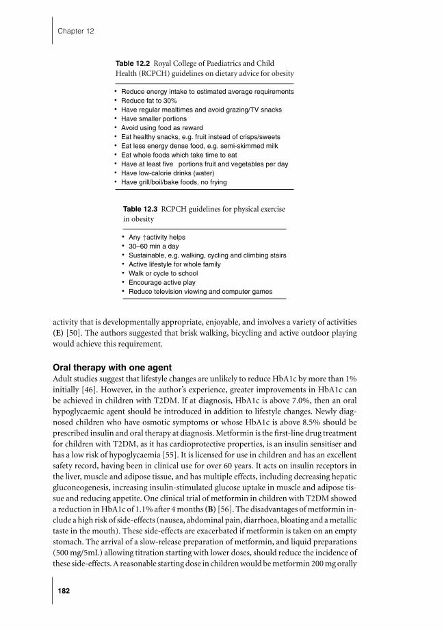
Chapter 12
Table 12.2 Royal College of Paediatrics and ChildHealth (RCPCH) guidelines on dietary advice for obesity
� Reduce energy intake to estimated average requirements� Reduce fat to 30%� Have regular mealtimes and avoid grazing/TV snacks� Have smaller portions� Avoid using food as reward� Eat healthy snacks, e.g. fruit instead of crisps/sweets� Eat less energy dense food, e.g. semi-skimmed milk� Eat whole foods which take time to eat� Have at least five portions fruit and vegetables per day� Have low-calorie drinks (water)� Have grill/boil/bake foods, no frying
Table 12.3 RCPCH guidelines for physical exercisein obesity
� Any ↑activity helps� 30–60 min a day� Sustainable, e.g. walking, cycling and climbing stairs� Active lifestyle for whole family� Walk or cycle to school� Encourage active play� Reduce television viewing and computer games
activity that is developmentally appropriate, enjoyable, and involves a variety of activities(E) [50]. The authors suggested that brisk walking, bicycling and active outdoor playingwould achieve this requirement.
Oral therapy with one agentAdult studies suggest that lifestyle changes are unlikely to reduce HbA1c by more than 1%initially [46]. However, in the author’s experience, greater improvements in HbA1c canbe achieved in children with T2DM. If at diagnosis, HbA1c is above 7.0%, then an oralhypoglycaemic agent should be introduced in addition to lifestyle changes. Newly diag-nosed children who have osmotic symptoms or whose HbA1c is above 8.5% should beprescribed insulin and oral therapy at diagnosis. Metformin is the first-line drug treatmentfor children with T2DM, as it has cardioprotective properties, is an insulin sensitiser andhas a low risk of hypoglycaemia [55]. It is licensed for use in children and has an excellentsafety record, having been in clinical use for over 60 years. It acts on insulin receptors inthe liver, muscle and adipose tissue, and has multiple effects, including decreasing hepaticgluconeogenesis, increasing insulin-stimulated glucose uptake in muscle and adipose tis-sue and reducing appetite. One clinical trial of metformin in children with T2DM showeda reduction in HbA1c of 1.1% after 4 months (B) [56]. The disadvantages of metformin in-clude a high risk of side-effects (nausea, abdominal pain, diarrhoea, bloating and a metallictaste in the mouth). These side-effects are exacerbated if metformin is taken on an emptystomach. The arrival of a slow-release preparation of metformin, and liquid preparations(500 mg/5mL) allowing titration starting with lower doses, should reduce the incidence ofthese side-effects. A reasonable starting dose in children would be metformin 200 mg orally
182

Type 2 diabetes mellitus
twice a day with meals, increasing gradually every 2 weeks until the maximum tolerateddose (E). Metformin is not usually tolerated in doses greater than 1 g 8- or 12-hourly. Thecontraindications to metformin use include renal and hepatic impairment (liver enzymes>twice the upper limit of normal). Lactic acidosis is a rare side-effect seen if metforminaccumulates in the body, such as in impaired renal function. Metformin is contraindicatedin people with uncontrolled heart failure (e.g. some children with Alstrom syndromeand cardiomyopathy), renal impairment, advanced liver disease and any predispositionto hypoxaemia, such as sepsis. Metformin may also enhance the likelihood of ovulationin teenagers with PCOS, so the risks of pregnancy should be explained and contraceptiveadvice offered.
Oral therapy with two agentsIf after 3 months of oral therapy with metformin the HbA1c remains above the target levelof 7.0%, a second agent should be introduced. The current National Institute of ClinicalExcellence (NICE) guidelines suggest the use of a glitazone as a replacement for either met-formin or an insulin secretagogue (E) [48]. However, these guidelines are currently beingrevised, and there is some logic to adding a glitazone to metformin, as this combinationtargets insulin resistance and consequently may benefit cardiovascular risk factors. This issupported by the results of the PROactive study (B) [57]. Thiazolidinediones act on the nu-clear receptor peroxisome proliferator-activated receptor gamma (PPARϒ). This receptoris strongly expressed in adipose tissue, and its downstream pathway increases transcriptionof various insulin-sensitive genes involved in lipid and glucose metabolism. The net effectis a lowering of insulin resistance. The original drug in this class troglitazone was with-drawn due to hepatotoxicity. The two drugs currently available in the UK are pioglitazoneand rosiglitazone. In adult studies, glitazones, either alone or in combination with met-formin, have been shown to reduce HbA1c by 1–1.5%, but the glucose-lowering effect maytake 3–6 months to reach maximum effect. There may also be benefits in blood pressureand cholesterol reduction. The side-effects include fluid retention, so are contraindicatedin heart failure (e.g. diabetes in cardiomyopathy such as Alstrom syndrome) and weightgain due to increased subcutaneous fat. Other contraindications include hepatic or renalimpairment.
Alternative second-line agents to glitazones include the sulphonylureas, meglitinideanalogues and alpha-glucosidase inhibitors. There is almost no evidence of the effects oftheir use in children.
SulphonylureasSulphonylureas bind to receptors on the K+/ATP channels, causing them to close; thisdepolarises the beta-cell membrane, which opens Ca++ channels, allowing influx of cal-cium and insulin exocytosis. These drugs work by increasing insulin secretion, so requireresidual pancreatic beta-cell function. Sulphonylurea binding persists on its receptors forlong periods, so that the earlier sulphonylureas have prolonged effects, including hypogly-caemia. Hypoglycaemia is still the main side-effect of this class of drugs, and families andchildren need to be educated about hypoglycaemia if sulphonylureas are being consideredas treatment. The first generation of these drugs was tolbutamide, and the second gener-ation include glibenclamide, gliclazide, glimepiride, glipizide and gliquidone. Studies inadults have shown them to reduce HbA1c by 1–2%. NICE guidelines recommend a sulpho-nylurea as initial therapy in adults with T2DM, who are normal weight or underweight (B)
183

Chapter 12
[48]. In children with obesity-related T2DM, sulphonylureas are a second-line treatmentin combination with metformin. Gliclazide is popular in the UK, and there is a once-a-daymodified release preparation available, which is associated with less hypoglycaemia thanthe standard generic 80 mg tablets.
Meglitinide analoguesThese are short-acting insulin secretagogues that also bind to the K+/ATP channels, causingthem to close and initiate insulin release from the pancreatic beta cells. They act within10–30 minutes and have a duration of action from 2 to 4 hours. They have been used withmeals to enhance insulin secretion. They may be less likely to cause hypoglycaemia due totheir shorter duration of action but are less effective in controlling basal hyperglycaemia.Repaglinide has been used in adults with T2DM alone or in combination with metformin.Nateglinide is used only with metformin. There are no published data on their use inchildren.
Alpha-glucosidase inhibitorsAcarbose is the only drug available in the UK in this class, and it inhibits gastrointestinalalpha-glucosidase, an enzyme that breaks down ingested carbohydrates. It can reduce thepostprandial rise in blood glucose but is associated with unacceptable side-effects for mostteenagers (flatulence, bloating and diarrhoea).
Insulin therapyInsulin has been available for use in children for over 80 years; paediatricians have extensiveexperience of its use in children with T1DM; and its effectiveness in glucose reduction andside-effects of hypoglycaemia are well known. The significant insulin resistance in T2DMmeans that hypoglycaemia is not as prevalent as in T1DM. Adults with T2DM are thoughtto have lost around 50% of pancreatic beta-cell function by the time of diagnosis, andmost require supplemental insulin within 5 years. In a study of children with T2DM fromNorth America, 50% had an insulin requirement by 5 years after diagnosis [58]. As well asan increase in hypoglycaemia, important side-effects in children with T2DM are increasedappetite and weight gain.
Young people should be considered for insulin therapy if 3 months of dual oral therapydo not achieve an HbA1c less than 7.0% (B). Treatment with metformin can be contin-ued, but any secretagogues should be discontinued if rapid-acting insulin is included inthe regime. There is a recognition in adult T2DM practice that patients have not beenmanaged aggressively enough and that the average adult with T2DM has been exposedto about 10 years of excess glycaemic burden, defined as an HbA1c above 7.0%, fromdiagnosis to initiation of insulin therapy as a result of ‘clinician inertia’ (B) [59]. It isbecoming increasingly recognised that elevated postprandial glucose makes an important,independent contribution to overall glucose control and plays a role in the pathogen-esis of diabetes complications [43]. There is evidence that the contribution to HbA1cis predominantly by fasting blood glucose concentrations as HbA1c rises above 7.3%,whereas postprandial blood glucose is the principal contributing factor at lower HbA1clevels [60].
The theoretical ideal insulin regime currently available to address these problems isbasal-bolus therapy using analogues (E). The basal component choices include insulinglargine and insulin detemir. These have a longer duration of action than the traditional
184

Type 2 diabetes mellitus
Table 12.4 Recommended actions for patients who are not achieving target HbA1cwith insulin regimes
� Review individualised care plan� Review lifestyle modifications� Review diet and carbohydrate counting� Ensure metformin dose is maximised� Titrate insulin doses up to achieve target fasting and postprandial glucose levels� Consider continuous blood glucose monitoring� Consider switching from twice daily premixed insulin to basal bolus if not already done so� Check compliance with oral and injected medication� Check insulin injection technique and device working properly� Check correct storage of insulin, in date, etc.� Check injection sites for lipohypertrophy
neutral pH Hagedorn (NPH) insulin and a flatter insulin profile over 24 hours. Therapid-acting insulin analogues aspart, lispro and glulisine are designed to mimic moreclosely the endogenous prandial insulin secretion compared with short-acting humaninsulin. The basal insulin dose should be titrated to achieve a target fasting glucose lessthan 7.0 mmol/L. Home blood glucose monitoring should include postprandial measure-ments; the International Diabetes Federation recommends a target postprandial glucoseof less than 7.5 mmol/L [61] and the American Diabetes Association a target of less than10 mmol/L (B) [62]. The major 2-hour postprandial blood glucose excursion shouldbe determined (usually after the main meal) and, if above target, the preceding dose ofrapid-acting insulin titrated up until target is achieved. Clearly, patients should be taughtcarbohydrate counting to facilitate appropriate insulin adjustment.
Although the above description is for a theoretical ideal insulin regimen, this may eitherbe unacceptable to patients or not suitable due to domestic, school or work difficulties.Acceptable alternatives would be twice daily administration of premixed insulins and abasal insulin with an oral secretagogue, such as a repaglinide before meals. Studies compar-ing basal insulin glargine/oral hypoglycaemic agent preparations with premixed insulinshave not shown a clear advantage of one regimen over the other. Some patients mightfind it more acceptable to have a stepwise progression of treatment, adding first a basallong-acting insulin to oral agents and then preprandial short-acting insulin analogues.For young people who are not achieving target HbA1c with either a basal-bolus regime ortwice daily premixed insulin, some suggested actions are given in Table 12.4.
Treatment of comorbiditiesT2DM is associated in children with a metabolic syndrome phenotype, and so is associatedwith several cardiovascular disease risk factors. All affected children should have regulareye examinations and urinary microalbumin excretion estimation. This review will discussonly dyslipidaemia and hypertension.
DyslipidaemiaDyslipidaemia in T2DM is characterised by raised triglycerides, low high-density lipo-protein (HDL) cholesterol and increased small dense, atherogenic low-density lipoprotein(LDL) cholesterol. Raised LDL cholesterol and lowered HDL cholesterol are establishedand modifiable risk factors for the development of cardiovascular disease. An adult with
185

Chapter 12
diabetes has a risk for cardiovascular disease close to that of a non-diabetic person witha previous myocardial infarction (A) [63]. Adults with T2DM have an absolute risk ofcardiovascular disease of 20% or more over 10 years. However, the risk is not known forchildren with T2DM. The closest comparison we have is a 10-year study of 589 patientswith childhood-onset T1DM aged over 18 years at baseline and followed for 10 years(B) [64]. The RR for cardiovascular disease was 1.8 with LDL cholesterol >2.6 mmol/L(>100 mg/dL) and 2.3 with LDL cholesterol >3.4 mmol/L (>130 mg/dL). For triglyc-erides the RR was 2.5 with triglyceride >2.3 mmol/L (>90 mg/dL) and 3.3 with triglyceride>3.9 mmol/L (>150 mg/dL). These data suggest that childhood-onset diabetes is asso-ciated with an increased risk for cardiovascular disease if dyslipidaemia is present, andtherefore treatment to lower lipids is logical.
The first-line treatment is always a reduced fat diet and exercise, weight reductionif growth is completed and glycaemic control. After non-pharmacological treatments,the most effective class of drugs are 3-hydroxy-3-methylglutaryl coenzyme A (HMG-CoA) reductase inhibitors (statins). These work by competitively inhibiting HMG-CoA,an enzyme involved in cholesterol synthesis, especially in the liver. They are particularlyeffective in lowering LDL cholesterol and increasing the concentrations of HDL cholesterol.However, they are not as effective as fibrates in reducing triglycerides.
NICE (E) [65] has recommended the use of statins as lipid-lowering agents for patientswho meet the following criteria:all patients over 18 years with diabetes plus one other risk factor:
� treated hypertension� retinopathy� nephropathy� features of the metabolic syndrome� high-risk family history.Clearly, many children under 18 years with T2DM have multiple risk factors and should
be considered for statin therapy. The treatment target is to achieve total cholesterol levels<4 mmol/L and LDL cholesterol <2 mmol/L (E) [65]. The side-effects include abdominalpain, flatulence, nausea and vomiting, and altered liver function tests. Atorvastatin, pravas-tatin and simvastatin are now all listed in the British National Formulary for Children forhyperlipidaemia.
Fibrates such as fenofibrate act mainly by decreasing serum triglycerides and raise HDL-cholesterol concentrations with less effect on LDL cholesterol. Some of the actions offibrates are mediated through PPARα receptors; consequently, they have multiple actions,including increasing insulin sensitivity and decreasing vascular inflammation. Althoughtheoretically they should be ideal for the dyslipidaemia in T2DM, a recent study of 10,000adults with T2DM showed a reduction in cardiovascular events, but no effect on totalmortality (A) [66]. They tend to have more side-effects than statins, and data on use inchildhood are very limited. Fibrates remain a second-line treatment in adults after statins.Liver function should be monitored 3-monthly and, as with statins, both these classes ofdrugs should be avoided in pregnancy.
HypertensionHypertension is a frequent accompaniment of T2DM in obese children and an independentrisk factor for progression to cardiovascular disease. In the UKPDS, control of hypertensionwas more important than blood glucose control in decreasing the frequency of cardiac
186

Type 2 diabetes mellitus
events (A) [47]. The same Pittsburgh study of childhood-onset T1DM quoted aboveshowed that a systolic blood pressure ≥120–129 mmHg was associated with an RR of 2.5for cardiovascular disease (B) [64].
All children with diabetes should have blood pressure estimated at least annually usingan appropriate-size cuff. This may mean using a thigh cuff in the most obese children.As body size is an important determinant of blood pressure in children, charts of normalvalues often give blood pressure values in relation to height centiles [67]. If the systolicor diastolic blood pressure is elevated above the 90th centile for age, sex and height ontwo or more occasions, an early morning urine collection should be collected to screenfor microalbuminuria. The first-line treatment is diet and exercise advice as for newlydiagnosed T2DM. If these measures have no effect after 3 months, then treatment with anangiotensin converting enzyme (ACE) inhibitor should be considered. These drugs blockACE, which reduces the production of angiotensin II, a potent vasoconstrictor. Lisinoprilis an example of this class of drug. The most important side-effects are severe hypotension,renal impairment and a dry cough. The risk of hypotension can be minimised by startingwith an initial low dose, such as 2.5 mg once daily. The usual maintenance dose is 10–20 mgonce daily. Newer drugs include the angiotensin-II-receptor antagonists, such as losartan,and do not seem to cause the dry cough that may occur with ACE inhibitors.
Non-alcoholic fatty liverNon-alcoholic fatty liver disease (NAFLD) is the term used to define a range of alcohol-likeliver injuries that occur without alcohol abuse and include non-alcoholic steatohepatitisand NAFLD-induced cirrhosis. The term NAFLD is usually used for the liver injuriesassociated with the metabolic syndrome, but a precise definition has not yet been agreed[68]. The prevalence is unknown in childhood, but it is closely linked to obesity, themetabolic syndrome and T2DM in childhood. Insulin resistance is believed to be theunderlying cause, as peripheral insulin resistance leads to influx of free fatty acids to theliver. This occurs by both decreased suppression of lipolysis and increased lipogenesis in theliver. The subsequent accumulation of fat within the hepatocytes leads to the developmentof hepatic insulin resistance. There is a lack of follow-up data on the outcomes of NAFLDin children; however, adult data show slow progression to cirrhosis over several yearsor decades. The children most at risk for NAFLD are likely to be those obese childrenwith abnormal glucose tolerance, or with multiple cardiovascular risk factors. The initialinvestigation should be liver function tests (LFTs): children with persistently (>1 yr)abnormal LFTs (pragmatically more than 1.5 times the upper limit of normal) shouldundergo a systematic assessment to exclude other forms of chronic liver disease [69]. Thediagnosis of NAFLD can only be confirmed by liver biopsy. The current best treatment islifestyle modification, in terms of increased physical activity and the promotion of a healthy,balanced diet. Vitamin E has been recommended as an antioxidant and metformin as aninsulin sensitiser.
Management algorithm (Figure 12.1)T1DM may be confused with T2DM. Any child in whom T2DM is suspected, who presentswith osmotic symptoms, ketonuria, dehydration or ketoacidosis, should be treated withinsulin immediately. Insulin can always be weaned off after the acute episode if neces-sary. These children can present with acute metabolic decompensation and withholdinginsulin may be fatal. Diabetic ketoacidosis in T2DM is treated in the same way as for
187

Chapter 12
Osmotic symptoms, ketoacidosis and dehydration
Asymptomatic
Commence insulin, diet, exercise and metformin
HbA1c<7.5% HbA1c ≥7.5%
Diet and exercise Diet, exercise andmetformin
3-monthly HbA1c
<7.0%continue
≥7.0% add sulphonylurea or
glitazone
≥7.0% switch to metformin plus long-acting insulin plus short-acting insulinboluses before meals. Alternatives:
twice daily premixed insulin analogues; or basal long-acting
insulin plus meglitinide
Figure 12.1 Algorithm for the diagnosis of T2DM (evidence of insulin resistance, autoantibodynegative).
T1DM. Metformin can be started when the diabetic ketoacidosis has resolved and insulincontinued or weaned off depending on glycaemic control. For asymptomatic patientswith only moderately raised HbA1c, a trial of diet and exercise is usually the best policy toobtain the maximum benefit from these lifestyle changes before adding in drug therapy.Metformin is the first-line drug treatment. If there is no response, it is worth checkingcompliance. Finally, insulin should be added for patients with persistently poor glycaemiccontrol. Insulin works best when used in combination with an agent that increases insulinsensitivity.
Polycystic ovarian syndrome
Polycystic ovarian syndrome (PCOS) is a heterogeneous clinical entity affecting 5–10% ofwomen worldwide, making it one of the commonest endocrine disorders of young womenand the commonest cause of infertility in this age group. The original description of amen-orrhoea with polycystic ovaries was made by Stein and Leventhal more than 60 years ago
188

Type 2 diabetes mellitus
[70]. The cardinal features were anovulation, presenting with oligomenorrhoea or amen-orrhoea and infertility, and hyperandrogenaemia, presenting with hirsutism, acne andalopecia. In retrospect, the features were probably the same as Achard–Thiers syndrome,the ‘diabetes of the bearded lady’ [71]. Since then it has become apparent that there arefundamental metabolic disturbances integral to the syndrome that lead to significant car-diovascular risk. These include obesity, insulin resistance, beta-cell dysfunction, impairedglucose tolerance and T2DM, dyslipidaemia, hypertension, obstructive sleep apnoea, aprocoagulant state and endothelial dysfunction. In one recent study, 40% of women withPCOS demonstrated glucose intolerance, and 7.5% of these women had overt T2DM [72].Conversely, 50% of women with T2DM have been reported to have evidence of polycysticovaries [73]. PCOS is a major risk factor for the development of T2DM in women of allages. A new area of concern is the recognition of PCOS in teenage girls with T1DM. Withincreasing weight gain, they become insulin resistant and are made hyperinsulinaemicwith exogenous insulin, especially with aggressive insulin regimens [74].
PathophysiologyThere is clearly a genetic component to the development of PCOS, based on observed famil-ial clustering [75]. Female first-degree relatives of women with PCOS have a significantlyincreased prevalence of PCOS (A) [76]. A more extreme version of hyperandrogenism,severe insulin resistance, and acanthosis nigricans, designated HAIR-AN, or type A insulinresistance, was described in adolescent girls (C) [77], due to mutations in the insulin recep-tor gene [78]. However, several studies failed to detect mutations in the insulin receptorsof women with the much commoner PCOS [79, 80]. Women with PCOS have significantlyhigher fasting insulin levels compared with age-matched and BMI-matched controls [81].The exact site of abnormality in the insulin-signalling cascade in PCOS is not known. Theinsulin resistance has some relation to overweight and obesity, and there is a hyperbolicrelation between insulin sensitivity and increasing BMI, with decreasing insulin sensitivitywith increasing BMI [82]. However, obesity is not the only cause of insulin resistance inPCOS: insulin resistance is higher and the glucose disposition index is lower in womenwith PCOS and a family history of T2DM [83]. In addition, insulin resistance also occurs innon-obese women with PCOS and normal glucose tolerance [84]. Even lean women withPCOS are as insulin resistant as obese women without PCOS, and obese PCOS women areas insulin resistant as patients with T2DM.
Insulin resistance and hyperandrogenaemia potentiate each other so that it was not clearwhich was the underlying abnormality. However, in experiments with gonadotrophin-releasing hormone agonists, it was shown that suppression of hyperandrogenaemia doesnot improve peripheral or hepatic insulin resistance in PCOS (B) [85]. However, if insulinlevels are lowered, for instance with metformin, then androgen levels are lowered (B) [86].What is clear is that the hyperinsulinism consistent with insulin resistance precedes andfacilitates the androgen excess observed in PCOS. Human ovaries have abundant insulinand IGF-1 receptors, and insulin stimulates ovarian androgen production: theca cells fromPCOS patients produce testosterone in response to insulin at higher levels compared withnon-PCOS theca cells (C) [87]. In PCOS, the ovaries appear to be hypersensitive to insulinaction and have augmented androgen production, which is surprising given that thereis widespread tissue insulin resistance (E) [88]. Sex-hormone-binding globulin (SHBG)is a glycoprotein produced in the liver which acts as a carrier for sex hormones, with ahigh binding affinity for testosterone and dihydrotestosterone. Insulin is thought to lower
189

Chapter 12
SHBG levels by acting directly to reduce hepatic SHBG synthesis: decreased basal levels ofSHBG are seen in PCOS, with a more significant reduction in obese women with PCOS.The hyperinsulinaemia in PCOS lowers the SHBG levels and enhances the expression ofhyperandrogenaemia [89].
Insulin resistance is known to rise during puberty, resolving by Tanner stage 5 [90]. How-ever, in girls with premature adrenarche, this pubertal rise in insulin resistance is greatlyexaggerated [91], and these girls appear to be at significantly higher risk for developingfunctional hyperandrogenism and PCOS [92].
DefinitionThere is not yet a consensus on the diagnostic criteria for PCOS, but minimal criteria areconsidered to be hyperandrogenism and ovulatory dysfunction in the absence of otherhypothalamopituitary, ovarian or adrenal disease. Of interest, polycystic ovaries on ultra-sound are not required to make the diagnosis. These have been defined as the presenceon ultrasound of eight or more subcapsular follicular cysts ≤10 mm in diameter andincreased ovarian stroma. Polycystic ovaries are present in 20–25% or randomly selectedwomen, so their presence in the absence of ovulatory dysfunction or hyperandrogenismis not sufficient to diagnose PCOS [93].
A recent consensus working group [94] has put forward a definition of PCOS based onthe presence of two out of three features:� oligo- or anovulation� clinical and/or biochemical signs of hyperandrogenism� polycystic ovaries and exclusion of other aetiologies (congenital adrenal hyperplasia,androgen-secreting tumours and the Cushing syndrome).
Hyperinsulinism may manifest with mild or moderate acanthosis nigricans. Hyperan-drogenism may present with excessive hirsutism and/or acne. Raised androgens (testos-terone and/or androstenedione) is the most consistent biochemical abnormality in PCOS.Increased adrenal androgen production (e.g. dehydroepiandrosterone sulphate) may of-ten coexist. SHBG is frequently reduced, and a threshold level of <37 nmol/L has beensuggested for the diagnosis of PCOS (sensitivity 87.5% and specificity 86.8%) [95].
A common presentation in adult practice is with anovulatory infertility. However inthe teenage age group, menstrual disturbance is often the presenting feature. Oligomen-orrhoea is an excellent marker for PCOS, and 85% of oligomenorrhoeic women can beassumed to have PCOS. Young women with PCOS have more pronounced irregularityof menstrual cycles than is usual for that age group, and the irregularity may include ei-ther amenorrhoea or prolonged heavy periods. Abnormalities of gonadotrophin secretionare also characteristic of PCOS, in particular an elevated luteinizing hormone (LH) oran elevated luteinizing hormone (LH)-to-follicle-stimulating hormone (FSH) ratio [96].However, serum LH levels may be normal in up to 40% of women with PCOS [97].
At least 60% of adolescent girls with features of the metabolic syndrome have concomi-tant PCOS, and multiple cardiovascular risk factors are two to three times more common inwomen with PCOS than in healthy controls. Of all women with PCOS, about 55–60% havenormal glucose tolerance, 30% have impaired glucose tolerance and 5–10% have T2DM.Between 30 and 80% of obese PCOS, women will develop T2DM by 30 years of age. Theprevalence of glucose intolerance increases with increasing BMI. Women with PCOS havea higher prevalence of cardiovascular disease on long-term follow-up than their healthypeers [98]. The cardiovascular abnormalities include lower HDL cholesterol, increased
190

Type 2 diabetes mellitus
carotid intima-media artery thickness, reduced vascular-flow-mediated dilatation and anRR for coronary heart disease of 1.5 and for fatal myocardial infarction of 1.9.
TreatmentTherapy for PCOS can be divided into acute and chronic therapy. Acute therapy is aimedat managing the infertility, improving the menstrual cycles and treating the acne andhirsutism. Chronic therapy is aimed at managing the cardiovascular risk factors includingglucose intolerance and T2DM.
Acute managementThe first principle of management is lifestyle intervention to reduce calorie intake, increaseexercise and achieve weight reduction. Several studies have shown the beneficial effects onmenstrual abnormalities, fertility rate, hirsutism and acanthosis nigricans (B, C) [99–101].These improvements seem to be maximised in women who lost more than 5% of theirbody weight. The intervention has to be a combination of diet and exercise programmes.The main beneficial effects of weight loss in PCOS appear to be mediated via reduction ofhyperinsulinism, improved insulin sensitivity and consequently reduced androgen levels.However, none of these parameters changed significantly in women who failed to restorea normal ovulation pattern (B) [99].
After lifestyle, the choices are between oral contraceptives, antiandrogens and insulinsensitisers. Oral contraceptives (OCP) can regularise the menstrual cycle and promoteovulation. They also reduce serum androgen levels, hirsutism and acne. In these respects,they are more effective than insulin sensitisers. The disadvantages of oral contraceptivesare that they may worsen insulin resistance, increase the risk of cardiovascular disease andincrease triglyceride levels. Cyproterone acetate has been popular as an antiandrogen inPCOS. In a study of the effectiveness of Dianette (OCP plus cyproterone) versus metformin(A) [102], Dianette caused worsening of insulin resistance. There is a concern about puttingyoung people on cyproterone at the age of 14 years and the long-term effects over decadesof use in terms of cardiovascular risk. If oral contraceptives are chosen as treatment forPCOS, it is advisable to undertake a baseline oral glucose tolerance test and fasting lipidsand repeat after 3–4 months on treatment. Insulin sensitisers have been shown to helpovulation in infertile women with PCOS, and metformin can enhance ovulation in 80%of women. Traditionally, women have been advised to stop metformin once they haveconceived. However, there is some evidence of an increased rate of miscarriage in womenwith PCOS once they have become pregnant and stopped metformin.
Chronic managementThe mainstay of chronic management is to reduce the long-term risks for cardiovasculardisease. All patients should be screened for cardiovascular risk factors at baseline, includingblood pressure and lipids and an oral glucose tolerance test. Insulin sensitisers in PCOShave been shown to reduce insulin resistance and improve glucose tolerance, and thereis evidence that they slow progression to T2DM and cardiovascular disease (B) [103].Metformin is the drug of choice: it improves peripheral insulin sensitivity and attenuateshyperandrogenaemia in both lean and obese women with PCOS, restoring normal mensesin a large proportion of previously amenorrhoeic women (B) [104]. The alternativesinclude thiazolidinediones; there is some evidence that these drugs can improve carotidintima-media artery thickness.
191

Chapter 12
References
1 World Health Organization Department of Noncommunicable Disease Surveillance. Definition,Diagnosis and Classification of Diabetes Mellitus and its Complications. Report of a WHO Con-sultation Part 1: Diagnosis and Classification of Diabetes Mellitus. Geneva, 1999. Available at:http://www.diabetes.com.au/pdf/who report.pdf
2 Field LL. Genetic linkage and association studies of type I diabetes: challenges and rewards. Diabetologia2002; 45: 21–35.
3 Medici F, Hawa M, Ianari A et al. Concordance rate for type II diabetes mellitus in monozygotic twins:actuarial analysis. Diabetologia 1999; 42: 146–50.
3a Knowler WC, Bennett PH, Hamman RF et al. Diabetes incidence and prevalence in Pima Indians: a19-fold greater incidence than in Rochester, Minnesota. Am J Epidemiol 1978; 108: 497–505.
4 Hopper JL. Is type II (non-insulin-dependent) diabetes mellitus not so ‘genetic’ after all? Diabetologia1999; 42: 125–7.
5 Poulsen P, Kyvik KO, Vaag A et al. Heritability of type II (non-insulin-dependent) diabetes mellitusand abnormal glucose tolerance – a population-based twin study. Diabetologia 1999; 42: 139–45.
5a Feltblower RG, Bodansky HJ, McKinney PA et al. Trends in the incidence of type 1 diabetes in SouthAsians in Bradford, UK. Diabet Med 2002; 19: 162–6.
6 Horikawa Y, Oda N, Cox NJ et al. Genetic variation in the gene encoding calpain-10 is associated withtype 2 diabetes mellitus. Nat Genet 2000; 26: 163–75.
7 Weedon MN, Schwarz PE, Horikawa Y et al. Meta-analysis and a large association study confirm a rolefor calpain-10 variation in type 2 diabetes susceptibility. Am J Hum Genet 2003; 73: 1208–12.
8 Turner MD, Cassell PG, Hitman GA. Calpain-10: from genome search to function. Diabetes MetabRes Rev 2005; 21: 505–14.
9 Yamagata K, Furuta H, Oda N et al. Mutations in the hepatocyte nuclear factor-4alpha gene in maturity-onset diabetes of the young (MODY1). Nature 1996; 384: 458–60.
10 Moller AM, Urhammer SA, Dalgaard LT et al. Studies of the genetic variability of the coding region ofthe hepatocyte nuclear factor-4alpha in Caucasians with maturity onset NIDDM. Diabetologia 1997;40: 980–3.
11 Rissanen J, Wang H, Miettinen R et al. Variants in the hepatocyte nuclear factor-1alpha and -4alpha genes in Finnish and Chinese subjects with late-onset type 2 diabetes. Diabetes Care 2000; 23:1533–8.
12 Thomas H, Jaschkowitz K, Bulman M et al. A distant upstream promoter of the HNF-4alpha geneconnects the transcription factors involved in maturity-onset diabetes of the young. Hum Mol Genet2001; 10: 2089–97.
13 Love-Gregory LD, Wasson J, Ma J et al. A common polymorphism in the upstream promoter region ofthe hepatocyte nuclear factor-4 alpha gene on chromosome 20q is associated with type 2 diabetes andappears to contribute to the evidence for linkage in an ashkenazi jewish population. Diabetes 2004;53: 1134–40.
14 Silander K, Mohlke KL, Scott LJ et al. Genetic variation near the hepatocyte nuclear factor-4 alphagene predicts susceptibility to type 2 diabetes. Diabetes 2004; 53: 1141–9.
15 Grant SF, Thorleifsson G, Reynisdottir I et al. Variant of transcription factor 7-like 2 (TCF7L2) geneconfers risk of type 2 diabetes. Nat Genet 2006; 38: 320–3.
16 Reynisdottir I, Thorleifsson G, Benediktsson R et al. Localization of a susceptibility gene for type 2diabetes to chromosome 5q34-q35.2. Am J Hum Genet 2003; 73: 323–35.
17 Duggirala R, Blangero J, Almasy L et al. Linkage of type 2 diabetes mellitus and of age at on-set to a genetic location on chromosome 10q in Mexican Americans. Am J Hum Genet 1999; 64:1127–40.
18 Kreymann B, Williams G, Ghatei MA et al. Glucagon-like peptide-1 7-36: a physiological incretin inman. Lancet 1987; 2: 1300–4.
19 Malecki MT, Jhala US, Antonellis A et al. Mutations in NEUROD1 are associated with the developmentof type 2 diabetes mellitus. Nat Genet 1999; 23: 323–8.
20 Ong KK, Phillips DI, Fall C et al. The insulin gene VNTR, type 2 diabetes and birth weight. Nat Genet1999; 21: 262–3.
192

Type 2 diabetes mellitus
21 Huxtable SJ, Saker PJ, Haddad L et al. Analysis of parent-offspring trios provides evidence for linkageand association between the insulin gene and type 2 diabetes mediated exclusively through paternallytransmitted class III variable number tandem repeat alleles. Diabetes 2000; 49: 126–30.
22 Hansen SK, Gjesing AP, Rasmussen SK et al. Large-scale studies of the HphI insulin gene variable-number-of-tandem-repeats polymorphism in relation to Type 2 diabetes mellitus and insulin release.Diabetologia 2004; 47: 1079–87.
23 Meigs JB, Dupuis J, Herbert AG et al. The insulin gene variable number tandem repeat and risk of type2 diabetes in a population-based sample of families and unrelated men and women. J Clin EndocrinolMetab 2005; 90: 1137–43.
24 Kim HI, Ahn YH. Role of peroxisome proliferator-activated receptor-gamma in the glucose-sensingapparatus of liver and beta-cells. Diabetes 2004; 53(suppl 1): S60–5.
25 Suter SL, Nolan JJ, Wallace P et al. Metabolic effects of new oral hypoglycemic agent CS-045 in NIDDMsubjects. Diabetes Care 1992; 15: 193–203.
26 Barroso I, Gurnell M, Crowley VE et al. Dominant negative mutations in human PPARgammaassociated with severe insulin resistance, diabetes mellitus and hypertension. Nature 1999; 402:880–3.
27 Hegele RA, Cao H, Frankowski C et al. PPARG F388L, a transactivation-deficient mutant, in familialpartial lipodystrophy. Diabetes 2002; 51: 3586–90.
28 Savage DB, Tan GD, Acerini CL et al. Human metabolic syndrome resulting from dominant-negativemutations in the nuclear receptor peroxisome proliferator-activated receptor-gamma. Diabetes 2003;52: 910–17.
29 Deeb SS, Fajas L, Nemoto M et al. A Pro12Ala substitution in PPARgamma2 associated with de-creased receptor activity, lower body mass index and improved insulin sensitivity. Nat Genet 1998; 20:284–7.
30 Yen CJ, Beamer BA, Negri C et al. Molecular scanning of the human peroxisome proliferator activatedreceptor gamma (hPPAR gamma) gene in diabetic Caucasians: identification of a Pro12Ala PPARgamma 2 missense mutation. Biochem Biophys Res Commun 1997; 241: 270–4.
31 Yamauchi T, Kamon J, Waki H et al. The mechanisms by which both heterozygous peroxisomeproliferator-activated receptor gamma (PPARgamma) deficiency and PPARgamma agonist improveinsulin resistance. J Biol Chem 2001; 276: 41245–54.
32 Altshuler D, Hirschhorn JN, Klannemark M et al. The common PPARgamma Pro12Ala polymorphismis associated with decreased risk of type 2 diabetes. Nat Genet 2000; 26: 76–80.
33 Aguilar-Bryan L, Clement JP, Gonzalez G et al. Toward understanding the assembly and structure ofKATP channels. Physiol Rev 1998; 78: 227–45.
34 Thomas P, Ye Y, Lightner E. Mutation of the pancreatic islet inward rectifier Kir6.2 also leads to familialpersistent hyperinsulinemic hypoglycemia of infancy. Hum Mol Genet 1996; 5: 1809–12.
35 Gloyn AL, Pearson ER, Antcliff JF et al. Activating mutations in the gene encoding the ATP-sensitivepotassium-channel subunit Kir6.2 and permanent neonatal diabetes. N Engl J Med 2004; 350: 1838–49.
36 Gloyn AL, Hashim Y, Ashcroft SJ et al. Association studies of variants in promoter and coding regionsof beta-cell ATP-sensitive K-channel genes SUR1 and Kir6.2 with Type 2 diabetes mellitus (UKPDS53). Diabet Med 2001; 18: 206–12.
37 Hani EH, Boutin P, Durand E et al. Missense mutations in the pancreatic islet beta cell inwardlyrectifying K+ channel gene (KIR6.2/BIR): a meta-analysis suggests a role in the polygenic basis of typeII diabetes mellitus in Caucasians. Diabetologia 1998; 41: 1511–15.
38 Gloyn AL. Glucokinase (GCK) mutations in hyper- and hypoglycemia: maturity-onset diabetes of theyoung, permanent neonatal diabetes, and hyperinsulinemia of infancy. Hum Mutat 2003; 22: 353–62.
39 Hansen SK, Nielsen EM, Ek J et al. Analysis of separate and combined effects of common variation inKCNJ11 and PPARG on risk of type 2 diabetes. J Clin Endocrinol Metab 2005; 90: 3629–37.
40 Nielsen EM, Hansen L, Carstensen B et al. The E23K variant of Kir6.2 associates with impaired post-OGTT serum insulin response and increased risk of type 2 diabetes. Diabetes 2003; 52: 573–7.
41 Florez JC, Burtt N, de Bakker PI et al. Haplotype structure and genotype-phenotype correlations ofthe sulfonylurea receptor and the islet ATP-sensitive potassium channel gene region. Diabetes 2004;53: 1360–8.
42 Savage PJ, Bennett PH, Senter RG et al. High prevalence of diabetes in young Pima Indians: evidenceof phenotypic variation in a genetically isolated population. Diabetes 1979; 28: 937–42.
193
Chapter 12
43 Gerich JE. Clinical significance, pathogenesis, and management of postprandial hyperglycemia. ArchIntern Med 2003; 163: 1306–16.
44 Lebovitz HE. Postprandial hyperglycaemic state: importance and consequences. Diabetes Res Clin Pract1998; 40(suppl): S27–8.
45 White N, Pyle L, Tamborlane W et al. Clinical characteristics and co-morbidities in a large cohort ofyouth with type 2 diabetes mellitus (T2DM) who volunteered for the Treatment Options for Type 2Diabetes in Adolescence and Youth (TODAY) study. Diabetes 2006; 55(suppl 1).
46 UKPDS. Intensive blood-glucose control with sulphonylureas or insulin compared with conventionaltreatment and risk of complications in patients with type 2 diabetes (UKPDS 33). UK ProspectiveDiabetes Study (UKPDS) Group. Lancet 1998; 352: 837–53.
47 UKPDS. Tight blood pressure control and risk of macrovascular and microvascular complications intype 2 diabetes: UKPDS 38. UK Prospective Diabetes Study Group. BMJ 1998; 317: 703–13.
48 McIntosh A, Hutchinson A, Home PD et al. Clinical Guidelines and Evidence Review for Type2 Diabetes: Management of Blood Glucose, 2001. Available at: http://www.nice.org.uk/pdf/NICEfull blood glucose.pdf
49 Nathan DM, Buse JB, Davidson MB et al. Management of hyperglycaemia in type 2 diabetes: a consensusalgorithm for the initiation and adjustment of therapy. A consensus statement from the AmericanDiabetes Association and the European Association for the Study of Diabetes. Diabetologia 2006; 49:1711–21.
50 Strong WB, Malina RM, Blimkie CJ et al. Evidence based physical activity for school-age youth. JPediatr 2005; 146: 732–7.
51 Knowler WC, Barrett-Connor E, Fowler SE et al. Reduction in the incidence of type 2 diabetes withlifestyle intervention or metformin. N Engl J Med 2002; 346: 393–403.
52 Tuomilehto J, Lindstrom J, Eriksson JG et al. Prevention of type 2 diabetes mellitus by changes inlifestyle among subjects with impaired glucose tolerance. N Engl J Med 2001; 344: 1343–50.
53 Wright A, Burden AC, Paisey RB et al. Sulfonylurea inadequacy: efficacy of addition of insulin over 6years in patients with type 2 diabetes in the U.K. Prospective Diabetes Study (UKPDS 57). DiabetesCare 2002; 25: 330–6.
54 Royal College of Paediatrics and Child Health. RCPCH Guideline Appraisal – Management ofObesity in Children and Young People. Available at: http://www.rcpch.ac.uk/publications/clinicaldocs/Obesity.pdf
55 UKPDS. Effect of intensive blood-glucose control with metformin on complications in overweightpatients with type 2 diabetes (UKPDS 34). UK Prospective Diabetes Study (UKPDS) Group. Lancet1998; 352: 854–65.
56 Jones KL, Arslanian S, Peterokova VA et al. Effect of metformin in pediatric patients with type 2diabetes: a randomized controlled trial. Diabetes Care 2002; 25: 89–94.
57 Dormandy JA, Charbonnel B, Eckland DJ et al. Secondary prevention of macrovascular events in pa-tients with type 2 diabetes in the Proactive Study (Prospective pioglitAzone Clinical Trial in macroVas-cular Events): a randomised controlled trial. Lancet 2005; 366: 1279–89.
58 Grinstein G, Aponte L, Vuguin P et al. Presentation and 5-year follow-up of non-insulin dependentdiabetes mellitus (NIDDM) in minority youth. Abstract presented at the 81st Annual Meeting of theEndocrine Society, San Diego, California. OR 25-5, 1999.
59 Brown JB, Nichols GA, Perry A. The burden of treatment failure in type 2 diabetes. Diabetes Care 2004;27: 1535–40.
60 Monnier L, Lapinski H, Colette C. Contributions of fasting and postprandial plasma glucose incrementsto the overall diurnal hyperglycemia of type 2 diabetic patients: variations with increasing levels ofHbA(1c). Diabetes Care 2003; 26: 881–5.
61 Alberti G. A desktop guide to type 2 diabetes mellitus. European Diabetes Policy Group 1998–1999. International Diabetes Federation European Region. Exp Clin Endocrinol Diabetes 1999; 107:390–420.
62 American Diabetes Association. Standards of medical care for patients with diabetes mellitus. DiabetesCare 1994; 17: 616–23.
63 Haffner SM, Lehto S, Ronnemaa T et al. Mortality from coronary heart disease in subjects with type2 diabetes and in nondiabetic subjects with and without prior myocardial infarction. N Engl J Med1998; 339: 229–34.
194

Type 2 diabetes mellitus
64 Orchard TJ, Forrest KY, Kuller LH et al. Lipid and blood pressure treatment goals for type 1 diabetes:10-year incidence data from the Pittsburgh Epidemiology of Diabetes Complications Study. DiabetesCare 2001; 24: 1053–9.
65 NICE. Statins for the Prevention of Cardiovascular Disease. Technology Appraisal 94. Available at:www.nice.org.uk/TA094
66 Keech A, Simes RJ, Barter P et al. Effects of long-term fenofibrate therapy on cardiovascular eventsin 9795 people with type 2 diabetes mellitus (the FIELD study): randomised controlled trial. Lancet2005; 366: 1849–61.
67 Rosner B, Prineas RJ, Loggie JM et al. Blood pressure nomograms for children and adolescents, byheight, sex, and age, in the United States. J Pediatr 1993; 123: 871–86.
68 McAvoy N, ferguson JW, Campbell IW et al. Non-alcoholic fatty liver disease: natural history, patho-genesis and treatment. Br J Diabetes Vasc Dis 2006; 6: 251–60.
69 Baumann U, Brown R. Non-alcoholic fatty liver in childhood. Br J Diabetes Vasc Dis 2006; 6: 264–8.70 Stein I, Leventhal M. Amenorrhoea associated with bilateral polycystic ovaries. Am J Obstet Gynecol
1935; 29: 181–91.71 Achard C, Thiers J. Le virilisme pilaire et son association a l’insuffisance glycolitique (diebete des
femmes a barb). Bull Acad Natl Med 1921; 86: 51–64.72 Legro RS, Kunselman AR, Dodson WC et al. Prevalence and predictors of risk for type 2 diabetes
mellitus and impaired glucose tolerance in polycystic ovary syndrome: a prospective, controlled studyin 254 affected women. J Clin Endocrinol Metab 1999; 84: 165–9.
73 Conn JJ, Jacobs HS, Conway GS. The prevalence of polycystic ovaries in women with type 2 diabetesmellitus. Clin Endocrinol (Oxf) 2000; 52: 81–6.
74 Codner E, Soto N, Lopez P et al. Diagnostic criteria for polycystic ovary syndrome and ovarianmorphology in women with type 1 diabetes mellitus. J Clin Endocrinol Metab 2006; 91: 2250–6.
75 Franks S, Gharani N, McCarthy M. Genetic abnormalities in polycystic ovary syndrome. Ann En-docrinol (Paris) 1999; 60: 131–3.
76 Govind A, Obhrai MS, Clayton RN. Polycystic ovaries are inherited as an autosomal dominant trait:analysis of 29 polycystic ovary syndrome and 10 control families. J Clin Endocrinol Metab 1999; 84:38–43.
77 Kahn CR, Flier JS, Bar RS et al. The syndromes of insulin resistance and acanthosis nigricans: insulin-receptor disorders in man. N Engl J Med 1976; 294: 739–45.
78 O’Rahilly S, Choi WH, Patel P et al. Detection of mutations in insulin-receptor gene in NIDDMpatients by analysis of single-stranded conformation polymorphisms. Diabetes 1991; 40: 777–82.
79 Conway GS, Avey C, Rumsby G. The tyrosine kinase domain of the insulin receptor gene is normal inwomen with hyperinsulinaemia and polycystic ovary syndrome. Hum Reprod 1994; 9: 1681–3.
80 Talbot JA, Bicknell EJ, Rajkhowa M et al. Molecular scanning of the insulin receptor gene in womenwith polycystic ovarian syndrome. J Clin Endocrinol Metab 1996; 81: 1979–83.
81 Chang RJ, Nakamura RM, Judd HL et al. Insulin resistance in nonobese patients with polycystic ovariandisease. J Clin Endocrinol Metab 1983; 57: 356–9.
82 Kahn SE. The relative contributions of insulin resistance and beta-cell dysfunction to the pathophysi-ology of type 2 diabetes. Diabetologia 2003; 46: 3–19.
83 Ehrmann DA, Sturis J, Byrne MM et al. Insulin secretory defects in polycystic ovary syndrome: rela-tionship to insulin sensitivity and family history of non-insulin-dependent diabetes mellitus. J ClinInvest 1995; 96: 520–7.
84 Dunaif A, Segal KR, Futterweit W et al. Profound peripheral insulin resistance, independent of obesity,in polycystic ovary syndrome. Diabetes 1989; 38: 1165–74.
85 Dunaif A, Green G, Futterweit W et al. Suppression of hyperandrogenism does not improve peripheralor hepatic insulin resistance in the polycystic ovary syndrome. J Clin Endocrinol Metab 1990; 70: 699–704.
86 Ehrmann DA, Cavaghan MK, Imperial J et al. Effects of metformin on insulin secretion, insulin action,and ovarian steroidogenesis in women with polycystic ovary syndrome. J Clin Endocrinol Metab 1997;82: 524–30.
87 Nestler JE, Jakubowicz DJ, de Vargas AF et al. Insulin stimulates testosterone biosynthesis by humanthecal cells from women with polycystic ovary syndrome by activating its own receptor and usinginositolglycan mediators as the signal transduction system. J Clin Endocrinol Metab 1998; 83: 2001–5.
195

Chapter 12
88 Poretsky L. On the paradox of insulin-induced hyperandrogenism in insulin-resistant states. EndocrRev 1991; 12: 3–13.
89 Rajkhowa M, Bicknell J, Jones M et al. Insulin sensitivity in women with polycystic ovary syndrome:relationship to hyperandrogenemia. Fertil Steril 1994; 61: 605–12.
90 Moran A, Jacobs DR, Jr, Steinberger J et al. Insulin resistance during puberty: results from clampstudies in 357 children. Diabetes 1999; 48: 2039–44.
91 Ibanez L, Potau N, Zampolli M et al. Hyperinsulinemia in postpubertal girls with a history of prematurepubarche and functional ovarian hyperandrogenism. J Clin Endocrinol Metab 1996; 81: 1237–43.
92 Oppenheimer E, Linder B, DiMartino-Nardi J. Decreased insulin sensitivity in prepubertal girls withpremature adrenarche and acanthosis nigricans. J Clin Endocrinol Metab 1995; 80: 614–18.
93 Farquhar CM, Birdsall M, Manning P et al. The prevalence of polycystic ovaries on ultrasound scanningin a population of randomly selected women. Aust N Z J Obstet Gynaecol 1994; 34: 67–72.
94 Rotterdam ESHRE & ASRM-sponsored PCOS Consensus Workshop Group. Revised 2003 consensuson diagnostic criteria and long-term health risks related to polycystic ovary syndrome. Fertil Steril2004; 81: 19–25.
95 Escobar-Morreale HF, Asuncion M, Calvo RM et al. Receiver operating characteristic analysis of theperformance of basal serum hormone profiles for the diagnosis of polycystic ovary syndrome inepidemiological studies. Eur J Endocrinol 2001; 145: 619–24.
96 Morales AJ, Laughlin GA, Butzow T et al. Insulin, somatotropic, and luteinizing hormone axes in leanand obese women with polycystic ovary syndrome: common and distinct features. J Clin EndocrinolMetab 1996; 81: 2854–64.
97 Conway GS, Jacobs HS, Holly JM et al. Effects of luteinizing hormone, insulin, insulin-like growthfactor-I and insulin-like growth factor small binding protein 1 in the polycystic ovary syndrome. ClinEndocrinol (Oxf) 1990; 33: 593–603.
98 Wild S, Pierpoint T, McKeigue P et al. Cardiovascular disease in women with polycystic ovary syndromeat long-term follow-up: a retrospective cohort study. Clin Endocrinol (Oxf) 2000; 52: 595–600.
99 Huber-Buchholz MM, Carey DG, Norman RJ. Restoration of reproductive potential by lifestyle modi-fication in obese polycystic ovary syndrome: role of insulin sensitivity and luteinizing hormone. J ClinEndocrinol Metab 1999; 84: 1470–4.
100 Kiddy DS, Hamilton-Fairley D, Bush A et al. Improvement in endocrine and ovarian function duringdietary treatment of obese women with polycystic ovary syndrome. Clin Endocrinol (Oxf) 1992; 36:105–11.
101 Pasquali R, Antenucci D, Casimirri F et al. Clinical and hormonal characteristics of obese amenorrheichyperandrogenic women before and after weight loss. J Clin Endocrinol Metab 1989; 68: 173–9.
102 Morin-Papunen LC, Vauhkonen I, Koivunen RM et al. Insulin sensitivity, insulin secretion, andmetabolic and hormonal parameters in healthy women and women with polycystic ovarian syndrome.Hum Reprod 2000; 15: 1266–74.
103 Sattar N, Hopkinson ZE, Greer IA. Insulin-sensitising agents in polycystic-ovary syndrome. Lancet1998; 351: 305–7.
104 Diamanti-Kandarakis E, Kouli C, Tsianateli T et al. Therapeutic effects of metformin on insulinresistance and hyperandrogenism in polycystic ovary syndrome. Eur J Endocrinol 1998; 138: 269–74.
196

CHAPTER 13
Rare forms of diabetes
Julian Shield, Maciej T. Malecki, Nicola A. Bridges &Jeremy Allgrove
The ability to ask questions is the greatest resource in learning the truth.—Anon
Neonatal diabetes (genetics and management)Julian Shield
IntroductionNeonatal diabetes is a rare condition affecting 1: 4–500,000 live births a year (E) [1]. Whilstrare, the last 10–15 years have witnessed dramatic advances in both the understanding ofits genetic basis and the treatment. Although still carrying the nomenclature suggestinga neonatal disease, it has become increasingly obvious that any child under 6 monthsof age developing diabetes is unlikely to have autoimmune-based type 1 diabetes mellitus(T1DM) and is liable to have one of the monogenic gene disorders linked to neonatal-onsetdiabetes (B) [2]. As might be expected, nearly all the genes associated with this conditionare intimately linked to fetal and infant pancreatic development. Neonatal diabetes can,for good prognostic and clinical reasons, be subdivided into a transient (TNDM) andpermanent (PNDM) form. The details are summarised in Table 13.1.
Insulin promoter factor 1 deficiency (OMIM 260370)
Frequency: rareStoffers et al. described a child with permanent neonatal diabetes and pancreatic exocrineinsufficiency due to pancreatic agenesis (C) [3]. The proband was homozygous for a mu-tation (Pro63fsdelC) in insulin promoter factor 1 (IPF-1), a gene involved in the mastercontrol of exocrine and endocrine pancreatic development, being responsible for the co-ordinated development of the pancreas in utero and also for the continued functionalintegrity of pancreatic beta islet cells. Within the extended family were eight individualsfrom six generations with early-onset diabetes akin to T2DM. These were identified as het-erozygotes for the same mutation. The illness resultant on heterozygosity was reassignedas maturity-onset diabetes of the young (MODY) 4 (C) [4]. Only one further case reportof pancreatic agenesis has been ascribed to an IPF-1 mutation and this was a compoundheterozygous mutation of the gene (C) [5].
197

Chapter 13
Tab
le13
.1M
ain
type
sof
neo
nat
aldi
abet
es,t
hei
rge
net
icba
sis,
com
orbi
diti
es,t
reat
men
t,ou
tcom
ean
dre
lati
vefr
equ
enci
esas
pote
nti
aldi
agn
oses
Fre
qu
ency
of
rep
ort
sO
MIM
Gen
eC
hro
mo
som
eA
sso
ciat
edfe
atu
res
Ou
tco
me
and
trea
tmen
tan
dcl
inic
allik
elih
oo
dn
um
ber
IPF-
113
q21.
1P
ancr
eatic
agen
esis
Insu
linan
dex
ocrin
ere
plac
emen
tth
erap
yfo
rlif
e,ou
tcom
ese
emin
gly
good
Rar
e#2
6037
0
Pro
bab
leZ
AC
6q24
Mac
rogl
ossi
aan
dum
bilic
alhe
rnia
eR
esol
utio
nus
ually
with
in3
mo;
rela
pse
arou
ndad
oles
cenc
ein
≈50%
Com
mon
estf
orm
ofT
ND
M%
6014
10
FOX
P3
Xp1
1.23
Ent
erop
athy
with
seve
refa
ilure
toth
rive;
othe
rau
toim
mun
eph
enom
ena
such
ashy
poth
yroi
dism
and
auto
imm
une
haem
olyt
ican
aem
ia
Poo
rpr
ogno
sis;
bone
mar
row
tran
spla
ntat
ion
and
imm
unos
uppr
essi
onha
vebe
entr
ied
with
equi
voca
lsuc
cess
Fai
rlyco
mm
on#3
0479
0
Glu
coki
nase
7p15
-13
Non
eid
entifi
edLi
felo
ngin
sulin
ther
apy
Rel
ativ
ely
rare
*138
079
EIF
2AK
32p
12R
enal
insu
ffici
ency
,men
tal
reta
rdat
ion,
hepa
tom
egal
y,sp
ondy
loep
iphy
seal
dysp
lasi
a
Ear
lyde
ath;
diab
etes
requ
ires
insu
linF
airly
unco
mm
on#2
2698
0
PTF
1A10
p12.
3Jo
intc
ontr
actu
res,
‘bea
ked
nose
’an
dlo
w-s
etdy
spla
stic
ears
:abs
ent
panc
reas
atpo
st-m
orte
m
Ear
lyde
ath
from
resp
irato
ryfa
ilure
Rar
e#6
0906
9
198

Rare forms of diabetes
KC
N11
J11
p15.
1S
ever
em
utat
ions
have
asso
ciat
edne
urol
ogic
alfe
atur
es,s
uch
asep
ileps
yan
dle
arni
ngdi
fficu
lties
(DE
ND
)
Maj
ority
resp
ond
totr
eatm
entw
ithsu
lpho
nylu
reas
(90%
)C
omm
ones
tfor
mof
PN
DM
and
rare
rfo
rmof
TN
DM
*600
937
AB
CC
811
p15.
1P
ossi
bly
min
orne
urol
ogic
alfe
atur
esin
som
eca
ses
Maj
ority
TN
DM
,per
man
entc
ases
resp
ond
tosu
lpho
nylu
reas
asdo
tran
sien
tcas
esin
rela
pse
Mod
erat
ely
freq
uent
#606
176
HN
F1ß
17ce
n-q2
1.3
Ren
alcy
sts
Long
-ter
min
sulin
ther
apy
Rar
e#1
3792
0
GLI
S3
9p24
.3-2
3C
onge
nita
lhyp
othy
roid
ism
and
glau
com
a;liv
erfib
rosi
san
dcy
stic
kidn
eydi
seas
e;fa
cial
dysm
orph
ism
50%
dead
by2
yrof
age;
olde
stsu
rviv
ing
case
now
aged
22yr
Rar
e(t
hree
fam
ilies
and
six
case
s)#6
1019
9
�
An
aste
risk
(*)
befo
rean
entr
ynu
mbe
rin
dica
tes
age
neof
know
nse
quen
ce.
�
Anu
mb
ersy
mb
ol(#
)be
fore
anen
try
num
ber
indi
cate
sth
atit
isa
desc
riptiv
een
try,
usua
llyof
aph
enot
ype,
and
does
notr
epre
sent
aun
ique
locu
s.T
here
ason
for
the
use
ofth
e‘#
’sig
nis
give
nin
the
first
para
grap
hof
the
OM
IMen
try.
Dis
cuss
ion
ofan
yge
ne(s
)re
late
dto
the
phen
otyp
ere
side
sin
anot
her
entr
y(ie
s),a
sde
scrib
edin
the
first
para
grap
h.�
Ap
lus
sign
(+)
befo
rean
entr
ynu
mbe
rin
dica
tes
that
the
entr
yco
ntai
nsth
ede
scrip
tion
ofa
gene
ofkn
own
sequ
ence
and
aph
enot
ype.
�
Ap
erce
ntsi
gn(%
)be
fore
anen
try
num
ber
indi
cate
sth
atth
een
try
desc
ribes
aco
nfirm
edM
ende
lian
phen
otyp
eor
phen
otyp
iclo
cus
for
whi
chth
eun
derly
ing
mol
ecul
arba
sis
isno
tkno
wn.
199
Chapter 13
TreatmentDue to the complete absence of pancreatic tissue, exocrine function is compromised,requiring the use of pancreatic enzyme supplementation in addition to lifelong insulintherapy. There are no extra-pancreatic-associated features.
Chromosome 6q anomalies (OMIM 601410)
Frequency: commonTransient diabetes in infancy that resolves within the first 3 months of life is a well-recognised phenomenon. Clinically, these children are born after significant intrauterinegrowth retardation, developing diabetes at a mean age of 3 days. The diabetes is charac-terised by severe hyperglycaemia and dehydration but little or no ketosis (B) [6]. Associatedanomalies include umbilical herniae and relative macroglossia. This condition remains themost frequent single cause of neonatal diabetes (E) [7].
In 1995 two patients were identified with paternal uniparental (iso)disomy (UPD)of chromosome 6q24, causing transient neonatal diabetes (C) [8]. Effectively, the childinherited two copies of the same chromosome 6 from the father, with no contributionfrom the mother. The process of UPD can uncover autosomal recessive conditions, as thechild can inherit two copies of a single mutation of a recessive gene carried on one of theparent of origin’s chromosomes or it can imply that a gene or genes are imprinted, wherebythe parent of origin affects a gene’s functional phenotype. In simple terms, imprinted genesare switched off by the addition of a methyl group(s) generally in the promoter region,preventing gene transcription that can be either on the maternal or paternal copy of a gene.Anomalies in 6q are due to disorders of imprinting. Two copies of paternal chromosome6, an unbalanced duplication (extra copy) of paternal 6q24 (B, C) [9, 10] that can beinherited, or loss of imprinting (loss of methylation) from maternal 6q24 all cause TNDM(A) [11] due to overexpression of gene(s) within the TNDM locus. At the AmericanDiabetes Association meeting in 2006, Polychronakos and Xiaoyu Du presented data onspecific and controlled ZAC overexpression on beta-cell function in the insulin 1 gene(INS-1) rat beta-cell line, demonstrating that it was likely to be the gene responsible forTNDM in 6q anomalies (B) [12].
All children with 6q anomalies have been associated with a transient form of diabetes ininfancy but this has been reported to relapse around the time of adolescence in a majority,although there is probably an element of reporting bias in the quoted levels of around 60%(B) [6].
TreatmentAll neonates presenting acutely with diabetes should be started on insulin therapy: usuallyon a continuous infusion so that normoglycaemia and any metabolic decompensationsuch as acidosis can be addressed swiftly. Once stabilised and an approximately daily re-quirement of insulin is established, conversion to either subcutaneous, intermittent insulinor continuous pump therapy can be initiated. Although long-acting insulins especially theanalogue glargine may prove useful in a basal-bolus regimen (E) [13], the best resultsseem to be obtained using continuous insulin-pump therapy (E) [14]. On occasion, whenpump therapy has proved logistically difficult, the author has used a three times a daymixed insulin regimen at mealtimes, with some success in those babies undergoing wean-ing. In the case of 6q anomalies diabetes will be transient, but insulin is required until
200

Rare forms of diabetes
remission to prevent dehydration and allow normal growth. If a molecular diagnosis of a6q anomaly is established early in a baby with reasonable glycaemic control, it is temptingto use intermittent subcutaneous injections until remission, as education of the parentsin pump therapy can be lengthy and complicated and the diabetes classically resolves overa few weeks or months.
Wolcott–Rallison syndrome (OMIM 226980)
Frequency: relatively uncommonThis is an autosomal recessive disorder characterised by infancy-onset (often within theneonatal period) diabetes associated with a spondyloepiphyseal dysplasia. In addition,there is a constellation of others features, such as hepatomegaly, mental retardation, renalfailure and early death (B) [15]. In 2000, Delepine et al. used two consanguineous familiesto map the condition to the locus 2p12. Within this locus lay the gene EIF2AK3 that ishighly expressed in islet cells acting as a regulator of protein synthesis. Further analysiswithin the consanguineous Wolcott–Rallison families confirmed frameshift or amino acidsubstitution mutations occurring in EIF2AK3 segregating with the disorder in each family(C) [16].
TreatmentLifelong insulin therapy is required for this condition and the disease is characterised byearly death from a number of causes, including renal failure and poorly controlled diabetes.
IPEX (FOXP3 mutations) (OMIM 304790)
Frequency: fairly commonIPEX is probably the only condition described in children developing diabetes underthe age of 6 months in which autoimmunity plays a part. Entitled ‘immune dysregulation,polyendocrinopathy, enteropathy, X-Linked syndrome’ (IPEX), its major features are early-onset diabetes (often neonatal) and diarrhoea, with small intestinal pathology such asvillous atrophy and inflammatory infiltrate. In 2001, the human disease was identifiedas due to mutations in FOXP3 (C) [17]. T1DM autoantibodies (GAD, IAA and ICA)are frequently described as those directed against the thyroid gland and various otherorgans (B) [18]. Other features frequently described in the human condition other thandiabetes, enteropathy and severe failure to thrive include eczema, haemolytic anaemia,thrombocytopaenia and hypothyroidism.
TreatmentGiven that immune dysregulation appears to underlie this condition, the obvious routesfor treatment have been applied with varying degrees of success. Immunosuppressionwith cyclosporin or tacrolimus has proved to be of limited value in general, although somepatients have responded better than others (B) [18]. The immunosuppression has oftenbeen at the cost of significant medication toxicity to other organs, such as the kidneys(E) [19]. As an alternative, bone marrow transplantation has been attempted in at leastfour cases with fairly promising effects over the short term on diarrhoea, weight gain andpossibly diabetes but three patients subsequently succumbed to infection with evidence ofan accompanying lymphoproliferative disorder (B, C) [18, 20]. The fourth patient did not
201

Chapter 13
have diabetes or a mutation in the coding region of FOXP3 but did have a deletion in thepromoter upstream of the gene and reduced levels of FOXP3 mRNA. This child remainswell 16 months after transplantation with complete clinical and immunological remission(C) [21]. Currently, the prognosis has to remain guarded for children with this condition.
Glucokinase mutations
Frequency: rareMODY-2 is caused by mutations in the glucokinase gene and usually leads to mild hyper-glycaemia in affected individuals. Glucokinase is a key regulator of glucose metabolism inislet cells controlling the levels of insulin secretion. However, within two families (Nor-wegian and Italian) with multiple forms of diabetes in their pedigree, two infants withclassical PNDM (presenting on day 1) were identified who were homozygous for missensemutations within the glucokinase gene rendering them completely deficient in glycolyticactivity, whilst their apparently consanguineous and mild-to-moderately glucose intol-erant parents were heterozygous for the same mutations (C) [22, 23]. Further cases ofpermanent neonatal diabetes secondary to homozygous mutations in glucokinase havenow been identified (C) [24], all in families with some consanguinity, although the con-dition is still very rare (E) [25]. When there is a history of maternal gestational diabetes,testing for fasting glucose levels in both parents is needed. If both parents have mild glucoseintolerance, a screen for glucokinase mutations is then warranted.
TreatmentLifelong insulin therapy is required.
KCNJ11 permanent neonatal diabetes (OMIM *600937)
Frequency: commonThe second most common cause of neonatal diabetes is that induced by heterozygousactivating mutations in the KCNJ11 gene, which encodes the Kir 6.2 subunit of theKATPchannel, a critical regulator of beta-cell insulin secretion (B) [26]. Although themajority have been identified to cause permanent neonatal diabetes, a few have beenassociated with a transient condition akin to that caused by 6q anomalies (B) [27].
The babies are born small for gestational age, but not as small as in some other neonataldiabetes conditions (mean birth weight 2.497 (690) g against 1.987 g (510) in 6q anoma-lies) (E) [7]. Thirty per cent have ketoacidosis and all display low insulin levels in the faceof quite dramatic hyperglycaemia. The median age of presentation is around 3–4 weeks ofage as opposed to 6q anomalies that tend to present in the first week of life, although thespectrum in both conditions is such that birth weight or the age of presentation cannot beused to distinguish between the two conditions. About 20% have associated neurologicaldisease with developmental delay and sometimes epilepsy or muscle weakness reflectinga role for the same potassium channel in the central nervous system. The most severe endof this spectrum has been entitled ‘DEND’, developmental delay, epilepsy and neonataldiabetes in which children can be severely affected. There is also an intermediate condition(i-DEND) with milder developmental delay and no epilepsy. The mutations causing iso-lated diabetes produce less change in ATP sensitivity than those associated with diabetesand neurological disease (Q52R and V59G) (B) [28].
202

Rare forms of diabetes
TreatmentSulphonylureas, a class of drugs often used in T2DM, close KATPchannels by an ATP-independent mechanism. A recent paper demonstrates that 90% (n = 44/49: ages 3 mo–36 yr) of patients with KCNJ11 gene mutations can be successfully transferred from insulinto oral sulphonylurea therapy, with a highly significant and sustained improvement inglycated haemoglobin (8.1–6.4%). The median dose of glibenclamide (the most commonlyused sulphonylurea) is 0.45 mg/kg/d but the range required is quite extensive from 0.05to 1.5. Although a successful switch to sulphonylureas is not mutation specific, it wouldappear that those with neurological features are less likely to be successful (B) [29]. Theeffect of sulphonylurea therapy on neurological outcome is currently under investigation.
ABCC8 gene mutations in PNDM (OMIM 606176)and TNDM (OMIM 601410)
Frequency: moderately frequentRecently, it has been identified that activating mutations in the ABCC8 gene that encodesthe sulphonylurea receptor component of the ATP-sensitive (KATP) channel together withthe inward-rectifying potassium-channel subunit Kir6.2 in the β cell can be responsiblefor both permanent and more commonly transient neonatal diabetes (B) [30, 31].
TreatmentThose few described with permanent neonatal diabetes (n = 2) respond effectively toglibenclamide therapy (0.22 and 0.59 mg/kg/d), whilst this has also proved effective inTNDM cases with later relapse of diabetes (B) [30]. Mutations in this gene may also beresponsible for some cases of apparent early-onset T2DM in later life.
Permanent neonatal diabetes with cerebellarhypoplasia (OMIM 609069)
Frequency: rareIn 1999, a new recessively inherited disease of neonatal diabetes associated with cerebellarhypoplasia was described (C) [32]. The three children of consanguineous Pakistani ori-gin had dysmorphic features (low-set ears, triangular facies, talipes equinovarus and jointstiffness) associated with neonatal diabetes, microcephaly, recurrent apnoeic attacks andabsent cerebellar tissue on brain imaging. A further child of North European descent waslater identified with an identical phenotype in whom an autopsy demonstrated completepancreatic agenesis. Further studies on this child and the original family led to the iden-tification of the gene responsible, PTF1A. This gene encodes the pancreas transcriptionfactor 1 alpha which is essential for normal pancreatic and cerebellar development (C) [33].So far, all children described with this condition have died in infancy, not from diabetesbut associated respiratory failure probably secondary to the brain anomalies.
Other very rare forms of neonatal diabetes1 In 2004, a probable new autosomal recessive condition of neonatal diabetes with intesti-nal and biliary anomalies was described. The five children were small for gestational agewith neonatal diabetes, atresia of duodenum, jejunum and gall bladder and absence of in-sulin, glucagon and somatostatin containing cells on pancreatic post-mortem examination.
203

Chapter 13
Four of the five died in the first year of life from seemingly multiorgan failure and/or sepsis(C) [34].2 A similar but possibly different disease has been described with pancreatic agenesis,absent gall bladder and cardiac septal anomalies (C) [35, 36].3 Senee et al. described a frameshift mutation or deletions in the transcription factorGLIS3 in three consanguineous families with neonatal diabetes, congenital hypothyroidismand facial dysmorphology (large, flat, square-shaped face with a thin and bird-shapedcurved nose). Additional features seen in some patients included congenital glaucoma,liver fibrosis and cystic kidneys (C) [37].4 In one family, a missense heterozygous mutation of HNF-1β was associated with perma-nent neonatal diabetes and some small cysts in one child but only transient hyperglycaemiawith more profound renal dysplasia in the other sibling (C) [38].
Maturity-onset diabetes of the youngMaciej T. Malecki
The progress in molecular genetics over last two decades made all those interested in di-abetes research and clinical practice aware of the enormous heterogeneity of this disease.However, the concept of its variety is not entirely new and the clinical differences amongstdiabetic patients have been observed over centuries. For example, in the middle of nine-teenth century, Bouchardet distinguished two major types of disease (C) [39]. The first,with bad prognosis, occurred in young, lean individuals and responded poorly to the onlytreatment available at that time, which was diet and physical exercise. On the other hand,older, obese persons with diabetes responded much better to this treatment. It took almostanother 100 years to realise that even young and lean subjects with diabetes are not all thesame. The majority of them required prompt and lifelong insulin treatment to save theirlives. There was, however, a much smaller group with a strikingly less severe clinical pictureand a better prognosis. Usually, in their families there were a lot of subjects with diabetesof similar clinical picture and the disease was typically passed to the next generations asan autosomal dominant trait. The acronym MODY was given to this form of diabetes (A)[40]. The mystery of MODY nature has been recently solved by molecular biology.
Genetics of MODYBased on the role of genetic factors, diabetes, as many other diseases such as arterialhypertension or hyperlipidaemia, may be divided into two major groups: polygenic andmonogenic forms (E) [41, 42]. The clinical picture of frequent, complex, multifactorialdiabetes, both T1DM and T2DM, is a result of the interaction between the environment andgenetic background understood as the contribution of many different genes (see Chapters3 and 12 respectively for further details). The susceptibility to polygenic forms of diabetesis associated with frequent polymorphisms that influence the expression of genes in theregulatory parts or result in amino acid variants in exons (E) [41, 42]. Alleles of thosepolymorphisms are present in both healthy individuals and diabetic patients, althoughwith different frequencies. Those sequence differences are associated with just a modestincrease in the risk of developing the disease. So they can be considered susceptibilityvariants, but certainly not causative factors that unequivocally determine the disease.
204

Rare forms of diabetes
Genotype
Phenotype
Environment
Genotype
Phenotype
Environment
Monogenic diabetes Polygenic diabetesFigure 13.1 Schematic comparison of theinfluence of genetic and environmentalfactors on the occurrence of monogenicand polygenic diabetes is shown. In amonogenic form, such as MODY, amutation with severe biological impact onthe function of one protein causes thephenotype. As a result of this mutation, thedisease is almost inevitable. Environmentalfactors are of less importance, they maymodify some characteristics of the clinicalpicture, e.g. the age of diagnosis.
In a polygenic form of disease, such asT2DM, both genetic and environmentalfactors are necessary for the occurrence ofthe phenotype. The impact of many geneticpolymorphisms and environmental factors,such as sedentary lifestyle and obesity, isbeing summarised.
In contrast, monogenic forms are a consequence of rare mutations in a single gene (E)[41, 42]. Those mutations cause significant changes in the structure and subsequently inthe function of a protein or, rarely, a tRNA. Monogenic diseases have enormous impacton the health of specific individuals. However, from the epidemiological perspective, theirimpact on entire societies is very limited. Genetic background plays a critical role in thepathogenesis of monogenic forms of diabetes, while the environment may only slightlymodify the elements of the clinical picture, e.g. age of disease onset (Figure 13.1). Theyare usually characterised by early age of diagnosis, sometimes in very early childhood,but it may be in adolescence as well, and occasionally by the presence of extra-pancreaticfeatures. Monogenic forms constitute just a small portion of diabetes, but scientific toolsavailable in recent years worked very well in finding those single genes with a big impact ondisease development (E) [41, 42]. This is a consequence of the fact that monogenic formshave a clearly defined inheritance model and the ascertainment of multigenerational fam-ilies is relatively straightforward, which makes genetic studies much easier (Figure 13.2).Monogenic diabetes is not frequently met among children and adolescents, unlike casesof autoimmune T1DM (B) [43].
However, the right differential diagnosis is of enormous practical importance. Theknown forms of monogenic diabetes are characterised by either, more frequently, a presenceof a defect in insulin secretion or, very rarely, a profound decrease in insulin sensitivity(E) [44]. The most frequent monogenic diabetes that is characterised by severe beta-celldefect is MODY. As mentioned above, several decades ago it was noticed that in somefamilies diabetes of moderate clinical picture was inherited as an autosomal dominanttrait (C) [45]. This means that it occurred in families in several subsequent generationswith equal frequency in both genders and was transmitted by both men and women (E)[46]. In the years that followed, many such families with diabetes were described andthat fact accelerated the genetic studies of this form of disease (E) [47]. In addition toan autosomal dominant mode of inheritance, the clinical features linked with MODYincluded early disease onset, usually in the second or third decade of life, lean body massas well as impairment in insulin secretion (E, A) [47–49].
205

Chapter 13
14Ins
20Diet
21Oral
19Oral
26Ins
15Ins
10Ins
14Diet
34Diet
Figure 13.2 An example of a family with MODY is shown. Closed and open symbols representsubjects with diabetes and normal glucose tolerance, respectively. The information about thediabetes age of diagnosis is shown in the first line. Below that is shown the method of treatment.Note that diabetes was diagnosed in four consecutive generations and that it was inherited onlyfrom one side of the family. The diagnosis was generally established in the second or third decadeof life. In this particular family, the variety of therapeutic options is seen: there are individuals ondiet, oral hypoglycaemic agents and insulin.
Even before the pathogenesis of MODY was defined by molecular genetics, it was ob-served that it did not constitute a uniform entity (E) [50]. Among the families withautosomal dominant early-onset diabetes there were some characterised by mild fasting hy-perglycaemia, with very moderate post-load glycaemic increment and almost total absenceof diabetic complications (B) [51]. In contrast, other families had a more severe clinical pic-ture of the disease with substantial impairment in insulin secretion, progressive hypergly-caemia and quite frequent presence of chronic vascular complications (A,B) [49, 52, 53]. In-deed, this observation of clinical diversity was later explained by the results of genetic studiesthat dissected MODY into several forms (E) [47]. The term MODY has been very commonlyused in hundreds of reports published worldwide every year. However, this name itself wasnot included in the new classification of diabetes by the American Diabetes Associationand World Health Organization (E) [54]. Instead of MODY, discrete subtypes of diabetesarising from mutations in specific beta-cell genes and designated as MODY in scientificliterature can be found in the section Other specific types of this classification (E) [54].
Presently, a consensus exists with respect to six genes linked with MODY (E) [48, 55].This list includes hepatocyte nuclear factor -4α,-1α,-1β (HNF-4α, -1α, -1β), glucokinase,IPF-1α and NEUROD1 (C, B) [56–61]. Five proteins coded by those genes are transcriptionfactors present in the beta-cell nucleus; the only exception is glucokinase, the enzymeexpressed in the exocrine pancreas and liver (Figure 13.3).
MODY associated with glucokinase gene mutationsGlucokinase is a key regulatory enzyme of the pancreatic beta cells, which catalyses glucosephosphorylation to glucose 6-phosphate. This step is crucial for the energy production
206

Rare forms of diabetes
Nucleus
Glucose
Glucosetransporter
Glucose
Glucokinase
Glucose-G-phosphate
Glycolysis
Mitochondrion
Krebscycle
ATPATP
Depolarisation
Voltage-dependentCa2+ channel
Intracellular Ca2+ stores
Insulinsecretion
Insulin-containinggranules
ATP-sensitiveK+ channel
Ca2+
Endoplasmicreticulum
HNF-4αHNF-1αIPF-1
HNF-1βNEUROD1
Figure 13.3 The schematic presentation of beta-cell structure and function is shown; thecellular localisation of proteins associated with MODY is marked. Once glucose is transportedinside the beta cell by glucose transporter 2 (GLUT-2) protein, a specific transporter, glucokinaseenzyme (MODY-2 gene) catalyses its phosphorylation to glucose 6-phosphate. The next steps ofglucose metabolism, glycolysis and the Krebs cycle result in the generation of ATP. The rise in itscellular concentration leads to closure of the ATP-sensitive potassium channels, depolarisation ofthe plasma membrane, opening of the voltage-dependent calcium channels and influx ofcalcium from extra- and intracellular sources. Subsequently, the fusion of secretory granulescontaining insulin with the plasma membrane and the release of this hormone into circulationoccurs. A heterozygous mutation in the glucokinase gene results in a reduction in activity of thisenzyme in the beta cell, causing impaired glucose phosphorylation and subsequently decreasedglucose-stimulated insulin release.
The transcription factors associated with MODY – HNF-4α (MODY-1), HNF-1α(MODY-3), IPF-1 (MODY-4), HNF-1β (MODY-5) and NEUROD1 (MODY-6) – function inthe nucleus of the beta cell. Each of them regulates the transcription of the insulin gene eitherdirectly or indirectly, through effects on the expression of other transcription factors. They alsoregulate the transcription of many other genes, e.g. encoding transporters and enzymes ofglucose metabolism. In addition, those transcription factors are involved in beta-celldevelopment. (Reproduced from Tajans [55] with permission.)
process and subsequently for ATP synthesis and insulin secretion (B) [62]. Mutationsidentified in the glucokinase gene are responsible for impaired glucose sensing by the betacell that results in an increase of the threshold for insulin secretion and subsequently amodest diabetic phenotype (E, B) [63–65]. Abnormalities of glucose metabolism, initiallyimpaired fasting glucose (usually between 6 and 8 mmol/L), are present already in earlychildhood and they are typically stable during the rest of life. It should be noted that theinsulin response to a glucose oral load or a mixed meal is well preserved, and subsequently
207

Chapter 13
the incremental rise of glucose level is modest, usually not larger than 3 mmol/L (B) [65].The HbA1c level is slightly elevated, rarely above 7%. The age of diagnosis may vary sub-stantially, even within one family. It depends very much on the moment when glucose ismeasured for the first time that the subtle abnormalities are noticed. For example, thismay occur in childhood in the course of infectious disease such as pneumonia, or dur-ing pregnancy with the clinical picture of gestational diabetes (B) [66]. Chronic diabetescomplications are rare. The expression of the glucokinase is not limited to the beta celland is also present in the liver. This does not seem to result in clinically noticeable symp-toms, although sophisticated investigations can show impairment in glycogen synthesis inglucokinase mutation carriers (B, A) [64, 65, 67].
Homozygous mutations in the glucokinase gene cause a complete deficiency of thisenzyme and lead to permanent neonatal diabetes mellitus (PNDM). This severe form ofPNDM requiring immediate insulin treatment develops in the first few weeks of life andis characterised by low birth weight (C) [23].
MODY associated with transcription factors mutationsTranscription factors are a very heterogeneous group of proteins. Their common featureis binding to a specific DNA sequence in the regulatory regions of various genes (E) [68].This way they regulate the expression of many genes and the synthesis of the proteins,including the beta cell’s insulin gene, Krebs cycle enzymes and glucose transporters (B)[69]. Moreover, it was shown in an animal model that some of these factors also regulate thedevelopment of insulin-secreting cells in the fetus (B) [70]. Thus, diabetes associated withtranscription factors has complicated biological mechanisms that involve the processes ofbeta-cell development and expression of many genes that impair insulin secretion. Thisresults in a severe phenotype that differs from the glucokinase form with its ‘biochemical’pathophysiology (B) [71].
Interestingly, there is some degree of heterogeneity among the forms linked with specifictranscription factors. Mutations in HNF -1α (MODY-3), the most frequent cause of auto-somal dominant diabetes, -4α (MODY-1) and -1β genes (MODY-5) cause diabetes that isusually diagnosed in the second and third decades of life and characterised by prominentpostprandial hyperglycaemia and the necessity of insulin treatment after some durationof disease (B) [71] (see section Management of MODY below). These forms of diabetes arefrequently accompanied by chronic diabetes complications, which is the consequence ofmore severe hyperglycaemia than in MODY-2.
Interestingly, some extra-pancreatic manifestations of the mutations in hepatocyte nu-clear factors have been described. This phenomenon is called pleiotropy and describesthe situation in which a single gene has multiple seemingly unrelated phenotypic effects.This is a result of the fact that HNFs are expressed in several organs, not just in the en-docrine pancreas. Some of these manifestations are very remarkable, e.g. developmentalabnormalities of uterus and kidney, such as agenesis or multiple cysts, seen frequentlyin MODY-5 (HNF-1β) but rarely in MODY-3 (C, B) [72–74]. The other extra-pancreaticfeatures associated with MODY, such as renal tubulopathy and lipid abnormalities, may berather subtle and thus more difficult to notice in clinical practice (C, A) [75, 76]. Diabetesthat is a consequence of IPF-1α (MODY-4) and NEUROD1 (MODY-6) genes is very rare,and our knowledge is based on the description of just a few families linked with thesesubtypes. Table 13.2 summarises the important pathophysiological and clinical features ofMODY subtypes.
208
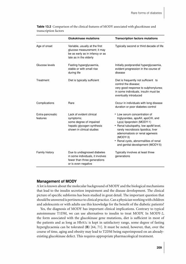
Rare forms of diabetes
Table 13.2 Comparison of the clinical features of MODY associated with glucokinase andtranscription factors
Glukokinase mutations Transcription factors mutations
Age of onset Variable, usually at the firstglucose measurement; it maybe as early as in infancy or aslate as in the elderly
Typically second or third decade of life
Glucose levels Fasting hyperglycaemia,stable or with small riseduring life
Initially postprandial hyperglycaemia,evident progression in the course ofdisease
Treatment Diet is typically sufficient Diet is frequently not sufficient tocontrol the disease;very good response to sulphonylurea;in some individuals, insulin must beeventually introduced
Complications Rare Occur in individuals with long diseaseduration or poor diabetes control
Extra-pancreaticfeatures
Lack of evident clinicalsymptoms;some degree of impairedhepatic glycogen synthesisshown in clinical studies
� Low serum concentration oftriglycerides, apoAII, apoCIII, andLp(a) lipoprotein (MODY-1)
� Renal tubulopathy, low apoM level,rarely necrobiosis lipoidica, liveradenomatosis or renal agenesis(MODY-3)
� Renal cysts, abnormalities of renaland genital development (MODY-5)
Family history Due to undiagnosed diabetesin some individuals, it involvesfewer than three generationsor is even negative
Typically involves at least threegenerations
Management of MODYA lot is known about the molecular background of MODY and the biological mechanismsthat lead to the insulin secretion impairment and the disease development. The clinicalpicture of specific subforms has been studied in great detail. The important question thatshould be answered is pertinence to clinical practice. Can a physician working with childrenand adolescents or with adults use this knowledge for the benefit of the diabetic patients?
Yes, the diagnosis of MODY has important clinical implications. Contrary to typicalautoimmune T1DM, we can use alternatives to insulin to treat MODY. In MODY-2,the form associated with the glucokinase gene mutations, diet is sufficient in most ofthe patients and as long as HbA1c is kept in satisfactory range, some degree of fastinghyperglycaemia can be tolerated (B) [64, 71]. It must be noted, however, that, over thecourse of time, aging and obesity may lead to T2DM being superimposed on an already-existing glucokinase defect. This requires appropriate pharmacological treatment.
209

Chapter 13
The situation is different in subtypes associated with transcription factors since the effi-cacy of diet is limited and thus pharmacological treatment must be implemented (B) [71].Since sulphonylureas, the oldest group of oral hypoglycaemic agents available worldwide,act on the beta cell, and their specific mechanism of action is associated with enhancing ofinsulin secretion (E) [77], the hypothesis that the latter group are drugs of choice in MODYtreatment seemed justified. Several early reports from various populations suggested thatthe patients with MODY-3 (HNF-1α), the most frequent autosomal dominant form ofdiabetes, be characterised by a specifically prominent therapeutic effect of sulphonylureas(C) [78, 79]. However, a clinical, double-blind, randomised study confirming this specifickind of pharmacogenetics in MODY was lacking. The breaking point was the publica-tion of the paper that compared the clinical efficacy of gliclazide (a sulphonylurea) andmetformin (a biguanide) in MODY-3 and in complex, late-onset T2DM (A) [80]. Thisstudy confirmed what had been suspected for years, that the differences in the clinicalresponse to various oral hypoglycaemic agents depend on the cause of diabetes. MODY-3patients showed better response to gliclazide than the patients with T2DM. The differencewas associated with their great insulin secretion increase. An additional factor was highinsulin sensitivity in these patients. The effect of metformin on metabolic compensationin MODY-3 patients was similar to that in T2DM and much weaker than for gliclazide.
The researchers attributed their findings to the nature of the beta-cell defect in MODY.HNF-1α is responsible for the expression of several genes involved in glucose uptake,glycolysis and mitochondrial metabolism (B) [69]. Sulphonylureas act downstream tothese defects by binding to the SUR-1 subunit of the beta-cell ATP-dependent potassiumchannel (E) [77]. Thus, they are able to bypass most of these defects and improve thebeta-cell functioning. So the genetic cause of MODY-3 is an important determinant ofresponse to oral hypoglycaemic drugs. The pathophysiological similarity suggests that ananalogous situation is likely in most of other MODY forms linked to transcription factors(C, B) [3, 61, 81].
The legal aspect of sulphonylurea use in children and adolescents should be brieflydiscussed since these drugs are licensed worldwide only for adults. The European Agencyfor the Evaluation of Medical Products statement dated 1995 says that, when justified, aphysician can use in children drugs licensed for adults (E) [82]. It is hard to find a betterjustification than a molecular diagnosis, such as MODY-3, which provides the basis forvery specific pharmacogenetics.
It should be remembered, however, that under these circumstances, sulphonylureas arebeing used in children and adolescents based on the results of genetic testing. This testingis more and more widely available in various European countries. In some of them testingmay by funded by the state health system and in some others it still constitutes part ofscientific projects.
The diagnosis of various form of MODY also has prognostic implications for a family.Since MODY is a disease of autosomal dominant inheritance, the probability of passingthe gene to a child by a mother or father is 50% (E) [46]. The penetration of the mutations,particularly in forms associated with HNFs is very high, which means that eventuallyalmost all the mutation carriers will develop diabetes, although familial factors, includingmodifying genes, parent of origin and whether or not a mutation carrier was exposed todiabetes in utero, may influence the age of diagnosis (B) [83, 84]. It should be understoodthat the efficacy of prevention of MODY, unlike in T2DM, is very limited. It seems, however,that maintaining lean body mass may delay disease onset. In addition, a regular surveillance
210

Rare forms of diabetes
of blood glucose in non-affected mutation carriers of MODY genes within the familyshould allow early diagnosis and intervention to maintain good metabolic control fromthe very onset of the disease in order to prevent chronic diabetic complications.
Other conditions associated with diabetes in childhoodNicola A. Bridges
There are several conditions in which diabetes features in one form or another. In themajority of cases this is either mild or does not usually occur until outside the paediatricage range. The ADA classification of diabetes (see Chapter 2) includes most of these.They are listed in Table 13.3 together with the principal features of the diabetes and withreference to their OMIM numbers. Further details on these rare conditions, together withthe relevant references relating to the various conditions, can be obtained from the OMIMwebsite (E/A) [85]. However, there a few which have a significant relevance to paediatricpractice and which bear more detailed discussion. The two most important of these arecystic fibrosis (CF) and Prader–Willi syndrome (P-WS).
Cystic-fibrosis-related diabetes
IntroductionCystic-fibrosis-related diabetes (CFRD) is a rare cause of diabetes, which has becomemore common, as improvements in CF care have resulted in increased life expectation.Recognition of the importance of impaired glucose tolerance and CFRD in prognosis hasmeant that detection and management of diabetes has assumed a greater relevance in thecare of individuals with CF.
Insulin secretion defects in CFCFRD is found only in individuals with exocrine pancreatic insufficiency. Pathologicalstudies have demonstrated loss of beta cells in CF even without diabetes (B) [86]. Loss ofinsulin secretion has been demonstrated from early childhood, and there is progressiveinsulin deficiency with age (A, B) [87–89]. Insulin secretion is delayed and reduced inresponse to a glucose load. Baseline insulin secretion is preserved at the onset of diabetes,so fasting glucose levels are usually normal (A) [90]. HbA1c can be low in established CFRD,although it still correlates with glucose levels (B) [91]. Studies using continuous glucose-monitoring systems have demonstrated significant hyperglycaemia in individuals whosefasting glucose, HbA1c and oral glucose tolerance test (OGTT) are all within normal limits(C, B) [92, 93]. HbA1c and fasting glucose are unhelpful in making the diagnosis of CFRD.The primary cause for CFRD is insulin deficiency, but insulin resistance is also a factor inabnormal glucose tolerance (related to chronic inflammation and steroid treatment) (A)[87, 94].
European and US register studies from the 1990s reported prevalence of CFRD of 3.1and 6.1% respectively in all ages (B) [95, 96]. Both these studies based prevalence data onwhether subjects reported treatment with insulin. If individuals are prospectively screenedusing OGTT, prevalence is much higher. In one study, 9% of those aged 5–9 years haddiabetes, rising to 43% in those over 30 years, with about a third in all age groups withimpaired glucose tolerance (B) [88]. In 2004 the US patient registry reported a 16.1%
211
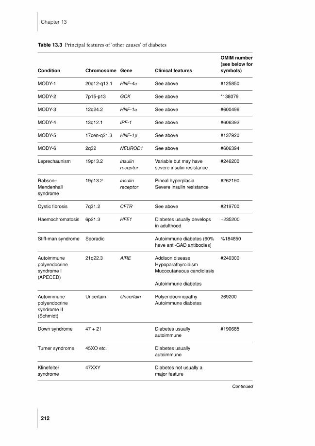
Chapter 13
Table 13.3 Principal features of ‘other causes’ of diabetes
OMIM number(see below for
Condition Chromosome Gene Clinical features symbols)
MODY-1 20q12-q13.1 HNF-4α See above #125850
MODY-2 7p15-p13 GCK See above *138079
MODY-3 12q24.2 HNF-1α See above #600496
MODY-4 13q12.1 IPF-1 See above #606392
MODY-5 17cen-q21.3 HNF-1β See above #137920
MODY-6 2q32 NEUROD1 See above #606394
Leprechaunism 19p13.2 Insulinreceptor
Variable but may havesevere insulin resistance
#246200
Rabson–Mendenhallsyndrome
19p13.2 Insulinreceptor
Pineal hyperplasiaSevere insulin resistance
#262190
Cystic fibrosis 7q31.2 CFTR See above #219700
Haemochromatosis 6p21.3 HFE1 Diabetes usually developsin adulthood
+235200
Stiff-man syndrome Sporadic Autoimmune diabetes (60%have anti-GAD antibodies)
%184850
Autoimmunepolyendocrinesyndrome I(APECED)
21q22.3 AIRE Addison diseaseHypoparathyroidismMucocutaneous candidiasis
Autoimmune diabetes
#240300
Autoimmunepolyendocrinesyndrome II(Schmidt)
Uncertain Uncertain PolyendocrinopathyAutoimmune diabetes
269200
Down syndrome 47 + 21 Diabetes usuallyautoimmune
#190685
Turner syndrome 45XO etc. Diabetes usuallyautoimmune
Klinefeltersyndrome
47XXY Diabetes not usually amajor feature
Continued
212
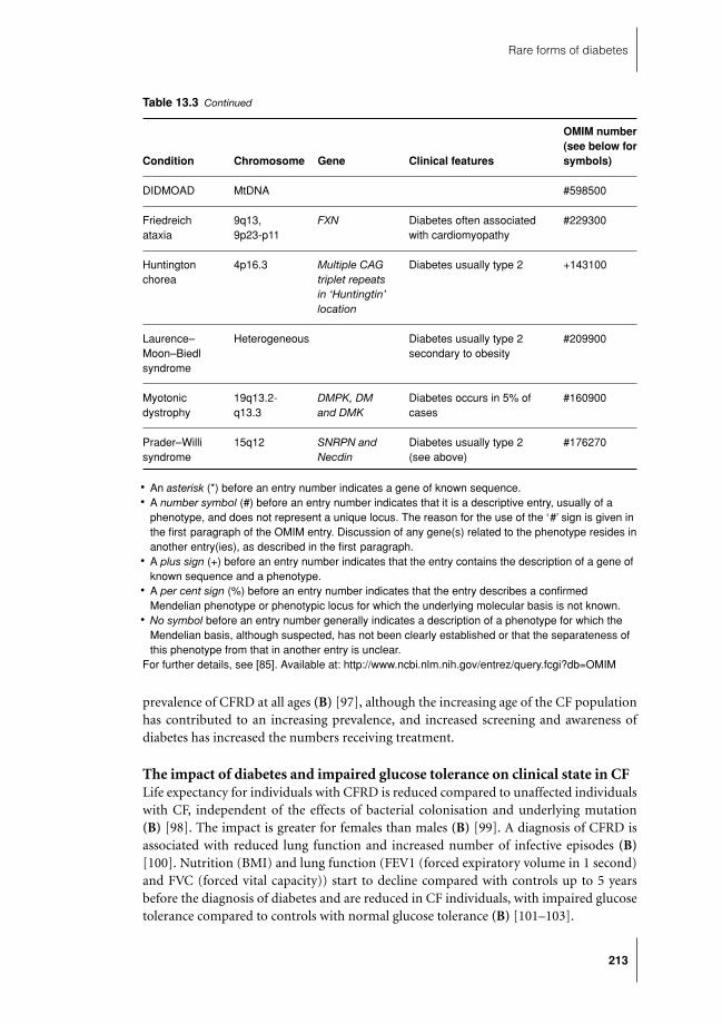
Rare forms of diabetes
Table 13.3 Continued
OMIM number(see below for
Condition Chromosome Gene Clinical features symbols)
DIDMOAD MtDNA #598500
Friedreichataxia
9q13,9p23-p11
FXN Diabetes often associatedwith cardiomyopathy
#229300
Huntingtonchorea
4p16.3 Multiple CAGtriplet repeatsin ‘Huntingtin’location
Diabetes usually type 2 +143100
Laurence–Moon–Biedlsyndrome
Heterogeneous Diabetes usually type 2secondary to obesity
#209900
Myotonicdystrophy
19q13.2-q13.3
DMPK, DMand DMK
Diabetes occurs in 5% ofcases
#160900
Prader–Willisyndrome
15q12 SNRPN andNecdin
Diabetes usually type 2(see above)
#176270
� An asterisk (*) before an entry number indicates a gene of known sequence.� A number symbol (#) before an entry number indicates that it is a descriptive entry, usually of a
phenotype, and does not represent a unique locus. The reason for the use of the ‘#’ sign is given inthe first paragraph of the OMIM entry. Discussion of any gene(s) related to the phenotype resides inanother entry(ies), as described in the first paragraph.
� A plus sign (+) before an entry number indicates that the entry contains the description of a gene ofknown sequence and a phenotype.
� A per cent sign (%) before an entry number indicates that the entry describes a confirmedMendelian phenotype or phenotypic locus for which the underlying molecular basis is not known.
� No symbol before an entry number generally indicates a description of a phenotype for which theMendelian basis, although suspected, has not been clearly established or that the separateness ofthis phenotype from that in another entry is unclear.
For further details, see [85]. Available at: http://www.ncbi.nlm.nih.gov/entrez/query.fcgi?db=OMIM
prevalence of CFRD at all ages (B) [97], although the increasing age of the CF populationhas contributed to an increasing prevalence, and increased screening and awareness ofdiabetes has increased the numbers receiving treatment.
The impact of diabetes and impaired glucose tolerance on clinical state in CFLife expectancy for individuals with CFRD is reduced compared to unaffected individualswith CF, independent of the effects of bacterial colonisation and underlying mutation(B) [98]. The impact is greater for females than males (B) [99]. A diagnosis of CFRD isassociated with reduced lung function and increased number of infective episodes (B)[100]. Nutrition (BMI) and lung function (FEV1 (forced expiratory volume in 1 second)and FVC (forced vital capacity)) start to decline compared with controls up to 5 yearsbefore the diagnosis of diabetes and are reduced in CF individuals, with impaired glucosetolerance compared to controls with normal glucose tolerance (B) [101–103].
213

Chapter 13
The treatment of diabetes and impaired glucose tolerance in CFInsulin treatment in CFRD benefits nutrition and lung function and reduces the num-ber of infective episodes (B, C) [102–104]. Oral hypoglycaemic agents have been usedextensively in CFRD but there is no evidence of benefit to lung function of nutritionalstatus. Sulphonylureas have been demonstrated to maintain adequate glycaemic control insome individuals (B) [105]. There have been no studies of insulin-sensitising agents suchas metformin or the thiazolidinediones because of concerns about complications in CF.
Studies of insulin treatment have used a variety of different insulin regimens and thereis no evidence favouring a particular approach. Improved diabetes management has beensuggested as one factor in the trend for longer survival in CF but it is not possible to extractthis effect from all the other areas of improved management that have made a difference.Microvascular complications of diabetes have been reported in the CF population (C)[106, 107] and, since many patients are likely to survive long enough with diabetes to beat risk of complications, this is a reason to aim for similar glycaemic targets as in otherdiabetic subjects.
In CF, insulin deficiency is present even in those with normal glucose tolerance ontesting (A) [90]. There appears to be a gradual progression to impaired glucose toleranceand then diabetes, with a range of clinical factors having an impact on the rate of thisprocess. Insulin deficiency has an adverse effect before the clinical criteria for a diagnosisof diabetes are met, and studies treating subjects with abnormal glucose tolerance but notdiabetes have demonstrated a positive effect of insulin on nutrition, lung function andrates of infectious exacerbations (C) [92, 108].
Conclusions
CFRD is associated with increased risk of adverse complications and shorter survival. Theaim of treatment with insulin is to reverse the adverse impact that loss of the anabolic effectof insulin has on nutrition and lung function. Insulin treatment results in improvementsin nutritional and respiratory status, but the longer term effects on survival are not yetknown. The diagnostic criteria for diabetes based on glucose levels may not be of help indetermining whether treatment is needed in CF, because there is evidence of the adverseeffects of insulin deficiency and benefit from treatment in individuals who do not meetthese criteria. It is clear that treating the insulin deficiency found in CF with insulin isof clinical benefit but further studies are needed to guide when to start treatment andoptimum treatment strategies.
Prader–Willi syndromeJeremy Allgrove
IntroductionPrader–Willi syndrome (P-WS) was first described in 1956 (C) [109]. The syndrome ischaracterised by an infant who remains very floppy and initially feeds poorly so much sothat most infants require tube feeding. Some time during the first year after birth, a changeoccurs, as a result of which the patients develop a large appetite, and it is often difficultto prevent these children from seeking food at the least opportunity. If this is allowed tocontinue unchecked, gross obesity may result.
214

Rare forms of diabetes
Diabetes, which usually resembles T2DM, has become increasingly recognised as acomplication of P-WS, has a mean age of onset of 20 years and is present in 25% of adults(B) [110]. It has been assumed that this is related to the gross obesity that frequentlydevelops. However, it seems that carbohydrate metabolism in P-WS is different from thatin exogenous obesity and that it is not related to increased insulin resistance in the sameway. Insulin resistance is not present to the same degree (A) [111, 112] but insulin releasein response to glucose is impaired and hepatic insulin extraction is increased (A) [111].This is perhaps not surprising, as there are no genes that are strongly linked to T2DMpresent on chromosome 15 (see Chapter 12).
Growth hormone (GH) is now used regularly for the treatment of short stature inP-WS and is thought to have a beneficial effect on body composition. In children who arenot diabetic before treatment, there is usually no adverse effect upon glucose metabolismeven after 5 years of treatment (B) [113, 114] and the impaired insulin release seen beforetreatment may improve possibly because of the reduction in total body fat and increasein lean mass. However, the onset of diabetes shortly after initiation of GH treatment isdescribed (C) [115] and it is not known whether or not diabetes is made worse by GHtherapy if it is already present.
Despite the differences between the diabetes of P-WS and T2DM, treatment of diabetesin P-WS is usually undertaken along the same lines as T2DM; i.e. it begins with oralhypoglycaemic agents, such as metformin. There is also some evidence to suggest that theglitazones may also be of value (C) [116] and, in the author’s experience, a combinationof metformin and roziglitazone has proved to be of value. Nevertheless, insulin may berequired, although this may have an adverse effect upon body weight.
Conclusion
P-WS is a cause of significant morbidity and mortality. Diabetes develops in an increasingproportion of patients and is being seen in childhood. The aetiology of the diabetes is notthe same as in exogenous obesity and is more dependent on insulin deficiency. Nevertheless,treatment usually starts with oral hypoglycaemic agents before adding insulin.
Acknowledgements
Maciej Malecki is grateful to his many colleagues in USA, UK, Poland and other countrieswith whom he has had the privilege to work and to collaborate with in the field of geneticdiabetes research and to the Polish Ministry of Science and Higher Education for supportingthe research on monogenic diabetes over recent years.
He also thanks Dr Paul Greenlaw for his editorial help.
References
1 Shield JP, Gardner RJ, Wadsworth EJ et al. Aetiopathology and genetic basis of neonatal diabetes. ArchDis Child Fetal Neonatal Ed 1997; 76: F39–42.
2 Iafusco D, Stazi MA, Cotichini R et al. Permanent diabetes mellitus in the first year of life. Diabetologia2002; 45: 798–804.
3 Stoffers DA, Zinkin NT, Stanojevic V et al. Pancreatic agenesis attributable to a single nucleotidedeletion in the human IPF1 gene coding sequence. Nat Genet 1997; 15: 106–10.
215

Chapter 13
4 Stoffers DA, Stanojevic V, Habener JF. Insulin promoter factor-1 gene mutation linked to early-onsettype 2 diabetes mellitus directs expression of a dominant negative isoprotein. J Clin Invest 1998; 102:232–41.
5 Schwitzgebel VM, Mamin A, Brun T et al. Agenesis of human pancreas due to decreased half-life ofinsulin promoter factor 1. J Clin Endocrinol Metab 2003; 88: 4398–406.
6 Temple IK, Gardner RJ, Mackay DJ et al. Transient neonatal diabetes: widening the understanding ofthe etiopathogenesis of diabetes. Diabetes 2000; 49: 1359–66.
7 Slingerland AS, Hattersley AT. Mutations in the Kir6.2 subunit of the KATP channel and permanentneonatal diabetes: new insights and new treatment. Ann Med 2005; 37: 186–95.
8 Temple IK, James RS, Crolla JA et al. An imprinted gene(s) for diabetes? Nat Genet 1995; 9: 110–12.9 Cave H, Polak M, Drunat S et al. Refinement of the 6q chromosomal region implicated in transient
neonatal diabetes. Diabetes 2000; 49: 108–13.10 Temple IK, Gardner RJ, Robinson DO et al. Further evidence for an imprinted gene for neonatal
diabetes localised to chromosome 6q22-q23. Hum Mol Genet 1996; 5: 1117–21.11 Gardner RJ, Mackay DJ, Mungall AJ et al. An imprinted locus associated with transient neonatal
diabetes mellitus. Hum Mol Genet 2000; 9: 589–96.12 Graded overexpression of ZAC impairs glucose stimulated insulin secretion in beta-cells. American
Diabetes Association, Washington, 2006, 190-OR.13 Jeha GS, Venkatesh MP, Edelen RC et al. Neonatal diabetes mellitus: patient reports and review of
current knowledge and clinical practice. J Pediatr Endocrinol Metab 2005; 18: 1095–102.14 Polak M, Shield J. Neonatal and very-early-onset diabetes mellitus. Semin Neonatol 2004; 9: 59–65.15 Senee V, Vattem KM, Delepine M et al. Wolcott–Rallison syndrome: clinical, genetic, and functional
study of EIF2AK3 mutations and suggestion of genetic heterogeneity. Diabetes 2004; 53: 1876–83.16 Delepine M, Nicolino M, Barrett T et al. EIF2AK3, encoding translation initiation factor 2-alpha kinase
3, is mutated in patients with Wolcott–Rallison syndrome. Nat Genet 2000; 25: 406–9.17 Wildin RS, Ramsdell F, Peake J et al. X-linked neonatal diabetes mellitus, enteropathy and endocrinopa-
thy syndrome is the human equivalent of mouse scurfy. Nat Genet 2001; 27: 18–20.18 Wildin RS, Smyk-Pearson S, Filipovich AH. Clinical and molecular features of the immunodysregu-
lation, polyendocrinopathy, enteropathy, X linked (IPEX) syndrome. J Med Genet 2002; 39: 537–45.19 Ruemmele FM, Brousse N, Goulet O. Autoimmune enteropathy: molecular concepts. Curr Opin
Gastroenterol 2004; 20: 587–91.20 Baud O, Goulet O, Canioni D et al. Treatment of the immune dysregulation, polyendocrinopathy,
enteropathy, X-linked syndrome (IPEX) by allogeneic bone marrow transplantation. N Engl J Med2001; 344: 1758–62.
21 Mazzolari E, Forino C, Fontana M et al. A new case of IPEX receiving bone marrow transplantation.Bone Marrow Transplant 2005; 35: 1033–34.
22 Njolstad PR, Sagen JV, Bjorkhaug L et al. Permanent neonatal diabetes caused by glucokinase deficiency:inborn error of the glucose-insulin signaling pathway. Diabetes 2003; 52: 2854–60.
23 Njolstad PR, Sovik O, Cuesta-Munoz A et al. Neonatal diabetes mellitus due to complete glucokinasedeficiency. N Engl J Med 2001; 344: 1588–92.
24 Porter JR, Shaw NJ, Barrett TG et al. Permanent neonatal diabetes in an Asian infant. J Pediatr 2005;146: 131–3.
25 Gloyn AL, Ellard S, Shield JP et al. Complete glucokinase deficiency is not a common cause of permanentneonatal diabetes. Diabetologia 2002; 45: 290.
26 Gloyn AL, Pearson ER, Antcliff JF et al. Activating mutations in the gene encoding the ATP-sensitivepotassium-channel subunit Kir6.2 and permanent neonatal diabetes. N Engl J Med 2004; 350: 1838–49.
27 Gloyn AL, Reimann F, Girard C et al. Relapsing diabetes can result from moderately activating muta-tions in KCNJ11. Hum Mol Genet 2005; 14: 925–34.
28 Proks P, Antcliff JF, Lippiat J et al. Molecular basis of Kir6.2 mutations associated with neonatal diabetesor neonatal diabetes plus neurological features. Proc Natl Acad Sci U S A 2004; 101: 17539–44.
29 Pearson ER, Flechtner I, Njolstad PR et al. Switching from insulin to oral sulfonylureas in patientswith diabetes due to Kir6.2 mutations. N Engl J Med 2006; 355: 467–77.
30 Babenko AP, Polak M, Cave H et al. Activating mutations in the ABCC8 gene in neonatal diabetesmellitus. N Engl J Med 2006; 355: 456–66.
31 Proks P, Arnold AL, Bruining J et al. A heterozygous activating mutation in the sulphonylurea receptorSUR1 (ABCC8) causes neonatal diabetes. Hum Mol Genet 2006; 15: 1793–1800.
216

Rare forms of diabetes
32 Hoveyda N, Shield JP, Garrett C et al. Neonatal diabetes mellitus and cerebellar hypoplasia/agenesis:report of a new recessive syndrome. J Med Genet 1999; 36: 700–4.
33 Sellick GS, Barker KT, Stolte-Dijkstra I et al. Mutations in PTF1A cause pancreatic and cerebellaragenesis. Nat Genet 2004; 36: 1301–5.
34 Mitchell J, Punthakee Z, Lo B et al. Neonatal diabetes, with hypoplastic pancreas, intestinal atresia andgall bladder hypoplasia: search for the aetiology of a new autosomal recessive syndrome. Diabetologia2004; 47: 2160–7.
35 Ashraf A, Abdullatif H, Hardin W et al. Unusual case of neonatal diabetes mellitus due to congenitalpancreas agenesis. Pediatr Diabetes 2005; 6: 239–43.
36 Verwest AM, Poelman M, Dinjens WN et al. Absence of a PDX-1 mutation and normal gastroduodenalimmunohistology in a child with pancreatic agenesis. Virchows Arch 2000; 437: 680–4.
37 Senee V, Chelala C, Duchatelet S et al. Mutations in GLIS3 are responsible for a rare syndrome withneonatal diabetes mellitus and congenital hypothyroidism. Nat Genet 2006; 38: 682–7.
38 Yorifuji T, Kurokawa K, Mamada M et al. Neonatal diabetes mellitus and neonatal polycystic, dysplastickidneys: phenotypically discordant recurrence of a mutation in the hepatocyte nuclear factor-1betagene due to germline mosaicism. J Clin Endocrinol Metab 2004; 89: 2905–8.
39 Bouchardet A. De la glycosuria ou diabete sucre. 1875.40 Tattersall RB, Fajans SS. A difference between the inheritance of classical juvenile-onset and maturity-
onset type diabetes of young people. Diabetes 1975; 24: 44–53.41 McCarthy MI, Hattersley AT. Molecular diagnostics in monogenic and multifactorial forms of type 2
diabetes. Expert Rev Mol Diagn 2001; 1: 403–12.42 McCarthy MI. Susceptibility gene discovery for common metabolic and endocrine traits. J Mol En-
docrinol 2002; 28: 1–17.43 Ehtisham S, Hattersley AT, Dunger DB et al. First UK survey of paediatric type 2 diabetes and MODY.
Arch Dis Child 2004; 89: 526–9.44 Porter JR, Barrett TG. Monogenic syndromes of abnormal glucose homeostasis: clinical review and
relevance to the understanding of the pathology of insulin resistance and beta cell failure. J Med Genet2005; 42: 893–902.
45 Tattersall RB. Mild familial diabetes with dominant inheritance. Q J Med 1974; 43: 339–57.46 Mange EJ, Mange AP. Human pedigrees. Basic Human Genetics, 2nd edn. Sinauer Associates Inc.,
Sunderland, MA, 1999, pp. 67–92.47 Tattersall R. Maturity-onset diabetes of the young: a clinical history. Diabet Med 1998; 15: 11–14.48 Hattersley AT. Maturity-onset diabetes of the young: clinical heterogeneity explained by genetic het-
erogeneity. Diabet Med 1998; 15: 15–24.49 Herman WH, Fajans SS, Ortiz FJ et al. Abnormal insulin secretion, not insulin resistance, is the genetic
or primary defect of MODY in the RW pedigree. Diabetes 1994; 43: 40–6.50 Tattersall RB, Mansell PI. Maturity onset-type diabetes of the young MODY: one condition or many?
Diabet Med 1991; 8: 402–10.51 Hattersley AT, Turner RC, Permutt MA et al. Linkage of type 2 diabetes to the glucokinase gene. Lancet
1992; 339: 1307–10.52 Bell GI, Xiang KS, Newman MV et al. Gene for non-insulin-dependent diabetes mellitus maturity-
onset diabetes of the young subtype is linked to DNA polymorphism on human chromosome 20q.Proc Natl Acad Sci U S A 1991; 88: 1484–8.
53 Velho G, Vaxillaire M, Boccio V et al. Diabetes complications in NIDDM kindreds linked to theMODY3 locus on chromosome 12q. Diabetes Care 1996; 19: 915–19.
54 Report of a WHO Consultation. Definition, Diagnosis and Classification of Diabetes Mellitus and ItsComplications. Geneva, 1999.
55 Fajans SS, Bell GI, Polonsky KS. Molecular mechanisms and clinical pathophysiology of maturity-onsetdiabetes of the young. N Engl J Med 2001; 345: 971–80.
56 Horikawa Y, Iwasaki N, Hara M et al. Mutation in hepatocyte nuclear factor-1 beta gene TCF2 associatedwith MODY. Nat Genet 1997; 17: 384–5.
57 Stoffers DA, Ferrer J, Clarke WL et al. Early-onset type-II diabetes mellitus MODY4 linked to IPF1.Nat Genet 1997; 17: 138–9.
58 Vionnet N, Stoffel M, Takeda J et al. Nonsense mutation in the glucokinase gene causes early-onsetnon-insulin-dependent diabetes mellitus. Nature 1992; 356: 721–2.
217

Chapter 13
59 Yamagata K, Furuta H, Oda N et al. Mutations in the hepatocyte nuclear factor-4alpha gene in maturity-onset diabetes of the young (MODY1). Nature 1996; 384: 458–60.
60 Yamagata K, Oda N, Kaisaki PJ et al. Mutations in the hepatocyte nuclear factor-1alpha gene inmaturity-onset diabetes of the young (MODY3). Nature 1996; 384: 455–8.
61 Malecki MT, Jhala US, Antonellis A et al. Mutations in NEUROD1 are associated with the developmentof type 2 diabetes mellitus. Nat Genet 1999; 23: 323–8.
62 Iynedjian PB, Mobius G, Seitz HJ et al. Tissue-specific expression of glucokinase: identification of thegene product in liver and pancreatic islets. Proc Natl Acad Sci U S A 1986; 83: 1998–2001.
63 Bell GI, Pilkis SJ, Weber IT et al. Glucokinase mutations, insulin secretion, and diabetes mellitus. AnnuRev Physiol 1996; 58: 171–86.
64 Froguel P, Zouali H, Vionnet N et al. Familial hyperglycemia due to mutations in glucokinase: definitionof a subtype of diabetes mellitus. N Engl J Med 1993; 328: 697–702.
65 Pearson ER, Velho G, Clark P et al. Beta-cell genes and diabetes: quantitative and qualitative differencesin the pathophysiology of hepatic nuclear factor-1alpha and glucokinase mutations. Diabetes 2001;50: S101–7.
66 Ellard S, Beards F, Allen LI et al. A high prevalence of glucokinase mutations in gestational diabeticsubjects selected by clinical criteria. Diabetologia 2000; 43: 250–3.
67 Velho G, Petersen KF, Perseghin G et al. Impaired hepatic glycogen synthesis in glucokinase-deficient(MODY-2) subjects. J Clin Invest 1996; 98: 1755–61.
68 Schrem H, Klempnauer J, Borlak J. Liver-enriched transcription factors in liver function and devel-opment. Part I: the hepatocyte nuclear factor network and liver- specific gene expression. PharmacolRev 2002; 54: 129–58.
69 Shih DQ, Screenan S, Munoz KN et al. Loss of HNF-1alpha function in mice leads to abnormalexpression of genes involved in pancreatic islet development and metabolism. Diabetes 2001; 50:2472–80.
70 Naya FJ, Huang HP, Qiu Y et al. Diabetes, defective pancreatic morphogenesis, and abnormal en-teroendocrine differentiation in BETA2/neuroD-deficient mice. Genes Dev 1997; 11: 2323–34.
71 Frayling TM, Evans JC, Bulman MP et al. Beta-cell genes and diabetes: molecular and clinical charac-terization of mutations in transcription factors. Diabetes 2001; 50: S94–100.
72 Bingham C, Ellard S, Cole T et al. Solitary functioning kidney and diverse genital tract malformationsassociated with hepatocyte nuclear factor-1beta mutations. Kidney Int 2002; 61: 1243–51.
73 Malecki M, Skupien J, Gorczynska K et al. Renal malformations may be linked to mutations in thehepatocyte nuclear factor-1alpha MODY3 gene. Diabetes Care 2005; 28: 2774–6.
74 Montoli A, Colussi G, Massa O et al. Renal cysts and diabetes syndrome linked to mutations of thehepatocyte nuclear factor-1 beta gene: description of a new family with associated liver involvement.Am J Kidney Dis 2002; 40: 397–402.
75 Menzel R, Kaisaki PJ, Rjasanowski I et al. A low renal threshold for glucose in diabetic patients with amutation in the hepatocyte nuclear factor-1alpha HNF-1alpha gene. Diabet Med 1998; 15: 816–20.
76 Shih DQ, Dansky HM, Fleisher M et al. Genotype/phenotype relationships in HNF-4alpha/MODY1:haploinsufficiency is associated with reduced apolipoprotein AII, apolipoprotein CIII, lipoproteina,and triglyceride levels. Diabetes 2000; 49: 832–7.
77 Gribble FM, Reimann F. Sulphonylurea action revisited: the post-cloning era. Diabetologia 2003; 46:875–91.
78 Pearson ER, Liddell WG, Shepherd M et al. Sensitivity to sulphonylureas in patients with hepatocytenuclear factor-1alpha gene mutations: evidence for pharmacogenetics in diabetes. Diabet Med 2000;17: 543–5.
79 Søvik O, Njølstad P, Følling I et al. Hyperexcitability to sulphonylurea in MODY3. Diabetologia 1998;41: 607–8.
80 Pearson E, Starkey B, Powell R et al. Genetic cause of hyperglycaemia and response to treatment indiabetes. Lancet 2003; 362: 1275–81.
81 Pearson ER, Pruhova S, Tack CJ et al. Molecular genetics and phenotypic characteristics of MODYcaused by hepatocyte nuclear factor 4alpha mutations in a large European collection. Diabetologia2005; 48: 878–85.
82 Committee for Proprietary Medicines (CPMP). Note for Guidance on Clinical Investigation ofMedicinal Products in Children. Human Medicines Evaluation Unit, London, 1997. Available at:http://www.emea.europa.eu/pdfs/human/ich/271199en.pdf
218
Rare forms of diabetes
83 Klupa T, Warram J, Antonellis A et al. Determinants of the development of diabetes maturity-onsetdiabetes of the young-3 in carriers of HNF-1alpha mutations: evidence for parent-of-origin effect.Diabetes Care 2002; 25: 2292–301.
84 Stride A, Shepherd M, Frayling T et al. Intrauterine hyperglycemia is associated with an earlier diagnosisof diabetes in HNF-1alpha gene mutation carriers. Diabetes Care 2002; 25: 2287–91.
85 NCBI. OMIM – Online Mendelian Inheritance in Man. Available at: http://www.ncbi.nlm.nih.gov/entrez/query.fcgi?db=OMIM
86 Iannucci A, Mukai K, Johnson D et al. Endocrine pancreas in cystic fibrosis: an immunohistochemicalstudy. Hum Pathol 1984; 15: 278–84.
87 Austin A, Kalhan SC, Orenstein D et al. Roles of insulin resistance and beta-cell dysfunction in thepathogenesis of glucose intolerance in cystic fibrosis. J Clin Endocrinol Metab 1994; 79: 80–5.
88 Moran A, Doherty L, Wang X et al. Abnormal glucose metabolism in cystic fibrosis. J Pediatr 1998;133: 10–17.
89 Solomon MP, Wilson DC, Corey M et al. Glucose intolerance in children with cystic fibrosis. J Pediatr2003; 142: 128–32.
90 Yung B, Noormohamed FH, Kemp M et al. Cystic fibrosis-related diabetes: the role of peripheralinsulin resistance and beta-cell dysfunction. Diabet Med 2002; 19: 221–6.
91 Brennan AL, Gyi KM, Wood DM et al. Relationship between glycosylated haemoglobin and meanplasma glucose concentration in cystic fibrosis. J Cyst Fibros 2006; 5: 27–31.
92 Dobson L, Hattersley AT, Tiley S et al. Clinical improvement in cystic fibrosis with early insulintreatment. Arch Dis Child 2002; 87: 430–1.
93 Jefferies C, Solomon M, Perlman K et al. Continuous glucose monitoring in adolescents with cysticfibrosis. J Pediatr 2005; 147: 396–8.
94 Hardin DS, Leblanc A, Marshall G et al. Mechanisms of insulin resistance in cystic fibrosis. Am J PhysiolEndocrinol Metab 2001; 281: E1022–8.
95 Konstan MW, Butler SM, Schidlow DV et al. Patterns of medical practice in cystic fibrosis: part I. Evalu-ation and monitoring of health status of patients. Investigators and Coordinators of the EpidemiologicStudy of Cystic Fibrosis. Pediatr Pulmonol 1999; 28: 242–7.
96 Navarro J, Rainisio M, Harms HK et al. Factors associated with poor pulmonary function: cross-sectional analysis of data from the ERCF. European Epidemiologic Registry of Cystic Fibrosis. EurRespir J 2001; 18: 298–305.
97 Cystic Fibrosis Foundation. Patient Registry 2004 Annual Report. Bethesda, Maryland, 2005. Availableat: http://www.cff.org/ID=4573/TYPE=2676/2004%20Patient%20Registry%20Report.pdf
98 Koch C, Rainisio M, Madessani U et al. Presence of cystic fibrosis-related diabetes mellitus is tightlylinked to poor lung function in patients with cystic fibrosis: data from the European EpidemiologicRegistry of Cystic Fibrosis. Pediatr Pulmonol 2001; 32: 343–50.
99 Milla CE, Billings J, Moran A. Diabetes is associated with dramatically decreased survival in femalebut not male subjects with cystic fibrosis. Diabetes Care 2005; 28: 2141–4.
100 Marshall BC, Butler SM, Stoddard M et al. Epidemiology of cystic fibrosis-related diabetes. J Pediatr2005; 146: 681–7.
101 Milla CE, Warwick WJ, Moran A. Trends in pulmonary function in patients with cystic fibrosis correlatewith the degree of glucose intolerance at baseline. Am J Respir Crit Care Med 2000; 162: 891–5.
102 Nousia-Arvanitakis S, Galli-Tsinopoulou A, Karamouzis M. Insulin improves clinical status of patientswith cystic-fibrosis-related diabetes mellitus. Acta Paediatr 2001; 90: 515–19.
103 Rolon MA, Benali K, Munck A et al. Cystic fibrosis-related diabetes mellitus: clinical impact of predi-abetes and effects of insulin therapy. Acta Paediatr 2001; 90: 860–7.
104 Franzese A, Spagnuolo MI, Sepe A et al. Can glargine reduce the number of lung infections in patientswith cystic fibrosis-related diabetes? Diabetes Care 2005; 28: 2333.
105 Rosenecker J, Eichler I, Barmeier H et al. Diabetes mellitus and cystic fibrosis: comparison of clinicalparameters in patients treated with insulin versus oral glucose-lowering agents. Pediatr Pulmonol 2001;32: 351–5.
106 Rosenecker J, Hofler R, Steinkamp G et al. Diabetes mellitus in patients with cystic fibrosis: theimpact of diabetes mellitus on pulmonary function and clinical outcome. Eur J Med Res 2001; 6: 345–50.
107 Yung B, Landers A, Mathalone B et al. Diabetic retinopathy in adult patients with cystic fibrosis-relateddiabetes. Respir Med 1998; 92: 871–2.
219

Chapter 13
108 Bizzarri C, Lucidi V, Ciampalini P et al. Clinical effects of early treatment with insulin glargine inpatients with cystic fibrosis and impaired glucose tolerance. J Endocrinol Invest 2006; 29: RC1–4.
109 Prader A, Labhart A, Willi H. Ein Syndrom von Adipositas, Kleinwuchs, Kryptorchismus undOligonephrie nach Myatonieartigem Zustand in Neugeborenenalter. Schweiz Med Wochenschr 1956;86: 1260–1.
110 Butler JV, Whittington JE, Holland AJ et al. Prevalence of, and risk factors for, physical ill-healthin people with Prader–Willi syndrome: a population-based study. Dev Med Child Neurol 2002; 44:248–55.
111 Schuster DP, Osei K, Zipf WB. Characterization of alterations in glucose and insulin metabolism inPrader–Willi subjects. Metabolism 1996; 45: 1514–20.
112 Zipf WB. Glucose homeostasis in Prader–Willi syndrome and potential implications of growth hor-mone therapy. Acta Paediatr Suppl 1999; 88: 115–17.
113 L’Allemand D, Eiholzer U, Schlumpf M et al. Carbohydrate metabolism is not impaired after 3 yearsof growth hormone therapy in children with Prader–Willi syndrome. Horm Res 2003; 59: 239–48.
114 Lindgren AC, Ritzen EM. Five years of growth hormone treatment in children with Prader-Willisyndrome. Swedish National Growth Hormone Advisory Group. Acta Paediatr Suppl 1999; 88:109–11.
115 Yigit S, Estrada E, Bucci K et al. Diabetic ketoacidosis secondary to growth hormone treatment in aboy with Prader–Willi syndrome and steatohepatitis. J Pediatr Endocrinol Metab 2004; 17: 361–4.
116 Yamakita T, Ishii T, Mori T et al. Troglitazone ameliorates insulin resistance in a diabetic patient withPrader–Willi syndrome. Diabetes Res Clin Pract 1998; 42: 205–8.
220

CHAPTER 14
Diabetes and information technology
Kenneth J. Robertson
What is laid down, ordered, factual is never enough to embrace the whole truth: lifealways spills over the rim of every cup.
—Boris Pasternak, Russian poet and writer (1890–1960)
Introduction
Eventually, information technology (IT) will be ubiquitous in health care, regarded as nomore remarkable than the electrical sockets in the wall or the taps on the sink. The culturalchange required for this to happen is as great, if not greater, than the improvements inthe technology to make possible the capture and sharing of clinical information whereverthat may be necessary. Note also that for this to happen in one hospital is challenging,but on a global scale it seems an unattainable goal. However, consider the internet – anew technology only a few years ago and now part of daily life for millions across theworld.
Obviously, the impact of IT on health and diabetes, in particular, is enormous andreaches all the way from drug manufacture to the meters used to measure blood glucose.This chapter will consider the main areas where IT is making a major contribution to theimprovement of care at the patient and professional level. Inevitably, the evidence base isweak, and the implications of this will also be explored.
Registers
While diabetes registers have been in place in many parts of the world for a long time,the majority are still dependent upon manual registration of patient data with centralentry onto a database. Modern IT opens up possibilities of having registers developed asa by-product of care. Registers are no longer just epidemiological or research tools. InScotland, the Scottish Care Information – Diabetes Collaboration (SCI-DC) has grownfrom a start in two Health Boards to be a national system that comprises a hospital-basedapplication to manage clinics and a network application that collates this with informationfrom general practice. This ‘shared’ record drives planning, forms the bedrock of annualnational audit (Scottish Diabetes Survey) and provides the demographic details to drivethe emergent Scottish Diabetes Retinopathy Screening Programme (E) [1]. The networkelement is now in place in every Health Board in Scotland (14 Boards) and the hospitalsystem is in all the adult services. The paediatric ‘module’ is being developed and is expectedto be implemented fully this year.
221

Chapter 14
In England and Wales, the National Diabetes Audit makes extensive use of IT to gatherand collate a minimum dataset about the care of individual patients. For the 2005 reporton children and young people (B) [2], 64 of 237 paediatric diabetes services provided databut this had doubled since the previous year. One of the most important lessons aboutthe process was that additional IT support will be required for centres so that such centralreporting can be a by-product of clinical care.
Clinical record
The mantra of health-care informatics is that ‘data for secondary uses should be availableas a by-product of care’. This rather utopian ideal can only be made a reality when clin-ical staff are involved in the design and implementation of the system from the start. Aseries of steps must be gone through to ensure that the project will be managed prop-erly and that the requirements are well understood. Some of the commonest issuesand pitfalls are presented here posed as questions. When contemplating the procure-ment/development/implementation of a clinical system, it would be reckless to proceedwithout being able to answer them all in some detail (see Table 14.1).
Data standards, data sharing and coding/terming
Traditionally, speciality clinical systems have been procured and implemented from theperspective only of the one speciality. However, given that patients frequently have comor-bidities and will, in any case, inevitably have encounters with other clinical professionals,it is increasingly important to be able to share data with others. Such sharing should beregarded as clinically dangerous unless aforethought has been given to establishing datastandards with risk of fields being misfiled, interpreted differently by alternative systems,etc. Properly constructed data standards will address:� the unambiguous clinical definition of a data item� the range of entries (numeric or text) that may be captured� validation rules� the code or term (often ICD-10 code or SNOMED-CT term) to be used� how the definition fits with the data requirements of other clinical groups – e.g. heightmay have been defined as ‘9.99 m’ where the 9 represents any number and the measurementis done with the patient in stocking soles.
For a child of 8 months, the numeric definition would have to specify three decimalplaces and state something about the measuring method. If these differences are not explicitand understood then transfer of the ‘height’ record from one system to another will beunsatisfactory. While this may not be a major issue, a similar approach to insulin dosescould be dangerous where an adult system would almost certainly record a dose of 0.5 U(in a young child) as 0 U since there is unlikely to be provision for decimal parts of units.Such considerations may appear esoteric but planning is vital.
Cross-speciality sharing will also mean that many generic data items will already havebeen defined. These will need to be viewed in the context of a child with diabetes and may beusable without modification or require qualification or a new data item. The introductionof systems such as the ‘Connecting for Health Care Record Service’ in England makes suchdiscussion urgent.
222

Diabetes and information technology
Table 14.1 Critical questions for a clinical system
1 What is (are) the purpose(s) of the proposed system?� It is likely that the principal purpose will be to create a clinical record of the encounters with
patients by members of the diabetes team.� Consideration must also be given to the need to provide access to information for other
professionals within and out with the current organisation, e.g. general practitioner, otherhospital staff, etc.
� Usually, there are defined local governance requirements and, increasingly, statistics arecollated at a regional or national level.
2 Is the record multidisciplinary and how will the different professionals view each other’s entries?� Paediatric diabetes teams generally work very closely and will benefit from having full access to
their patients’ records.3 Who will enter data into the system?
� Data quality is likely to be higher where clinicians are using the system as their record –mistakes are noted and resolved – and ad hoc queries are more likely to produce usefulanswers.
� Where clerical staff enter data (often from handwritten records), there is a high likelihood oftranscription errors and misinterpretation of the clinician’s intended meaning.
� Clinicians may argue that they do not have time to enter data directly but this has to bechallenged. If one of the requirements is to have real-time data capture then the ergonomics ofthe system has to be able to support this – logical screen layout, ability to move aroundsections easily (patients do not always present problems and issues in the expected order) andresponsiveness. After all, it is a contractual and ethical obligation to record the clinical process,so time would normally be devoted to do this on paper. It may also be appropriate to considerthe type of computer hardware being used – peripatetic staff such as diabetes nurse specialistsand dietitians may be better served by the use of mobile devices.
4 What training will be required and who will provide it?� Most clinical staff now have basic computer skills but these may require to be enhanced before
system-specific training.5 What benefits are expected?
� This may seem like a silly question but identification of perceived benefits will focus both thedevelopment and implementation of the system and allow objective testing of their achievementat various stages. This may be especially pertinent to securing revenue funding (see below).
6 What quality assurance processes are in place to ensure that data are being captured accurately?� The system should have validation at a number of levels – input validation should prevent, for
example, ridiculous blood pressures such as 1000/800 being entered.� Appropriate fields may be mandatory before progressing to the next stage – care must be taken
if using this method, that the appropriate information will always be available to the user.� Data integrity is another critical concept. Where definitions and options for data fields change, it
is essential that historical information can still be presented in its original context; e.g. if a fieldis recorded as a number from a list, then changes to the list must not result in the recorded datachanging meaning.
7 What will be the outputs?� A common benefit of clinical applications is the production of discharge or clinic letters for
primary care clinicians. This is fine but increasingly the latter expect to receive such informationin electronic form.
� Email solutions may be a short-term approach, although they should only be used within anappropriate security model, e.g. encryption.
� A better ploy would be to ensure that the data are passed in a form that can be incorporatedinto other systems and this can be achieved only when technical and semantic (meaning)interoperability has been addressed from the outset.
Continued
223
Chapter 14
Table 14.1 Continued
� It may be possible to set up ‘standard reports’ that can be run regularly but most clinicians willalso wish to have an ad hoc reporting tool. The functionality and ease of use of such toolsshould be evaluated carefully before selecting a system because many a system has beenpraised for its ability to absorb information but damned for its reluctance to give it up.
8 What clinical leadership will be provided?� The introduction of any new technology into the clinical process requires planning and support.
Time and again, software programmes have been deemed to fail, not because of inadequaciesin the system but because there was no clear leadership providing guidance, encouragementand occasionally coercion to ensure that the difficulties are overcome and success assured. Itis a truism in eHealth that many excellent applications are ‘failures’, while many mediocre onessucceed because of leadership. Note, however, that this has to be true leadership (appropriateseniority and peer respect) with the new process becoming embedded in the clinical workflowor when the leader moves on, the use of the system will cease. Larger projects must also haveformal project management and a ‘senior responsible owner’ who can ensure that progress ismaintained.
9 How will the system be administered?� Any clinical system will have to be fully supported so that continuity of service is assured
(commonly described as 24/7 business continuity), and this is expensive and new to thehealth-care community.
� User management will be necessary – simple when there are only a few users on one site butmuch more complex when users, unknown to the system administrator, are to be registered.
� Consideration must be given to access rights – often based upon role. Login must be strictly byauthenticated users so that audit trails are accurate – there is no point in having sophisticatedaudit functionality in the system if the first person into the diabetes centre in the morning logson for everyone.
10 How will the system be funded?� It is common for clinicians to purchase software with ‘soft’ money. While this may be
straightforward, it is almost invariably a bad move unless supported fully by the local/regional ITprofessionals who will also ensure fit with the strategy.
� It is very unusual for clinical software to have only a capital cost and the revenue consequencesmust be considered too – maintenance and upgrades, as well as training costs as appropriate.
� The licensing model will very often mean that additional users incur additional costs. There isalso the cost of the hardware (computers and servers) to factor in.
Germany offers an excellent example of how coordination at the national level can leadto the provision of high-quality data for the purposes of benchmarking and governance.The DPV (B) [3] approach comprises three elements – diabetes documentation software(DPV), semi-annual benchmarking (QC-DPV) and a data pool for diabetes research (DPV-SCIENCE). Improvements in the quality of care have been demonstrated with 183 centresenrolled in the project by 2005 covering some 24,000 patients – more than 75% of childrenwith diabetes in Germany (personal communication).
Electronic reminders and algorithms
One of the features of real-time data-entry clinical systems is that they can be set up toprovide support for the process of care. Based upon agreed criteria, the system can offerprompts; e.g. when eye screening or urine testing for microalbuminuria is due. In ourclinic, introduction of such a system improved adherence to eye-screening targets from 61to 89% in 1 year. This is not 100%, however, and the finding was similar to a study by Sequist
224

Diabetes and information technology
et al. in Boston (A) [4], who also found that 76% of clinicians felt that such remindersimproved quality of care. This serves to emphasise that the introduction of computerisedrecords without consideration of the processes of care and workflow management, coupledwith staff training, will fail to deliver the benefits expected.
Confidentiality and consent
Few areas of eHealth are more fraught and contentious just now than the debate aboutconsent in relation to the use of electronic medical records. European Union countries allhave their Data Protection Law based upon the European Directive on Data Protection(95/46/EC) (E) [5] but the details vary, so readers should be sure that any advice is basedupon local legislation. In the UK (E) [6], patients should be informed about the usesto which their information will be put but it is acceptable to assume implied consentwhen sharing clinical information for the specific purpose of direct care (assuming thatthe patient has not specified any caveats in relation to particularly sensitive information).However, there has been a debate around this principle in relation to the NHS Care RecordsService, in England, where the intention is to make summary information available on aSpine record so that it can be accessed wherever the patient is receiving care. The BritishMedical Association argues that this record should only be created on an opt-in basis(explicit consent), while the NHS argues that it should be created unless the patient hasopted out. While such arguments may appear abstruse, they are at the heart of the moveto patients having electronic records. Some legislatures have experimented with givingpatients their own records on smartcards but this has not been successful at more thana small pilot level. There is absolute clarity, however, around the use of clinical recordsfor research purposes. Unless the data are anonymised, explicit consent must be sought.To be even more specific – when data are used outside of direct care and the patients areidentifiable, it is illegal to do this without the explicit (and preferably written) consent ofthe patients.
Comorbidity
Patients often have other medical problems besides diabetes and this becomes almostuniversal during adult years. It is not, therefore, in the best interests of the patients or theirhealth professionals if information about each of their problems is held within independentsilos. This is a key problem and central to the emergence of pan-health IT solutions beingdeveloped throughout the world. It is fair to say that there is no general solution yet butcompliance with the appropriate local/regional technical and information standards willoffer the best chance of later compatibility.
Patient access
This is too large a topic to be discussed here but suffice to say that most eHealth programmesin the developed world have adopted strategies that give patients access to their healthrecords in some way. Numerous examples exist of allowing patients to disease-specificelements, e.g. diabetes, renal disease, etc., but these are almost always bespoke solutions thathave not addressed the problem (that already besets clinical staff) of how the patient can getaccess to all of the appropriate information without having to memorise a host of usernames
225

Chapter 14
and passwords. What is required is a large-scale authentication and identification systemto ensure that the patients see only what relates to them. Mechanisms must also be inplace to make it impossible for the patients to directly change the information capturedby the clinicians as part of their professional record – there should, however, be a processfor reconciliation of errors. Too often also, no thought is given to supporting patients inunderstanding the meaning of what they have accessed. These are complex issues thatwill have to be addressed in the next few years. Increasingly, there is a perception that themedical record ‘belongs’ to the patient but experiments in letting patients be the custodianof their own records (outside of specific elements such as maternity records), e.g. withsmartcards such as the European DiabCard (B) [7], have had very limited success.
Telemedicine
This is an area of diabetes care that has proved attractive, especially in remote and ruralcommunities. The prospect of being able to maintain a therapeutic relationship withpatients from afar has stimulated research into the feasibility and acceptability of usingteleconferencing and remote monitoring. Smith et al. in Queensland (B) [8] successfullyintroduced teleconsultation for children with diabetes and other endocrine abnormalities.They delivered support for acute crises, education to patients and staff and routine clinicalcontact and emphasised the importance of this multimodality approach. Telemedicinemust be used to solve identified problems rather than as an excuse to use new technology.The hardware is becoming more reliable and flexible in its mobility and use and so we arelikely to see it being introduced more widely. It may have particular value in linking tosmall A&E departments without paediatric expertise so that the immediate care of childrenwith diabetic ketoacidosis can be expedited.
Numerous schemes have been established with a view to encouraging patients (oftenadolescents) to monitor their blood glucose more effectively and to support them in usingthe results. These have ranged from downloading data from blood glucose meters inte-grated into mobile phones (Nokia, Finland) to emailing of results to the clinic. The ‘SweetTalk’ programme in Dundee has introduced a system of patient-tailored text messagesdesigned to improve adherence behaviour generally (A) [9].
In 2005, Farmer et al. published a meta-analysis of telemedicine interventions to supportblood glucose monitoring in diabetes (A) [10]. Most of the studies they reviewed were ofshort duration and had small numbers of patients, so their conclusion, ‘Telemedicinesolutions for diabetes care are feasible and acceptable, but evidence for their effectivenessin improving HbA1c or reducing costs while maintaining HbA1c levels, or improving otheraspects of diabetes management is not strong’, was perhaps unsurprising. There is a recurrenttendency to introduce such technology without consideration of proper evaluation.
Conclusion
Diabetes care is supported in numerous other ways by IT and doubtlessly many more willemerge in the coming years. We have yet to see the real impact of genomics and proteomicson medicine but when the genetics and phenotypes of diabetes are better understood, itwill become even more critical to have technological support to ensure that individualpatients receive best care. Early attempts better to organise research findings and literature
226

Diabetes and information technology
to support care (see www.elib.scot.nhs.uk/portal/diabetes/Pages/) will become more tightlyintegrated with patient records.
Quite beyond the remit of this chapter but still a potent use of IT is the physical linkageof continuous blood glucose monitoring with insulin-pump algorithms which will, in thenear future, close the loop so that patients can leave the technology to monitor and managetheir blood glucose.
eHealth is a relatively new discipline and the application of the rigour which computersdemand of record keeping will certainly be a challenge to a traditionally rather anarchicprofession. Nonetheless, the potential benefits are huge for clinicians and their patientsalike.
References
1 Morris AD, Boyle DI, MacAlpine R et al. The diabetes audit and research in Tayside Scotland (DARTS)study: electronic record linkage to create a diabetes register. DARTS/MEMO Collaboration. BMJ 1997;315: 524–8.
2 The Information Centre. National Diabetes Audit Report for the Audit Period 2004/05: Key Findings aboutthe Quality of Care for Children and Young People with Diabetes in England, Incorporating Registrationsfrom Wales. Available at: www.ic.nhs.uk
3 Grabert M, Schweiggert F, Holl RW. A framework for diabetes documentation and quality managementin Germany: 10 years of experience with DPV. Comput Methods Programs Biomed 2002; 69: 115–21.
4 Sequist TD, Gandhi TK, Karson AS et al. A randomized trial of electronic clinical reminders to improvequality of care for diabetes and coronary artery disease. J Am Med Inform Assoc 2005; 12: 431–7.
5 Directive 95/46/EC of the European Parliament and the Council of 24 October 1995 on the Protectionof Individuals with Regard to the Processing of Personal Data and on the Free Movement of Such Data.Available at: http://www.cdt.org/privacy/eurodirective/EU Directive .html
6 Corporate Author: Great Britain. Data Protection Act (UK) 1998: Elizabeth II. Chapter 29: 8th ImpressionOctober 2003 (Incorporating Corrections). Reprinted April 2003. Available at: http://www.opsi.gov.uk/ACTS/acts1998/19980029.htm
7 Engelbrecht R, Hildebrand C. Diabcard: Improved Communication in Diabetes Care Based on Chip CardTechnology. Available at: http://www-mi.gsf.de/diabcard/nf index.html
8 Smith AC, Batch J, Lang E et al. The use of online health techniques to assist with the delivery of specialistpaediatric diabetes services in Queensland. J Telemed Telecare 2003; 9(suppl 2): S54–7.
9 Franklin VL, Waller A, Pagliari C et al. A randomized controlled trial of Sweet Talk, a text-messagingsystem to support young people with diabetes. Diabet Med 2006; 23: 1332–8.
10 Farmer A, Gibson OJ, Tarassenko L et al. A systematic review of telemedicine interventions to supportblood glucose self-monitoring in diabetes. Diabet Med 2005; 22: 1372–8.
227

Abbreviations
AAP American Academy of PediatricsACE Angiotensin converting enzymeACR Albumin creatinine ratioADA American Diabetes AssociationAER Albumin excretion rateAGREE Appraisal of Guidelines, Research and Evaluation for EuropeAIRE Autoimmune regulatorALAD La Asociacion Latino Americana de DiabetesAPS I & II Autoimmune polyendocrine syndromes 1 and 2AUC Area under the curve
BFST Behavioural family systems therapyBG Blood glucoseBMI Body mass indexBP Blood pressureBPA British Paediatric AssociationBSPED British Society for Paediatric Endocrinology and Diabetes
CBT Cognitive behavioural therapyCD Compact diskCF Cystic fibrosisCFRD Cystic-fibrosis-related diabetesCGMS Continuous glucose-monitoring systemCNS Central nervous systemCRF Chronic renal failureCSII Continuous subcutaneous insulin infusionCTLA-4 Cytotoxic T lymphocyte antigen-4CV Cardiovascular
DAFNE Diet adjustment for normal eatingDARTS Diabetes Audit and Research in Tayside StudyDBP Diastolic blood pressureDCCT Diabetes Control and Complications TrialDESG Diabetes Education Study GroupDH Department of HealthDIDMOAD Diabetes Insipidus, Diabetes Mellitus, Optic Atrophy
and DeafnessDKA Diabetic ketoacidosisDNA Deoxyribonucleic acid
228
Abbreviations
DSME Diabetes self-management educationDUK Diabetes UK
EASD European Association for the Study of DiabetesED Eating disorderEDIC Epidemiology of Diabetes Interventions and ComplicationsELISA Enzyme-linked immunosorbent assayEMA Endomyseal antibodiesENDIT European Nicotinamide Diabetes Intervention TrialESPE European Society for Pediatric EndocrinologyESPGHAN European Society of Paediatric Gastroenterology, Hepatology and
Nutrition
FACTS Families Adolescents and Children’s Teamwork StudyFinnDiane Finnish Diabetes Nephropathy StudyFPG Fasting plasma glucosefT4 Free thyroxine
GAD-65/GADA Glutamic acid dehydrogenaseGDG Guideline development groupGFD Gluten-free dietGH Growth hormoneGI Glycaemic indexGL Glycaemic loadGLP-1 Glucagon-like peptide 1GRID Genetic resource investigating diabetesGWB2 Glucowatch Biographer 2
HbA1c Glycated haemoglobin A1cHBGM Home blood glucose monitoringHCP Health care professionalHDL High-density lipoproteinHLA Human leucocyte antigenHMG-CoA 3-Hydroxy-3-methyl-glutaryl-CoA reductaseHNF Hepatic nucleocyte factorHSG Hvidøre Study GroupHTA Health technology assessment
IA-2 Tyrosine phosphatase-like moleculeIAA Insulin autoantibodiesICA Islet cell antibodiesICD International classification of diseaseIDDM1 Insulin-dependent diabetes mellitus susceptibility locus 1IDDM2 Insulin-dependent diabetes mellitus susceptibility locus 2IDDM12 Insulin-dependent diabetes mellitus susceptibility locus 12IDF International Diabetes FederationIFG Impaired fasting glucose
229

Abbreviations
IFIH1 Interferon-induced helicase 1IGF Insulin-like growth factorIGT Impaired glucose toleranceIPEX Immune dysregulation, polyendocrinopathy, enteropathy X-linked
syndromeIPF Insulin promoter factorISPAD International Society for Pediatric and Adolescent DiabetesIT Information technology
KCNJ11 Potassium inwardly rectifying channel, subfamily J, member 11
LDL Low-density lipoproteinLJM Limited joint mobilityLWPES Lawson Wilkins Pediatric Endocrine Society
MDI Multiple daily injectionsMI Motivational interviewingMMR Measles, mumps and rubellaMODY Maturity-onset diabetes of the youngMUFA Monounsaturated fatty acid
NAFL Non-alcoholic fatty liverNASH Non-alcoholic steato-hepatitisNDA National Diabetes AuditNH Nocturnal hypoglycaemiaNHS National Health ServiceNPH Neutral pH HagedornNICE National Institute for Clinical ExcellenceNICE TA NICE Technology AssessmentNSF National Service Framework
OFSTED Office for Standards in EducationOGTT Oral glucose tolerance testOMIM Online Mendelian inheritance in manOR Odds ratio
PCOS Polycystic ovarian syndromePCR Polymerase chain reactionPNDM Permanent neonatal diabetes mellitusPPARγ Peroxisome proliferator-activated receptor gammaPTPN22 Protein tyrosine phosphatase non-receptor type 22P-WS Prader–Willi syndrome
RCP Royal College of PhysiciansRCPCH Royal College of Paediatrics and Child HealthRCT Randomised controlled trialRNA Ribonucleic acid
230

Abbreviations
SBP Systolic blood pressureSCI-DC Scottish Care Information – Diabetes CollaborationSDS Standard deviation scoreSH Severe hypoglycaemiaSIGN Scottish Intercollegiate Guideline NetworkSNP Single nucleotide polymorphismSPE Structured patient educationSSGCYD Scottish Study Group for the Care of the Young with DiabetesSUR Sulphonyl urea receptor
T1DM Type 1 diabetes mellitusT2DM Type 2 diabetes mellitusT3 Tri-iodothyronineTCF Transcription factorTgA Thyroglobulin antibodiesTh1 T helper 1TNDM Transient neonatal diabetes mellitusTODAY Treatment options for T2DM in adolescence and youthTPO Thyroid peroxidaseTreg Regulatory T cellsTSH Thyroid stimulating hormoneTTG Tissue transglutaminase
UKPDS United Kingdom Prospective Diabetes Study
VNTR Variable number of tandem repeats
WHO World Health Organization
231

Index
Page numbers in italic refer to figures; page numbers in bold refer to tables.
ABCC8 genemutations in PNDM and TNDM, 203See also neonatal diabetes
acanthosis nigricans, 189activity aspects, T1DM in very young children and,
66acute management, polycystic ovarian syndrome
(PCOS), 191adolescence and diabetes, 76
action research in, 83clinical perspectives
adolescents with T1DM, 86anxiety and depression, 80associated diseases, 77behavioural problems, 79–80clinic non-attendance, 85eating disorders, 78emotional problems, 79–80glycaemic control deterioration
aspects, 76insulin disorders, 78insulin omission, 77–8long-term consequences of diabetes, 79paediatric to adult service, transition of care
from, 86physiological changes of puberty, 76psychological interventions, 80, 81psychological issues, 77transfer-related aspects, 87transition of care, 87–90weight disorders, 78young adult clinics, effectiveness of, 86–7
conceptualisations of, 82cultural perspectives, 80–3diagnostic criteria, 9insulin
free mixing, 54management, 49, 54
issues related to adolescence, 84management team structure, 17–18social perspectives, 80social support networks, 85study results, 83–4See also type 1 diabetes mellitus (T1DM); type 2
diabetes mellitus (T2DM)aetiology, T1DM, 26alcohol
consumption and diabetes, 97–8, 108See also dietary management
algorithms, electronic reminders and, 224alpha-glucosidase
inhibitors, 184See also type 2 diabetes mellitus (T2DM)
treatmentanalogues, meglitinide, 184antibody
anti-CD3, 35antibody-negative child, 15autoantibodies, 15, 31islet cell, 31
antigen-presenting cells (APCs), 29antioxidant
nutrients, 108See also dietary management
anxietyadolescence considerations, 80psychological aspects, 148–9
autoantibodyislet cell, 31markers, diabetes-associated, 15
autoimmune diseasesscreening for, 161T1DM, 30
behavioural approachesadolescence and diabetes, 79–80in diabetes education, 117–18psychology and, 144, 146See also dietary management
bicarbonatetherapy, 45See also diabetic ketoacidosis (DKA)
blood glucosemonitoring, 69See also glucose monitoring; type 1 diabetes
mellitus (T1DM) in very youngchildren
body mass index (BMI), 162British Society for Paediatric Endocrinology and
Diabetes (BSPED) guidelines, 5
calpain-10, 177candidate genes for T2DMHNF1A, 178insulin gene, 178KCNJ11 gene, 179NEUROD1, 178PPARγ gene, 179
232

Index
carbohydrate, 107assessment, 111counting, 111, 114counting (modern), 114
carbohydrate to insulin ratios (level 3),114–15
consistent carbohydrate intake (level 1), 114pattern management principles (level 2), 114
See also dietary managementcardiovascular considerations
diabetes and, 115–16See also dietary management
carer education, T1DM in very young childrenand, 67–8
carotenoids, 108cataracts
screening for associated conditions, 164See also ophthalmic complications
CD4 helper T cells, 32CD8 T cells, 32cerebellar hypoplasia, permanent neonatal
diabetes with, 203cerebral oedema, 46–8
management, 47–8pathophysiology, 47See also diabetic ketoacidosis (DKA)
children and diabetes, 123continuous subcutaneous insulin infusion
(CSII), 49, 50–53, 54diagnostic criteria, 9enterovirus infection in, 37insulin management
free mixing, 54prepubertal, 49preschool, 49
insulin regimens, 48, 50–53management team structure, 17–18rare forms of diabetes in
cystic-fibrosis-related diabetes, 211, 213–14Prader–Willi syndrome (P-WS), 214–15
See also dietary management; education inchildhood diabetes; screening forassociated conditions; type 1 diabetesmellitus (T1DM) in very young children
chromosome 6qanomalies, 200See also neonatal diabetes
chronic management, polycystic ovarian syndrome(PCOS), 191
chronic renal failure (CRF), 161clinical
assessment, diabetic ketoacidosis and, 44record, 222See also information technology
clinical perspectives (adolescence and diabetes)adolescents with T1DM, 86anxiety and depression, 80associated diseases, 77behavioural problems, 79–80clinic non-attendance aspects, 85eating disorders, 78emotional problems, 79–80
glycaemic control deterioration aspects, 76insulin
disorders, 78omission, 77–8
long-term consequences of diabetes, 79paediatric to adult service, transition of care
from, 86physiological
changes of puberty, 76interventions, 80, 81issues, 77
transfer-related aspects, 87transition of care, 87–90weight disorders, 78young adult clinics, effectiveness of, 86–7
Cochrane databases, 7coeliac disease
background and definition, 159complications, 159presentation and diagnosis, 159prevalence, 159screening, 160
for autoimmune conditions, 161for associated conditions, 159
treatment, 160comorbidities treatment
dyslipidaemia, 185–6hypertension, 186–7information technology and, 225See also type 2 diabetes mellitus (T2DM)
treatmentcomplications, screening procedures performed for
dental abnormalities, 167foot care, 165growth, 162hypertension, 163limited joint mobility, 166lipids profile, 165macrovascular disease, 165–6necrobiosis lipoidica, 167nephropathy, 162–3neuropathy, 166ophthalmic complications
cataracts, 164retinopathy, 164
skin changes, 166smoking, 166
confidentialityand consent, 225See also information technology
continuous glucose monitoring system (CGMS),56
See also home blood glucose monitoring(HBGM); glucose monitoring
continuous subcutaneous insulin infusion (CSII)children and, 49, 50–3special situations management and, 94See also insulin management
coordinated transfer process, 151cow’s milk
proteins, 36See also trigger factors (T1DM)
233

Index
C-peptide measurement, 15CTLA-4 gene, 28, 29cultural perspectives (adolescence and diabetes),
80, 82–3cystic-fibrosis-related diabetes (CFRD), 211
diabetes and impaired glucose tolerance inimpact of, 213treatment, 214
insulin secretion defects in, 211
datasources, 6See also evidence-based health care
databasesCochrane, 7MEDLINE, 6
dentalabnormalities, 167See also screening for associated conditions
depressionadolescence and diabetes, 80eating disorders and, 116psychology, 148–9See also dietary management
diabetesantibody-negative child and, 15childhood. See children and diabetesclassification, 9, 14C-peptide measurement, 15defined, 9diagnostic criteria, 10
adolescence, 9childhood, 9
education. See education in childhood diabetesepidemiology, 9fasting insulin measurement, 15glycaemia, aetiological classification of, 11impaired fasting glycaemia (IFG), 10impaired glucose tolerance (IGT), 10information technology and, 221, 223–4
clinical record, 222coding/terming, 222comorbidity, 225confidentiality and consent, 225data sharing, 222data standards, 222diabetes registers, 221electronic reminders and algorithms, 224patient access, 225–6telemedicine, 226
long-term consequences in adolescents, 79management problems, 142management team structure, 17
adolescence, 17–18childhood, 17–18glycaemic goals, 21multidisciplinary team approach, 18role of, 19stabilisation in ambulatory versus in-hospital
care, evidence for, 20maturity-onset diabetes of the young (MODY),
17
monogenic, 15, 16neonatal. See neonatal diabetesoral glucose tolerance test (OGTT), 9–10rare forms. See rare forms of diabetesscreening and, 161special situations management in, 93
alcohol consumption, 97–8exercise, 95–7fasting and feasting, 99–100illness, 93–4immunisation advice, 99recreational drug use, 98–9surgery, 97travel, 95
stress hyperglycaemia, 17type 1 diabetes mellitus (T1DM), 9
epidemiology, 12, 13, 14prevention and cure, 34–5
typesT1DM, 14T2DM, 14
See also adolescence and diabetes; dietarymanagement; type 1 diabetes mellitus(T1DM); type 2 diabetes mellitus (T2DM)
Diabetes Education Study Group (DESG), 125diabetes self management education (DSME),
125–6diabetes-associated autoantibody markers, 15diabetic ketoacidosis (DKA), 5
bicarbonate therapy, 45clinical assessment, 44fluid management
fluid rate, 44fluid type, 44–5
general issues-associated with, 44insulin therapy, 45management, 43monitoring, 46morbidity, 46mortality, 46pathophysiology, 42phosphate replacement, 45potassium replacement, 45prevention, 48resuscitation, 44subcutaneous insulin therapy and oral fluids,
transition to, 46See also cerebral oedema; type 1 diabetes
mellitus (T1DM)diagnosis (psychological aspects)
severity of presentation, 142treatment at home or in hospital, 142
dietaryfat, 106–7See also carbohydrate
dietary managementaims, 104–5alcohol intake, 108antioxidant nutrients intake, 108behavioural approaches in diabetes education,
117–18carbohydrate
234

Index
assessment, 111counting, 111, 114counting (modern), 114–15intake, 107
cardiovascular considerations, 115–16development aspects, 105dietary fat intake, 106–7dietary guidelines, 116eating disorders, 116–17education and, 110
education tools, 111structured patient education, 110–11
energy balance, 105food
plate, 113pyramid, 112
glycaemic control influence on, 109–10glycaemic index (GI), 115glycaemic load (GL), 115growth potential, 105non-starch polysaccharide (fibre) intake, 107nutrients balance, 105practical management, 109protein intake, 106psychological difficulties, 116–17salt intake, 108sucrose intake, 107weight management, 108–9
dietitian role in diabetes management, 19disordered eating
behaviour, 116–17See also dietary management
Dose Adjustment for Normal Eating (DAFNE), 126drug use and diabetes, recreational, 98–9dyslipidaemia, 185–6
See also type 2 diabetes mellitus (T2DM)treatment
eating disordersadolescence and diabetes, 78dietary management and, 116–17psychology, 149–50
eatingdisordered, 116–17disturbance, 149–50See also psychology
educationbehavioural approaches in diabetes and, 117dietary management and
education tools, 111structured patient education, 110–1
education in childhood diabetes, 123, 127–30barriers and consequences, 134–5defined, 124–5Diabetes Education Study Group (DESG), 125diabetes self management education (DSME),
125–6Dose Adjustment for Normal Eating (DAFNE),
126educational interventions, 125implementation aspects, 130–2, 135–6levels
level 1 (at diagnosis), 132, 133level 2 (continuing education), 132, 133, 134
modes of education, 125principles and practice of, 131structured patient education (SPE), 125–6T1DM and, 123T2DM and, 123therapeutic patient education, 125
electronic reminders and algorithms, 224emotional
problems, 79–80See also adolescence and diabetes
endomyseal antibodies (EMA), 160energy balance, 105energy intake
carbohydrate, 107dietary fat, 106–7non-starch polysaccharide (fibre), 107protein, 106sucrose, 107See also dietary management
enterovirusinfection, 37See also mumps; rubella
environmental factors (T2DM), 175–6epidemiology, T1DM, 12, 13, 14evidence-based health care, 1
data sourcesCochrane databases, 7MEDLINE, 6–7
evidence grading aspects, 3good practice points (GPP), 3guideline development groups (GDG), 3
Fermat’s last theorem proof and, 2guidelines, 3
British Society for Paediatric Endocrinologyand Diabetes (BSPED) guidelines, 5
International Society for Pediatric andAdolescent Diabetes (ISPAD) guidelines,4, 6
Lawson Wilkins Pediatric EndocrineSociety/British Society of PaediatricEndocrinology and Diabetes(LWPES/BSPED) guidelines, 5–6
National Institute for Clinical Excellence(NICE), 4, 6
mathematical proof behind, 2scientific proof behind, 2–3
exercise and diabetes, 95–7experience and expectations, understanding, 151
familyconflict, 147–8interventions, 144, 146See also psychology
fastingand diabetes, 99–100insulin measurement, 15
fatdietary, 106–7See also dietary management
feasting and diabetes, 99–100
235

Index
Fermat’stheorem, 2See also evidence-based health care
fibrenon-starch polysaccharide, 107See also dietary management
flavanoids, 108flitting insulitis, 31fluid management
diabetic ketoacidosis and, 44fluid rate, 44fluid type, 44–5
oral, 46food
advice, T1DM in very young children and,65–6
plate, 113pyramid, 112See also dietary management
footcare, 165See also screening for associated conditions
FOXP3 mutations, 201free mixing
insulin, 54See also insulin management
frequency, neonatal diabetesABCC8 gene mutations in PNDM and TNDM,
203chromosome 6q anomalies, 200glucokinase mutations, 202insulin promoter factor 1 (IPF-1), 197IPEX (FOXP3 mutations), 201KCNJ11 permanent neonatal diabetes, 202permanent neonatal diabetes with cerebellar
hypoplasia, 203Wolcott–Rallison syndrome, 201
GADA, 31gene mutations
ABCC8, 203glucokinase, 206
geneticsHLA haplotypes and genotypes, 27maturity-onset diabetes of the young (MODY),
204–6glucokinase gene mutations, 206–8transcription factors mutations, 208
neonatal diabetes, 197type 1 diabetes mellitus (T1DM), 26, 27type 2 diabetes mellitus (T2DM), 175–6
calpain-10, 177candidate genes investigations, 178genome-wide scanning, 176HNF1A, 178HNF4A promoter variants, 177insulin gene, 178KCNJ11 gene, 179NEUROD1, 178PPARγ gene, 179TCF7L2, 177
genome-wide scanning
calpain-10, 177HNF4A promoter variants, 177TCF7L2, 177See also type 2 diabetes mellitus (T2DM)
genotypes, HLA, 27glucokinase mutations
MODY and, 206neonatal diabetes and, 202
glucose monitoringcontinuous glucose monitoring system
(CGMS), 56Glucowatch biographer (GWB2), 56–7glycaemic control and, 55T1DM in very young children and blood
glucose, 69glucose tolerance in cystic-fibrosis (CF), impaired
impact of, 213treatment of, 214
Glucowatch biographer (GWB2), 56–7See also glucose monitoring
gluten-free diet (GFD), 160glycaemia, aetiological classification of, 11glycaemic
controldeterioration aspects in adolescents, 76dietary management and, 109–10glucose monitoring and, 55T1DM in very young children and, 71targeting of, 144
goals, 21index (GI), 115load (GL), 115See also diabetes management
glycated haemoglobin, 70–1good practice points (GPP), 3growth
complications, screening procedures and, 162potential, 105See also dietary management
guideline development groups (GDG), 3guidelines
dietary, 116evidence-based health care, 3
British Society for Paediatric Endocrinologyand Diabetes (BSPED) guidelines, 5
International Society for Pediatric andAdolescent Diabetes (ISPAD) guidelines,4, 6
Lawson Wilkins Pediatric EndocrineSociety/British Society of PaediatricEndocrinology and Diabetes(LWPES/BSPED) guidelines, 5
National Institute for Clinical Excellence(NICE), 4, 6
type 2 diabetes mellitus (T2DM) treatment, 180See also dietary management
haemoglobin, glycated, 70–1haplotypes, HLA, 27HbA1c
diabetes management and, 143–4levels, 18
236

Index
health careevidence-based. See evidence-based helath careproviders, T1DM in very young children and,
66–7See also education in childhood diabetes
heterozygous mutation of HNF-1β, 204HNF1A
gene, 178See also type 2 diabetes mellitus (T2DM)
HNF-1γ , heterozygous mutation of, 204HNF4A promoter variants, 177home blood glucose monitoring (HBGM), 55
See also continuous glucose monitoring system(CGMS); insulin management
human leucocyte antigen (HLA)haplotypes and genotypes, 27See also type 1 diabetes mellitus (T1DM)
hyperglycaemia, stress, 17hypertension
complications, screening procedures and, 163type 2 diabetes mellitus (T2DM) treatment and,
186–7hyperthyroidism, 158
See also screening for associated conditionshypoglycaemia
avoidance, 58insulin regimen and, 54–5prevalence, 57risk factors, 57T1DM in very young children and, 69–70
glycated haemoglobin and, 70–1monitoring goals, 70
treatment, 57–8See also insulin management
hypoplasiacerebellar, 203See also neonatal diabetes
IDDM1 susceptibility locus for T1DM, 28IDDM12, 28IDDM2, 28illness and diabetes, 93–4immunisation advice and diabetes, 99immunology, T1DM, 29, 31–34impaired fasting glycaemia (IFG), 10impaired glucose
tolerance (IGT), 10tolerance in cystic-fibrosis (CF)
impact of, 213treatment, 214
infective factors (T1DM)virus infections
enterovirus infection, 37mumps, 36rubella, 36
See also trigger factors (T1DM)information technology, 221–2, 223–4, 225–6
clinical record, 222coding/terming, 222comorbidity, 225confidentiality and consent, 225data
sharing, 222standards, 222
diabetes registers, 221electronic reminders and algorithms, 224patient access, 225–6telemedicine, 226
inhibitorsalpha-glucosidase, 184See also type 2 diabetes mellitus (T2DM)
treatmentinsulin
carbohydrate and insulin ratios, 114disorders, 78gene, 178hypoglycaemia and, 54–5management. See insulin managementomission, 77–8regimen, 54–5secretion defects in cystic-fibrosis (diabetes and
impaired glucose tolerance in CF), 211impact of, 213treatment of, 214
therapydiabetic ketoacidosis, 45–6T1DM in very young children and, 64–5T2DM treatment, 184–5
See also adolescence and diabetes; type 2diabetes mellitus (T2DM)
insulin autoantibodies (IAA), 27insulin management, 48
adolescents, 49, 54children
continuous subcutaneous insulin infusion(CSII), 49, 50–3, 54
continuous glucose monitoringcontinuous glucose monitoring system
(CGMS), 56Glucowatch biographer (GWB2), 56–7
continuous subcutaneous insulin infusion, 54free mixing aspects, 54glucose monitoring and glycaemic control, 55home blood glucose monitoring (HBGM), 55hypoglycaemia and insulin regimen, 54–5insulin regimens, 48–9, 50–3prepubertal children, 49preschool children, 49T1DM, 48–9See also diabetic ketoacidosis
insulin promoter factor 1 (IPF-1) deficiencyneonatal diabetes and, 197treatment, 200
International Society for Pediatric and AdolescentDiabetes (ISPAD) guidelines, 4, 6
IPEX (FOXP3 mutations), 201See also neonatal diabetes
islet in T1DM, pathology of, 29
joint mobility, limited, 166
KCNJ11 geneneonatal diabetes and, 202T2DM and, 179
237

Index
ketone monitoring, 69See also type 1 diabetes mellitus (T1DM) in very
young children
Lawson Wilkins Pediatric EndocrineSociety/British Society of PaediatricEndocrinology and Diabetes(LWPES/BSPED) guidelines, 5–6
lifestyle changes, T2DM treatment and, 181limited joint
mobility, 166See also screening for associated conditions
lipids profile, 165lipoidica, necrobiosis, 167low-density lipoprotein (LDL), 165
macrovasculardisease, 165–6See also screening for associated conditions
managementcerebral oedema, 47–8diabetic ketoacidosis, 43–4neonatal diabetes, 197
mathematical proof, evidence-based health care’s, 2maturity-onset diabetes of the young (MODY), 17,
204genetics, 204–6glucokinase gene mutations, 206–8management, 209–10T2DM and, 17, 177transcription factors mutations, 208See also neonatal diabetes
medicine, evidence-based. See evidence-basedhealth care
MEDLINE database, 6, 7meglitinide
analogues, 184See also type 2 diabetes mellitus (T2DM)
treatmentmicroalbuminuria, 163mixing, insulin, 54
See also insulin managementmobility, limited joint, 166monitoring
diabetic ketoacidosis, 46glucose, 55
continuous glucose monitoring system(CGMS), 56
Glucowatch biographer (GWB2), 56–7glycaemic control and, 55
home blood glucose monitoring (HBGM), 55hypoglycaemia, 70T1DM in very young children and, 69–70
blood glucose, 69ketone, 69
monogenic diabetes, 15, 16morbidity, diabetic ketoacidosis and, 46multidisciplinary team approach, 18mumps, 36
See also enterovirus infection; rubellamutations
FOXP3, 201
glucokinase, 202transcription factors, 208
National Institute for Clinical Excellence (NICE),3, 4, 6
necrobiosis lipoidica, 161, 167See also screening for associated conditions
neonatal diabetesABCC8 gene mutations in PNDM and TNDM
treatment, 203chromosome 6q anomalies treatment, 200genetics, 197glucokinase mutations treatment, 202insulin promoter factor 1 (IPF-1), 197, 200IPEX (FOXP3 mutations) treatment, 201KCNJ11 permanent neonatal diabetes
treatment, 203management, 197permanent neonatal diabetes with cerebellar
hypoplasia, 203very rare forms of, 203–4Wolcott–Rallison syndrome treatment, 201See also maturity-onset diabetes of the young
(MODY)nephropathy complications, screening procedures
and, 162–3NEUROD1
gene, 178See also type 2 diabetes mellitus (T2DM)
neuropathy, 166autonomic, 167See also screening for associated conditions
non-alcoholic fatty liver disease (NAFLD), 187See also type 2 diabetes mellitus (T2DM)
treatmentnutrients
balance, 105antioxidant, 108See also dietary management
nutritional factors, type 1 diabetes mellitus(T1DM), 36
oedema, cerebral, 46–8management, 47–8pathophysiology, 47See also diabetic ketoacidosis (DKA)
ophthalmic complications, screening proceduresand
cataracts, 164retinopathy, 164
oralfluids, 46therapy
with one agent, 182–3with two agents, 183
See also diabetic ketoacidosis (DKA); type 2diabetes mellitus (T2DM) treatment
oral glucose tolerance test (OGTT), 9, 10
paediatric to adult service, transition of care from,86
organisation of transition, 151
238

Index
psychosocial aspects of, 150See also adolescence and diabetes; psychology
parent and carer education, T1DM in very youngchildren and, 67–8
pathophysiologycerebral oedema, 47diabetic ketoacidosis, 42polycystic ovarian syndrome, 189–90T1DM management, 42
patientaccess, 225–6See also information technology
peerrelationships, 148See also psychology
permanent neonatal diabetes with cerebellarhypoplasia, 203
phosphate replacement, diabetic ketoacidosis and,45
physiological changes of puberty in adolescents, 76PNDM, ABCC8 gene mutations in, 203polycystic ovarian syndrome (PCOS), 188–9
defined, 190pathophysiology, 189–90treatment
acute management, 191chronic management, 191
See also type 2 diabetes mellitus (T2DM)treatment
polysaccharide (fibre)non-starch, 107See also dietary management
potassium replacement, diabetic ketoacidosis and,45
PPARγgene, 179See also type 2 diabetes mellitus (T2DM)
practitioner–patient relationship, 150Prader–Willi syndrome (P-WS), 214–15pre-diabetes, 10prepubertal children, insulin management in, 49preschool children, insulin management in, 49primary care involvement, transition and, 151protein
intake, 106See also dietary management
psychiatricdisorders, 148See also psychology
psychoeducational interventionspsychology, 142See also education
psychologist role in diabetes management, 19psychology
adolescence and diabetes, 77, 80, 81anxiety, 148–9behavioural interventions, 144, 146depression, 148–9diabetes management, problems with, 142diagnosis aspects
severity of presentation, 142treatment at home or in hospital, 142
dietary, 116–17eating
disorders, 149–50disturbance, 149–50
everyday life problems, 147family
conflict, 147–8interventions, 144, 146
glycaemic control, targeting of, 144HbA1c, improvement in, 143–4independence, emergence of, 147paediatric to adult services, transition from
organisation of transition, 151psychosocial aspects of transition, 150
peer relationships, 148psychiatric disorders and, 148psychoeducational interventions, 142psychosocial interventions, 144T1DM, 141See also adolescence and diabetes; dietary
managementpuberty
physiological changes in adolescents of, 76See also adolescence and diabetes
rare forms of diabetes, 197in children
cystic-fibrosis-related diabetes (CFRD), 211,213–14
Prader–Willi syndrome (P-WS), 214–15maturity-onset diabetes of the young (MODY),
204genetics, 204–6glucokinase gene mutations, 206–8management, 209–10transcription factors mutations, 208
neonatal diabetes, 197ABCC8 gene mutations in PNDM and
TNDM, 203chromosome 6q anomalies, 200glucokinase mutations, 202insulin promoter factor 1 (IPF-1) deficiency,
200insulin promoter factor 1 (IPF-1) treatment,
197IPEX (FOXP3 mutations), 201KCNJ11 permanent neonatal diabetes, 202permanent neonatal diabetes with cerebellar
hypoplasia, 203very rare forms of, 203–4Wolcott–Rallison syndrome, 201
recreational drug use and diabetes, 98–9resuscitation, diabetic ketoacidosis and, 44retinopathy
screening for associated conditions, 164See also ophthalmic complications
rubella, 36See also enterovirus infection; mumps
salt, 108See also dietary management
scientific proof, evidence-based health care’s, 2, 3
239
Index
screening, T2DM, 16, 17screening for associated conditions
coeliac disease, 159background and definition, 159complications, 159presentation and diagnosis, 159prevalence, 159screening for autoimmune conditions, 161treatment, 160
complications and, 162–167, 168dental abnormalities, 167foot care, 165growth, 162hypertension, 163limited joint mobility, 166lipids profile, 165macrovascular disease, 165–6necrobiosis lipoidica, 167nephropathy, 162–3neuropathy, 166ophthalmic complications, 164skin changes, 166smoking, 166
diabetes complications, screening for, 161hyperthyroidism, 158thyroid disease, 157–8
severityof presentation, 142See also psychology
skinchanges, 166See also screening for associated conditions
smoking, 166social
perspectives (adolescence and diabetes), 80support networks, 85worker role in diabetes management, 19
special situations management, 93alcohol consumption, 97–8exercise, 95–7fasting and feasting, 99–100illness, 93–4immunisation advice, 99recreational drug use, 98–9surgery, 97travel, 95
storm-and-stress conceptualisation, 82stress hyperglycaemia, 17structured patient education (SPE), education in
childhood diabetes and, 125–6subcutaneous
insulin therapy, 46See also diabetic ketoacidosis (DKA)
sucrose, 107See also dietary management
sulphonylureas, 183See also type 2 diabetes mellitus (T2DM)
treatmentsurgery and diabetes, 97susceptibility locus for type 1 diabetes mellitus
(T1DM), 27IDDM1, 28
IDDM12, 28–9IDDM2, 28–9
T cellsCD4 helper, 32CD8, 32responses, 32
telemedicine, 226See also information technology
therapeutic patient education, 125thyroglobulin (TgA), 157thyroid disease
T1DM and, 157–8TPO antibodies, 158See also screening for associated conditions
thyroid peroxidase (TPO), 157tissue transglutaminase (TTG), 160TNDM, ABCC8 gene mutations in, 203tocopherols, 108toxic factors, T1DM, 35TPO antibodies, 158transcription factors mutations, MODY and, 208transition from paediatric to adult services
organisation of transition, 151psychosocial aspects of transition, 150See also psychology
transition of care aspects in adolescentshow to transfer, 88post transition aspects, 89–90recommendations for transition, 88–9transition service formation, 89where and when to transfer, 87See also adolescence and diabetes
travel and diabetes, 95treatment
at home/hospital, 142See also psychology
trigger factors (T1DM), 35infective
enterovirus infection, 37mumps infections, 36rubella infections, 36virus infections, 36
nutritionalcow’s milk proteins, 36
toxic, 35See also infective factors (T1DM)
type 1 diabetes mellitus (T1DM), 9adolescents and, 86–7aetiology, 26alcohol consumption and, 98autoimmune disease, 30cardiovascular considerations and, 116in children. See type 1 diabetes mellitus (T1DM)
in very young childrenclinical characteristics of, 16dietary management and, 104education in childhood diabetes, 123, 126,
128–30epidemiology, 12, 13, 14exercise and, 96genetics, 26–7
240
Index
HLA haplotypes and genotypes, 27hyperthyroidism and, 158IDDM1 susceptibility locus for, 28IDDM2 susceptibility locus for, 28immune pathogenesis in, 28immunology
diabetes prevention and cure, 34–5immune pathogenesis of, 31–4islet cell autoantibodies, 31pathology of the islet in, 29
management. See type 1 diabetes mellitus(T1DM) management
psychological aspects, 141screening for associated conditions and, 157–8susceptibility loci
IDDM1, 28IDDM12, 28–9IDDM2, 28–9with LOD scores, 30
thyroid disease and, 157–8trigger factors, 35
cow’s milk proteins, 36enterovirus infection, 37infective, 36mumps infections, 36nutritional, 36rubella infections, 36toxic, 35virus infections, 36
See also adolescence and diabetes; type 2diabetes mellitus (T2DM)
type 1 diabetes mellitus (T1DM) in very youngchildren, 63
activity aspects and, 66food advice for, 65–6glycaemic control, 71glycated haemoglobin, 70–1health-care providers for, 66–7hypoglycaemia, 69–70insulin therapy, 64–5monitoring of
blood glucose, 69goals, 70ketone, 69
parent and carer education, 67–8treatment goals, 64
type 1 diabetes mellitus (T1DM) management, 42cerebral oedema, 47
management, 47, 48pathophysiology, 47
diabetic ketoacidosisbicarbonate therapy, 45cerebral oedema, 46clinical assessment, 44fluid management, 44fluid rate, 44fluid type, 44, 45general issues-associated with, 44insulin therapy, 45management, 43monitoring, 46morbidity, 46
mortality, 46pathophysiology, 42phosphate replacement, 45potassium replacement, 45prevention, 48resuscitation, 44subcutaneous insulin therapy and oral fluids,
transition to, 46hypoglycaemia
avoidance, 58prevalence, 57risk factors, 57treatment, 57–8
insulin management, 48adolescents, 49, 54continuous glucose monitoring system
(CGMS), 56continuous subcutaneous insulin infusion, 54free mixing aspects, 54glucose monitoring and glycaemic control, 55Glucowatch biographer (GWB2), 56, 57home blood glucose monitoring (HBGM), 55hypoglycaemia and insulin regimen, 54–5insulin regimens, 48–9, 50–3prepubertal children, 49preschool children, 49
type 2 diabetes mellitus (T2DM), 175alcohol consumption and, 98clinical characteristics of, 16education in childhood diabetes, 123, 128–30environmental factors in, 175–6fasting and feasting aspects, 100genetics, 175–6
calpain-10, 177candidate genes investigations, 178genome-wide scanning, 176goals, 181HNF1A, 178HNF4A promoter variants, 177importance of, 175insulin gene, 178investigations, 176KCNJ11 gene, 179NEUROD1, 178PPARγ gene, 179TCF7L2, 177
guidelines, 180management. See type 2 diabetes mellitus
(T2DM) treatmentMODY and, 17screening for, 16, 17
type 2 diabetes mellitus (T2DM) treatment, 180alpha-glucosidase inhibitors, 184comorbidities treatment
dyslipidaemia, 185–6hypertension, 186–7
insulin therapy, 184–5lifestyle changes and, 181management algorithm, 187–8meglitinide analogues, 184non-alcoholic fatty liver disease (NAFLD),
187
241

Index
type 2 diabetes mellitus (T2DM) treatment (cont.)oral therapy with one agent, 182–3oral therapy with two agents, 183polycystic ovarian syndrome, 188–9
acute management, 191chronic management, 191defined, 190pathophysiology, 189–90treatment, 191
sulphonylureas, 183
variable number of tandem repeats (VNTR),28
virus infectionsenterovirus infection, 37mumps, 36
rubella, 36See also trigger factors (T1DM)
vitamins, 108See also dietary management
weightdisorders, 78management, 108–9See also adolescence and diabetes; dietary
managementWolcott–Rallison syndrome, neonatal diabetes
and, 201
young adult clinicseffectiveness of, 86–7See also adolescence and diabetes
242