Evidence of Phvtoalexins in Cucumber Leaves lnfected · PDF fileEvidence of Phvtoalexins in...
9
Plant Physiol. (1997) 113: 719-727 Evidence of Phvtoalexins in Cucumber Leaves lnfected with Powdery Mildew following Treatment with Leaf Extracts of Re ynoutria sa chalinensis’ Fouad Daayf, Annegret Schmitt, and Richard R. Bélanger* Laboratoire de Biocontrôle, Centre de Recherche en Horticulture, Departement de Phytologie, Université Laval, Québec, Canada G1 K 7P4 (F.D., R.R.B.); and Federal Biological Research Centre for Agriculture and Forestry, lnstitute for Biological Control, Heinrichstrasse 234, 64287 Darmstadt, Cermany (A.S.) Phenolic compounds extracted from cucumber (Cucumis sativus 1.) leaves were separated and analyzed for their differential pres- ente and fungitoxicity in relation to a prophylactic treatment with Milsana (Compo, Münster, Cermany) against powdery mildew (Sphaerotheca fuliginea). Based on our extraction and purification procedures, at least eight separate phenolic compounds with anti- funga1 activity were identified as intrinsic components of cucumber plants. O f these compounds, six displayed a significant increase in concentration as a result of elicitation with Milsana, this being particularly evident when the plant was stressed by the pathogen. The combined amounts of these antifungal compounds in treated plants was nearly five times the leve1 found in control plants. One week after Milsana application, some of the antifungal compounds obtained through hydrolysis of their glycosidic links were also detected in their free form, indicating that they are likely liberated from conjugated phenolics by enzymatic hydrolysis in planta. To our knowledge, these results provide the first direct evidence that cucumber plants produce elevated levels of phytoalexins in re- sponse to an eliciting treatment after infection. Severa1 defense mechanisms are known to be induced in plants challenged by pathogens (Lyon et al., 1995). Such mechanisms culminate in a number of physical and bio- chemical changes, including lignification and suberization of the plant cell wall (Hammerschmidt and Kuc, 1982; Hammerschmidt et al., 1985; Stein et al., 1993), deposition of callose (Benhamou, 1992), de novo synthesis of pathogenesis-related proteins (Carr and Klessig, 1989), and biosynthesis and accumulation of secondary metabolites (Bennett and Wallsgrove, 1994), namely phytoalexins (Bailey and Mansfield, 1982; Darvill and Albersheim, 1984; Ebel, 1986). With the exception of the latter, such reactions have been reported to occur as components of systemic acquired resistance in cucumber (Siegrist et al., 1994). Sur- prisingly, although they are commonly found in many plants, phytoalexins have never been reported to play a role in the resistance expressed by cucumber upon chal- ’ This work was supported by the Natural Sciences and Engi- neering Research Council of Canada, the Program Synergie, and Deutsche Fonschungsgemeinschaft HU369/4-2. This is CRH con- tribution no. 170. * Corresponding author; e-mail [email protected]; fax 1-418-656-7856. lenge with a microorganism (Dixon and Paiva, 1995). This situation contrasts with the concept that plants have devel- oped a generalized response system to stress. In recent studies, evidence was provided that Milsana (Compo, Miinster, Germany), a commercial formulation of extracts from leaves of the giant knotweed Reynoutria sa- ckalinensis F. Schmidt (Nakai), significantly reduced the incidence of powdery mildew (Spkaerotkeca fuliginea Schlecht. ex. Fr Poll.) on cucumber (Cucumis sativus L.) plants under both small- and large-scale conditions (Herger et al., 1988; Daayf et al., 1995; Dik and Van der Straay, 1995). The active ingredients are believed to be natural elicitors that induce the plant’s natural defense mechanisms. Although a number of morphological and biochemical modifications have been reported to take place in cucum- ber leaves in response to Milsana, no direct association has been made with the reported prophylactic properties. Some biochemical modifications such as increased chloro- phyll values (Herger and Klingauf, 1990) may explain changes in leaf morphology following Milsana application (Daayf et al., 1995), but these are unlikely to be related to the protective properties of the extracts. In contrast, other biochemical changes, including increases in the activity of peroxidases, polyphenoloxidases, and Phe ammonia-lyase (Herger and Klingauf, 1990; Schneider and Ullrich, 1994), imply the capacity of the plant to stimulate its phenylpro- panoid pathway. However, no phytoalexins could be de- tected in cucumber plants treated with Milsana (Kow- alewski, 1993). In a recent study, we were able to show that Milsana stimulated the production of fungitoxic phenolic com- pounds in cucumber (Daayf et al., 1995). The bulk of the fungitoxic activity was found in phenolics in their aglycone form. The presence of these aglycones appeared to corre- late with the prophylactic properties of the product. In an attempt to determine whether these compounds were phy- toalexins and could play a role in the resistance induced by Milsana in cucumber, the objectives of the present study Abbreviations: FII, fraction 11; FIII, fraction 111; LW, long- wavelength UV light; p-CAME, para-coumaric acid methyl ester; Rt, retention time (in min) as observed by HPLC in a water- acétonitrile gradient; SW, short-wavelength UV light. 71 9 www.plantphysiol.org on May 24, 2018 - Published by Downloaded from Copyright © 1997 American Society of Plant Biologists. All rights reserved.
-
Upload
phungxuyen -
Category
Documents
-
view
215 -
download
1
Transcript of Evidence of Phvtoalexins in Cucumber Leaves lnfected · PDF fileEvidence of Phvtoalexins in...

Plant Physiol. (1997) 113: 719-727
Evidence of Phvtoalexins in Cucumber Leaves lnfected with Powdery Mildew following Treatment with Leaf Extracts of
Re ynoutria sa chalinensis’
Fouad Daayf, Annegret Schmitt, and Richard R. Bélanger*
Laboratoire de Biocontrôle, Centre de Recherche en Horticulture, Departement de Phytologie, Université Laval, Québec, Canada G1 K 7P4 (F.D., R.R.B.); and Federal Biological Research Centre for Agriculture and Forestry,
lnstitute for Biological Control, Heinrichstrasse 234, 64287 Darmstadt, Cermany (A.S.)
Phenolic compounds extracted from cucumber (Cucumis sativus 1.) leaves were separated and analyzed for their differential pres- ente and fungitoxicity in relation to a prophylactic treatment with Milsana (Compo, Münster, Cermany) against powdery mildew (Sphaerotheca fuliginea). Based on our extraction and purification procedures, at least eight separate phenolic compounds with anti- funga1 activity were identified as intrinsic components of cucumber plants. O f these compounds, six displayed a significant increase in concentration as a result of elicitation with Milsana, this being particularly evident when the plant was stressed by the pathogen. The combined amounts of these antifungal compounds in treated plants was nearly five times the leve1 found in control plants. One week after Milsana application, some of the antifungal compounds obtained through hydrolysis of their glycosidic links were also detected in their free form, indicating that they are likely liberated from conjugated phenolics by enzymatic hydrolysis in planta. To our knowledge, these results provide the first direct evidence that cucumber plants produce elevated levels of phytoalexins in re- sponse to an eliciting treatment after infection.
Severa1 defense mechanisms are known to be induced in plants challenged by pathogens (Lyon et al., 1995). Such mechanisms culminate in a number of physical and bio- chemical changes, including lignification and suberization of the plant cell wall (Hammerschmidt and Kuc, 1982; Hammerschmidt et al., 1985; Stein et al., 1993), deposition of callose (Benhamou, 1992), de novo synthesis of pathogenesis-related proteins (Carr and Klessig, 1989), and biosynthesis and accumulation of secondary metabolites (Bennett and Wallsgrove, 1994), namely phytoalexins (Bailey and Mansfield, 1982; Darvill and Albersheim, 1984; Ebel, 1986). With the exception of the latter, such reactions have been reported to occur as components of systemic acquired resistance in cucumber (Siegrist et al., 1994). Sur- prisingly, although they are commonly found in many plants, phytoalexins have never been reported to play a role in the resistance expressed by cucumber upon chal-
’ This work was supported by the Natural Sciences and Engi- neering Research Council of Canada, the Program Synergie, and Deutsche Fonschungsgemeinschaft HU369/4-2. This is CRH con- tribution no. 170.
* Corresponding author; e-mail [email protected]; fax 1-418-656-7856.
lenge with a microorganism (Dixon and Paiva, 1995). This situation contrasts with the concept that plants have devel- oped a generalized response system to stress.
In recent studies, evidence was provided that Milsana (Compo, Miinster, Germany), a commercial formulation of extracts from leaves of the giant knotweed Reynoutria sa- ckalinensis F. Schmidt (Nakai), significantly reduced the incidence of powdery mildew (Spkaerotkeca fuliginea Schlecht. ex. Fr Poll.) on cucumber (Cucumis sativus L.) plants under both small- and large-scale conditions (Herger et al., 1988; Daayf et al., 1995; Dik and Van der Straay, 1995). The active ingredients are believed to be natural elicitors that induce the plant’s natural defense mechanisms.
Although a number of morphological and biochemical modifications have been reported to take place in cucum- ber leaves in response to Milsana, no direct association has been made with the reported prophylactic properties. Some biochemical modifications such as increased chloro- phyll values (Herger and Klingauf, 1990) may explain changes in leaf morphology following Milsana application (Daayf et al., 1995), but these are unlikely to be related to the protective properties of the extracts. In contrast, other biochemical changes, including increases in the activity of peroxidases, polyphenoloxidases, and Phe ammonia-lyase (Herger and Klingauf, 1990; Schneider and Ullrich, 1994), imply the capacity of the plant to stimulate its phenylpro- panoid pathway. However, no phytoalexins could be de- tected in cucumber plants treated with Milsana (Kow- alewski, 1993).
In a recent study, we were able to show that Milsana stimulated the production of fungitoxic phenolic com- pounds in cucumber (Daayf et al., 1995). The bulk of the fungitoxic activity was found in phenolics in their aglycone form. The presence of these aglycones appeared to corre- late with the prophylactic properties of the product. In an attempt to determine whether these compounds were phy- toalexins and could play a role in the resistance induced by Milsana in cucumber, the objectives of the present study
Abbreviations: FII, fraction 11; FIII, fraction 111; LW, long- wavelength UV light; p-CAME, para-coumaric acid methyl ester; Rt, retention time (in min) as observed by HPLC in a water- acétonitrile gradient; SW, short-wavelength UV light.
71 9 www.plantphysiol.orgon May 24, 2018 - Published by Downloaded from
Copyright © 1997 American Society of Plant Biologists. All rights reserved.

720 Daayf et al. Plant Physiol. Vol. 11 3 , 1997
were (a) to isolate and purify phenolic compounds pro- duced by powdery mildew-infected cucumber leaves in response to Milsana treatment; (b) to assess their fungitox- icity; and (c) to evaluate their concentrations in infected plants treated with Milsana in comparison with control plants.
MATERIALS A N D M E T H O D S
Extraction and Fractionation o f Compounds
Seeds of cucumber (Cucumis sativus L. cv Mustang) were sown in LC-1 Horticubes (Smith-Oasis, Kent, OH) and fertilized daily with a nutrient solution of N-P-K (7:11:27, v /v ) for 3 weeks under greenhouse conditions. Plants were then transferred to gullies fed by a base nutrient solution, as described by Chérif and Bélanger (1992).
Plant material used for extraction and fractionation of compounds came from cucumber plants that were part of an experiment (experiment 1) aimed at evaluating the pro- tective role of Milsana against powdery mildew. This ex- periment comprised a control treatment, in which plants were sprayed with water, or a treatment with Milsana, in which plants were given the recommended dosage of con- centrated extracts diluted in water (2%). For each treat- ment, four rows of eight plants each were used, and each section of 32 plants was separated by a guard row. Plants were placed in a greenhouse and observed daily for the presence of Spkaerotheca fuliginea colonies. In this experi- ment, the natural presence of powdery mildew spores in the greenhouse ensured leaf infection without artificial inoculation. Upon appearance of the first signs of infection, the treatments were applied weekly, and disease incidence was assessed as a percentage of leaf infected area by cal- culating the percentage for all leaves on each plant and the mean for each treatment, as described by Daayf et al. (1995).
Four to five lower cucumber leaves (approximately 100 g) were sampled 1 d after treatment from leaves that showed no signs of infection prior to the water (M-S-) or Milsana application (M+S-) and leaves that were infected (4%) prior to the treatments (M-S+ and M+S+). A mod- ified extraction method of Dercks and Buchenauer (1986) was adopted, as described by Daayf et al. (1995), allowing for the determination of free and glycoside-linked pheno- lics. The foliar material was homogenized with 80% meth- ano1 at 10 mL/g, protected from oxidation by replacing oxygen with nitrogen and eliminating light, and extracted for 48 h on a rotary shaker. After extraction, the methanolic homogenate was filtered, and the residue was washed with 20 mL of 80% methanol. Chlorophyll, carotenoids, lipids, and waxes were removed by partitioning against light petroleum ether three times (fraction I). The methanolic fraction containing the phenolic constituents was evapo- rated at 38"C, and the aqueous residue was partitioned three times with 30 mL of anhydrous ethyl ether.
Free phenolic compounds were found in the ether frac- tion (FII). The aqueous fraction was diluted with an equal volume of 4 N HC1 and acid hydrolysis was performed for 90 min in a water bath at 100°C. After cooling, the hydl'o-
lysate was partitioned against anhydrous ethyl ether (FIII). Concentrations of FII and FIII were adjusted to 20 g of fresh material per milliliter of methanol.
Detection, Separation, and Quantification of Compounds
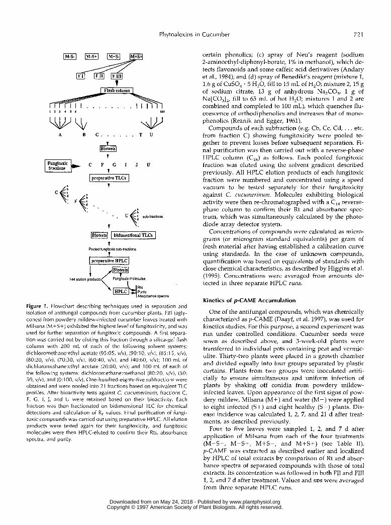
The scheme of separation of compounds of interest is shown in Figure 1. FIII (aglycones) from infected plants treated with Milsana (M+S+) was separated first because it had exhibited the highest leve1 of fungitoxicity on TLC bioassay (Daayf et al., 1995). To this end, a concentrate of this fraction was eluted through a silica-gel flash column (40 x 3 cm), with 200 mL of each of the following solvent systems: dich1oromethane:ethyl acetate (95:5, v/v), (90:10, v/v), (85:15, v/v), (80:20, v/v), (70:30, v/v), (60:40, v/v), and (40:60, v/v); 100 mL of dich1oromethane:ethyl acetate (20:80, v/v); and 100 mL of each of the following systems: dich1oromethane:methanol (80:20, v / v), (50:50, v / v), and (0:100, v /v) . This separation led to 185 subfractions, and those showing bands with equivalent R, values in TLC assays were pooled together to yield 21 subfractions (A-U). Detection of these bands was carried out using the univer- sal detection reagent (75 g of ammonium molybdate, 4 g of ceric sulfate, and 500 mL of 10% sulfuric acid:H,O, v/v), which showed dark spots after heating treated thin-layer chromatograms.
Each fraction was tested for fungitoxic activity directly on thin-layer chromatograms against Cladosporium cucume- rinum Ellis and Arth., as described previously (Daayf et al., 1995). The fractions (20- to 50-pL aliquots) were spotted on silica-gel TLC plates (Silica Gel 60 F-254, Merck) and de- veloped with dich1oromethane:hexane:methanol (BDH, Inc., Toronto, Canada). After drying for 1 to 2 h, the plates were covered with a concentrated conidial suspension of C. cucumerinum mixed (l:l, v / v ) in a 20 g /L solution of potato dextrose agar. The plates were then incubated in a humid chamber for 48 to 72 h, and zones of inhibition appeared as white spots against a dark background formed by spores and mycelium of C. cucumerinum. To assess whether fun- gitoxicity would result from Milsana itself, simultaneous TLC and bioassays were performed with Milsana alone and with Milsana hydrolyzed under the same conditions as the plant extracts at corresponding concentrations.
The bioactive fractions were then chromatographed by preparative TLC for further separation. Bands were re- isolated and eluted, each resulting in corresponding sub- fractions ( e g Ca, Cb, . . . from fraction C). These were again tested for their bioactivity against C. cucumerinum. Subfractions showing no fungitoxicity were eliminated, and those showing fungitoxicity were kept for two- dimensional TLC with dich1oromethane:hexane:methanol (6:4:1, v /v ) for the first chromatography dimension, and with different solvents for the second dimension, both of which depend on the polarity of compounds to be sepa- rated. For each active subfraction (Ca, Cb, . . . from bioac- tive fractions), four simultaneous two-dimensional TLC steps were carried out for the following purposes: (a) ob- servation of their behavior under SW and LW, R, estima- tion, and fungitoxicity; (b) application of ammonium hy- droxide vapors, which reinforce or modify fluorescence of
www.plantphysiol.orgon May 24, 2018 - Published by Downloaded from Copyright © 1997 American Society of Plant Biologists. All rights reserved.
Phytoalexins in Cucumber 721
t
1 2 3 4 1 6 115
ulv A B C . . . . . . T U
1 I Fungltoric k C F G I J U 1 fractions
I ureuarative T L C ~ I 1
d . . . . . . . Uf sub-franiom
T
.. 1
preparative BPLC J
@i&+%nr Absorbance specva
Figure 1. Flowchart descrihing techniques used in separation and isolation of antifungal compounds from cucumber plants. Fl l l (agly- cones) from powdery mildew-infected cucumber leaves treated with Milsana (M+S+) exhihited the highest leve1 of fungitoxicity, and was used for further separation of fungitoxic compounds. A first separa- tion was carried out by eluting this fraction through a silica-gel flash column with 200 mL of each of the following solvent systems: dich1oromethane:ethyI acetate (95:05, v/v), (9O:l O, v/v), (85:15, v/v), (80:20, v/v), (70:30, v/v), (60:40, v/v), and (40:60, v/v); 100 mL of dich1oromethane:ethyI acetate (20:80, v/v); and 100 mL of each of the following systems: dich1oromethane:methanol (80:20, v/v), (50: 50, v/v), and (0:100, v/v). One-hundred-eighty-five suhfractions were obtained and were pooled into 21 fractions hased on equivalent TLC profiles. After hioactivity tests against C. cucumerinum, fractions C, F, C, I , J , and U were retained based on their bioactivity. Each fraction was then fractionated on bidimensional TLC for chemical detections and calculation of R, values. Final purification of fungi- toxic compounds was carried out using preparative HPLC. All elution products were tested again for their fungitoxicity, and fungitoxic molecules were then HPLC-eluted to confirm their Rts, absorbance spectra, and purity.
certain phenolics; (c) spray of Neu’s reagent (sodium 2-aminoethyl-diphenyl-borate, 1% in methanol), which de- tects flavonoids and some caffeic acid derivatives (Andary et al., 1984); and (d) spray of Benedikt’s reagent (mixture 1, 1.6 g of CuSO, . 5 H,O, fill to 15 mL of H,O; mixture 2,15 g of sodium citrate, 13 g of anhydrous Na,C03, 1 g of Na[CO,],, fill to 65 mL of hot H,O; mixtures 1 and 2 are combined and completed to 100 mL), which quenches flu- orescence of orthodiphenolics and increases that of mono- phenolics (Reznik and Egger, 1961).
Compounds of each subfraction (e.g. Cb, Cc, Cd, . . . etc. from fraction C) showing fungitoxicity were pooled to- gether to prevent losses before subsequent separation. Fi- nal purification was then carried out with a reverse-phase HPLC column (CJ as follows. Each pooled fungitoxic fraction was eluted using the solvent gradient described previously. A11 HPLC elution products of each fungitoxic fraction were numbered and concentrated using a speed vacuum to be tested separately for their fungitoxicity against C. cucumerinum. Molecules exhibiting biological activity were then re-chromatographed with a C,, reverse- phase column to confirm their Rt and absorbance spec- trum, which was simultaneously calculated by the photo- diode array detector system.
Concentrations of compounds were calculated as micro- grams (or microgram standard equivalents) per gram of fresh material after having established a calibration curve using standards. In the case of unknown compounds, quantification was based on equivalents of standards with close chemical characteristics, as described by Higgins et al. (1995). Concentrations were averaged from amounts de- tected in three separate HPLC runs.
Kinetics of p-CAME Accumulation
One of the antifungal compounds, which was chemically characterized as p-CAME (Daayf, et al. 1997), was used for kinetics studies. For this purpose, a second experiment was run under controlled conditions. Cucumber seeds were sown as described above, and 3-week-old plants were transferred to individual pots containing peat and vermic- ulite. Thirty-two plants were placed in a growth chamber and divided equally into four groups separated by plastic curtains. Plants from two groups were inoculated artifi- cially to ensure simultaneous and uniform infection of plants by shaking off conidia from powdery mildew- infected leaves. Upon appearance of the first signs of pow- dery mildew, Milsana (M+) and water (M-) were applied to eight infected ( S + ) and eight healthy (S-) plants. Dis- ease incidence was calculated 1, 2, 7, and 21 d after treat- ments, as described previously.
Four to five leaves were sampled 1, 2, and 7 d after application of Milsana from each of the four treatments (M-S-, M-S+, M+S-, and M+S+) (see Table 11). p-CAME was extracted as described earlier and localized by HPLC of total extracts by comparison of Rt and absor- bance spectra of separated compounds with those of total extracts. Its concentration was followed in both FII and FIII 1,2, and 7 d after treatment. Values and SDS were averaged from three separate HPLC runs.
www.plantphysiol.orgon May 24, 2018 - Published by Downloaded from Copyright © 1997 American Society of Plant Biologists. All rights reserved.
722 Daayf et al. Plant Physiol. Vol. 113, 1997
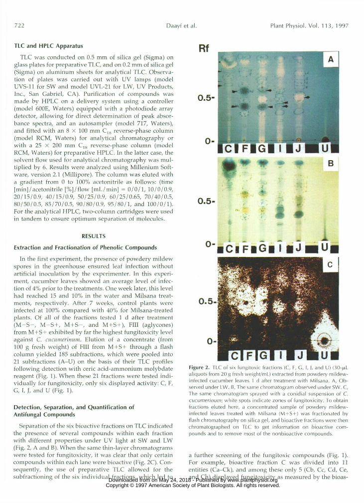
TLC and HPLC Apparatus
TLC was conducted on 0.5 mm of silica gel (Sigma) onglass plates for preparative TLC, and on 0.2 mm of silica gel(Sigma) on aluminum sheets for analytical TLC. Observa-tion of plates was carried out with UV lamps (modelUVS-11 for SW and model UVL-21 for LW, UV Products,Inc., San Gabriel, CA). Purification of compounds wasmade by HPLC on a delivery system using a controller(model 600E, Waters) equipped with a photodiode arraydetector, allowing for direct determination of peak absor-bance spectra, and an autosampler (model 717, Waters),and fitted with an 8 x 100 mm C,K reverse-phase column(model RCM, Waters) for analytical chromatography orwith a 25 X 200 mm C1K reverse-phase column (modelRCM, Waters) for preparative HPLC. In the latter case, thesolvent flow used for analytical chromatography was mul-tiplied by 6. Results were analyzed using Millenium Soft-ware, version 2.1 (Millipore). The column was eluted witha gradient from 0 to 100% acetonitrile as follows: (time[min]/acetonitrile [%]/flow [mL/min] = 0/0/1, 10/0/0.9,20/15/0.9, 40/15/0.9, 50/25/0.9, 60/25/0.65, 70/40/0.5,80/50/0.5, 85/70/0.5, 90/80/0.9, 95/80/1, and 100/0/1).For the analytical HPLC, two-column cartridges were usedin tandem to ensure optimum separation of molecules.
RESULTS
Extraction and Fractionation of Phenolic Compounds
In the first experiment, the presence of powdery mildewspores in the greenhouse ensured leaf infection withoutartificial inoculation by the experimenter. In this experi-ment, cucumber leaves showed an average level of infec-tion of 4% prior to the treatments. One week later, this levelhad reached 15 and 10% in the water and Milsana treat-ments, respectively. After 7 weeks, control plants wereinfected at 100% compared with 40% for Milsana-treatedplants. Of all of the fractions tested 1 d after treatment(M-S-, M-S+, M+S-, and M+S+), Fill (aglycones)from M+S+ exhibited by far the highest fungitoxicity levelagainst C. cucumerinum. Elution of a concentrate (from100 g fresh weight) of Fill from M+S+ through a flashcolumn yielded 185 subfractions, which were pooled into21 subfractions (A-U) on the basis of their TLC profilesfollowing detection with eerie acid-ammonium molybdatereagent (Fig. 1). When these 21 fractions were tested indi-vidually for fungitoxicity, only six displayed activity: C, F,G, I, J, and U (Fig. 1).
Detection, Separation, and Quantification ofAntifungal Compounds
Separation of the six bioactive fractions on TLC indicatedthe presence of several compounds within each fractionwith different properties under UV light at SW and LW(Fig. 2, A and B). When the same thin-layer chromatogramswere tested for fungitoxicity, it was clear that only certaincompounds within each lane were bioactive (Fig. 2C). Con-sequently, the use of preparative TLC allowed for thesubfractioning of the six individual fractions, which led to
0.5-
0- •ClFlG|T|J
0.5-
Figure 2. TLC of six fungitoxic fractions (C, F, C, I, ), and U) (30-jx.Laliquots from 20 g fresh weight/ml) extracted from powdery mildew-infected cucumber leaves 1 d after treatment with Milsana. A, Ob-served under LW. B, The same chromatogram observed under SW. C,The same chromatogram sprayed with a conidial suspension of C.cucumerinum; white spots indicate zones of fungitoxicity. To obtainfractions eluted here, a concentrated sample of powdery mildew-infected leaves treated with Milsana (M + S + ) was fractionated byflash chromatography on silica gel, and bioactive fractions were thenchromatographed on TLC to get information on bioactive com-pounds and to remove most of the nonbioactive compounds.
a further screening of the fungitoxic compounds (Fig. 1).For example, bioactive fraction C was divided into 11entities (Ca-Ck), and among these only 5 (Cb, Cc, Cd, Ce,and Ch) displayed fungitoxicity as measured by the bioas- www.plantphysiol.orgon May 24, 2018 - Published by Downloaded from
Copyright © 1997 American Society of Plant Biologists. All rights reserved.

Phytoalexins in Cucumber 723
say (Fig. 3). These subfractions from fractions C, F, G, I, J,and U presented different chemical characteristics follow-ing two-dimensional TLC (Fig. 1), which are summarizedin Table I.
When all of the active subfractions were pooled intocorresponding fractions and concentrated, the latter wereeluted by preparative HPLC, leading to a total of 144elution products (Fig. 1), which were each assigned a num-ber (1-144). They were further tested for bioactivity and 8were found to yield Rts of these bioactive molecules (TableI). Comparison of their Rts and absorbance spectra withthose observed for total Fill from all four sets of sampledleaves (S-M-, S-M+, S+M-, and S+M+) led to theprecise localization of their peaks on the total high-performance liquid chromatograms (Fig. 4).
For quantitative and comparative purposes, these chro-matograms were analyzed based on equivalent amounts offresh leaf material, as shown for total Fill from S-M- andS+M+ (Fig. 4). From these chromatograms, at least 21peaks were identified as being common to both, but only 6of them (1, 4, 10, 12, 16, and 20) contained active moleculeswhen tested for fungitoxicity (Fig. 1; Table I). Many ofthese peaks displayed a marked increase in concentrationin M+S+ compared with M — S — (Fig. 4), which indicatesthat they behaved like phytoalexins. For instance, bioactivepeaks 10, 12, 16, and 20 showed an increase in concentra-tion ranging from 2- to 9-fold compared with their coun-terpart in M-S-.
One day after Milsana treatment, bioactive compound 72(peak 10) displayed a 7-fold increase in concentration inM+S+ (65 ± 5 fig m-coumaric acid equivalents/g freshweight) compared with M — S — (9 ± 4 fig m-coumaric acidequivalents/g fresh weight) (Fig. 4). Compound 57 (peak12) was roughly twice as high in M+S+ (15 ± 2.5 figp-coumaric acid equivalents/g fresh weight) comparedwith M — S — (7 ± 1.5 fig p-coumaric acid equivalents/gfresh weight). Peak 16, containing three active compounds,exhibited a 9-fold increase in M+S+ (45 ± 8 fig p-coumaric
0.5-
Figure 3. Chromatogram inhibition assay with C. cucumerinum ofsubfractions from fraction C extracted from infected cucumber leavestreated with Milsana after separation with dichloromethane:hexane:methanol (6:7:1, v/v). White spots indicate zones of fungitoxicity.From the 11 subfractions tested, only 5 exhibited bioactivity (Cb, Cc,Cd, Ce, and Ch).
acid equivalents/g fresh weight) compared with M — S —(5 ± 4 jug p-coumaric acid equivalents/g fresh weight).Compound 17 (peak 20), recently identified as p-CAME(Daayf, et al., 1997) showed a 2.5-fold increase in M+S+(30 ± 5 /xg/g fresh weight) compared with the control(13 ± 2 fig/g fresh weight). A sum calculation of all of thecompounds with phytoalexin properties in this experiment(given as equivalents of phenolic compounds) gave ap-proximately 155 p,g equivalents/g fresh weight in M+S+versus 34 fig equivalents/g fresh weight in M—S—, whichtranslated into a nearly 5-fold increase.
Analysis of active molecules in the other two treatments(M-S+ and M+S-) indicated a similar pattern of pres-ence. The difference was that peaks that increased inM+S- and M—S+ were the same ones that increased inM+S+, but did so to a smaller extent (results not shown).
In general, higher amounts of antifungal compoundswere consistently observed 1 d after Milsana application inFill from M+S+. Analysis of compounds in FII at this timeshowed no such differential accumulation among treat-ments. On the other hand, some of these active moleculesseparated from Fill were present in their free form in FIIextracted 1 week after Milsana treatment.
Kinetics of p-CAME Accumulation
In a second experiment, plants were kept in a growthchamber and artificially inoculated, and the kinetics ofp-CAME accumulation were followed in both its free (FII)and conjugated form (Fill) over 7 d (Fig. 5). From theseresults, it is clear that the compound reached its highestconcentration in M+S+ in both FII and Fill. Another pointof interest was the negative correlation between the in-crease of p-CAME in FII and Fill over time. In particular, inFill of all tested materials, p-CAME reached its highestconcentration within the first 2 d (Fig. 5). Concentrationsthen dropped to initial values 7 d after treatment. By con-trast, p-CAME in FII remained low on d 1 and 2 aftertreatment and reached its highest level on d 7. The concen-tration in FII after 7 d was comparable to that found earlyin Fill, suggesting a conversion into the active form inplanta. The rapid accumulation of p-CAME in Fill ofM+S+ and subsequent conversion into its free form cor-related with a mere 4% increase in visible infection over thenext 6 d (Table II). In contrast, infection increased 34% inM-S+ plants, which displayed a much lower accumula-tion of p-CAME in both FII and Fill over the same time.This trend was maintained over a period of 3 weeks.Whereas 90% of the leaf surface was visibly infected inM-S+ plants, only 45% was infected in M+S+ plants.
DISCUSSION
Results presented in this study provide the first directevidence, to our knowledge, that cucumber plants produceelevated levels of phytoalexins in response to an elicitingtreatment after infection. Considering that Milsana (a) is anefficient prophylactic treatment against cucumber pow-dery mildew (Merger et al., 1988; Daayf et al., 1995; Dik andVan der Straay, 1995); (b) contains no fungitoxic com- www.plantphysiol.orgon May 24, 2018 - Published by Downloaded from
Copyright © 1997 American Society of Plant Biologists. All rights reserved.

724 Daayf et al. Plant Physiol. Vol. 1 1 3 , 1997
Table 1. Chromatographic and other chemical characteristics of fungitoxic compounds isolated from Milsana-treated and infected cucumber leaves and of some phenolic standards
Peak CPd LW sw RF Rt NH,OH Neu Benedikt A”,,,
1 4
10 1 2 16 16 16 20
p-Coumaric m-Coumaric o-Coumaric p-CAME Caffeic Ferulic Sinapic Narinaenin
121 1 O0 fl 72 57 81 37 dk 38 dk 17 bl*
bl* fl Y
bl* bl bl bl
-
V
+ 0.05 6 0.37 30
+ 0.32 42 + 0.35 52 + 0.4 75 + 0.34 75 + 0.50 76 + 0.70 92
-
+ 0.35 + 0.25 + 0.32 + 0.7 + 0.22 + 0.43 + 0.36 + 0.80
51 63 73 92 38 59
93 58
gb O + + + + + +
- 285 (330”) 285 -
310.7 310 360 325 310.7
-
- -
-
-
310.7
2751325 310.7
325 (295”) 325 (295”)
290 (330”)
281
325
L”
240 240 250 250 270 265 250
250 250
24013 1 O 250 265 260 265 250
~
Cpd, Compound; -, not detectable; +, detectable; NH,OH, ammonium hydroxide vapors; Neu, flavonoids’ reagent (-, no reaction, i-, modification of fluorescence), Benedikt, orthopiphenolic’s reagent (-, no quenching, +, quenching); A,,,, maximum absorbance wavelength (nm); A,,,; minimum absorbance wavelength (nm); bl, blue fluorescent; dk, dark; fl, fluorescent; gb, green-blue fluorescent; o, orange; y, yellow. . , Not determined; “, shoulder; *, pale blue. R, values correspond to migration of compounds on silica gel-aluminum plates with solvent system
(dichloromethane:hexane:methanol, 6:4:1, v/v).
pounds, as indicated here and previously (Herger and Klingauf, 1990); and (c) induces accumulation of fungitoxic phenolic compounds, it appears that these compounds con- tribute to cucumber’s increased resistance to powdery mil- dew, at least in the context of a prophylactic treatment with Milsana. This was shown in the reduction of powdery
2o:oo 4o:oo ’ ’ s0:oo ’ ’ 8o:oo ’ ’ ’ lOd.00 0.00
Retention Time (min.)
Figure 4. Reverse-phase HPLC analysis on C,8 column of aglycone fractions from control (M-S-, top) and infected Milsana-treated (M+S+, bottom) long English cucumber leaves 1 d after Milsana treatment. Elution was performed with a gradient of O to 100% acetonitrile. Volumes of M-S- and M+S+ used for these injections (25 pL) correspond to the same quantity of fresh leaf material (20 g fresh weight/mL).
mildew infection in both experiments 1 and 2, in the rapid accumulation of antifungal phenolics in cucumber leaves in response to infection and a treatment with Milsana, and in the kinetics of p-CAME accumulation. These results are of particular significance, since they provide support for the production of antifungal compounds with induction of cucumber defense mechanisms. Previously, lignification and production of chitinases were the main defense reac- tions reported to occur in cucumber (Hammerschmidt and Kuc, 1982; Hammerschmidt et al., 1985; Siegrist et al., 1994).
Based on our extraction and purification procedures, at least eight separate phenolic compounds with antifungal activity were identified as intrinsic components of cucum- ber plants. In addition, severa1 of them increased in con- centration as a result of an elicitation with Milsana, this being particularly true when the plant was stressed by the pathogen. This phenomenon is a classical manifestation of induced resistance, in which an elicitor will magnify, in time and amount, the defense response of the plant to fend off a pathogen (Benhamou, 1996). As such it is somewhat surprising that cucumber, albeit a thoroughly studied model of induced resistance (Siegrist et al., 1994), was never before reported to produce phytoalexins or antifun- gal compounds.
One explanation may reside in the diversity of fungitoxic phenolics observed in the present study. Although our work has led to the chemical characterization of one of these phenolics (Daayf et al., 1997) it also indicates that a minimum of five or six additional compounds increased in response to infection, which is characteristic of phytoalex- ins. In earlier studies in which phytoalexins were found to be important components of resistance, the activity was usually ascribed to one molecule or a few closely related
www.plantphysiol.orgon May 24, 2018 - Published by Downloaded from Copyright © 1997 American Society of Plant Biologists. All rights reserved.

Phytoalexins in Cucumber 725
30 4
" I
O 4
Days after treatment
Figure 5. Concentrations of p-CAME over time in its conjugated (FIII) and free (FII) form in cucumber leaves obtained from healthy control (M-S-), powdery mildew-infected (M-S+), healthy Milsana- treated (M+S-), and powdery mildew-infected and Milsana-treated (M+S+) plants. Each value is the mean ? SD of three separate HPLC runs. FM, Fresh weight.
molecules. For example, the Pisum sativum-pisatin model is probably the best known and the most studied because the phytoalexin is unique to pea, it has very high specific activity, and its presence correlates very well with resis- tance (Cruickshank, 1963; Kuc, 1995). In contrast, the con- cept that severa1 phytoalexins can be simultaneously in- volved in resistance has been well documented recently, notably in groundnut (Strange and Subba Rao, 1994). In this regard, we believe that the cumulative presence of a11 of the phytoalexins observed here is probably necessary for them to play an important part in resistance in cucumber. For instance, in the specific case of p-CAME, our results have shown that it has antifungal properties and that its kinetics of accumulation correlate well with cucumber re- sistance. However, in situ accumulation probably does not reach sufficient levels to account for all of the observed resistance. On the other hand, the combined amount of antifungal compounds detected (155 p g / g fresh weight) is in agreement with previous reports associating the concen- tration of a single phytoalexin or a group of phytoalexins with plant resistance (Yoshikawa et al., 1978; Dahiya and
Rimmer, 1988; Beimen et al., 1992; Edwards et al., 1995). In addition, the nearly 5-fold increase in concentration (155 versus 34 p g / g fresh weight) falls well within the range of reported increases for a number of phytoalexins (Graham et al., 1990; Rouxel et al., 1991; Strange and Subba Rao, 1994; Edwards et al., 1995). In this context, we have shown previously that the combined aglycones from the M+S+ treatment induced a significant reduction in germination of S. fuliginea conidia compared with all other treatments (Daayf et al., 1996). Previous efforts to isolate phytoalexins from cucumber may have been hampered by attempts to focus on one individual molecule (e.g. coniferyl alcohol) that would confer a11 of the activity (Hammerschmidt and Kuc, 1982).
From a biological point of view, this multiplicity of active compounds may confer a distinct advantage to the plant. Recent studies have shown that fungi can develop the ability to detoxify a phytoalexin (VanEtten et al., 1989, 1995), and that phytoalexin degradation could be impor- tant in determining compatibility (Kuc, 1995). Conceivably, pathogens are less likely to develop simultaneous resis- tance against a variety of antifungal molecules.
In this study, hydrolysis of the phenolics prior to testing their fungitoxicity was instrumental in identifying the mol- ecules of interest, and thus may also explain prior difficul- ties in identifying phytoalexins in cucumber. Hydrolyzed fractions (FIII) were considerably more active than free phenolics (FII), especially shortly after treatment. In the case of p-CAME, the subsequent presence in its free form (FII) (Fig. 5) in leaf extracts indicates that conversion into an active form takes place in planta, presumably through the activity of P-glucosidases. Previous work by our re- search group has established that postinfection synthesis of P-glucosidases was stimulated in cucumber following elic- itation (Chérif et al., 1994). Our findings are consistent with reports linking conjugated defense-related phenolics with resistance in an increasing number of plants (Higgins et al., 1995). In fact, other studies have suggested that conjugated phytoalexins or conjugated phytoalexin precursors that are present constitutively in the cell prior to infection are more important for resistance than phytoalexins that are pro- duced de novo in response to infection (Graham et al., 1990; Graham and Graham, 1991; Weidemann et al., 1991; Geibel and Feucht, 1993).
Very little is known about the constituents of the Milsana formulation that confer its prophylactic properties. Al-
Table II. fercent S. fuliginea infection of long fnglish cucumber plants in response to a treatment with Milsana (extracts from R. sachalinensis)
Controla Milsana Days after
Treatment M - ~ - M-S+ M+S- M+S+
1 O ? O 5 2 3 0 5 0 5 ? 3 2 1 2 2 8 2 2 1 2 2 5 2 3 7 2 ? 2 39 ? 3 1.5 i 3 9 ? 2
a Control (M-S- and M-S+) consisted of water application. M, Milsana; S, powdery mildew. S+ plants were artificially inoculated with S. fuliginea 3 d prior to treatments. Each value represem the mean percent infection of eight plants 2 SD.
www.plantphysiol.orgon May 24, 2018 - Published by Downloaded from Copyright © 1997 American Society of Plant Biologists. All rights reserved.

726 Daayf et al. Plant Physiol. Vol. 11 3 , 1997
though a direct fungicidal activity has been ruled out, it would be interesting to identify the active molecule(s) that acts as elicitor. In this sense, one may question if the production of phytoalexins i n cucumber is specific to a treatment with Milsana, since previous work with soluble silicon h a d already suggested the presence of antifungal, soluble phenolics in the general defense responses of cu- cumber (Chérif e t al., 1992, 1994; Bélanger et al., 1995). One m a y also ask whether Milsana-mediated, induced resis- tance occurs locally a n d systemically. Should it provide systemic protection, it could represent a n interesting model in which to s tudy signal perception a n d transduction i n plant disease response (Lamb, 1994) and the role of signal molecules such as salicylic acid and jasmonic acid (Farmer a n d Ryan, 1990; Klessig and Malamy, 1994).
ACKNOWLEDCMENTS
We thank Caroline Labbé and Tyler Avis for technical assistance and Drs. Nicole Benhamou and Serge Yelle for helpful suggestions and critica1 review of the manuscript.
Received August 15, 1996; accepted November 27, 1996. Copyright Clearance Center: 0032-0889/ 97/ 113/ 0719/ 09.
LITERATURE ClTED
Andary C, Roussel JL, Rascol JP (1984) Micromethods for analysis of heterosidic esters of caffeic acid. J Chromatogr 303: 312-317
Bailey JA, Mansfield JW (1982) Phytoalexins. Halsted Press, New York
Beimen A, Bermpohl A, Meletzus D, Eichenlaub R, Barz W (1992) Accumulation of phenolic compounds in leaves of tomato plants after infection with Clavibacter michiganense subsp. michi- gnnense strains differing in virulence. Z Naturforsch 47c: 898-909
Bélanger, RR, Bowen PA, Ehret DL, Menzies JG (1995) Soluble silicon: its role in crop and disease management of greenhouse crops. Plant Dis 79: 329-336
Benhamou N (1992) Ultrastructural detection of p-1,3-glucans in tobacco root tissues infected by Phytophthora infestam var. nico- tianne using a gold-complexed tobacco P-1,3-glucanase. Physiol Mo1 Plant Pathol 41: 351-370
Benhamou N (1996) Elicitor-induced plant defence pathways. Trends Plant Sci 1: 233-240
Bennett RN, Wallsgrove RM (1994) Secondary metabolites in plant defense mechanisms. New Phytol 127: 617-633
Carr JP, Klessig DF (1989) The pathogenesis-related proteins of plants. In JK Setlow, eds, Genetic Engineering, Principles and Methods. Plenum Press, New York, pp 65-89
Chérif M, Asselin A, Bélanger RR (1994) Defense responses in- duced by soluble silicon in cucumber roots infected by Pythium spp. Phytopathology 84: 236-242
Chérif M, Bélanger RR (1992) Use of potassium silicate amend- ments in recirculating nutrient solutions to suppress Pythium ultimutn on long English cucumber. Plant Dis 76: 1008-1011
Chérif M, Benhamou N, Menzies JG, Bélanger RR (1992) Silicon induced resistance in cucumber plants-against Pythium ultimum. Physiol Mo1 Plant Pathol 41: 411425
Cruickshank IAM (1963) Phytoalexins. Annu Rev Phytopathol 1:
Daayf F, Bélanger RR, Schmitt A (1996) Alteration of cucumber leaf physiology by treatment with extracts of Reynoutria sachali- nensis. In H Lyr, PE Russel, HD Sisler, eds, Modern Fungicides and Antifungal Compounds. Intercept Ltd., Andover, UK, pp 245-251
Daayf F, Bel-Rhlid R, Bélanger RR (1997) Methyl ester of p-coumaric acid: a phytoalexin-like compound from long En- glish cucumber leaves. J chem Eco1 (in press)
351-374
Daayf F, Schmitt A, Bélanger RR (1995) The effects of plant extracts of Reynoutria sachalinensis on powdery mildew develop- ment and leaf physiology of long English cucumber. Plant Dis
Dahiya JS, Rimmer SR (1988) Phytoalexin accumulation in tissues of Brassica napus inoculated with Leptosphnerin maculans. Phyto- chemistry 27: 3105-3107
Darvill AG, Albersheim P (1984) Phytoalexins and their elicitors: a defense against microbial infection in plants. Annu Rev Plant Physiol 35: 243-276
Dercks W, Buchenauer H (1986) Untersuchungen zum Einfluss von Aluminiumfosetyl auf den pflanzlichen Phenolstoffwechsel in den Pathogen-Wirt Beziehungen PhytophthoralFragaria- Erdbeere und BreminlLactuca-Salat. J Phytopathol 115: 37-55
Dik AJ, Van der Straay (1995) The effect of Milsana on cucumber powdery mildew under Dutch conditions. Med Fac Landbouww Rijksuniv Gent 59: 1027-1034
Dixon RA, Paiva NL (1995) Stress-induced phenylpropanoid me- tabolism. Plant Cell 7: 1085-1097
Ebel J (1986) Phytoalexin synthesis: the biochemical analysis of the induction process. Annu Rev Phytopathol 2 4 235-264
Edwards R, Mizen T, Cook R (1995) Isoflavonoid conjugate accu- mulation in the roots of lucerne (Medicago sntiva) seedlings fol- lowing infection by the stem nematode (Ditylenchus dispnci). Nematologica 41: 51-66
Farmer EE, Ryan CA (1990) Interplant communication: airborne methyl jasmonate induces synthesis of proteinase inhibitors in plant leaves. Proc Natl Acad Sci USA 87: 7713-7716
Geibel M, Feucht W (1993) The characteristic weak glycosidic bonding of flavonoid 5-glucosides from Prunus cerasus bark and their contribution to the resistance against Cytospora personii (abstract L15). In International Scientific Symposium on Natural Phenols in Plant Resistance, International Society for Horticul- tural Science, Faculty of Agriculture and Horticulture, Munich, Germany
Graham MY, Graham TL (1991) Rapid accumulation of anionic peroxidases and phenolic polymers in soybean cotyledon tissues following treatment with Phytophthora megasperma f.sp. glycinea wall glucan. Plant Physiol 97: 1445-1455
Graham TL, Kim JE, Graham MY (1990) Role of constitutive isoflavone conjugates in the accumulation of glyceollin in soy- bean infected with Phytophthora nzegasperma. Mo1 Plant Microbe Interact 3: 157-166
Hammerschmidt R, Bonnen AM, Bergstrom GC, Baker KK (1985) Association of epidermal lignification with non-host resistance of cucurbits to fungi. Can J Bot 63: 2393-2398
Hammerschmidt R, Kuc J (1982) Lignification as a mechanism for induced systemic resistance in cucumber. Physiol Plant Pathol 20: 61-71
Herger G, Klingauf F (1990) Control of powdery mildew fungi with extracts of the giant knotweed, Reynoutria sachalinensis (Polygonaceae). Med Fac Landbouww Rijksuniv Gent 55: 1007- 1014
Herger G, Klingauf F, Mangold D, Pommer EH, Scherer M (1988) Efficacy of extracts of Reynoutria sachalinensis (F. Smidt) Nakai (Polygonaceae) against funga1 diseases, especially powdery mil- dews (in German). Nachrichtenbl Dtsch Pflanzenschutzdienst (Braunschweig) 40: 56-60
Higgins VJ, Hollands J, Bates DK (1995) Phytoalexins in forage legumes: studies on detoxification by pathogens and the role of glycosidic precursors in roots. In M Daniel, RP Purkayastha, eds, Handbook of Phytoalexin Metabolism and Action. Marcel Dek- ker, New York, pp 391403
Klessig DF, Malamy J (1994) The salicylic acid signal in plants. Plant Mo1 Biol 26: 1439-1458
Kowalewski A (1993) Investigations about the chemical nature and the mode of action of the resistance inducing ingredients from Reynoutria sachalinensis (F. Schmidt) Nakai. In B Fritig, M Legrand, eds, Mechanisms of Plant Defense Responses. Kluwer Academic Publishers, Dordrecht, The Netherlands, p 182
Kuc J (1995) Phytoalexins, stress metabolism, and disease resis- tance in plants. Annu Rev Phytopathol 33: 275-297
79: 577-580
www.plantphysiol.orgon May 24, 2018 - Published by Downloaded from Copyright © 1997 American Society of Plant Biologists. All rights reserved.
Phytoalexins in Cucumber 72 7
Lamb CJ (1994) Plant disease resistance genes in signal perception and transduction. Cell 76: 419422
Lyon GD, Reglinski T, Newton AC (1995) Nove1 disease control compounds: the potential to immunize plants against infection. Plant Pathol 44: 407-427
Reznik H, Egger K (1961) Benedikts reagents als indicator fur phenolishe orthodihydroxygruppe. Z Ana1 Chem 183: 196-199
Rouxel T, Kollmann A, Boulidard L, Mithen R (1991) Abiotic elicitation of indole phytoalexins and resistance to Leptosphaeria maculans within Brassiceae. Planta 184: 271-278
Schneider S, Ullrich W (1994) Differential induction of resistance and enhanced enzyme activities in cucumber and tobacco caused by various abiotic and biotic inducers. Physiol Mo1 Plant Pathol45: 291-304
Siegrist J, Jeblick W, Kauss H (1994) Defense responses in in- fected and elicited cucumber (Cucumis sativus L.) hypocotyl segments exhibiting acquired resistance. Plant Physiol 105:
Stein BD, Klomparens KL, Hammerschmidt R (1993) Histochem- istry and ultrastructure of the induced resistance response of
1365-1374
cucumber plants to Collefofrichum lagenarium. J Phytopathol137: 177-188
Strange RN, Subba Rao PV (1994) The phytoalexin response of groundnut and its role in disease resistance. Oleagineux 49: 227-233
VanEtten HD, Matthews DE, Matthews PS (1989) Phytoalexin detoxification: importance for pathogenicity and practical impli- cations. Annu Rev Phytopathol 27: 143-164
VanEtten HD, Sandrock RW, Wasmann CC, Soby SD, McClus- key K, Wang P (1995) Detoxification of phytoanticipins and phytoalexins by phytopathogenic fungi. Can J Bot Suppl 1 73:
Weidemann C, Tenhaken R, Hohe U, Barz W (1991) Medicarpin and maackiain-3-0-glucoside-6’-0 malonate conjugates are con- stitutive compounds of chickpea (Cicer arietinum L.) cell cultures. Plant Cell Rep 10: 371-374
Yoshikawa M, Yamauchi K, Masago H (1978) Glyceollin: its role in restricting funga1 growth in resistant soybean hypocotyls infected with Phyfophfhora megasperma var. sojae. Physiol Plant Pathol 12: 73-82
518-525
www.plantphysiol.orgon May 24, 2018 - Published by Downloaded from Copyright © 1997 American Society of Plant Biologists. All rights reserved.