
EVIDENCE FOR A NOVEL BACTERIOPHAGE FROM MORAXELLA CATARRHALIS · EVIDENCE FOR A NOVEL BACTERIOPHAGE...
76
EVIDENCE FOR A NOVEL BACTERIOPHAGE FROM MORAXELLA CATARRHALIS a thesis submitted to the department of molecular biology and genetics and the institute of engineering and science of bilkent university in partial fulfillment of the requirements for the degree of master of science BY G ¨ ULC ¸ ˙ IN C ¸ AKAN AUGUST 2005
Transcript of EVIDENCE FOR A NOVEL BACTERIOPHAGE FROM MORAXELLA CATARRHALIS · EVIDENCE FOR A NOVEL BACTERIOPHAGE...

EVIDENCE FOR A NOVELBACTERIOPHAGE FROM MORAXELLA
CATARRHALIS
a thesis submitted to
the department of molecular biology and genetics
and the institute of engineering and science of
bilkent university
in partial fulfillment of the requirements
for the degree of master of science
BY
GULCIN CAKAN
AUGUST 2005
I certify that I have read this thesis and that in my opinion it is fully adequate,
in scope and in quality, as a thesis for the degree of Master of Science.
Dr.Kamruddin Ahmed
I certify that I have read this thesis and that in my opinion it is fully adequate,
in scope and in quality, as a thesis for the degree of Master of Science.
Assist. Prof. Dr. Ozlen Konu
I certify that I have read this thesis and that in my opinion it is fully adequate,
in scope and in quality, as a thesis for the degree of Master of Science.
Prof.Dr. Gulay Ozcengiz
Approved for the Institute of Engineering and Science:
Prof. Dr. Mehmet B. BarayDirector of the Institute Engineering and Science
ii
ABSTRACT
EVIDENCE FOR A NOVEL BACTERIOPHAGE FROMMORAXELLA CATARRHALIS
Gulcin Cakan
M.S. in Molecular Biology and Genetics
Supervisors: Dr.Kamruddin Ahmed , Assist.Prof.Dr. Ozlen Konu
August 2005, 76 Pages
Moraxella catarrhalis is one of the major causes of RTI and otitis media, which
was known as a harmless inhabitant of upper respiratory tract until 1980s. The
knowledge on virulence factors, pathogenesis of this bacterium is scarce and the
reason/(s) for recent pathogenic conversion of M. catarrhalis is/are not known.
Several examples demonstrate that bacteriophages control bacterial virulence in
almost every step of pathogenesis. Number of bacteriophages are known to be
solely responsible from bacterial virulence. We hypothesize that a bacteriophage
may be responsible for pathogenic conversion of M. catarrhalis, and we inves-
tigated whether a bacteriophage is present in M. catarrhalis. In this study,
evidence for a bacteriophage from M.catarrhalis is presented. Supernatants of
M.catarrhalis broth cultures were shown to cause bacterial cell lysis on soft agar
cultures, indicating presence of bacteriophages released from them. Two particles
having different morphologies and different size ranges (p<0. 05) were co-purified
from these supernatants. One segment of dsDNA molecule and three segments of
ssRNA molecules were extracted from these particles. The comparison between
pathogenic and non-pathogenic strains of M.catarrhalis demonstrated marked
differences in the quantity of these RNA and DNA molecules.Future studies are
necessary to determine the origins of RNA and DNA molecules.
Keywords: Moraxella catarrhalis, bacteriophage, virulence.
iii

OZET
MORAXELLA CATARRHALIS ’DEN EDINILEN YENIBIR BAKTERIYOFAJ’A DAIR BULGULAR
Gulcin Cakan
Molekuler Biyoloji ve Genetik, Yuksek Lisans
Tez Yoneticileri: Dr.Kamruddin Ahmed , Assist. Prof. Dr. Ozlen Konu
Agustos 2005, 76 Sayfa
1980’li yıllara kadar solunum yollarında yerlesik zararsız bir bakteri olarak bili-
nen Moraxella catarrhalis, solunum yolları enfeksiyonu ve otitis media’nın baslıca
nedenlerinden biridir. Bu bakterinin virulens faktorleri ve patojenezi hakkındaki
bilgiler az sayıda olup son zamanlarda gerceklesen patojenik donusumunun
ardında yatan nedenler bilinmemektedir. Bakteriyofajların bakteri virulensini,
patojenezin hemen her asamasında kontrol edebildigi orneklenmistir. Bazı bak-
teriyofajlar ise bakteriyel virulensin tek sorumlusudurlar. M. catarrhalis ’in
yakın zamanda gecirdigi patojenik donusumun bakteriyofaj kaynaklı oldugunu
varsayarak M. catarrhalis ’in bakteriyofaj icerip icermedigini arastırdık. Bu
calısmada, M. catarrhalis ’in bakteriyofaja sahip olduguna dair kanıtlar sunulmak-
tadır. Varolan bakteriyofajların salındıgını destekleyecek sekilde, M. catarrhalis
sıvı (broth) kulturlerinden elde edilen supernatantların, yumusak agar bakteriyal
kulturlerinin dagılmasına (lysis) yol actıgı gosterilmistir. Bu supernatantlardan
farklı morfoloji ve boyutlara sahip olan iki ayrı yapı birlikte izole edilmistir.
Bu yapılardan biri dsDNA diger ucu ise ssRNA olan nukleik asit fragmanları
elde edilmistir. Patojenik ve patojenik-olmayan M. catarrhalis turleri arasinda
yapılan karsılastırmalar, bu iki grup arasında RNA ve DNA miktarlarında belir-
gin farklılıklar oldugunu gostermistir. Elde edilen RNA ve DNA molekullerinin
kaynaklarının ortaya cıkarılması icin arastırmaların surdurulmesi gerekmektedir.
Anahtar sozcukler : Moraxella catarrhalis, bakteriyofaj, virulens.
iv

Acknowledgement
I would like to extend my gratitudes to;
...my family, for everything...
...Yasar, for his continuous support and help...
...Dr.Kamruddin Ahmed, for his continuous guidance, and invaluable help
during my stay in Nagasaki...
...Dr.Ozlen Konu, for her invaluable support and friendship...
...Canan and Mustafa, for their efforts in this project...
...Iraz, for her help in thesis writing, and her friendship...
...Ceren, Jale, Betul and Gulcan, for their friendship...
...Prof.Osamu Nakagomi, for supporting my visit to his department...
...members of Nakagomi lab, for their support and friendship...
...Izumi and Mika, for their hospitality...
...Past and present members of MBG Department; where I learned a lot and
made great friends during last 6 years.
v
Table of Contents
Signature Page . . . . . . . . . . . . . . . . . . . . . . . . . . . . . . . ii
Abstract . . . . . . . . . . . . . . . . . . . . . . . . . . . . . . . . . . . iii
Ozet . . . . . . . . . . . . . . . . . . . . . . . . . . . . . . . . . . . . . iv
Acknowledgement . . . . . . . . . . . . . . . . . . . . . . . . . . . . . . v
Table of Contents . . . . . . . . . . . . . . . . . . . . . . . . . . . . . . vi
List of Tables . . . . . . . . . . . . . . . . . . . . . . . . . . . . . . . . ix
List of Figures . . . . . . . . . . . . . . . . . . . . . . . . . . . . . . . . x
Abbreviations . . . . . . . . . . . . . . . . . . . . . . . . . . . . . . . . xi
1 Introduction 1
1.1 Moraxella catarrhalis . . . . . . . . . . . . . . . . . . . . . . . . . 1
1.1.1 Infections . . . . . . . . . . . . . . . . . . . . . . . . . . . 2
1.1.2 Virulence Mechanisms and Factors . . . . . . . . . . . . . 3
1.1.3 Antibiotic Resistance . . . . . . . . . . . . . . . . . . . . 5
1.2 Role of Horizontal Gene Transfer in M.catarrhalis Virulence . . . 7
vi

1.2.1 Mechanisms of Horizontal Gene Transfer . . . . . . . . . . 8
1.3 Bacteriophage Control of Bacterial Virulence . . . . . . . . . . . 10
1.3.1 Adhesion . . . . . . . . . . . . . . . . . . . . . . . . . . . . 12
1.3.2 Invasion . . . . . . . . . . . . . . . . . . . . . . . . . . . . 13
1.3.3 Resistance to serum and phagocytes . . . . . . . . . . . . 14
1.3.4 Exotoxin production . . . . . . . . . . . . . . . . . . . . . 14
1.3.5 Susceptibility to antibiotics . . . . . . . . . . . . . . . . . 15
1.3.6 Transmission of bacterial pathogens . . . . . . . . . . . . . 15
1.4 Aim of the Study . . . . . . . . . . . . . . . . . . . . . . . . . . . 15
2 Materials and Methods 17
2.1 Materials . . . . . . . . . . . . . . . . . . . . . . . . . . . . . . . 17
2.1.1 Chemicals . . . . . . . . . . . . . . . . . . . . . . . . . . . 17
2.1.2 Bacterial Strains . . . . . . . . . . . . . . . . . . . . . . . 18
2.1.3 Enzymes . . . . . . . . . . . . . . . . . . . . . . . . . . . . 18
2.1.4 Oligonucleotides . . . . . . . . . . . . . . . . . . . . . . . . 18
2.1.5 Commercially Available Kits . . . . . . . . . . . . . . . . . 19
2.1.6 DNA and RNA Size Markers . . . . . . . . . . . . . . . . . 19
2.1.7 Apparatus and Equipment . . . . . . . . . . . . . . . . . . 19
2.1.8 Solutions and Buffers . . . . . . . . . . . . . . . . . . . . . 20
2.2 Methods . . . . . . . . . . . . . . . . . . . . . . . . . . . . . . . . 21
vii

2.2.1 Growth and Maintenance of Bacteria . . . . . . . . . . . . 21
2.2.2 Bacteriophage Detection, Purification and Visualization . . 22
2.2.3 Nucleic Acid Extraction . . . . . . . . . . . . . . . . . . . 25
2.2.4 Nucleic Acid Characterization . . . . . . . . . . . . . . . . 26
2.2.5 Preparation and Optimization for Sequencing of RNA . . . 27
3 Results 30
3.1 Plaque Assay . . . . . . . . . . . . . . . . . . . . . . . . . . . . . 30
3.2 Phage Isolation . . . . . . . . . . . . . . . . . . . . . . . . . . . . 31
3.3 Nucleic Acid Extraction and Characterization . . . . . . . . . . . 33
3.4 Ligation, Reverse Transcription and PCR . . . . . . . . . . . . . . 35
3.5 Comparison of Different Strains . . . . . . . . . . . . . . . . . . . 36
4 Discussion 39
5 Future Perspectives 47
6 References 49
viii
List of Tables
3.1 Summary of Plaque Assay Results . . . . . . . . . . . . . . . . . . 31
4.1 Classification of Bacteriophages . . . . . . . . . . . . . . . . . . . 42
ix

List of Figures
3.1 Plaque Assay . . . . . . . . . . . . . . . . . . . . . . . . . . . . . 30
3.2 TEM Images of Particles . . . . . . . . . . . . . . . . . . . . . . . 32
3.3 Size Variation and Difference of Particles . . . . . . . . . . . . . . 33
3.4 DnaseI & RNaseIII Treatments . . . . . . . . . . . . . . . . . . . 34
3.5 S1 Nuclease Treatment . . . . . . . . . . . . . . . . . . . . . . . . 35
3.6 Phage Nucleic Acid on Agarose Gel . . . . . . . . . . . . . . . . . 36
3.7 Phage Nucleic Acid on Rnase-free Gels . . . . . . . . . . . . . . . 37
3.8 PCR Products . . . . . . . . . . . . . . . . . . . . . . . . . . . . . 37
3.9 Comparison of Different Strains . . . . . . . . . . . . . . . . . . . 38
4.1 Schematic Representation of Major Phage Groups . . . . . . . . 41
x
Abbreviations
BHI Brain Hearth Infusion
cDNA complementary Deoxyribonucleic Acid
CHIPS Chemotaxis Inhibitory Protein
ddH2O distilled deionized water
DEPC Diethylpyrocarbonate
dH2O distilled water
DNA Deoxyribonucleic Acid
DNase Deoxyribonuclease
dsDNA double-stranded DNA
EDTA ethylenediaminetetraacetate
GAS Group A Streptococci
GTPase Guanidine triphosphatase
HMW High Molecular Weight
Hfr High Frequency of Recombination
ICTV International Committee for Taxonomy of Viruses
LOS Lipooligosaccharide
LRTI Lower Respiratory Tract Infections
lt liter
M Molar
µg Microgram
µl Microliter
µM Micromolar
MVs Membrane Vesicles
mg Milligram
ml Milliliter
mM Millimolar
MOPS 3-(N-Morpholino)propanesulfonic acid 4-Morpholinepropanesulfonic acid
OD Optical Density
xi
OMP Outer Membrane Protein
PBS Phosphate Buffered Saline
PCR Polymerase Chain Reaction
PVL Panton-Valentine Leukocidin
RFLP Restriction Fragment Length Polymorphism
RNA Ribonuclease
RTI Respiratory Tract Infections
SM Suspension Medium
SodC Superoxide Dismutase
ssRNA single-stranded RNA
TAE Tris-Acetate-EDTA
tRNA transfer RNA
RNase Ribonuclease
RT-PCR Reverse Transcription-Polymerase Chain Reaction
SCID Severe Combined Immunodeficient
SDS Sodium Dodecyl Sulfate
SM1 Streptococcus mitis Phage
SPI Salmonella Pathogenicity Island
PAGE Polyacrylamide Gel Elecrophoresis
TCP Toxin Co-regulated Pilus
Tm Melting Temperature
Usp Ubiquitous Surface Protein
VPI Vibrio Pathogenicity Island
xii

Chapter 1
Introduction
1.1 Moraxella catarrhalis
Moraxella catarrhalis is a gram-negative, aerobic, diplococcus initially de-
scribed in 1896 with the name Mikrokokkus catarrhalis (Frosch, 1896). The or-
ganism has also been known as Micrococcus catarrhalis, Neisseria catarrhalis (En-
right & McKenzie, 1997), and Branhamella catarrhalis (Catlin ,1990). To date
the argument over the nomenclature of this bacterium is unresolved, although
most investigators now use Moraxella catarrhalis.
M. catarrhalis is frequently found as a harmless inhabitant of human respira-
tory tract, however since the first pathogenic strains of M. catarrhalis has been
identified by Jonhson in 1981 the number of infections caused by this bacterium
increased significantly (Johnson & Schilievert, 1981). Today, the organism is ac-
cepted as a common and important pathogen, although the factors responsible for
its pathogenicity and the increase in the prevalence of M. catarrhalis infections
are poorly understood (McGregor et al., 1998).
1
1.1.1 Infections
In infants, colonization of upper respiratory tract by M. catarrhalis is com-
mon; upto 78% of infants being colonized during the first two years of life. The
number of colonized individuals decreases gradually as the children get older and
finally only 1-5% of adults are colonized by this organism (McGregor et al., 1998).
In children, this bacterium is a major cause of otitis media preceeded by
H.influenzae and S. pneumoniae (Catlin 1990, Murphy 1996, Christensen 1999).
Various groups from Finland, Norway, United States, Japan and Spain demon-
strated that about 15-20% of acute otitis media cases in children are caused by
M. catarrhalis (Karalus & Campagnari, 2000). Acute otitis media is a common
problem in infants and children, and recurrent episodes of infections are also com-
mon. This may result in hearing loss associated with learning and developmental
problems as children reach school age (Murphy, 1996). In U.S. an estimated 3-4
million cases of otitis media are caused by M. catarrhalis alone (Murphy & Sethi,
1992) which cost an estimated 2 billion every year (Murphy, 1996).
M. catarrhalis is the third most common cause of respiratory tract infections
(RTI), after H.influenzae and S.pneumoniae. M. catarrhalis can cause pneumo-
nia (Wright 1990, West 1982), sinusitis (Brorson 1976), bronchitis, laryngitis and
tracheistis (Verduin et al., 1992). In adults, M. catarrhalis infections are found
in presence of predisposing factors such as chronic obstructive pulmonary disease
(Wright 1990, Murphy, 1996), immunodeficient state (McNeely 1976, Diamond
1984), viral damage to respiratory tract epithelium (Arola 1990) or smoking (Mc-
Gregor, 1998).
M. catarrhalis causes RTI predominantly in elderly (McGregor, 1998), and
cause a significant number of morbidity in infected individuals. It is reported
that 81% of the patients infected with M. catarrhalis are over 55 years old where
45% of elderly patients died within 3 months of acquiring M. catarrhalis (Wright
et al., 1990). The reason for low rate of M. catarrhalis infections in healthy adults
is thought to be the strong immune system in these individuals. Hence, children
2
whose immune system is immature and old people whose immune system is weak
are more prone to M. catarrhalis infections (Verduin et al., 2002). In children
, mostly under one year of age, it can cause lower respiratory tract infections
(LRTI) in absence of predisposing factors (McGregor, 1998).
There have been sporadic reports of meningitis (Cochi 1968, Feign 1969, Doern
1981, Nagyi et al., 1988) and a case of fatal neonatal meningitis (Daoud et al.,
1996) caused by M. catarrhalis. This bacterium is also associated with nosocomial
infections and very rarely causes keratitis, endocardititis (Douer et al., 1977),
urogenital tract infection (Elbasnier & Desphande, 1993) and septic arthritis
(Craig et al., 1983).
1.1.2 Virulence Mechanisms and Factors
Our knowledge regarding the virulence factors and pathogenesis of M.
catarrhalis is scarce. Although two decades has past since this organism was
recognized as a pathogen, we do not yet know the mechanism/(s) of its patho-
genic conversion.
M. catarrhalis appears to be a strict human pathogen, therefore a good animal
model to investigate the pathogenesis does not exist. The best models available
are the Murine Pulmonary Clearance Model (Unhanand et al., 1992) and the
SCID (Severe Combined Immunodeficient) mouse model (Harkness et al., 1993).
In the pulmonary clearance model, mice are challenged by introducing bacteria
into their lungs and the rate of clearance is followed as a measure of the immune
response. In SCID mouse model, bacteria are introduced by multiple routes in-
cluding intranasal and intravenous, but the symptoms produced in these animals
do not mimic those seen in human infection.
Fortunately identification of bacterial colonization factors responsible for at-
tachment of M. catarrhalis to human cells is possible by doing attachment ex-
periments. Since the attachment of bacteria to human cells is the first step
for colonization and pathogenesis, adhesins are accepted as virulence factors.
3

Bacterial attachment is a complex process which might include more than one
adhesin-receptor interaction. Fimbriae (Ahmed 1992a), hemagglutinins, UspA1
(Aebi 1998) and CD protein (Murphy 1993) are the adhesions; ganglioside GM2
(Ahmed & Matsumoto 1996) and asialoganglioside GM1 (Ahmed et al., 2002) are
the receptors for M. catarrhalis identified so far.
1.1.2.1 Fimbriae
Fimbriae have been described on the surface of M. catarrhalis by electron mi-
croscopy (Ahmed et al., 1994). Recently, type IV pili expression by M. catarrhalis
has been demonstrated by RT-PCR (Luke et al.,2004). Fimbriae are composed
of repeating subunits; the part that interacts with the host cell is on the tip of
the pilus (London & Kolenbrander, 1996). In M. catarrhalis, fimbriae are re-
sponsible for adherence and hemagglutination, therefore thought to be important
virulence factors (Ahmed 1992a). It was also found that fimbriae are present on
all fresh isolates of M. catarrhalis, and it decreases in number upon repeated in
vitro passages (Ahmed et al., 1992). Since fimbriae have not been isolated, little
information is available regarding the characterization of these structures.
1.1.2.2 Lipooligosaccharide(LOS)
Another prominent bacterial surface component of M. catarrhalis is the LOS.
There are three major LOS serotypes in M. catarrhalis : A, B and C; these struc-
tures encompass 95% of all strains studied to date (Rahman & Holme, 1996).
M. catarrhalis LOS shares homology with LOS of other gram-negative bacte-
ria. These shared LOS epitopes have been implicated as virulence factors for
M. catarrhalis i.e. resistance to bactericidal activity mediated by normal human
serum (Zaleski et al., 2000).
Recently, it has been found that LOS is not an adhesin of M. catarrhalis
however plays an important role in attachment by providing surface charge of the
4
bacteria (Akgulet al., 2005).
1.1.2.3 CD Protein
Outer Membrane Protein (OMP) CD is a heat-modifiable protein that was
initially reported as OMP C and OMP D. Later it was realized that these two
proteins were the two forms of a single antigen. This protein has homology with
Pseudomonas aeruginosa porin protein (Murphy et al., 1993). Sequence analysis
and RFLP studies on OMP CD show high degree of conservation among different
strains of M. catarrhalis.
The antibody against OMP CD elicits complement-mediated bactericidal ac-
tivity against various strains of M. catarrhalis which indicates that it may stim-
ulate a protective immune response if used as a vaccine.
1.1.2.4 UspA1 and UspA2
Initially a ubiquitous protein in the outer membrane of M. catarrhalis was
defined with the name High Molecular Weight OMP (HMW-OMP).Subsequently
it was realized that the HMW-OMP was actually an oligomeric complex of two
proteins and they were named as ubiquitous surface protein A1 and A2 (UspA1
and UspA2). They were initially characterized as a single protein not only because
of a single band in SDS-PAGE, but also because they share a common epitope
which is a 140-aminoacid region that is 93% identical (Cope 1999).
1.1.3 Antibiotic Resistance
M. catarrhalis is currently treated with following antibiotics: amoxicillin-
clavulunic acid, second generation and third generation cephalosporins,
trimethoprim-sulfamethaxole, fluoroquinolones and newer macrolides such as
5

azithromcycin or clarithromycin (Spach & Black, 1998). However the organism
has acquired resistance to β-lactam antibiotics over the last 25-30 years. The first
β-lactamase-positive clinical strain was isolated in 1977 β (Hoi-Dang et al., 1978).
Subsequently, resistance to β-lactams has increased at a rate unprecedented for
any bacterial species (Karalus & Campagnari, 2000), and currently more than
90% of clinical isolates are β-lactamase positive (Felmingham 1999). In North
America and Europe almost 100% of the strains were reported as -lactamase pro-
ducers (Clavo-Sanchez 1997). However, the ratio of β-lactamase producer strains
varies from country to country; in Japan it is 94% (Martinez et al., 1998), in
Kuwait it is 57% (Ahmed et al., 1999), in Uganda it is 100% (Yoshimine et al.,
2001) and in Turkey it is 100% (Ozyilmaz et al., 2005). In a study conducted
in five centers and four cities of Turkey, 81% of the M. catarrhalis isolates ob-
tained respiratory tract specimens were found to produce β-lactamase. Rates of
β-lactamase production varied from 38 to 90% in five centers (Gur et al., 2002).
The cause of the increase in number of β-lactamase producer strains is not
known, but it is thought that increased use of antibiotics has contributed to this
dramatic increase. However, such increase has not been demonstrated in other
bacteria under the same antibiotic selective pressure (Felmingham & Washington,
1999).
Although M. catarrhalis is treatable with non-β-lactam antibiotics, β-
lactamase production by this organism causes problems since M. catarrhalis can
act as a co-pathogen, providing a source of β-lactamase that renders β-lactamase
unstable drugs ineffective against β-lactamase-negative respiratory pathogens
(Wardle 1986, Enright 1997).
The β-lactamase enzymes of M. catarrhalis are named BRO-1 and BRO-
2, differ by a single amino acid substitution [BRO-2 contains a Gly residue at
position 294 whereas BRO-1 contains Asp residue] (Bootsma , 1996). They
confer resistance to penicillin, ampicillin and amoxycillin and are inactivated
by β-lactamase inhibitors (Livermore 1995). The enzymes differed from other
known β-lactamases of gram-negative bacteria by substrate profile and isoelec-
tric point (Farmer 1982, Buu HoiDang 1978, Simpson 1983). BRO β-lactamases
6
have been identified only in 2 closely related Moraxella species (Moraxella nonliq-
uefaciens and Moraxella lacunata) (Wallace , 1989), they are quite different from
β-lactamases of other pathogenic bacteria, and the origin is unknown (Hoi-Dang
1978, Karalus 2000). They have been suggested to be lipoproteins originated
from gram-positive bacteria (Bootsma, 1996), however this observation has not
been yet confirmed with further studies.
Exceptional spread of BRO -lactamases suggests that these bacteria devel-
oped an efficient transfer mechanism (McGregor. 1998). The genetic hetero-
geneity seen on molecular typing of M. catarrhalis strains argues against clonal
expansion as a mechanism of transferring β-lactamase enzymes (McGregor , 1998
his comment). Differences between β-lactamase producing strains were demon-
strated by restriction endonuclease typing (Patterson, 1988; Patterson 1989). In
addition, G+C content of the bro gene was shown to be 31% and was significantly
lower than that of the flanking genes (47% and 50%),and overall G+C content of
M. catarrhalis (41%) (Bootsma, 1996). These findings implicates horizontal gene
transfer in the acquisition of the BRO β-lactamases (McGregor, 1998).
The emergence of β-lactamase-positive strains motivated scientists to find a
vaccine against M. catarrhalis. But vaccine development for M. catarrhalis is
only in the antigen identification stage (McMichael, 2001).
1.2 Role of Horizontal Gene Transfer in
M.catarrhalis Virulence
Advances in the understanding of the molecular basis of microbial pathogen-
esis have led to the hypothesis that more virulent bacterial strains can emerge
through recent acquisition of virulence factors. A virulent pathogenic bacterium
may differ from its avirulent relative by only a small number of genes. These genes
are called virulence genes and they are generally clustered together in patho-
genicity islands (PIs) (Groisman 1996, Hacker 1997). A variety of pathogenicity
7
islands show evidence of recent acquisition by horizontal gene transfer. Mostly,
the horizontal gene transfer is obvious since the virulence genes are encoded in
mobile elements such as plasmids, transposons or bacteriophages. In some cases,
although there is no evidence of currently functional transfer machinery the site
of chromosomal integration of PIs indicates that they were acquired by horizon-
tal gene transfer (Hacker 1997, Ochman 2000). For example, the pathogenicity
islands often reside at tRNA and tRNA-like loci which appear to be common
sites of foreign DNA integration (Hacker 1997, Ochman 2000). And frequently,
the flanking sequences around the pathogenicity islands include short direct re-
peats reminiscent of those generated during mobile genetic element integration
(Ochman 2000). In other cases, the difference between the nucleotide composition
of virulence genes and the genes encoding highly conserved essential functions is
a sign of horizontal transfer of the virulence genes (Miao 1999).
1.2.1 Mechanisms of Horizontal Gene Transfer
1.2.1.1 Conjugation
It is the most widespread mechanism of gene transfer between bacteria of same
or different species. It requires cell to cell contact mediated by sex pili, for transfer
of DNA. Transmission of DNA from donor to recipient can be mediated by a
plasmid, which is self transmissible or mobilizable or which can integrate into the
chromosome forming a high frequency of recombination (Hfr) strain (Buchanan-
Wollaston 1987, Heinemann 1989). Conjugation may also take place through
conjugative transposons which encode proteins required for excision, formation
of a conjugative bridge and transposition into the recipient strain (Dutta 2002).
8
1.2.1.2 Transformation
With this mechanism naked DNA can be transmitted between related or un-
related bacterial species provided that the donor and recipient cells are present
at the same place at the same time. Some species like Bacillus subtilis and Strep-
tococcus pneumoniae can undergo high levels of transformation only when they
reach specific physiological stages in their life-cycles (Dubnau 1999). But species
like Neiserria gonnhoroae and Haemophillus influenzae are perpetually able to
take up foreign DNA by transformation (Pifer 1985, Goodman 1988, Elkins 1991).
Although transformation of M. catarrhalis is possible under experimental condi-
tions where naked DNA is spotted on bacterial colonies (Skerker 2001), presence
of an endogenous and very active endonuclease in M. catarrhalis (Kamme, 1984)
suggests that natural transformation is difficult to occur in this bacterium.
1.2.1.3 Transduction
Transduction is the bacteriophage mediated transfer of genetic materials be-
tween organisms recognized by the phage (Schicklmaier 1995, Jiang 1998). In this
process, phage proteins can promote the integration of the transferred DNA into
host chromosome to protect from degradation by host restriction endonucleases
(Dutta 2002). Two types of transduction may occur; specialized transduction
and generalized transduction. Specialized transduction occurs when a prophage
is imprecisely excised from the host and small segments of flanking bacterial DNA
is co-packaged with the phage DNA and transferred to other bacteria in each in-
fection. In the case of pathogenic bacteria these extra genes frequently encode
important virulence factors like bacterial toxins (Baba 2002, Beres 2002, Smoot
2002). Generalized transduction occurs when a segment of bacterial DNA is pack-
aged into phage particle instead of phage DNA and incorporated into the genome
of next host. Phages such as Salmonella phage P22 or coliphage Mu occasionally
commit the error to package even a head-full of bacterial DNA instead of phage
DNA (Canchaya 2003).
9
Among the mechanisms for transfer of DNA, transduction by bacteriophages
appears to be advantageous. Transduction is efficient, and in contrast to con-
jugative transfer of plasmids, does not require intimate contact between bacteria.
Bacteriophages can carry large blocks of DNA and can survive harsh conditions
that eliminate bacterial populations. Therefore, DNA important to a popula-
tion can be preserved until a host for lysogenic conversion is reintroduced into
an environmental niche. Bacteriophages can also spread DNA directly to an en-
tire population of bacteria, eliminating the need for clonal expansion of a specific
population. Bacteriophages encoding virulence factors can convert their bacterial
host, in a process known as lysogenic conversion, from a non-pathogenic strain
to a pathogenic strain or a strain with increased virulence (Boyd 2002).
1.3 Bacteriophage Control of Bacterial Viru-
lence
Bacteriophages are viruses that infect bacteria, discovered at the beginning of
20th century. In 1896, Hankin reported that something which could pass through
a very fine porcelain filter in the waters of the Ganges and Jamuna rivers in India
had marked antibacterial action. Later in 1915, a British bacteriologist Fred-
erick Twort, actually isolated filterable entities capable of destroying bacterial
cultures and producing small cleared areas on Micrococcus cultures. Felix Hu-
bert d’Herelle, a French Canadian microbiologist working at the Pasteur Institute
in Paris, reported the lysis of Shigella cultures in broth in 1917. D’Herelle recog-
nized the viral nature of his agent and named it ’bacteriophage’ or bacteria-eater
(from the Greek phago meaning to eat). He postulated the intracellular multipli-
cation of viruses and introduced phage therapy of infectious diseases (Ackermann
2003).
Thousands of varieties of phage exist, each of which may infect only one
type or a few types of bacteria. ICTV classified phages into 13 families, and 30
genera (Regenmortel 2000). Phage virions are tailed, filamentous, polyhedral or
10

pleomorphic (Ackermann 2003). Like all viruses, phages are simple organisms
that consist of a core of genetic material surrounded by a protein capsid. The
nucleic acid may be either DNA or RNA and may be double-stranded or single-
stranded, linear or circular. RNA bearing bacteriophages may have segmented
or unsegmented genomes.
During infection a phage attaches to a bacterium and inserts its genetic mate-
rial into the cell. After this, a phage follows one of two life cycles, lytic (virulent)
or lysogenic (temperate) (Madigan 2003). Lytic phages utilize the machinery
of the cell to produce phage components and lyse the cell, releasing new phage
particles. Lysogenic phages incorporate their nucleic acid into the chromosome
of the host cell and replicate with it as a unit without destroying the cell. In this
case, an induction event, such as stress to the host, can trigger a switch to lytic
infection (Fuhrman, 1999).
A less well-defined type of phage-host interaction is termed pseudolysogeny,
in which the phage nucleic acid may remain within a host cell for some time (pos-
sibly for a few generations) before lysis, or cell destruction, occurs (Lenski 1998).
Following infection, bacteriophage can either enter a dormant intracellular phase
or proceed with lytic infection (Wommack, 2000). In this respect, pseudolysogeny
resembles true lysogeny. Unlike in true lysogeny, however, the phage genome does
not integrate into host cellular replicons (Williamson et al., 2001).
Pseudolysogeny may be related to host starvation, in which the virus adopts
an inactive state, unable to initiate viral gene expression owing to the low energy
state of the cell; normal viral activity returns when the cell is fed (Ripp & Miller,
1997). Alternatively, pseudolysogeny may be regarded as a transient state of host
immunity, apparently induced by an immunizing agent (perhaps a polysaccharide
depolymerase) released from infected cells, and helping to foster coexistence of
host and virus (Moebus 1996, Moebus 1997). Viruses or virus-like particles may
also be involved in killing cells by mechanisms that do not result in virus repro-
duction ( Chiura 1997) Pseudolysogens are often characterized by the sustained
production of phage along with a thriving population of host cells (Ackermann,
1987). Another indicative characteristic of pseudolysogeny is the failure to induce
11
prophage into a lysis cycle through the introduction of a mutagenic agent, such
as mitomycin C (Wommack & Colwell, 2000).This result is caused by the lack of
chromosomal integration.
Bacteriophages’ contribution to the bacterial pathogenicity began to be un-
covered as early as 1927, when Frobisher and Brown discovered that nontoxigenic
streptococci acquired the ability to produce scarlatinal toxin when exposed to fil-
tered supernatants of toxigenic streptococcal cultures. Much later the scarlatinal
toxin has been proven to be phage encoded (Zabriske 1964; Weeks 1984; John-
son 1984), and the list of phage-encoded virulence factors contributing bacterial
pathogenicity has been growing since then. Today, we know that bacteriophages
can alter the host bacterial properties relevant to all stages of infection; including
adhesion, colonization, invasion, and spread of bacteria among human tissues;
exotoxin production, resistance to antibiotics and immune defenses; and trans-
missibility among humans. (Patrick & Mathew, 2002).
1.3.1 Adhesion
One group of phage-encoded virulence factors are proteins involved in bacterial
attachment to host cells.
Streptococcus mitis adheres to platelets, fibrin, and exposed valvular matrix
components, giving rise to the vegetations that damage heart valves and provide
a source of bacteremia and septic emboli in infective endocarditis. Recently, a
bacterial loci encoding two surface proteins, PblA and PblB has been shown to
be involved in platelet adherence (Bensing et al., 2001a). These genes which
were initially found to have similar sequences with phage capsid and tail fiber
genes, reside on an inducible prophage, SM1 (Bensing et al., 2001b), and encode
proteins present in the SM1 phage particle. Whether and by what mechanisms
the phage-particle-associated and/or the bacterial-membrane-bound fractions of
PblA and PblB contribute directly to vegetation formation by S. mitis remain
unknown(Patrick 2002).
12
In Vibrio cholerae the toxin co-regulated pilus (TCP) plays an essential role
in colonizing the human host. The tcp gene cluster is required for biogenesis
and regulation of TCP production, and was initially described as part of Vibrio
pathogenicity island (VPI) (Kovach et al. 1996, Karaolis et al. 1998). However,
it is now proposed that the 39.5kb VPI corresponds to the genome of an unusual
filamentous phage, VP, and the major pilin protein TcpA is also the bacteriophage
coat protein (Karaolis 1999).
1.3.2 Invasion
Some bacteria invade through human tissues by help of enzymes degrad-
ing extracellular matrix components, such as collagenases, hyaluronidases and
hemolysins (Patrick 2002). The hyaluronidase of group A streptococci (GAS) is
phage encoded (Hynes 1989) and the hyaluronidase activity is associated with
the phage particles (Benchetrit 1977; Benchetrit 1978). This enzyme may func-
tion in aiding phage to penetrate the streptococcal capsule which is composed
solely of hyaluronic acid; it may also have a role in spread of the streptococci
through human connective tissue, which is suggested by presence of phage-
encoded hyaluronidase in sera of GAS-infected patients (Halperin 1987). How-
ever, this hypothesis has not been tested yet.
Salmonella enterica serovar thyphimirium uses Salmonella pathogenicity is-
land 1 (SPI1)-encoded type III secretion system, to inject effector proteins directly
in to the cytoplasm of host cells aiding invasion (Hueck 1998). SopE is one of these
secreted proteins, which activates human RhoGTPases and contributes to efficient
entry of Salmonella into tissue culture cells (Hardt 1998a, Hardt 1998b, Wood
1996). The sopE gene is encoded in a temperate phage, SopE?. Thus lysogeny
of this phage aids in adaptation of Salmonella to its mammalian host, by inter-
action of SPI1 and SopE? (Patrick 2002). Another early step during Salmonella
infection is its localization to Peyer’s patches, and the gene called gipA which is
encoded by the phage Gifsy-1 is expressed specifically in the Peyer’s pathces and
is necessary for optimal survival in the patches (Stanley 2000).
13
1.3.3 Resistance to serum and phagocytes
Staphylococci produce a number of proteins involved in phagocyte invasion,
two of which have been shown to be phage-encoded. The first one is a re-
cently discovered chemotaxis inhibitory protein (CHIPS) that inhibits phago-
cytosis by binding to and attenuating the activity of neutrophil receptors for
complement and formylated peptides (Veldkamp 2000). The second gene is the
Panton-Valentine leukocidin (PVL), a cytotoxin with direct activity against hu-
man phagocytes (Kaneko 1997,Vijver 1972).
Some bacteria can reduce effectiveness of the toxic compounds released into
the phagolysosome by producing enzymes such as catalase and superoxide dismu-
tase that detoxify reactive forms of oxygen (Bacterial Pathogenesis, Farrant et al.,
1997). Superoxide dismutase (SodC) of S.enterica is encoded by the functional
phage Gifsy-2; isogenic Salmonella derivatives lacking Gifsy-2 were attenuated in
a mouse model (Figueroa-Bossi & Bossi, 1999).
1.3.4 Exotoxin production
Exotoxins are the most widely recognized phage-encoded virulence factors;
and exotoxin production is the major pathogenic mechanism for several bacte-
ria. Since the discovery in 1951 that the diptheria toxin is encoded on the -
phage genome of Corynebacterium diptheriae, bacteriophage encoded toxins have
been found in a range of both gram-negative and gram-positive bacteria, in-
cluding Staphylococcus aureus (Betley 1985), Streptococcus pyogenes (Goshorn
& Schilievert, 1989), Escherichia coli (O’Brien 1984,Huang 1987), Pseudomonas
aeruginosa (Nakayama et al., 1999), Vibrio cholerae, Clostridium tetani, Clostrid-
ium botulinum and Shigella spp. (Barksdale & Arden, 1974).
14
1.3.5 Susceptibility to antibiotics
Although no example of phage-encoded resistance gene is known yet, phages
may play an important role in transduction of resistance genes encoded in
plasmids of Staphylococci (Firth &Skurray, 2000) and Streptococci (Caparon,
2000). Resistance of Streptococci to tetracycline, chloramphenicol, macrolides,
lincomycin, and clindamycin (Ubukata et al., 1975) and streptomycin (Hyder &
Streitfeld, 1978) were shown to be transferred by streptococcal phages. This trans-
fer occurs probably via generalized transduction of non-phage-encoded resistance
genes.
1.3.6 Transmission of bacterial pathogens
Phage-encoded cholera toxin (Waldor & Mekalanos, 1996) up-regulates en-
terocyte adenylate cyclase activity, this leads to watery diarrhea (Finkelstein,
1992) which is widely assumed to contribute significantly to the fecal-oral trans-
mission of V. cholerae (Mintz et al., 1994). This is an example of a bacteriophage
contributing to the transmission of its bacterial host among humans.
1.4 Aim of the Study
Recent pathogenic conversion of M.catarrhalis and rapid spread of β-
lactamase producer strains increased importance of this organism and motivated
us to study the reasons behind pathogenicity of M.catarrhalis. Since bacterio-
phages have been shown to contribute bacterial pathogenesis at all stages of
infection, and they are responsible for pathogenic conversion of several bacte-
ria, we hypothesized that a bacteriophage may be found in M.catarrhalis which
contributes to its pathogenicity.
15

The primary aim of the project was to investigate whether M. catarrhalis has
any bacteriophages. Secondary aim of the project was to investigate whether a
phage is responsible for the pathogenicity of the bacterium.
16
Chapter 2
Materials and Methods
2.1 Materials
2.1.1 Chemicals
All chemicals were of molecular biology grade and purchased from Sigma-
Aldrich Chemie GmbH (Steinheim, Germany) except;
• Hydrogen Peroxide, 8M Sodium Hydroxide Solution and 2-Propanol were
purchased from WAKO Pure Chemical Industries Ltd. (Osaka, Japan)
• NorthernMax Formaldehyde Load Dye,Agarose LE was purchased from
Ambion Inc. (TX, USA)
• TRI LS reagent was purchased from Invitrogen Corporation (CA, USA).
• Tris, EDTA and sucrose were purchased from Nacalai Tesque Inc. (Kyoto,
Japan).
17
2.1.2 Bacterial Strains
In this study M. catarrhalis was used for all purposes. B-88-152, B-88-83, B-
87-75, B-87-94, 34, 133 pathogenic strains, and B-91-06,B-91-07, B-91-09, B-91-
22, B-91-25, B-91-26, B-91-27, B-91-28 non-pathogenic strains of M. catarrhalis
were used. B-88-152 was used unless otherwise stated.
2.1.3 Enzymes
• DnaseI, Lysozyme, RNaseA were purchased from WAKO (Kyoto, Japan).
• ProteinaseK was purchased from MERCK (NJ, USA).
• RNaseIII was purchased from New England Biolabs (MA, USA).
• S1 Nuclease, T4 RNA ligase were purchased from Promega (WI, USA).
• BD Powerscript Reverse Transcriptase, KlenTaq LA polymerase mix were
purchased from BD Biosciences (CA, USA).
• PCR Master Mix was purchased from Promega (WI, USA).
2.1.4 Oligonucleotides
All oligonucleotides were custom synthesized and purchased from Gene Net
(Fukuoka,Japan)
• Primer A: 5’PO4-AGGTCTCGTAGACCGTGCACC-NH2-3’
• Primer B: 5’-GGTGCACGGTCTACGAGACCT-3’
• CapSwitch oligo (Clontech, CA, USA): 5’-TACGCTGCGAGAAGACGACAGAAGGG-
3’
• 5’PCR primer: 5’-TACGCTGCGAGAAGACGACAGAA-3’
18
2.1.5 Commercially Available Kits
• RNaid Kit (Q-BioGene, CA, USA) was used to purify RNA from agarose
gel and ligation reactions.
• Qiaquick PCR purification Kit (Qiagen, CA, USA) was used to purify PCR
products.
2.1.6 DNA and RNA Size Markers
• M-VIII (Roche Applied Sciences, IN, USA), DNA-HindIII digest (TaKaRa,
Shiga, Japan) were used as DNA size standards.
• RNA Millenium Markers (Ambion Inc. TX, USA) was used as single-
stranded RNA size standards.
2.1.7 Apparatus and Equipment
• Horizontal Gel Electrophoresis Machines Mupid 2 and Mupid EX-U were
purchased from Advance Co. (Tokyo, Japan).
• Thermal Cycler Perkin Elmer 9600 was purchased from GMI Inc. (MN,
USA).
• Electron microscope JEM-1230 was purchased from Jeol (Tokyo, Japan).
• UV/Visible Spectrophotometer, Ultracentrifuge, Benchtop Ultracentrifuge
were purchased from Beckman Coulter Inc (CA, USA).
19
2.1.8 Solutions and Buffers
PBS -
Working solution was prepared by dissolving 1 tablet (Dulbecco’s PBS,
Dainippon Pharmaceutical Co. Ltd.)in 100ml water. Solution was treated with
0.1% DEPC and autoclaved.
TAE
Stock solution (50XTAE) was prepared by adding 121g Tris, 18.6g EDTA, and
28.55ml glacial acetic acid. The total volume was brought to 500ml with ddH2O.
Working solution (1XTAE) was prepared by diluting 50XTAE by 50 times.
BHI Broth (1lt)
37g BHI broth powder (Difco) was dissolved in water. After preparation
medium was sterilized by autoclaving.
BHI-Agar (1lt)
52g BHI agar powder (Difco) was dissolved in water. After preparation
medium was sterilized by autoclaving.
Blood-Agar
7% rabbit blood was added into BHI agar.
Soft-Agar
0.8% BactoAgar was dissolved in BHI broth and sterilized by autoclaving.
30% Sucrose
300g sucrose was dissolved in 1lt 0.01M Tris by stirring.Sterilized by auto-
claving.
20

Suspension Medium(SM) (1lt)
5.8gr NaCl, 100mg gelatin, 2g magnesium hepta hydrate was dissolved in 60ml
1M Tris (pH 7.5)and 945ml ddH2O. Sterilized by autoclaving.
1M Tris (pH=8.0)
60.55g Tris was dissolved in ∼300ml ddH2O, 21ml 37% HCl was added. pH
was adjusted to 8.0 with HCl, and the total volume was brought to 500ml by
adding ddH2O.
0.5M EDTA (pH=8.0)
93.05g EDTA was dissolved in ∼300ml ddH2O by adjusting the pH to 8.0
with NaOH. After pH is adjusted, total volume was brought to 500ml by adding
ddH2O.
MOPS Electrophoresis Buffer
10X stock solution was prepared by mixing 0.2M MOPS (pH 7.0), 20mM
sodium acetate and 10mM EDTA (pH 8.0).
DnaseI Reaction Buffer
10X stock solution was prepared mixing 25mM MgCl2, 5mM CaCl2 and 0.1M
Tris-HCl (pH 7.5) in ddH2O.
2.2 Methods
2.2.1 Growth and Maintenance of Bacteria
M. catarrhalis strains were stocked in BHI broth containing 5% rabbit blood
and stored at -80◦C until use. For stocking, the bacteria were cultured on 7%
21
rabbit blood agar plates and the colonies were taken with a sterile cotton swab
and transferred to the cryotube containing 1 ml of media. During experiments,
the bacteria were cultured on BHI agar or BHI broth. The plates were incubated
at 37◦C in presence of 5% CO2.
2.2.2 Bacteriophage Detection, Purification and Visual-
ization
2.2.2.1 Plaque Assay
Phage suspension was prepared from 16 hr bacterial culture in broth.Bacterial
cells were removed by 15min centrifugation at 3,000rpm at 4◦C and subsequent
filtration of supernatant through 0.45µm millipore filter.
675µl of 6 hr broth culture was mixed with 5ml soft agar and the mixture
was overlayed on BHI agar plates. 10-20 µl of phage suspension was spotted on
soft agar-bacteria mixture, and the plates were incubated overnight at 37 ◦C in
presence of 5% CO2.
2.2.2.2 Bacteriophage Purification
Purification from agar culture over 15% sucrose cushion
M.catarrhalis was cultured on BHI agar and grown at 37◦C for 16 hr with 5%
CO2. Bacteria from surface of 40 agar plates were collected with sterile cotton
swabs, and harvested in 15ml SM buffer. The suspension was aliquoted into 2 ml
eppendorf tubes and mixed well after adding 3% (v/v) chloroform. The organic
and aqueous phases were separated by centrifugation at 6000xg for 5 min and the
aqueous phase was transferred into a sterile tube. The treatment with chloroform
was repeated once more and the aqueous phase was added on 15% sucrose very
22
slowly by sterile pasteur pipette. Bacteriophage particles were precipitated by
centrifugation at 100,000xg for 2 hr in Beckman Ultracentrifuge with rotor NVT
100. The supernatant was discarded and the pellet was resuspended in 150µl SM
(Barbian 2000).
Standard Method (30% sucrose cushion)
3 ml of 6 hr starter culture was used to subculture into 300ml BHI broth
which was incubated at 37◦C in a shaking incubator for 16hr.Bacterial cells were
pelleted by centrifugation at 3,000rpm for 15min and discarded. Bacteriophage
particles were pelleted from supernatant at 30,000rpm in Beckman 45Ti rotor
at 4◦C for 1hr. Resulting pellet was suspended in PBS- and the Bacteriophage
particles were purified further by centrifugation over 30% sucrose at 30,000rpm
in Beckman SW 41 Ti rotor for 2hr at 4◦C. The final pellet was suspended in
PBS- and stored at -20◦C until needed.
CsCl Isopycnic Centrifugation
30,000 rpm pellet obtained from 16hr bacterial supernatant was suspended in
SM buffer containing cesium chloride in a density of 1.45g/cm. The suspension
was centrifuged for 16 hr at 50,000 rpm in Beckman bench top ultracentrifuge
rotor TLS 55, at 4◦C. At the end of centrifugation a band and a precipitate
was formed. The particles which formed the band, precipitate were collected
separately. Also fractions from above and below of the band were collected to
separate tubes. CsCl was removed by centrifugation at 50,000rpmn for 90 min.
The pellets were suspended in PBS - , and spectrometric analysis was performed
to find the nucleic acid containing fractions. Absorbance of each sample was
measured in a scan from wavelength 220 to 350 nm the samples with highest
absorbance at 260 nm wavelength were used for nucleic acid extraction.
30-60%Sucrose Gradient
Bacteriophage particles were pelleted from supernatant of 6 hr broth culture
by at 30,000rpm for 1 hr, and suspended in PBS-. This suspension was centrifuged
over a sucrose gradient formed by adding 2 ml of 30% sucrose over 2 ml of 60%
23

sucrose. Centrifugation was performed at 30000rpm for 90min, in a Beckman
Ultracentrifuge using rotor SW41Ti . The pellet formed at the bottom of the
tube and the band formed at 60%-30% boundary was diluted in PBS- (final
volume 30 ml). The diluted samples were centrifuged at 30,000 rpm in rotor 60
Ti for 90 min in order to remove excess sucrose. The final pellets were suspended
in 700µl PBS- and 500µl of the suspension was used to extract nucleic acid using
phenol-chloroform extraction method.
60-80% Sucrose Gradient
Bacteriophage particles were pelleted and suspended in PBS- as described
in the above section. Sucrose gradient was prepared by overlaying 80%, 70%
and 60% sucrose solutions on top of each other, using 2 ml of each solution.
Bacteriophage suspension was centrifuged as described in the above section and
the bands formed at 80-70% boundary and in the 60% as well as the diffused
band in the 70% region were diluted in PBS- and precipitated as described in
the above section. Each pellet was suspended in 500µl PBS- and used for nucleic
acid extraction using phenol-chloroform extraction method.
2.2.2.3 Electron Microscopy
One drop of phage suspension was applied on a cooper-grid and then stained
with 3% uranyl acetate for 30 sec. Excess of uranyl acetate was removed with a
filter paper tip. Specimens were examined with a JEM-1230 (JEOL Ltd., Tokyo,
Japan) at a magnification of 100,000 times.
2.2.2.4 Morphometric Analysis
The sizes of each type of particle were determined by measuring the longest
distance between two points on borders of the particles. The distances were
measured on original pictures with a millimetric ruler. The sizes of hexagonal
24
and vesicle-like (pleopmorphic) particles were compared with Mann-Whitney test
using Minitab 14.
2.2.3 Nucleic Acid Extraction
2.2.3.1 Phenol Extraction
500µl phenol:chloroform:isoamylalcohol (25:24:1) was added on 500µl phage
suspension in PBS- and mixed by vortexing and hand mixing for a total of 3 min.
The mixture was centrifuged at 12,000 rpm for 15 min at 25◦C in a Beckman
Avanti 30 centrifuge with rotor F 3602. The upper phase was transferred into a
new tube and 0.7 volume 2-propanol was added. Nucleic acid was precipitated
by centrifugation at 12,000 rpm at 4◦C for 20 min. After the supernatant was
discarded the pellet was washed with 70% ethanol at 12,000 rpm at 4◦C for 15
min. The supernatant was discarded and the pellet was dried in MicroVac, the
pellet was resuspended in 100µl water and stored at -80◦C.
2.2.3.2 TRI LS Reagent Extraction
TRI LS Reagent was used according to manufacturer’s instructions. 750µl
TRI reagent was added to 250µl of phage suspension, and the phage was lysed by
repeated pipeting. Homogenized samples were incubated at room temperature
for 5 min. 200µl chloroform was added and the tubes were shaken vigorously
in hand for 15min, the tubes were incubated at room temperature for 15 min.
500µl 2-propanol was added to the aqueous phase , mixed and incubated at room
temperature for 10min and RNA was precipitated at 12,000xg for 10 min at 4◦C.
After removing the supernatant, RNA pellet was washed with 1ml of 75% ethanol
and centrifugation at 7,500xg for 5 min at 4◦C. The supernatant was discarded
by a micropipette and the pellet was air dried/vacuum dried. The pellet was
suspended in 100% formamide and stored at -80◦C.
25
2.2.3.3 SDS-ProtK lysis
Bacteriophage particles were lysed with EDTA (final concentration: 20
mmol/L), proteinase K (final concentration: 50µg/ml) and SDS (final concen-
tration: 0.5%) at 56C for 1hr. Equal volume of phenol at 60C was added to
the lysate, mixed and incubated at 60C for 30min. The suspension was cen-
trifuged at 5,000xg for 10 min and the aqueous phase was transferred to a sterile
tube. Subsequent two extractions were performed using an equal volume of phe-
nol:chloroform (1:1) and chloroform at same centrifugation conditions. Finally
nucleic acids were precipitated overnight at -20C after adding 2 volumes of 95%
ethanol and 0.1 volume of 3M sodium acetate. Nucleic acids were pelleted at
13,000 rpm for 15 min and the pellet was washed with 1 ml of 70% ethanol. The
pellet was air-dried and dissolved in water (Oakey 2000).
2.2.4 Nucleic Acid Characterization
2.2.4.1 DnaseI Treatment
DnaseI was used according to manufacturer’s instructions as follows; 0.5µg
nucleic acid was treated with 2 units enzyme in a 10µl reaction mixture containing
1xDnaseI reaction buffer. Reaction was performed for 30 min at 37◦C.
2.2.4.2 S1 Nuclease Treatment
S1 nuclease was used according to manufacturer’s instructions as follows;
0.5µg nucleic acid was treated with 50units enzyme in a 10µl reaction mixture
containing 1xS1 nuclease reaction buffer. Reaction was performed for 10 and 30
min at 25◦C.
26
2.2.4.3 RNAseIII Treatment
RNaseIII was used according to manufacturer’s instructions as follows; 1µg
nucleic acid was treated with 0.15unit enzyme a 10µl reaction mixture containing
1x RNaseIII reaction buffer. Reaction was performed for 20 min at 37◦C.
2.2.4.4 Agarose Gel Electrophoresis
Phage nucleic acids were separated on 1.6 % agarose gel for routine use. Re-
quired amount of Agarose was completely dissolved in 1x TAE buffer by heating
in microwave, and then ethidium bromide was added to a final concentration of
30ng/ml. The nucleic acid samples were mixed with 1/5 volume 6x loading buffer
and loaded onto gels. The gel was run at room temperature in 1x TAE at 50
Volts.
2.2.4.5 Formaldehyde/Agarose Gel Electrophoresis
1.5% gel was used for separation of RNA fragments. 0.45 g agarose was dis-
solved in 21.6 ml DEPC-treated water by heating in a microwave oven. When the
agarose was dissolved completely, it was cooled down to 60◦C by incubating in a
water bath. 3 ml 10xMOPS and 5.4 ml formaldehyde was added to the solution
and the gel was poured and kept in a fume hood at least 1hr to solidify. RNA
samples were mixed with 3 volumes of formaldehyde gel loading buffer and ethid-
ium bromide was added to a final concentration of 0.05µg/µl. RNA millennium
size marker was used according to the manufacturer’s instructions. Samples were
loaded to the gel and the gel was run at 50 volts at room temperature.
2.2.5 Preparation and Optimization for Sequencing of
RNA
27
2.2.5.1 Gel Extraction
Isolated phage RNA was run on 1.2% Formaldehyde RNA gel and the sep-
arated segments were extracted from gel by RNAid kit as follows: RNA bands
were excised from gel using a sterile RNAse-free scalpel and 0.3ml RNA binding
salt (pH 5.0) was added per 0.1g of gel slice. Gel slice was melted by incu-
bating at 37◦C approximately 10min. When the gel was completely melted the
vial was transferred to room temperature and 2µl RNA matrix per µg of RNA
was added. RNA was allowed to bind RNA Matrix for 10min at room temper-
ature. RNA/RNA Matrix complex was precipitated at 13000rpm for 1 min and
the supernatant was removed. The pellet was suspended in 500µl RNA Wash
Buffer and the mixture was centrifuged at 13000rpm for 1 min to wash the pellet.
The washing step was repeated two times more. The pellet was suspended in
15µl DEPC-treated water and RNA was eluted by incubating at 70◦C for 10min.
The RNA matrix was removed by 2min of centrifugation at 13000rpm and RNA
containing supernatant was transferred to a sterile vial for further experiments.
2.2.5.2 Linker Ligation
100ng of RNA was used to ligate Linker A to 3’ end of the RNA. The ligation
reaction contained 50 mM Tris-HCl (pH 7.5), 10mM MgCl2, 5mM dithiothreitol,
1mM ATP, 100µM Linker A and 10 units of T4 RNA ligase. The reaction was
performed in 25µl reaction mixture,at 16◦C for 10 hr (Attoui 2000). The oligo-
ligated RNA was recovered from the reaction using RNaid kit as follows: 75µl
RNA binding salt was added to the ligation reaction mixture and mixed well.
5µl of RNA matrix was added to the mixture and incubated 10 min at room
temperature to allow RNA binding to RNA Matrix. RNA/RNA Matrix complex
was precipitated at 13,000 rpm for 1 min and the supernatant was removed. The
pellet was suspended in 500µl RNA Wash Buffer and the mixture was centrifuged
at 13,000 rpm for 1 min to wash the pellet. The washing step was repeated two
times more. The pellet was resuspended in 15µl DEPC-treated water and RNA
28
was eluted by incubating at 50◦C for 5 min. The RNA matrix was removed
by 2 min of centrifugation at 13,000 rpm and RNA containing supernatant was
transferred to a sterile vial for further experiments.
2.2.5.3 First Strand cDNA Synthesis
3µl RNA solution was mixed 1µM Capswitch oligo and 1µM primerB and heat
denatured at 70◦C for 5min. First strand cDNA was synthesized in a 10µl reaction
mixture containing 50 mM Tris-HCl (pH 8.3), 75 mM KCl, 6 mM MgCl2, 2 mM
DTT, 1 mM of each dNTP, and 100Units BD-Powerscript Reverse Transcriptase
(BD Biosciences, USA). Reaction was completed in 90 min at 42◦C and stopped
by incubating on ice (Attoui 2000). The optimum cDNA synthesis conditions
were searched by changing the denaturation step (72◦C for 2 min, 80◦C for 10
min) and duration of synthesis reaction (60 min, 90 min).
2.2.5.4 Optimization of PCR Amplification
5’PCR primer and primer B were used to directly PCR amplify the cDNA,
using KlenTaq polymerase mixture (BD Biosciences, USA). 100µl reaction mix-
ture contained 2µl cDNA solution, 0.2 mM each dNTP, 0.2µM Primer B, 0.2µM
5’PCR primer, 10µl of 10x KlenTaq PCR buffer and 2µl of 50x advantage KlenTaq
polymerase mix. PCR amplification was carried out using a cycle of denaturation
at 95◦C for 2 min and 35 cycles of incubation at 95◦C for 15 sec and 68◦C for 5
min (Attoui 2000). PCR optimization was performed by varying the extension
time from 5 min to 3min using KlenTaq polymerase mixture. PCR amplification
was also carried out with Taq polymerase (Promega Master Mix) using a cycle
of initial denaturation at 95◦C for 2 min and 35 cycles of incubation at 95◦C
for 30sec, 64/68◦C for 45 sec and 72◦C for 90/60sec followed by final extension
of 5min at 72◦C. The amplification product was analyzed at 1.5% agarose gel
in 1x TAE. The PCR product was purified using Qiaquick PCR purification kit
according to manufacturer’s instructions.
29
Chapter 3
Results
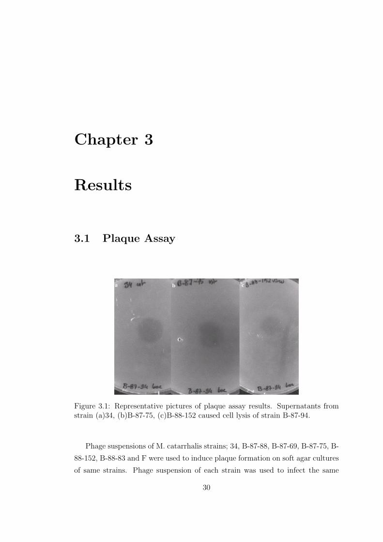
3.1 Plaque Assay
Figure 3.1: Representative pictures of plaque assay results. Supernatants fromstrain (a)34, (b)B-87-75, (c)B-88-152 caused cell lysis of strain B-87-94.
Phage suspensions of M. catarrhalis strains; 34, B-87-88, B-87-69, B-87-75, B-
88-152, B-88-83 and F were used to induce plaque formation on soft agar cultures
of same strains. Phage suspension of each strain was used to infect the same
30

strain and/or other strains (Figure 3.1). Plaque formation was observed on each
plate except two. However all possible combinations were not performed and not
all plaque assays were repeated.(Table 3.1)
Table 3.1: Summary of plaque assay results. Lytic effects were measured bydiameters of the clear zones produced on agar plates: +,< 15mm; ++, 15 to18mm; +++,> 18mm; N/A, not available; -, no plaque formation.
3.2 Phage Isolation
The particles isolated over 15% sucrose cushion and 30% sucrose cushion
were visualized by TEM. Several particles with different shapes and sizes were
copurified when 15% sucrose cushion was used. Particles resembling fimbriae
were co-purified with nonuniform vesicle-like particles and hexagonal particles
were copurified (Figure 3.2A ). When 30% sucrose cushion was used, only two
types of particles were copurified. Hexagonal particles with varying sizes were
co-purified with vesicle-like particles. However the vesicles had more uniform size
and shape compared to the previous isolate. With this method very big vesicle-
like particles and fimbriae-like particles were removed and the hexagonal particles
were obtained with higher concentration. Aggregations of particles were observed
frequently and the two types of particles were co-existed in these aggregations.
31
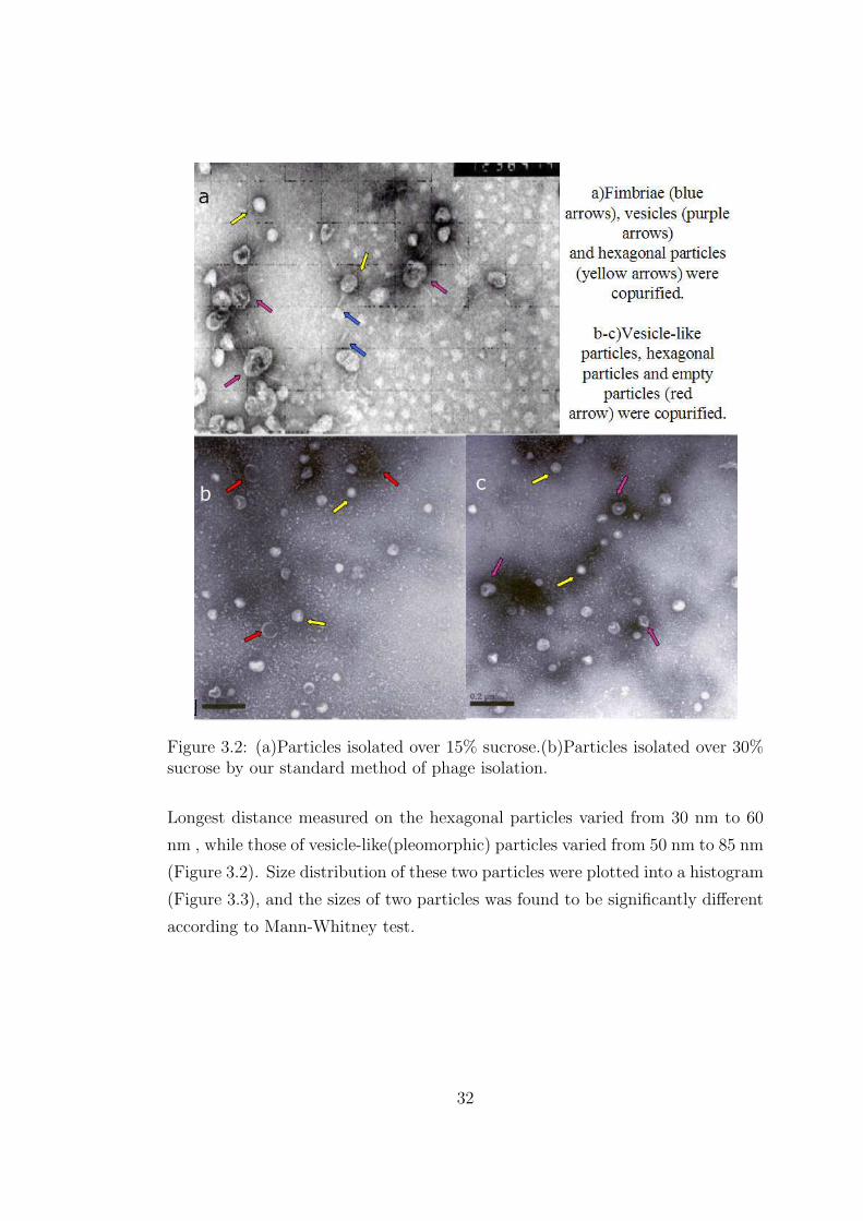
Figure 3.2: (a)Particles isolated over 15% sucrose.(b)Particles isolated over 30%sucrose by our standard method of phage isolation.
Longest distance measured on the hexagonal particles varied from 30 nm to 60
nm , while those of vesicle-like(pleomorphic) particles varied from 50 nm to 85 nm
(Figure 3.2). Size distribution of these two particles were plotted into a histogram
(Figure 3.3), and the sizes of two particles was found to be significantly different
according to Mann-Whitney test.
32
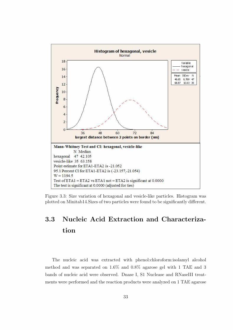
Figure 3.3: Size variation of hexagonal and vesicle-like particles. Histogram wasplotted on Minitab14.Sizes of two particles were found to be significantly different.
3.3 Nucleic Acid Extraction and Characteriza-
tion
The nucleic acid was extracted with phenol:chloroform:isolamyl alcohol
method and was separated on 1.6% and 0.8% agarose gel with 1 TAE and 3
bands of nucleic acid were observed. Dnase I, S1 Nuclease and RNaseIII treat-
ments were performed and the reaction products were analyzed on 1 TAE agarose
33

Figure 3.4: (a)DnaseI caused degradation of M.catarrhalis genomic DNA andhigh molecular weight nucleic acid extracted from phage particles.(b)RNaseIIIcaused degradation of rotavirus Wa genomic RNA(dsRNA) but phage RNA wasresistant to RNaseIII
gel to understand the nature of nucleic acids isolated. High molecular weight ( 9
kb) nucleic acid was found to be double stranded DNA since it was sensitive to
DNAseI (Figure 3.4A) and resistant to S1 (Figure 3.5) nuclease and RNAse III
(Figure 3.4B). However the smaller bands were found to be single-stranded RNA
since they were resistant to DNAse I and RNAse III treatment whereas they were
digested completely by S1 nuclease.
Particles isolated with different methods we used, all contained double-
stranded DNA together with single-stranded RNA with same mobility and band-
ing pattern as visualized on agarose gels. Particles isolated from M.catarrhalis
with different sucrose gradients also contained 3 bands of nucleic acid with the
same mobilities (Figure 3.6). It was hard to tell the molecular size of the ssRNA
bands by looking at their mobility in agarose gels since they do not have a con-
sistent speed. The relative mobility of the RNA bands varied a lot depending
on the concentration of the gel, run time and room temperature. When agarose
gel was prepared free of contaminating ribonucleases three RNA segments with
mobilities of 0.4kb, 0.7kb and 1.1kb were observed in every isolate (Figure 3.7
A).
TRI LS extracted RNA was visualized on 1.5% formaldehyde/agarose gels
34

Figure 3.5: S1 nuclease caused degradation of RNA extracted from mousespleen(totalRNA), and low molecular weight nucleic acids extracted from phageparticles. gDNA:genomic DNA, Wa: Rotavirus Wa strain genomic RNA(dsRNA), totalRNA: total RNA extracted from mouse spleen, phage:nucleic acidextracted from isolated phage particles.
and 3 RNA segments with sizes of 0.5kb, 1.5kb and 2.4kb compared to RNA
millennium markers were observed (Figure 3.7 B).
3.4 Ligation, Reverse Transcription and PCR
Ligation products were reverse transcribed with primer B in presence of cap-
switch oligo, and PCR was performed with primerB and 5’PCR primer. PCR
products were visualized on 1.5% agarose gel in 1TAE. All PCRs performed with
KlenTaq polymerase mix yielded a smear, which did not contain any band/bands.
Only in one condition banding was observed, the products were combined and
cleaned with PCR purification kit. The purified product formed a smear on 1.5%
agarose gel, and did not yield any bands. PCRs performed with Taq polymerase
did not yield any smear or product , but only the primer dimers (Figure 3.8).
35

Figure 3.6: a)- Agarose gel in 1xTAE. lane1;Phage particles were isolated withstandard method and nucleic acid was phenol extracted. lane2;HindIII digestwas loaded as molecular size standard. b) lane1; M8 as molecular weight marker.lane2-7;Phage particles isolated from different fractions of sucrose gradients, nu-cleic acid was phenol extracted .
3.5 Comparison of Different Strains
Our standard method for purification of bacteriophage particles was used to
isolate bacteriophage from other pathogenic and non-pathogenic strains we had.
Although no visible pellet was obtained from non-pathogenic strains visible pel-
lets were obtained from pathogenic strains, after centrifugation over 30% sucrose
cushion. However the experiment was carried on as if pellets were visible in all
strains, and nucleic acid was extracted and visualized on ribonuclease-free agarose
gel. All strains yielded a high molecular weight band and 3 or more low molecu-
lar weight band as visualized on 1.6% agarose gel prepared free of contaminating
ribonucleases (Figure 3.9).
36
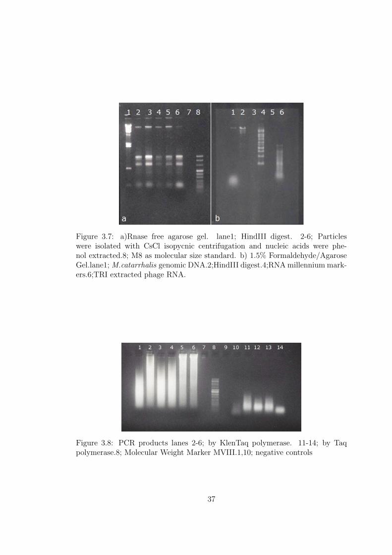
Figure 3.7: a)Rnase free agarose gel. lane1; HindIII digest. 2-6; Particleswere isolated with CsCl isopycnic centrifugation and nucleic acids were phe-nol extracted.8; M8 as molecular size standard. b) 1.5% Formaldehyde/AgaroseGel.lane1; M.catarrhalis genomic DNA.2;HindIII digest.4;RNA millennium mark-ers.6;TRI extracted phage RNA.
Figure 3.8: PCR products lanes 2-6; by KlenTaq polymerase. 11-14; by Taqpolymerase.8; Molecular Weight Marker MVIII.1,10; negative controls
37

Figure 3.9: Nucleic acids isolated from phage and vesicles of pathogenic andnonpathogenic strains were loaded to the gel.Lanes (1-5): 34,B-87-75, B-88-83,B-87-94, B-88-133; (8-14): B-91-6, B-91-7, B-91-9, B-91-22, B-91-25, B-91-26,B-91-27 (Samples were diluted 10-60 times).Lanes (6-7): HindIII Digest, MVIIIas molecular weight markers.
38

Chapter 4
Discussion
Bacteriophages are the most abundant life forms in the biosphere. Thousands
of varieties of phage exist, each of which may infect only one type or a few
types of bacteria. Even though no bacteriophage from M.catarrhalis has been
yet defined, it is very unlikely that it would not have one. In this study, evidence
for a bacteriophage from M.catarrhalis is presented.
Supernatants ofM.catarrhalis broth cultures were shown to cause bacterial cell
lysis on soft agar cultures, indicating presence of bacteriophages in them. Two
particles having different morphologies (hexagonal and pleopmorphic(vesicle-like)
and different size ranges (p<0. 05) were co-purified from these supernatants.
Two methods for isolation of bacteriophage particles were used. The 15% sucrose
cushion method yielded an impure isolate containing low number of hexagonal
phage particles, however the 30% sucrose cushion method yielded purer isolate
containing larger number of hexagonal phage particles due to increased sucrose
concentration and usage of broth culture as starting material. The sizes of the
two particles were represented by the largest distance between two points on
these particles. However this may not reflect the real sizes of the particles, and
the measurement should be improved by including multiple distances on each
particle and combining those in a more complex analysis.
39
One segment of dsDNA molecule and three segments of ssRNA molecules
were extracted from the isolated particles. Since several attempts to separate
two particles from each other failed, it was not possible to determine the origins
of RNA and DNA molecules. These two particles are believed to attract each
other and form aggregates of different sizes, since small and large aggregates
containing the two particles together were visualized under electron microscope.
Most likely, the aggregates of various sizes have different densities therefore they
are collected from different fractions of the sucrose and CsCl gradients. Usage of
another buffer during phage isolation (instead of PBS- or SM) which can block
the attraction between the hexagonal and pleomorphic(vesicle-like) particles will
resolve this problem.
Optimization of RNA sequencing reactions also are performed. There might
be several reasons causing failure of RNA sequencing trials. Ligation of linker
to template can be improved by using phosphatases to prevent self ligation and
concatenation of the RNA template, and also by changing linker/template ra-
tio, incubation time and temperature. Moreover the cDNA synthesis and PCR
amplification should be optimized.
The nucleic acid content particles isolated from pathogenic and non-
pathogenic strains also indicated differences in quantity suggesting possible in-
volvement of the identified particles in the pathogenesis, which has to be investi-
gated further.
Although sufficient information to determine the classification of the phages
isolated in this study is not yet available, apparently the morphology and nucleic
acids of these particles are not extraordinary. Currently, International Commit-
tee for taxonomy of viruses (ICTV) classifies phages into 13 families, and 30
genera (Regenmortel et al., 2000). Phage families are chiefly defined by particle
morphology and nature of nucleic acid. Phages with different morphologies have
been discovered including tailed, filamentous, polyhedral or pleomorphic phages
(Ackermann, 2003). In this study polyhedral (hexagonal) particles and vesicle-
like pleomorphic particles were co-purified. Polyhedral phages which have been
identified until now contain ss/dsDNA in circular, linear or supercoiled form;
40
single-stranded linear RNA; or double-stranded segmented linear RNA (Acker-
mann, 2003).(Table 4.1) The polyhedral phages have been classified into 5 fami-
lies consist of corticoviridae, leviviridae, microviridae, podoviridae and tectiviri-
dae(Figure 4.1) .
Figure 4.1: Schematic Representation of Major Phage Groups (Ackermann 2003).
In order to determine whether the polyhedral bacteriophages released from
M.catarrhalis belong to one of these families, these particles need to be isolated
and the nature of nucleic acid in them needs to be defined. Pleomorphic bac-
teriophages yet discovered all contain ds, supercoiled or circular DNA and they
are grouped into two families being plasmaviridae and fuselloviridae. Plasmaviri-
dae virions are enveloped, their shapes are from spherical to pleomorphic; and
they are 50 to 125 nm in diameter with a loose fitting membrane (baggy mem-
brane). A regular capsid structure is not detectable (ICTVdB descriptions). The
vesicle-like particles isolated in this study resemble plasmaviridae since they have
41
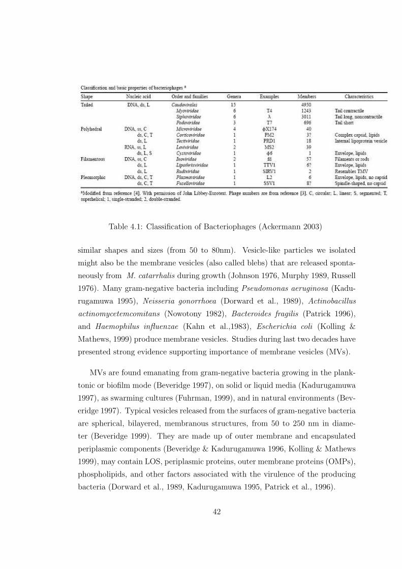
Table 4.1: Classification of Bacteriophages (Ackermann 2003)
similar shapes and sizes (from 50 to 80nm). Vesicle-like particles we isolated
might also be the membrane vesicles (also called blebs) that are released sponta-
neously from M. catarrhalis during growth (Johnson 1976, Murphy 1989, Russell
1976). Many gram-negative bacteria including Pseudomonas aeruginosa (Kadu-
rugamuwa 1995), Neisseria gonorrhoea (Dorward et al., 1989), Actinobacillus
actinomycetemcomitans (Nowotony 1982), Bacteroides fragilis (Patrick 1996),
and Haemophilus influenzae (Kahn et al.,1983), Escherichia coli (Kolling &
Mathews, 1999) produce membrane vesicles. Studies during last two decades have
presented strong evidence supporting importance of membrane vesicles (MVs).
MVs are found emanating from gram-negative bacteria growing in the plank-
tonic or biofilm mode (Beveridge 1997), on solid or liquid media (Kadurugamuwa
1997), as swarming cultures (Fuhrman, 1999), and in natural environments (Bev-
eridge 1997). Typical vesicles released from the surfaces of gram-negative bacteria
are spherical, bilayered, membranous structures, from 50 to 250 nm in diame-
ter (Beveridge 1999). They are made up of outer membrane and encapsulated
periplasmic components (Beveridge & Kadurugamuwa 1996, Kolling & Mathews
1999), may contain LOS, periplasmic proteins, outer membrane proteins (OMPs),
phospholipids, and other factors associated with the virulence of the producing
bacteria (Dorward et al., 1989, Kadurugamuwa 1995, Patrick et al., 1996).
42
Interestingly, vesicles have been shown to carry DNA and deliver it to other
bacteria (Kadurugamuwa 1995, Patrick 1996). Specific DNA-binding peptides
were reported to be present in vesicle lysates of H. influenzae vesicles (Kahn
1982, Kahn 1983) and to be associated with vesicles from Neisseria gonorrhoea
(Dorward et al., 1989). Dorward et al. has shown that vesicles released by
N. gonorrhoea harbor both linear and circular DNA, including 4.2- and 7.1-kb
plasmids; and MV-mediated transfer of an antibiotic resistance plasmid can occur
between two strains of N. gonorrhoea (Dorward et al.,1989).Vesicles produced by
Pseudomonas aeruginosa were reported to contain DNA (Kadurugamuwa 1995).
Chromosomal and bacteriophage-associated virulence genes were detected in
Escherichia coli O157:H7 vesicles (Kolling & Mathews 1999). Yaron et. al.
demonstrated that E. coli O157:H7 vesicles contain genes of chromosomal, plas-
mid and phage origin; and vesicles mediate the transfer of these virulence genes,
which are subsequently expressed by recipient enteric bacteria (E. coli JM109 and
Salmonella serovar Enteritidis) (Yaron et al., 2000). Incubation of MVs from E.
coli O157 : H7, which carried ampicillin resistance (AmpR) and green fluorescent
protein genes on a plasmid (pGFP), with E. coli lacking this plasmid resulted in
fluorescent transformants, which also suggests that E. coli MVs act as vectors of
DNA transport (Yaron et al., 2000).
There are certain advantages for DNA to encapsulate itself in MVs. DNA
would be protected from exonucleases. Furthermore, MVs should also enhance
the efficiency of DNA delivery and uptake into a recipient cell as they fuse into
the outer membrane (OM). Because naturally competent bacteria are genetically
programmed to permit the efficient uptake of macromolecular DNA (Dubnau,
1999) and non-competent bacteria lack such a mechanism, MVs could possibly
overcome such genetic barriers.
MVs of M.catarrhalis were not previously investigated for presence of DNA
in them; however our findings support this possibility. If the DNA molecule we
isolated is actually encapsulated in blebs, this might have important function in
horizontal transfer of virulence genes. Three main mechanisms of gene transfer
have been identified in bacteria; transformation, conjugation and transduction (as
43

explained in introduction). Possibly, membrane vesicles constitute an alternative
mode of gene transfer among bacteria (Yaron et al., 2000). MVs of M.catarrhalis
may aid in transformation of this organism and spread of virulence genes such as
bro β-lactamases.
Several findings (as explained in introduction) implicate horizontal gene trans-
fer in the acquisition of the bro β-lactamases (McGregor, 1998). The suspected
location of the bro genes on the chromosome and the high efficiency of trans-
fer tend to support theory of a conjugal transposon as a method for spread of
resistance (Wallace1989). Although there are indications suggesting presence of
conjugative process in transfer of bro genes, transfer factors required for conju-
gation have not been reported in this organism (McGregor 1998). Transforma-
tion has been suggested as a mechanism for interbacterial transfer of bro genes
(Catlin,1990). Transformation of streptomycin resistance is known to occur be-
tween M. catarrhalis and M. nonliquefaciens strains (Catlin 1964). Transforma-
tion by naked DNA is unlikely to occur between M. catarrhalis strains since
this organism secretes very active endonuclease to its environment (Kamme et
al., 1984). Transformation mediated by MVs containing DNA would protect the
DNA from endogenous and exogenous nucleases.
MVs have been shown to be important in bacterial pathogenesis in various
ways, therefore the MVs of M. catarrhalis should be investigated in detail to
elucidate their role in pathogenesis of this organism. Since the knowledge regard-
ing pathogenic mechanisms and virulence factors of this organism is scarce, the
information on MVs might become very significant.
Vesicles facilitate pathogenesis of bacteria by improving attachment to eukary-
otic cells. Grenier and Mayrand (1987) demonstrated that B. gingivalis vesicles
adhere to epithelial cells and act as an intermediate for the attachment of bacteria
to host cells. Vesicles produced by A. actinomycetemcomitans were reported to
promote the adherence of the parent cell to epithelial cells (Meyer 1993). Con-
tribution of vesicles to the bacterial pathogenesis is proposed to be more than
improving adherence only. Since vesicles have ability to integrate into the mem-
branes of foreign bacteria and to adhere to or be engulfed by eukaryotic cells and
44
integrate into the membranes, they are believed to function as secretory vesicles
involved in delivery of virulence factors to heterologous and homologous bacteria
or to host cells (Beveridge 1999, Kadurugamuwa 1999). In fact, virulence factors
associated with the parent strain, including proteases, phospholipases, autolysin,
hemolysins, and Shiga toxins, have been isolated from vesicles (Beveridge 1999,
Kadurugamuwa &Beveridge 1995, Kolling & Mathews 1999, Li 1998). The MVs
of P. aeruginosa, Proteus mirabilis, and Serratia marcescens package phospholi-
pase C, proteases, proelastase, and hemolysins (Beveridge 1999). It is probable
that MVs from Borrelia, enterotoxigenic E. coli, Haemophilus, Neisseria, and
Vibrio species also contribute to the virulence of these other pathogens (Bev-
eridge 1999). Research suggests that vesicles released by other pathogens possess
enzymatic and toxic activity towards prokaryotic and eukaryotic cells (Patrick et
al. 1996, Wai et al. 1995). Vesicles released from P. aeruginosa are able to fuse
with the membranes of gram-negative and gram-positive organisms, whereupon
they release autolysins, resulting in cell lysis of the targeted organism (Kaduruga-
muwa 1996, Li 1996). Lysis of competing bacteria, for instance, would provide
nutrients for the vesicle-producing strain and limit competition from other bac-
teria.
Recently, it has been discovered that a chromosome-encoded β-lactamase in
P. aeruginosa can also be packaged into MVs (Ciofu et al., 2000). This raises the
intriguing possibility that β-lactamase-containing MVs could be discharged from
pathogens at their infection sites in tissue to increase the breakdown of β-lactam
antibiotics in the local tissue environment. Because MVs contain porins as part of
their OMP complement, the antibiotic readily diffuses into these MVs and is in-
activated. This could be a general strategy for reducing antibiotic concentrations
at infection sites, and it could also be one of the ways in which chronic, hard-to-
treat infections (such as cystic fibrosis) are maintained by the infecting bacterium
(Ciofu et al., 2000). Since MVs can readily fuse to the outer membranes of a wide
range of other gram-negative bacterial pathogens (Kadurugamuwa 1999), thereby
emptying their luminal contents into the periplasm of recipient cells, β-lactamase
(and other antibiotic-inactivating enzymes) could be physically transferred from
a donor cell to a non-β-lactamase-producing recipient (e.g., following the cystic
45
fibrosis analogy, from P. aeruginosa [donor] to Burkholderia cepacia [recipient]).
In this way, a varied group of gram-negative non-producers in close proximity
to a donor cell could withstand antibiotic treatment and contribute to the in-
fection without these same bacteria having the genetic capacity to produce the
inactivating enzyme themselves (Beverigde 1999). Transportation of the active
β-lactamase to β-lactamase sensitive strains may explain the high rate of β-
lactamase producer M. catarrhalis strains found in clinical studies.
46
Chapter 5
Future Perspectives
Here we presented evidence for a novel bacteriophage from M. catarrhalis,
however investigation should continue in order to demonstrate the presence of this
phage. Number of questions have to be answered, with new sets of experiments.
First of all, the two types of particles have to be isolated, and the nucleic acid
content of each has to be determined.
Second of all, the DNA and RNA have to be sequenced. If our current method
of RNA sequencing will not yield results, reverse transcription with random
primers will be used to form a cDNA library of the RNA. This cDNA library
can be sequenced with conventional methods, and the complete sequence can be
retrieved by alignment of the partial sequences. For sequencing of DNA, a ge-
nomic library has to be formed using at least two different restriction enzymes
cutting the DNA in sufficient number of locations. Once the genomic library
is formed, each clone will be sequenced and the complete sequence will be ob-
tained by alignment of all partial sequences. Sequences of the RNA and DNA are
valuable to determine the nature of the particles we isolated. Presence of phage
genes on these nucleic acids will ultimately prove the presence of bacteriophage in
M.catarrhalis. Moreover, the sequence homology searches and phylogenetic tree
analysis will let us comment on classification of the phage(s). The sequence data
47
will let us understand the nature of the vesicle-like particles. If the vesicle-like
particles are the MVs, and they contain the DNA; this DNA most likely carries
important virulence genes. The proteins and enzymes carried on or in these MVs
are also candidate virulence factors of M.catarrhalis, and one can investigate the
importance of them. In addition, the biological properties of the phage such as
burst size, growth curve, life cycle have to be studied. Potential of phage in phage
therapy has to be considered in the future.
48
Chapter 6
References
Ackermann HW. Bacteriophage observations and evolution Res Microbiol.
2003. 154(4):245-51.
Ackermann, HW., Dubow M.S. General properties of bacteriophages. In.
Viruses of prokaryotes, vol. 1 CRC Press, Inc., Boca Raton, Fla.1987.
Aebi C, LaFontaine ER, Cope LD, Latimer JL, Lumbley SL, McCraken
GH, Hansen EJ. Phenotypic effect of isogenic uspA1 and uspA2 mutations on
Moraxella catarrhalis strainO35E. Infect Immun.1998. 66: 3113-9.
Ahmed K. Fimbriae of Branhamella catarrhalis as possible mediator of adher-
ence to pharyngeal epithelial cells. Acta Pathol Microbiol Immunol Scand.1992a.
100:1066-107.2
Ahmed K, Masaki H, Dai TC, Ichinose A, Utsunomiya Y, Tao M, Nagatake
T, Matsumoto K. Expression of fimbriae and host response in Branhamella
catarrhalis respiratory infections. Microbiol Immunol. 1994. 38(10):767-71.
Ahmed K, Matsumato K. 1996. Attachment of Moraxella catarrhalis to pha-
ryngeal epithelial cells is mediated by glycosphingolipid receptor. FEMS Micro-
bial.Lett. 135: 305-309.
49
Ahmed K, Nagawaka T, Nakano Y, Martinez G, Ichinose A, Zheng CH, Akaki
M, Aikawa M, Nagatake T. Attachment of Moraxella catarrhalis occurs to the
positively charged domains of pharyngeal epithelial cells. Microb Pathog.2000.
28: 203-209
Ahmed K, Rikitomi N, Nagatake T, Matsumoto K. 1990. Electron microscopic
observation of Branhamella catarrhalis. Microbiol Immunol. 34: 967-975
Ahmed K, Rikitomi N and Matsumoto K. 1992. Fimbriation, hemagglutina-
tion and adherence properties of fresh clinical isolates of Branhamella catarrhalis.
Microbiol Immunol. 36:1009-1017
Ahmed K, Suzuki Y, Miyamoto D, Nagatake T. Asialo-GM1 and asialo-GM2
are putative adhesion molecules for Moraxella catarrhalis Med Microbiol Immunol
(Berl). 2002. 191(1):5-10.
Arola M, Ruuskanen O, Ziegler T, Mertsola J, Nanto-Salonen K, Putto-
Laurila A, Viljanen M K, Halonen P. Clinical role of respiratory virus infection
in acute otitis media. Pediatrics 1990. 86 :848-855.
Baba T, Takeuchi F, Kuroda M, Yuzawa H, Aoki K, Oguchi A, Nagai Y,
Iwama N, Asano K, Naimi T et al. Genome and virulence determinants of high
virulence community-acquired MRSA. Lancet 2002. 359:1819-1827.
Baquero F, Barrett JF, Courvalin P, Morrissey I, Piddock L, Novick WJ.
Epidemiology and mechanisms of resistance among respiratory tract pathogens.
Clin. Microbiol. Infect. 1998. 4(Suppl.2):19-26.
Barbian KD and Minnick MF. A bacteriophage-like particle from Bartonella
bacilliformis. Microbiol. 2000. 146:599-609.
Barksdale, L. and Arden, S.B. Persisting bacteriophage infections, lysogeny,
and phage conversions. Annu. Rev. Microbiol. 1974.28: 265-299
Benchetrit, L. C., E. D. Gray, and L. W. Wannamaker. Hyaluronidase activity
of bacteriophages of group A streptococci. Infect. Immun. 1977.15:527-532.
50
Benchetrit, L. C., E. D. Gray, R. D. Edstrom, and L. W. Wannamaker. Pu-
rification and characterization of a hyaluronidase associated with a temperate
bacteriophage of group A, type 49 streptococci. J. Bacteriol. 1978. 134:221-228.
Bensing, B. A., Rubens C. E., Sullam P.M. Genetic loci of Streptococcus mitis
that mediate binding to human platelets. Infect. Immun. 2001a. 69:1373-1380.
Bensing, B. A., Siboo I.R., Sullam P.M. Proteins PblA and PblB of Strepto-
coccus mitis, which promote binding to human platelets, are encoded within a
lysogenic bacteriophage. Infect. Immun.2001b. 69:6186-6192.
Beres SB, Sylva GL, Barbian KD, Lei B, Hoff JS, Mammarella ND,Liu MY,
Smoot JC, Porcella SF, Parkins LD et al. Genome sequence of a serotype M3
strain of group A Streptococcus : phage-encoded toxins, the high-virulence phe-
notype, and clone emergence. Proc Natl Acad Sci USA 2002. 99:10078-10083.
Betley, M.J. and Mekalanos, J.J. Staphylococcal enterotoxin A is encoded by
a phage. Science 1985. 229: 185-187.
Beveridge, T. J., and J. L. Kadurugamuwa. Periplasm, periplasmic spaces,
and their relation to bacterial wall structure: novel secretion of selected periplas-
mic proteins from Pseudomonas aeruginosa. Microb. Drug Resist. 1996. 2:1-8.
Beveridge, T. J. Structures of gram-negative cell walls and their derived mem-
brane vesicles. J. Bacteriol. 1999.181:4725-4733
Bootsma HJ, van Dijk H, Verhoef J, Fleer A, Mooi FR. Molecular characteri-
zation of the BRO b-lactamases of Moraxella (Branhamella) catarrhalis. Antimi-
crob. Agents Chemother. 1996. 40:966-972.
Boyd EF, Waldor MK. Evolutionary and functional analyses of variants of
the toxin-coregulated pilus protein TcpA from toxigenic Vibrio cholerae. non-
O1/non-O139 serogroup isolates Microbiology. 2002.148(6):1655-66.
Brorson JE, Axelsson A, Holm SE. 1976. Studies on Branhamella catarrhalis
with special reference to maxillary sinusitis. Scand J Infect Dis. 8:151-155
51
Buu Hoi-Dang Van A, Brive-Le Bouquence B, Barthelemy M, Labia R. Novel
b-lactamases from Branhamella catarrhalis. Ann. Microbiol. 1978. 129B: 6-10.
Canchaya C, Fournous G, Chibani-Chennoufi S, Dillmann ML, Brussow H.
Phage as agents of lateral gene transfer. Curr Opin Microbiol. 2003. 6(4):417-24.
Caparon, M. 2000. Genetics of group A streptococci , p. 53-64. In V. A.
Fischetti, R. P. Novick, J. J. Ferretti, D. A. Portnoy, and J. I. Rood (ed.), Gram-
positive pathogens. ASM Press, Washington, D.C.
Catlin BW . Reciprocal genetic transformation between Neisseria catarrhalis
and Moraxella nonliquefaciens. Journal of General Microbiology 1964. 37 :369-
379.
Catlin, BW. Branhamella catarrhalis : an organism gaining respect as a
pathogen. Clin Microbiol Rev 1990: 3; 293-320
Christensen, JJ. 1999. Branhamella catarrhalis : clinicalmicrobiological and
immunological features in lower respiratory tract infections. APMIS Suppl 88:1-
36.
Ciofu O, Beveridge TJ, Kadurugamuwa J, Walther-Rasmussen J, Hoiby N.
Chromosomal beta-lactamase is packaged into membrane vesicles and secreted
from Pseudomonas aeruginosa. Antimicrob Chemother. 2000 Jan;45(1):9-13.
Clavo-Sanchez AZ, Giron-Gonzalez JA, Lopez-Prieto D, Canueto-Quintero
J, Sanchez-Porto A, Vergara-Campos A, Marin-Casanova P, Cordoba-Dona JA.
Multivariate analysis of risk factors for infection due to penicillin resistant and
multi-drug resistant Streptococcus pneumoniae: a multicenter study. Clin Infect
Dis. 1997. 24: 1052-1059.
Cocchi P, Ulivelli A. Meningitis caused by Neisseria catarrhalis. Acta Pedi-
atrica Scandanavia 1968. 57 : 451-453.
Cope LD, Lafontaine RTSlaughter CA, HasemannJr CA, Aebi C, Henderson
FW, Mc CrackenGH Jr, Hansen EJ. Characterization of the Moraxella catarrhalis
52
UspA1 and UspA2 genes and their encoded products. J Bacteriol 1999.181: 4026-
4034
Craig DB, Wehrle PA. Branhamella catarrhalis septic arthritis. J. Rheumatol.
1983. 10 : 985-986.
Daoud A, Abuekteish F, Masaadeh H. Neonatal meningitis due to Moraxella
catarrhalis and review of the literature. Ann. Trop. Paed. 1996.16 : 199-201.
Diamond LA, Lorber B. Branhamella catarrhalis pneumonia and im-
munoglobulin abnormalities: a new association? Am. Rev. Resp. Dis. 1984.
129:876-878.
Doern GV, Miller MJ, Winn RE. Branhamella (Neisseria) catarrhalis sys-
temic disease in humans. case reports and review of the literature. Arch. Inter.
Med. 1981. 141:1690-1692.
Dorward, D. W., C. F. Garon, and R. C. Judd. Export and intercellular
transfer of DNA via membrane blebs of Neisseria gonorrhoeae. J. Bacteriol.
1989. 171:2499-2505.
Douer D, Danziger Y, Pinkhas J. Neisseria catarrhalis endocarditis. Ann
Intern Med. 1977. 86 :116.
Dubnau D. DNA uptake in bacteria Annu Rev Microbiol. 1999. 53:217-44.
Elbasnier AM, Deshpande H. Recurrent urinary tract infection with haematuria
caused by Moraxella (Branhamella) catarrhalis. J. Infect. 1993. 27 :216-217.
Eliasson I, Kamme C, Vang M, Waley SG. Characterization of cell-bound
papain-soluble β-lactamases in BRO-1 and BRO-2 producing strains of Moraxella
(Branhamella) catarrhalis and Moraxella nonliquefaciens. Eur J Clin Microbiol
Infect Dis 1992. 11:313-321.
Elkins C, Thomas CE, Seifert HS, Sparling PF. Species-specific uptake of
DNA by gonococci is mediated by a 10-base-pair sequence. J Bacteriol. 1991.
173(12):3911-3.
53
Enright MC, McKenzie H. Moraxella (Branhamella) catarrhalis–clinical and
molecular aspects of a rediscovered pathogen. J Med Microbiol. 1997
May.46(5):360-71.
Farmer T, Reading C. β-lactamases of Branhamella catarrhalis and their in-
hibition by clavulanic acid. Antimicrob Agents Chemother 1982. 21:506-508.
Farrant, J. L., A. Sansone, J. R. Canvin, M. J. Pallen, P. R. Langford, T.
S. Wallis, G. Dougan, and J. S. Kroll. Bacterial copper- and zinccofactored
superoxide dismutase contributes to the pathogenesis of systemic salmonellosis.
Mol. Microbiol. 1997. 25:785-796.
Feign RD, San Joaqin V, Middelkamp JN. Purpura fulminans associated with
Neisseria catarrhalis septicemia and meningitis. Pediatrics 1969. 44 :120-123.
Felmingham D.,Washington J. Trends in the antimicrobial susceptibility of
bacterial respiratory tract pathogens- findings of the Alexander Project 1992-
1996. J. Chemother. 1999. 11(Suppl. 1):5-21.
Figueroa-Bossi, N., and L. Bossi. Inducible prophages contribute to
Salmonella virulence in mice. Mol. Microbiol. 1999. 33:167-176.
Finkelstein, R. A. 1992. Cholera enterotoxin (choleragen): a historical per-
spective. perspective, p. 155-187. In D. Barua and W. B. Greenough III (ed.),
Cholera. Plenum Press, New York, N.Y.
Firth, N., and R. A. Skurray. 2000. Genetics: accessory elements and genetic
exchange , p. 326-338. In V. A. Fischetti, R. P. Novick, J. J. Ferretti, D. A.
Portnoy, and J. I. Rood (ed.), Gram-positive pathogens. ASM Press,Washington,
D.C.
Frobisher, M., and J. Brown. Transmissible toxicogenicity of streptococci.
Bull. Johns Hopkins Hosp.1927. 41:167-173.
Frosch P.W.1896.Kolle, Die Mikrokokken, in die Mikroorganism. Vol2 Flugge,
Verlag von Vogel, Leipzig. 154-155.
54
Fuhrman JA. Marine viruses and their biogeochemical and ecological effects
Nature. 1999 Jun 10;399(6736):541-8. Review.
Goshorn, S.C. and Schlievert, P.M. Bacteriophage association of streptococcal
pyrogenic exotoxin type C. J. Bacteriol. 1989. 171: 3068-3073.
Groisman EA, Ochman H. Pathogenicity islands: bacterial evolution in quan-
tum leaps. Cell. 1996. 87(5):791-4.
Gur D, Ozalp M, Sumerkan B, Kaygusuz A, Toreci K, Koksal I, Over U, Soyle-
tir G. Prevalence of antimicrobial resistance in Haemophilus influenzae, Strepto-
coccus pneumoniae, Moraxella catarrhalis and Streptococcus pyogenes : results of
a multicentre study in Turkey. International J Antimicrob Agents 2002. 19:207-
211.
Halperin, S. A., P. Ferrieri, E. D. Gray, E. L. Kaplan, and L. W. Wannamaker.
Antibody response to bacteriophage hyaluronidase in acute glomerulonephritis
after group A streptococcal infection. J. Infect. Dis. 1987.155:253-261.
Hardt, W. D., L. M. Chen, K. E. Schuebel, X. R. Bustelo, and J. E. Galan.
S. typhimurium encodes an activator of Rho GTPases that induces membrane
ruffling and nuclear responses in host cells. Cell 1998a. 93:815-826.
Hardt, W. D., H. Urlaub, and J. E. Galan. A substrate of the centisome
63 type III protein secretion system of Salmonella typhimurium is encoded by a
cryptic bacteriophage. Proc. Natl. Acad. Sci. USA 1998b. 95:2574-2579.
Harkness RE, Guimond MJ, McBey BA, Klein MH, Percy DH, Croy BA.
1993. Branhamella catarrhalis pathogenesis in SCID and SCID/ beige mice.
Brief report. APMIS 101:805-810
Hoban DJ, Doern GV, Fluit AC, Roussel-Delvallez M, Jones RN. Worldwide
prevalence of antimicrobial resistance in Streptococcus pneumoniae, Haemophilus
influenzae, and Moraxella catarrhalis in the SENTRY antimicrobial surveillance
program, 1997-1999. Clin Infect Dis 2001. 32(Suppl. 2):S81-93.
55
Hoi-Dang A.B., Brive-Le Bouguenec C., Barthelemy M. ,Labia R. Novel beta-
lactamase from Branhamella catarrhalis. Ann. Microbiol. 1978. 129:397-406.
Huang, A. et al. Characterization of a bacteriophage that carries the genes
for production of Shiga-like toxin 1 in Escherichia coli. J. Bacteriol. 1987. 169:
4308-4312.
Hueck, C. J. Type III protein secretion systems in bacterial pathogens of
animals and plants. Microbiol. Mol. Biol. Rev. 1998. 62:379-433.
Hyder, S. L., and M. M. Streitfeld. Transfer of erythromycin resistance from
clinically isolated lysogenic strains of Streptococcus pyogenes via their endogenous
phage. J. Infect. Dis. 1978. 138: 281-286.
Hynes, W. L., and J. J. Ferretti. Sequence analysis and expression in Es-
cherichia coli of the hyaluronidase gene of Streptococcus pyogenes bacteriophage
H4489A. Infect. Immun 1989. 57:533-539.
Jiang SC, Paul JH. Gene transfer by transduction in the marine environment
Appl Environ Microbiol. 1998. 64(8):2780-7.
Johnson KG, McDonald IJ, Perry MB Studies on the cellular and free
lipopolysaccharides from Branhamella catarrhalis. Can. J. Microbiol.1976. 22
:460-467.
Johnson, L. P., Schlievert P.M. Group A streptococcal phage T12 carries the
structural gene for pyrogenic exotoxin type A. Mol. Gen. Genet. 1984. 194:52-56.
Johnson, M.A.,Drew, W.L. and Roberts, M. (1981) Branhamella catarrhalis
a lower respiratory tract pathogen? J. Clin. Microbiol. 13:1066-9.
Kadurugamuwa, J. L., and T. J. Beveridge. Virulence factors are released from
Pseudomonas aeruginosa in association with membrane vesicles during normal
growth and exposure to gentamicin: a novel mechanism of enzyme secretion. J.
Bacteriol. 1995. 177:3998-4008.
56
Kadurugamuwa, J. L., and T. J. Beveridge. Bacteriolytic effect of mem-
brane vesicles isolated from Pseudomonas aeruginosa on other bacteria including
pathogens: conceptually new antibiotics. J. Bacteriol. 1996. 178:2767-2774.
Kahn, M. E., G. Maul, and S. H. Goodgal. Possible mechanism for donor
DNA binding and transport in Haemophilus. Proc. Natl. Acad. Sci.USA 1982.
79:6370-6374.
Kahn, M. E., F. Barany, and H. O. Smith. Transformasomes: specialized
membranous structures that protect DNA during Haemophilus transformation.
Proc. Natl. Acad. Sci. USA 1983. 80:6927-6931.
Kamme C, Wang M, Stahl S. Intrageneric and intergeneric transfer of
Branhamella catarrhalis beta-lactamase production Scand J Infect Dis. 1984.
16(2):153-5.
Kaneko, J., T. Kimura, Y. Kawakami, T. Tomita, and Y. Kamio. Panton-
valentine leukocidin genes in a phage-like particle isolated from mitomycin C-
treated Staphylococcus aureus V8 (ATCC 49775). Biosci. Biotechnol.Biochem.
1997.61:1960-1962.
Karalus R, Campagnari A. M. Catarrhalis :a review of an important human
mucosal pathogen Microb Infect. 2000. 2: 547-559.
Karaolis, D.K. et al. A Vibrio cholerae pathogenicity island associated with
epidemic and pandemic strains. Proc. Natl. Acad. Sci USA 1998. 95:3134-3139.
Karaolis, D.K. et al. A bacteriophage encoding a pathogenicity island, a type-
IV pilus and a phage receptor in cholera bacteria. Nature 1999. 399: 375-379.
Kolling, G. L., and K. R. Matthews. Export of virulence genes and Shiga toxin
by membrane vesicles of Escherichia coli O157:H7. Appl. Environ. Microbiol.
1999. 65:1843-1848.
Kovach, M.E. et al. A putative integrase gene defines the distal end of a large
cluster of ToxR-regulated colonization genes in Vibrio cholerae. Microbiology
57

1996. 142: 2165-2174.
Lenski, R. E. Dynamics of interactions between bacteria and virulent bacte-
riophage Adv. Microb.Ecol. 1988. 10:1-44.
Li, Z., A. J. Clarke, and T. J. Beveridge. A major autolysin of Pseudomona
aeruginosa: subcellular distribution, potential role in cell growth and division,
and secretion in surface membrane vesicles. J. Bacteriol. 1996. 178:2479-2488.
Li, Z., A. J. Clarke, and T. J. Beveridge. Gram-negative bacteria produce
membrane vesicles which are capable of killing other bacteria. J.Bacteriol. 1998.
180:5478-5483.
Livermore DM. -lactamases in laboratory and clinical resistance. Clin Micro-
biol Rev 1995. 8:557-84.
London J, Kolenbrander PE. Coaggregation: enhancing colonization in a
fluctuating environment. In: Molecular and ecological diversity. Fletcher
M(Ed.)Wiley-Liss, 1996 New York, pp249-279.
Luke NR, Howlett AJ, Shao J, Campagnari A. Expression of Type IV Pili by
Moraxella catarrhalis Is Essential for Natural Competence and Is Affected by Iron
Limitation Infection and Immunity. 2004. 6262-6270. Madigan MT, Martinko
JM, Parker J. Essentials of Virology. In Brock Biology of Microorganisms 10th
edit. Parentice Hall.2003
Martinez G, Ahmed K, Watanabe K, Tao M, and Nagatake T. Changes in
antimicrobial susceptibility to Moraxella catarrhalis over a ten year period J
Infect Chemother. 1998.4:139-141.
McNeely DJ, Kitchens CS, Kluge RM. Fatal Branhamella(Neisseria)
catarrhalis pneumonia in an immunodeficient host. Am. Rev. Resp. Dis. 1976.
114:399-402.
McGregor K., Chang B.J., Mee B.J. , Riley T.V. Moraxella catarrhalis : Clin-
ical Significance , Antimicrobial Susceptibility and BRO Beta-Lactamases Eur J
58
Clin Microbiol Infect Dis 1998. 17 :219-234.
McMichael JC. Vaccines for Moraxella catarrhalis Vaccine 2001. 19:101-107.
Meyer, D. H., and P. M. Fives-Taylor. Evidence that extracellular components
function in adherence of Actinobacillus actinomycetemcomitans to epithelial cells.
Infect. Immun. 1993. 61:4933-4936.
Mintz, E. D., T. Popovic, and P. A. Blake. 1994. Transmission of Vibrio
cholerae O1 , p. 345-356. In I. K. Wachsmuth, P. A. Blake, and O. Olsvik (ed.),
Vibrio cholerae and cholera: molecular to global perspectives. American Society
for Microbiology, Washington, D.C.
Mirold, S., W. Rabsch, M. Rohde, S. Stender, H. Tschape, H. Russmann, E.
Igwe, and W. D. Hardt. Isolation of a temperate bacteriophage encoding the
type III effector protein SopE from an epidemic Salmonella typhimurium strain.
Proc. Natl. Acad. Sci. USA 1999. 96:9845-9850.
Moebus, K. Investigations of the marine lysogenic bacterium H24. II. Devel-
opment of pseudolysogeny in nutrient rich broth Mar. Ecol. Prog. Ser. 1997.
148:229-240.
Moebus, K. Marine bacteriophage reproduction under nutrient-limited growth
of host bacteria. 2.Investigations with phage-host system [H3:H3/1] Mar. Ecol.
Prog. Ser. 1996. 144:13-22.
Murphy, TF. 1996. Branhamella catarrhalis : epidemiology, surface antigenic
structure and immune response. Microbiol Rev. 60; 267-79.
Murphy, T. F., and M. R. Loeb. Isolation of the outer membrane of Bran-
hamella catarrhalis. Microb. Pathogen. 1989. 6:159-174.
Murphy TF, Sethi S. Bacterial infection in chronic obstructive pulmonary
disease. Am. Rev. Resp. Dis. 1992. 146 :1067-1083.
Murphy TF, Kirkham C, Lesse AJ. The major heat-modifiable outer mem-
brane protein CD is highly conserved among strains of Branhamella catarrhalis.
59
Mol Microbiol. 1993. 10(1):87-97.
Nakayama, K. et al. The complete nucleotide sequence of phi CTX, a
cytotoxin-converting phage of Pseudomonas aeruginosa: implications for phage
evolution and horizontal gene transfer via bacteriophages. Mol. Microbiol. 1999.
31: 399-419.
Naqvi SH, Kilpatrick B, Bouhasin J. Branhamella catarrhalis meningitis fol-
lowing otolaryngologic surgery. APMIS 1988. Supplement 3: 74-75.
Nowotony, A., U. H. Beling, B. Hammond, C. H. Lai, M. Listgarten, P.
H.Pham, and F. Sanavi. Release of toxic microvesicles by Actinobacillus actino-
mycetemcomitans. Infect. Immun. 1982. 37:151-154.
Oakey H.J. and Owens L. A new bacteriophage, VHML, isolated from a toxin-
producing strain of Vibrio harveyi in tropical Australia Journal of Applied Mi-
crobiology 2000. 89: 702-709
O’Brien, A.D. et al. Shiga-like toxinconverting phages from Escherichia coli
strains that cause hemorrhagic colitis or infantile diarrhea. Science 1984.226:
694-696.
Ochman H, Lawrence JG, Groisman EA. Lateral gene transfer and the nature
of bacterial innovation Nature 2000. 405(6784):299-304.
Ozyilmaz E, Akan OA, Gulhan M, Ahmed K, Nagatake T. Major bacteria of
community-acquired respiratory tract infections in Turkey Jpn. J. Infect. Dis.
2005. 58:50-52.
Patrick, S., J. P. McKenna, S. O’Hagan, and E. Dermott. A comparison of
the haemagglutinating and enzymic activities of Bacteroides fragilis whole cells
and outer membrane vesicles. Microb. Pathog. 1996.20:191-202.
Patrick L. W., Matthew K.W. Bacteriophage Control of Bacterial Virulence
Infect. and Immun. 2002. 3985-3993.
60
Patterson, T. F., J. E. Patterson, B. L. Masecar, G. E. Burden,W. J. Hier-
holzer, Jr., and M. J. Zervos. A noscomial outbreak of Branhamella catarrhalis
confirmed by restriction endonuclease analysis. J. Infect. Dis.1988. 157:996-1001.
Patterson, J. E., T. F. Patterson, P. Farrel, W. J. Hierholzer,Jr., and M. J.
Zervos. Evaluation of restriction endonuclease analysis as an epidemiologic typing
system for Branhamella catarrhalis. J. Clin. Microbiol. 1989. 27:944-946.
Pifer ML, Smith HO. Processing of donor DNA during Haemophilus influenzae
transformation: analysis using a model plasmid system Proc Natl Acad Sci U S
A. 1985. 82(11):3731-5.
Rahman M, Holme T. Antibody response in rabbits to serotype specificde-
terminants in lipopolysaccarides from Moraxella catarrhalis J Med Microbiol.
1996.44: 348-354.
Regenmortel M.H.V. van, Fauquet C.M. , Bishop D.H.L., Carstens E.B.,
Estes M.K., Lemon S.M., Maniloff J., McGeoch D.J., Pringle C.R., Wickner
R.B. Virus Taxonomy, Seventh Report of the International Committee on Tax-
onomy of Viruses Academic Press, San Diego, 2000, pp. 63-136, 267-284, 389-393,
645-650.
Ripp, S., Miller, R. V. The role of pseudolysogeny in bacteriophage-host inter-
actions in a natural freshwater environment Microbiology 1997. 143: 2065-2070.
Russell, R. R. B., and I. J. McDonald. Comparison of the cell envelope pro-
teins of Micrococcus cryophilus with those of Neisseria and Branhamella species.
Can. J. Microbiol. 1976 22:309-312.
Schicklmaier P, Schmieger H. Frequency of generalized transducing phages in
natural isolates of the Salmonella typhimurium complex Appl Environ Microbiol.
1995. 61(4):1637-40.
Simpson IN, Plested SJ. The origin and properties of β-lactamase satellite
bands seen in isoelectric focusing. Journal of Antimicrobial Chemotherapy 1983.
12: 127-131.
61
Smoot JC, Barbian KD, Van Gompel JJ, Smoot LM, Chaussee MS,Sylva GL,
Sturdevant DE, Ricklefs SM, Porcella SF, Parkins LD et al. Genome sequence and
comparative microarray analysis of serotype M18 group A Streptococcus strains
associated with acute rheumatic fever outbreaks. Proc Natl Acad Sci USA 2002.
99:4668-4673.
Spach DH, Black D. Antibiotic resistance in community-acquired respiratory
tract infections: current issues Ann Allergy Asthma Immunol. 1998. 81(4):293-
302
Stanley, R. L., C. D. Ellermeier, and J. M. Slauch. Tissue-specific gene expres-
sion identifies a gene in the lysogenic phage Gifsy-1 that affects Salmonella enter-
ica serovar typhimurium survival in Peyer’s patches. J.Bacteriol. 2000.182:4406-
4413.
Swerdlow D. L., Ries, A. A. Vibrio cholerae non-O1-the eighth pandemic?
Lancet 1993. 342: 382-383.
Ubukata, K., M. Konno, and R. Fujii. Transduction of drug resistance
to tetracycline, chloramphenicol, macrolides, lincomycin and clindamycin with
phages induced from Streptococcus pyogenes. J. Antibiot. (Tokyo) 1978. 28:681-
688.
Unhanand M, Maciver I, Ramilo O, Arencibia-Mireless O, Argyle JC, Mc-
Cracken GH, Hansen EJ. 1992. Pulmonary clearance of Moraxella catarrhalis in
an animal model. J Infect dis. 165: 644-650.
Vijver J. C.,Es-Boon E, Michel M.F. Lysogenicconversion in Staphylococcus
aureus to leucocidin production. J. Virol.1972. 10:318-319.
Veldkamp, K. E., H. C. Heezius, J. Verhoef, J. A. van Strijp, and K. P. Van
Kessel. Modulation of neutrophil chemokine receptors by Staphylococcus aureus
supernate. Infect. Immun. 2000. 68:5908-5913.
Verduin CM, Hol C, Fleer A, Dijk H, Belkum A. Moraxella catarrhalis :from
emerging to established pathogen Clin Micobiol Rev. 2002. 15:125-144.
62

Yoshimine H, Oishi K, Mubiru F, Nalwoga H, Takahashi H, Amano H, Ombasi
P, Watanabe K, Joloba M, Aisu T, Ahmed K, Shamada M, Muger WA and
Nagatake T. Community acquired pneumonia in Ugandan adults: short-term
parenteral ampicillin therapy for bacterial pneumonia Am J Trop Med Hyg. 2001.
64:171-177
Wai, S. N., A. Takade, and K. Amako. The release of outer membrane vesicles
from the strains of enterotoxigenic Escherichia coli. Microbiol. Immunol. 1995.
39:451-456.
Waldor, M. K., and J. J. Mekalanos. Lysogenic conversion by a filamentous
phage encoding cholera toxin. Science 1996.272:1910-1914.
WallaceJr R.J., Steingrube V.A., Nash D.R., Hollis D.G., Flanagan C., Brown
B.A., Labidi A., Weaver R.E. BRO beta-lactamases of Branhamella catarrhalis
and Moraxella subgenus Moraxella, including evidence for chromosomal beta-
lactamase transfer by conjugation in B.catarrhalis, M. nonliquefaciens and M.
lacunata. Antimicrob. Agents Chemother. 1989. 33: 1845-1854.
Wardle JK. Branhamella catarrhalis as an indirect pathogen. Drugs 1986.
31(suppl 3):S93-S96.
Weeks, C. R., and J. J. Ferretti. The gene for type A streptococcal exotoxin
(erythrogenic toxin) is located in bacteriophage T12. Infect. Immun. 1984.
46:531-536.
West M, Berk SL, Smith JK. Branhamella catarrhalis pneumonia. Southern
Medical Journal 1982. 75: 1021-1023.
Williamson SJ, McLaughlin MR, Paul JH. Interaction of the FHSIC Virus
with Its Host: Lysogeny or Pseudolysogeny? Appl Environ Microbiol. 2001.
67(4):1682-8.
Wood, M. W., R. Rosqvist, P. B. Mullan, M. H. Edwards, and E. E. Galyov.
SopE, a secreted protein of Salmonella dublin, is translocated into the target
eukaryotic cell via a sip-dependent mechanism and promotes bacterial entry. Mol.
63
Microbiol. 1996. 22:327-338.
Wommack, K. E., and R. R. Colwell. Virioplankton: viruses in aquatic ecosys-
tems Microbiol. Mol. Biol. Rev. 2000. 64:69-114.
Wright PW, Wallace RJ, Shepherd JR. A descriptive study of 42 cases of Bran-
hamella catarrhalis pneumonia. American Journal of Medicine 1990. 88(Suppl.
5A): 2-8.
Zabriskie, J. B. The role of temperate bacteriophage in the production of
erythrogenic toxin by group A streptococci. J. Exp. Med. 1964. 119:761-779.
Zaleski A, Scheffler NK, Densen P, Lee FK, Campagnari AA, Gibson BW,
Apicella MA. Lipooligosaccharide P(k)(Gal?1-4Galβ1-4Glc) epitope of Moraxella
catarrhalis is a factor in resistance to bactericidal activity mediated by normal
human serum. Infect Immun. 2000. 68: 5261-5268
64


![BMC Microbiology BioMed Central · 2017. 8. 27. · Moraxella catarrhalis is an exclusively human, mucosal res-piratory tract commensal and pathogen causing between 5% [1] and 20%](https://static.fdocuments.in/doc/165x107/60b9d1b25ab06638794a37be/bmc-microbiology-biomed-central-2017-8-27-moraxella-catarrhalis-is-an-exclusively.jpg)


![BACTERIOPHAGE-RESISTANT AND BACTERIOPHAGE-SENSITIVE ...halsmith/phagemutantsubmitted_2.pdf · BACTERIOPHAGE-RESISTANT AND BACTERIOPHAGE-SENSITIVE BACTERIA IN A CHEMOSTAT ... [22],](https://static.fdocuments.in/doc/165x107/5b3839687f8b9a5a518d2ce1/bacteriophage-resistant-and-bacteriophage-sensitive-halsmithphagemutantsubmitted2pdf.jpg)
