Evidence-Based Prevention of Pressure Ulcers in the...
12
T he development of hospital-acquired pressure ulcers is a great concern in health care today. Pressure ulcer treatment is costly, and the development of pressure ulcers can be prevented by the use of evidence-based nursing practice. In 2008, the Centers for Medicare and Medicaid Services announced that they will not pay for additional costs incurred for hospital-acquired pressure ulcers. 1 The development of a stage III or IV pressure ulcer is now considered a “never event.” 2 This change has resulted in an increased focus on preventive strategies and institutional scrutiny of pressure ulcers that develop in patients after hospital admission. The cost of 1 stage III or IV pressure ulcer may be between $5000 and $50000. 2 The actual cost of pressure ulcers is not known because it is unclear what costs were included in estimates, such as nursing care costs, material costs, and added acute care days related to the devel- opment of a pressure ulcer. 3 In the intensive care unit (ICU), patients have multiple factors that increase the risk of pressure ulcers developing. Typically the patient has respiratory equipment, urinary catheters, sequential compression devices, multiple intravenous catheters, and the infusion of vasoactive agents for hypotension that may contribute to inability to turn patients and increase the risk of pressure ulcer development. This article discusses the multiple risk factors present in critical care for the development of pressure ulcers, current practices, and evidence for interven- tions aimed at preventing pressure ulcers. Evidence-Based Prevention of Pressure Ulcers in the Intensive Care Unit KAREN L. COOPER, RN, MSN, CCRN, CNS, WOCN ©2013 American Association of Critical-Care Nurses doi: http://dx.doi.org/10.4037/ccn2013985 Skin and Wound Care The development of stage III or IV pressure ulcers is currently considered a never event. Critical care patients are at high risk for development of pressure ulcers because of the increased use of devices, hemodynamic instabil- ity, and the use of vasoactive medications. This article addresses risk factors, risk scales such as the Norden, Braden, Waterlow, and Jackson-Cubbin scales used to determine the risk of pressure ulcers in critical care patients, and prevention of device-related pressure ulcers in patients in the critical care unit. (Critical Care Nurse. 2013;33[6]:57-67) www.ccnonline.org CriticalCareNurse Vol 33, No. 6, DECEMBER 2013 57 This article has been designated for CNE credit. A closed-book, multiple-choice examination follows this article, which tests your knowledge of the following objectives: 1. Identify factors that place critically ill patients at increased risk for pressure ulcers 2. Describe the pressure risks associated with commonly used devices in the critical care setting 3. Apply evidence-based strategies for the prevention of pressure ulcers in critical care patients CNE Continuing Nursing Education by AACN on June 21, 2018 http://ccn.aacnjournals.org/ Downloaded from
Transcript of Evidence-Based Prevention of Pressure Ulcers in the...

The development of hospital-acquired pressure ulcers is a great concern in healthcare today. Pressure ulcer treatment is costly, and the development of pressure ulcerscan be prevented by the use of evidence-based nursing practice. In 2008, the Centersfor Medicare and Medicaid Services announced that they will not pay for additionalcosts incurred for hospital-acquired pressure ulcers.1 The development of a stage III or
IV pressure ulcer is now considered a “never event.”2 This change has resulted in an increased focuson preventive strategies and institutional scrutiny of pressure ulcers that develop in patients afterhospital admission. The cost of 1 stage III or IV pressure ulcer may be between $5000 and $50000.2
The actual cost of pressure ulcers is not known because it is unclear what costs were included inestimates, such as nursing care costs, material costs, and added acute care days related to the devel-opment of a pressure ulcer.3 In the intensive care unit (ICU), patients have multiple factors thatincrease the risk of pressure ulcers developing. Typically the patient has respiratory equipment,urinary catheters, sequential compression devices, multiple intravenous catheters, and the infusionof vasoactive agents for hypotension that may contribute to inability to turn patients and increasethe risk of pressure ulcer development. This article discusses the multiple risk factors present incritical care for the development of pressure ulcers, current practices, and evidence for interven-tions aimed at preventing pressure ulcers.
Evidence-Based Prevention of PressureUlcers in the IntensiveCare UnitKAREN L. COOPER, RN, MSN, CCRN, CNS, WOCN
©2013 American Association of Critical-Care Nurses doi: http://dx.doi.org/10.4037/ccn2013985
Skin and Wound Care
The development of stage III or IV pressure ulcers is currently considered a never event. Critical care patientsare at high risk for development of pressure ulcers because of the increased use of devices, hemodynamic instabil-ity, and the use of vasoactive medications. This article addresses risk factors, risk scales such as the Norden,Braden, Waterlow, and Jackson-Cubbin scales used to determine the risk of pressure ulcers in critical carepatients, and prevention of device-related pressure ulcers in patients in the critical care unit. (Critical CareNurse. 2013;33[6]:57-67)
www.ccnonline.org CriticalCareNurse Vol 33, No. 6, DECEMBER 2013 57
This article has been designated for CNE credit. A closed-book, multiple-choice examination follows this article,which tests your knowledge of the following objectives:
1. Identify factors that place critically ill patients at increased risk for pressure ulcers2. Describe the pressure risks associated with commonly used devices in the critical care setting3. Apply evidence-based strategies for the prevention of pressure ulcers in critical care patients
CNE Continuing Nursing Education
by AACN on June 21, 2018http://ccn.aacnjournals.org/Downloaded from

Incidence of Pressure Ulcers in ICUsMultiple studies of the prevalence and incidence of pres-
sure ulcers have been done. Prevalence studies involve asnapshot of current pressure ulcers in a given unit on agiven day.3 Typically, the hospital assesses all patients’ skinto determine if each patient exhibits the physical signs ofa pressure ulcer, and if so, the pressure ulcer is staged. Theincidence of pressure ulcers indicates the number of patientsin whom pressure ulcers develop in a given health caresetting.3 Multiple studies1,4-7 show that the incidence ofpressure ulcers in the ICU ranges from 10% to 41%.
Classification of Pressure UlcersThe National Pressure Ulcer Advisory Panel (NPUAP)
revised its pressure ulcer classification in 20078 (Table 1).Previously, pressure ulcers were classified as stage I throughstage IV, or as unstageable. In areas such as the heels, scalp,
malleolus, or ears, the lack of subcutaneous fat layersmakes progression of pressure ulcers from stage II tostage III or IV a concern (Figures 1 and 2). A new classifi-cation, deep tissue injury, is now included. Suspecteddeep tissue injury is described as a bluish or purple areaof discoloration over an area of pressure or shear that maybe difficult to discern in patients with dark skin. It mayalso appear as a blood-filled blister. Deep tissue injurymay develop into a full- or partial-thickness pressure ulcer.8
The depth of injury in a suspected deep tissue injury maynot be evident at the time of identification. This injurymay resolve or develop into a stageable pressure ulcer.(Table 2 lists websites offering additional informationand pictures of pressure ulcers.)
In 1989, Karen Lou Kennedy, RN, CS, FNP, first describeda pressure ulcer seen in terminal patients receiving long-term care. This ulcer is a rapidly progressing pressureulcer seen in terminal patients with hours or days beforedeath.9,10 The Kennedy terminal ulcer is not currentlydescribed in national or international pressure ulcerguidelines, but critical care nurses should be aware ofthis ulcer classification as a potentially unpreventablepressure ulcer that may be seen in patients in whom deathis imminent. The Kennedy terminal ulcer is most oftenseen in patients admitted to the ICU from long-term–care
58 CriticalCareNurse Vol 33, No. 6, DECEMBER 2013 www.ccnonline.org
Author
Karen L. Cooper is a clinical nurse specialist at Sutter Auburn FaithHospital in Auburn, California.Corresponding author: Karen L. Cooper, RN, MSN, CCRN, CNS, WOCN, Sutter Auburn FaithHospital, 11815 Education St, Auburn, CA 95602 (e-mail: [email protected]).
To purchase electronic or print reprints, contact the American Association of Critical-Care Nurses, 101 Columbia, Aliso Viejo, CA 92656. Phone, (800) 899-1712 or (949)362-2050 (ext 532); fax, (949) 362-2049; e-mail, [email protected].
Classification
Stage I
Stage II
Stage III
Stage IV
Unstageable
Deep tissue injury
Kennedy terminal ulcer
Description
Nonblanchable area of redness over a bony prominence. If a stage I pressure ulcer is suspected, the nurseshould reevaluate the reddened area at the next skin inspection or turning activity to determine if the red-ness is still present. Reactive hyperemia is a common condition that occurs with localized tissue pressuresuch as occurs when legs are crossed, and normal tissue colors returns when the pressure is relieved. Ifthe reddened area is still nonblanchable, it should be considered to be a stage I pressure ulcer.
Partial-thickness skin loss (limited to the epidermis) that may be described as a clear fluid-filled blister orshallow wound with a pink-red wound base.
Full-thickness wound, loss of the epidermis, and invasion into the dermis. Stage III pressure ulcers do notinvolve loss of muscle, nor do they expose tendon, muscle, or bone tissue. In body areas that do not havesubcutaneous fat layers such as the ears, nose, scalp, or malleolus, pressure ulcers that appear to be par-tial thickness should be considered a stage III pressure ulcer (Figure 1).
Full-thickness loss of the epidermis and dermis and extension into muscle layers. Bone, tendon, and muscle maybe exposed. If cartilage, bone, or tendon is exposed in body areas that do not have layers of subcutaneousfat such as the ear, nose, scalp, or malleolus, the wound should be classified as a stage IV pressure ulcer.8
Ulcers in which the wound bed is covered with eschar or slough. Eschar is a hard, thick, black, brown, ortan scablike covering of the wound. Slough is a white, tan, gray, or green tissue or mucuslike substancecovering the wound bed8 (Figure 2).
A bluish or purple area of discoloration over an area of pressure or shear that may also be described as ablood-filled blister.
An ulcer that rapidly develops into a full-thickness wound. A pear, butterfly, or “U”-shaped ulcer in the sacrum,or a very small stage I or II area that rapidly progresses to a stage III or stage IV ulcer within hours.
Table 1 Classification of pressure ulcers
by AACN on June 21, 2018http://ccn.aacnjournals.org/Downloaded from
facilities. I have seen 2 rapidly developing pressure ulcerswith the butterfly pattern in patients with septic shockwho died within hours of admission to the ICU.
Pressure Ulcer Risk Assessment ScalesMultiple risk factor scales have been developed, but
they do not reflect the additional risk factors present inthe ICU. The most common risk scales used in the UnitedStates are the Braden Scale and the Norton Scale. These2 pressure ulcer risk scales are recommended by theAgency for Health Care Policy and Research.11 The mostcommon pressure ulcer risk scales used in Britain are theWaterlow and Braden Scales.12 The Jackson Cubbin RiskAssessment Score is a pressure ulcer risk tool specific toEuropean critical care units (Table 3).
Other studies of risk factors have examined comor-bid conditions such as diabetes and peripheral vasculardisease, score on the Acute Physiology and ChronicHealth Evaluation, ICU length of stay, presence ofmechanical ventilation, use of sedatives, and the use ofinstruments to measure the pressure at the interfacebetween bed and patient.13-18 Unfortunately, the inde-pendent and dependent variables in these studies do notpoint to a single predictor for pressure ulcers in theintensive care environment. Researchers have also stud-ied patients’ mobility and risk for pressure ulcer develop-ment. Progressive mobility protocols decrease the risk ofpressure ulcers developing, but do not address theinability of patients to follow a progressive mobility pro-tocol owing to hemodynamic instability or other physi-cal restraints.19 Prolonged stays in the ICU are related toincreased incidence of pressure ulcers, but study variables
www.ccnonline.org CriticalCareNurse Vol 33, No. 6, DECEMBER 2013 59
Figure 1 Stage III pressure ulcer on heel.
Figure 2 Pressure ulcer with eschar and slough on heel.
Website
http://www.npuap.org/resources.htm
http://woundconsultant.com/sitebuilder/staging.pdf
http://www.slideboom.com/presentations/82699/Test-Your-Pressure-Ulcer-Staging-Skills
http://emedicine.medscape.com/article/1293614-overview
http://www.bedsorefaq.com/what-is-a-kennedy-terminal-ulcer/
http://www.kennedyterminalulcer.com/
Key information
Pictures of various pressure ulcer stagesQuick reference guide to evidence-based preventionQuick reference guide for treatment
Pictures of pressure ulcer stages
Self-test of staging pressure ulcers
Pictures of pressure ulcersHistory and assessment factorsCauses of pressure ulcersNonsurgical treatment options for pressure ulcers
Picture and information on Kennedy terminal ulcer
Pictures for purchase onlyCurrent information on Kennedy terminal ulcer
Table 2 Websites providing information on pressure ulcers
by AACN on June 21, 2018http://ccn.aacnjournals.org/Downloaded from

do not discriminate if increased length of stay is due to thepatient’s acuity or lack of bed availability in an appropri-ate lower level of care. Duration of mechanical ventilationis also associated with increased risk of pressure ulcerdevelopment.20-22
Variables in acuity in ICUs and lack of definitive stud-ies addressing use of mechanical ventilation, vasoactive
medi cations,and mobilityaffect the resultsin studies of
pressure ulcer development. Even if the opinion is that aparticular scale overestimates risk, it still identifies that thepatient is at risk and encourages the use of preventive meas-ures to prevent pressure ulcers from developing.23 Researchdoes show that increased nursing care directed at preven-tion decreases the development of pressure ulcers.20,21,23
Individual Risk Factors and Strategies forPrevention of Pressure Ulcers
Pressure ulcers occur over bony prominences. The mostcommon areas for pressure ulcers include the sacrum, coc-cyx, heels, and ear.1,4 In addition to pressure, moisture, fric-tion, and shear contribute to the development of pressureulcers.3 Pressure over a bony prominence causes tissueischemia in the skin, muscle, and the fascia between the skinsurface and bone. The pressure compresses small vesselsand prevents both supply of oxygen and nutrients at thecapillary interface as well as venous return of metabolicwastes. Metabolic wastes accumulate and cause local vasodi-latation, which contributes to edema, which further com-presses small vessels and increases edema and ischemia.Local tissue death then occurs, resulting in a pressure ulcer.Moisture contributes to maceration, which may make epi-dermal layers more vulnerable to breakdown from pressure.
Friction and shear may remove epidermal layers and makethe skin more vulnerable to injury and pressure effects.Advanced age and nutritional deficiency also contribute torisk for pressure ulcer development.24 Elderly persons haveless subcutaneous fat, decreased dermal thickness, anddecreased sensory perception. These factors makes elderlypatients prone to more rapid tissue injury and less likely torespond to tissue cues to change position. Poor nutritionalstatus causes a decrease in protein and renders tissue moresusceptible to the effects of pressure24 (Table 4).
Pressure on bony prominences such as the coccyx,trochanters, heels, and occiput have traditionally beenminimized by using turning schedules every 2 hours andelevating patients’ heels off of the mattress; however, the2-hour repositioning regimen is not based on scientificstudy.3 It is suggested that the patient be turned every 2hours to alternating lateral and supine positions. Thepatient’s body should be turned laterally 30º and the headof the bed elevated no higher than 30º to prevent pressureon the coccyx.24 This position may promote ventilator-associated pneumonia in intubated patients and patientsreceiving enteral feeding. To prevent ventilator-associatedpneumonia, it is suggested that the head of the bed beelevated higher than 30º.25 Frequently, intubated patientsare restrained or treated with sedatives to prevent removalof the endotracheal tube. Such precautions prevent thepatient from changing position, and if the patient is alsohemodynamically unstable, he or she may not toleratelateral position changes.
In addition, patients with femoral sheaths, intra-aortic balloon pumps, and low blood pressure have restric-tions in repositioning and mobility. For these patients, itmay be necessary to use preventive measures to decreasepressure between the mattress and the patient. Low-air-loss mattresses and pressure redistribution mattresses
In addition to pressure, moisture, friction, and shear contribute to thedevelopment of pressure ulcers.
60 CriticalCareNurse Vol 33, No. 6, DECEMBER 2013 www.ccnonline.org
Braden scale
Sensory perceptionMoistureActivityMobilityNutrition statusFriction/shear
Norton scale
Physical conditionMental statusActivityMobility Continence
Waterlow scale
SexAgeBuildAppetiteNurses’ visual assessment of skin conditionMobilityContinenceFactors contributing to tissue malnutritionNeurologic deficitsMajor surgery or traumaMedication
Jackson Cubbin scale
AgeWeightSkin conditionMental statusMobilityNutritionRespirationContinenceHygieneHemodynamic status
Table 3 Comparison of variables considered in various scales used to assess risk of pressure ulcers
by AACN on June 21, 2018http://ccn.aacnjournals.org/Downloaded from
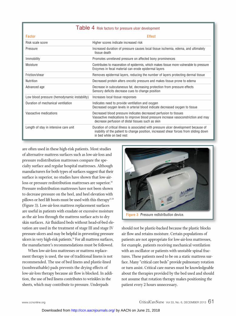
are often used in these high-risk patients. Most studiesof alternative mattress surfaces such as low-air-loss andpressure redistribution mattresses compare the spe-cialty surface and regular hospital mattresses. Althoughmanufacturers for both types of surfaces suggest that theirsurface is superior, no studies have shown that low-air-loss or pressure redistribution mattresses are superior.26
Pressure redistribution mattresses have not been shownto decrease pressure on the heel, and heel elevation withpillows or heel lift boots must be used with this therapy11,26
(Figure 3). Low-air-loss mattress replacement surfacesare useful in patients with exudate or excessive moistureas the air loss through the mattress surface acts to dryskin surfaces. Air fluidized beds without head-of-bed ele-vation are used in the treatment of stage III and stage IVpressure ulcers and may be helpful in preventing pressureulcers in very high-risk patients.24 For all mattress surfaces,the manufacturer’s recommendations must be followed.
When low-air-loss mattresses or mattress replace-ment therapy is used, the use of traditional linens is notrecommended. The use of bed linens and plastic-lined(nonbreathable) pads prevents the drying effects oflow-air-loss therapy because air flow is blocked. In addi-tion, the use of bed linens contributes to wrinkles in thesheets, which may contribute to pressure. Underpads
should not be plastic-backed because the plastic blocksair flow and retains moisture. Certain populations ofpatients are not appropriate for low-air-loss mattresses,for example, patients receiving mechanical ventilationwith an oscillator or patients with unstable spinal frac-tures. These patients need to be on a static mattress sur-face. Many “critical care beds” provide pulmonary rotationor turn assist. Critical care nurses must be knowledgeableabout the therapies provided by the bed used and shouldnot assume that rotation therapy makes positioning thepatient every 2 hours unnecessary.
www.ccnonline.org CriticalCareNurse Vol 33, No. 6, DECEMBER 2013 61
Factor
Risk scale score
Pressure
Immobility
Moisture
Friction/shear
Nutrition
Advanced age
Low blood pressure (hemodynamic instability)
Duration of mechanical ventilation
Vasoactive medications
Length of stay in intensive care unit
Effect
Higher scores indicate increased risk
Increased duration of pressure causes local tissue ischemia, edema, and ultimatelytissue death
Promotes unrelieved pressure on affected bony prominences
Contributes to maceration of epidermis, which makes tissue more vulnerable to pressureEnzymes in fecal material can erode epidermal layers
Removes epidermal layers, reducing the number of layers protecting dermal tissue
Decreased protein alters oncotic pressure and makes tissue prone to edema
Decrease in subcutaneous fat, decreasing protection from pressure effectsSensory deficits decrease cues to change position
Increases local tissue responses
Indicates need to provide ventilation and oxygenDecreased oxygen levels in arterial blood indicate decreased oxygen to tissue
Decreased blood pressure indicates decreased perfusion to tissuesVasoactive medications to improve blood pressure increase vasoconstriction and may
decrease perfusion of distal tissues such as skin
Duration of critical illness is associated with pressure ulcer development because ofinability of the patient to change position, increased shear forces from sliding downin bed while on bed rest
Table 4 Risk factors for pressure ulcer development
Figure 3 Pressure redistribution device.
by AACN on June 21, 2018http://ccn.aacnjournals.org/Downloaded from
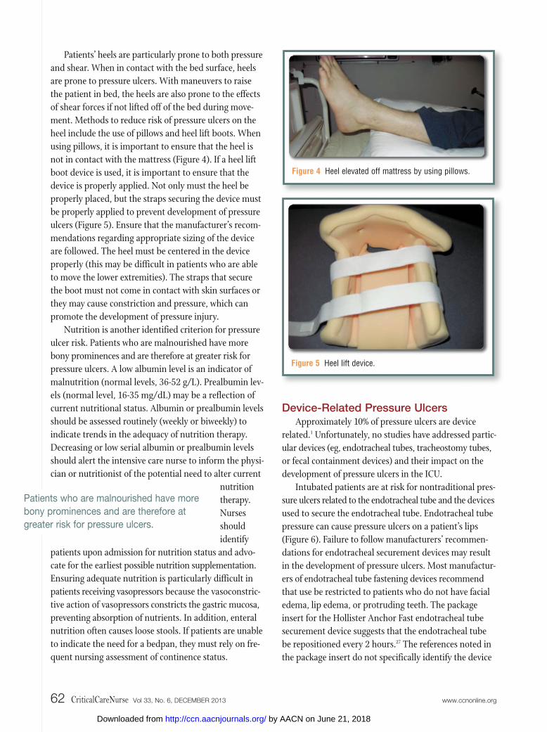
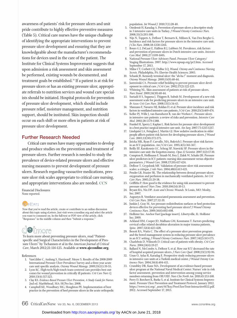
Patients’ heels are particularly prone to both pressureand shear. When in contact with the bed surface, heelsare prone to pressure ulcers. With maneuvers to raisethe patient in bed, the heels are also prone to the effectsof shear forces if not lifted off of the bed during move-ment. Methods to reduce risk of pressure ulcers on theheel include the use of pillows and heel lift boots. Whenusing pillows, it is important to ensure that the heel isnot in contact with the mattress (Figure 4). If a heel liftboot device is used, it is important to ensure that thedevice is properly applied. Not only must the heel beproperly placed, but the straps securing the device mustbe properly applied to prevent development of pressureulcers (Figure 5). Ensure that the manufacturer’s recom-mendations regarding appropriate sizing of the deviceare followed. The heel must be centered in the deviceproperly (this may be difficult in patients who are ableto move the lower extremities). The straps that securethe boot must not come in contact with skin surfaces orthey may cause constriction and pressure, which canpromote the development of pressure injury.
Nutrition is another identified criterion for pressureulcer risk. Patients who are malnourished have morebony prominences and are therefore at greater risk forpressure ulcers. A low albumin level is an indicator ofmalnutrition (normal levels, 36-52 g/L). Prealbumin lev-els (normal level, 16-35 mg/dL) may be a reflection ofcurrent nutritional status. Albumin or prealbumin levelsshould be assessed routinely (weekly or biweekly) toindicate trends in the adequacy of nutrition therapy.Decreasing or low serial albumin or prealbumin levelsshould alert the intensive care nurse to inform the physi-cian or nutritionist of the potential need to alter current
nutritiontherapy.Nursesshouldidentify
patients upon admission for nutrition status and advo-cate for the earliest possible nutrition supplementation.Ensuring adequate nutrition is particularly difficult inpatients receiving vasopressors because the vasoconstric-tive action of vasopressors constricts the gastric mucosa,preventing absorption of nutrients. In addition, enteralnutrition often causes loose stools. If patients are unableto indicate the need for a bedpan, they must rely on fre-quent nursing assessment of continence status.
Device-Related Pressure UlcersApproximately 10% of pressure ulcers are device
related.1 Unfortunately, no studies have addressed partic-ular devices (eg, endotracheal tubes, tracheostomy tubes,or fecal containment devices) and their impact on thedevelopment of pressure ulcers in the ICU.
Intubated patients are at risk for nontraditional pres-sure ulcers related to the endotracheal tube and the devicesused to secure the endotracheal tube. Endotracheal tubepressure can cause pressure ulcers on a patient’s lips(Figure 6). Failure to follow manufacturers’ recommen-dations for endotracheal securement devices may resultin the development of pressure ulcers. Most manufactur-ers of endotracheal tube fastening devices recommendthat use be restricted to patients who do not have facialedema, lip edema, or protruding teeth. The packageinsert for the Hollister Anchor Fast endotracheal tubesecurement device suggests that the endotracheal tubebe repositioned every 2 hours.27 The references noted inthe package insert do not specifically identify the device
Patients who are malnourished have morebony prominences and are therefore atgreater risk for pressure ulcers.
62 CriticalCareNurse Vol 33, No. 6, DECEMBER 2013 www.ccnonline.org
Figure 4 Heel elevated off mattress by using pillows.
Figure 5 Heel lift device.
by AACN on June 21, 2018http://ccn.aacnjournals.org/Downloaded from

but are based on general 2-hour positioning schedules toprevent development of pressure ulcers.27
Cervical collars are another device that increases therisk for pressure ulcer development at contact points onthe chin, shoulder, and ear. In studies of trauma patientswith cervical collars, longer duration of collar use wasassociated with increased risk of pressure ulcer develop-ment. Use of cervical collars for more than 5 days is asso-ciated with a 38% to 55% risk of pressure ulcersdeveloping.28 Rigid collars made of foam or plastic areassociated with a higher risk of pressure ulcer develop-ment than are padded collars.28 Padded collars such asthe Aspen or Miami cervical collar may prevent pressureulcer development if used appropriately28 (Figure 7). Themanufacturer’s guidelines should be used to ensure propersizing and cleansing of removable padding. The neck isparticularly prone to sweating, and moisture may macer-ate skin and make it vulnerable to pressure. When paddedcollars are used, it is advisable to order an extra set ofpads to replace used pads after cleansing so that they cancompletely dry. The skin surfaces under the collar shouldbe visualized by the nurse according to hospital’s policyto determine if redness indicating pressure is present. Ifredness is present, the patient should be evaluated forappropriate size and application of the cervical collar.
Tracheostomy tubes also have the potential to con-tribute to pressure ulcer development, especially forpatients receiving mechanical ventilation. Turning and posi-tioning may cause tension on tubing, which can promotedisplacement of the faceplate of the tracheostomy tube orcause movement of the tube. Faceplate pressure may causepressure ulcers over the bony prominence of the clavicles(s)at the sternal junction. The risk for pressure is higher whenthe tracheostomy tube is still sutured because the tra-cheostomy dressing cannot be easily inserted. Secretionsfrom the stoma site and tracheostomy tube may collectunder the tube and promote maceration of the skin. Afterthe sutures are removed, if a patient has excessive secretionsfrom the stoma, a foam dressing may be used to absorb exu-date and prevent pressure from the faceplate. To preventpressure caused by ventilator tubing from causing torque onthe tracheostomy tube, place a rolled towel under ventilatortubing near the connection to the tracheostomy tube sothat the tubing does not deflect downward, causing ten-sion and thus deflecting the faceplate downward.
Bilevel positive airway pressure and continuous positiveairway pressure masks also predispose patients to the
development of pressure ulcers at the points where themask touches the patient’s face. These masks both havepressure points over the ears from the straps, and partialmasks have pressure points over the nasal prominence.Although no published studies have described preventivemeasures other than making sure that the straps are nottoo tight, alternating a partial face mask (Figure 8) with afull face mask (Figure 9) may be helpful in preventingfurther skin breakdown. Other strategies to prevent pres-sure ulcer development on fragile nasal skin include theuse of foam dressings to decrease pressure. Thin foamdressings may be helpful as they not only decrease pres-sure but also prevent air leaks that may compromise oxy-genation. Hydrocolloid dressings do not relieve pressure,but they do reduce friction and shear.
Rigid transfer boards may produce shear injuriesbecause the patient slides over a rigid surface. Multiple
www.ccnonline.org CriticalCareNurse Vol 33, No. 6, DECEMBER 2013 63
Figure 6 Endotracheal tube securement device.
Figure 7 Padded cervical collar.
by AACN on June 21, 2018http://ccn.aacnjournals.org/Downloaded from

ergonomic products are available to prevent shear effectsduring transfer and repositioning of patients. Amongthese products are the Hover Matt (HoverTech Interna-tional), Ergo Nurse (Ergonurse Inc), and slip sheets. TheHover Matt is a lateral transfer device that uses an inflat-able mattress that enables the patient to float on the mat-tress during transfer. Use of this device reduces nursing
effort and shear forces on the patient. When the patientis transferred and repositioned, the mattress is deflatedand the patient is turned from side to side to remove thedeflated mattress. The Ergo Nurse is a rigid frame withstraps that are connected to bar connectors for thepatient’s bed sheets. When the bed is lowered, the patientis lifted and can be repositioned up in bed or from sideto side, preventing friction or shear injuries (Figure 10).The Ergo Nurse device is also available in bariatric size.Slip or slide sheets are made of material that slides overbed surfaces and does not have edges or a rigid surfacethat can cause friction or shear. There is no evidencethat one product is superior to another.
Fecal incontinence can contribute to skin breakdownbecause of the enzymes present in fecal matter.5,29 Enzymesin and the pH of fecal matter may act in conjunctionwith moisture to promote skin maceration and epider-mal erosion. Topical skin barriers assist in providing abarrier between moisture and skin; however, frequentcleansing because of diarrhea reduces the effectivenessof the skin barrier. Fecal containment devices are aneffective way to prevent skin damage due to moistureand enzyme action on perianal tissues. Indwelling fecalcontainment devices include products such as the Flexi-Seal (ConvaTec Inc), Actiflo (Hollister Global), and Zassi(Zassi Medical Evolutions Inc) devices. Topical fecalcontainment pouches are also available. Appropriate
64 CriticalCareNurse Vol 33, No. 6, DECEMBER 2013 www.ccnonline.org
Figure 8 Partial bilevel positive airway pressure (BiPAP) mask.
Figure 9 Full bilevel positive airway pressure (BiPAP) mask.
Figure 10 Ergo Nurse device.
by AACN on June 21, 2018http://ccn.aacnjournals.org/Downloaded from

application and use of these devices may prevent pres-sure ulcers from developing by preventing skin contactwith fecal enzymes and moisture.29
Bariatric PatientsBariatric patients present a unique challenge to criti-
cal care nurses and may be at increased risk for pressureulcers because of moisture in skin folds, device pressure,and inability to perform position changes due to issuesrelated to staffing and appropriate equipment.30 Bariatricpatients may be at higher risk for development of pressureulcers because adipose tissue typically has a decreasedblood supply compared with muscle tissue and the increasein weight increases pressure on tissues. Adhering to man-ufacturers’ guidelines for the use of equipment as well asusing appropriate equipment and sufficient personnelfor repositioning and lifting patients should assist inreducing risk.
Use of Preventive Measures Many recently published quality improvement arti-
cles indicate that unit-based quality assurance projectsthat identify effectiveness of preventive measures and
prevalence of pressure ulcers are particularly helpful inpreventing pressure ulcers in patients.31-33
Unit-based performance activities include teachingnursing staff how to identify risk factors and how tostage pressure ulcers, but the most important aspect ofthe quality initiatives appears to be in communicatingthe effectiveness of the therapy in terms of success indays without pressure ulcer development.31-33
Unit-based quality initiatives that document the num-ber of days that have passed between occurrences ofhospital-acquired pressure ulcers are one way to commu-nicate thissuccess inpreventingpressureulcers fromdeveloping. Using a 2-nurse handoff report and assess-ment on admission and shift change, which includesconducting a skin assessment, reinforces individualaccountability in interventions to prevent developmentof pressure ulcers. These activities are a demonstratedquality tool for identifying pressure areas before theybecome stage I or greater pressure ulcers.32-34 Heightened
www.ccnonline.org CriticalCareNurse Vol 33, No. 6, DECEMBER 2013 65
Who is at risk?
Identification of risk
Devices
Friction/shear
Pressure
Moisture
Nutrition
Vasoactive medications, patientunable to be turned every 2 hours
Comorbid conditions that contributeto pressure ulcer formation
Quality assurance
Interventions
Use a risk-identification scale each shift to identify which patients are at risk
Become familiar with the manufacturer’s recommendation for devices used in your unit (endotracheal securement devices, bilevel and continuous positive airway pressure masks, heel lift devices, mattresses)
Ensure that tubing and devices are not placed between skin surfaces; make sure ventilator tubingis not causing tension on tracheostomy tube and faceplate
Is your unit using appropriate lift and turning devices?Use assistive devices that reduce friction and shear; ensure the appropriate number of staff are
present to lift/turn patient
Consider the use of pressure-relief devices including specialty mattress surfaces, padded cervicalcollars, heel lift devices, and pillows
Use skin barrier creams, topical or indwelling fecal containment devices
Identify patients at risk and promote feeding at the earliest time possible; request specialtymattresses as soon as risk is identified
Provide a specialty mattress to reduce skin interface pressure
Flag the patient at risk and use critical thinking to determine appropriate therapy: heel lift device,specialty mattress, device
Identify unit-specific statisticsTrack prevalence and report to staffCelebrate decreases in occurrenceIdentify best practicesContribute to critical care–specific research in pressure ulcer prevention and risk
Table 5 Risk factors and interventions to prevent pressure ulcers
Skin inspection should occur on each shiftor more often in patients at risk of pressureulcer development.
by AACN on June 21, 2018http://ccn.aacnjournals.org/Downloaded from
awareness of patients’ risk for pressure ulcers and unitpride contribute to highly effective preventive measures(Table 5). Critical care nurses have the unique challengeof identifying the appropriate interventions to preventpressure ulcer development and ensuring that they areknowledgeable about the manufacturer’s recommenda-tions for devices used in the care of the patient. TheInstitute for Clinical Systems Improvement suggests thatupon admission a risk assessment and skin assessmentbe performed, existing wounds be documented, andtreatment goals be established.34 If a patient is at risk forpressure ulcers or has an existing pressure ulcer, appropri-ate referrals to nutrition services and wound care special-ists should be initiated. Hospital protocols for preventionof pressure ulcer development, which should includepressure relief, moisture management, and nutritionsupport, should be instituted. Skin inspection shouldoccur on each shift or more often in patients at risk ofpressure ulcer development.
Further Research NeededCritical care nurses have many opportunities to develop
and produce studies on the prevention and treatment ofpressure ulcers. Further research is needed to study theprevalence of device-related pressure ulcers and effectivenursing measures to prevent development of pressureulcers. Research regarding vasoactive medications, pres-sure ulcer risk scales appropriate to critical care nursing,and appropriate interventions also are needed. CCN
Financial DisclosuresNone reported.
References1. VanGilder C, Amlung S, HarrisonP, Meyer S. Results of the 2008-2009
International Pressure Ulcer Prevalence Survey and a three year acutecare unit specific analysis. Ostomy Wound Manage. 2009;55(11):39-55.
2. Lyon KC. High-tech/high-touch team-centered care provides best out-comes for wound prevention in critically ill patients. Crit Care Nurs Q.2010;33(4):317-323.
3. Clay KS. Evidence-Based Pressure Ulcer Prevention, A Study Guide for Nurses.2nd ed. Marblehead, MA: HCPro Inc; 2008.
4. Campbell KE, Woodbury MG, Houghton PE. Implementation of bestpractice in the prevention of heel pressure ulcers in the acute orthopedic
population. Int Wound J. 2010;7(1):28-40.5. Ozedemit H, Karadag A. Prevention of pressure ulcers a descriptive study
in 3 intensive care units in Turkey. J Wound Ostomy Continence Nurs.2008;35(3):293-300.
6. Nijs N, Toppets A, Defloor T, Bernarts K, Milisen K, Van Den Berghe G.Incidence and risk factors for pressure ulcers in the intensive care unit.J Clin Nurs. 2008;18:1258-1266.
7. Bours G J, DeLaat E, Halfens R J, Lubbers M. Prevalence, risk factorsand prevention of pressure ulcers in Dutch intensive care units. IntensiveCare Med. 2001;27:1599-1605.
8. National Pressure Ulcer Advisory Panel. Pressure Ulcer Category/Staging Illustrations. 2007. http://www.npuap.org/pr2.htm. AccessedSeptember 17, 2013.
9. Milne CT, Corbett CQ, DuBuc LQ. Wound, Ostomy and Continence NursingSecrets. Philadelphia, PA: Elsevier Health Sciences; 2003.
10. Schank JE. Kennedy terminal ulcer: the “ah-ha !” moment and diagnosis.Ostomy Wound Manage. 2009;55(9):40-44.
11. Jastremski CA. Pressure relief bedding to prevent pressure ulcer devel-opment in critical care. J Crit Care. 2002;17(2):122-125.
12. Whiteing NL. Skin assessment of patients at risk of pressure ulcers.Nurs Stand. 2009;24(10):40-44.
13. Suriadi H S, Sugama J, Thigpen B, Subuh M. Development of a new riskassessment scale for predicting pressure ulcers in an intensive care unit.Br Assoc Crit Care Nurs. 2008;13(1):34-43.
14. Manzano F, Navarro MJ, Roldan D, et al. Pressure ulcer incidence and riskfactors in ventilated intensive care patients. J Crit Care. 2010;25(3):469-476.
15. Keller PJ, Wille J, van Ramshorst B, van der Werken C. Pressure ulcersin intensive care patients: a review of risks and prevention. Intensive CareMed. 2002;28:1379-1388.
16. Frankel H, Sperry J, Kaplan L. Risk factors for pressure ulcer developmentin a best practice surgical intensive care unit. Am Surg. 2007;73:1215-1217.
17. Lindquist LA, Feinglass J, Martin GJ. How sedative medication in olderpeople affects patient risk factors for developing pressure ulcers. J WoundCare. 2003;12(30):272-275.
18. Jiricka MK, Ryan P, Carvallo, MA, Bukvich J. Pressure ulcer risk factorsin an ICU population. Am J Crit Care. 1995;4(5):361-367.
19. Reilly EF, Karakousis GC, Schrag SP, Stawicki SP. Pressure ulcers in theintensive care unit: the forgotten enemy. Opus 12 Scientist. 2007;1(2):17-30.
20. Compton R, Hoffmann F, Straub M, Frey J, Zidek W, Schafer JH. Pressureulcer predictors in ICU patients: nursing skin assessment versus objectiveparameters. J Wound Care. 2008;17(10):417-424.
21. Defloor T, Grypdonk MF. Validation of pressure ulcer risk assessmentscales: a critique. J Adv Nurs. 2004;48(6):613-621.
22. Pender LR, Frazier SK. The relationship between dermal pressure ulcers,oxygenation and perfusion in mechanically ventilated patients. Int CritCare Nurs. 2005;21:29-38.
23. Griffiths P. How good is the evidence for using risk assessment to preventpressure ulcers? Nurs Time. 2010;106(14):10-13.
24. Bryant RA, Nix DP. Acute and Chronic Wounds. St Louis, MO: Mosby,Inc; 2007.
25. Augustyn B. Ventilator associated pneumonia assessment and prevention.Crit Care Nurs. 2007;27:32-39.
26. Junkin J, Gray M. Are pressure redistribution surfaces or heel protectiondevices effective for preventing heel pressure ulcers? J Wound OstomyContinence Nurs. 2009;36(6):602-608.
27. Hollister Inc. Anchor Fast [package insert]. Libertyville, IL: HollisterInc; 2009.
28. Ackland HM, Cooper JD, Malham GM, Kossmann T. Factors predictingcervical collar related decubitus ulceration in major trauma patients.Spine. 2007;32(4):423-428.
29. Benoit RA, Watts C. The effect of a pressure ulcer prevention programand the bowel management system in reducing pressure ulcer prevalencein an ICU setting. J Wound Ostomy Continence Nurs. 2007;34(2):163-175.
30. Charlebois D, Wilmoth D. Critical care of patients with obesity. Crit CareNurse. 2004;24(4):19-27.
31. Ballard N, McCombs A, DeBoor S, et al. How our ICU decreased the rateof hospital acquired pressure ulcers. J Nurs Care Qual. 2008;23(1):92-96.
32. Uzun O, Aylaz R, Karadag E. Prospective study reducing pressure ulcersin intensive care units at a Turkish medical center. J Wound Ostomy Con-tinence Nurs. 2004;36(4):404-411.
33. Crumbley DR, Kane MA. Development of an evidence-based pressureulcer program at the National Naval Medical Center: Nurses’ role in riskfactor assessment, prevention and intervention among young servicemembers returning from OIF/OEF. Nurs Clin North Am. 2010;45:153-168.
34. Perry D, Borchert K, Burke S, et al. Institute for Clinical Systems Improve-ment. Pressure Ulcer Prevention and Treatment Protocol. January 2012.https://www.icsi.org/_asset/6t7kxy/PresUlcerTrmt-Interactive0112.pdf.2012. Accessed September 17, 2013.
66 CriticalCareNurse Vol 33, No. 6, DECEMBER 2013 www.ccnonline.org
Now that you’ve read the article, create or contribute to an online discussionabout this topic using eLetters. Just visit www.ccnonline.org and select the articleyou want to comment on. In the full-text or PDF view of the article, click“Responses” in the middle column and then “Submit a response.”
To learn more about preventing pressure ulcers, read “Patient-specific and Surgical Characteristics in the Development of Pres-sure Ulcers” by Tschannen et al in the American Journal of CriticalCare, March 2012;21:116-125. Available at www.ajcconline.org.
by AACN on June 21, 2018http://ccn.aacnjournals.org/Downloaded from
CNE Test Test ID C1363: Evidence-Based Prevention of Pressure Ulcers in the Intensive Care UnitLearning objectives: 1. Identify factors that place critically ill patients at increased risk for pressure ulcers 2. Describe the pressure risks associated withcommonly used devices in the critical care setting 3. Apply evidence-based strategies for the prevention of pressure ulcers in critical care patients
Program evaluation Yes No
Objective 1 was met q qObjective 2 was met q qObjective 3 was met q qContent was relevant to my
nursing practice q qMy expectations were met q qThis method of CNE is effective
for this content q qThe level of difficulty of this test was:
q easy q medium q difficultTo complete this program,
it took me hours/minutes.
1. Which of the following describes why there is increased concern over the development of hospital-acquired pressure ulcers (HAPUs)?a. There is little that can be done to treat a pressure ulcer once it occurs.b. Medicare and Medicaid Services will not pay for costs associated with a HAPU.c. Development of a stage I or II pressure ulcer is now considered a “never event.”d. The established cost of a pressure ulcer is more than $50 000 per event.
2. Which of the following factors does not specifically place critically ill patients at increased risk for pressure ulcers?a. Presence of multiple devices and equipmentb. Infusion of vasoactive agents for hypotensionc. Length of time receiving mechanical ventilationd. Increased incidence of urinary incontinence
3. Which of the following statements correctly describes deep tissue injury?a. The injury always progresses to a full-thickness pressure ulcer.b. This classification excludes superficial blood blisters.c. The injury appears as a bluish or purple discoloration over an area of pressure.d. The depth of the injury is clearly apparent at the time of identification.
4. The Kennedy terminal ulcer describes which of the following?a. A nationally recognized ulcer that is unique to the critical care settingb. A rapidly progressing ulcer seen in terminal patients just before death c. A chronic ulcer that develops primarily in long-term care facilitiesd. A preventable ulcer generally associated with patients in septic shock
5. Which statement is true regarding the 4 most common pressure ulcer riskassessment scales?a. None of the scales fully reflect the additional risk factors present in ICU patients.b. All of the scales are recommended by the Agency for Health Care Policy and Research.c. Only the Waterlow Scale specifically addresses hemodynamic instability.d. The Braden Scale is most effective for assessing risk in critically ill patients.
6. Which of the following statements does not describe the pathophysiology underlying the development of pressure ulcers? a. Compression of vessels prevents the supply of oxygen and nutrients to the tissues.b. Metabolic wastes accumulate at the tissues, leading to further vasoconstriction.c. Moisture contributes to maceration, making the skin more vulnerable to pressure.d. Friction and shear may remove epidermal layers, making the skin vulnerable to injury.
7. Positioning strategies to prevent pressure ulcers include which of the following?a. Turning patients every 4 hours b. Maintaining the head of bed at an elevation greater than 30°c. Elevating patients’ heels off the mattressd. Avoiding the supine position whenever possible
8. Which of the following should be considered when selecting a mattress toreduce the risk of pressure ulcers?a. Low-air-loss mattresses are beneficial for patients with excessive moisture.b. Air fluidized beds are preferred for patients receiving mechanical ventilation.c. Mattresses with pressure redistribution are considered superior to low-air-loss surfaces.d. Rotational surfaces eliminate the need for turning.
9. Which of the following indicates an increased nutritional risk for developmentof pressure ulcers?a. An admission albumin level of 38 g/Lb. Initiation of enteral nutritionc. A decreasing trend in prealbumin levelsd. Infusion of vasodilators
10. Which of the following statements is true regarding device-related pressureulcers?a. They account for approximately 10% of pressure ulcers. b. They only occur when the manufacturer’s directions are not followed.c. They occur more frequently with endotracheal tubes than other devices.d. They have been well-defined in a number of research studies.
11. Which of the following interventions is recommended to reduce pressureulcers in patients with medical devices? a. Repositioning of the endotracheal tube every 4 hoursb. Removing cervical collars every shift to perform a thorough skin assessmentc. Supporting ventilator tubing to prevent torque on the tracheostomy tubed. Applying hydrocolloid dressings on the face to reduce pressure from continuous positive airway pressure/bilevel positive airway pressure masks
12. Bariatric patients are at higher risk for pressure ulcers because of which ofthe following?a. Prolonged need for mechanical ventilationb. Decreased blood supply to adipose tissuec. Impaired gastrointestinal absorption of nutrientsd. Increased reluctance to perform position changes
13. Which of the following is true regarding unit-based quality improvementprojects for pressure ulcer prevention?a. They have little impact on pressure ulcer outcomes.b. They focus primarily on teaching staff how to stage ulcers.c. They are effective in heightening staff awareness of pressure ulcer risk.d. They help identify staff who are not following hospital policies.
For faster processing, takethis CNE test online at
www.ccnonline.org or mail this entire page to:
AACN, 101 Columbia Aliso Viejo, CA 92656.
Test ID: C1363 Form expires: December 1, 2016 Contact hours: 1.0 Pharma hours: 0.0 Fee: AACN members, $0; nonmembers, $10 Passing score: 10 correct (77%) Synergy CERP Category A Test writer: Joni L. Dirks, RN-BC MS CCRN
Name Member #
Address
City State ZIP
Country Phone
RN Lic. 1/St RN Lic. 2/St
Payment by: q Visa q M/C q AMEX q Discover q Check
Card # Expiration Date
SignatureThe American Association of Critical-Care Nurses is accredited as a provider of continuing nursing education by the American Nurses Credentialing Center’s Commission on Accreditation.
AACN has been approved as a provider of continuing education in nursing by the State Boards of Nursing of Alabama (#ABNP0062), California (#01036), and Louisiana (#ABN12). AACN programming meets the standards for most other states requiring mandatory continuing education credit for relicensure.
Test answers: Mark only one box for your answer to each question. You may photocopy this form.
1. qa qb qc qd
10. qa qb qc qd
13. qa qb qc qd
12. qa qb qc qd
11. qa qb qc qd
9. qa qb qc qd
8. qa qb qc qd
7. qa qb qc qd
6. qa qb qc qd
5. qa qb qc qd
4. qa qb qc qd
3. qa qb qc qd
2. qa qb qc qd
by AACN on June 21, 2018http://ccn.aacnjournals.org/Downloaded from

Karen L. CooperEvidence-Based Prevention of Pressure Ulcers in the Intensive Care Unit
http://ccn.aacnjournals.org/Published online ©2013 American Association of Critical-Care Nurses
10.4037/ccn2013985 57-66 33 2013;Crit Care Nurse
http://ccn.aacnjournals.org/cgi/external_ref?link_type=PERMISSIONDIRECTPersonal use only. For copyright permission information:
http://ccn.aacnjournals.org/subscriptions/Subscription Information
http://ccn.aacnjournals.org/misc/ifora.xhtmlInformation for authors
http://www.editorialmanager.com/ccn Submit a manuscript
http://ccn.aacnjournals.org/subscriptions/etoc.xhtmlEmail alerts
362-2049. Copyright ©2016 by AACN. All rights reserved. bimonthly by AACN, 101 Columbia, Aliso Viejo, CA 92656. Telephone: (800) 899-1712, (949) 362-2050, ext. 532. Fax: (949) Critical Care Nurse is an official peer-reviewed journal of the American Association of Critical-Care Nurses (AACN) published
by AACN on June 21, 2018http://ccn.aacnjournals.org/Downloaded from