EVERYTHING YOU ALWAYS WANTED TO KNOW … cleft palate ... words/sentences with the nose open then...
124
EVERYTHING YOU ALWAYS WANTED TO KNOW ABOUT VOICE AND RESONANCE DISORDERS BUT WERE AFRAID TO ASK… (I.E. THE MARY AND MARC VELUM AND LARYNX SHOW) Marc Haxer M.A.CCC and Mary K Berger M.S.CCC University of Michigan Health Systems Ann Arbor, MI
Transcript of EVERYTHING YOU ALWAYS WANTED TO KNOW … cleft palate ... words/sentences with the nose open then...

EVERYTHING YOU ALWAYS
WANTED TO KNOW ABOUT VOICE
AND RESONANCE DISORDERS BUT
WERE AFRAID TO ASK… (I.E. THE
MARY AND MARC VELUM AND LARYNX
SHOW)
Marc Haxer M.A.CCC
and
Mary K Berger M.S.CCC
University of Michigan Health Systems
Ann Arbor, MI

So where do we start?

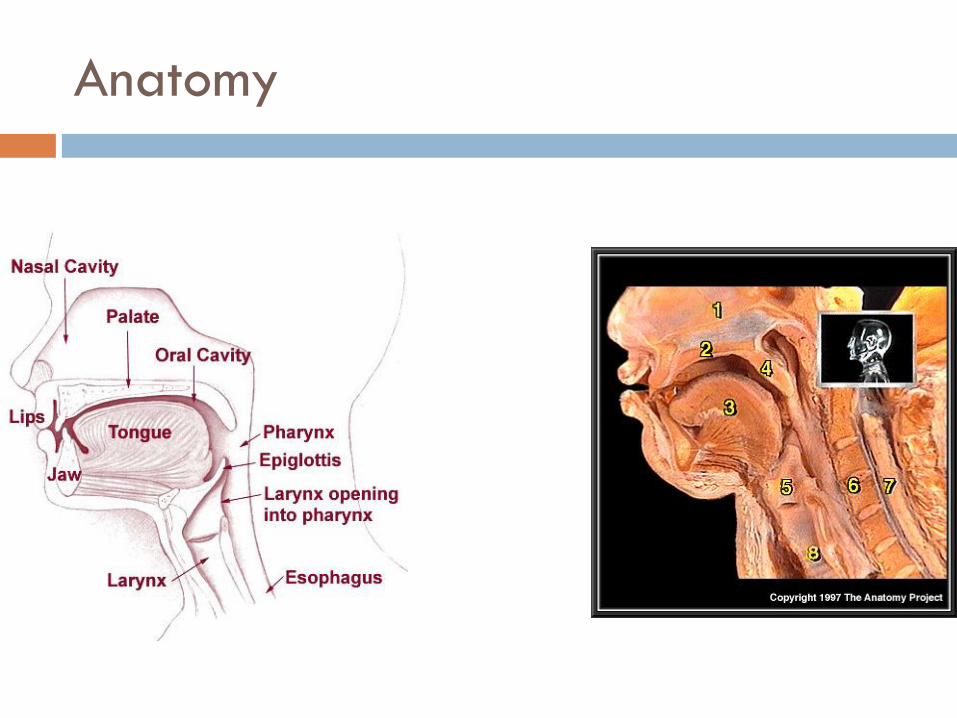
Anatomy
Structures
Hard palate
Palatine raphe/posterior
nasal spine
Soft palate/velum
Uvula/musculus uvulae
Anterior faucial arches
(palatoglossus)
Posterior faucial arches
(palatopharyngeus)
Palatine tonsils

TYPES OF CLEFTING
Anatomy

Anatomy/physiology
Muscles
Palatine aponeurosis
Tensor veli palatini
Levator veli palatini-muscle repaired at the time of surgery
Palatoglossus
Palatopharyngeus (palatothyroideus)
Musculus uvulae
Superior constrictor-lateral wall movement


Perceptual exam
Performed by a Speech Pathologist
The Gold Standard for the diagnosis of
velopharyngeal dysfunction
Need imaging to determine intervention.

Oral motor exam
Head/neck: ears, torticollis, cranial vault
Face: structures, symmetry, sensation
Mandible: structure, ROM, strength testing
Dentition: occlusion, missing teeth, dentures
Tongue: structure, strength, ROM, RAM
Palate: uvula, fistula, length, distance to posterior pharyngeal wall, Passavant’s ridge, mesial movement of lateral pharyngeal walls, gag response.

Left unilateral cleft lip and palate

Bilateral cleft lip and palate

Cleft palate only

Oronasal fistula
Communication between the oral and nasal cavities
Can complain of nasal regurgitation
May be difficult to see or tell if truly communicates
Often no effect on speech resonance
Gum test

Submucous cleft palate
bifid uvula
zona pellucida (thin, blue-
tinged mucosa)
posterior nasal spine
notching
lateral levator muscle
bulges
+/- hypernasality
nasal regurgitation with
liquids
Occult cleft-absent
musculus uvulae

Velopharyngeal valve
Soft palate (velum) contacts posterior pharyngeal wall to transmit air pressure and sound energy into the oral cavity for oral consonant and vowel productions. Normal valving allows adequate intra-oral air pressure, normal oral resonance and sufficient breath support for normal length of utterance. Structural defect MAY interfere with closure.

Speech perceptual exam
Articulation-place and manner, compensatory
misarticulations
Resonance-nasality
Phonation-quality, consistency
Prosody-inflection, timing, rate
Speech intelligibility rating (4 point scale)
Speech acceptability rating (4 point scale)

Velopharyngeal dysfunction- VPD
Velopharyngeal dysfunction (VPD) or
Velopharyngeal Inadequacy (VPI)- absence of
closure of velopharyngeal port with
hypernasality, nasal air emission. Variety of
causes. Average 5%-25% of cleft children
have resonance disorder. Needs further
workup.
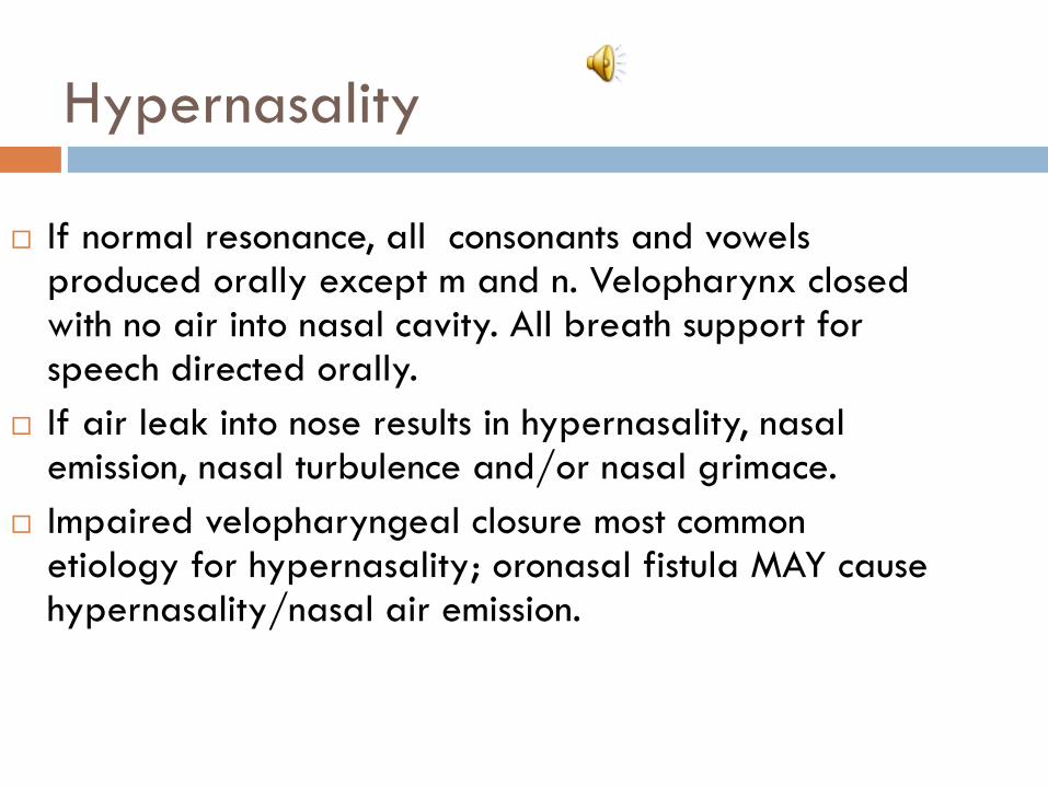
Hypernasality
If normal resonance, all consonants and vowels produced orally except m and n. Velopharynx closed with no air into nasal cavity. All breath support for speech directed orally.
If air leak into nose results in hypernasality, nasal emission, nasal turbulence and/or nasal grimace.
Impaired velopharyngeal closure most common etiology for hypernasality; oronasal fistula MAY cause hypernasality/nasal air emission.

Velopharyngeal dysfunction
Velopharyngeal dysfunction (VPD) or
velopharyngeal inadequacy (VPI) without
presence of a cleft:
R/o submucous or occult cleft
Articulation disorder
S/p adenoidectomy
Velocardiofacial syndrome (DiGeorge
syndrome, 22q11 deletion) “the black hole”

Velopharyngeal insufficiency
Palate too short
Structural problem
Not enough tissue at time of initial cleft repair
Submucous cleft (SMC)
Deep pharynx due to cranial base anomalies
Following adenoidectomy
Needs surgery to correct.

Velopharyngeal closure using adenoid

Velopharyngeal incompetence
Reduced movement of soft palate
Physiological cause
Poor muscle function
Pharyngeal hypotonia
Velar paralysis or paresis
Dysarthria
Apraxia

Manifestations of VPD
Nasal regurgitation
Inappropriate air flow
Nasal rustles/ turbulence
Hypernasal resonance
Compensatory
misarticulations
Poor speech intelligibility
Nasal grimace
Hoarseness
Vocal cord nodules
Short utterance length
Soft voice syndrome
Primary Manifestations Secondary Manifestations

Resonance disorders
Resonance descriptors (don’t say “nasally speech”)
Hypernasality-especially on vowels and voiced oral consonants
Hyponasality/denasality-too nasal resonance /m, n, ng/
Cul de sac resonance (hypertrophied tonsils/adenoids)
Mixed resonance-hyper- and hyponasality (congestions, deviated/deflected septum)
Audible nasal turbulence; nasal rustle
-Phoneme specific VPD

Hypernasality
If normal resonance, all consonants and vowels produced orally except m and n. Velopharynx closed with no air into nasal cavity. All breath support for speech directed orally.
If air leak into nose results in hypernasality, nasal emission, nasal turbulence and/or nasal grimace.
Impaired velopharyngeal closure most common etiology for hypernasality; oronasal fistula MAY cause hypernasality/nasal air emission.

Oral consonants
High pressure consonants:
p,b,t,d,k,g,s,z,sh,ch,j
Low pressure consonants:
r,l,w,h,y and vowels
High pressure consonants may be weakened or nasalized with VPD.

Hyponasality
Nasal consonants /m/ /n/ /ng/
Velopharynx open allowing consonant to resonant
through nose
Sensitive to nasal obstruction
Only nasal consonants effected by hyponasality
Causes /m/ to sound like /b/, /n/ to sound like /d/
and /ng/ to sound like /g/.
Reduced nasal emission with mirror on nasal productions

Mixed resonance
Evidence for both hypernasality due to
velopharyngeal dysfunction and hyponasality
due to often nasal obstruction
Often reduced nasal air emission from one
naris on mirror exam

Phoneme specific nasal emission (PSNAE)
Hypernasality or nasal air emission due to articulation error pattern
Not physical etiology; otherwise normal resonance; competent VP mechanism
Causes phoneme-specific nasal emission (usually sibilant sounds).
Never surgery-always speech tx

Assessing resonance with
oral consonants
Cul de sac testing (Bzoch,
1979) produce the oral
words/sentences with the
nose open then with the nose
closed. If normal resonance,
will be identical productions.
Nasal mirror testing under
nares for nasal emission
(speech and non speech
tasks).

Assessing resonance
Modified tongue anchor technique: puff cheeks around
protruded tongue (Dalston, 1990).
See-scape
Nasal tubing/stethoscope
Assess for presence of compensatory misarticulations
**Repeat standardized words, sentences, serial counting
and spontaneous speech sampling .
**See word/sentences lists at end of presentation

Articulation issues and clefting
Cleft lip/ alveolus only-normal incidence of articulation
errors unless untreated hearing deficits
Hearing impairment
Incidence of developmental articulation errors similar
Compensatory misarticulation errors (generally backing of
place of articulation in vocal tract with nasal air flow for oral
productions)
Abnormal dentition/occlusion
Due to inability to achieve velopharyngeal closure
Mislearning errors (structural or unrelated to cleft)
Substitutions and omissions more common than distortions

Compensatory misarticulations
Maladaptive articulation pattern that occurs in individuals who have velopharyngeal dysfunction. Articulation valving occurs more posterior in the vocal tract to compensate for reduced intraoral air pressure.
Glottal stop
Pharyngeal stop
Pharyngeal fricative
Laryngeal fricative
Velar fricative
Mid-dorsum palatal stop
Posterior nasal fricative/nasal turbulence
Often accompanied by a nasal grimace.

Compensatory misarticulations

Active vs. passive errors
Obligatory vs. compensatory
Both have structural origins (esp. VPD)
Passive/obligatory errors: hypernasality, nasalized oral
consonants, weak pressure consonants. Disappear when
structure corrected.
Active/compensatory errors: e.g. glottal stops. Active attempt
to compensate for structural deficit. Persist when structure
corrected.
Harding and Grunwell, 1998; Hutters and Bonsted, 1987.

Phonation and VPD
Hoarseness associated with hyperfunction of the
larynx=vocal abuse, check for nodules
Hypophonia- due to nasal emission have reduced
loudness or may be masking audible nasal
turbulence/emission
Periods of aphonia

Decision time…
Speech Perceptual Exam
Sound-specific VPI
Speech Therapy
VP Insufficiency
Surgery
VP incompetence
Almost but not quite closed/ Inconsistently
closed
Speech Therapy
Moderate to large gap/
never closes
Palatal Lift or surgery

Medical interventions for
resonance disorders
Surgical Management is indicated when:
hypernasality is caused by structural or
physiological abnormality
moderate to large velopharyngeal gap
velopharyngeal insufficiency
hyponasality.

Velopharyngeal Insufficiency:
needs surgery

Velopharyngeal Insufficiency:
needs surgery

VPD: Needs resonance therapy
(pre- and post-tx example)

Speech language pathology management
hypernasal resonance is associated with oral-motor
dysfunction/dysarthria
hypernasality occurs primarily when the child is tired
the child is status-post secondary palatal surgery and
needs therapy to increase lateral wall motion, closure
of DSP port or increase elevation of the palate
during speech
cooperative with adequate cognitive skills

Resonance therapy techniques
Don’t blow it! Blowing and sucking exercises
help blowing and sucking
Blowing exercises do not improve velopharyngeal strengthening
Blowing can be used to assist with the idea of oral air stream that can then be valved with articulators
Blowing bubbles to stimulate for bilabials and oral air flow

Resonance therapy techniques
Auditory discrimination-
hypernasality
audible nasal turbulence
nasal snort
hyponasality

Resonance therapy techniques
Exaggerated articulation-
increasing ROM of the articulators may
assist with increasing palatal closure with
increased muscle recruitment
generally slow down rate of speech to
improve velopharyngeal closure and
coordination

Resonance therapy techniques
Visual feedback
See-Scape (AliMed, SuperDuper, Pro-Ed) or nasal mirror for monitoring nasal air emission
tissue, tissue paper or paper paddle for oral air flow with plosives
feather for oral air flow with fricatives
Nasometer (acoustic measurement with visual feedback-Kay Pentax)
biofeedback nasoendoscopy (direct visual feedback of velopharyngeal closure).
Resonance therapy techniques
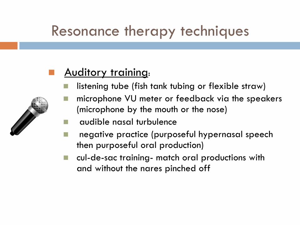
Auditory training:
listening tube (fish tank tubing or flexible straw)
microphone VU meter or feedback via the speakers (microphone by the mouth or the nose)
audible nasal turbulence
negative practice (purposeful hypernasal speech then purposeful oral production)
cul-de-sac training- match oral productions with and without the nares pinched off

Resonance therapy techniques
Tactile training:
feel airflow on hands
feel nasal air flow from nares
feel vibration on side of nose with audible nasal
turbulence only with voiced consonants
yawning followed by vowel-target consonant
(flattens base of tongue and elevations soft palate)

Resonance therapy techniques
Awareness training:
Teach concepts that child can understand to describe
oral/nasal airflow for example:
-Mr. Mouth/Mr. Nose
-mouth and nose sound
-”make the wind come out of your mouth”
-throat sound or voice box sound

Resonance therapy
Therapy note: If persisting hypernasality or
nasal emission after a few months of tx, child
should be referred to a specialist for further
assessment and consideration of physical
management. Don’t keep in tx and continually
asked to perform a speech task that is
impossible to do.

Compensatory misarticulation therapy
Articulation therapy note: Articulation therapy
can be effective for place of articulation even
if a surgery is still needed to reduce
velopharyngeal dysfunction or a oronasal
fistula.

Compensatory misarticulation therapy
Accuracy training:
Reinforcing place of articulation with
exaggerated articulation, may recruit palatal
musculature to increase ROM. Has potential
to achieve velopharyngeal closure ONLY if
competent.

Compensatory misarticulation therapy
Phoneme hierarchy in therapy:
Train front sounds prior to back sounds
Voiceless before voiced phonemes
Basic articulation therapy rules apply too (introduce
sounds in developmental hierarchy, begin with
sounds in isolation then C-V, V-C and C-V-C
contexts, etc)
Note: work with sounds the child can produce to
identify target sound selection

Compensatory misarticulation therapy
Techniques:
Whispering (eliminates glottal stops)
Forward tongue placement (eliminate
pharyngeal fricatives)
Pair /h/ with target phonemes
Introduce new sound that changes one feature
of sound child can produce (t→d, m→b)
OK to use nonsense words briefly for early
practice

Compensatory misarticulation therapy
Techniques-continued
Build list of short words with correctly
produced sounds to practice as warm
up and to “remind” child of correct
productions
Target at least 50 correct productions
in a 30 minute session with toddlers;
100 correct productions with school
aged children

Compensatory misarticulation therapy
Glottal stops
Whisper with over aspiration
Emphasize fronted productions
Voicing at the end of syllable with gradual VOT
/h/ plus target labial or lingual oral placement
Produce nasal counterpart then plug nose
(m→b, n→d, ng→g); then use partial nares occlusion
Use awareness training and be specific where to place tongue/lips and how to direct air stream

Home program
Parent(s) need training to hear correct
productions for reinforcing
Daily practice
Short practice sessions (30-60 seconds) several
times per day
Reinforcing for self-monitoring/corrections
Craniofacial Team
Plastic Surgery
Maxillofacial Surgery
Orthodontics
Pediatric Dentistry
Prosthodontics
Speech Pathology
Audiology
Social Work
Psychology
Genetics
Dietitian
Nursing
Neurosurgery
Otolaryngology
Coordinator
Community Professionals

Larynx
Cartilaginous tube
Connects inferiorly to respiratory system
Trachea, lungs
Connects superiorly to vocal tract
Pharynx, oropharynx, nasopharynx
Anatomic orientation important
Highlights interactive relationship between vocal subsystems
Pulmonary mechanism
Laryngeal valve
Supraglottic vocal tract resonator

Larynx
Communicative function of larynx relies heavily on
integration of these vocal subsystems
Lungs
Provides aerodynamic tracheal pressure that blows vocal folds apart
and sets them into vibration
Vocal folds
Provide sound source for phonation as vocal folds repeatedly
oscillate during vibration
Vocal tract
Shapes and filters acoustic energy to produce sound recognized as
human voice

Larynx
Differential dx of voice disorders requires assessment of these parameters
Laryngeal health/function will influence quality of voice production
Compromised respiratory support will decrease potential for adequate VF vibration
Alteration in shape/size of vocal tract will affect resonance
Compromise in any area will adversely affect optimal production of voice and increase risk for development of voice problem
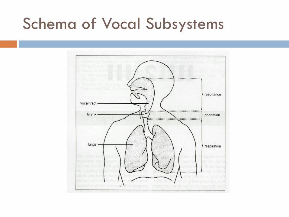
Schema of Vocal Subsystems

Larynx
Complex arrangement of cartilages, muscles, mucous membranes, and connective tissues Allows for wide degrees of variation in position,
movement, and tension to support three basic functions Airway preservation for ventilation Airway protection to block/repel environmental
infiltrates Phonation for communication or singing
Cartilage housing serves as columnar protective shield for laryngeal valve

Larynx
Laryngeal valve achieves these functions through three levels of “folds”
Aryepiglottic
False vocal folds
True vocal folds

Neurologic Supply
Cranial nerve X innervates larynx peripherally
Vagus = “wanderer”
Innervates sites from skull to abdomen
Innervates larynx through two important branches
Superior laryngeal nerve (SLN)
Branches off vagus near nodose ganglia in neck
Courses alongside carotid arteries
Forms internal/external branches Internal branch provides all sensory information to larynx
External branch is motor nerve to cricothyroid (CT) muscle
Recurrent laryngeal nerve (RLN)
Extends to thorax
Forms long loop under heart before coursing superiorly under thyroid gland and into larynx
Different on right/left sides of body
Nerves (especially left) susceptible to injury
Supplies all sensory information to area below VFs and all motor innervation to PCA, TA, LCA, and IA muscles

Schema of Laryngeal Innervation

Respiration for Phonation
VF vibration is sound source for phonation Phonation dependent on respiratory power provided by lungs
and abdominal/thoracic musculature Inferior border of lungs attached to diaphragm by double-walled
pleural lining During inhalation, diaphragm contracts thus compressing viscera and
pulling lungs inferiorly allowing for expanded lung volume As lung volume expands, air passively drawn into lungs
During exhalation, passive elastic recoil/other musculature forces air out of lungs
Air moved superiorly through VFs/vocal tract
During exhalation for voice/speech, VFs adduct to midline thus constricting outgoing airflow
Resulting aerodynamic energy sets VFs into oscillation creating vibratory sound that comprises phonation

Vocal Tract Resonance
Sound waves from larynx pass through supraglottic
air passage into pharynx, oropharynx, and
nasopharynx
Also move articulators including velum, hard palate,
tongue, and teeth
Excitation of air molecules within these spaces
creates phenomenon called resonance

Etiologies of Voice Disorders
Etiology: the science that deals with the causes or origin of diseases or conditions e.g., "the etiology is unknown" translates into we don’t
know the cause
Medicine.net. Accessed 2/8/10

Etiologies of Voice Disorders
West, Kennedy, and Carr (1937)
There is always a reason for a voice disorder
SLP’s job is to find cause(s)
Sometimes easy
VF nodules in a screamer
Sometimes hard
Differential Dx of ADSD vs. Functional dysphonia vs. subtle VF motion
disorder
Successful outcome dependent on clinician’s ability to
determine cause(s) based on number of reference points

Major Categories of Etiologic Correlates
Voice misuse
Phonotrauma
Inappropriate vocal components
Medically related disorders
Direct surgery
Indirect surgery
Chronic illnesses and disorders
Primary disorder etiologies
Personality-related disorders

Medically related disorders
Direct surgery Laryngectomy
Total, hemi-, supraglottic, supracricoid Glossectomy
Hemi-, anterior, posterior, total Mandibulectomy
Total, partial Maxillectomy
Total, partial Other head and neck surgeries
Composite resection, Radical neck dissection

Medically related disorders
Indirect surgery
Thyroidectomy Partial versus total
Hysterectomy May result in temporary/permanent lowering of pitch
secondary to hormonal changes General anesthesia/endotracheal tube
Can result in mechanical trauma to VFs/trauma to posterior larynx
Cardiac Surgery
Emergent versus planned
Cervical spine surgery
Carotid endarterectomy
Skull base surgery

Chronic illnesses and disorders
Sinusitis/URIs
Asthma, COPD, lung CA
Allergies
Laryngopharyngeal reflux disease
Emotional Disorders
Endocrine Dysfunction
Smoking, alcohol, and drug abuse

Personality-related Disorders
Environmental stress
Dysphonia secondary to occurrences that cause emotional/physical stresses
Loss of employment
Death of spouse/significant other
Family conflict
Conversion behaviors
Dysphonia as psychological reaction to stressful situation
Avoidance behavior(s) developed to counteract stressful situation(s) Whispering, muteness, unusual dysphonias
Identity conflict
Dysphonia secondary to difficulty in establishing individual’s personality
High-pitched falsetto in post-pubescent adolescent
Weak, juvenile, thin-sounding voice of adult female
Increase in fundamental frequency in male-to-female transsexual patient

Pathologies of the Laryngeal
Mechanism
Voice disorder present when perceptual attributes of individual’s voice differ from those of similar age, geographical location, and cultural background
Range of etiologies of voice disorders large; differences may arise from variety of factors
Structural, medical, or neurological changes in respiratory, laryngeal, and vocal tract mechanisms
Maladaptive or inappropriate voice use
Psychogenic factors

Pathologies of the Laryngeal
Mechanism
Complimentary relationship among various physical,
voice use, and psychogenic influences ensures that . .
Most voice disorders/laryngeal pathologies will have
contributions from more than one etiologic factor
There is considerable overlap among these three
groupings

Pathologies of the Laryngeal
Mechanism
Inappropriate vocal behaviors/excessive vocal
demands may generate organic pathology
Psychological trauma/excessive emotional stress
may accompany onset of laryngeal dystonia
(ADSD/ABSD)
Voice/laryngeal disorders emerging after URI may
persist long after same has resolved

Pathologies of the Laryngeal
Mechanism
Previous examples highlight overlap of original
etiologic factor with secondary behaviors that
maintain voice problem
Because of the above, intervention strategies are also
eclectic and can include:
Medical/surgical management
Voice rehabilitation
Psychological intervention
Combination of the above

Pathology Classifications
Structural changes in the vocal fold
Neurogenic voice disorders
Systemic disease contributors to laryngeal
pathologies
Disorders of voice use
Idiopathic voice disorders

Structural Changes in the Vocal Folds
Pathologies of the VFs include any that cause alteration in histological structure of VFs
Changes in mucosal layers or vocal fold muscle body will affect: Mass, size, stiffness, flexibility, and tension of vibrating
mechanism Glottic closure pattern during vibration
Any one of these vocal fold changes has potential to alter: Vocal quality Pitch Intensity

Structural changes of the vocal folds
**Nodules
Polyps
Vocal fold hemorrhage/varix
**Reinke’s edema/polypoid degeneration
Laryngitis: acute/chronic
Granuloma/contact ulcer
Congenital/acquired cysts
Papilloma
Congenital/acquired webs
**Sulcus vocalis
**Presbylaryngeus
Leukoplakia and hyperkeratosis
VF carcinoma
** Voice therapy primary intervention

Vocal Fold Polyps

Vocal Fold Varix/Hemorrhage

Reinke’s Edema

Laryngitis

Congenital/Acquired Cysts

Laryngeal Papilloma

Congenital/Acquired Webs

Sulcus Vocalis

Presbylaryngeus/Vocal Fold Bowing

Leukoplakia/Hyperkeratosis

Laryngeal Carcinoma

Pathology Classifications
Neurogenic voice disorders **Unilateral vocal fold
paralysis
Bilateral vocal fold paralysis
Adductor spasmodic dysphonia
Abductor spasmodic dysphonia
**Essential vocal tremor
**Myasthenia gravis
Multiple sclerosis
Huntington’s chorea
**Parkinson’s disease
Amyotrophic lateral sclerosis
** Voice therapy primary intervention

Pathology Classifications
Systemic disease influences on the larynx and voice
Pharmaceutical effects
Growth hormone influences
Thyroid function influences
Sex hormonal imbalances
Rheumatoid arthritis
Allergies
Candida
Respiratory diseases
Reflux disease

Pathology Classifications
Disorders of voice use
Muscle tension dysphonia
Vocal Fatigue
Vocal abuse/misuse
Ventricular phonation
Puberphonia/mutational falsetto
Transgender voice
Conversion aphonia

Pathology Classifications
Idiopathic voice disorders
Paradoxical vocal fold motion
Chronic cough
Above are components of Irritable Larynx Syndrome along
with Muscle Tension Dysphonia and Globus sensation
Subglottic stenosis
Laryngomalacia

The Voice Evaluation
Primary objectives:
Discover etiologic factors associated w/development of
voice problem
Describe deviant vocal symptoms

The Voice Evaluation
Components of the diagnostic voice evaluation
Medical examination
Patient interview
Perceptual evaluation of voice
Instrumental analysis
Acoustic/aerodynamic analyses
Functional evaluation of vocal fold movement

The Voice Evaluation
Team members
Otolaryngologist
Voice pathologist
Singing voice specialist
Neurologist
Allergist
Endocrinologist
Pulmonologist

The Voice Evaluation
Otolaryngologist
Examines larynx for pathology
Provide Dx for voice problem
Determines mode of treatment
Voice pathologist
Identifies cause(s) of voice disorder
Evaluates vocal symptoms
Establishes improved voice through use of various therapeutic techniques
Singing voice specialist
Evaluates efficiency/correctness of performance technique
Suggests modifications as needed

The Voice Evaluation
Neurologist
Endocrinologist
Allergist
Pulmonologist
Above medical professionals consulted as needed to aid
in diagnosis/management of vocal issues

Medical Examination
Detailed history of problem
Head and neck examination Otoscopic evaluation
Examination of oral/nasal cavities
Palpation of salivary/thyroid glands and lymph nodes
Visual examination of larynx Indirect laryngoscopy
Fiberoptic laryngoscopy
Direct laryngoscopy
Pertinent medical history
Other tests as indicated Radiographs, CT, MRI of head/neck
Blood analyses
Swallowing studies

Voice Pathology Evaluation
Patient interview
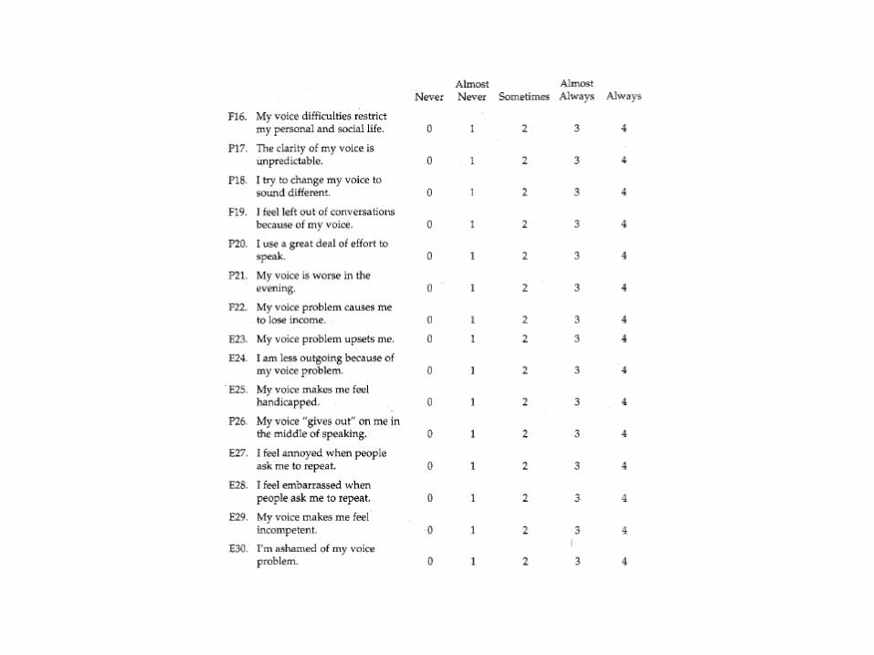
Questionnaire/QOL Survey
History of problem
Medical history
Social history
Oral mechanism examination
Evaluation of voice components
Respiration, phonation, resonance, pitch, intensity, rate
Instrumental analysis Acoustic/aerodynamic analyses
Functional evaluation of vocal fold movement Flexible endoscopy
Videostroboscopy
Impressions
Prognosis
Recommendations

History
History of Problem
Place all of the information obtained during your patient interview into chronological order
Medical History
Patient interview
Medical chart review
Information from physician consult
Review of outside records
Social History
Obtain information from social/work spheres that may have adverse impact on vocal/laryngeal function
Change in family dynamics
Change in employment status
Death in family
Levels of stress secondary to above?

Evaluation of Vocal Components
Respiration
Diaphragmatic versus clavicular/thoracic focus
Degree of upper chest/shoulder/neck tension
Phonation
Qualitative disturbances
Ability to maintain steady pitch
Consistent versus inconsistent
Resonance
Hypo/hypernasal, cul-de-sac, etc.
Pitch
Too high/too low
Limited in range?
Can patient vary the same?
Does the voice change with changes in pitch?
Intensity
Too loud/too soft
Patient’s ability to vary intensity
Any associated vocal changes with variation?
Rate
Too fast/too slow, changes prior to evaluation, coordination w/respiration

Case Study
64-year-old male
Medical dx of acute necrotic pancreatitis
2-month hospitalization
Subsequently underwent elective cholycystectomy
Multiple complications
Extended second hospitalization
Multiple intubations/extubations
Decline in voice subsequent to hospital discharge
Seen by Otolaryngology/Head and Neck Surgery
Dx of right vocal fold granuloma
Tx’d with anti-reflux measures
Referral to Speech Pathology for videostroboscopy

Videostroboscopy
Vocal fold mobility within functional limits bilaterally
Right vocal fold markedly erythematous
Irregular configuration to body of right vocal fold
Edge of right vocal fold irregular
Glottic closure incomplete given edge irregularity of right vocal fold
Amplitudes of vibration/mucosal waves absent on right vocal fold
Vocal fold vibration asymmetric

Marc’s Fab Video Clip

Recommendations
Follow-up with Otolaryngology/Head and Neck
Surgery given concerning appearance of right
vocal fold
Biopsy right vocal fold
Continue with anti-reflux measures per physician
orders
Follow-up with Speech Pathology per
Otolaryngology

Results
Biopsy positive for T1a N0 M0 moderately differentiated squamous cell carcinoma of the right vocal fold
Underwent definitive XRT using IMRT
Post-tx, seen by Speech Pathology X2 for voice therapy
Vocal warm-ups/cool downs
Lip trills
Pitch glides/steps on /oo/
Vocal Function Exercises (Stemple)

References
Bzoch KR (Ed.): Communicative Disorders Related to Cleft
Lip and Palate, 5th Ed., Pro-Ed, Austin, 2004.
Golding-Kushner, K. Therapy Techniques for Cleft palate Speech & Related Disorders, Singular Thomas Learning, San Diego, 2001
Kummer A, Cleft Palate & Craniofacial Anomalies, 2nd Ed., Delmar Cengage Learning, New York, 2007
Peterson-Falzone, S., Hardin-Jones,M., Karnell, M., Cleft Palate Speech, 3RD Ed., Mosby, St. Louis, 2001
Peterson-Falzone, S., Trost-Cardamone, J., Karnell, M., Hardin-Jones, M., The Clinician’s Guide to Treating Cleft Palate Speech, Mosby, St. Louis, 2006
Stemple, J., Glaze, L., Klaben, B., Clinical Voice Pathology Theory and Management, 4th Ed., Plural Publishing, San Diego, 2010
www.cleftline.org (Cleft Palate Foundation)

Thank you
C.S. Mott Children’s
Hospital- Opening 2011

Nasal words and sentence stimuli
Mama, me me, new new, no no, inga inga Mama made some lemon jam. Nancy is a nurse. The monkey had a banana. Hand the mean dog some meat. The swing is neat and clean. Many men walked many miles. Amanda came from Maine. Santa came when the snow fell. Jane came in when the phone rang.

Oral sentences
Buy a baby bib.
Pop a bubble.
Purple paper
Daddy did it.
It’s too tight.
Go get it.
Cookie and cake
Chocolate chip cookies
Dick took Patty.
Peter had a puppy.
Buy a baby bib.
Tell Dad to do it.
Katy had a cookie.
Go get a big egg.
I see a black dog.
Zippers are easy.

Oral sentences
Sissy sees the sky.
Shoes and socks
Stop the bus.
Should I wash the dishes?
Zippers are easy to close.
Jack had a magic badge.
Chad’s teacher was at church.
Check your watch.
Chocolate chip cookies are delicious.
Go get a big egg.