Every Diagnosis Counts HiFISH
7
HiFISH Digital FISH Analysis Platform Every Diagnosis Counts
Transcript of Every Diagnosis Counts HiFISH
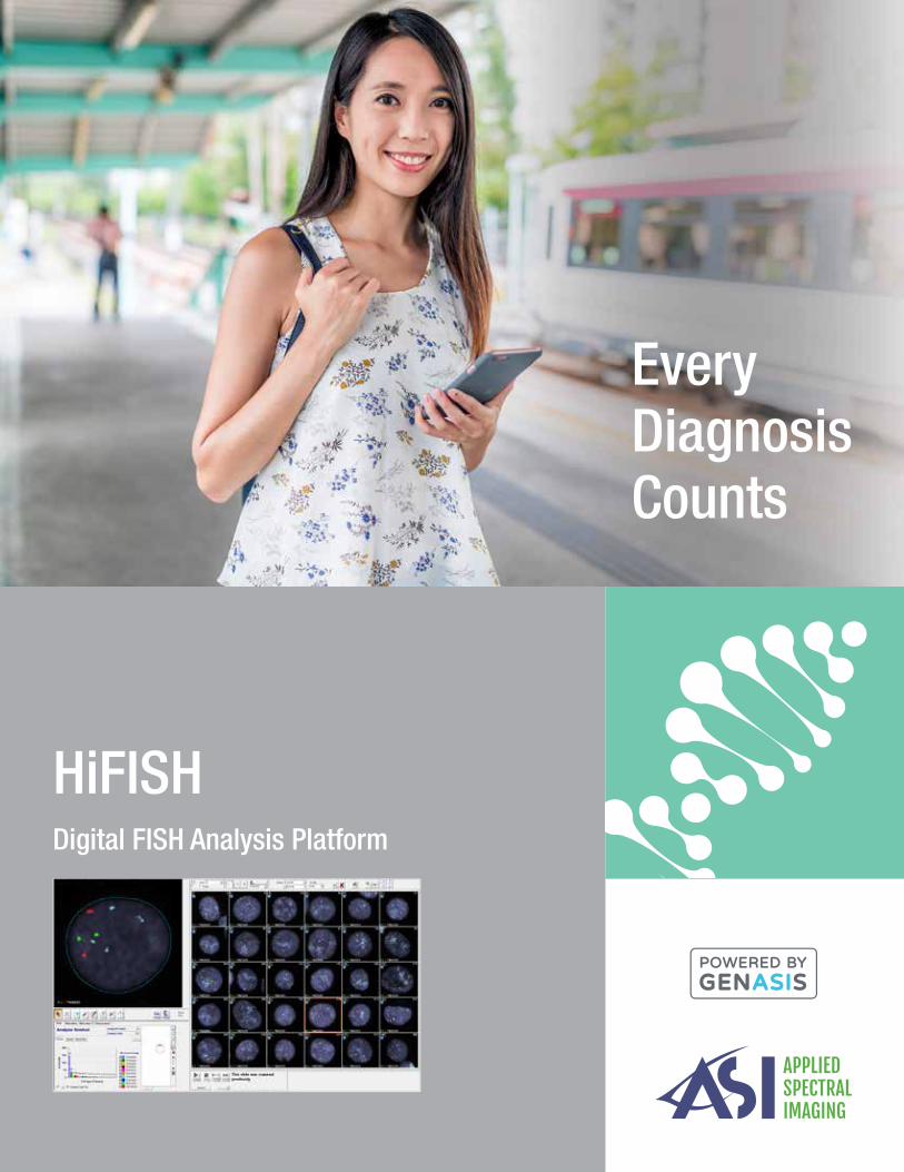
HiFISHDigital FISH Analysis Platform
EveryDiagnosisCounts

1 2 3 4The Ultimate Digital FISH Workflow
Digitize slides, analyze cells and report
Welcome to your optimized FISH testing solution:
Automated FISH scan and capture with real-time analysis
Onscreen review and classification adjustment
Review and approval of final statistics Create report with final statistics and cell images
Scan & Capture
View& Analyze
Review& Approve
Report
Scan Analyze Review Report Complete!

-78% -71% -58%
Accurate computer-assisted analysis provides higher
confidence in patient assessment
DiagnosticConfidence
Onscreen Analysis
Lab Productivity
Digital workflows enable labor efficiencies and time savings every
step of the way
Onscreen FISH analysis in the light significantly improves
technologist user experience
“HiFISH allows us to view much more cells than we would with a manual microscope, which leads to more accurate and reliable results. Manual FISH analysis is also tedious
and leads to technologist fatigue but HiFISH never gets tired.”
Dr. Yarin Hadid, Bnai Zion Medical Center
Cutting-edge scanning instrumentation for in-depth cell imaging and analysis
Exceptional On-screen Image Quality
Increased Lab Productivity with Automation
Wide Application Coverage
Any Sample
Any Probe
Digitize Your SlidesEfficiency, Precision, Versatility
Over 55% Time Savings (Minutes / Case)
HER2/neu (Breast)
Hematology Enumeration
ALK (Lung)
Hematology Fusion
UroVysion (Bladder)
“Digital FISH analysis provides more efficient and accurate results and better patient care in comparison to traditional FISH methods.”
Liew M, Rowe L, Clement PW, Miles RR, Salama ME., J Pathol Inform
HiFISH
Manual Workflow
ALK CLL AML
45
10
50
21
45
13
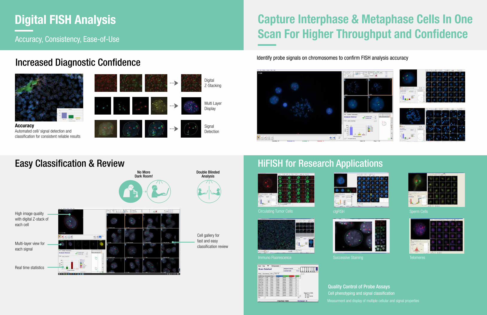
Easy Classification & Review
Increased Diagnostic Confidence
Digital Z-Stacking
Multi Layer Display
Signal Detection
Digital FISH Analysis Accuracy, Consistency, Ease-of-Use
AccuracyAutomated cell/ signal detection and classification for consistent reliable results
High image quality with digital Z-stack of each cell
Multi-layer view for each signal
Cell gallery for fast and easy classification review
Real time statistics
No MoreDark Room!
Double BlindedAnalysis
Quality Control of Probe AssaysCell phenotyping and signal classification
HiFISH for Research Applications
Measurment and display of multiple cellular and signal properties
Circulating Tumor Cells clgFISH
Successive StainingImmuno Fluorescence Telomeres
Sperm Cells
Capture Interphase & Metaphase Cells In One Scan For Higher Throughput and Confidence
Identify probe signals on chromosomes to confirm FISH analysis accuracy

Case Data Management (CDM)
GenASIs AnyWhereTM for Remote AccessLab Connectivity Anytime, Anywhere
Review, analyze and sign off case information from any location via a secured network
Data Management and ConnectivityModern Paperless Workflow
Central Portal and Database Easily Integrates with Lab LIS
LIS Connectivity
Performance
Security
Data Integrity
HIPAA Compliant
Advanced Reporting 1D/2D Barcode Reader
Efficient
Comprehensive
Eliminates human error
Work from Home
Compare performance year on year and make data driven decisions
Annual analysisand review
Calculate performance benchmarks and
track your KPIs. Meet certification and
regulatory requirements
Benchmarks
Justify investment in additional capital
equipment for the lab
Growth
Identify best practices to increase ROI per case and focus improvement
efforts
Optimization
Generate lab performance statisticLabLifeTM for Lab Management s
Become a Data-Driven Lab withLabLife
NEW
NEW
Personalized Levels of AutomationScanning Protocols for All Probes, Samples and Preparation Techniques
Slide Staining
ScanProtocol
Start & Walkaway Scanning
Onscreen Analysis
Digital Report
User selected Fields of View
Semi Automated
High Cellular Density
User pre-configured scanning pattern and stop criteria
Fully Automated
High Cellular Density
DensityScan™ automatic detection and ranking of Fields of View
Fully Automated
Low Cellular Density
Dual scan automatic detection & classification of metaphases & interphases
Fully Automated
Low Cellular Density

4,500 Systems installed
worldwide
60Third party distribution
partners
71Countries through direct & indirect sales forces
About Applied Spectral Imaging
Applied Spectral Imaging (ASI) is a global leader in biomedical imaging with a comprehensive product portfolio and a global distribution footprint.
Founded in 1993, ASI markets, services and supports its products in nearly 60 countries. The Company's technology, powered by GenASIs, enables pathology, cytogenetics and research laboratories to provide advanced diagnostics to patients through superior digital diagnostic tools.
ASI has a wide portfolio of dedicated solutions for brightfield, fluorescence and spectral imaging and analysis, including HiPath Pro, PathFusion, HiBand, HiFISH, CytoPower and Rainbow.
ASI’s wide FDA clearance portfolio includes: FDA clearance for BandView, FISHView, SpotScan for CEP XY, UroVysion, ALK and HER2/neu FISH, and for HiPath IHC Family for HER2, ER, PR, and Ki67, on the manual configuration
The Company has offices in the US and Asia and a global network of distribution partners.
Global Presence
The Company’s Product Portfolio
Diverse platforms can accommodate all laboratory needs
ASI Ltd Asia
ASI Inc USA
Direct sales
Indirect sales
9-Slide Scanning System 1-Slide Capture System99-Slide Tray Loader
HyperSpectral System Review & Analysis Station Remote Connectivity
Imaging & Analysis Solutions for Laboratories
Cytogenetics Pathology Research
HiFISH RainbowPathFusionHiPath ProCytoPowerHiBand

SCAN ME
www.spectral-imaging.com
SCAN ME
System Specifications
North AmericaApplied Spectral Imaging Inc.Tel: +1 760 929 [email protected]
HeadquartersApplied Spectral Imaging Ltd.Tel: +1 817 886 [email protected]
DOC000348 Rev. G
Manual 1 Slide 9 Slide Motorized Stage 99 Slide Tray Loader
Microscope Support
FL upright microscopes OLYMPUS BX61 FLOLYMPUS BX63 FLZEISS AxioImager Z2 FL
OLYMPUS BX61 FLOLYMPUS BX63 FLZEISS AxioImager Z2 FL
Objectives
Olympus10x/0.3NA60x/1.25NA
ZEISS10x/0.3NA63x/1.25NA
Olympus10x/0.3NA40x/1.4NA60x/1.25NA
ZEISS10x/0.3NA40x/1.3NA63x/1.25NA
Olympus4x/0.16NA10x/0.3NA40x/1.4NA60x/1.25NA
ZEISS5x/0.16NA10x/0.3NA40x/1.3NA63x/1.25NA
Camera 5MP CMOS Monochrome 5MP CMOS Monochrome 5MP CMOS Monochrome
Slide Capacity 1 slide 9 slides 99+ slides
Barcode Reader Handheld 1D/2D Handheld 1D/2D Integrated 1D/2D
Automatic Oil Dispenser
N/A Optional Integrated
DimensionsAccording to clients microscope
61cm x 69cm x 85cm(24” x 27.2” x 33.5”)
100cm x 90cm x 90cm (39.4” x 35.5” x 35.5”)
WeightAccording to clients microscope
45Kg99.2lb
80Kg176.4lb