
Evaluation of two biological matrices for repairing of ...
12
The Iraqi Journal of Veterinary Medicine, 42(2):21-32. 2018 21 Evaluation of two biological matrices for repairing of ventral hernia in bucks A.K. Mahdi and Ahmed H. F. AL-Bayati Department of Surgery and Obstetrics, College of Veterinary Medicine, University of Baghdad, Iraq. E-mail: kamelareeg@yahoo.com. Received: 1/3/2018 Accepted: 9/4/2018 Publishing: 31/1/2019 Summary The aim of this study was to estimate the changes in ventral hernia repairing in Iraqi bucks by using two biological matrixes derived from bovine (pericardium and urinary bladder matrix) through histopathological examination. All bucks right lower flank awas prepared surgically, sedation were done by using (2% Xylazine hydrochloride) at a dose of 0.2mg/kg intramuscular, and surgical site anesthetized through an inverted (L) shape local infiltration technique using lidocaine hydrochloride (2%) at a dose of 8mg/kg. Ventral abdominal hernias were induced in (24) bucks through elliptical resection of abdominal muscles to made hernia ring (6-8cm) with avoiding peritoneum perforation. After 30 days of operation bucks were divided into two equal groups. cellular Bovine pericardium group and cellular urinary bladder matrix group. In two groups hernias were treated with only replacement of acellular bovine pericardium and cellular urinary bladder matrix respectively which fixed with interrupted horizontal mattress 2cm far from hernia ring by Polypropylene (No.1) suture material. Histopathological biopsies were taken at 2 nd , 8 th and 16 th week post treatment (4 bucks\ period). Both groups’ successes in reconstruction of large hernia in bucks through prevent recurrent or others post-operative complications. In addition the histopathological examination showed that the acellular urinary bladder matrix superior than acellular bovine pericardium matrix in enhance healing based on acellular urinary bladder matrix role in augment early initiation of inflammatory cells infiltration, fibroblast proliferation and marked collagen deposition, in addition to its early degradation, incorporation and remodeling. Keywords: Hernia, Bucks, Bioimplants, Histopathological.. ------------------------------------------------------------------------------------------------------------------------ Introduction Abdominal wall defects (hernia) correspond a difficult trouble, considered a common acquired condition in ruminant's which has some harmless effects, such as lowering the productivity and reproductively (1 and 2). Furthermore, large full-thickness, abdominal wall defects secondary to wide resection of malignant tumors, traumatic injuries or congenital abnormalities cannot be closed primarily (3). Surgical intervention (herniorrhaphy) is useful in these cases but large ventral abdominal hernia may require hernioplasty such as (Mesh repairs) to minimize the amount of tension that must be put on the abdominal wall in order to cover the hernia (4). However, these materials may in addition to cause infection and chronic pain in the surgical area (5), it may perhaps contribute to the dysfunction of other organs, and cause complications such as internal adherence, obstructions, and fistula development (6). To avoid the potential squeal of synthetic non absorbable materials, biological materials are being developed by the surgeons and used for abdominal wall defect repairs and other applications, such as porcine small intestinal sub mucosa, a cellular dermal matrix and human dura mater (5). The composition and structure of these biomaterials contrast according to the source of the tissue that can provide a reservoir of active molecules like growth factors that can be rapidly mobilized following injury to stimulate cell proliferation and migration (7).
Transcript of Evaluation of two biological matrices for repairing of ...
The Iraqi Journal of Veterinary Medicine, 42(2):21-32. 2018
21
Evaluation of two biological matrices for repairing of ventral hernia in bucks
A.K. Mahdi and Ahmed H. F. AL-Bayati
Department of Surgery and Obstetrics, College of Veterinary Medicine, University of Baghdad,
Iraq.
E-mail: [email protected].
Received: 1/3/2018
Accepted: 9/4/2018 Publishing: 31/1/2019
Summary
The aim of this study was to estimate the changes in ventral hernia repairing in Iraqi bucks by using two
biological matrixes derived from bovine (pericardium and urinary bladder matrix) through histopathological
examination. All bucks right lower flank awas prepared surgically, sedation were done by using (2%
Xylazine hydrochloride) at a dose of 0.2mg/kg intramuscular, and surgical site anesthetized through an
inverted (L) shape local infiltration technique using lidocaine hydrochloride (2%) at a dose of 8mg/kg.
Ventral abdominal hernias were induced in (24) bucks through elliptical resection of abdominal muscles to
made hernia ring (6-8cm) with avoiding peritoneum perforation. After 30 days of operation bucks were
divided into two equal groups. cellular Bovine pericardium group and cellular urinary bladder matrix group.
In two groups hernias were treated with only replacement of acellular bovine pericardium and cellular
urinary bladder matrix respectively which fixed with interrupted horizontal mattress 2cm far from hernia ring
by Polypropylene (No.1) suture material. Histopathological biopsies were taken at 2nd, 8th and 16th week post
treatment (4 bucks\ period). Both groups’ successes in reconstruction of large hernia in bucks through
prevent recurrent or others post-operative complications. In addition the histopathological examination
showed that the acellular urinary bladder matrix superior than acellular bovine pericardium matrix in
enhance healing based on acellular urinary bladder matrix role in augment early initiation of inflammatory
cells infiltration, fibroblast proliferation and marked collagen deposition, in addition to its early degradation,
incorporation and remodeling.
Keywords: Hernia, Bucks, Bioimplants, Histopathological..
------------------------------------------------------------------------------------------------------------------------
Introduction
Abdominal wall defects (hernia) correspond
a difficult trouble, considered a common
acquired condition in ruminant's which has
some harmless effects, such as lowering the
productivity and reproductively (1 and 2).
Furthermore, large full-thickness, abdominal
wall defects secondary to wide resection of
malignant tumors, traumatic injuries or
congenital abnormalities cannot be closed
primarily (3). Surgical intervention
(herniorrhaphy) is useful in these cases but
large ventral abdominal hernia may require
hernioplasty such as (Mesh repairs) to
minimize the amount of tension that must be
put on the abdominal wall in order to cover the
hernia (4). However, these materials may in
addition to cause infection and chronic pain in
the surgical area (5), it may perhaps contribute
to the dysfunction of other organs, and cause
complications such as internal adherence,
obstructions, and fistula development (6). To
avoid the potential squeal of synthetic non
absorbable materials, biological materials are
being developed by the surgeons and used for
abdominal wall defect repairs and other
applications, such as porcine small intestinal
sub mucosa, a cellular dermal matrix and
human dura mater (5). The composition and
structure of these biomaterials contrast
according to the source of the tissue that can
provide a reservoir of active molecules like
growth factors that can be rapidly mobilized
following injury to stimulate cell proliferation
and migration (7).

The Iraqi Journal of Veterinary Medicine, 42(2):21-32. 2018
22
The current study aimed to compare the
efficacies of using of two natural bio-scaffolds
including; acellular bovine pericardium matrix
(BP) and acellular urinary bladder matrix
(UBM) in the reconstruction of experimentally
induced large ventral hernias in Iraqi bucks.
Materials and Methods
After obtaining an official approval from the
ethical committee of the college. Twenty four
apparently healthy adult local bucks aged 1-2
years and weighing (25-30) kg were used.
Bucks were acclimatized for 2 weeks in pens
at the College of Veterinary Medicine,
University of Baghdad, Iraq before operation.
The animals were divided and numbered
according to the experimental design and each
animal was injected s.c with Ivermectin as
antihelminthic in a dose 0.2mg/kg., B.W and
s.c vaccinated with 5ml of enterotoxaemia
vaccine.Ventral abdominal hernias were
induced in the right lower flanks of 24 bucks
before herniation, food was withheld for 24
hours and water for 12 hours. Buck was
sedated with (Xylazine hydrochloride 2%) at a
dose of 0.2mg/kg B.W IM. In addition surgical
site were anesthetized locally by using
lidocaine hydrochloride (2%) at a dose of
8mg/kg B.W through an inverted (L) shape
local infiltration technique. A vertical incision
10-12cm was done through skin and
subcutaneous tissue, then abdominal muscles
were separated bluntly, to create hernia 6-8cm
of full-thickness abdominal muscles were
resected with avoiding opening the peritoneum
(Fig.1). Finally, skin and subcutaneous layers
were sutured using simple interrupted pattern
with non-absorbable suture materials (Silk
No.1) and wounds were covered with sterile
gauze.
Figure, 1: Shows hernia ring after excision of
abdominal wall muscles.
Whole fresh urinary bladders of cows were
excised immediately after slaughtered at local
abattoir. The matrix was prepared as described
by (7 and 8) to make it acellular to prevent
rejection. The urinary bladder was filled with
tap water to facilitate trimming of external
connective tissues and adipose tissue with
scissors and washed in tap water. The tunica
serosa, tunica muscularis and most of the
muscularis mucosa were mechanically
delaminated from bladder tissue by scraping
with blunt knife. The remaining basement
membrane of the tunica mucosa and the
subjacent tunica propria became a urinary
bladder matrix (UBM), decellularized and
disinfected by immersion in peracetic acid
(0.1%) and ethanol (4%) for two hours. Traces
of peracetic acid were removed and pH
adjusted to ~7.4 by rinsing in phosphate
buffered saline (PBS) with 100 IU/ml
penicillin, 100 µg/ml streptomycin and 100
µg/ml amphotericin. At room temperature the
scaffold was rinsed and shaken in deionized
water twice, and once in PBS for 15min. The
scaffold was then sterilized by immersion in
peracetic acid solution (0.1%) titrated to pH
7.0 at room temperature for five hours.
Bovine pericardium was excised from
slaughtered cows at local abattoir and
submerged in PBS (pH 7.4) and rinsed in
saline until clean of blood. Fat and connective
tissues was removed with dry gauze. The
tissue was decellularized as mentioned by (9)
with the same steps were done for UBM
decellularization. After 30 days post inducing
of hernia, all animals were allocated into two
main equal groups. In Bovine pericardium
group (BP), the hernias ring was closed with a
sheets of acellular BP sheets. Under the effect
of sedation and local anesthesia, as mention
before, the area of the operation was ready for
aseptic operation and vertical skin incision was
made parallel to the original skin incision.
Gentle blunt dissections of the underlying
tissues to free the adhesions until defined the
borders of the hernial ring and then the hernial
sac was inverted into the abdominal cavity.
Implants were soaked in sterile PBS
(containing antibiotics and antifungal) for

The Iraqi Journal of Veterinary Medicine, 42(2):21-32. 2018
23
three hours before operation, onlay repair
technique was done and BP implant firmly
fixed with the muscular layers of the hernia
ring by interrupted stitches of horizontal
mattress with Polypropylene (No.1) no less
than 2cm far from hernia edges. The access
skin and s.c tissue was sharply excised then
skin closed. The same procedures as
mentioned for first group were performed in
urinary bladder group (UBM); with exception
that the hernias were closed with acellular
UBM implant (Fig.2). Post-treatment of
hernia, sterile bandage truss were used for
each buck to care the surgical site and to
lessen postoperative edema. To reduce the
activity of animals and avoid the trauma all
animals were housed in separated pen. During
first five days, the animals received soft food
which increased gradually with the time
associated with daily administration IM of a
combination of penicillin and streptomycin in
a dose of 10.000 IU/ kg and 10 mg/kg body
weight respectively for 5days.
Figure 2: Shows the onlay fixation of the UBM
sheet with interrupted horizontal mattress using
Polypropylene (No.1).
The histopathological evaluation was
performed at 2, 8, and 16 weeks post-
implantation (four animals/period). Segments
of (1x1) cm3 were collected from three areas;
edge of the implant, center and between them.
The samples were fixed in neutral buffered
formalin solution (10%) and set in paraffin and
then sectioned longitudinally and transversally
at (5-7) µm and staining with Hematoxylin-
Eosin (H&E) and Mallory trichrome stain
which stain collagen blue color and
granulation tissue yellowish color (10).
The results of the histopathological score of
progression of healing process in the site of
hernias of both treatment groups were
evaluated through using a scoring system, as
mentioned in (Table,1), illustrated by (11).
Higher scores within the scoring system
represent more approving outcomes with
rating to remodeling, as evidenced by cellular
infiltration, host ECM deposition,
neovascularization, scaffold degradation,
fibrous encapsulation and cell types that
represent low levels of inflammation.
Data were analyzed using SAS (Statistical
Analysis System-Version 9.1). Two ways
ANOVA and least significant differences
(LSD) post hoc test were performed to assess
significant difference among means P≤0.05
was considered statically significant.
Table,1: Histopathological scores of progression of healing process at the site of hernias (11).
scores 0 1 2 3
Cellular infiltration Zero cells in contact
with Scaffold.
Cells contact periphery, no
penetration into scaffold.
Cells infiltrate scaffold,
not reach center
Cells penetrate into
center of scaffold
Cell types (neutrophiles,
macrophages ,and foreign
body giant cells
Inflammatory cells
present, no fibroblasts
Primarily inflammatory cells,
few fibroblasts
Primarily fibroblasts
few inflammatory cells
Fibroblasts only, no
inflammatory cells
Host extracellular matrix
(ECM) deposition.
No host ECM
deposition
Host ECM deposited
atperiphery of scaffold
Host ECM deposited
Inside scaffold, but not at the center
Host ECM deposited
inside scaffold, including the center
Scaffold degradation Original scaffold
intact, Borders clearly
demarcated
Scaffold partially degraded,
layers separated by cells,
blood vessels, host tissue, etc.
Scaffold extremely
degraded, difficult to
distinguish scaffold
from host tissue
Scaffold completely degraded, no
evidence of original scaffold.
Fibrous encapsulation Extensive
encapsulation (50–100%) of
periphery)
Moderate encapsulation (25–
50% of periphery)
Mild encapsulation
(≤25% of periphery)
No fibrous encapsulation
Neovascularization Zero blood vessels
presence
Vessels present at scaffold
periphery, no penetration into scaffold
Vessels infiltrate
scaffold but not reach center of scaffold
Vessels penetrate into
center of scaffold

The Iraqi Journal of Veterinary Medicine, 42(2):21-32. 2018
24
Results and Discussion
Clinically, all experimental bucks were kept
under inspection along the period of the study
to record general health, behavior and
alertness, with no death of any animal. Ventral
hernia was noticed directly post-operations of
inducing hernias, there were appeared as clear
tense swelling in the right low flank of the
abdominal wall. Post-treatment of hernias, the
hernia sac was disappeared, and local
inflammatory reaction was gradually
disappeared within 5 to 7 days, as well as,
clinical followed-up of both treatment groups
not recorded any complications, except
different sizes of seromas were formed in three
bucks of each treatment groups which were
detected during 3 days post-implantation by
gradual formation of sac like swelling at the
site of implantation and treated by exploratory
aspiration of the serous fluids under aseptic
technique. Along the period of the study, one
case of reherniation was noticed in UBM
group at 5 days may be due to severe and
continues coughing which associated to
aspiration pneumonia.
In the present study, hernias were treated
after 4th weeks post-inducing to create
complicated hernia and fibrosis of hernia ring.
All implants fixation were success without
tearing through suturing with ring site. This
result closed to (12), whom referred that the
hernia repairing should be performed at least
four weeks post-inducing may be due to it
suitable time for resolution of active
inflammation.
Alternatively, only replacement was used in
this study is a successful technique with this
biological implant and hernia size, it gave
improvement of tissue healing as showed in
histopathological results, without
complications or recurrent. The same
observations were noticed by (13), whom
explained that onlay implantation allows of
tissue in growth by two directions and (14),
demonstrated that the contact of biomaterial
with the fascia over or under the rectus
muscles stimulates re-growth of the host
tissue, and promotes the production and
deposition of collagen on the suture line in
addition to acceleration of wound healing.
In the current study, seroma was occurred in
three animals for each treated group during 72
hours post-hernia treatment. Many previous
studies like (15 and 16) recorded the formation
of seromas in more than one case at the same
period of the present study post-hernioplasty.
The causes of seroma formation in the current
study may be due to dissections that form
separation between skin and subcutaneous
tissue, as well as, the manipulation at surgical
site for implantation that cause aseptic
inflammation. These reasons are the same as
recorded by (17), who referred to seroma
formation due to local inflammatory response
to a mechanical injury during surgical
dissection and the presence of foreign bodies.
While, (18) noticed that the abdominal wall
approaching (ventral incision) lead to
accumulation of blood and serum among the
various level of wound resulting in hematoma
or seroma or dead space.other authors (19)
referred to the seroma formation are, the blood
and lymphatic vessel injury during dissection,
dead-space formation, shear forces among
layers and the release of inflammatory
mediators .
Wound infections weren’t noticed along the
fallowed up period, it may be related to the
biological implants components. Many studies
were confirmed the antimicrobial effects of
natural bio-scaffolds therefore increased
frequency of their using to repair contaminated
abdominal wall defects (20 and 21). Other
result chers (22) suggested the presence of
several different low molecular weight
peptides with antibacterial activity exist within
ECM and such peptides can resist the bacterial
infection, and there were differences between
antimicrobial peptides in each bioscaffolds
type . Histopathological section at 2weeks post
implantation in BP group revealed presence of
highly celluar and vascular immature
granulation tissue with severe hemorrhage and
numerous congested capillary and moderate
mononuclear cells (MNCs) (Fig.3), in another
section there were a multifocal MNCs
aggregation consist mainly of macrophages
and lymphocytes with proliferation of
fibroblasts producing collagen fibers (Fig.4),
and identification of a fragment of muscles
fibers observed within the necrotic area with
calcium precipitation (Fig.5).

The Iraqi Journal of Veterinary Medicine, 42(2):21-32. 2018
25
Figure, 6: BP-treatment group at 8th week post
implantation shows fragments of pericardial implant
(long arrow) and high vascularization, presence of
immature collagen fibers and blood vesesles in the
host tissue(short arrow) (Mallory stain, 200X).
Figure, 7: BP-treatment group at 8th week post
implantation shows vascular (immature) granulation
tissue (H&E stain, ).
Figure, 8: BP-treatment group at 8th week post
implantation, shows presence of thick collagen fibers
(long arrow) with moderate vascular granulation
tissue (short arrow) (Mallory stain, 200X).
The histopathological section at 8 weeks post
implantation in BP group showed a fragments of
BP and high vascularization, presence of immature
collagen fibers and blood vessels (BVs) in the host
Figure, 3.: Histopathological section in BP-
treatment group at 2nd week post implantation,
shows the presence of highly celluar vascular
immature granulation tissue with severe
hemorrhage(long arrow) numerous congested
capillary(short arrow) and moderate
mononuclear cells (MNCs) (H&E stain, 100X)
.
Figure, 4: BP-treatment group at 2nd week post
implantation, shows a multifocal MNCs
aggregation (long arrow) consist mainly of
macrophages and lymphocyte with
proliferation of fibroblast producing collagen
fibers (H&E stain, 100X) .
Figure, 5: BP-treatment group at 2nd
week post implantation, shows fragment
of muscles fibers observed within the
necrotic area (long arrow) with calcium
precipitation (short arrew) (Mallory
stain, 100X).
The Iraqi Journal of Veterinary Medicine, 42(2):21-32. 2018
26
tissue (Fig.6), in another section, vascular
(immature) granulation tissue was seen (Fig.7),
with presence of thick collagen fibers with
moderate vascular granulation tissue (Fig.8).
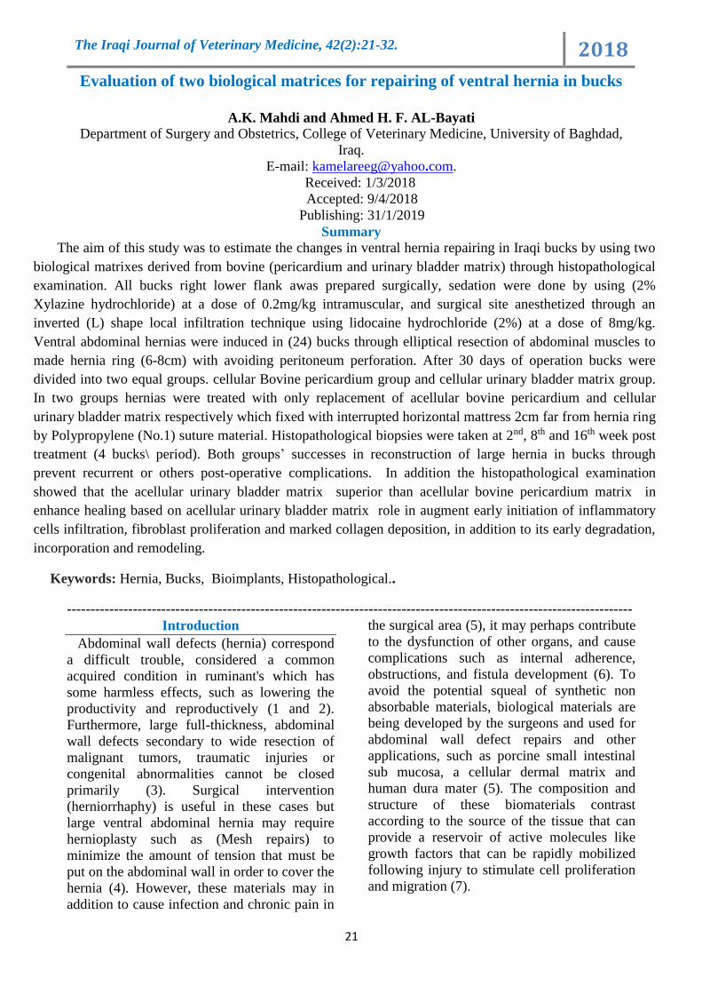
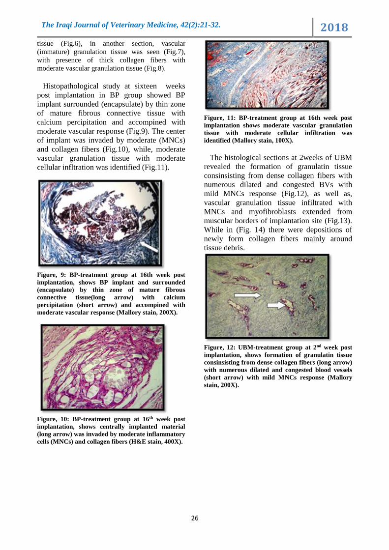
Histopathological study at sixteen weeks
post implantation in BP group showed BP
implant surrounded (encapsulate) by thin zone
of mature fibrous connective tissue with
calcium percipitation and accompined with
moderate vascular response (Fig.9). The center
of implant was invaded by moderate (MNCs)
and collagen fibers (Fig.10), while, moderate
vascular granulation tissue with moderate
cellular infltration was identified (Fig.11).
Figure, 9: BP-treatment group at 16th week post
implantation, shows BP implant and surrounded
(encapsulate) by thin zone of mature fibrous
connective tissue(long arrow) with calcium
percipitation (short arrow) and accompined with
moderate vascular response (Mallory stain, 200X).
Figure, 10: BP-treatment group at 16th week post
implantation, shows centrally implanted material
(long arrow) was invaded by moderate inflammatory
cells (MNCs) and collagen fibers (H&E stain, 400X).
Figure, 11: BP-treatment group at 16th week post
implantation shows moderate vascular granulation
tissue with moderate cellular infiltration was
identified (Mallory stain, 100X).
The histological sections at 2weeks of UBM
revealed the formation of granulatin tissue
consinsisting from dense collagen fibers with
numerous dilated and congested BVs with
mild MNCs response (Fig.12), as well as,
vascular granulation tissue infiltrated with
MNCs and myofibroblasts extended from
muscular borders of implantation site (Fig.13).
While in (Fig. 14) there were depositions of
newly form collagen fibers mainly around
tissue debris.
Figure, 12: UBM-treatment group at 2nd week post
implantation, shows formation of granulatin tissue
consinsisting from dense collagen fibers (long arrow)
with numerous dilated and congested blood vessels
(short arrow) with mild MNCs response (Mallory
stain, 200X).

The Iraqi Journal of Veterinary Medicine, 42(2):21-32. 2018
27
Figure, 13: UBM-treatment group at 2nd week post
implantation, shows vascular granulation tissue was
infiltrated with MNCs and myofibroblasts extended
from muscular borders of implantation site (H &E
stain, 400).
Figure, 14: UBM-treatment group at 2nd week post
implantation shows deposition of newly form
collagen fibers mainly around tissue debris (long
arrow) (Mallory stain, 100X).
The host tissue at 8 weeks post implantation
exhibited peripheral aggregation of MNCs
forming granulomatous like reaction as shown
in (Fig.15). In addition shows the construction
of implant with delicate irregular collagen
fibers (Fig. 16).
Figure, 15: UBM-treatment group at 8th week post
implantation, shows that host tissue (long arrow)
exhibited peripheral aggregation of MNCs (short
arrow) forming granulomatous like reaction (H&E
stain, 200X)
Figure, 16: UBM-treatment group at 8th week post
implantation shows the construction of implant with
delicate irregular collagen fibers (long arrow) (H&E
stain, 400X).
At sixteen weeks post implantation, the
histopathological finding characterized by
formation of thick regular collagen fibers
infiltrated with MNCs which attached to host
muscular layer and incorperation of new
collagen bandle within muscular coat (Fig.17).
In addition, areminante of implant near the
suture material surrounded by new fibrous
tissue (Fig.18).
Figure,17: UBM-treatment group at 16th week post
implantation, shows regular collagen fibers
infiltrated with MNCs which attached to host
muscular layer and incorporation of new collagen
(H&E stain,200X)
Figure, 18: UBM-treatment group at 16th week post
implantation, shows the remnant of implant (long
arrow) near the suture material surrounded by new
fibrous tissue (Mallory stain, 400X).

The Iraqi Journal of Veterinary Medicine, 42(2):21-32. 2018
28
The analysis results of the histopathological
score (Table, 2) gives well information about
the role of both implants in enhanced healing,
and reflected that despite the significant
difference (P≤0.05) between both group at the
2nd and 8th week, while no significant
differences was confirmed at 16th week.
Table ,2 : Shows the Means± SE of histopathological score results for both treatment groups.
LSD 16 weeks 8 weeks 2 weeks Group Reaction score
0.9945
A 3.00±0.57a B 2.00±0.14b C 1.00±0.28b BP Cellular
Infiltration A 3.00±0.14a A 3.00±0.28a B 2.00±0.14a UBM
0.6022 A2.00±0.28b A 2.00±0.28b B 1.00±0.14b BP Cell type
(inflammatory cells) A 3.00±0.14a A 3.00±0.00a B 2.00±0.14a UBM
0.9945 A2.00±0.28b A 2.00±0.28b B 1.00±0.28b BP Host (ECM)
deposition A 3.00±0.14a A 3.00±0.28a B 2.00±0.28a UBM
0.7032 A2.00±0.28b B 2.00±0.28a C 1.00±0.14b BP Scaffold
Degradation A3.00±0.28a B 2.00±0.14a B 2.00±0.14a UBM
0.629 B 2.00±0.00b C 1.00±0.00a A 3.00±0.28a BP Fibrous
Encapsulation
A 3.00±0.28a C 1.00±0.00a B 2.00±0.28b UBM
0.7263 A 3.00±0.28a A 3.00±0.00a B 2.00±0.00a BP
Neovascularization.
C 1.00±0.28b A 3.00±0.28a B 2.00±0.28a UBM
The results of the histopathological score of
cellular infiltration along the experimental
study confirmed significant differences
(P≤0.05) in both treatment groups especially
fibroblasts, at 2nd week PO in UBM treated
animals compared with BP treated animals and
progress with time. Awell progression of the
healing process in UBM treatment group
compared to BP treatment group which was
manifested by vascular granulation tissue
formation, peripherial fibroblasts activity
which produced dense collagen fibers
formation and myofibroblasts infiltration.
These results enclosed with (7) who confirmed
that the using of UBM-implants enhanced the
cellular integrity and induces early
inflammatory cells infiltration in early phase
of implantation for hernia repair in rabbits
compared to the untreated tissues., as well as,
(23) confirmed the ability of UBM type of
bioscaffolds to facilitate the restoration of
normal site appropriate tissue when used for
hernia repair. While (24), observed that BP-
implant were replaced with immature fibrous
tissue and later newly developed of layer of
connective tissue consisting of delicate
collagen fibers after
The analysis results of ECM deposition and
host degradation illustrated significant
differences (P≤0.05) in both groups along the
period of the study the mean values were
lower at 2nd week and increased with time in
both treatment groups. In addition in UBM-
implants were faster than BP-implants,
histopathological sections appeared the
deposition or invited of new tissue to replace
the scaffold in UBM group at 2nd week.
While, in BP treated group the deposition
showed periphery and not reach the center till
16th week post-implantation . Alternatively,
histopathological score of presence study
showed different between both treatment
groups in inflammatory cells infiltration, host
ECM deposition and matrix degradation in
which UBM-implants were faster than BP-
implants in degradation and host deposition, as
well as, early cellular infiltration. These results
enclosed with (25 and 26) referred that the
degradation of BP implants occurs in uniform
and gradual. While (27), confirmed that the
UBM-implants rapidly degraded and turnover
(remodeling). In addition (28), referred that the
physical and chemical properties of
biomaterials could influence on the intensity
and duration of the inflammatory response.The

The Iraqi Journal of Veterinary Medicine, 42(2):21-32. 2018
29
result of degradation of the implants showed
significant differences (P≤0.05) in both group
for each period of study. The mean values in
both groups were lower at 2nd week and
progress with time. The means in BP animals
group were lower than UBM treated group for
all periods. The result of fibrous encapsulation
also recorded significant differences (P≤0.05)
for both groups along the studied periods, that
mean early encapsulation in UBM treated
group compared to BP treated group that mean
well incorporation were occur in early time in
UBM group .
The analysis neovascularization score reflect
the site of blood vessels were distributed, there
were a significant differences (P≤0.05) along
the studied period in both groups, while no
significant differences between both groups at
each period except that at 16th week which
shows higher means in BP group than that in
UBM group. In the current study showed early
and faster degradation of UBM implants
compared to BP implants, this fact may related
to early infiltrations of inflammatory cells in
UBM treatment group that may be enhanced
more attraction to fibroblasts cells and
collagen deposition. Realy bioscaffolds
degradation can stimulates the releasing of the
inherent bioactive constituents and promote
host cells deposition. This fact was noticed by
(29), referred the degradation process has a
significant biological activity which stimulates
the releasing of the inherent bioactive
constituents subsequently promote tissue
neovascularization and host-cell deposition (30
and 31) through encouraging cells attachment,
proliferation, differentiation, maturation and
angiogenesis .
Histopathological results also reflect the role
of UBM implant in augmented remodeling
process, it may be related to UBM and BP
matrix composition and contains like growth
factors or cytokines which retained in implant
that causes change in the cells quantity and
quality that’s precipitated in the healing
process and augment remodeling when
compared to BP implants group. This fact
explained by (22 and 32), indicated about the
different in the products of each ECM that
related to its source, these products have
multiple biological properties including
angiogenic, chemotactic and antibacterial
activity when liberating growth factors and
cytokines during their degradation process by
the host proteolytic enzymes .
The results of histopathological score of the
present study appeared that UBM implant
encapsulated started at 2nd week post-
implantation prior to BP and progress with
time to thick regular collagen fibers while in
BP treated group showed it encapsulated by
thin zone of mature fibrous connective tissucs
with calcium percipitation, irregular collagen
fibers. This result support the idea of UBM
implant cause or induced early initiation of
inflammatory response and fibroblast
attraction compared to BP implants. The same
outcomes were recognized by (33 and 34),
who proved that the biological scaffold
encapsulation indicates an accelerated
progression of the inflammatory response. In
addition (24), confirmed that the dense fibrous
capsule which forms around implant during
remodeling was responsible for the firm
incorporation between the implant and the host
tissue, as well as, the zone of capsule did not
interfere with the progression of implant
degradation . All that change in the results of
histological score included rate of
inflammatory cells infiltration and implant
degradation or deposition may be related to the
change in the implant ultra-structure. This
result agree with results of a study by (35 and
36), were mentioned that UBM implants
allowed or augmented fibroblasts proliferation
and collagen production due to it contains
collagen more than 90 % of whole matrix,
specially collagen type-III that consider a
homo-trimeric fibrillar collagen which
providing initial support for cell migration and
adherence and facilitates ECM turnover and
remodeling. While, BP implants augments
neovascularization due to collagen type-I is the
predominant component of pericardial tissue
which arranged hierarchically in different
levels, as well as, it has been shown to induce
activation of mitogen activate protein kinase
pathways that promote angiogenesis (37).
In addition, differences in collagen
orientation between BP and UBM-implants
may have a role in change of host response
and healing process, BP matrix has a wavy
shape of collagen fibers in all species, more
intense curly appearance in bovine than other

The Iraqi Journal of Veterinary Medicine, 42(2):21-32. 2018
30
species of mammals but, the collagen in UBM
implants have unidirectional as showed by
(38). While (39 and 40), explained, that the
orientation of ECM components, such as
collagen fibers, can profoundly influence the
directed migration of cells, possibly by
potentiating growth factor receptor signaling
or by mechanically reinforcing cells migration.
It is concluded that, the present study
demonstrated that the only implantation
technique of acellular BP and UBM were a
simple, less cost-effective and safe methods
with minor non-serious complications to
support large abdominal wall hernias. In
addition UBM implant superior than BP
implant in enhance healing based on early
initiation of inflammatory cells infiltration,
fibroblast proliferation and marked collagen
deposition, in addition to its early degradation,
incorporation and remodeling.
References
1. Jettennavar, P.S., Kalmath, G.P. and
Anilkumar, M.C. (2010). Ventral
Abdominal Hernia in a Goat. J.
Veterinary World, 3(2):93.
2. Das, B.C., Nath, B.K.; Pallab, M.S.;
Mannan, A. and Biswas, D. (2012).
Successful management of ventral
abdominal hernia in goat: a case report
.International J. of Natural Sci.
2(2):60-62
3. Cavallaro, A.; Lo Menzo, E.; Di Vita,
M.; Zanghì, A.; Cavallaro, V. and
Veroux, P.F. (2010).Use of biological
meshes for abdominal wall
reconstruction in highly contaminated
fields. World J. Gastroenterol.,
16:1928-1933.
4. Abdin-Bey, M.R. and Ramadan, R.O.
(2001). Retrospective Study of Hernias
in Goats. Scientific J. of King Faisal
University (Basic and Applied
Sciences). 2(1):77-88.
5. Song, Z.; Peng, Z.; Liu, Z. and Yang, J.
(2013). Reconstruction of abdominal
wall musculofascial defects with small
intestinal submucosa scaffolds seeded
with tenocytes in rats. Tissue
Engineering, Part A, 19(13-14):1543–
1553.
6. Jacob, B.P.; Hogle, N.J.; Durak, E.;
Kim, T. and Fowler, D.L. (2007).
Tissue ingrowth and bowel adhesion
formation in animals comparative
study polypropelene versus proceed
versus paroete composite. Surgical
Endoscopy, 3(1):23-29.
7. Eberli, D.; Atala, A.; Yoo, J.J. (2011).
One and four layer acellular bladder
matrix for fascial tissue reconstruction.
J. of Materials Science., 22(3):741-751.
8. Rosen, M.J. (2010). Biological mesh
for abdominal wall reconstruction; a
critical appraisal. Am. Surg., 76(1):1-6.
9. Freytes, D.O; Tullius, R.S. and
Badylak, S.F. (2006). Effect of storage
upon material properties of lyophilized
porcine extracellular matrix derived
from the urinary bladder. J. of
Biomedical Materials Res. Part B:
Applied Biomaterials, 78(2):327-333.
10. Bancroft, J.D. (2008). Theory and Practice
of histological technique. 6th ed, Churchill
Livingstone Elsevier, pp:8.
11. Valentin, JE. Badylak, JS.; McCabe,
GP. and Badylak, SF. (2006).
Extracellular matrix bioscafolds for
orthopaedic applications. A
comparative histological study. J. Bone
Joint Surg. Am., 88(12):2673–2686.
12. Monteiro, J.A.; Delossantos, A.I.;
Rodriguez, N.L.; Michael, P.P., Franz, G.
and Wagnerm, C.H.T. (2013). Porcine
incisional hernia model: Evaluation of
biologically derived intact extracellular
matrix repairs. J of Tissue Eng., 4:1-7.
13. Holihan, J.L.; Nguyen, D.H.; Nguyen,
M.T.; Mo, J.; Kao, L.S. and Liang,
M.K. (2015). Mesh Location in open
ventral hernia repair: A systematic
review and network meta-analysis.
World J. Surg., 40(1):89-99.
14. .Gurrado, A.; Franco, I.F.; Lissidini,
G.; Greco, G.; De Fazio, M.; Pasculli,
A. (2014). Impact of pericardium
bovine patch (Tutomesh®) on
incisional hernia treatment in
contaminated or potentially
contaminated fields: retrospective
comparative study. Hernia, 19(2):259-
266.
15. Al-Asadi, R.N. (2005). A comparative
study of three surgical techniques for
reconstruction of experimentally
The Iraqi Journal of Veterinary Medicine, 42(2):21-32. 2018
31
induced large ventral hernia in goats.
Ph.D. Thesis in Veterinary Medicine.
Univ. of Baghdad, Baghdad-Iraq.
16. Hummadi, S.K. (2011). Hernioplasty
of experimentally induced ventro-
lateral hernia in bucks using silk suture
versus polypropelen suture. MSc.
Thesis in Veterinary Medicine. Univ.
of Baghdad, Baghdad-Iraq.
17. Bendavid, R. and Kux, M. (2001).
Seromas In abdominal wall hernias:
Principles and management. Edited by
Bendavid, R.; Abrahamson, J.;
Arregui, M.E.; Flament, J.B.; Phillips,
E.H. New York: Springer, Pp:753-756.
18. Schessel, S.; Ralph, G. and Ran, K.
(2002). The management of
postoperative disrupted abdominal
wall. Am. J. Surg., 184(3): 263-268.
19. Westphalen, A.P.; Araújo, A.C.F;
Zacharias, P.; Rodrigues, E.S.; Fracaro,
G.B. and Filho, G.D. (2015). Repair of
large incisional hernias. To drain or not
to drain.Randomized clinical trial.
Acta. Cir. Bras., 30(12): 31-38.
20. Cole, W.C.; Balent, E.M.; Masella,
P.C.; Kajiura, L.N.; Matsumoto, K.W.
and Pierce, L.M. (2015). An
experimental comparison of the effects
of bacterial colonization on biologic
and synthetic meshes. Hernia,
19(2):197–205.
21. Köckerling, F.; Alam, N.N.; Narang,
S.K.; Daniels, L.R. and Smart, N.J.
(2015). Biological Meshes for Inguinal
Hernia Repair–Review of the
Literature. Front.Surg.,(2):48-54.
22. .Brennan, E.P.; Reing, J.; Chew, D.;
Myers-Irvin, J. M.; Young, E.J. and
Badylak, S.F. (2006). Antibacterial
activity within degradation products of
biological scaffolds composed of
extracellular matrix. Tissue
Engineering, 12:2949-2955.
23. Sasse, K.C.; Warner, D.L.; Ackerman,
E. and Brandt, J. (2016). Hiatal hernia
repair with novel biological graft
reinforcement. JSLS., 20(2):
e2016.00016.
24. Abouelnasr, K.S.; Zaghloul, A.E. and
Karrouf, G.I. (2014). Comparative
evaluation of glycerolized bovine
pericardium implant with prolene mesh
for closure of large abdominal wall
defects in dogs. Iranian J. of Veterinary
Research., 15(3) 211-217.
25. Hafeez, Y.M.; Zuki, A.B.Z.; Yusof,
N.; Asnah, H.; Loqman, M.Y. and
Noordin, M.M. (2005). Effect of
freeze-drying and gamma irradiation
on biomechanical properties of bovine
pericardium. Cell and tissue banking.
6(2): 85-89.
26. Eva, G.; Babuci, S.; Tica, C.;
Petrovici, V.; Nacu, V.; Ionescu, C.
and Negru, I. (2017). Comparative
cellular local response in abdominal
defect plastic surgery with bovine
pericardium and bovine fascia
preserved in formaldehyde in
experimental rabbits. ARS Medica
Tomitana, 2(23):83-93.
27. Remlinger, N.T.; Gilbert, T.W.;
Yoshida, M.; Guest, B.N.; Hashizume,
R. and Weaver, M.L. (2013). Urinary
bladder matrix promotes site
appropriate tissue formation following
right ventricle outflow tract repair .J of
Organogenessis, 9(3):149–160.
28. Di Vita, G.; Milano, S.; Frazzetta, M.;
Patti, R.; Palazzolo, V. and Barbera, C.
(2000). Tension-free hernia repair is
associated an increase in inflammatory
response markers against the mesh. Am
J Surg., 180: 203-207..
29. Badylak, S.F.; Freytes, D.O. and
Gilbert, T.W. (2009). Extracellular
matrix as a biological scaffold
material: structure and function. Acta.
Biomater., 5:1-13. 321-342.
30. Record, R.D.; Hillegonds, D.;
Simmons, C.; Tullius, R.; Rickey, F.;
Elmore, D. and Badylak, S.F. (2001).
In vivo degradation of 14C-labeled
small intestinal submucosa (SIS) when
used for urinary bladder repair.
Biomaterials, :2653–2659.
31. Gilbert, T.W.; Stewart-Akers, A.M.;
Simmons-Byrd, A. and Badylak, S.F.
(2007). Degradation and remodeling of
small intestinal sub mucosa in canine
achilles tendon repair. J. Bone Joint
Surg. Am., 89:621-630.

The Iraqi Journal of Veterinary Medicine, 42(2):21-32. 2018
32
32. Li, F.; Li, W.; Johnson, S.; Ingram, D.;
Yoder, M. and Badylak, S. (2004).
Low-molecular-weight peptides
derived from extracellular matrix as
chemo attractants for primary
endothelial cells. Endothelium, 11:
199-206.
33. Anderson, JM.; Cook, G.; Costerton,
B.; Hanson, SR.; Pettersen, AH. And
Jacobsen, N. (2004). Host Reactions to
Biomaterials and Their Evaluation. 2nd
ed, Chapter four .Biomaterials Sci.,
Elsevier Inc, Pp: 26-87. 35. Anderson,
J.M.; Rodriguez, A. and Change, D.T.
(2008). Fogin Body Reaction to
Biomaterials. Semin Immunol., 20(2):
86–100.
34. .Anderson, J.M.; Rodriguez, A. and
Change, D.T. (2008). Fogin Body
Reaction to Biomaterials. Semin
Immunol., 20(2): 86–100.
35. .Brown, B.; Lindberg, K.; Reing, J.;
Beer Stolz, B. and Badylak, S.F.
(2006). The basement membrane
component of biologic scaffolds
derived from extracellular matrix
.Tissue Engineering. 12(3): 519-526.
36. .Gould, L.J. (2016). Topical Collagen-
based biomaterials for chronic wounds:
Rationale and clinical application.
Advances in Wound Care. 5(1):19-31.
37. Mendoza-Novelo, B.; Alvarado-Castro,
D.I.; Mata-Mata, J. L.; Cauich-
Rodríguez, J. V.; Vega-González, A.
and Jorge-Herrero, E. (2016) Stability
and mechanical evaluation of bovine
pericardium cross-linked with
polyurethane prepolymer in aqueous
medium. Materials Science and
Engineering: C., 33(4):2392-2398.
38. .Ambra, L.; Berti, S.; Feleppa C.;
Magistrelli, P.; Bonfante, P. and Falco,
E. (2012). Use of bovine pericardium
graft for abdominal wall reconstruction
in contaminated fields. World J.
Gastrointest. Surg., 171:174-176.
39. .Hynes R. O. (2009). The extracellular
matrix: not just pretty fibrils. Science,
326:1216-1219.
40. Egeblad, M.; Rasch, MG. and Weaver,
VM. (2010). Dynamic interplay
between the collagen scaffold and
tumor evolution. Cell Biol., 22(5):697-
706.
لترميم الفتوق البطنية في ذكور الماعز الإحيائية تقييم لأثنين من الأغشية
حمد حميد فتح الله البياتيأو ريج كامل مهديأ
، العراق.جامعة بغداد ،كلية الطب البيطري ،فرع الجراحة والتوليد
E-mail: [email protected].
الخلاصة
ة باستخدام اثنين من الهدف من هذه الدراسه هو تقدير التغيرات في ترميم الفتوق البطنية في ذكور الماعز العراقي
ن خلال الفحص النسجي المرضي. بقارشغاف القلب والنسيج الغشائي للمثانة البولية مخوذة من الأأحيائية المغشية الأالأ
كغم \ملغم 0.2الزايلازين هايدروكلورايد( بجرعة 2سفل الخاصرة اليمنى لجميع ذكور الماعز ، التهدئة تمت باستخدام) %أر ض ح
2وكلورايد %بطريقة التخدير الترشيحي بشكل حرف اللام بالمقلوب باستخدام الليدوكايين هايدر حقنا بالعضلة وخدر مكان العملية
سم( 8×6ذكر من الماعز من خلال قطع بيضاوي لعضلات البطن لعمل حلقة الفتق ) 24فتق بطني في ث حد أكغم .\ملغم 8بجرعة
ذكور الماعز الى مجموعتين متساويه. مجموعه المعالجه بغشاء حداث قسمت يوم من الإ 30غشاء البريتون. بعد مع عدم اختراق
ية فق الغشاء بطريقة الخياطة الأ شغاف القلب اللاخلوي البقري ومجموعة الغشاء اللاخلوي للمثانة البولية البقرية على التوالي وثبت
جل الفحص النسجي المرضي عند الخزع لأ(. اخذت 1ي بروبلين عدد سم بعيدا عن حافة الحلقة بخيط )البول2المتقطعة المرتبة
عدم ب لفتوق الكبيرة في ذكور الماعز فترة(. كلا الغشائين نجحت في اعادة بناء ا \حيوانات 4بعد المعالجة ) 16و 8و2الاسبوع
من غشاء فضل أاء المثانه البقري اللاخلوي كان ان الفحص النسجي المرضي بين ان غش فضلاً عناع الفتق بعد العملية. استرج
شغاف الفلب البقري في تسريع الالتئام مستندا على دور غشاء المثانه في تشجيع البدء المبكر لارتشاح الخلايا الالتهابية ، تكاثر
خلايا الارومات الليفية وترسب الكولاجين المتميز اضافة الى التاكل ، الاندغام واعادة البناء المبكر. مقارنه مع مجموعة غرسة
غاف القلب اللاخلوي. نسيج ش
الكلمات المفتاحية: الفتق ، ذكور الماعز، الغرس الاحيائي، النسجي المرضي


![Journal of Chromatography Abib.irb.hr/datoteka/599559.Mutavdzic_Pavlovic_et_all.pdfof organic environmental pollutants from food and biological matrices [12], but to our knowledge](https://static.fdocuments.in/doc/165x107/6045b9bed0b23b4f355fc694/journal-of-chromatography-abibirbhrdatoteka-of-organic-environmental-pollutants.jpg)

