Evaluation of Low-Grade Squamous Intraepithelial...
8
528 The Korean Journal of Pathology 2010; 44: 528-35 DOI: 10.4132/KoreanJPathol.2010.44.5.528 Background : We examined cervicovaginal smears that contained definite low-grade squa- mous intraepithelial lesion (LSIL) cells and rare atypical cells suggestive of high-grade SIL (HSIL) (ASC-H) or contained borderline dysplastic cells between LSIL and HSIL. Such lesions were classified as LSIL-H. This study aimed to investigate the cytologic and histologic char- acteristics of LSIL-H category and we evaluated the associated clinical risk. Methods : The histologic outcomes of LSIL-H were compared with those of LSIL and ASC-H. Both the cyto- logic and histologic findings of LSIL-H that were confirmed as cervical intraepithelial neopla- sia 2 (CIN2) or greater (CIN2+) were investigated. Results : LSIL-H accounted for 0.09% of the Pap tests. On the follow-up histology, the most frequent outcome was CIN2, and the risk of CIN2+ was higher than that for ASC-H. In the cases of LSIL-H that was histologically con- firmed as CIN2+, most of the atypical cells suggestive of HSIL were cytologically similar to those of CIN2, and the corresponding cervical tissues were characterized by small CIN2+ lesions in a large background of flat condyloma/CIN1. The LSIL-H cases not confirmed on initial colposcopically-directed biopsy required further follow-up. Conclusions : LSIL-H may be a valid diagnostic category with distinctive features that are different from LSIL or ASC-H. LSIL-H needs further follow-up for the proper management. Key Words : Cervix uteri; Cytology; Vaginal Smears; Cervical intraepithelial neoplasia Sung Ran Hong Bock Man Kim Hye Sun Kim Yi Kyeong Chun Hy Sook Kim 528 Evaluation of Low-Grade Squamous Intraepithelial Lesions, Cannot Exclude High-Grade Squamous Intraepithelial Lesions on Cervical Smear 528 528 Corresponding Author Hy Sook Kim, M.D. Department of Pathology, Cheil General Hospital, 1-19 Mukjeong-dong, Jung-gu, Seoul 100-380, Korea Tel: +82-2-2000-7660 Fax: +82-2-2000-7779 E-mail: [email protected] Department of Pathology, Cheil General Hospital & Women’s Healthcare Center, Kwandong University College of Medicine, Seoul, Korea Received : September 8, 2009 Accepted : August 5, 2010 The 2001 Bethesda System (TBS) for reporting on the cervi- cal cytology of squamous intraepithelial lesions (SIL) has been simplified into two categories: low-grade (LSIL) and high-grade (HSIL). 1 The rationale for the two tiers of LSIL and HSIL is par- tly based on the principle that a reduced number of diagnostic categories improves the interobserver and intraobserver repro- ducibility. The TBS atlas has been updated and it provides de- tailed interpretative criteria to improve the reproducibility of cytologic reports. 2 Yet there are occasional cases that cannot be clearly categorized as LSIL or HSIL. One report on the repro- ducibility of the subclassifications of SIL found that the agree- ment rate between LSIL and HSIL ranged from 81% for con- ventional smears to 93% for ThinPrep cytology preparations. 3 Most of the non-consensus cases were SILs that were difficult to grade. For these SILs of an indeterminate grade, the TBS atlas recommends an interpretation of ‘SIL, grade cannot be deter- mined.’ 2 Recent studies have documented cases of SIL of an indetermi- nate grade that typically exhibit unequivocal cells of LSIL and a small number of atypical cells suggestive of HSIL (ASC-H) un- der the terminology of ‘mild-to-moderate dysplasia’, 4 ‘LSIL, can- not exclude HSIL (LSIL-H)’ 5-7 and ‘LSIL with ASC-H (LSIL- H)’. 8,9 In our department, for the Papanicolaou (Pap) smears that show cells that are definitely LSIL and rare atypical cells that are suggestive of HSIL, or for the smears that show border- line cells between LSIL and HSIL, our reporting system has in- cluded the categories of ‘LSIL, cannot exclude HSIL’ and ‘SIL, consistent with (c/w) cervical intraepithelial neoplasia 1 to 2 (CIN1-2)’, but there are no detailed cytologic criteria to differ- entiate between the two categories. For this study, we pooled these categories as ‘LSIL-H’. The objective of the current study was to evaluate the clinical risk for LSIL-H on the follow-up cer- vical histology and to investigate the cytologic and histologic characteristics associated with the LSIL-H category.
Transcript of Evaluation of Low-Grade Squamous Intraepithelial...

528
The Korean Journal of Pathology 2010; 44: 528-35DOI: 10.4132/KoreanJPathol.2010.44.5.528
Background : We examined cervicovaginal smears that contained definite low-grade squa-mous intraepithelial lesion (LSIL) cells and rare atypical cells suggestive of high-grade SIL(HSIL) (ASC-H) or contained borderline dysplastic cells between LSIL and HSIL. Such lesionswere classified as LSIL-H. This study aimed to investigate the cytologic and histologic char-acteristics of LSIL-H category and we evaluated the associated clinical risk. Methods : Thehistologic outcomes of LSIL-H were compared with those of LSIL and ASC-H. Both the cyto-logic and histologic findings of LSIL-H that were confirmed as cervical intraepithelial neopla-sia 2 (CIN2) or greater (CIN2+) were investigated. Results : LSIL-H accounted for 0.09% ofthe Pap tests. On the follow-up histology, the most frequent outcome was CIN2, and the riskof CIN2+ was higher than that for ASC-H. In the cases of LSIL-H that was histologically con-firmed as CIN2+, most of the atypical cells suggestive of HSIL were cytologically similar tothose of CIN2, and the corresponding cervical tissues were characterized by small CIN2+lesions in a large background of flat condyloma/CIN1. The LSIL-H cases not confirmed oninitial colposcopically-directed biopsy required further follow-up. Conclusions : LSIL-H maybe a valid diagnostic category with distinctive features that are different from LSIL or ASC-H.LSIL-H needs further follow-up for the proper management.
Key Words : Cervix uteri; Cytology; Vaginal Smears; Cervical intraepithelial neoplasia
Sung Ran Hong Bock Man KimHye Sun Kim Yi Kyeong ChunHy Sook Kim
528
Evaluation of Low-Grade Squamous Intraepithelial Lesions, Cannot
Exclude High-Grade Squamous Intraepithelial Lesions on Cervical Smear
528 528
Corresponding AuthorHy Sook Kim, M.D.Department of Pathology, Cheil General Hospital, 1-19Mukjeong-dong, Jung-gu, Seoul 100-380, KoreaTel: +82-2-2000-7660Fax: +82-2-2000-7779E-mail: [email protected]
Department of Pathology, Cheil GeneralHospital & Women’s Healthcare Center,Kwandong University College ofMedicine, Seoul, Korea
Received : September 8, 2009Accepted : August 5, 2010
The 2001 Bethesda System (TBS) for reporting on the cervi-cal cytology of squamous intraepithelial lesions (SIL) has beensimplified into two categories: low-grade (LSIL) and high-grade(HSIL).1 The rationale for the two tiers of LSIL and HSIL is par-tly based on the principle that a reduced number of diagnosticcategories improves the interobserver and intraobserver repro-ducibility. The TBS atlas has been updated and it provides de-tailed interpretative criteria to improve the reproducibility ofcytologic reports.2 Yet there are occasional cases that cannot beclearly categorized as LSIL or HSIL. One report on the repro-ducibility of the subclassifications of SIL found that the agree-ment rate between LSIL and HSIL ranged from 81% for con-ventional smears to 93% for ThinPrep cytology preparations.3
Most of the non-consensus cases were SILs that were difficult tograde. For these SILs of an indeterminate grade, the TBS atlasrecommends an interpretation of ‘SIL, grade cannot be deter-mined.’2
Recent studies have documented cases of SIL of an indetermi-
nate grade that typically exhibit unequivocal cells of LSIL and asmall number of atypical cells suggestive of HSIL (ASC-H) un-der the terminology of ‘mild-to-moderate dysplasia’,4 ‘LSIL, can-not exclude HSIL (LSIL-H)’5-7 and ‘LSIL with ASC-H (LSIL-H)’.8,9 In our department, for the Papanicolaou (Pap) smearsthat show cells that are definitely LSIL and rare atypical cellsthat are suggestive of HSIL, or for the smears that show border-line cells between LSIL and HSIL, our reporting system has in-cluded the categories of ‘LSIL, cannot exclude HSIL’ and ‘SIL,consistent with (c/w) cervical intraepithelial neoplasia 1 to 2(CIN1-2)’, but there are no detailed cytologic criteria to differ-entiate between the two categories. For this study, we pooledthese categories as ‘LSIL-H’. The objective of the current studywas to evaluate the clinical risk for LSIL-H on the follow-up cer-vical histology and to investigate the cytologic and histologiccharacteristics associated with the LSIL-H category.
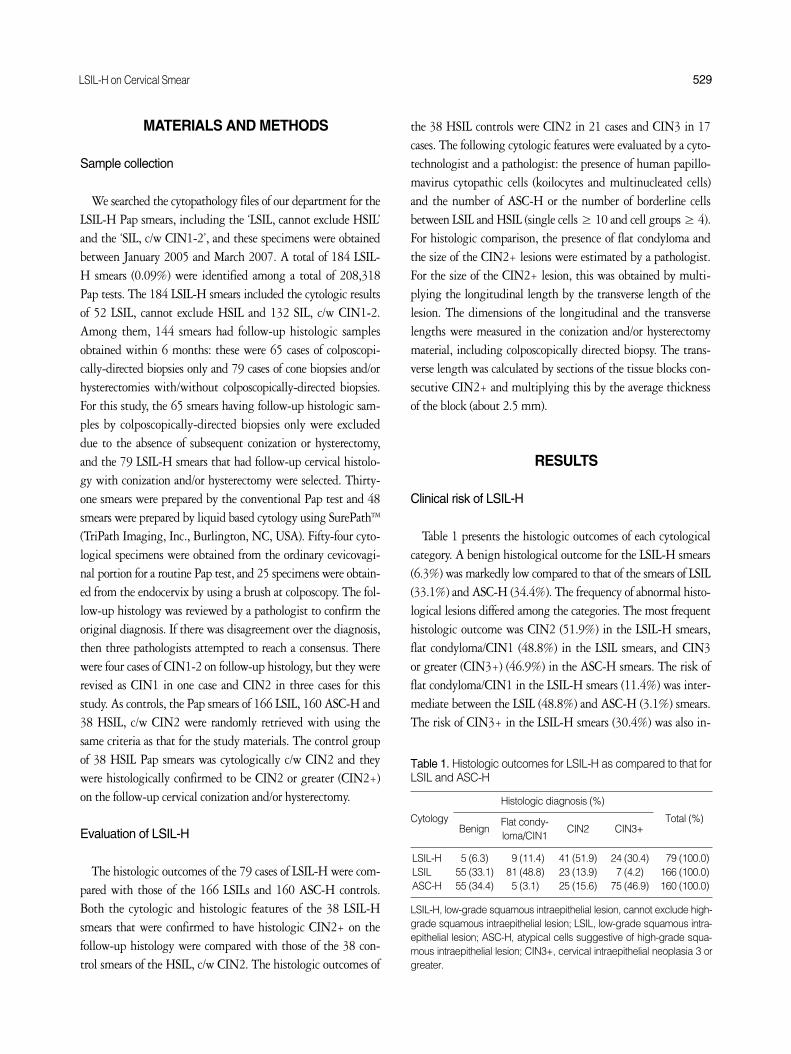
LSIL-H on Cervical Smear 529
MATERIALS AND METHODS
Sample collection
We searched the cytopathology files of our department for theLSIL-H Pap smears, including the ‘LSIL, cannot exclude HSIL’and the ‘SIL, c/w CIN1-2’, and these specimens were obtainedbetween January 2005 and March 2007. A total of 184 LSIL-H smears (0.09%) were identified among a total of 208,318Pap tests. The 184 LSIL-H smears included the cytologic resultsof 52 LSIL, cannot exclude HSIL and 132 SIL, c/w CIN1-2.Among them, 144 smears had follow-up histologic samplesobtained within 6 months: these were 65 cases of colposcopi-cally-directed biopsies only and 79 cases of cone biopsies and/orhysterectomies with/without colposcopically-directed biopsies.For this study, the 65 smears having follow-up histologic sam-ples by colposcopically-directed biopsies only were excludeddue to the absence of subsequent conization or hysterectomy,and the 79 LSIL-H smears that had follow-up cervical histolo-gy with conization and/or hysterectomy were selected. Thirty-one smears were prepared by the conventional Pap test and 48smears were prepared by liquid based cytology using SurePathTM
(TriPath Imaging, Inc., Burlington, NC, USA). Fifty-four cyto-logical specimens were obtained from the ordinary cevicovagi-nal portion for a routine Pap test, and 25 specimens were obtain-ed from the endocervix by using a brush at colposcopy. The fol-low-up histology was reviewed by a pathologist to confirm theoriginal diagnosis. If there was disagreement over the diagnosis,then three pathologists attempted to reach a consensus. Therewere four cases of CIN1-2 on follow-up histology, but they wererevised as CIN1 in one case and CIN2 in three cases for thisstudy. As controls, the Pap smears of 166 LSIL, 160 ASC-H and38 HSIL, c/w CIN2 were randomly retrieved with using thesame criteria as that for the study materials. The control groupof 38 HSIL Pap smears was cytologically c/w CIN2 and theywere histologically confirmed to be CIN2 or greater (CIN2+)on the follow-up cervical conization and/or hysterectomy.
Evaluation of LSIL-H
The histologic outcomes of the 79 cases of LSIL-H were com-pared with those of the 166 LSILs and 160 ASC-H controls.Both the cytologic and histologic features of the 38 LSIL-Hsmears that were confirmed to have histologic CIN2+ on thefollow-up histology were compared with those of the 38 con-trol smears of the HSIL, c/w CIN2. The histologic outcomes of
the 38 HSIL controls were CIN2 in 21 cases and CIN3 in 17cases. The following cytologic features were evaluated by a cyto-technologist and a pathologist: the presence of human papillo-mavirus cytopathic cells (koilocytes and multinucleated cells)and the number of ASC-H or the number of borderline cellsbetween LSIL and HSIL (single cells ≥ 10 and cell groups ≥ 4).For histologic comparison, the presence of flat condyloma andthe size of the CIN2+ lesions were estimated by a pathologist.For the size of the CIN2+ lesion, this was obtained by multi-plying the longitudinal length by the transverse length of thelesion. The dimensions of the longitudinal and the transverselengths were measured in the conization and/or hysterectomymaterial, including colposcopically directed biopsy. The trans-verse length was calculated by sections of the tissue blocks con-secutive CIN2+ and multiplying this by the average thicknessof the block (about 2.5 mm).
RESULTS
Clinical risk of LSIL-H
Table 1 presents the histologic outcomes of each cytologicalcategory. A benign histological outcome for the LSIL-H smears(6.3%) was markedly low compared to that of the smears of LSIL(33.1%) and ASC-H (34.4%). The frequency of abnormal histo-logical lesions differed among the categories. The most frequenthistologic outcome was CIN2 (51.9%) in the LSIL-H smears,flat condyloma/CIN1 (48.8%) in the LSIL smears, and CIN3or greater (CIN3+) (46.9%) in the ASC-H smears. The risk offlat condyloma/CIN1 in the LSIL-H smears (11.4%) was inter-mediate between the LSIL (48.8%) and ASC-H (3.1%) smears.The risk of CIN3+ in the LSIL-H smears (30.4%) was also in-
Cytology Total (%)
Histologic diagnosis (%)
BenignFlat condy-loma/CIN1
CIN2 CIN3+
LSIL-H 5 (6.3) 9 (11.4) 41 (51.9) 24 (30.4) 79 (100.0)LSIL 55 (33.1) 81 (48.8) 23 (13.9) 7 (4.2) 166 (100.0)ASC-H 55 (34.4) 5 (3.1) 25 (15.6) 75 (46.9) 160 (100.0)
LSIL-H, low-grade squamous intraepithelial lesion, cannot exclude high-grade squamous intraepithelial lesion; LSIL, low-grade squamous intra-epithelial lesion; ASC-H, atypical cells suggestive of high-grade squa-mous intraepithelial lesion; CIN3+, cervical intraepithelial neoplasia 3 orgreater.
Table 1. Histologic outcomes for LSIL-H as compared to that forLSIL and ASC-H

530 Sung Ran Hong Bock Man Kim Hye Sun Kim, et al.
termediate between the LSIL (4.2%) and ASC-H (46.9%) sme-ars. One patient with LSIL-H had a focal, superficial microin-vasive squamous cell carcinoma that was confirmed on hysterec-tomy after the diagnosis of CIN3 in the colposcopic biopsy. Thecorresponding LSIL-H smear of this case showed many keratiniz-ing dysplastic cells, grade cannot be determined, as well as LSILcells. There was no case of microinvasive carcinoma among thecontrols of LSIL or ASC-H.
Cytologic and histologic characteristics of LSIL-H
Tables 2 and 3 present the cytologic features of the LSIL-Hsmears that were confirmed to have histologic CIN2+ (Fig. 1),and these smears were compared with the control smears of HS-IL, c/w CIN2 (Fig. 2). All of these study and control cases wereconfirmed to have histologic CIN2+ on the follow-up conization
and/or hysterectomy. Evidence of LSIL such as koilocytotic atyp-ia and multinucleated cells were frequently found in the LSIL-H smears (78.9% and 71.0%, respectively) compared to that ofthe smears of HSIL, c/w CIN2 (18.4% and 18.4%, respective-ly). The number of abnormal cells that were atypical cells sug-gestive of HSIL or borderline cells between LSIL and HSIL wasless in the LSIL-H smears (single cells, 28.9%; clusters, 0%)than that in the smears of HSIL, c/w CIN2 (single cells, 76.3%;clusters, 50%) (Table 2). With regard to the degree of nuclearatypia, the atypical cells suggestive of HSIL or the borderlinecells between LSIL and HSIL in the LSIL-H were less severe thanthose of the HSIL, c/w CIN2 (Table 3). The most common nu-clear features of the LSIL-H smears were a moderate degree ofatypia such as an increased N/C ratio, hyperchromasia and coarsechromatin, and a mild degree of nuclear membrane irregulari-ty. However, the most common nuclear features of the HSILsmears, c/w CIN2 were a severe degree of atypia such as an in-creased N/C ratio, hyperchromasia and coarse chromatin, and amoderate degree of nuclear membrane irregularity.
Table 4 shows the histologic characteristics of the LSIL-Hsmears that were confirmed to represent CIN2+ on the follow-up conization and/or hysterectomy. Condylomatous change in
Cytolgic featuresLSIL-H
(n = 38) (%)HSIL, c/w CIN2
(n = 38) (%)
Evidences of LSIL Koilocyte (+) 30 (78.9) 7 (18.4)Multinucleation (+) 27 (71.0) 8 (18.4)
Suggestive of HSIL Single cells (≥ 10) 11 (28.9) 29 (76.3)Clusters (≥ 4) 0 (0.0) 19 (50.0)
LSIL-H, low-grade squamous intraepithelial lesion, cannot exclude high-grade squamous intraepithelial lesion; CIN2+, cervical intraepithelialneoplasia 2 or greater; HSIL, high-grade squamous intraepithelial lesion;c/w CIN2, consistent with CIN2; LSIL, low-grade squamous intraepithe-lial lesion.
Table 2. The overall cytologic features of LSIL-H confirmed ashistologic CIN2+ and as compared with those of HSIL, c/w CIN2
Nuclear atypiaDegree of atypia (%)
Absent or Mild Moderate Severe
LSIL-H (n = 38)Increased N/C ratio 5 (13.2) 25 (65.8) 8 (21.1)Hyperchromasia 7 (18.4) 24 (63.2) 7 (18.4)Coarse chromatin 14 (36.8) 20 (52.6) 4 (10.5)Nuclear membrane irregularity 23 (60.5) 14 (36.8) 1 (2.6)
HSIL, c/w CIN2 (n = 38)Increased N/C ratio 2 (5.3) 14 (36.8) 22 (57.9)Hyperchromasia 1 (2.6) 13 (34.2) 24 (63.2)Coarse chromatin 3 (7.9) 17 (44.7) 18 (47.4)Nuclear membrane irregularity 4 (10.5) 23 (60.5) 11 (28.9)
HSIL, high-grade squamous intraepithelial lesion; LSIL-H, low-gradesquamous intraepithelial lesion, cannot exclude high-grade squamousintraepithelial lesion; c/w CIN2, consistent with cervical intraepithelialneoplasia 2.
Table 3. Nuclear features of the atypical cells suggestive of HSIL
CytologyPresence of flat
CIN2+ size, mm2
condyloma (%)
LSIL-H (n = 38) 37 (97.4) 52.9HSILa (n = 38) 29 (76.3) 116.7
aHSIL, consistent with CIN2.LSIL-H, low-grade squamous intraepithelial lesion, cannot exclude high-grade squamous intraepithelial lesion; CIN2+, cervical intraepithelialneoplasia 2 or greater; HSIL, high-grade squamous intraepithelial lesion.
Table 4. The histologic characteristics of LSIL-H confirmed ashistologic CIN2+ and as compared to those of HSIL, consistentwith CIN2
Conization
Initial colposcopic biopsyBenign
Flat condy-loma/CIN1
CIN2 CIN3+
Benign (n = 13) 4 1 4 4Flat condyloma/CIN1 (n = 10) 5 2 3 0CIN2 (n = 32) 10 5 14 3CIN3+ (n = 17) 0 0 2 15Total (n = 72) 19 8 23 22
LSIL-H, low-grade squamous intraepithelial lesion, cannot excludehigh-grade squamous intraepithelial lesion; CIN3+, cervical intraepithe-lial neoplasia 3 or greater.
Table 5. Follow-up conization results after colposcopically-direct-ed biopsy for LSIL-H

LSIL-H on Cervical Smear 531
the histologic tissues was more frequently found in the LSIL-Hsmears (97.4%) than that in the smears of HSIL, c/w CIN2(76.3%). The average size of the histologic CIN2+ lesions wassmaller in the LSIL-H smears (52.9 mm2) than that in the smearsof HSIL, c/w CIN2 (116.7 mm2).
There were 72 patients who underwent both colposcopic bio-psy and subsequent conization, and we evaluated the clinicalsignificance of the conization in the LSIL-H smears of these pati-ents (Table 5). Eleven (47.8%) of 23 patients who had benignor flat condyloma/CIN1 at the initial colposcopically-directed
biopsy were found to have CIN2 or CIN3 on conization; espe-cially, four patients were confirmed to have CIN3 on conization.Among 32 patients diagnosed as CIN2 according to the initialcolposcopically-directed biopsy, three were up-graded to CIN3on conization. From these results, fourteen (31.1%) of all 45patients who were confirmed to have CIN2 and CIN3+ had anup-graded diagnosis of high grade CIN2 or CIN3 by the sub-sequent conization.
Fig. 1. Low-grade squamous intraepithelial lesion (LSIL), cannotexclude high-grade squamous intraepithelial lesion. There are atyp-ical koilocytes of LSIL or cervical intraepithelial neoplasia (CIN) 1to 2 in the background (A, B), and an individual atypical cell thatis suspicious for high grade squamous intraepithelial lesion (arrow)(B) (Papanicolaou stain). The subsequent biopsy shows CIN2 withflat condyloma (C).
A B
C

532 Sung Ran Hong Bock Man Kim Hye Sun Kim, et al.
DISCUSSION
We studied 79 cases of LSIL-H smears that were followed byhistological confirmation (conization and/or hysterectomy) with-in 6 months. The LSIL-H smears showed a different clinical riskfrom the control LSIL and ASC-H smears. First, the detectionrate of benign lesions on the follow-up histology was markedlylower in the LSIL-H smears (6.3%) than that in the smears ofLSIL (33.1%) and ASC-H (34.4%). As compared with the LSILand the ASC-H smears, the main reason for this may be the fact
that the cytological characteristics of LSIL-H typically exhibitunequivocal cells of LSIL and a small number of atypical cellssuggestive of HSIL. It is reasonable that the presence of unequiv-ocal cells of LSIL on cytology can cause an increased detectionrate of flat condyloma or more severe lesion on histology. There-fore, the detection rate of benign lesions on the follow-up his-tology will be low in the LSIL-H smears. Second, the most com-mon histological diagnosis on the follow-up cervical histologywas CIN2 for the LSIL-H smears. This finding is different fromthose of the LSIL and ASC-H smears, for which the most com-
Fig. 2. High-grade squamous intraepithelial lesion (HSIL), consis-tent with cervical intraepithelial neoplasia (CIN) 2. There is a groupof HSIL cells, consistent with CIN2, in the upper part of the fieldand a few cells (atypical koilocytes) in the lower part of the field(A). Higher magnification discloses a greater nuclear/cytoplasmicratio and more prominent irregularities of the nuclear envelope thanthose of low-grade squamous intraepithelial lesion (B) (Papanico-laou stain). The tissue is CIN2 with koilocytotic change in the upperlayer (C).
A B
C

LSIL-H on Cervical Smear 533
mon histological diagnosis was flat condyloma/CIN1 and CIN-3+, respectively. Third, for the LSIL-H smears, the detectionrate of flat condyloma/CIN1 or CIN3+ on the follow-up his-tology was intermediate between the cases of LSIL and ASC-Hsmears. Therefore, LSIL-H is considered to be a distinct cyto-logic category.
In the current study, the detection rate of CIN2+ on the fol-low-up histology of the LSIL-H smears (82.3%) was higherthan that for the LSIL (18.1%) and ASC-H (62.5%) smears.This was caused by the predominantly high rate of histologicalCIN2 (51.9%) in the LSIL-H. These findings are slightly dis-similar to other studies that reported an intermediate risk inthe LSIL-H smears for the detection of high-grade CIN on thefollow-up histology. Elsheikh et al.6 reported that the LSIL-Hsmears (40.7%) had an intermediate risk of CIN2+ betweenthe LSIL smears (13%) and the HSIL smears (74%) on the fol-low-up histology, but the risk was similar to that of the ASC-Hsmears (44%). A recent paper shows generally similar findingsto those of our data, even though the positive rates are differentbetween the cytologic categories.10 They reported that the CIN-2+ rate (33.14%) observed in association with LSIL-H was high-er than the rate of LSIL (16.11%) but it was lower than the rateof HSIL (69.03%) and it was similar or slightly higher than therate observed in the ASC-H (26.33%).10 They also showed thatthe rate of CIN3+ observed on the follow-up of LSIL-H (11.24%)was lower than the rate of CIN3+ in the ASC-H (17.79%). Ourstudy showed a high detection rate of CIN2+ in the LSIL-H, ascompared to the previous studies.6,10 The dissimilarity betweenthe current study and the previous studies may be due to differ-ent study materials. In the previous studies, the follow-up his-tological materials were obtained by biopsy specimens (includ-ing cervical biopsy, endocervical curetting, loop electrosurgicalexcision and cone excision), yet in the current study, the histol-ogy materials included only confirmatory histologic materialsof cone biopsies and/or hysterectomies. It is reasonable that thedetection rate of high-grade CIN should be higher in the fol-low-up conization/hysterectomy than that in the colposcopicbiopsy only. In fact, if the materials of the colposcopically-direct-ed biopsies only are included in this study, the detection rate ofCIN2+ on the follow-up histology of the LSIL-H smears (58.3%)was similar or slightly lower than that of the ASC-H smears(62.5%) (data not shown).
Certain Pap smears could be classified as LSIL-H for two rea-sons. Firstly, there are occasional ‘borderline’ cases that lie bet-ween LSIL and HSIL on both the Pap smears and the histologi-cal samples, although examination of the morphologic features
usually classifies the smears as either LSIL or HSIL. In a studythat focused on the cervical biopsy-cytology correlation and thatwas performed by the College of American Pathologists, thediagnosis SIL of an indeterminate grade occupied 3.3% of allthe SIL diagnoses.11 LSIL-H smears may belong to SIL, gradecannot be determined. Nasser et al.5 revealed that dysplastic cellswith a borderline N/C ratio appeared in 18% of the LSIL-Hsmears. Similarly, we used the cytologic result of ‘LSIL, cannotexclude HSIL’ together with ‘SIL, c/w CIN1-2,’ without detailedcytologic criteria to differentiate between the two categories,and we found four histologic results of CIN1-2 on the follow-up histology. They were revised to CIN1 or CIN2 for this study.Second, for the cases of LSIL-H that contain numerous cells ofLSIL and rare cells suggestive of HSIL, it could be assumed thatthe cervical tissue has a small portion of high-grade CIN in alarge background of low-grade lesion of flat condyloma or CIN1.Our follow-up histology data showed that the size of CIN2+on the follow-up histology was smaller in the LSIL-H cases ascompared with that of the pure cases of HSIL, c/w CIN. At thesame time, condylomatous change on the follow-up histologywas more frequently found in the cases of LSIL-H than that inthe cases of HSIL, c/w CIN. Taken together, our results supportthe assumption that in many cases of HSIL-H, the cervix has asmall, high-grade CIN admixed with large flat condyloma/CIN1.
In the current study, the LSIL-H smears accounted for 0.09%(184/208,318) of all the Pap tests, which was a somewhat lowerfrequency than that reported by Elsheikh et al.6 (0.15%, 194/126,911), Shidham et al.9 (0.19%, 146/77,979), McGrath etal.4 (0.2%, 108/48,687) and Owens et al.7 (0.5%, 113/21,220).Elsheikh et al.6 described that LSIL-H was the least frequent ofall squamous cell abnormalities and it accounted for 0.15% ofall Pap test interpretations and 2.5% of the squamous cell car-cinomas. Although the prevalence of LSIL-H has varied acrossthe reports, there was no significant difference among them.The similar prevalence indicates that the cytologic definition ofLSIL-H is relatively well established as a Pap smear that typi-cally contains definitive cells of LSIL and a few atypical cellsthat are suggestive, but not diagnostic of HSIL, and/or the Papsmear has borderline dysplastic cells that are between LSIL andHSIL. However, the diagnostic criteria for cells suggestive ofHSIL were not exactly same between the previous studies. Forexample, Nasser et al.5 described atypical squamous metaplasticcells (62%), atypical keratinized cells (20%) and dysplastic cellswith a borderline N/C ratio (18%) as the cytologic criteria ofthe cells suggestive of HSIL. However, Elsheikh et al.6 excludedthe cells of dyskeratosis or keratinizing dysplasia, and included

534 Sung Ran Hong Bock Man Kim Hye Sun Kim, et al.
rare dysplastic cells (usually < 5 cells), atypical metaplastic squa-mous cells and cells with an N/C ratio intermediate betweenLSIL and HSIL as the criteria. The cytological details for themorphologic criteria of LSIL-H need to be better defined to setup LSIL-H as a valid diagnostic category.
We observed that the nuclear atypia in LSIL-H cells sugges-tive of HSIL was less severe than that of the pure cases of HSIL,c/w CIN2. The most frequently encountered morphologic find-ings of the LSIL-H cells suggestive of HSIL were a moderatedegree of nuclear atypia such as an increased N/C ratio, hyper-chromasia and coarse chromatin, and a mild degree of nuclearmembrane irregularity. These cytologic findings resemble thoseof CIN2. Accordingly, we agree with McGrath et al.4 that LSIL-H cells with features suggestive of HSIL usually represent CIN2.
The 2001 consensus guidelines of the American Society forColposcopy and Cervical Pathology (ASCCP) recommendedcolposcopy for women with a Pap smear diagnosis of LSIL orHSIL. However, the ASCCP guidelines do not address the man-agement of women with a Pap smear diagnosis of SIL of an inde-terminate grade. Elsheikh et al.6 suggested that the same man-agement guidelines for ASC-H should be applied to patientswith LSIL-H because both tend to have poorly reproduciblecytological interpretation and a clinical risk of CIN2+ on thefollow-up histology that is intermediate between LSIL and HSIL.Shidham et al.9 reported that a management algorithm compa-rable to that for ASC-H and HSIL would be appropriate for theLSIL-H cases, and they suggested a management algorithm forwomen with LSIL-H. Similarly, based on our data, the manage-ment of the LSIL-H cases that are diagnosed as benign or flatcondyloma/CIN1 on the initial colposcopically-directed biopsyneeds to be emphasized. A sizable percentage (47.8%) of patientswho had LSIL-H and who were confirmed to have CIN2+ onthe follow-up histology were identified by conizaton after orig-inally being diagnosed with benign or flat condyloma/CIN1 onthe initial colposcopic biopsy. Accordingly, we suggest that themanagement of women with LSIL-H that is not confirmed onthe initial colposcopically-directed biopsy may require furtherfollow-up, just like cases of ASC-H or HSIL. However, becausethe LSIL-H cases appeared to have small CIN2 on the cytologyand histology, further large studies are necessary to avoid excessmanagement and to identify the real nature of the LSIL-H entity.
In conclusion, the Pap smears of LSIL-H accounted for 0.09%of all the Pap tests in this current study and these smears hadcharacteristic findings. On the follow-up histology, the risk ofdetecting CIN2+ was higher than that for ASC-H, but the mostcommon histological outcome was CIN2. The cytological fea-
tures of the atypical cells suggestive of HSIL were usually thoseof CIN2, and most cervical lesions of the LSIL-H smears had asmall focus of high-grade CIN admixed with a relatively largearea of flat condyloma/CIN1. For proper management, the LSIL-H smears not confirmed on the initial colposcopic biopsy needfurther follow up. Therefore, LSIL-H represents a valid diag-nostic category that is defined as a LSIL smear containing rareatypical cells suggestive, but not diagnostic of HSIL.
REFERENCES
1. Solomon D, Davey D, Kurman R, et al. The 2001 Bethesda System:
terminology for reporting results of cervical cytology. JAMA 2002;
287: 2114-9.
2. Solomon D, Nayar R. The Bethesda System for reporting cervical
cytology: definitions, criteria, and explanatory notes. 2nd ed. New
York: Springer, 2004.
3. Woodhouse SL, Stastny JF, Styer PE, Kennedy M, Praestgaard AH,
Davey DD. Interobserver variability in subclassification of squamous
intraepithelial lesions: results of the College of American Patholo-
gists Interlaboratory Comparison Program in Cervicovaginal Cytol-
ogy. Arch Pathol Lab Med 1999; 123: 1079-84.
4. McGrath CM, Kurtis JD, Yu GH. Evaluation of mild-to-moderate
dysplasia on cervical-endocervical (Pap) smear: a subgroup of pati-
ents who bridge LSIL and HSIL. Diagn Cytopathol 2000; 23: 245-8.
5. Nasser SM, Cibas ES, Crum CP, Faquin WC. The significance of
the Papanicolaou smear diagnosis of low-grade squamous intraep-
ithelial lesion cannot exclude high-grade squamous intraepithelial
lesion. Cancer 2003; 99: 272-6.
6. Elsheikh TM, Kirkpatrick JL, Wu HH. The significance of “low-grade
squamous intraepithelial lesion, cannot exclude high-grade squa-
mous intraepithelial lesion” as a distinct squamous abnormality cat-
egory in Papanicolaou tests. Cancer 2006; 108: 277-81.
7. Owens CL, Moats DR, Burroughs FH, Gustafson KS. “Low-grade
squamous intraepithelial lesion, cannot exclude high-grade squa-
mous intraepithelial lesion” is a distinct cytologic category: histo-
logic outcomes and HPV prevalence. Am J Clin Pathol 2007; 128:
398-403.
8. Kir G, Cetiner H, Gurbuz A, Karateke A. Reporting of “LSIL with
ASC-H” on cervicovaginal smears: is it a valid category to predict
cases with HSIL follow-up? Eur J Gynaecol Oncol 2004; 25: 462-4.
9. Shidham VB, Kumar N, Narayan R, Brotzman GL. Should LSIL
with ASC-H (LSIL-H) in cervical smears be an independent catego-
ry? A study on SurePath specimens with review of literature. Cyto-
journal 2007; 4: 7.

LSIL-H on Cervical Smear 535
10. Alsharif M, Kjeldahl K, Curran C, Miller S, Gulbahce HE, Pambuc-
cian SE. Clinical significance of the diagnosis of low-grade squa-
mous intraepithelial lesion, cannot exclude high-grade squamous
intraepithelial lesion. Cancer Cytopathol 2009; 117: 92-100.
11. Jones BA, Novis DA. Cervical biopsy-cytology correlation: a Col-
lege of American Pathologists Q-Probes study of 22 439 correlations
in 348 laboratories. Arch Pathol Lab Med 1996; 120: 523-31.


![Chapter 19 Basics of the Differential Geometry of Curvescis610/gma-v2-chap19.pdf530 19 Basics of the Differential Geometry of Curves wecanthinkoftheparametert ∈]a,b[ astime,andthefunction](https://static.fdocuments.in/doc/165x107/60c2171126d3013b755dc7ff/chapter-19-basics-of-the-differential-geometry-of-curves-cis610gma-v2-530-19-basics.jpg)