Evaluation of Hypertelorism in Children with Genetic ... · Compared to Normal Egyptian Children....
12
Journal of American Science 2010;6(10) http://www.americanscience.org [email protected] 160 Evaluation of Hypertelorism in Children with Genetic Syndromes Compared to Normal Egyptian Children. Amira A. Abdel Azeem *1 ; Manal H. Abu EL Ela 2 ; Shahira R. Nowier 1 ; Amr HAFEZ 3 and Tarek Saleh 3 Ophthalmogenetics 1 , Public Health 2 , Ophthalmology 3 Departments, Research Institute of Ophthalmology, Cairo Egypt * [email protected] Abstract: A case control study was carried out to evaluate hypertelorism in genetic syndromes and to start setting up standards for orbital parameters among Egyptian children. Head circumference, inner canthal, outer canthal and interpupillary distances were measured in 279 children; 49 patients with syndromes involving hypertelorism and 230 normal control children within the same age group. Controls were classified into 13 age groups (from1to 13 years) and mean values of the measured orbital parameters were estimated for the corresponding thirteen groups. Normal values were presented and compared with other populations and with Egyptian children with syndromes having hypertelorism. No significant sex differences were observed in different orbital measurements. Egyptian orbital morphometry did not resemble those of African Americans but showed similarities to the Turkish. Craniofacial anomalies had the greatest measurements of hypertelorism followed by the miscellaneous syndromes and skeletal displasias. The study will acquaint the geneticists on the need to the actual measurement, in relation to age, sex and racial standards for accurate diagnosis of syndromes. [Journal of American Science. 2010; 6(10):160-172]. (ISSN: 1545-1003). Key words: Hypertelorism, syndromes, canthal distance, interpupillary distance, canthal index, interpupillary index, and circumference-interorbital index. 1. Introduction The clinical observation of the face especially the orbital region remains an essential part for the clinical evaluation of phenotypic anomalies, which can be either qualitative or quantitative anomalies. Qualitative anomalies are relatively easy to define as present or absent compared to an “ideal” human phenotype. For diagnosis of quantitative anomalies as hypertelorism an objective definition of an abnormal phenotype requires the knowledge of the normal variation of the trait in a population of a given ethnic background at a certain age [1]. In many circumstances, normal standards for parameters are unavailable or, when published data are available, they include mostly the Western populations [2]. The normal distance between the orbits varies during embryogenesis and after birth in accordance with the general craniofacial development and increases with age [3]. The word hypertelorism is used to describe increased interorbital distance, a condition that is etiologically and pathogenically heterogeneous. Hypertelorism is not an isolated syndrome by itself. It is an anomaly that may occur as either part of a syndrome or malformation sequence [4]. Three possible pathogenic mechanisms lesser wings of the sphenoid, fixing the orbits in fetal position or failure of development of the nasal capsule, allowing the primitive brain vesicle to protrude into the space normally occupied by the capsule resulting in morphokinetic arrest in the position of the eyes; and disturbance in the development of the skull base as in craniosynostosis syndromes or in mid-facial malformations [3]. Evaluation of the interocular distances in clinical practice is based on the measurement of interpupillary, inner canthal and outer canthal distances; that can be easily compared to normal values [5]. A number of indices as canthal index, interpupillary index and circumference-interorbital index have also been proposed for greater accuracy due to variations in fronto-occipital circumference among healthy subjects [6]. Consideration of the position of eyes is relevant for the diagnosis of a large number of syndromes, permitting adequate preoperative evaluation, genetic counseling and treatment [7]. Searching in literature we could not find any data related to the normal orbital values among Egyptian children. So, we constructed a case -control
Transcript of Evaluation of Hypertelorism in Children with Genetic ... · Compared to Normal Egyptian Children....

Journal of American Science 2010;6(10)
http://www.americanscience.org [email protected] 160
Evaluation of Hypertelorism in Children with Genetic Syndromes Compared to Normal Egyptian Children.
Amira A. Abdel Azeem*1; Manal H. Abu EL Ela 2; Shahira R. Nowier 1; Amr HAFEZ3 and Tarek Saleh 3
Ophthalmogenetics1, Public Health2, Ophthalmology3 Departments, Research Institute of Ophthalmology, Cairo
Egypt *[email protected]
Abstract: A case control study was carried out to evaluate hypertelorism in genetic syndromes and to start setting up standards for orbital parameters among Egyptian children. Head circumference, inner canthal, outer canthal and interpupillary distances were measured in 279 children; 49 patients with syndromes involving hypertelorism and 230 normal control children within the same age group. Controls were classified into 13 age groups (from1to 13 years) and mean values of the measured orbital parameters were estimated for the corresponding thirteen groups. Normal values were presented and compared with other populations and with Egyptian children with syndromes having hypertelorism. No significant sex differences were observed in different orbital measurements. Egyptian orbital morphometry did not resemble those of African Americans but showed similarities to the Turkish. Craniofacial anomalies had the greatest measurements of hypertelorism followed by the miscellaneous syndromes and skeletal displasias. The study will acquaint the geneticists on the need to the actual measurement, in relation to age, sex and racial standards for accurate diagnosis of syndromes. [Journal of American Science. 2010; 6(10):160-172]. (ISSN: 1545-1003). Key words: Hypertelorism, syndromes, canthal distance, interpupillary distance, canthal index, interpupillary index,
and circumference-interorbital index. 1. Introduction
The clinical observation of the face especially the orbital region remains an essential part for the clinical evaluation of phenotypic anomalies, which can be either qualitative or quantitative anomalies. Qualitative anomalies are relatively easy to define as present or absent compared to an “ideal” human phenotype. For diagnosis of quantitative anomalies as hypertelorism an objective definition of an abnormal phenotype requires the knowledge of the normal variation of the trait in a population of a given ethnic background at a certain age [1]. In many circumstances, normal standards for parameters are unavailable or, when published data are available, they include mostly the Western populations [2].
The normal distance between the orbits varies during embryogenesis and after birth in accordance with the general craniofacial development and increases with age [3]. The word hypertelorism is used to describe increased interorbital distance, a condition that is etiologically and pathogenically heterogeneous. Hypertelorism is not an isolated syndrome by itself. It is an anomaly that may occur as either part of a syndrome or malformation sequence [4]. Three possible pathogenic mechanisms
lesser wings of the sphenoid, fixing the orbits in fetal position or failure of development of the nasal capsule, allowing the primitive brain vesicle to protrude into the space normally occupied by the capsule resulting in morphokinetic arrest in the position of the eyes; and disturbance in the development of the skull base as in craniosynostosis syndromes or in mid-facial malformations [3]. Evaluation of the interocular distances in clinical practice is based on the measurement of interpupillary, inner canthal and outer canthal distances; that can be easily compared to normal values [5].
A number of indices as canthal index, interpupillary index and circumference-interorbital index have also been proposed for greater accuracy due to variations in fronto-occipital circumference among healthy subjects [6]. Consideration of the position of eyes is relevant for the diagnosis of a large number of syndromes, permitting adequate preoperative evaluation, genetic counseling and treatment [7].
Searching in literature we could not find any data related to the normal orbital values among Egyptian children. So, we constructed a case - control
Journal of American Science 2010;6(10)
http://www.americanscience.org [email protected] 161
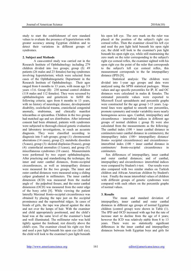
study to start the establishment of new standard values to evaluate the presence of hypertelorism with greater accuracy among Egyptian children and to detect their variations in different groups of syndromes. 2. Subject and Methods
A caseـcontrol study was carried out in the Research Institute of Ophthalmology including 279 children divided into two groups; group (I): 49 patients (26 males and 23 females) having syndromes involving hypertelorism; which were selected from cases of the Ophthalmogenetic Department in the Research Institute of Ophthalmology. Their ages ranged from 6 months to 13 years, with mean age 3.9 years ±3.6. Group (II): 230 normal control children (118 males and 112 females). They were screened by ophthalmologists and geneticists to fulfill the following criteria; ages from 6 months to 13 years, with no history of neurologic disease, developmental disability, oculofacial trauma, craniofacial congenital anomaly, strabismus, and clinically manifest telecanthus or epicanthus. Children in the two groups had matched age and sex distribution. After informed consent had been obtained, patients with syndromes were subjected to thorough clinical genetic evaluation and laboratory investigations, to reach an accurate diagnosis. They were classified according to diagnosis into 5 sub-groups; group (1): chromosomal aberrations (14 cases), group (2): metabolic disorders (5cases), group (3): skeletal displasias (9cases), group (4): craniofacial anomalies (11cases), and group (5): miscellaneous syndromes (10 cases). Measurements were performed by two expert ophthalmologists. After practicing and standardizing the technique, the inner and outer canthal distances, fronto-occipital circumferences, as well as interpupillary distance were measured for the two groups. The inner and outer canthal distances were measured using a sliding caliper graduated in millimeters. The inner canthal dimension (ICD) was measured from the medial angle of the palpebral fissure, and the outer canthal dimension (OCD) was measured from the outer edge of the bony orbit [8]. While viewing the patient laterally Maximal fronto-occipital circumference was obtained by placing the tape just on the occipital prominence and the supraorbital ridges. In cases of braids of girls, the tape was placed against the skin and not over the lumps of hair. With the examiner and child appropriately positioned, and the child’s head was at the same level of the examiner’s head and well illuminated. The millimeter ruler was held tightly against the forehead, rest directly above the child's eyes. The examiner closed his right eye first and used a pen light beneath his open eye (left eye), the child will look to the examiner's pen light beneath
his open left eye. The zero mark on the ruler was placed at the position of the subject's right eye corneal reflex. Then the examiner closed his left eye and used the pen light held beneath his open right eye, the child will look to the examiner's pen light beneath his open right eye, while still maintaining the zero mark on the ruler corresponding to the subject's right eye corneal reflex, the examiner sighted with his open right eye the point of the ruler that corresponds to the subject's left eye corneal reflex, this measurement corresponds to the far interpupillary distance (IPD) [9].
Statistical analysis: The children were divided into 1-year age groups and data were analyzed using the SPSS statistical packages. Mean values and age specific percentiles for IP, IC and OC distances were calculated in males & females. The estimated percentile values were exported to Microsoft Excel spreadsheets and percentile graphs were constructed for the age group 1-13 years. Log-trend lines were applied to smooth raw percentiles, promote symmetry and render variability more nearly homogenous across ages. Canthal, interpupillary and circumference - interorbital indices in different age groups of normal children ( group II) were also calculated from the measurements as the following: The canthal index (100 × inner canthal distance in centimeters/outer canthal distance in centimeters), the interpupillary index (100× interpupillary distance/ fronto- occipital circumference) and circumference-interorbital index (100 × inner canthal distance in centimeters/ fronto-occipital circumference in centimeters. Sex differences of interpupillary, inner canthal and outer canthal distances; and of canthal, interpupillary and circumference- interorbital indices were compared by Student's t-test. Our results were also compared with two similar studies on Turkish children and African American children by Student's t-test. Finally the mean interorbital values of children with different groups of genetic syndromes were compared with each others on the percentile graphs of normal values. 3. Results
The mean and standard deviation of interpupillary, inner canthal and outer canthal distances in different age groups of normal Egyptian children (control group) were shown in Table (1). The IPD and OCD increased with age but the rate of increase start to decline from the age of 4 years; however the ICD was relatively stable from 8 to 13 years. There were no statistically significant differences in the inner canthal and interpupillary distances between both Egyptian boys and girls for

Journal of American Science 2010;6(10)
http://www.americanscience.org [email protected] 162
the same mean age (P values were > 0.05). Outer canthal distances showed also no significant Table [1]: Mean and SD of interpupillary, inner canthal and outer canthal distances in different age groups of normal Egyptian children.
IPD (mm) ICD (mm) OCD (mm)
Age (years) no Mean SD Mean SD Mean SD
1 15 44.07 3.67 26.60 1.84 73.47 2.59
2 17 46.12 2.23 27.94 2.77 75.82 2.27
3 16 49.06 3.00 27.88 3.79 77.19 2.43
4 15 51.87 3.09 27.93 3.10 79.57 5.05
5 22 52.89 2.31 28.05 1.89 80.14 4.82
6 25 53.36 3.12 28.70 2.50 81.36 3.85
7 16 53.00 1.86 29.00 1.83 82.13 3.32
8 20 53.70 2.69 30.30 1.30 83.15 3.73
9 19 55.68 2.13 29.21 1.13 85.84 5.80
10 19 56.11 3.05 29.37 1.34 85.42 4.06
11 13 57.69 3.09 30.62 0.96 85.62 5.25
12 16 58.25 1.65 30.00 1.10 89.44 4.18
13 17 58.41 3.59 30.12 2.32 88.76 3.38
IPD= interpupillary distance ICD= inner canthal distance OCD=outer canthal distance
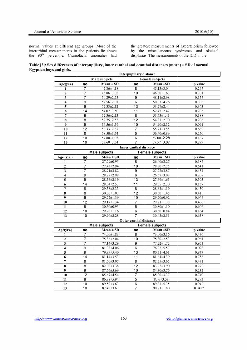
differences between boys and girls except at the age of 13 years there was increased distances among girls (P = 0.042) as shown in Table (2). Canthal, interpupillary and circumference - interorbital indices in different age groups of normal Egyptian children were shown in Table (3). There were no statistically significant sex differences of canthal, interpupillary and circumference- interorbital indices (P values were > 0.05) as shown in Tables (4). Comparing our measurements with two similar studies on Turkish children [10] and African American children [11], the mean inner canthal and outer canthal distances in both males and females were similar to that of the Turkish children ( P values were >0.05). The interpupillary distances showed similarity in female children but male children showed different measurements especially at the ages of (7, 8, 10, 12 and 13 years old). P values were
<0.01, <0.01, <0.05, <0.01 and <0.05 respectively. However, African Americans have statistically significant wider inner canthal distances than Egyptian children (P values were <0.05), but they have smaller outer canthal measurements in both male and female children.
Percentile normal values of the measured distances were calculated to aid in syndrome diagnosis. The percentile values of interpupillary, inner canthal and outer canthal distances in normal Egyptian children were graphed in fig. 1, 2 and 3 respectively.
Each group of patients with syndromes involving hypertelorism was sub-classified according to age into 1-year age subgroups and compared with the percentile normal values. Table (5) and fig. 4 showed the significant deviation of the mean orbital measurements in patients with syndromes from
Journal of American Science 2010;6(10)
http://www.americanscience.org [email protected] 163
normal values at different age groups. Most of the interorbital measurements in the patients lie above the 90th percentile. Craniofacial anomalies had
the greatest measurements of hypertelorism followed by the miscellaneous syndromes and skeletal displasias. The measurements of the ICD in the
Table [2]: Sex differences of interpupillary, inner canthal and ocanthal distances (mean) ± SD of normal Egyptian boys and girls.
Interpupillary distance Female subjects Male subjects
p value Mean ±SD no Mean ± SD no Age(yrs.) 0.247 45.13±3.04 8 42.86±4.18 7 1 0.701 46.30±1.63 10 45.86±3.02 7 2 0.157 48.11±2.98 9 50.29±2.75 7 3 0.308 50.83±4.26 6 52.56±2.01 9 4 0.363 53.27±2.44 13 52.33±2.12 9 5 0.205 52.45±2.42 11 54.07±3.50 14 6 0.188 53.63±1.41 8 52.36±2.13 8 7 0.206 54.33±2.70 12 52.75±2.55 8 8 0.091 54.90±2.32 10 56.56±1.59 9 9 0.682 55.71±3.55 7 56.33±2.87 12 10 0.250 56.40±0.89 5 58.50±3.74 8 11 0.167 59.00±2.28 6 57.80±1.03 10 12 0.279 59.57±3.87 7 57.60±3.34 10 13
Inner canthal distance Female subjects Male subjects
p value Mean ±SD no Mean ± SD no Age (yrs.) 0.187 26.00±2.27 8 27.29±0.95 7 1 0.541 28.30±2.75 10 27.43±2.94 7 2 0.454 27.22±3.87 9 28.71±3.82 7 3 0.208 26.67±3.08 6 28.78±2.99 9 4 0.303 27.69±1.65 13 28.56±2.19 9 5 0.137 29.55±2.30 11 28.04±2.53 14 6 0.430 28.63±1.19 8 29.38±2.33 8 7 0.415 30.50±1.45 12 30.00±1.07 8 8 0.967 29.20±0.92 10 29.22±1.39 9 9 0.406 29.71±1.38 7 29.17±1.34 12 10 0.606 30.80±1.10 5 30.50±0.93 8 11 0.164 30.50±0.84 6 29.70±1.16 10 12 0.658 30.43±2.51 7 29.90±2.28 10 13
Outer canthal distance Female subjects Male subjects
p value Mean ±SD no Mean ± SD no Age(yrs.) 0.476 73.00±3.16 8 74.00±1.83 7 1 0.961 75.80±2.53 10 75.86±2.04 7 2 0.951 77.22±1.72 9 77.14±3.29 7 3 0.098 76.92±5.57 6 81.33±4.06 9 4 0.847 80.31±4.61 13 79.89±5.40 9 5 0.758 81.64±4.39 11 81.14±3.53 14 6 0.471 82.75±3.65 8 81.50±3.07 8 7 0.272 83.92±3.90 12 82.00±3.38 8 8 0.232 84.30±3.76 10 87.56±5.69 9 9 0.740 85.00±3.37 7 85.67±4.54 12 10 0.293 83.6±3.58 5 86.88±5.94 8 11 0.942 89.33±5.35 6 89.50±3.63 10 12 0.042* 90.71±1.80 7 87.40±3.63 10 13

Journal of American Science 2010;6(10)
http://www.americanscience.org [email protected] 164
Table [3]: Canthal, interpupillary and circumference - interorbital indices of normal Egyptian children
Canthal index IP index Circumference-interorbital index
Age(years) no Mean SD Mean SD Mean SD
1 15 36.17 1.55 9.54 1.06 5.74 0.30
2 17 36.82 3.10 9.61 0.41 5.82 0.56
3 16 36.12 4.78 9.78 0.70 5.55 0.75
4 15 35.18 3.78 10.41 0.69 5.61 0.68
5 22 35.15 3.50 10.67 0.46 5.66 0.47
6 25 35.34 3.32 10.67 0.61 5.74 0.50
7 16 35.37 2.67 10.45 0.39 5.72 0.42
8 20 36.51 2.20 10.42 0.44 5.88 0.30
9 19 34.21 3.04 10.64 0.25 5.59 0.32
10 19 34.44 1.99 10.74 0.55 5.62 0.26
11 13 35.87 2.25 10.93 0.44 5.80 0.19
12 16 33.60 1.82 10.99 0.26 5.66 0.21
13 17 34.00 3.17 11.01 0.57 5.68 0.46

Journal of American Science 2010;6(10)
http://www.americanscience.org [email protected] 165
Table [4]: Sex differences of canthal, interpupillary and circumference- interorbital indices in normal Egyptian boys and girls
Canthal index Female subjects Male subjects
p value Mean ±SD no Mean ± SD no Age(yrs.) 0.102 35.56±1.76 8 36.87±0.95 7 1 0.466 37.29±2.86 10 36.14±3.52 7 2 0.454 35.29±5.27 9 37.17±4.21 7 3 0.676 34.65±2.82 6 35.53±4.44 9 4 0.387 34.59±2.91 13 35.94±4.27 9 5 0.249 36.22±2.55 11 34.65±3.76 14 6 0.289 34.64±1.87 8 36.10±3.25 8 7 0.818 36.42±2.29 12 36.66±2.21 8 8 0.371 34.82±3.07 10 33.53±3.03 9 9 0.353 35.01±2.22 7 34.10±1.86 12 10 0.200 36.91±2.34 5 35.22±2.08 8 11 0.315 34.21±1.59 6 33.24±1.93 10 12 0.659 33.57±3.08 7 34.29±3.35 10 13
Interpupillary index female subjects Male subjects
p value Mean ±SD no Mean ± SD no Age(yrs.) 0.291 9.83±1.04 8 9.22±1.08 7 1 0.653 9.65±0.32 10 9.55±0.54 7 2 0.107 9.53±0.69 9 10.10±0.62 7 3 0.796 10.35±0.97 6 10.45±0.51 9 4 0.423 10.73±0.50 13 10.57±0.42 9 5 0.130 10.46±0.58 11 10.83±0.61 14 6 0.712 10.49±0.45 8 10.41±0.34 8 7 0.859 10.43±0.43 12 10.40±0.50 8 8 0.199 10.57±0.25 10 10.72±0.23 9 9 0.557 10.64±0.65 7 10.80±0.50 12 10 0.303 10.76±0.01 5 11.03±0.55 8 11 0.144 11.12±0.31 6 10.92±0.20 10 12 0.565 11.11±0.67 7 10.94±0.52 10 13
Circumference- interorbital index Female subjects Male subjects
p value Mean ±SD no Mean ± SD no Age(yrs.) 0.162 5.64±0.39 8 5.86±0.09 7 1 0.553 5.89±0.49 10 5.72±0.67 7 2 0.339 5.39±0.77 9 5.76±0.72 7 3 0.440 5.44±0.71 6 5.73±0.68 9 4 0.355 5.58±0.39 13 5.78±0.57 9 5 0.199 5.89±0.44 11 5.62±0.53 14 6 0.244 5.60±0.25 8 5.85±0.52 8 7 0.725 5.86±0.35 12 5.91±0.21 8 8 0.588 5.63±0.30 10 5.55±0.36 9 9 0.524 5.68±0.26 7 5.59±0.27 12 10 0.274 5.88±0.26 5 5.76±0.13 8 11 0.222 5.74±0.13 6 5.61±0.23 10 12
0.980 5.68±0.48 7 5.68±0.48 10 13

Journal of American Science 2010;6(10)
http://www.americanscience.org [email protected] 166
Table [5]: Deviation of the mean orbital measurements from the normal values; in patients with syndromes having hypertelorism
IPD
Miscellaneous Craniofacial Skeletal Metabolic Chromosomal Mean Age(yrs) 55>90th(n=2) 52.5> 90th(n=6) 44.07 1 55.5>90th(n=2) 57>90th(n=2) 46.12 2 60.5>90th(n=3) 49.06 3 66 >90th(n=6) 62>90th(n=2) 61 > 90th(n=3) 51.87 4 61.5 > 90th(n=3) 52.89 5 63.5>90th(n=2) 53.36 6 66>90th(n=3) 53.00 7 67>90th(n=2) 53.70 8 67>90th(n=2) 55.68 9 66.5>90th(n=2) 72> 90th(n=3) 56.11 10 57.69 11 73.5>90th(n=2) 58.25 12 67.5> 90th(n=2) 67>90th(n=2) 58.41 13
ICD Miscellaneous Craniofacial Skeletal Metabolic Chromosomal Mean Age(yrs.)
32.5 >90th 30 = 90th 26.60 1 31>90th 30(75th-90th) 27.94 2
30(75th90th) 27.88 3 35.5>90th 32>90 31 (75th-90th) 27.93 4 31(75th-90th) 28.05 5
32>90th 28.70 6 33>90th 29.00 7
35>90th 30.30 8 32= 90th 29.21 9
36>90th 39>90th 29.37 10 30.62 11 40>90th 30.00 12
35 >90th 33 >90th 30.12 13
OCD Miscellaneous Craniofacial Skeletal Metabolic Chromosomal Mean Age(yrs.)
77 >90th 75 >90th 73.47 1 80>90th 84> 90th 75.82 2
91>90th 77.19 3 96>90th 92>90th 91>90th 79.57 4 92>90th 80.14 5
95>90th 81.36 6 99>90th 82.13 7
99>90th 83.15 8 102>90th 85.84 9
97>90th 105>90th 85.42 10 85.62 11 107>90th 89.44 12
100>90th 101>90th 88.76 13

Journal of American Science 2010;6(10)
http://www.americanscience.org [email protected] 167
10th
25th50th
90th75th
R2 = 0.93
R2 = 0.98
R2 = 0.91
R2 = 0.87
R2 = 0.95
35.00
37.00
39.00
41.00
43.00
45.00
47.00
49.0051.00
53.00
55.00
57.00
59.00
61.00
63.00
65.00
1 2 3 4 5 6 7 8 9 10 11 12 13
Age group(yrs)
IPD(m
m)
Fig. [1]: The Percentile values (log- trend line) of interpupillary distance in normal Egyptian children (control
group)
10th25th
50th
75th
90th
R2 = 0.73
R2 = 0.54
R2 = 0.77
R2 = 0.30
R2 = 0.29
20.00
21.00
22.00
23.00
24.00
25.00
26.00
27.00
28.00
29.00
30.00
31.00
32.00
33.00
34.00
1 2 3 4 5 6 7 8 9 10 11 12 13
Age group(yrs)
ICD(m
m)
Fig. [2]: The Percentile values (Log trend line) of inner canthal distances in normal Egyptian children
(control group)
10th
25th
50th
75th
90th
R2 = 0.80
R2 = 0.81
R2 = 0.78
R2 = 0.86
R2 = 0.93
64.0066.0068.0070.0072.0074.0076.0078.0080.0082.0084.0086.0088.0090.0092.0094.0096.0098.00
1 2 3 4 5 6 7 8 9 10 11 12 13
Age group(yrs)
OCD(m
m)
Fig. [3]: The Percentile values (log trend line) of outer canthal distances in normal Egyptian children (control
group)

Journal of American Science 2010;6(10)
http://www.americanscience.org [email protected] 168
10th
50th
90th
35.00
39.00
43.00
47.00
51.00
55.00
59.00
63.00
67.00
71.00
75.00
79.00
1 2 3 4 5 6 7 8 9 10 11 12 13
Age group(yrs)
IPD
(mm
)
MetabolicChromosomalSkeletalMiscelleneousCraniofacial
10th
50th
90th
20.00
22.00
24.00
26.00
28.00
30.00
32.00
34.00
36.00
38.00
40.00
42.00
1 2 3 4 5 6 7 8 9 10 11 12 13Age group(yrs)
ICD
(mm
)
ChromosomalMetabolicCraniofacialMiscellaneousSkeletal
10th
64.00
68.00
72.00
76.00
80.00
84.00
88.00
92.00
96.00
100.00
104.00
108.00
112.00
1 2 3 4 5 6 7 8 9 10 11 12 13Age group(yrs)
OC
D(m
m)
ChromosomalMetabolicSkeletalCraniofacialMiscellaneous
50th
90th
Fig. [4]: Deviation of the mean orbital measurements from normal values; in patients with syndromes having
hypertelorism
Journal of American Science 2010;6(10)
http://www.americanscience.org [email protected] 169
chromosomal disorders lies equal to directly under the 90th percentile in all age groups while in the metabolic disorders lies under the 90 th percentile in young children till the age of 3 years. On the other hand, in skeletal displasias the measurements of the ICD lies above the 90th percentile till the age of 7 years then follow the 90th percentile thereafter. 4. Discussion:
Hypertelorism is a manifestation of a complex deformity that affects several skeletal and soft- tissue structures and relies on the evaluation of certain anthropometric facial measurements [12]. Ethnic variation often renders anthropometric reference values obtained in one population unsuitable for use in others. Racial differences exist for certain of globe and orbital position [13]. Egyptian society, while appearing on the surface to be a homogenous “race” was in actuality internally diverse and integrated with the nations around them [14]. The argument that Ancient Egypt was African deserves to be put, but of course, there was also mixing with the Semitic-speaking peoples (the Akkadians, Phoenicians and Hyksos, the people of Babylonia and Assyria, and later the Arabs) and with Europeans [15].
Once the common measures used by scientists (inner canthal distance, outer canthal distance, and fronto-occipital circumference) were provided, a true appreciation of the degree of hypertelorism could be made. In this study, normative values for these parameters and for the canthal, interpupillary and circumference-interorbital indices of Egyptian children were presented. In addition, the relationship between these craniofacial dimensions and advancing age was explored. Measurements were age related however the rate of increase (from 4 to 13 years) was less in IPD and OCD and nearly stable from 8 to 13 years in ICD. These was in accordance with Lakshminarayana et al. [17] who demonstrated that interpupillary distance increases from birth to 5 years, with negligible changes thereafter and Zhang et al. [16] who documented that there is only one rush –increase stage of ocular development which is before the 8 years old in children. However Brückner et al. [18] showed that the increase of interpupillary distance continues until 30 years of age.
There were no statistically significant differences in the inner canthal and interpupillary distances between both Egyptian boys and girls for the same mean age (P values were > 0.05). Also outer canthal distances showed no significant differences
between boys and girls except at 13 years there was increased distances among girls (P =0.042). This increase may be related to the small sample size. In spite of some authors documented sex differences between orbital measurements, [10] others did not find statistically significant differences between male and female children for the mean age [19, 20].
The values obtained in our sample of Egyptian children were compared with those in another two races; Turkish and African American children. The mean inner canthal and outer canthal distances in both males and females were similar to that of the Turkish children. The interpupillary distances showed similarity with female children but male children showed different measurements. In spite of that African Americans have wider inner canthal distances than Egyptian children they have smaller outer canthal measurements. However the values for the different populations perhaps should be compared with some caution because, whereas we obtained the interpupillary dimension by direct measurement, others obtained it by manipulation of the two intercanthal distances; and photographic techniques were also used in other studies.
In clinical genetics, inner and outer canthal measurements are the simplest to use for standard clinical workups and patients' values should be compared to their own racial norms. The canthal index is useful for consultations by correspondence when facial photographs are provided even if no measurements are given with clinical examination. Because the canthal index is based on the ratio of the inner and outer canthal distances, it can be calculated from clinical photographs alone regardless of size [3].
Because not all wide-set eyes are the same, accurate terminology is critical for understanding and management. Orbital hypertelorism signifies an increased distance between both medial sides and lateral sides of orbits. Interorbital hypertelorism denotes increased distance between the inner orbital walls [21]. Isolated hypertelorism is rare and usually occurs in a sporadic form. Apparently dominant and recessive cases have been reported. Syndromic hypertelorism has many causes as chromosomal abnormalities, single gene disorders, developmental abnormalities of the skull or brain, and rare syndromes of unknown cause [22].
The mean values of orbital measurements of 49 Egyptian patients (26 males and 23 females) with different genetic syndromes involving hypertelorism were compared with measurements of normal

Journal of American Science 2010;6(10)
http://www.americanscience.org [email protected] 170
children in the same age groups. Table (5) and fig. 4 showed that most of the interorbital measurements in the chromosomal aberrations, metabolic disorders, skeletal displasias, craniofacial anomalies and miscellaneous syndromes lie above the 90 th
percentile. However, the measurements of the ICD in the chromosomal disorders lie equal to directly under the 90 th percentile in all age groups. Illusory hypertelorism may occur with a flat nasal bridge, epicanthic folds, exotropia, widely spaced eyebrows, narrow palpebral fissures, and dystopia canthorum. Two or more of these features enhance the illusion of hypertelorism further. In Down syndrome, epicanthic folds and low nasal bridge both contribute to the illusion of ocular hypertelorism, although measurements actually may show normal values [23].
The metabolic disorders curves lied under the 90th percentile in young children till the age of 3 years then they increases with age; this may be related to the progressive course of skull enlargement in our examined cases with metabolic disorders associated with hypertelorism (3different types of mucopolysaccharide disorders). Craniofacial anomalies had the greatest measurements of hypertelorism followed by the miscellaneous syndromes. On the other hand, in skeletal displasias the measurements of the ICD lies above the 90 th centile till the age of 7 years then follow the 90 th percentile thereafter. This work showed that the mean interorbital values of certain syndromes could be compared to each others using the percentile graphs to evaluate the degree of hypertelorism in different genetic syndromes and the measurements about the eyes and the derived indices should be clinically applicable in assessment of the patients with genetic syndromes with facial and orbital features.
5. Conclusion
Examination of a patient with developmental anomalies includes examination of the orbital region as well as other parts of the face and the body. Measurement in a careful manner and comparing the values with normal standard measurements specific for the patient's race, age and sex would lead to the precise analysis of these features with early diagnostic evaluation. This would guide molecular investigations if available and surgical intervention which may be useful in the overall care of the patient and his family.
This study will acquaint the geneticists on the need to actually measure the features, rather than rely on one impression on physical features in diagnosing syndromes and also may help in providing standards for specific measurements of Egyptian children to diagnose hypertelorism. We
recommend other studies with larger sample size of Egyptian population to obtain more accurate estimates as we consider this study as a pilot study for further set up of a national project. Corresponding author Amira A. Abdel Azeem Ophthalmogenetics Dept. Research Institute of Ophthalmology, Cairo Egypt [email protected] 6. References
1. Dollfus H, Verloes A. Dysmorphology and the Orbital Region: A Practical Clinical Approach. Surv Ophthalmol, 2004, 49: 547–561.
2. Barone CM, Jimenez DF, Laskey A, Alcantara BG, Braddock SR. Bony orbital distances among the Fiiilipino population. Journal of Craniofacial surgery, 2002, 13(2): 258-61.
3. Cohen MM, Richieri-Costa A, Guion-Almeida ML, Saavedra D. Hypertelorism: interorbital growth, measurements, and pathogenic considerations. Int Journal of Oral Maxillofac Surg., 1995, 24(6): 387–395.
4. Fearon JA, Bartlett SP, Whitaker LA. The skeletal treatment of orbital hypertelorism. Neurosurg Clin N Am., 1991, 2(3):673-81.
5. Farkas LG, Posnick JC, Hreczko TM. Anthropometric growth study of the head. Cleft Palate Craniofac J., 1992, 29:303–308.
6. Evereklioglu C, Doganay S, Er H, Tercan M, Gunduz A, Balat A, Borazan M. Interpupillary index: a new parameter for hypo- hypertelorism. Craniomaxillofac surg. J., 2001, 29 (4):191-194.
7. Wu KH, Tsai FJ, Li TC, Tsai CH, Peng CT, Wang TR. Normal values of inner canthal distance, interpupillary distance and palpebral fissure length in normal Chinese children in Taiwan. Acta paediatrica taiwanica, 2000, 41 (1): 22-27.
8. Laestadius ND, Aase JM, Smith DW. Normal inner canthal and outer orbital dimensions. Pediatrics, 1969, 74: 465-468.
9. Anderson, A. L. (1954). Accurate clinical means of measuring intervisual axis distance. Arch. Ophthal. 52, 349-352.
10. Evereklioglu C, Doganay S, Er H, Gunduz A, Tercan M, Balat A, Cumurcu T. Craniofacial Anthropometry in a Turkish Population. The Cleft Palate-Craniofacial Journal, 2002, 39 (2): 208–218.

Journal of American Science 2010;6(10)
http://www.americanscience.org [email protected] 171
11. Juberg RC, Sholte FG, Touchstone WJ. Normal values of intercanthal distances of 5-11- year -old American Blacks. Pediatrics, 1975, 55(3): 431-436.
12. Ortiz-Monasterio F, Molina F. Orbital hypertelorism. Clin Plast Surg. 1994, 21(4):599-612.
13. Douglas TS, Viljoen DL. Eye measurements in 7-years-old black South African children. Ann Hum Biol., 2006, 33(2): 241-254.
14. Jackson K. Ancient Egyptian Self-Identity. 2004,www. focusanthro.org/essays/Jackson-03-04.html.
15. kWEKU. To every Egyptian. 2009, raceandhistory ry.com/cgi-bin/forum/ webbbs_config.pl/noframes/read/1624
16. Zhang M, Hong R, Fu Z, Ye M, Yang H. The measurement of normal values of exophthalmos, interpupillary distance and interorbital distance of children and adolescence in Xiamen and the rule of their development. Zhonghua Yan Ke Za Zhi. 2000 Nov;36(6):462-466
17. Lakshminarayana P, Janardhan K, David HS. Anthropometry for syndromology. Indian J Pediatr. 1991; 58: 253–258.
18. Brückner R, Batschelet E, Hugenschmidt F. Basal longitudinal study on aging: ophthalmogerontological research results. Doc Ophthalmol. 1987; 64:235–310.
19. Kiambo DK, Kayembe D. Orbital measurements in Zairian children. Inner canthal, outer orbital, inter-pupillary distances and proptosis. J Fr Ophthalmol, 1994, 17(8-9): 496-500.
20. Saheeb BD, Umweni AA, Obuekwe ON, Folaranmi N. Normal values of medial and lateral canthal distances in 3 to 18 year-old Nigerians. West Afr J Med. 2004 Apr-Jun; 23(2):156-61.
21. Tan S T, Mulliken, J B. Hypertelorism: Nosologic Analysis of 90 Patients. Plastic & Reconstructive Surgery, 1997, 99 (2) 317-327.
22. Gripp K, Escobar FL. Facial bones. In: Human malformations and related anomalies, 2006, ed. By Stevenson RE, Hall JG, 2nd ed., Ch. 8, P. 267, Oxford University Press.
23. Gerald BE, Silverman FN. Normal and abnormal interorbital distances, with special reference to mongolism. Am J Roentgenol, 1965, 95:154-161.
5/9/2010