Evaluation of Chronic Autonomic Symptoms in Gulf … of Chronic Autonomic Symptoms in Gulf War...
29
Evaluation of Chronic Autonomic Symptoms in Gulf War Veterans with Unexplained Fatigue • Principal Investigator: Dr. Mian Li, WRIISC-DC and Neurology Service at VAMC-DC; Associate Clinical Professor of Neurology • Co-Investigators: Drs Han Kang, Pamela Karasik, Clara Mahan, Friedhelm Sandbrink, Ping Zhai • Post-doctoral fellow: Changqing Xu • Study Coordinator (Volunteer Status): Wenguo Yao • Funded by VA Merit Review Clinical Science R & D • Research approved by local IRB/R & D and conducted in VAMC- DC with compliance of stipulated human research regulations and VA policies regarding report or dissemination of research and non- research information. 1
Transcript of Evaluation of Chronic Autonomic Symptoms in Gulf … of Chronic Autonomic Symptoms in Gulf War...

Evaluation of Chronic Autonomic Symptoms in Gulf War Veterans with Unexplained Fatigue
• Principal Investigator: Dr. Mian Li, WRIISC-DC and Neurology
Service at VAMC-DC; Associate Clinical Professor of Neurology
• Co-Investigators: Drs Han Kang, Pamela Karasik, Clara Mahan,
Friedhelm Sandbrink, Ping Zhai
• Post-doctoral fellow: Changqing Xu
• Study Coordinator (Volunteer Status): Wenguo Yao
• Funded by VA Merit Review Clinical Science R & D
• Research approved by local IRB/R & D and conducted in VAMC-
DC with compliance of stipulated human research regulations and
VA policies regarding report or dissemination of research and non-
research information.
1

Rationale
• GW veterans (7.7%) reported dizziness/imbalance. blurred
vision, excessive fatigue, or tremor (Kang HK, et al. Illnesses
among United States veterans of the Gulf War: a population-
based survey of 30,000 veterans. J Occup Environ Med 2000;
42(5): 491-501
• Reported neurological symptoms are similar or identical to those
from patients with diseases of autonomic nervous system
• Ill group (deployed) with post-exertion fatigue
• Control (deployed) without fatigue
• Objective of this study
• Autonomic parameters useful in treatment
2

Peripheral Autonomic Nervous System
• Afferent Pathways
• Efferent Components
parasympathetic and sympathetic
• Neurotoxin may preferentially affect small nerve fiber
• Small fiber neuropathy vs Autonomic neuropathy
• Long delay in diagnosing autonomic system disorder:
a) unclear nature course of acquired autonomic disorders
b) lack of appropriate investigative team
3
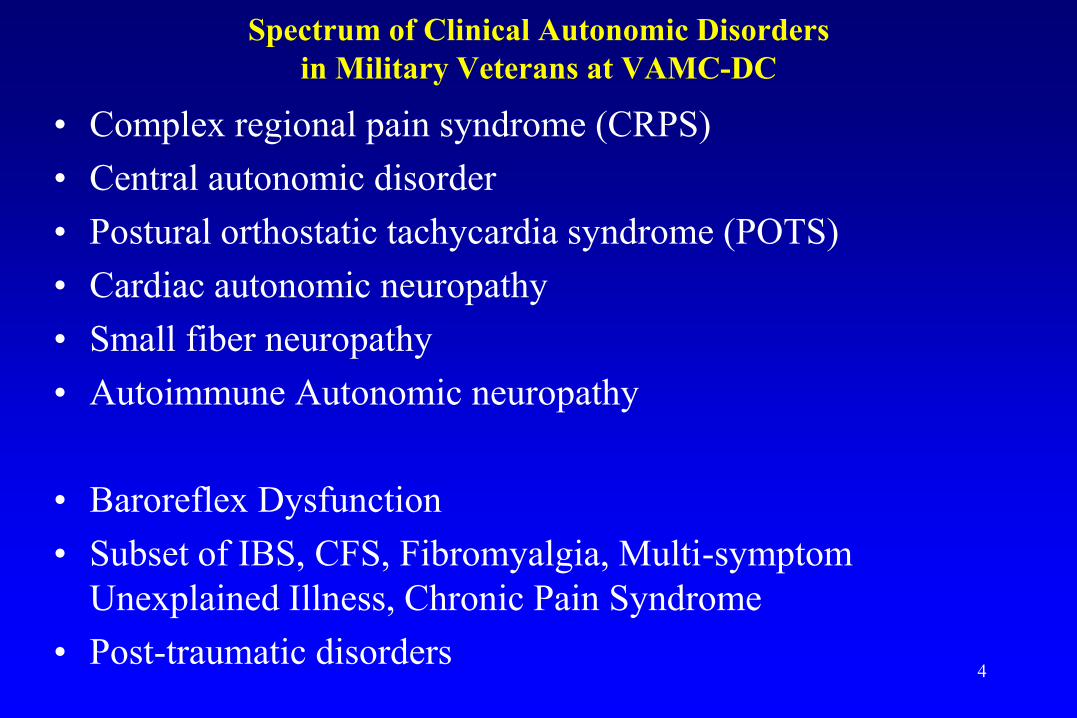
Spectrum of Clinical Autonomic Disorders
in Military Veterans at VAMC-DC
• Complex regional pain syndrome (CRPS)
• Central autonomic disorder
• Postural orthostatic tachycardia syndrome (POTS)
• Cardiac autonomic neuropathy
• Small fiber neuropathy
• Autoimmune Autonomic neuropathy
• Baroreflex Dysfunction
• Subset of IBS, CFS, Fibromyalgia, Multi-symptom
Unexplained Illness, Chronic Pain Syndrome
• Post-traumatic disorders
4

Autonomic Testing in literatures about GW Research
Heart Rate Variability
• cardiovagal Normal Valsalvar maneuver
•
• • Normal HRV to deep breathing
High Resting Rate
Abnormal HRV to deep breathing
Screening tool for cardiovagal function, interpretation needs beat-to-beat BP measurement
5

Panel 1: normal; 2: β-blocker; 3: reduced PP; 4: α-blocker
6

“Valsalva ratio”: why so low? What’s the next to do?
Sharief MK, et al. Neurology, 2002;59:1518--1525 7

Table 1. Autonomic symptoms of study participants
Group Ill (n=17) Control (n=13)
age (years; range) 47.7 ± 1.3 (39-58) 47.8 ± 1.9 (40-64)
Sex distributions (M/F) 14/3 12/1
BMI (M ± SEM) 33.6 ± 2.1 28.8 ± 1.4
Night diarrhea 3 0
Sexual dysfunction 4 3
Syncope 4 1
Abdominal pain 6 2
Dry eye /dry mouth 6 1
Anxiety 8 1
Tremor 8 1
Dizziness 8 1
Blurred vision 11 1
Weakness 11 1
Excessive fatigue 17 0
Values are mean ± SEM. M/F: male/female. 8

Ill Control P Value
Supine systolic BP 128.4 ± 1.9 132.6 ± 2.2 0.299
Supine diastolic BP 76.8 ± 1.2 77.4 ± 1.7 0.780
Supine heart rate 78.9 ± 2.4 ** 65.6 ± 2.8 <0.01
Standing systolic BP 125.7 ± 2.4 129.1 ± 2.3 0.189
Standing diastolic BP 79.8 ± 1.1 83.1 ± 2.3 0.629 Standing heart rate 93.1 ± 3.6 ** 75.1 ± 3.5 <0.01
Maximum-minimum HR 15.2 ± 2.2 11.5 ± 2.2 0.236
Standing systolic BP fall -5.5 ± 3.1 -5.6 ± 1.9 0.972
Standing diastolic BP increase 4.0 ± 1.2 5.8 ± 1.7 0.367
Tilt table headup systolic BP fall 10.4 ± 3.3 (n=16) 2.9 ± 4.4 0.179
Tilt table headup diastolic BP fall 10.9 ± 2.5 (n=16) 1.4 ± 3.8 0.843
Tilt table maximum HR 93.6 ± 3.1 * (n=16) 81.5 ± 4.6 <0.05
Tilt table HR increase 17.9 ± 2.4 (n=16) 17.7 ± 3.9 0.956
HRVdb Ratio 1.38 ± 0.04 1.33 ± 0.03 0.759
HRVvm Ratio 1.64 ± 0.03 1.84 ± 0.08 0.057
Test results expressed as mean ± SEM. HR: heart rate (beats/min). BP: blood pressure (mmHg).
* p<0.05, **P < 0.01.
Table 2. The alteration of heart rate and blood pressure in standing, tilt table,
deep breath and Valsalvar maneuver in ill (n=17) and control(n=13) groups.
9

Autonomic Testing in literatures about GW Research
• Quantitative Sensory Testing for Small Fiber Neuropathy
Comments on QST
∙ Nerve root or other pathology complicates the
interpretation (Veterans with Low Back Pain)
∙ Sensitivity and specificity varies among Labs
∙ Less optimal in “positive symptoms”
10

Left Hand Right Hand
Sk
in S
ensi
tiv
ity t
o
Tem
per
atu
re (
°C)
A B
C
Sk
in S
ensi
tiv
ity t
o
Vib
rati
on
(un
its)
Left Hand Right Hand
D
0
0.5
1
1.5
2
2.5
Left Foot Right Foot 0
0.5
1
1.5
2
2.5
3
3.5
4
4.5
0
0.2
0.4
0.6
0.8
1
1.2
1.4
1.6
1.8
2 *
(n =17)
(n =13)
Left Foot Right Foot 0
1
2
3
4
5
6
Sk
in S
ensi
tiv
ity t
o
Tem
per
atu
re (
°C)
Sk
in S
ensi
tiv
ity t
o
Vib
rati
on
(un
its)
(n =17)
(n =13)
Quantitative Sensory Testing (QST)
11

How do you interpret: more focal (compression) neuropathy in Control, more
or less distal symmetrical sensory in this group?
Davis, LE, et al. Neurology 2004;63(6):1070-1077 12

Neurological symptom score
Score 1 point for presence of a symptom
Dyck PJ. et al. Ann Neurol 8; 590-596, 1980
•Sensory disturbances
A. Negative symptoms
9. Difficulty identifying objects in mouth ________
10. Difficulty identifying objects in hands ________
11. Unsteadiness in walking ________
B. Positive symptoms
12. “Numbness,” “asleep feeling,” like Novocain,”
“ prickling,” at any site ________
13. Pain burning, deep aching,
Render ness at any location ________
•Autonomic symptoms
14. Postural fainting _________
15. Impotence in male _________
16. Loss of urinary control _________
17. Night diarrhea __________
•Symptoms of muscle weakness
A. Bulbar
1. Extraocular ________
2. Facial ________
3. Tongue ________
4. Throat ________
B. Limbs
5. Shoulder girdle and upper arm ________
6. Hand ________
7. Glutei and thigh ________
8. Legs _________

Score
Parameter 0 1 2 3 4
Sensory symptoms None Symptoms limited symptoms limited to Symptoms Symptoms above
To fingers or toes ankle or wrist extend to knee knees or elbows,
Or elbow or functionally disabling
Motor symptoms None Slight difficulty Moderate difficulty Require Paralysis
Help/assistance
Autonomic symptoms, n 0 1 2 3 4 or 5
Pin, sensibility Normal Reduced in Reduced up to Reduced up to Reduced to
fingers/Toes wrist/ankle elbow/knee above elbow/ Knee
Vibration sensibility Normal Reduced in Reduced up to Reduced up to Reduced to
finger/ Toes wrist/ankle elbow/knee above elbow/ Knee
Strength Normal Mild weakness Moderate weakness Severe weakness Paralysis
Tendon reflexes Normal Ankle reflex Ankle reflex absent Ankle reflex absent All reflexes absent
reduced Others reduced
Vibration sensation Normal to 126% to 150% 151% to200 201 to 300% >300% ULN
(QST vibration) 125% ULN ULN ULN ULN
Sural amplitude Normal/reduced 76 to 95% 51 to 75% 26 to 50% 0 to 25% of LLN
To <5% LLN of LLN of LLN of LLN
Personal amplitude Normal/reduced 76 to 95% 51 to 75% 26 to 50% 0 to 25% of LLN
To <5% LLN of LLN of LLN of LLN
QST = quantitative sensory test; ULN = upper limit of normal; LLN = lower limit of normal
Total Neuropathy Score
14

Total Neuropathy Score
• Initially designed for neurotoxin related neuropathy
• Useful outcome measures for length-dependant
symmetrical polyneuropathy
• Components of sensory, motor, and autonomic symptoms
• Objective measurement based on large fiber data
• Diabetic Neuropathy (n=35)
mild moderate severe control
Mean 12 20 25 0.4
SD 5.2 4.9 7.4 0.5
(Cornblath, DR, et al. Neurology 1999;53:1660-4)
15

QSART Neural Pathway (P256, Clinical Neurophysiology)
Our research method: Quantitative Sensory Axonal Reflex Testing
Clinical significance: correlated c somatic small nerve fiber on skin
biopsy 16

Normal Control
17
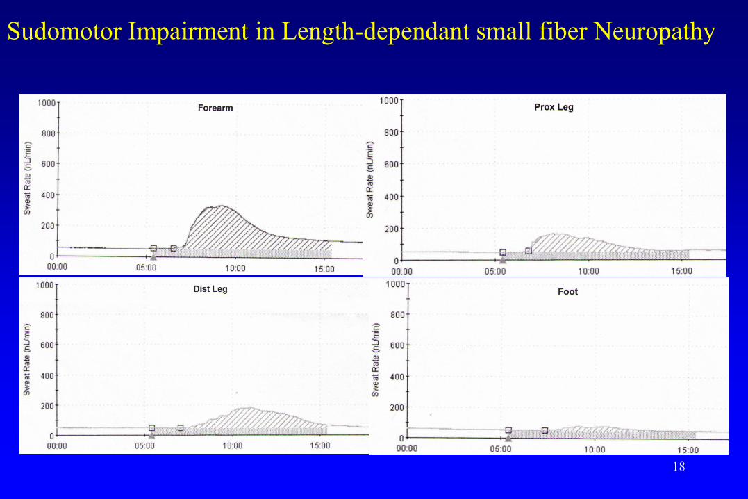
Sudomotor Impairment in Length-dependant small fiber Neuropathy
18

Group SFN LFN FN Myopathy NMT
Ill (n=17) 2 1 5 0 0
Control (n=13) 0 0 3 0 0
Table 4. Clinical Neuromuscular Disorders
SFN: Small fiber neuropathy. LFN: Large fiber neuropathy.
FN: Focal neuropathy. NMT: Neuromuscular transmission disorder.
19

Autonomic Testing in Chronic Fatigue Research H
R/B
P
Tilt Head down
Classic neurocardiogenic(vasovagal) response
BP HR
HR
/BP
Tilt Head down
Dysautonomic response
HR
/BP
Tilt Head down
POTS response H
R/B
P
Tilt Head down
Hypoadrenergic with
normal vagal response
Tilt Head down
Hyperadrenergic
POTS response H
R/B
P
HR
/BP
Tilt Head down
Normal response
Haemodynamic patterns during tilt table test 20

Compound Autonomic Severity Scales (CASS)
• Sudomotor Subscore (3 points)
Quantitative Sudomotor Axonal Reflex Test
• Adrenergic Subscore (4 points)
Valsavar Maneuvar or Tilt Table
• Cardiovagal Subscore (3 points)
Heart Rate Variability
Mean SD
• Multisystem Atrophy (n=18): 8.5 (1.3)
Autonomic Neuropathy (n=20): 8.6 (1.2)
Parkinson disease (n=20): 1.5 (1.1)
Asymptomatic Peripheral Neuropathy (n=20) 1.7 (1.3)
(Low PA. Mayo Clin Proc 1993;68:748-52)
21

Sudomotor Index
1. Single QSART site reduced, or Length-dependent pattern (distal sweat volume <1/3 of proximal value, or
TST anhidrosis present but < 25%
2. Single QSART site <50% of lower limit, or TST anhidrosis 25-50%
3. Two or more QSART sites <50% of lower limit, or TST anhidrosis >50%
Adrenergic Index
1. Phase IIE reduction <40 >25 mm Hg MBP, or Reduced phase IIL, or
Pulse pressure reduction to <50% of baseline
Increased PRT (4 - 5 sec)
Absent phase IV
2. Phase IIL absent or increased PRT (6 - 9 sec)
3. Absent phases IIL and IV and increased PRT (>10 sec)
4. 3 + OH (SBP reduction > 30 mm Hg; MBP > 20mm Hg)
If the Valsalvar maneuver is normal, a score of 1 can be assigned if the following changes occur on tilt up:
a. Excessive oscillations in MBP (>20mmHg occupying at least 50% of the duration of tilt up)
b. Fall in pulse pressure >50%
c. Transient fall in SPB >20 mmHg with recovery (within 1 minute)
d. SBP reduction>20mmHg beyond 1 min
e. DBP reduction>10mmHg beyond 1 min
f. Overshoot >20mmHg following tilt back
Cardiovascular HR Index
1. HRDB or VR reduced but >50% of minimum or reduced BRSV
2. HRDB or VR reduced to >50% of minimum or BRSV (≤3 ms/mm Hg)
3. HRDB or VR reduced to >50% of minimum and BRSV (≤3 ms/mm Hg)
HRDB ,heart response to deep breathing; MBP, mean blood pressure; OH, orthostatic hypotension; PRT, pressure
recovery time; QSART, quantitative sudomotor axon reflex test; SBP, systolic blood pressure; TST, thermoregulatory
sweat test; VR, Valsalvar ratio.
Composite Autonomic Severity Score (CASS)
22

0
5
10
15
20
Ill Control
Tota
l N
euro
pat
hy
Sco
re
0
1
2
3
4
5
6
CA
SS
Ill Control
Fig. Total Neuropathy and CASS Score in Ill and Control
groups

Ill (n = 17) Control (n = 13)
Mean/ ± SEM 95% CI Mean/± SEM 95% CI P Value
SNAP (sensory)
Amplitude (μV) 12.48 ± 1.91 (n = 15) 8.38—16.58 12.53 ± 2.46 (n = 9) 6.85—18.0 0.988
Latency (ms) 3.31 ± 0.12 (n = 12) 3.06—3.56 3.71 ± 0.19 (n = 9) 3.26— 4.17 0.073
CMAP (motor)
Amplitude (mV) 4.45 ± 0.51 (n = 13) 3.34—5.55 6.13 ± 0.72 (n = 9) 4.48—7.79 0.061
Latency (ms) 4.79 ± 0.13 (n = 13) 4.51—5.07 4.79 ± 0.21(n = 9) 4.30—5.28 0.989
TNS 6.29 ±1.10 3.95—8.63 3.50 ± 0.98 (n = 12) 1.34—5.66 0.083
CASS Median SD Median SD
Total 2.00 0.83 (n = 16) 1.00 0.92 0.011*
Sudomotor 1.00 0.75 1.00 0.63 0.069
Cardiovagal 0.50 0.62 0.00 0.50 0.482
Adrenergic 0.00 0.51 0.00 0.37 0.113
Table 3. TNS and CASS
SNAP: sensory nerve action potential from sural sensory nerve.
CMAP: compound muscle action potential from peroneal nerve.
TNS: total neuropathy score. CASS: composite autonomic severity score. 24

Disorder Profiles
• Hx + PE + Neurophysiology/Autonomic function tests (n =30)
Diagnosis depending on the definition
• Clinical autonomic disorders n = ?
• Small fiber dysfunction n = 4
• Autonomic neuropathy
CASS=> 3 + Abnormal QSART n = 2
• Cardiac autonomic neuropathy n = ?
• POTS n = 2
• CRPS n = 1
• Metabolic disorders n = 7
• Cardiovascular disorder n = ?
• Autoimmune disorder n = 4

Case 1: Hydrocarbon Neuropathy
• Middle age Veteran (fuel technician) had exposure to JP 8 for a total estimated
duration of 2 yrs. Evaluated for “PTSD” . NL Glucose. Negative SS-A/B,
nAch-R Ab, ACE, infec panel, and immune panel.
• Normal Nerve Conduction Study
• Needle EMG: L S1 radiculopathy
• QST: Abnormal at feet
Threshold (L/R)
Vibration Thermal-Cold
Finger 1.6/1.4 1-2/1-2
Great Toe 8.6/3.1 6.0/3.0
• Total Neuropathy Score: 9
• Abnormal QSART
26

Case 2: Post-Vaccination POTS
• Young veteran had near syncope events with episodic “sinus tachycardia” 10
days after vaccination. Evaluated for “panic attack”.
• Panel A BP HR Panel B MinHR Max Ratio (low cutoff)
Supine 108/72 45 HRdb 46 77 1.67 (> 1.20)
Standing 98/59 77
Tilting (HU) HRvm 44 84 1.91 (> 1.50)
3 min 98
• NCS: Normal
• QST: Normal
• TNS: 4 (subjective autonomic symptoms)
• QSART: Mildly abnormal
• CASS: 5
27

Conclusion
• Self reported unexplained neurological symptoms
can be confirmed on a battery of objective
autonomic testing in selected GW veterans
• Objective parameters on autonomic testing may
be useful in guiding the treatment of selected
multi-symptom illnesses in an appropriate clinical
context
• Ill group (deployed) defined in this study has
impaired autonomic nervous system function by
objective autonomic testing compared to Control
(deployed) 28

∙ Special Thanks to both Veterans participating in the
Study and Volunteer researchers
∙ Clinical content reflects PI and Co-PIs (FS, PZ, PK)’s
opinions
∙ The research results are preliminary and hypothesis
driven with small sample size of highly selected study
participants. Alternative hypothesis and interpretation
for these finding exist
∙ Cautious application of autonomic testing in individual
cases requires an appropriate research or clinical
context.
29