Evaluating the evidence for transmission distortion in ... · Evaluating the evidence for...
49
1 Evaluating the evidence for transmission distortion in human pedigrees Wynn K. Meyer * , Barbara Arbeithuber § , Carole Ober *, ** , Thomas Ebner §§ , Irene Tiemann- Boege § , Richard R. Hudson ***, **** , Molly Przeworski *, ***, §§§, **** * Dept. of Human Genetics, University of Chicago, Chicago, IL, USA § Dept. of Biophysics, Johannes Kepler University, Linz, Austria ** Dept. of Obstetrics and Gynecology, University of Chicago, Chicago, IL, USA §§ Kinderwunsch Zentrum, Landes- Frauen- und Kinderklinik, IVF-Unit, Linz, Austria *** Dept. of Ecology and Evolution, University of Chicago, Chicago, IL, USA §§§ Howard Hughes Medical Institute **** Co-supervised this work Genetics: Published Articles Ahead of Print, published on March 6, 2012 as 10.1534/genetics.112.139576 Copyright 2012.
Transcript of Evaluating the evidence for transmission distortion in ... · Evaluating the evidence for...
1
Evaluating the evidence for transmission distortion in human pedigrees
Wynn K. Meyer*, Barbara Arbeithuber§, Carole Ober*, **, Thomas Ebner§§, Irene Tiemann-
Boege§, Richard R. Hudson***, ****, Molly Przeworski*, ***, §§§, ****
*Dept. of Human Genetics, University of Chicago, Chicago, IL, USA §Dept. of Biophysics, Johannes Kepler University, Linz, Austria **Dept. of Obstetrics and Gynecology, University of Chicago, Chicago, IL, USA §§Kinderwunsch Zentrum, Landes- Frauen- und Kinderklinik, IVF-Unit, Linz, Austria ***Dept. of Ecology and Evolution, University of Chicago, Chicago, IL, USA §§§ Howard Hughes Medical Institute **** Co-supervised this work
Genetics: Published Articles Ahead of Print, published on March 6, 2012 as 10.1534/genetics.112.139576
Copyright 2012.
2
Running title: Transmission distortion in human pedigrees
Key words: transmission distortion, segregation distortion, natural selection, human evolution,
genome-wide scan
Corresponding author:
Wynn Meyer
Department of Human Genetics
920 E. 58th St., CLSC 416
Chicago, IL 60637
Phone: 773-834-9838
Fax: 773-834-0505
Email: [email protected]
IRB information: Research on FHS and AGRE data was approved by University of Chicago IRB
10-674-B, "Population genetic analyses of the Framingham and AGRE Data". Because we only
analyzed data that had been previously collected by other researchers for other purposes and was
then made available to us, our IRB granted a waiver of consent. Research on HUTT data was
approved by University of Chicago IRB numbers 5444, “Studies of Fertility in Hutterite
Couples,” and 8073, “Genetic Studies of Complex Phenotypes in the Hutterites.” For the sperm
genotyping, blood and semen from anonymous donors were provided by the IVF clinic of the
Landes- Frauen- und Kinderklinik, Linz, following protocols approved by the Ethics Committee
of Upper Austria (EK-Number: 1-11[2.1.6]).

3
ABSTRACT
Children of a heterozygous parent are expected to carry either allele with equal
probability. Exceptions can occur, however, due to meiotic drive, competition among gametes,
or viability selection, which we collectively term “transmission distortion” (TD). Although there
are several well-characterized examples of these phenomena, their existence in humans remains
unknown. We therefore performed a genome-wide scan for TD by applying the transmission
disequilibrium test (TDT) genome-wide to three large sets of human pedigrees of European
descent: the Framingham Heart Study (FHS), a founder population of European origin (HUTT),
and a subset of the Autism Genetic Resource Exchange (AGRE). Genotyping error is an
important confounder in this type of analysis. In FHS and HUTT, despite extensive quality
control, we did not find sufficient evidence to exclude genotyping error in the strongest signals.
In AGRE, however, many signals extended across multiple SNPs, a pattern highly unlikely to
arise from genotyping error. We identified several candidate regions in this dataset, notably a
locus in 10q26.13 displaying a genome-wide significant TDT in combined female and male
transmissions and a signature of recent positive selection, as well as a paternal TD signal in
6p21.1, the same region in which a significant TD signal was previously observed in 30
European males. Neither region replicated in FHS, however, and the paternal signal was not
visible in sperm competition assays or as allelic imbalance in sperm. In maternal transmissions,
we detected no strong signals near centromeres or telomeres, the regions predicted to be most
susceptible to female-specific meiotic drive, but we found a significant enrichment of top signals
among genes involved in cell junctions. These results illustrate both the potential benefits and the
challenges of using the TDT to study transmission distortion and provide candidates for
investigation in future studies.
INTRODUCTION
According to Mendel’s Law of Segregation, diploid organisms that are heterozygous at a
locus are equally likely to transmit either allele to their offspring. Yet cases occur in which one
allele is observed among offspring at greater than 50% frequency. This phenomenon of observed
“transmission distortion” (TD), also known as transmission ratio distortion, can result from two
distinct biological processes. The first, which we call “segregation distortion”, includes meiotic
4
drive, in which the functional products of meiosis preferentially carry one allele, and competition
among gametes. Meiotic drive is more likely to occur in asymmetric meioses, such as those in
human female germ cells (Malik 2009; Pardo-Manuel de Villena and Sapienza 2001). Examples
include the B chromosomes most commonly observed in insects and plants and the “knob”
chromosomes of maize (Jones and Rees 1982; Peacock et al. 1981; Östergren 1945). In turn,
segregation distorters like the t-haplotype in mice confer an advantage in competition for
fertilization between gametes carrying different alleles (Lyon 2003). The second process that
could lead to observed TD is ongoing viability selection; if an allele confers a viability advantage
to gametes or individuals, it will appear to be transmitted to more than 50% of the surviving
offspring of heterozygous parents. With the exception of viability selection on diploids, these
phenomena are more likely to produce TD in gametes of only one sex (see Lyttle 1993).
In several known cases of segregation distortion, the advantage to the distorter allele is
strong, with as many as 99% of offspring inheriting this allele (Lyttle 1993). Such an allele is
unlikely to be observed as segregating within a population if not maintained by some
countervailing force, because it would drive to fixation rapidly. Yet there are numerous examples
of polymorphic drivers across multiple taxa. Their maintenance in the population can often be
explained by reduced fertility or fitness of adults homozygous for the driver (see Hartl 1972 and
Carvalho and Vaz 1999), as in the well-known segregation distorter (SD) system in Drosophila
and t-haplotypes in mice. The SD system disrupts a signaling pathway involved in nuclear
localization, preventing SD+ sperm – those that do not carry the distorter – from developing
normally, thus leading to eventual transmission of nearly 100% SD sperm (Kusano et al. 2003).
Males homozygous for SD have severely reduced fertility (Hartl 1973; Hartl 1974), and it is
presumably this deleterious effect, in combination with suppressors of distortion, that permits the
observation of polymorphism at the SD locus in natural populations of Drosophila (Hartl 1975;
Hiraizumi and Thomas 1984; Presgraves 2009). In mice, interactions between t-haplotype
distorters and responder loci reduce motility of non-t-haplotype-bearing sperm in heterozygotes,
and males homozygous for the t-haplotype are sterile (Lyon 2003; Veron et al. 2009). In these
cases, the distorter allele enhances its own transmission at the expense of the organism and can
thus be seen as a selfish genetic element. Beyond these two cases, segregation distortion has
been detected in a wide variety of organisms, including many species of insects, plants, fungi,

5
and vertebrates, suggesting that deleterious effects of drivers may be common (de la Casa-
Esperón and Sapienza 2003; Lyttle 1993; Pardo-Manuel de Villena and Sapienza 2001).
The prevalence of distorters in natural populations has important implications for genome
evolution, as well as for speciation. In particular, asymmetric female meiosis provides the
opportunity for meiotic drive loci to influence the outcome of oötid competition, i.e., competition
among the four products of meiosis to be included in the oocyte pronucleus. An allele affecting
the orientation of chromosomes toward the pronucleus could lead either to distortion or to non-
disjunction; therefore, common appearances of such alleles could potentially explain the high
rates of non-disjunction observed in female Drosophila and humans (Hassold and Hunt 2001;
Zwick et al. 1999). This type of meiotic drive has also been proposed as a powerful force in the
evolution of centromeres, given their central importance to chromosome positioning during
meiosis. Specifically, the rapid evolution of repetitive DNA in centromeres is thought to be due
to competition among centromeres to bind spindle elements, with longer repeats favored. This
“centromeric drive” hypothesis predicts frequent segregation distortion at the centromere in
females (Henikoff et al. 2001; Malik and Henikoff 2002). The telomere may also be involved in
determining orientation toward the meiotic spindle and has therefore been proposed as another
potential target of female-specific meiotic drive (Novitski 1951; Anderson et al. 2008; Axelsson
et al. 2010).
The dynamics of distorter alleles may also influence local patterns of meiotic
recombination. In several known cases, distortion results from an interaction wherein the “drive”
allele at the distorter locus acts on a “sensitive” allele at a responder locus. This dynamic
produces indirect selection on linked recombination rate modifiers, whereby linked mutations on
the drive/insensitive background that decrease recombination between distorter and responder
will be favored (Charlesworth and Hartl 1978). Conversely, at unlinked sites, modifiers that
increase recombination will be beneficial because they uncouple the distorter and responder,
thereby suppressing the costly drive (Haig and Grafen 1991; Thomson and Feldman 1974).
There may also be selection on modifiers of recombination that influence the stage of meiosis at
which distorters gain a transmission advantage (Haig 2010, Brandvain and Coop 2012).
Moreover, because systems of distortion loci and their responders co-evolve rapidly and can
generate Dobzhansky-Muller incompatibilities, they may play an important role in the evolution
6
of reproductive isolation (Frank 1991; Hurst and Pomiankowski 1991). On the X chromosome,
segregation distortion loci can influence sex ratios and even lead to novel sex-determining
mechanisms (Gileva 1998; Hurst and Werren 2001; Jarrell 1995). Thus, understanding the
prevalence of TD is important for many aspects of evolutionary genetics.
Although there are numerous examples from other organisms, the extent and influence of
TD in humans remains unknown. One study found a genome-wide excess of allele sharing
among siblings, suggestive of TD, in a founder population of European origin (Zöllner et al.
2004), but another reported a deficit of allele sharing in Australian and Dutch dizygotic twins
(Montgomery et al. 2006). A more direct way of assessing TD is by testing the null hypothesis
that the transmission rates of both alleles from heterozygous parents are equal to 50%. The
transmission disequilibrium test (TDT), originally designed for family-based association tests
using an affected-only design, can be used to test for TD in genotyping data from pedigrees
(Spielman et al. 1993). One limitation of the TDT (and tests for excess allele sharing) is that
even relatively low levels of genotyping error can strongly enrich for apparent TD. For example,
mistyping of major allele homozygote parents as heterozygotes can lead to apparent over-
transmission of the major allele (Mitchell et al. 2003), as can a large proportion of missed calls
among heterozygotes (see Box 4 in Hirschhorn and Daly 2005). Several authors have proposed
modifications or alternatives to the TDT that are more robust to genotyping errors (Cheng and
Chen 2007; Gordon et al. 2001; Gordon et al. 2004), but they suffer from a number of limitations
when applied genome-wide: for instance, they cannot be used for tests in only one sex, do not
address the problem of differential fractions of missing data among genotype classes, and/or are
not robust to population stratification (a benefit of the original TDT). An additional challenge for
genome-wide scans is that correction for multiple testing leads to stringent cutoffs for
significance, such that extremely large sample sizes are required to detect moderate TD; for
example, 2,839 transmissions are required to achieve 50% power to detect distortion strength
(deviation from 50% transmission) of 5% at α = 10-7 (Evans et al. 2006). The best power for
detecting TD genome-wide, therefore, exists at loci with strong TD and high minor allele
frequency (MAF), because, for a given sample size, these provide the most observable
transmissions from heterozygotes. A strongly distorting locus experiences a trajectory similar to
7
that of a beneficial allele, so in order to observe a TD locus with high MAF, distortion must be
either extremely common or counter-balanced, as is often observed in other organisms.
To date, three studies have looked for TD in human pedigrees using the TDT. Santos et
al. (2009) applied the TDT across chromosome 6p in fathers, mothers, and both sexes of 30
HapMap Yoruba in Ibadan, Nigeria (YRI) and 30 CEPH (Utah residents with ancestry from
northern and western Europe) (CEU) trios (Frazer et al. 2007) and found one experiment-wide
significant region in CEU males. This study reduced the impact of multiple testing correction by
using tag SNPs and investigating a small region of the genome, selected in part because it is
largely syntenic with mouse chromosome 17, where t-haplotypes lie, and contains the major
histocompatibility complex (MHC) region. The power of the study was limited for all but very
strong TD; even if 43 parents were heterozygous – the maximum number for which a SNP would
not be filtered due to deviation from Hardy-Weinberg equilibrium (HWE) – distortion strength
of 27.9% would be required to achieve 50% power for experiment-wide significance (p = 2 x 10-
4). The one region that these authors identified as significant at this level showed 17 of 18
transmissions of the same allele. Given the small sample, the result could be due to chance
fluctuations in male transmission rate; thus, replication is necessary for the finding to be well
supported and, because of the winner’s curse (Bazerman and Samuelson 1983, Göring et al.
2001), to estimate its strength. In a second study, the TDT was extended to the whole genome in
the HapMap; the authors reported 200 candidate genes containing markers in the top 0.1% of
signals in one or both parents, none of which met genome-wide significance (Deng et al. 2009).
None of these top signals met genome-wide significance, which is unsurprising given the small
sample size of this study. Finally, Paterson et al. (2009) conducted a genome-wide assessment of
TD using parents of both sexes in the Framingham Heart Study (FHS), an outbred population of
European descent. They attributed most strong signals to the confounding effects of genotyping
error but reported eight cases where genotypes appeared to have been called more reliably, one
of which had p < 10-7.
As these studies demonstrate, determining the full extent of TD in the human genome is
hampered by the pervasive effects of genotyping error and the large sample sizes needed to
obtain power for all but very strong effects. Here we used a large set of genotyped families to
address the following questions: 1) Are there any well-supported examples of strong TD in
8
contemporary human populations? 2) Are there any developmental or molecular processes that
tend to be over-represented in regions with signals of TD? and 3) Is there evidence for TD near
human female centromeres or telomeres, the locations proposed to be most susceptible to drive
in asymmetric meioses? To this end, we applied the TDT genome-wide to three large,
independent European cohorts with at least 800 parent-offspring pairs each, using multiple
approaches to try to overcome the problems posed by genotyping error.
MATERIALS AND METHODS
Genome-wide scan for TD
Samples: We used three sets of pedigrees:
(1) The Framingham Heart Study (FHS) is a longitudinal study of individuals of European
ancestry from Framingham, Massachusetts (Cupples et al. 2007; Dawber et al. 1951; Dawber et
al. 1963). The study includes three generations of individuals, collected beginning in 1948.
(2) The Hutterites (HUTT) are a founder population of European ancestry. The HUTT samples
included in this study were collected in South Dakota (Ober et al. 2001).
(3) The Autism Genetic Resource Exchange (AGRE) is a set of families in which more than one
member has been diagnosed with an Autism Spectrum Disorder (Geschwind et al. 2001). The
AGRE families come from several self-reported race and ethnicity categories.
Quality Controls (QC) On Individuals: For FHS and AGRE, we removed individuals
with < 90% call rate. No individuals had > 5% SNPs with Mendelian errors; to enrich for high
quality samples in AGRE, we removed the 1% of individuals with the most Mendelian errors.
We confirmed reported relationships with family members using identity by state (IBS); we used
p(IBS1) > 0.75 for parent-offspring relationships to allow for variation around the expectation of
p(IBS) = 1. We removed all individuals whose IBS information indicated that they were
unrelated to the other individuals in their reported pedigree. We identified monozygotic twins
and mis-labeled duplicates using p(IBS2) > 0.9 for full siblings and kept only the individual with
the highest call rate. We checked individuals’ sexes by confirming that they had the correct X
chromosome homozygosity (F). The expectation for F is near 0 in females and 1 in males; we
switched the sex labels for a parent pair whenever F was greater for the mother than the father
(this occurred in three cases in AGRE only). In total, this resulted in the exclusion of 142
9
individuals in FHS and 90 in AGRE. All above steps were conducted using PLINK v1.07
(Purcell et al. 2007, http://pngu.mgh.harvard.edu/purcell/plink/). QC on individuals in AGRE
was performed following principal component analysis (PCA) to define a European subset
(described below). The HUTT data were pre-processed to remove any individuals with < 95%
call rate, >4% Mendelian error rate, sample mis-specification, low concordance between
Affymetrix platforms, or sex mismatch. We additionally removed one individual that IBS data
suggested was a twin or sample duplicate.
The TDT is not sensitive to population stratification; however, heterogeneity in ancestry
could dilute the signal of a geographically restricted segregation distorter or selected allele. We
therefore attempted to construct a subset of individuals with fairly homogeneous ancestry,
without drastically reducing the sample size. To this end, we performed PCA on HapMap CEU
genotype data (Frazer et al. 2007) using Eigenstrat (Price et al. 2006) and projected the data from
AGRE and FHS pedigree founders separately onto these PCs. We plotted PC1 against PC2, and
we defined the “CEU ellipse” as the ellipse whose focus was the mean of HapMap CEU points,
and whose axes extended to the maxima and minima of these points. We then removed
FHS/AGRE individuals whose (PC1, PC2) points fell outside a concentric ellipse that was 500%
the size of this “CEU ellipse,” with the same axis proportions.
Quality Controls (QC) On SNPs: Within each dataset, we retained only SNPs that met
the following criteria: > 90% call rate, < 20 Mendelian errors, and HWE p-value (calculated
using only dataset founders) ≥ 10-4 (this filter was not applied in HUTT, due to the inter-
relatedness of the founders). In FHS, we also filtered individual genotypes whose BRLMM
confidence score was in the top (i.e., worst) 5% of all scores (Affymetrix 2006). To reduce
genotyping error further by eliminating genotypes that appeared unlikely according to HapMap
data, we imputed FHS genotypes using Impute v1 with HapMap CEU as an imputation panel
(Marchini et al. 2007). We excluded all SNPs whose concordance was less than 0.25 + 0.65*I,
where I represents information (this cut-off was based on the distribution of high-quality data on
an imputation-concordance plot, as suggested by Bryan Howie, pers. comm.). This imputation-
based filtering did not completely eliminate problematic SNPs with poor genotype clustering, as
determined by visual inspection (results not shown). To exclude SNPs at which power is limited,
we removed SNPs with fewer than 200 (FHS, AGRE) or 50 (HUTT) transmissions from
10
heterozygous parents of the relevant type (with the reduced transmission requirement in HUTT
due to its inclusion as a replication panel).
TDT: The TDT is a McNemar’s test of the binomial (H0: pA1 = pA2 = ½), where pA1 is
probability of transmitting the A1 allele and pA2 is the probability of transmitting the A2 allele.
The test statistic, X = (b-c)2/(b+c), where b and c are the numbers of observed transmissions of
the A1 and A2 alleles, respectively, is asymptotically chi-square distributed with 1 degree of
freedom (Spielman et al. 1993). We performed the TDT in all datasets for 1) all parental
transmissions (“combined”), 2) paternal transmissions only (“paternal”), and 3) maternal
transmissions only (“maternal”), using PLINK (Purcell et al. 2007), with all individuals in the
pedigrees coded as “affected.” Raw data (transmission counts and p-values for all SNPs) for all
tests and all datasets are provided in Table S1.
For cases in which all members of a trio are heterozygous at a locus, the allele
transmitted by each parent is not identifiable without phase information. In these instances, 0.5
was added to both b and c when calculating the paternal and maternal test statistics. This biases
the test statistic towards the null and produces estimates of allele transmission rates that are
closer to 50% than they would be in the presence of TD. An alternative method for estimating
transmission rates, which we implement when estimating the strength of TD (see Discussion), is
to calculate the maximum likelihood estimate, θp1, of the probability of transmitting the over-
represented allele from the parental sex of interest, when the opposite parent’s transmission rate
is set to θp2 = 0.5.
We considered loci to be “maternal-specific” if they reached a particular significance
threshold in the maternal TDT but were not significant at p < 0.01 in the paternal TDT or had p <
0.01 in the paternal TDT, but with the opposite allele over-transmitted. The reverse comparison
was used to identify “paternal-specific” loci.
Permutations: In order to maintain the pattern of linkage within parents contributing to
the test, we permuted the data as follows: for all offspring within a family, for each chromosome,
with 50% probability, we flipped which allele was transmitted, and with 50% probability, we
kept the transmitted allele as observed. We performed this permutation for all loci with sufficient
number of transmissions (see above) that passed QC, and we determined permutation test
statistics, recording the lowest p-value genome-wide. We then selected the 5%-tile of minimum

11
p-values across permutations as the genome-wide significance threshold. In HUTT, because of
the large number of children within each family and small overall sample size, permuting in this
way does not substantially change the minimum p-value; we therefore used a Bonferroni
correction to estimate genome-wide significance in this dataset.
Replication: Because of the prevalence of genotyping error in FHS, we looked for
replication of the top FHS combined TDT signals in HUTT in order to gain confidence that some
of these signals were truly due to TD. We defined SNPs as “replicating” if they reached genome-
wide significance in FHS and had p < 0.01 in HUTT. We tested whether more of the FHS
genome-wide significant SNPs replicated in HUTT than expected by chance, by examining (1 -
Fbinom((1-x); n, p))/2, where Fbinom represents the cumulative distribution function of the
binomial, x the observed number of replicating SNPs, n the number of independent (r2 < 0.2)
SNPs with sufficient sample size in HUTT, and p the empirical probability of any SNP having p
< 0.01 in HUTT. We divided by two because chance over-transmission is equally likely to occur
for either allele.
Validation: Because of our concerns that many of the top signals in FHS and HUTT
(both genotyped on Affymetrix platforms) might be driven by genotyping error, we attempted to
validate the top HUTT signals using an independent technology. Specifically, we genotyped a
subset of 384 HUTT on the Sequenom iPLEX® Gold platform with a multiplex designed to
contain five of the six genome-wide significant maternal-specific TDT SNPs and the top five
combined TDT SNPs from HUTT, along with eight other SNPs.
From the iPLEX® output, we eliminated individuals with fewer than 50% of genotypes
successfully called. All remaining individuals were called at ≥ 12 of the 17 successfully typed
SNPs. We removed individuals involved in at least one Mendelian error at a SNP for which the
yield, peak, and clustering for that individual did not suggest genotyping error, because the
identity of these individuals was uncertain. This resulted in the elimination of all but two
Mendelian errors. We then removed the individuals most likely responsible for the errors at these
particular SNPs, using peak height and genotype clustering (by eye) to determine which
individual was of poorest quality. We removed one SNP that failed, producing yields similar to
the negative controls. All remaining SNPs had call rates > 94%. We additionally removed
genotypes with yield below 0.7.
12
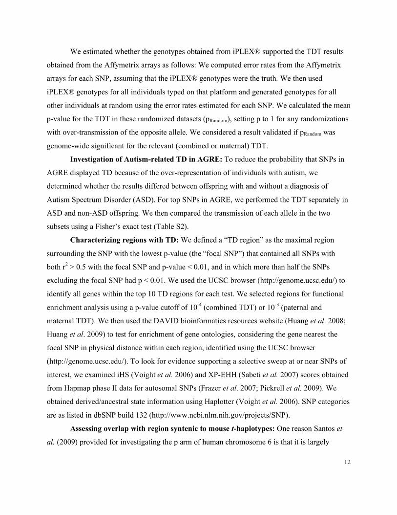
We estimated whether the genotypes obtained from iPLEX® supported the TDT results
obtained from the Affymetrix arrays as follows: We computed error rates from the Affymetrix
arrays for each SNP, assuming that the iPLEX® genotypes were the truth. We then used
iPLEX® genotypes for all individuals typed on that platform and generated genotypes for all
other individuals at random using the error rates estimated for each SNP. We calculated the mean
p-value for the TDT in these randomized datasets (pRandom), setting p to 1 for any randomizations
with over-transmission of the opposite allele. We considered a result validated if pRandom was
genome-wide significant for the relevant (combined or maternal) TDT.
Investigation of Autism-related TD in AGRE: To reduce the probability that SNPs in
AGRE displayed TD because of the over-representation of individuals with autism, we
determined whether the results differed between offspring with and without a diagnosis of
Autism Spectrum Disorder (ASD). For top SNPs in AGRE, we performed the TDT separately in
ASD and non-ASD offspring. We then compared the transmission of each allele in the two
subsets using a Fisher’s exact test (Table S2).
Characterizing regions with TD: We defined a “TD region” as the maximal region
surrounding the SNP with the lowest p-value (the “focal SNP”) that contained all SNPs with
both r2 > 0.5 with the focal SNP and p-value < 0.01, and in which more than half the SNPs
excluding the focal SNP had p < 0.01. We used the UCSC browser (http://genome.ucsc.edu/) to
identify all genes within the top 10 TD regions for each test. We selected regions for functional
enrichment analysis using a p-value cutoff of 10-4 (combined TDT) or 10-3 (paternal and
maternal TDT). We then used the DAVID bioinformatics resources website (Huang et al. 2008;
Huang et al. 2009) to test for enrichment of gene ontologies, considering the gene nearest the
focal SNP in physical distance within each region, identified using the UCSC browser
(http://genome.ucsc.edu/). To look for evidence supporting a selective sweep at or near SNPs of
interest, we examined iHS (Voight et al. 2006) and XP-EHH (Sabeti et al. 2007) scores obtained
from Hapmap phase II data for autosomal SNPs (Frazer et al. 2007; Pickrell et al. 2009). We
obtained derived/ancestral state information using Haplotter (Voight et al. 2006). SNP categories
are as listed in dbSNP build 132 (http://www.ncbi.nlm.nih.gov/projects/SNP).
Assessing overlap with region syntenic to mouse t-haplotypes: One reason Santos et
al. (2009) provided for investigating the p arm of human chromosome 6 is that it is largely

13
syntenic to mouse chromosome 17, where t-haplotypes are located. Given that we also find a
paternal signal on chromosome 6p, we assessed whether this region shared sequence similarity to
the t-haplotype region; locations of shared sequence similarity with the mouse genome for the
paternal-specific TD region in 6p21.1 were determined using the UCSC genome browser
conversion tool (http://genome.ucsc.edu). The only region of the mouse genome with sequence
similarity spanning the entire TD region identified here is outside the annotated boundaries of t-
haplotypes (Silver 1993; Wallace and Erhart 2008).
Comparing AGRE signal in 6p21.1 between Affymetrix and Illumina platforms: We
considered only those individuals who had been genotyped on both platforms and were included
in the original TDT in AGRE (using Illumina data). We merged the datasets, setting any
genotypes differing between Affymetrix and Illumina to missing data. Aside from elimination of
Mendelian errors, no quality control steps were performed for Affymetrix genotyping data. We
considered SNPs with at least 141 transmissions, the minimum sample size required for 80%
power to detect TD at p < 0.05, using the estimated distortion strength of 0.1187. We
additionally used this dataset to calculate pairwise LD between Affymetrix and Illumina SNPs in
AGRE founders.
Analysis of maternal TD near centromeres and telomeres: We calculated genetic
distance to the centromere for all SNPs using the HapMap phase II genetic map (Frazer et al.
2007) and gaps in the assembly annotated as centromeres by the UCSC Genome Browser
(http://genome.ucsc.edu/), using build HG18. We also calculated genetic distance to the most
telomeric SNP in the HapMap phase II (Frazer et al. 2007) and physical distance between the
most telomeric SNPs in our dataset and gaps in the assembly annotated as telomeres by the
UCSC Genome Browser (http://genome.ucsc.edu/) using build HG19 (telomere locations were
not available for all chromosomes in HG18). We determined the genetic distance to the
centromere and most telomeric HapMap SNP, as well as the number of SNPs between the SNP
and the centromere and telomere, for all maternal-specific (i.e., paternal TDT p > 0.01) SNPs
with p < 10-3. We additionally checked for marginally significant maternal TDT p-values (p <
0.05) at the SNP closest to the centromere and telomere on both arms of metacentric
chromosomes and the q arm of acrocentric chromosomes.
Sperm typing to test for TD in sperm production or motility
14
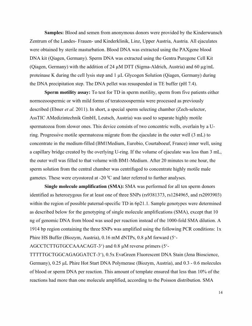
Samples: Blood and semen from anonymous donors were provided by the Kinderwunsch
Zentrum of the Landes- Frauen- und Kinderklinik, Linz, Upper Austria, Austria. All ejaculates
were obtained by sterile masturbation. Blood DNA was extracted using the PAXgene blood
DNA kit (Qiagen, Germany). Sperm DNA was extracted using the Gentra Puregene Cell Kit
(Qiagen, Germany) with the addition of 24 µM DTT (Sigma-Aldrich, Austria) and 60 µg/mL
proteinase K during the cell lysis step and 1 µL Glycogen Solution (Qiagen, Germany) during
the DNA precipitation step. The DNA pellet was resuspended in TE buffer (pH 7.4).
Sperm motility assay: To test for TD in sperm motility, sperm from five patients either
normozoospermic or with mild forms of teratozoospermia were processed as previously
described (Ebner et al. 2011). In short, a special sperm selecting chamber (Zech-selector,
AssTIC AMedizintechnik GmbH, Leutsch, Austria) was used to separate highly motile
spermatozoa from slower ones. This device consists of two concentric wells, overlain by a U-
ring. Progressive motile spermatozoa migrate from the ejaculate in the outer well (3 mL) to
concentrate in the medium-filled (BM1Medium, Eurobio, Courtaboeuf, France) inner well, using
a capillary bridge created by the overlying U-ring. If the volume of ejaculate was less than 3 mL,
the outer well was filled to that volume with BM1-Medium. After 20 minutes to one hour, the
sperm solution from the central chamber was centrifuged to concentrate highly motile male
gametes. These were cryostored at -20 ⁰C and later referred to further analyses.
Single molecule amplification (SMA): SMA was performed for all ten sperm donors
identified as heterozygous for at least one of three SNPs (rs9381373, rs1284965, and rs2093903)
within the region of possible paternal-specific TD in 6p21.1. Sample genotypes were determined
as described below for the genotyping of single molecule amplifications (SMA), except that 10
ng of genomic DNA from blood was used per reaction instead of the 1000-fold SMA dilution. A
1914 bp region containing the three SNPs was amplified using the following PCR conditions: 1x
Phire HS Buffer (Biozym, Austria), 0.16 mM dNTPs, 0.8 µM forward (5‘-
AGCCTCTTGTGCCAAACAGT-3‘) and 0.8 µM reverse primers (5‘-
TTTTTGCTGGCAGAGGATCT-3‘), 0.5x EvaGreen Fluorescent DNA Stain (Jena Bioscience,
Germany), 0.25 µL Phire Hot Start DNA Polymerase (Biozym, Austria), and 0.3 - 0.6 molecules
of blood or sperm DNA per reaction. This amount of template ensured that less than 10% of the
reactions had more than one molecule amplified, according to the Poisson distribution. SMA

15
reactions were set up in a dedicated laminar flow hood decontaminated with UV light and 10%
chlorine before the start of each experiment. No-template controls were included in each
experiment to screen for contamination. The PCR was performed in 10 µL volumes in a real-
time PCR Thermocycler (CFX384 System, Biorad), using an initial heating step of 94˚ for 2 min
followed by 5 cycles at 94˚ for 15 sec, 65˚ for 15 sec, and 72˚ for 30 sec, and then 35 cycles at
94˚ for 15 sec, 68˚ for 15 sec, and 72˚ for 30 sec. We considered the amplification curve and the
melting curve profile to identify PCR reactions that amplified our region of interest. We verified
the amplification of the correct product by acrylamide gel electrophoresis in the initial stages
when appropriate experimental conditions were optimized. Approximately 20% of our SMA
reactions had the wrong product size, probably due to amplification of other genomic regions.
These false positives were identified by a different melt curve profile and did not render a
genotyping result, so they could be easily excluded.
Genotyping single molecule amplifications: SMA reactions that amplified the region of
interest were diluted 1,000-fold and genotyped by allele-specific PCR in combination with a
real-time PCR machine (CFX384 System, Biorad), as described previously (Tiemann-Boege et
al. 2006). The last three phosphodiester bonds at the 3’ end of the allele-specific primers were
substituted by phosphorothioate bonds to increase allele-specific selectivity. The allele-specific
PCR reactions were carried out in 10 µL volumes containing 5 µL of the SMA dilution, 0.4 µM
allele-specific primer and 0.4 µM outside primer, and either 1x OneTaq Reaction Buffer (NEB),
1x SYBR Green I (Invitrogen), and 0.125U OneTaq Hot Start DNA Polymerase or 1x AmpliTaq
Gold Buffer, 1.5 mM MgCl2, 0.16 mM dNTPs, 1x SYBR Green I (Invitrogen), and 0.25U Z05
DNA Polymerase (Roche, Austria). The primer sequences are shown in Table S3.
The reactions were carried out with an initial heating step of 94˚ for 2 min, followed by
40 cycles at 94˚ for 30 sec, 65˚ for 30 sec, and 72˚ for 15 sec when using OneTaq Hot Start DNA
Polymerase or with 95˚ for 2 min, followed by 40 cycles at 95˚ for 30 sec, 56˚ for 30 sec, and 72˚
for 15 sec when using Z05 DNA Polymerase. For each sample two reactions were amplified: one
for each allele, differing only by the allele-specific primer. The genotype was assessed using the
difference between the quantification cycles (Cqs) of the two allele-specific reactions;
homozygote samples presented a large difference compared to heterozygote samples. In order to
verify the genotyping data, we genotyped two SNPs for each sample, except for one donor who
16
was heterozygous at only one of the three SNPs; for this donor, we genotyped the same SNP
twice. In 98% of cases, the alleles at the two SNPs conformed to the expected haplotypes, and
for rare non-matching genotypes between SNPs (mostly occurring for samples with more than
one molecule), we repeated the genotyping and corrected the false genotyping call. Reactions
that resulted in a heterozygous genotype at each SNP, indicating that more than one molecule
had been amplified, were eliminated from the analysis. Equivalence of the two PCR methods
(OneTaq Hot Start and Z05 DNA Polymerase) was determined by typing 48 samples from one
donor using both polymerases; both methods yielded the same genotype for all samples.
Testing allelic ratios: Blood genotyping was performed in a subset of donors as a
measure of noise. We performed a two-sided test of the binomial for blood samples because we
had no expectation for which allele would be over-represented. In sperm, we tested for
deviations from 50% occurrence of each allele independently for each donor, as well as for all
donors combined (because a chi-squared test of homogeneity did not demonstrate significant
heterogeneity among donors). Given that we expected a particular allele to be over-represented
in sperm under the alternative hypothesis of TD, we performed a one-sided test of the binomial
(i.e., the p-value is the probability of observing at least as many of the TDT-based over-
transmitted allele as we observed in the sperm genotyping, assuming a binomial with parameter
0.5). We tested unselected sperm (not selected using the motility assay) and fast sperm (the
fastest 0.005 - 5.155% of sperm from the motility assay) separately. We additionally tested
whether the transmission rate from sperm typing was compatible with that inferred from the
TDT, using the following likelihood ratio test: LR = Lik(θsperm = θTDT )/Lik(θsperm, θTDT), where θ
represents the paternal transmission rate of the over-represented allele inferred from the TDT at
SNP rs9381373; for this test, we combined allele counts from unselected and selected sperm.
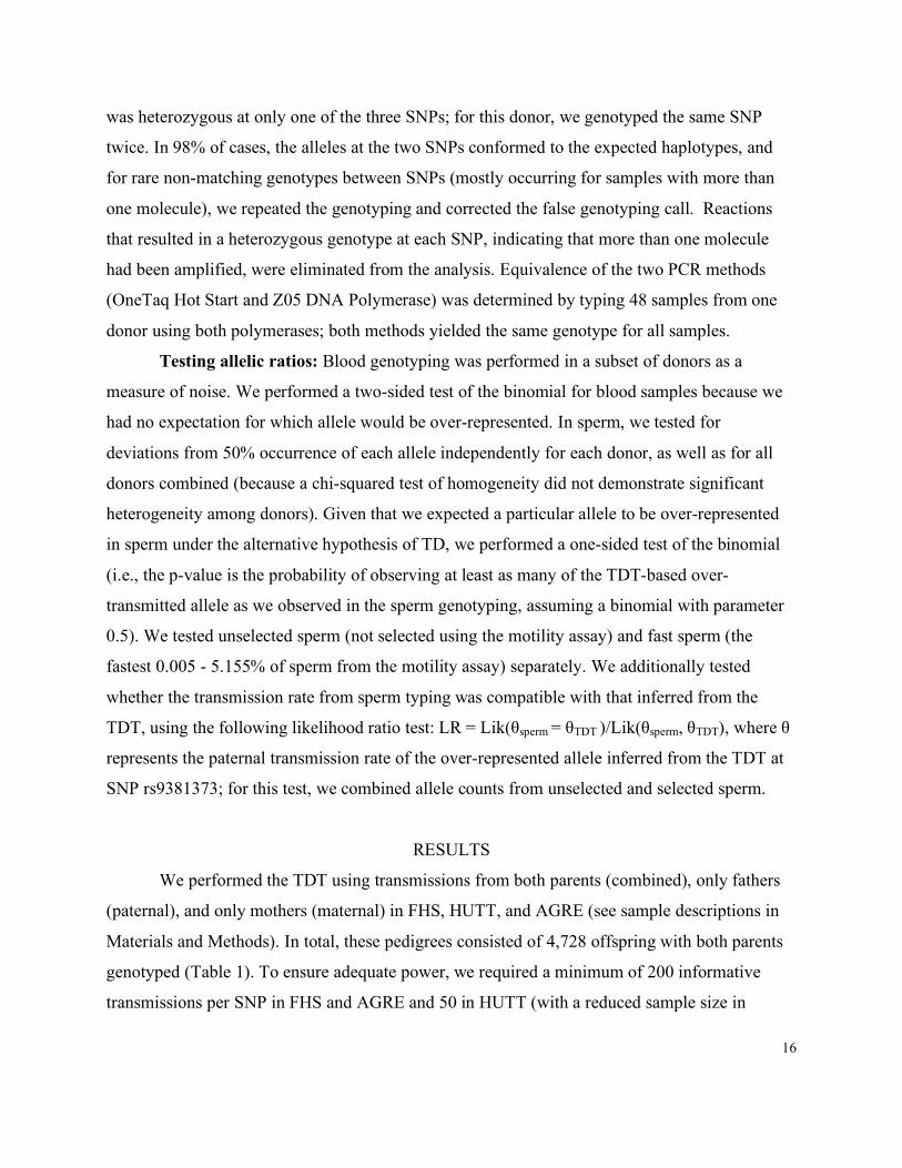
RESULTS
We performed the TDT using transmissions from both parents (combined), only fathers
(paternal), and only mothers (maternal) in FHS, HUTT, and AGRE (see sample descriptions in
Materials and Methods). In total, these pedigrees consisted of 4,728 offspring with both parents
genotyped (Table 1). To ensure adequate power, we required a minimum of 200 informative
transmissions per SNP in FHS and AGRE and 50 in HUTT (with a reduced sample size in
17
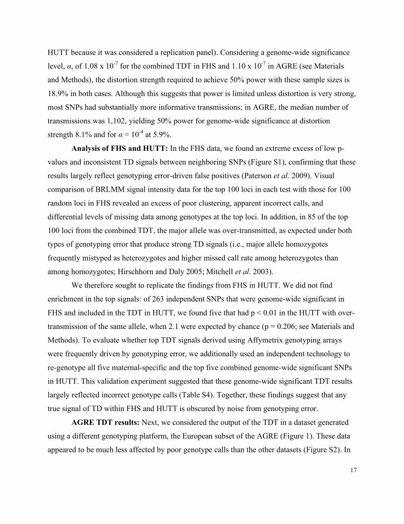
HUTT because it was considered a replication panel). Considering a genome-wide significance
level, α, of 1.08 x 10⁻⁷ for the combined TDT in FHS and 1.10 x 10⁻⁷ in AGRE (see Materials
and Methods), the distortion strength required to achieve 50% power with these sample sizes is
18.9% in both cases. Although this suggests that power is limited unless distortion is very strong,
most SNPs had substantially more informative transmissions; in AGRE, the median number of
transmissions was 1,102, yielding 50% power for genome-wide significance at distortion
strength 8.1% and for α = 10-4 at 5.9%.
Analysis of FHS and HUTT: In the FHS data, we found an extreme excess of low p-
values and inconsistent TD signals between neighboring SNPs (Figure S1), confirming that these
results largely reflect genotyping error-driven false positives (Paterson et al. 2009). Visual
comparison of BRLMM signal intensity data for the top 100 loci in each test with those for 100
random loci in FHS revealed an excess of poor clustering, apparent incorrect calls, and
differential levels of missing data among genotypes at the top loci. In addition, in 85 of the top
100 loci from the combined TDT, the major allele was over-transmitted, as expected under both
types of genotyping error that produce strong TD signals (i.e., major allele homozygotes
frequently mistyped as heterozygotes and higher missed call rate among heterozygotes than
among homozygotes; Hirschhorn and Daly 2005; Mitchell et al. 2003).
We therefore sought to replicate the findings from FHS in HUTT. We did not find
enrichment in the top signals: of 263 independent SNPs that were genome-wide significant in
FHS and included in the TDT in HUTT, we found five that had p < 0.01 in the HUTT with over-
transmission of the same allele, when 2.1 were expected by chance (p = 0.206; see Materials and
Methods). To evaluate whether top TDT signals derived using Affymetrix genotyping arrays
were frequently driven by genotyping error, we additionally used an independent technology to
re-genotype all five maternal-specific and the top five combined genome-wide significant SNPs
in HUTT. This validation experiment suggested that these genome-wide significant TDT results
largely reflected incorrect genotype calls (Table S4). Together, these findings suggest that any
true signal of TD within FHS and HUTT is obscured by noise from genotyping error.
AGRE TDT results: Next, we considered the output of the TDT in a dataset generated
using a different genotyping platform, the European subset of the AGRE (Figure 1). These data
appeared to be much less affected by poor genotype calls than the other datasets (Figure S2). In
18
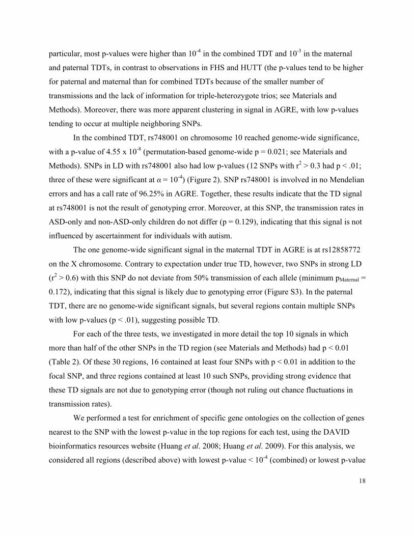
particular, most p-values were higher than 10-4 in the combined TDT and 10-3 in the maternal
and paternal TDTs, in contrast to observations in FHS and HUTT (the p-values tend to be higher
for paternal and maternal than for combined TDTs because of the smaller number of
transmissions and the lack of information for triple-heterozygote trios; see Materials and
Methods). Moreover, there was more apparent clustering in signal in AGRE, with low p-values
tending to occur at multiple neighboring SNPs.
In the combined TDT, rs748001 on chromosome 10 reached genome-wide significance,
with a p-value of 4.55 x 10⁻⁸ (permutation-based genome-wide p = 0.021; see Materials and
Methods). SNPs in LD with rs748001 also had low p-values (12 SNPs with r2 > 0.3 had p < .01;
three of these were significant at α = 10-4) (Figure 2). SNP rs748001 is involved in no Mendelian
errors and has a call rate of 96.25% in AGRE. Together, these results indicate that the TD signal
at rs748001 is not the result of genotyping error. Moreover, at this SNP, the transmission rates in
ASD-only and non-ASD-only children do not differ (p = 0.129), indicating that this signal is not
influenced by ascertainment for individuals with autism.
The one genome-wide significant signal in the maternal TDT in AGRE is at rs12858772
on the X chromosome. Contrary to expectation under true TD, however, two SNPs in strong LD
(r2 > 0.6) with this SNP do not deviate from 50% transmission of each allele (minimum pMaternal =
0.172), indicating that this signal is likely due to genotyping error (Figure S3). In the paternal
TDT, there are no genome-wide significant signals, but several regions contain multiple SNPs
with low p-values (p < .01), suggesting possible TD.
For each of the three tests, we investigated in more detail the top 10 signals in which
more than half of the other SNPs in the TD region (see Materials and Methods) had p < 0.01
(Table 2). Of these 30 regions, 16 contained at least four SNPs with p < 0.01 in addition to the
focal SNP, and three regions contained at least 10 such SNPs, providing strong evidence that
these TD signals are not due to genotyping error (though not ruling out chance fluctuations in
transmission rates).
We performed a test for enrichment of specific gene ontologies on the collection of genes
nearest to the SNP with the lowest p-value in the top regions for each test, using the DAVID
bioinformatics resources website (Huang et al. 2008; Huang et al. 2009). For this analysis, we
considered all regions (described above) with lowest p-value < 10-4 (combined) or lowest p-value

19
< 10-3 (paternal or maternal, considering sex-specific results only). The most enriched categories
were, for the combined test, “alternative splicing” (p = 0.0459; 1.59-fold enrichment); for the
paternal test, “vitamin metabolic process” and “cell maturation” (p = 0.0593; 30.06-fold
enrichment); and for the maternal test, “vinculin, conserved site” (p = 5.27 x 10⁻³; 362-fold
enrichment), with additional related functional categories also enriched (see Table 3; p-values
are uncorrected for multiple testing but presented for comparison among categories). The top GO
categories related to combined TD signals were broad and difficult to interpret, and none of those
related to paternal TD signals were enriched at p < 0.05. Intriguingly, however, the maternal-
specific TD signals tagged vinculin and an alpha-catenin, which are unlinked but share the
capacity to bind actin and are involved in cytoskeletal integrity and cell spreading. If variants in
these genes influence cell division or early development, this would provide a candidate
mechanism for distortion in females.
A suggestive signal of paternal TD in AGRE: In the paternal-specific TDT, there was a
strong signal of TD (p = 1.77 x 10⁻⁵) in a region where experiment-wide TD was previously
identified in HapMap CEU males (Santos et al. 2009). The region of TD in AGRE spans
approximately 711 kb on chromosome 6 surrounding SNP rs12199720, which is the strongest
regional signal of paternal-specific TD in the AGRE (Table 2, Figure 3). The finding of TD in
this region in the paternal but not the maternal TDT (minimal pMaternal = 0.1037) both indicates
that this TD is due to a male-specific process and suggests that the signal is not due to subtle
genotyping error affecting calls for parents of both sexes.
If there is truly distortion in this region, the causal SNP is likely to be regulatory: The
only three non-synonymous SNPs in this region known to be polymorphic in CEU have minor
allele frequencies of < 0.07 (The 1000 Genomes Project Consortium 2010; Frazer et al. 2007),
making them unlikely to be driving the observed TD signal. The transcription factors RUNX2
and SUPT3H and the miRNA MIRN586 all fall within the region, and top SNP rs12199720 is
within an intron of both RUNX2 and SUPT3H, with the nearest exon in RUNX2. These two genes
play an important role in human growth; RUNX2 is involved in osteoblastic differentiation and
skeletal morphogenesis (Ducy et al. 2000; Otto et al. 1997; Wheeler et al. 2000), defects in
RUNX2 cause the autosomal dominant skeletal disorder cleidocranial dysplasia (CLCD)
(Mundlos et al. 1997), and a SNP in an intron of SUPT3H was suggestively associated with
20
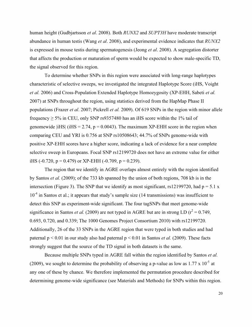
human height (Gudbjartsson et al. 2008). Both RUNX2 and SUPT3H have moderate transcript
abundance in human testis (Wang et al. 2008), and experimental evidence indicates that RUNX2
is expressed in mouse testis during spermatogenesis (Jeong et al. 2008). A segregation distorter
that affects the production or maturation of sperm would be expected to show male-specific TD,
the signal observed for this region.
To determine whether SNPs in this region were associated with long-range haplotypes
characteristic of selective sweeps, we investigated the integrated Haplotype Score (iHS, Voight
et al. 2006) and Cross-Population Extended Haplotype Homozygosity (XP-EHH, Sabeti et al.
2007) at SNPs throughout the region, using statistics derived from the HapMap Phase II
populations (Frazer et al. 2007; Pickrell et al. 2009). Of 619 SNPs in the region with minor allele
frequency ≥ 5% in CEU, only SNP rs9357480 has an iHS score within the 1% tail of
genomewide |iHS| (iHS = 2.74, p = 0.0043). The maximum XP-EHH score in the region when
comparing CEU and YRI is 0.756 at SNP rs10508643; 44.7% of SNPs genome-wide with
positive XP-EHH scores have a higher score, indicating a lack of evidence for a near complete
selective sweep in Europeans. Focal SNP rs12199720 does not have an extreme value for either
iHS (-0.720, p = 0.479) or XP-EHH (-0.709, p = 0.239).
The region that we identify in AGRE overlaps almost entirely with the region identified
by Santos et al. (2009); of the 733 kb spanned by the union of both regions, 708 kb is in the
intersection (Figure 3). The SNP that we identify as most significant, rs12199720, had p = 5.1 x
10-4 in Santos et al.; it appears that study’s sample size (14 transmissions) was insufficient to
detect this SNP as experiment-wide significant. The four tagSNPs that meet genome-wide
significance in Santos et al. (2009) are not typed in AGRE but are in strong LD (r2 = 0.749,
0.693, 0.720, and 0.339; The 1000 Genomes Project Consortium 2010) with rs12199720.
Additionally, 26 of the 33 SNPs in the AGRE region that were typed in both studies and had
paternal p < 0.01 in our study also had paternal p < 0.01 in Santos et al. (2009). These facts
strongly suggest that the source of the TD signal in both datasets is the same.
Because multiple SNPs typed in AGRE fall within the region identified by Santos et al.
(2009), we sought to determine the probability of observing a p-value as low as 1.77 x 10⁻⁵ at
any one of these by chance. We therefore implemented the permutation procedure described for
determining genome-wide significance (see Materials and Methods) for SNPs within this region.
21
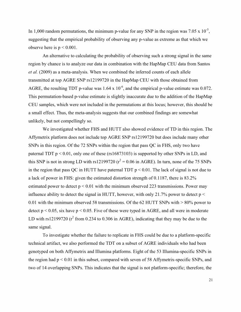
In 1,000 random permutations, the minimum p-value for any SNP in the region was 7.05 x 10⁻⁵,
suggesting that the empirical probability of observing any p-value as extreme as that which we
observe here is p < 0.001.
An alternative to calculating the probability of observing such a strong signal in the same
region by chance is to analyze our data in combination with the HapMap CEU data from Santos
et al. (2009) as a meta-analysis. When we combined the inferred counts of each allele
transmitted at top AGRE SNP rs12199720 in the HapMap CEU with those obtained from
AGRE, the resulting TDT p-value was 1.64 x 10⁻⁶, and the empirical p-value estimate was 0.072.
This permutation-based p-value estimate is slightly inaccurate due to the addition of the HapMap
CEU samples, which were not included in the permutations at this locus; however, this should be
a small effect. Thus, the meta-analysis suggests that our combined findings are somewhat
unlikely, but not compellingly so.
We investigated whether FHS and HUTT also showed evidence of TD in this region. The
Affymetrix platform does not include top AGRE SNP rs12199720 but does include many other
SNPs in this region. Of the 72 SNPs within the region that pass QC in FHS, only two have
paternal TDT p < 0.01, only one of these (rs16873103) is supported by other SNPs in LD, and
this SNP is not in strong LD with rs12199720 (r2 = 0.06 in AGRE). In turn, none of the 75 SNPs
in the region that pass QC in HUTT have paternal TDT p < 0.01. The lack of signal is not due to
a lack of power in FHS: given the estimated distortion strength of 0.1187, there is 83.2%
estimated power to detect p < 0.01 with the minimum observed 223 transmissions. Power may
influence ability to detect the signal in HUTT, however, with only 21.7% power to detect p <
0.01 with the minimum observed 58 transmissions. Of the 62 HUTT SNPs with > 80% power to
detect p < 0.05, six have p < 0.05. Five of these were typed in AGRE, and all were in moderate
LD with rs12199720 (r2 from 0.234 to 0.306 in AGRE), indicating that they may be due to the
same signal.
To investigate whether the failure to replicate in FHS could be due to a platform-specific
technical artifact, we also performed the TDT on a subset of AGRE individuals who had been
genotyped on both Affymetrix and Illumina platforms. Eight of the 53 Illumina-specific SNPs in
the region had p < 0.01 in this subset, compared with seven of 58 Affymetrix-specific SNPs, and
two of 14 overlapping SNPs. This indicates that the signal is not platform-specific; therefore, the
22
lack of replication in FHS is particularly worrisome and suggests that the signal in the other
datasets is unlikely to be driven by real TD.
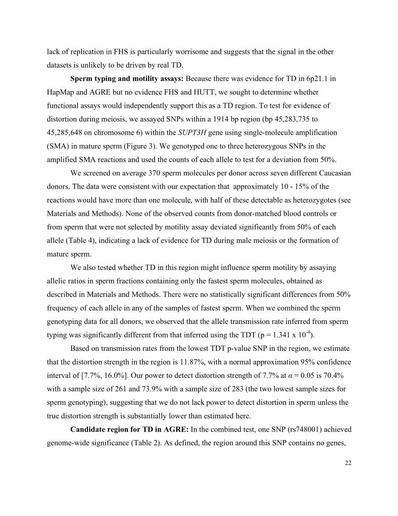
Sperm typing and motility assays: Because there was evidence for TD in 6p21.1 in
HapMap and AGRE but no evidence FHS and HUTT, we sought to determine whether
functional assays would independently support this as a TD region. To test for evidence of
distortion during meiosis, we assayed SNPs within a 1914 bp region (bp 45,283,735 to
45,285,648 on chromosome 6) within the SUPT3H gene using single-molecule amplification
(SMA) in mature sperm (Figure 3). We genotyped one to three heterozygous SNPs in the
amplified SMA reactions and used the counts of each allele to test for a deviation from 50%.
We screened on average 370 sperm molecules per donor across seven different Caucasian
donors. The data were consistent with our expectation that approximately 10 - 15% of the
reactions would have more than one molecule, with half of these detectable as heterozygotes (see
Materials and Methods). None of the observed counts from donor-matched blood controls or
from sperm that were not selected by motility assay deviated significantly from 50% of each
allele (Table 4), indicating a lack of evidence for TD during male meiosis or the formation of
mature sperm.
We also tested whether TD in this region might influence sperm motility by assaying
allelic ratios in sperm fractions containing only the fastest sperm molecules, obtained as
described in Materials and Methods. There were no statistically significant differences from 50%
frequency of each allele in any of the samples of fastest sperm. When we combined the sperm
genotyping data for all donors, we observed that the allele transmission rate inferred from sperm
typing was significantly different from that inferred using the TDT (p = 1.341 x 10⁻⁴).
Based on transmission rates from the lowest TDT p-value SNP in the region, we estimate
that the distortion strength in the region is 11.87%, with a normal approximation 95% confidence
interval of [7.7%, 16.0%]. Our power to detect distortion strength of 7.7% at α = 0.05 is 70.4%
with a sample size of 261 and 73.9% with a sample size of 283 (the two lowest sample sizes for
sperm genotyping), suggesting that we do not lack power to detect distortion in sperm unless the
true distortion strength is substantially lower than estimated here.
Candidate region for TD in AGRE: In the combined test, one SNP (rs748001) achieved
genome-wide significance (Table 2). As defined, the region around this SNP contains no genes,

23
but it does contain several regions that are among the most highly conserved elements in
vertebrates (i.e., within the 5,000 most highly conserved elements out of 1.31 million total
conserved elements; Siepel et al. 2005) (Figure 2). The maximum range at which loci are in LD
(r2 > 0.1; The 1000 Genomes Project Consortium 2010) with the focal SNP contains all of
LOC100169752 and approximately 22 kb of the 3’ end of C10ORF122, including one exon.
Notably, focal SNP rs748001 is associated with a signal of recent directional selection, falling
within the tail of Integrated Haplotype Score (iHS) signals (iHS = 2.181, p = 0.021) in the
HapMap II CEU (Frazer et al. 2007; Pickrell et al. 2009) (Figure 2). This SNP is also in LD with
two SNPs that have strongly negative iHS: rs4962310 (iHS = -2.05, p = 0.014, r2 with rs748001
in CEU = 0.469) and rs11244542 (iHS = -2.01, p = 0.016, r2 with rs748001 in CEU = 0.45), and
the over-transmitted allele at rs748001 is in phase with the derived allele at both of these SNPs
(Frazer et al. 2007). We note, however, that none of the seven SNPs within this region that were
genotyped in the FHS and HUTT had TDT p < 0.01 in these datasets, despite apparently
sufficient power. True distortion of strength 3.8% in FHS and 6.2% in HUTT would be required
for 80% power at two or more SNPs. Because of the winner’s curse (see Ioannidis et al. 2001,
Göring et al. 2001, and Lohmueller et al. 2003), estimating effect size from our data would yield
a substantial over-estimate, so it is unclear whether the true distortion strength is large enough to
achieve power in these other datasets; nevertheless, the failure to replicate in FHS suggests the
absence of strong TD in this region.
Analysis of maternal TD near centromeres and telomeres: We evaluated the
prevalence of TD at loci closely linked to centromeres, because these sites are likely to segregate
with the untyped centromeric repeats proposed to be subject to female-specific meiotic drive. We
found only one example of a maternal-specific (paternal TDT p > 0.01) SNP with p < 10-3
within 1 cM of the centromere (on chromosome 10) in AGRE, with 116 SNPs separating this
SNP from the centromere; the next nearest SNP with p < 10-3 was separated from the centromere
by 256 SNPs (Figure 4). Across all chromosomes, the nearest SNPs to all centromeres had
maternal TDT p > 0.05 except chromosome arms 3q (p = 0.021) and 19q (p = 5.63 x 10⁻³). The
only other SNP in strong LD (r2 > 0.6) with the most centromeric 3q SNP had maternal p = 0.261
and a stronger signal in the paternal TDT (p = 0.030), so this may be a spurious signal due to
genotyping error. The SNP on chromosome 19q also had a lower p-value in the paternal TDT (p
24
= 2.18 x 10⁻³), so if it is a true signal of TD, it is unlikely to be due to a mechanism specific to
asymmetric meioses. With the possible exception of chromosome 22, the lack of signal was not
due to SNP sparsity near centromeres; at least 25 SNPs within 1 cM of the centromere passed
QC on all chromosomes except 15 (four SNPs within 1 cM) and 22 (minimum distance 2.22
cM).
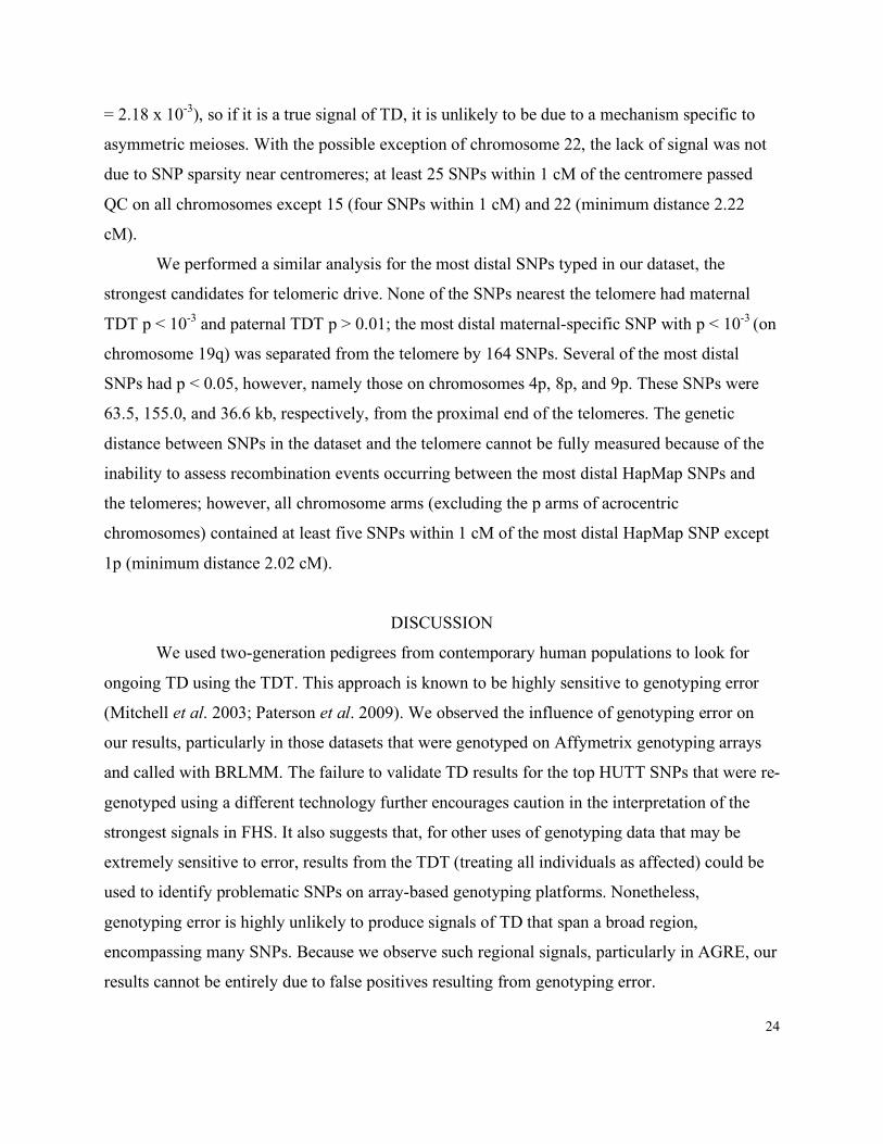
We performed a similar analysis for the most distal SNPs typed in our dataset, the
strongest candidates for telomeric drive. None of the SNPs nearest the telomere had maternal
TDT p < 10-3 and paternal TDT p > 0.01; the most distal maternal-specific SNP with p < 10-3 (on
chromosome 19q) was separated from the telomere by 164 SNPs. Several of the most distal
SNPs had p < 0.05, however, namely those on chromosomes 4p, 8p, and 9p. These SNPs were
63.5, 155.0, and 36.6 kb, respectively, from the proximal end of the telomeres. The genetic
distance between SNPs in the dataset and the telomere cannot be fully measured because of the
inability to assess recombination events occurring between the most distal HapMap SNPs and
the telomeres; however, all chromosome arms (excluding the p arms of acrocentric
chromosomes) contained at least five SNPs within 1 cM of the most distal HapMap SNP except
1p (minimum distance 2.02 cM).
DISCUSSION
We used two-generation pedigrees from contemporary human populations to look for
ongoing TD using the TDT. This approach is known to be highly sensitive to genotyping error
(Mitchell et al. 2003; Paterson et al. 2009). We observed the influence of genotyping error on
our results, particularly in those datasets that were genotyped on Affymetrix genotyping arrays
and called with BRLMM. The failure to validate TD results for the top HUTT SNPs that were re-
genotyped using a different technology further encourages caution in the interpretation of the
strongest signals in FHS. It also suggests that, for other uses of genotyping data that may be
extremely sensitive to error, results from the TDT (treating all individuals as affected) could be
used to identify problematic SNPs on array-based genotyping platforms. Nonetheless,
genotyping error is highly unlikely to produce signals of TD that span a broad region,
encompassing many SNPs. Because we observe such regional signals, particularly in AGRE, our
results cannot be entirely due to false positives resulting from genotyping error.

25
Unlike other types of genotyping error, unidentified copy number variants (CNVs) could
produce spurious TDT signals that span multiple SNPs; however, there are a number of lines of
evidence against CNVs underlying our strongest signals. First, CNVs common enough to yield
strong TD signals should produce numerous Mendelian errors and cause deviations from HWE;
therefore, SNPs in these regions should be eliminated in our QC steps. Some rare cases, for
instance duplications with more than one polymorphic paralog, may be more difficult to detect.
To rule out the possibility of an inter-chromosomal duplication or paralog causing the genome-
wide significant signal on chromosome 10, we verified that there was no LD between the region
and other segments of the genome. Additionally, there are no CNVs in this region in the
Database of Genomic Variants, an online database of published CNVs
(http://projects.tcag.ca/variation/). CNVs can only produce a sex-specific signal of distortion if
one of the copies resides on a sex chromosome, and in this case, the distortion should differ
between male and female offspring. We determined that there was no difference in the distortion
rate between male and female offspring in the region of suggestive paternal-specific TD on
chromosome 6p (p = 0.5379), and therefore this signal cannot be attributed to a duplication or
paralog on a sex chromosome.
In addition to false positives, another concern is that our loose filter based on deviation
from HWE (p < 10-4) could cause us to miss true signals of TD. We used this filter to eliminate
SNPs with unusual genotype proportions due to genotyping error; however, strong viability
selection or segregation distortion could also produce a deviation from HWE. With this in mind,
we investigated the strength of selection/distortion necessary to create a deviation of p < 10-4. We
generated genotypes at random for all founders, using the expected frequencies under viability
selection or sex-specific drive (assuming a 1:1 sex ratio). We then performed the exact test of
Hardy-Weinberg on 1,000 such simulated datasets. We found that, even with s = 0.5 and h = 0.5,
fewer than 0.1% of cases of viability selection generate a deviation from HWE strong enough to
be detected by our filter. In contrast, 18.2% of cases with (unbalanced) sex-specific distortion
equal to 30% generated such a deviation. We conclude that loci experiencing very strong
segregation distortion and not subject to a counter-balancing force may occasionally deviate
from HWE and be excluded from our analysis, but that the effects of viability selection on HWE
are negligible.
26
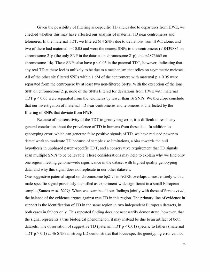
Given the possibility of filtering sex-specific TD alleles due to departures from HWE, we
checked whether this may have affected our analysis of maternal TD near centromeres and
telomeres. In the maternal TDT, we filtered 614 SNPs due to deviations from HWE alone, and
two of these had maternal p < 0.05 and were the nearest SNPs to the centromere: rs10439884 on
chromosome 21p (the only SNP in the dataset on chromosome 21p) and rs2873665 on
chromosome 14q. These SNPs also have p < 0.05 in the paternal TDT, however, indicating that
any real TD at these loci is unlikely to be due to a mechanism that relies on asymmetric meioses.
All of the other six filtered SNPs within 1 cM of the centromere with maternal p < 0.05 were
separated from the centromere by at least two non-filtered SNPs. With the exception of the lone
SNP on chromosome 21p, none of the SNPs filtered for deviations from HWE with maternal
TDT p < 0.05 were separated from the telomeres by fewer than 16 SNPs. We therefore conclude
that our investigation of maternal TD near centromeres and telomeres is unaffected by the
filtering of SNPs that deviate from HWE.
Because of the sensitivity of the TDT to genotyping error, it is difficult to reach any
general conclusion about the prevalence of TD in humans from these data. In addition to
genotyping error, which can generate false positive signals of TD, we have reduced power to
detect weak to moderate TD because of sample size limitations, a bias towards the null
hypothesis in unphased parent-specific TDT, and a conservative requirement that TD signals
span multiple SNPs to be believable. These considerations may help to explain why we find only
one region meeting genome-wide significance in the dataset with highest quality genotyping
data, and why this signal does not replicate in our other datasets.
One suggestive paternal signal on chromosome 6p21.1 in AGRE overlaps almost entirely with a
male-specific signal previously identified as experiment-wide significant in a small European
sample (Santos et al. 2009). When we examine all our findings jointly with those of Santos et al.,
the balance of the evidence argues against true TD in this region. The primary line of evidence in
support is the identification of TD in the same region in two independent European datasets, in
both cases in fathers only. This repeated finding does not necessarily demonstrate, however, that
the signal represents a true biological phenomenon; it may instead be due to an artifact of both
datasets. The observation of suggestive TD (paternal TDT p < 0.01) specific to fathers (maternal
TDT p > 0.1) at 46 SNPs in strong LD demonstrates that locus-specific genotyping error cannot
27
be responsible for the signal in AGRE. In addition, the genotyping data supporting the signal in
AGRE derives from the Illumina Hap550 platform, which tends to have lower rates of error-
driven false positives than Affymetrix platforms (Figure S2).
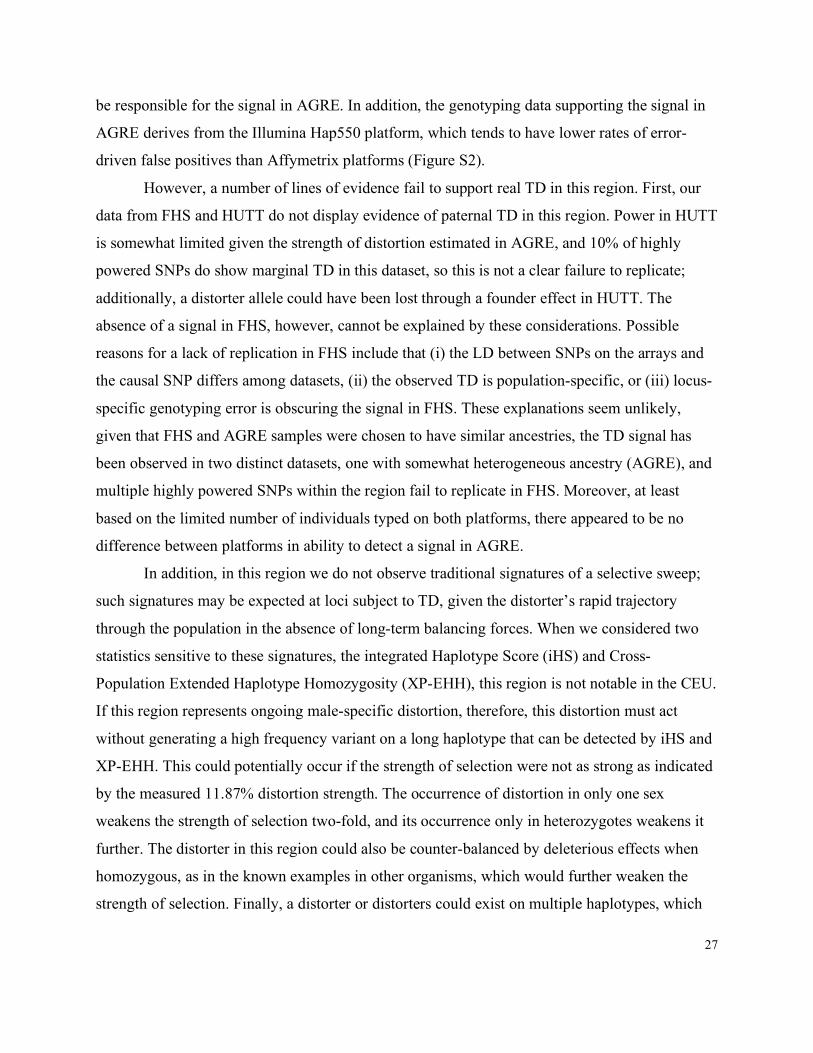
However, a number of lines of evidence fail to support real TD in this region. First, our
data from FHS and HUTT do not display evidence of paternal TD in this region. Power in HUTT
is somewhat limited given the strength of distortion estimated in AGRE, and 10% of highly
powered SNPs do show marginal TD in this dataset, so this is not a clear failure to replicate;
additionally, a distorter allele could have been lost through a founder effect in HUTT. The
absence of a signal in FHS, however, cannot be explained by these considerations. Possible
reasons for a lack of replication in FHS include that (i) the LD between SNPs on the arrays and
the causal SNP differs among datasets, (ii) the observed TD is population-specific, or (iii) locus-
specific genotyping error is obscuring the signal in FHS. These explanations seem unlikely,
given that FHS and AGRE samples were chosen to have similar ancestries, the TD signal has
been observed in two distinct datasets, one with somewhat heterogeneous ancestry (AGRE), and
multiple highly powered SNPs within the region fail to replicate in FHS. Moreover, at least
based on the limited number of individuals typed on both platforms, there appeared to be no
difference between platforms in ability to detect a signal in AGRE.
In addition, in this region we do not observe traditional signatures of a selective sweep;
such signatures may be expected at loci subject to TD, given the distorter’s rapid trajectory
through the population in the absence of long-term balancing forces. When we considered two
statistics sensitive to these signatures, the integrated Haplotype Score (iHS) and Cross-
Population Extended Haplotype Homozygosity (XP-EHH), this region is not notable in the CEU.
If this region represents ongoing male-specific distortion, therefore, this distortion must act
without generating a high frequency variant on a long haplotype that can be detected by iHS and
XP-EHH. This could potentially occur if the strength of selection were not as strong as indicated
by the measured 11.87% distortion strength. The occurrence of distortion in only one sex
weakens the strength of selection two-fold, and its occurrence only in heterozygotes weakens it
further. The distorter in this region could also be counter-balanced by deleterious effects when
homozygous, as in the known examples in other organisms, which would further weaken the
strength of selection. Finally, a distorter or distorters could exist on multiple haplotypes, which

28
would reduce the power of iHS or XP-EHH to detect TD. The lack of strong LD between the
focal SNP and several other SNPs within the region with p-values < 10-3 may indicate the
presence of at least two haplotypes contributing to the TD signal (Figure 3).
Also arguing against a real effect, the available functional data from sperm do not support
a role for this region in spermatogenesis or sperm motility. When we genotyped both unselected
sperm and the fastest sperm from heterozygous males, we observed no significant deviation from
50% of each allele in any of the sperm samples from 10 Austrian donors. Furthermore, the allele
ratios inferred from sperm typing were significantly different from those inferred from the TDT.
There are at least two scenarios involving real TD that could produce this discrepancy between
sperm typing and the TDT. First, the distortion could be heterogeneous among males, with
distorters and non-distorters differing in genetic background or environmental conditions.
Second, the distortion could occur through a mechanism that the assays performed here do not
sufficiently capture, such as influencing sperm survival in the female reproductive tract or
capacity to fertilize the egg. Absence of real TD in this region can obviously also explain the
discrepancy between the TDT and sperm typing results.
Altogether, given the lack of replication in FHS, a long haplotype-based signature of
selection, or functional validation in sperm, the most parsimonious explanation of the TD signal
in 6p21.1 is chance fluctuation in male transmission rates in both HapMap and AGRE.
Nonetheless, the detection of nearly identical large regions displaying TD in fathers only in two
independent datasets is intriguing, and, in our view, warrants further investigation of this region
in future pedigree or sperm analyses.
Our scan also revealed a candidate region for TD in both parents on chromosome 10,
surrounding SNP rs748001, which achieves genome-wide significance in AGRE (Figure 2). The
presence of multiple SNPs with low p-values (p < 0.01) in strong LD with this SNP provides
evidence that this signal is highly unlikely to be driven by genotyping error. This SNP is also
within the tail of Integrated Haplotype Score (iHS) signals in the HapMap II CEU (empirical p =
0.021) (Frazer et al. 2007; Pickrell et al. 2009). Interestingly, the over-transmitted haplotype in
the TDT contains the ancestral allele at the focal SNP. The iHS is designed to identify selective
sweeps on new mutations, so the ancestral allele at rs748001 may be in LD with a derived allele
that is experiencing a selective sweep. The SNPs rs4962310 and rs11244542, both of which have
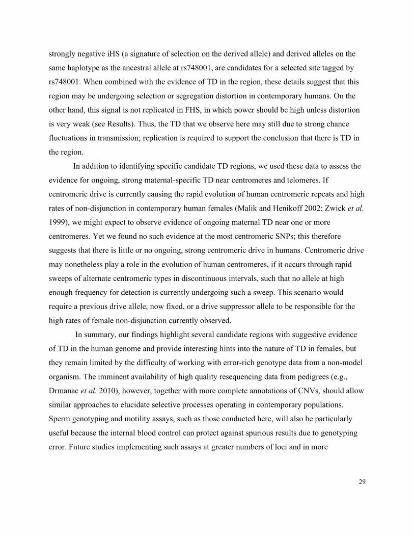
29
strongly negative iHS (a signature of selection on the derived allele) and derived alleles on the
same haplotype as the ancestral allele at rs748001, are candidates for a selected site tagged by
rs748001. When combined with the evidence of TD in the region, these details suggest that this
region may be undergoing selection or segregation distortion in contemporary humans. On the
other hand, this signal is not replicated in FHS, in which power should be high unless distortion
is very weak (see Results). Thus, the TD that we observe here may still due to strong chance
fluctuations in transmission; replication is required to support the conclusion that there is TD in
the region.
In addition to identifying specific candidate TD regions, we used these data to assess the
evidence for ongoing, strong maternal-specific TD near centromeres and telomeres. If
centromeric drive is currently causing the rapid evolution of human centromeric repeats and high
rates of non-disjunction in contemporary human females (Malik and Henikoff 2002; Zwick et al.
1999), we might expect to observe evidence of ongoing maternal TD near one or more
centromeres. Yet we found no such evidence at the most centromeric SNPs; this therefore
suggests that there is little or no ongoing, strong centromeric drive in humans. Centromeric drive
may nonetheless play a role in the evolution of human centromeres, if it occurs through rapid
sweeps of alternate centromeric types in discontinuous intervals, such that no allele at high
enough frequency for detection is currently undergoing such a sweep. This scenario would
require a previous drive allele, now fixed, or a drive suppressor allele to be responsible for the
high rates of female non-disjunction currently observed.
In summary, our findings highlight several candidate regions with suggestive evidence
of TD in the human genome and provide interesting hints into the nature of TD in females, but
they remain limited by the difficulty of working with error-rich genotype data from a non-model
organism. The imminent availability of high quality resequencing data from pedigrees (e.g.,
Drmanac et al. 2010), however, together with more complete annotations of CNVs, should allow
similar approaches to elucidate selective processes operating in contemporary populations.
Sperm genotyping and motility assays, such as those conducted here, will also be particularly
useful because the internal blood control can protect against spurious results due to genotyping
error. Future studies implementing such assays at greater numbers of loci and in more

30
individuals, with more single molecules per individual, could provide mechanistic insights into
loci influencing regional transmission rates in males.
ACKNOWLEDGMENTS
We thank Graham Coop, Martin Kreitman, Guy Sella, Andrew Skol, Matthew Stephens,
and members of the Pritchard, Przeworski, and Stephens labs for helpful discussions, as well as
Stephen Wright, David Cutler and two anonymous reviewers. We are grateful to Jonathan
Pritchard for the suggestion of experimental assays in sperm, Adi Fledel-Alon and Ellen Leffler
for help working with the FHS, HUTT, and AGRE datasets, Cord Melton for help with the 1000
Genomes data, Joe Pickrell for data and discussion of tests for selection, Bryan Howie for
assistance with imputation, and Kevin Ross for performing iPlex genotyping and help
interpreting the resulting data. Finally, we gratefully acknowledge the resources provided by the
Framingham Heart Study and the Autism Genetic Resource Exchange Consortium* and the
participating families. The Framingham Heart Study and the Framingham SHARe project are
conducted and supported by the National Heart, Lung, and Blood Institute (NHLBI) in
collaboration with Boston University. The Framingham SHARe data used for the analyses
described in this manuscript were obtained through dbGaP (accession numbers phs000007.v5.p3
and phs000007.v6.p3). This manuscript was not prepared in collaboration with investigators of
the Framingham Heart Study and does not necessarily reflect the opinions or views of the
Framingham Heart Study, Boston University, or the NHLBI. The Autism Genetic Resource
Exchange is a program of Autism Speaks and is supported, in part, by grant 1U24MH081810
from the National Institute of Mental Health to Clara M. Lajonchere (PI).
*The AGRE Consortium:
Dan Geschwind, M.D., Ph.D., UCLA, Los Angeles, CA;
Maja Bucan, Ph.D., University of Pennsylvania, Philadelphia, PA;
W.Ted Brown, M.D., Ph.D., F.A.C.M.G., N.Y.S. Institute for Basic Research in Developmental
Disabilities, Staten Island, NY;
Joseph Buxbaum, Ph.D., Mt. Sinai School of Medicine, NY, NY;
Rita M. Cantor, Ph.D., UCLA School of Medicine, Los Angeles, CA;

31
John N. Constantino, M.D., Washington University School of Medicine, St. Louis, MO;
T.Conrad Gilliam, Ph.D., University of Chicago, Chicago, IL;
Clara Lajonchere, Ph.D, Cure Autism Now, Los Angeles, CA;
David H. Ledbetter, Ph.D., Emory University, Atlanta, GA;
Christa Lese-Martin, Ph.D., Emory University, Atlanta, GA;
Janet Miller, J.D., Ph.D., Cure Autism Now, Los Angeles, CA;
Stanley F. Nelson, M.D., UCLA School of Medicine, Los Angeles, CA;
Gerard D. Schellenberg, Ph.D., University of Washington, Seattle, WA;
Carol A. Samango-Sprouse, Ed.D., George Washington University, Washington, D.C.;
Sarah Spence, M.D., Ph.D., UCLA, Los Angeles, CA;
Matthew State, M.D., Ph.D., Yale University, New Haven, CT.
Rudolph E. Tanzi, Ph.D., Massachusetts General Hospital, Boston, MA.
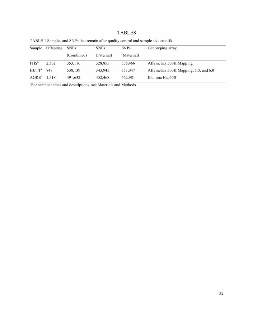
32
TABLES TABLE 1 Samples and SNPs that remain after quality control and sample size cutoffs.
Sample Offspring SNPs
(Combined)
SNPs
(Paternal)
SNPs
(Maternal)
Genotyping array
FHSa 2,362 353,116 328,855 335,466 Affymetrix 500K Mapping
HUTTa 848 538,139 343,945 353,047 Affymetrix 500K Mapping, 5.0, and 6.0
AGREa 1,518 491,632 452,468 462,981 Illumina Hap550 aFor sample names and descriptions, see Materials and Methods.

33
TABLE 2 Top 10 SNPs in each test with regional signals of TD
TDTa Focal SNP Ch p Rateb OAc Nd MAFe Lengthf
(kb)
Startg
(HG17)
#h Genes
C rs748001 10 4.55 x 10⁻⁸ 0.585 Anc 1036 0.248 27.586 127199116 8 None
C rs12661087 6 3.65 x 10⁻⁶ 0.601 Anc 524 0.124 203.859 120861096 6 None
C rs13112011 4 4.90 x 10⁻⁶ 0.624 Anc 338 0.074 20.956 25233826 4 None
C rs1941852 11 7.91 x 10⁻⁶ 0.613 Der 388 0.069 40.57 97380183 4 None
C rs10766778 11 9.74 x 10⁻⁶ 0.570 Anc 1002 0.210 2.297 21219167 2 NELL1
C rs1249667 1 1.33 x 10⁻⁵ 0.637 Der 251 0.051 39.316 75491425 2 SLC44A5
C rs16910190 11 1.35 x 10⁻⁵ 0.578 Der 773 0.158 52.365 22773148 5 GAS2
SVIP
C rs12582514 12 1.59 x 10⁻⁵ 0.558 Der 1374 0.483 99.547 21469826 13 PYROXD1
RECQL
GOLT1B
C rs10935427 3 1.80 x 10⁻⁵ 0.556 Anc 1445 0.396 97.379 142396949 9 ACPL2
C rs10136259 14 1.94 x 10⁻⁵ 0.566 Anc 1059 0.220 7.736 32663208 3 NPAS3
P rs12199720 6 1.77 x 10⁻⁵ 0.593 Anc 532 0.409 710.622 44779449 46 SUPT3H
MIR586
RUNX2
P rs10795851 10 2.28 x 10⁻⁵ 0.596 Der 482 0.198 31.105 11265343 8 CUGBP2
P rs2921031 8 3.29 x 10⁻⁵ 0.601 Der 419 0.190 22.967 8365511 5 None
P rs2166013 5 4.11 x 10⁻⁵ 0.606 Der 371 0.147 109.672 119407151 8 None
P rs1377210 4 4.11 x 10⁻⁵ 0.627 Der 259 0.100 40.571 110022271 2 AGXT2L1
P rs6084148 20 6.62 x 10⁻⁵ 0.584 Der 567 0.237 36.55 2632924 5 EBF4
P rs4261974 4 7.97 x 10⁻⁵ 0.586 Der 532 0.220 9.772 120221079 3 SYNPO2
P rs295263 9 8.46 x 10⁻⁵ 0.603 Der 364 0.134 19.792 4829511 4 RCL1
MIR101-2
P rs10506080 12 9.63 x 10⁻⁵ 0.592 Der 453 0.174 69.518 31825919 5 H3F3C
P rs321202 5 1.10 x 10⁻⁴ 0.570 Anc 765 0.414 122.722 143107350 6 HMHB1
M rs17807087 1 6.14 x 10⁻⁶ 0.633 Anc 290 0.106 21.172 229092422 4 None
M rs2282315 6 1.60 x 10⁻⁵ 0.645 Anc 220 0.086 53.056 132808430 3 STX7
M rs12229163 12 2.43 x 10⁻⁵ 0.649 Anc 202 0.076 24.574 113358516 2 None
M rs4646421 15 2.58 x 10⁻⁵ 0.621 Der 301 0.108 302.84 72503662 6 6i
M rs10868142 9 1.38 x 10⁻⁴ 0.597 Anc 387 0.171 14.718 84150265 6 SLC28A3
M rs952893 2 1.41 x 10⁻⁴ 0.589 Der 453 0.175 125.338 51727031 13 None
M rs17066329 4 1.54 x 10⁻⁴ 0.614 Anc 277 0.112 11.921 179845588 2 None

34
TDTa Focal SNP Ch p Rateb OAc Nd MAFe Lengthf
(kb)
Startg
(HG17)
#h Genes
M rs1372679 /
rs1372680
4 1.64 x 10⁻⁴ 0.616 Anc 262 0.094 20.547 100658226 3 None
M rs10216366 8 1.75 x 10⁻⁴ 0.593 Anc 410 0.157 64.351 126776817 4 None
M rs16973745 18 1.82 x 10⁻⁴ 0.627 Anc 216 0.081 124.932 36610407 5 None aCategory of TDT (C = combined; P = paternal; M = maternal) bRate at which the over-transmitted allele was transmitted. cOver-transmitted allele (Anc=ancestral; Der=derived) dSample size (number of transmissions) eMinor allele frequency in AGRE founders fLength of the region of TD (see Materials and Methods) gStart of the region of TD (see Materials and Methods) hNumber of SNPs within the TD region that have p < 0.01 iThis region contains six genes: SEMA7A, UBL7, ARID3B, CLK3, EDC3, and CYP1A1

35
TABLE 3 Gene ontology terms most enriched among top TD signals
Test Term # Genes Fold Enrichment p-value* FDR (%) Annotation Source
Combined alternative splicing 13 1.590 0.0459 38.91 SP_PIR_KEYWORDS
Combined splice variant 13 1.586 0.0467 41.31 UP_SEQ_FEATURE
Paternal vitamin metabolic
process
2 30.062 0.0593 54.26 GOTERM_BP_FAT
Paternal cell maturation 2 30.062 0.0593 54.26 GOTERM_BP_FAT
Maternal Vinculin, conserved site 2 362.152 0.00527 5.31 INTERPRO
Maternal Vinculin/alpha-catenin 2 289.722 0.00659 6.59 INTERPRO
Maternal fascia adherens 2 159.775 0.0117 10.95 GOTERM_CC_FAT
Maternal intercalated disc 2 106.517 0.0175 15.97 GOTERM_CC_FAT
Maternal cell-cell junction 3 12.614 0.0203 18.35 GOTERM_CC_FAT
Maternal cell-cell adherens
junction
2 45.65 0.0403 33.39 GOTERM_CC_FAT
*Uncorrected p-values are presented for comparative purposes.

36
TABLE 4 Sperm typing data from single DNA molecules characterized for transmission distortion
Blood Unselected Sperm Fast Sperm Donor
ID
SNPs OHap/
UHapa Ob Uc pd Ob Uc pe Ob Uc pe
Prop Fast Sperm (%)f
26 rs9381373
rs2093903
TA/CC 129 153 0.171 142 119 0.0866 NA NA NA NA
1 rs9381373
rs2093903
TA/CC 134 141 0.718 143 140 0.453 NA NA NA NA
1006 rs9381373
rs2093903
TA/CC 123 131 0.661 150 155 0.634 NA NA NA NA
19 rs9381373
rs1284965
TG/CA 186 210 0.248 249 251 0.553 NA NA NA NA
21 rs9381373
rs2093903
TA/CC NA NA NA 173 200 0.926 NA NA NA NA
37 rs9381373
rs2093903
TA/CC NA NA NA 109 124 0.853 101 111 0.775 0.922
45 rs1284965 G/A NA NA NA 222 211 0.361 121 116 0.398 5.155
2 rs9381373
rs2093903
TA/CC NA NA NA NA NA NA 123 136 0.808 0.015
6 rs9381373
rs1284965
rs2093903
TGA/CAC NA NA NA NA NA NA 112 134 0.929 0.007
8 rs9381373
rs1284965
rs2093903
TGA/CAC NA NA NA NA NA NA 110 149 0.994 0.005
Total 572 635 0.0743 1067 1084 0.651 567 646 0.989
aHaplotype expected to be over-represented/Haplotype expected to be under-represented based on the TDT in
AGRE bCount of haplotype expected to be over-represented based on the TDT cCount of haplotype expected to be under-represented based on the TDT dProbability of observing at least as strong a deviation from 50% each allele (2-sided binomial) eProbability of observing at least as many of the allele expected to be over-represented as observed (1-sided
binomial) eProportion of total sperm selected by motility assay for fast sperm
37
LITERATURE CITED
The 1000 Genomes Project Consortium, 2010 A map of human genome variation from
population-scale sequencing. Nature 467: 1061-1073.
Affymetrix, 2006 BRLMM: An Improved Genotype Calling Method for the GeneChip Human
Mappking 500K Array Set [White paper]. Retrieved from
http://www.affymetrix.com/support/technical/whitepapers/brlmm_whitepaper.pdf.
Anderson, J. A., Y. S. Song and C. H. Langley, 2008 Molecular population genetics of
drosophila subtelomeric DNA. Genetics 178: 477-487.
Axelsson, E., A. Albrechtsen, A. P. van, L. Li, H. J. Megens et al, 2010 Segregation distortion in
chicken and the evolutionary consequences of female meiotic drive in birds. Heredity 105: 290-
298.
Bazerman, M. H., and W. F. Samuelson, 1983 I won the auction but don’t want the prize. J.
Conflict Resolut. 27: 618-634.
Brandvain, Y., and G. Coop, 2012 Scrambling eggs: Meiotic drive and the evolution of female
recombination rates. Genetics 190: 709-723.
Carvalho, A. B., and S. C. Vaz, 1999 Are drosophila SR drive chromosomes always balanced?
Heredity 83: 221-228.
Charlesworth, B., and D. L. Hartl, 1978 Population dynamics of the segregation distorter
polymorphism of Drosophila Melanogaster. Genetics 89: 171-192.
Cheng, K. F., and J. H. Chen, 2007 A simple and robust TDT-type test against genotyping error
with error rates varying across families. Hum. Hered. 64: 114-122.
38
Cupples, L. A., H. T. Arruda, E. J. Benjamin, S. D'Agostino Ralph B., S. Demissie et al, 2007
The Framingham Heart Study 100K SNP genome-wide association study resource: Overview of
17 phenotype working group reports. BMC Medical Genetics 8: S1-19.
Dawber, T. R., G. F. Meadors and F. E. Moore, 1951 Epidemiological Approaches to Heart
Disease: The Framingham Study. American Public Health Association, New York, N.Y.
Dawber, T. R., W. B. Kannel and L. P. Lyell, 1963 An approach to longitudinal studies in a
community: The framingham study. Ann. N. Y. Acad. Sci. 107: 539-556.
de la Casa-Esperón, E., and C. Sapienza, 2003 Natural selection and the evolution of genome
imprinting. Annu. Rev. Genet. 37: 349-370.
Deng, L., D. Zhang, E. Richards, X. Tang, J. Fang et al, 2009 Constructing an initial map of
transmission distortion based on high density HapMap SNPs across the human autosomes.
Journal of Genetics and Genomics 36: 703-709.
Drmanac, R., A. B. Sparks, M. J. Callow, A. L. Halpern, N. L. Burnes et al, 2010 Human
genome sequencing using unchained base reads on self-assembling DNA nanoarrays. Science
327: 78-81.
Ducy, P., T. Schinke and G. Karsenty, 2000 The osteoblast: A sophisticated fibroblast under
central surveillance. Science 289: 1501-1504.
Ebner, T., O. Shebl, M. Moser, R. B. Mayer, W. Arzt et al, 2011 Easy sperm processing
technique allowing exclusive accumulation and later usage of DNA-strandbreak-free
spermatozoa. ReprodBiomed Online 22: 37-43.

39
Evans, D. M., A. P. Morris, L. R. Cardon and P. C. Sham, 2006 A Note on the Power to Detect
Transmission Distortion in Parent-Child Trios via the Transmission Disequilibrium Test. Behav.
Genet. 36: 947-950.
Frank, S. A., 1991 Divergence of meiotic drive-suppression systems as an explanation for sex-
biased hybrid sterility and inviability. Evolution 45: 262-267.
Frazer, K. A., D. G. Ballinger, D. R. Cox, D. A. Hinds, L. L. Stuve et al, 2007 A second
generation human haplotype map of over 3.1 million SNPs. Nature 449: 851-861.
Geschwind, D. H., J. Sowinski, C. Lord, P. Iversen, J. Shestack et al, 2001 The autism genetic
resource exchange: A resource for the study of autism and related neuropsychiatric conditions.
Am. J. Hum. Genet. 69: 463-466.
Gileva, E. A., 1998 Inbreeding and Sex Ratio in Two Captive Colonies of Dicrostonyx Torquatus
Pall., 1779: A Reply to G. H. Jarrell. Munksgaard International Publishers.
Gordon, D., S. C. Heath, X. Liu and J. Ott, 2001 A Transmission/Disequilibrium test that allows
for genotyping errors in the analysis of single-nucleotide polymorphism data. The American
Journal of Human Genetics 69: 371-380.
Gordon, D., C. Haynes, C. Johnnidis, S. B. Patel, A. M. Bowcock et al, 2004 A transmission
disequilibrium test for general pedigrees that is robust to the presence of random genotyping
errors and any number of untyped parents. European Journal of Human Genetics 12: 752-761.
Göring, H. H., Terwilliger, J. D. and J. Blangero, 2001 Large upward bias in estimation of locus-
specific effects from genome-wide scans. Am. J. Hum. Genet. 69: 1357-1369.
40
Gudbjartsson, D. F., G. B. Walters, G. Thorleifsson, H. Stefansson, B. V. Halldorsson et al, 2008
Many sequence variants affecting diversity of adult human height. Nat. Genet. 40: 609-615.
Haig, D., 2010 Games in tetrads: Segregation, recombination, and meiotic drive. Am. Nat. 176:
404-413.
Haig, D., and A. Grafen, 1991 Genetic scrambling as a defence against meiotic drive. J. Theor.
Biol. 153: 531-558.
Hartl, D. L., 1972 Population Dynamics of Sperm and Pollen Killers. Theor. Appl. Genet. 42:
81-88.
Hartl, D. L., 1973 Complementation analysis of male fertility among the segregation distorter
chromosomes of Drosophila melanogaster. Genetics 73: 613-629.
Hartl, D. L., 1974 Genetic dissection of segregation distortion. I. Suicide combinations of SD
genes. Genetics 76: 477-486.
Hartl, D. L., 1975 Segregation distortion in natural and artificial populations of Drosophila
melanogaster, pp. 83-91 in Gamete competition in plants and animals, edited by D. L. Mulcahy.
North-Holland Publishing, Amsterdam.
Hassold, T., and P. Hunt, 2001 To err (meiotically) is human: The genesis of human aneuploidy.
Nat. Rev. Genet. 2: 280-291.
Henikoff, S., K. Ahmad and H. S. Malik, 2001 The centromere paradox: Stable inheritance with
rapidly evolving DNA. Science 293: 1098.
Hirschhorn, J. N., and M. J. Daly, 2005 Genome-wide association studies for common diseases
and complex traits. Nature Reviews Genetics 6: 95-108.
41
Huang, D. W., B. T. Sherman and R. A. Lempicki, 2009 Bioinformatics enrichment tools: Paths
toward the comprehensive functional analysis of large gene lists. Nucleic Acids Research 37: 1-
13.
Huang, D. W., B. T. Sherman and R. A. Lempicki, 2008 Systematic and integrative analysis of
large gene lists using DAVID bioinformatics resources. Nat. Protocols 4: 44-57.
Hurst, G. D. D., and J. H. Werren, 2001 The role of selfish genetic elements in eukaryotic
evolution. Nature Reviews Genetics 2: 597-606.
Hurst, L. D., and A. Pomiankowski, 1991 Causes of sex ratio bias may account for unisexual
sterility in hybrids: A new explanation of Haldane's rule and related phenomena. Genetics 128:
841-858.
Ioannidis, J. P., E. E. Ntzani, T. A. Trikalinos and D. G. Contopoulos-Ioannidis 2001 Replication
validity of genetic association studies. Nat. Genet. 29: 306-309.
Jarrell, G. H., 1995 A Male-Biased Natal Sex-Ratio in Inbred Collared Lemmings, Dicrostonyx
Groenlandicus. Munksgaard International Publishers.
Jeong, J. H., J. S. Jin, H. N. Kim, S. M. Kang, J. C. Liu et al, 2008 Expression of Runx2
transcription factor in non-skeletal tissues, sperm and brain. J. Cell. Physiol. 217: 511-517.
Jones, R. N., and H. Rees, 1982 B Chromosomes. Academic Press, London ;New York. Kusano, A., C. Staber, H. Y. Chan and B. Ganetzky, 2003 Closing the (ran)GAP on segregation
distortion in drosophila. Bioessays 25: 108-115.
Lohmueller, K. E., C. L. Pearce, M. Pike, E. S. Lander and J. N. Hirschhorn, 2003 Meta-analysis
of genetic association studies supports a contribution of common variants to susceptibility to
common disease. Nat. Genet. 33: 177.
42
Lyon, M. F., 2003 Transmission ratio distortion in mice. Annu. Rev. Genet. 37: 393-408.
Lyttle, T. W., 1993 Cheaters sometimes prosper: Distortion of mendelian segregation by meiotic
drive. Trends Genet. 9: 205-210.
Malik, H. S., 2009 The centromere-drive hypothesis: A simple basis for centromere complexity.
Prog. Mol. Subcell. Biol. 48: 33-52.
Malik, H. S., and S. Henikoff, 2002 Conflict begets complexity: The evolution of centromeres.
Curr. Opin. Genet. Dev. 12: 711-718.
Marchini, J., B. Howie, S. Myers, G. McVean and P. Donnelly, 2007 A new multipoint method
for genome-wide association studies by imputation of genotypes. Nat. Genet. 39: 906-913.
Mitchell, A. A., D. J. Cutler and A. Chakravarti, 2003 Undetected genotyping errors cause
apparent overtransmission of common alleles in the transmission/disequilibrium test. Am. J.
Hum. Genet. 72: 598-610.
Montgomery, G. W., G. Zhu, J. J. Hottenga, D. L. Duffy, A. C. Heath et al, 2006 HLA and
genomewide allele sharing in dizygotic twins. Am. J. Hum. Genet. 79: 1052-1058.
Mundlos, S., F. Otto, C. Mundlos, J. B. Mulliken, A. S. Aylsworth et al, 1997 Mutations
involving the transcription factor CBFA1 cause cleidocranial dysplasia. Cell 89: 773-779.
Novitski, E., 1951 Non-random disjunction in drosophila. Genetics 36: 267-280.
Ober, C., M. Abney and M. S. McPeek, 2001 The genetic dissection of complex traits in a
founder population. The American Journal of Human Genetics 69: 1068-1079.
Östergren, G., 1945 Parasitic nature of extra fragment chromosomes. Botaniska Notiser 2: 157.
43
Otto, F., A. P. Thornell, T. Crompton, A. Denzel, K. C. Gilmour et al, 1997 Cbfa1, a candidate
gene for cleidocranial dysplasia syndrome, is essential for osteoblast differentiation and bone
development. Cell 89: 765-771.
Pardo-Manuel de Villena, F., and C. Sapienza, 2001 Nonrandom Segregation during Meiosis:
The Unfairness of Females. Mamm. Genome. 12: 331-339.
Paterson, A. D., D. Waggott, A. Schillert, C. Infante-Rivard, S. B. Bull et al, 2009 Transmission-
ratio distortion in the Framingham Heart Study. BMC Proc. 3 Suppl 7: S51.
Peacock, W. J., E. S. Dennis, M. M. Rhoades and A. J. Pryor, 1981 Highly repeated DNA
sequence limited to knob heterochromatin in maize. Proc. Natl. Acad. Sci. U. S. A. 78: 4490-
4494.
Pickrell, J. K., G. Coop, J. Novembre, S. Kudaravalli, J. Z. Li et al, 2009 Signals of recent
positive selection in a worldwide sample of human populations. Genome Res. 19: 826-837.
Presgraves, D. C., P. R. Gérard, A. Cherukuri, and T. W. Lyttle, 2009 Large-Scale Selective
Sweep among Segregation Distorter Chromosomes in African Populations of Drosophila
melanogaster. PLoS Genet. 5: e1000463.
Price, A. L., N. J. Patterson, R. M. Plenge, M. E. Weinblatt, N. A. Shadick et al, 2006 Principal
components analysis corrects for stratification in genome-wide association studies. Nat. Genet.
38: 904-909.
Purcell, S., B. Neale, K. Todd-Brown, L. Thomas, M. A. Ferreira et al, 2007 PLINK: A tool set
for whole-genome association and population-based linkage analyses. Am. J. Hum. Genet. 81:
559-575.

44
Sabeti, P. C., P. Varilly, B. Fry, J. Lohmueller et al, 2007 Genome-wide detection and
characterization of positive selection in human populations. Nature 449: 913-918.
Santos, P. S., J. Hohne, P. Schlattmann, I. R. Konig, A. Ziegler et al, 2009 Assessment of
transmission distortion on chromosome 6p in healthy individuals using tagSNPs. Eur. J. Hum.
Genet. 17: 1182-1189.
Santos, P.S., J. Hohne, F. Poerner, M. d. G. Bicalho, B. Uchanska-Ziegler et al, 2011 Does the
new HapMap throw the baby out with the bath water? Eur. J. Hum. Genet. 19: 733-734.
Siepel, A., G. Bejerano, J. S. Pedersen, A. S. Hinrichs, M. Hou et al, 2005 Evolutionarily
conserved elements in vertebrate, insect, worm, and yeast genomes. Genome Research 15: 1034-
1050.
Silver, L. M., 1993 The peculiar journey of a selfish chromosome: Mouse t haplotypes and
meiotic drive. Trends Genet. 9: 250-254.
Spielman, R. S., R. E. McGinnis and W. J. Ewens, 1993 Transmission test for linkage
disequilibrium: The insulin gene region and insulin-dependent diabetes mellitus (IDDM). Am. J.
Hum. Genet. 52: 506-516.
Thomson, G. J., and M. W. Feldman, 1974 Population genetics of modifiers of meiotic drive. II
linkage modification in the segregation distortion system. Theor. Popul. Biol. 5: 155-162.
Tiemann-Boege, I., P. Calabrese, D. M. Cochran, R. Sokol and N. Arnheim, 2006 High-
resolution recombination patterns in a region of human chromosome 21 measured by sperm
typing. PLoS Genet. 2: e70.

45
Veron, N., H. Bauer, A. Y. Weisse, G. Luder, M. Werber et al, 2009 Retention of gene products
in syncytial spermatids promotes non-Mendelian inheritance as revealed by the t complex
responder. Genes Dev. 23: 2705-2710.
Voight, B. F., S. Kudaravalli, X. Wen, and J. K. Pritchard, 2006 A Map of Recent Positive
Selection in the Human Genome. PLoS Biology 4: e72.
Wallace, L. T., and M. A. Erhart, 2008 Recombination within mouse t haplotypes has replaced
significant segments of t-specific DNA. Mamm. Genome 19: 263-271.
Wang, E. T., R. Sandberg, S. Luo, I. Khrebtukova, L. Zhang et al, 2008 Alternative isoform
regulation in human tissue transcriptomes. Nature 456: 470-476.
Wheeler, J. C., K. Shigesada, J. Peter Gergen and Y. Ito, 2000 Mechanisms of transcriptional
regulation by runt domain proteins. Semin. Cell Dev. Biol. 11: 369-375.
Zöllner, S., X. Wen, N. A. Hanchard, M. A. Herbert, C. Ober et al, 2004 Evidence for extensive
transmission distortion in the human genome. Am. J. Hum. Genet. 74: 62-72.
Zwick, M. E., J. L. Salstrom and C. H. Langley, 1999 Genetic variation in rates of
nondisjunction: Association of two naturally occurring polymorphisms in the chromokinesin nod
with increased rates of nondisjunction in Drosophila melanogaster. Genetics 152: 1605-1614.

46
FIGURES

47
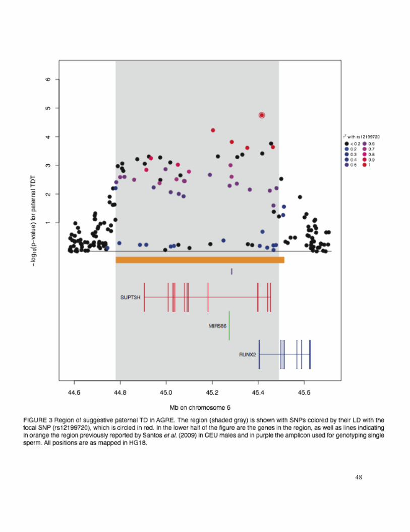
48

49