EUROSPINE 2017 Scientific Programme oral presentations · 2017-10-11 · EUROSPINE 2017 Scientific...
13
EUROSPINE 2017 Scientific Programme oral presentations Thursday, 12 October, 2017 15:50 – 17:05 Cervical Spine 42 COMPARISON OF POSTERIOR FOSSA DECOMPRESSION WITH AND WITHOUT DURAPLASTY FOR THE TREATMENT OF CHIARI I MALFORMATION: A PROSPECTIVE INVESTIGATION Zezhang Zhu, Enze Jiang, Zhen Liu, Bin Wang, Yang Yu, Yong Qiu Spine Surgery, Drum Tower Hospital of Nanjing University Medical School, Nanjing, China Background/ introduction: Chiari malformation Type I (CM-I) is a common and often debilitating neurological disorder. Whether to treat CM-I patients with a traditional posterior fossa decompression with duraplasty (PFDD) or a less invasive way without duraplasty (PFD) remains controversial. Purpose of the study: The aim of this study was to prospectively compare the radiological and clinical outcomes between the two procedures. Materials and Methods: Ninety patients with CMI were randomly assigned to undergo either PFDD or PFD alone. Data were analyzed for clinical outcome (Chicago Chiari Outcome Scale, CCOS), complications, and syrinx resolution. In both groups, a dissection from the occipital bone and a C1 laminectomy were performed. In addition, the dura mater was opened and expanded in the PFDD group. Results: The age, gender, and pre-operative neurologic status were similar between the two groups. Compared to the PFD group, patients undergoing PFDD had significantly longer operation time (176min vs. 149min, P=0.025), longer postoperative drainage time in days (2.5d vs. 1.9d, P<0.001), and higher postoperative drainage volume (413ml to 73ml, P<0.001). The improvement rate of clinical symptoms was 77.5% in the PFD group and 66.7% in the PFDD group, respectively (P=0.330). At the latest exanimation, no statistically significant difference was found between the PFD and the PFDD groups in terms of syrinx resolution (82.5% vs. 90.5%, P=0.344). Total CCOS score averaged 15.07 and 15.30, respectively, in the PFDD and PFD groups (P=0.229). Compared with the PFD group, patients in the PFDD group had a higher incidence of cerebrospinal fluid (CSF) leak (23.8% vs. 2.5%, P=0.007). Conclusion: Compared to the more invasive decompression with duraplasty, PFD without duraplasty produces comparable radiological and clinical outcomes while being associated with a lower risk of complications. Disclosures: Author 1: none; Author 2: none; Author 3: none; Author 4: none; Author 5: none; Author 6: none
Transcript of EUROSPINE 2017 Scientific Programme oral presentations · 2017-10-11 · EUROSPINE 2017 Scientific...

EUROSPINE2017ScientificProgrammeoralpresentations
Thursday, 12 October, 2017 15:50 – 17:05 Cervical Spine
42
COMPARISON OF POSTERIOR FOSSA DECOMPRESSION WITH AND WITHOUT DURAPLASTY FOR THE TREATMENT OF CHIARI I MALFORMATION: A PROSPECTIVE INVESTIGATION Zezhang Zhu, Enze Jiang, Zhen Liu, Bin Wang, Yang Yu, Yong Qiu Spine Surgery, Drum Tower Hospital of Nanjing University Medical School, Nanjing, China Background/ introduction: Chiari malformation Type I (CM-I) is a common and often debilitating neurological disorder. Whether to treat CM-I patients with a traditional posterior fossa decompression with duraplasty (PFDD) or a less invasive way without duraplasty (PFD) remains controversial. Purpose of the study: The aim of this study was to prospectively compare the radiological and clinical outcomes between the two procedures. Materials and Methods: Ninety patients with CMI were randomly assigned to undergo either PFDD or PFD alone. Data were analyzed for clinical outcome (Chicago Chiari Outcome Scale, CCOS), complications, and syrinx resolution. In both groups, a dissection from the occipital bone and a C1 laminectomy were performed. In addition, the dura mater was opened and expanded in the PFDD group. Results: The age, gender, and pre-operative neurologic status were similar between the two groups. Compared to the PFD group, patients undergoing PFDD had significantly longer operation time (176min vs. 149min, P=0.025), longer postoperative drainage time in days (2.5d vs. 1.9d, P<0.001), and higher postoperative drainage volume (413ml to 73ml, P<0.001). The improvement rate of clinical symptoms was 77.5% in the PFD group and 66.7% in the PFDD group, respectively (P=0.330). At the latest exanimation, no statistically significant difference was found between the PFD and the PFDD groups in terms of syrinx resolution (82.5% vs. 90.5%, P=0.344). Total CCOS score averaged 15.07 and 15.30, respectively, in the PFDD and PFD groups (P=0.229). Compared with the PFD group, patients in the PFDD group had a higher incidence of cerebrospinal fluid (CSF) leak (23.8% vs. 2.5%, P=0.007). Conclusion: Compared to the more invasive decompression with duraplasty, PFD without duraplasty produces comparable radiological and clinical outcomes while being associated with a lower risk of complications. Disclosures: Author 1: none; Author 2: none; Author 3: none; Author 4: none; Author 5: none; Author 6: none

43
NECK PAIN ASSOCIATION WITH MODIC CHANGES OF THE CERVICAL SPINE,PREVALENCE, RISK FACTORS, AND DISC DEGENERATION:A CROSS-SECTIONAL, LARGE-SCALE POPULATION-BASED STUDY En Xie, Dingjun Hao Hong–Hui Hospital,Xi 'an Jiaotong University College of Medicine, Xi’an, Shaanxi, China Background Context Modic changes (MC) are bone marrow lesions on magnetic resonance imaging (MRI), suggestive of being associated with neck pain (neck pain). Data on determinants of MC and their association with disc degeneration and other spinal phenotypes, as well as that of neck pain, rely mostly on small-scale patient populations and remain controversial. Purpose This study addressed the potential determinants of MC and their association with disc degeneration and Neck pain. Study Design/Setting Patient Sample This study consisted of 3717 volunteers. Methods Sagittal T2-weighted MRIs of the cervical spine were assessed for the presence of MC and other spinal phenotypes (eg, disc degeneration, disc displacement, Schmorl nodes) in all individuals. Subjects' demographics, occupation, lifestyle, and clinical profiles were measured. Results The overall prevalence of MC was 5.8% (n=141), which increased with advancing age. Modic changes predominantly occurred at the lowest two Cervical levels (83%). In the multivariate analyses, only the presence of disc displacement and a higher disc degeneration score were involved in MC at the upper Cervical levels (C1-C4) (p<0.05). The presence of MC at the lowest Cervical levels (C4-C7) were associated with age, the presence of Schmorl nodes, disc degeneration or displacement, and historical cervical injury (p<0.05). Subjects who were both smokers and overweight or obese had an increased likelihood of MC in the lower Cervical levels (OR: 2.37; 95% CI: 1.71-3.90). The presence of MC at the lower Cervical levels were associated with historical neck pain (OR: 1.76; 95% CI: 1.07-4.07) and with severity and duration of symptoms (p<0.05). Conclusions Based on one of the largest MRI studies to assess Cervical MC, we noted that MC were associated with both disc degeneration and the presence and severity of neck pain. Determinants and association of MC with disc degeneration and clinical symptoms in the upper versus the lower cervical spine were different. Our study further stresses the significance of MC as important imaging phenotypes associated with neck pain. Disclosures: Author 1: none; Author 2: none;

44
STRATEGIES FOR MAINTAINING HORIZONTAL GAZE IN ASYMPTOMATIC SUBJECTS Ayman Assi, Ziad Bakouny, Nour Khalil, Michel Salameh, Naji Bou Zeid, Fares Yared, Joeffroy Otayek, Aren Joe Bizdikian, Renaud Lafage, Virginie Lafage, Ismat Ghanem, Wafa Skalli, Khalil Kharrat, Gaby Kreichati Laboratory of Biomechanics and Medical Imaging, Faculty of Medicine, University of Saint-Joseph, Beirut, Lebanon Introduction Maintaining horizontal gaze is essential for activities of daily living. In subjects with postural malalignment, compensatory mechanisms are recruited to maintain horizontal gaze, especially at the cervical level [1]. While the cervical curvature had been typically considered to be lordotic, recent studies have shown that different forms of curvatures can occur even in asymptomatic subjects [2]. It is still not known how asymptomatic subjects achieve horizontal gaze. Our hypothesis was that subjects employ different cervical curvature forms in order to maintain horizontal gaze. Purpose To investigate the different cervical strategies for maintaining horizontal gaze. Methods 144 asymptomatic subjects (age:29±11years,71F) filled the SF36 quality of life (QoL) questionnaire and underwent full body biplanar x-rays in free standing position. Typical global postural and spino-pelvic alignment parameters were assessed. Head and cervical parameters were measured: radiographic chin brow vertical angle (CBVA), C0C2, C1C2, C2C7, C2 slope, C2C7 SVA, T1 slope, neck tilt, thoracic inlet angle (TIA). Subjects were divided into 3 groups of cervical curvature forms per C2C7 angle: kyphotic (K:<-5°), straight (S:[-5°,5°]), lordotic (L:>5°). Demographics, SF36 parameters and CBVA were compared between groups (Kruskal-Wallis test). Global postural, spino-pelvic, head and cervical parameters were compared between groups, while controlling for confounding factors (ANCOVA). Results 46 (32%) subjects had kyphotic (-12±7°), 39 (27%) straight (0±3°) and 59 (41%) lordotic (12±7°) cervical spines. While demographic and SF36 parameters did not differ between groups, CBVA was found to be significantly different between lordotic and kyphotic groups (2° vs 6.5° resp., p=0.002); therefore, subsequent comparisons were controlled for CBVA. Subjects in the 3 groups had similar PI, PT, SS, LL and knee flexion (p>0.05). However, SVA (L:-2, S:-11, K:-26 mm, p<0.001), TK (L:51, S:47, K:40°, p<0.001), other postural parameters, as well as head and cervical parameters, significantly differed between groups (Fig.1). Conclusion Kyphotic cervical spine subjects presented more posterior global alignment, with smaller TK and decreased T1 slope. These subjects compensated at the lower cervical level with a kyphotic C2C7 and a hyperlordotic upper cervical spine (increased C0C2 and C1C2) in order to maintain horizontal gaze. In contrast, lordotic cervical spine subjects had more anterior global alignment, with larger TK and increased T1 slope. These subjects compensated at the lower cervical level with a lordotic C2C7 and a hypolordotic upper cervical spine (decreased C0C2 and C1C2). Straight cervical spine subjects had an intermediate profile. Asymptomatic subjects can employ different cervical strategies in order to compensate for differences in global alignment in order to maintain horizontal gaze, without alteration of QoL.

[1] Barrey,Eur Spine J,2013 [2] Scheer,JNS spine,2013 Disclosures: Author 1: none; Author 2: none; Author 3: none; Author 4: none; Author 5: none; Author 6: none; Author 7: none; Author 8: none; Author 9: none; Author 10: none
45
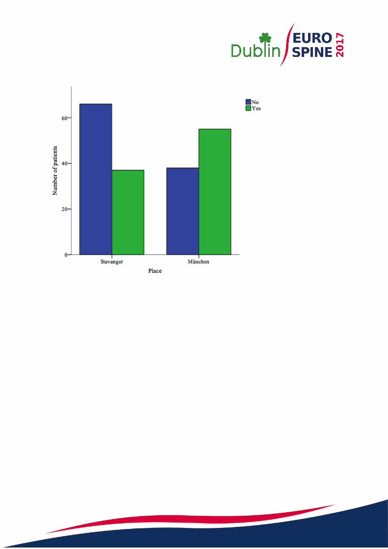
PATIENTS BELIEFS ABOUT DIAGNOSIS AND TREATMENT OF CERVICAL SPONDYLOSIS Clemens Weber, Maziar Behbahani, Roald Baardsen, Jens Lehmberg, Bernhard Meyer, Ehab Shiban Dept. of Neurosurgery, Stavanger, Norway Introduction: Clinical results of surgical spine care may be influenced by the patients' understanding and knowledge of his or her condition, the diagnosing, the treatment options and the decision making process. Purpose of the study: The aim of this patient survey was to evaluate certain factors of patients' understanding of degenerative cervical spondylosis, including the importance of magnetic resonance imaging, risk factors, treatment alternatives and effectiveness. Methods: An anounymous questionnaire survey was performed on patients with degenerative cervical spondylosis referred to the outpatients' clinics of the Department of Neurosurgery at Klinikum rechts der Isar, Technical University of München, Germany, and the Department of Neurosurgery at Stavanger University Hospital, Norway. All patients presented with neck pain with or without radiating to the arm. The survey included eight questions; four questions about the respondents’ gender and age, if she or he has received previous spine surgery and/or conservative treatment for his or her cervical disorder, and four questions about the importance of imaging in the decision-making process, if she or he would be willing to undergo cervical surgery even with little or no symptoms only based on imaging findings, and about the effectiveness and the risk of surgical or conservative treatment. Results: 211 patients answered the questionnaire, 102 in München, Germany, and 109 in Stavanger, Norway. A high portion (67%) of patients with a degenerative cervical condition believes that results from MRI studies are more important than clinical findings in the decision making process whether to offer surgery or not, 47% were willing to undergo surgery based on MRI showing abnormalities even without no or little symptoms (image 1). Half of all patients that answered the questionnaire belief that surgery is more effective in the treatment of axial neck pain. A large portion of patients (93%) believes that surgery has a higher risk than physical therapy. Discussion: The results of this survey study on patients’ beliefs on certain aspects of the diagnosing and management of degenerative cervical spondylosis demonstrates the existence of misconceptions concerning these aspects in a large portion of patients referred to neurosurgical outpatient clinics both in Germany and Norway. Disclosures: Author 1: none; Author 2: none; Author 3: none; Author 4: none; Author 5: grants/research support: Medtronic, Ulrich Medical, consultant: BrainLab, Medtronic, Spineart, Ulrich,royalties: spineart,other financial report: Nexstim, relievant Author 6: consultant: Invibio
46
SURGICAL OUTCOME OF ELDERLY PATIENTS OVER 80 YEARS WITH CERVICAL SPONDYLOTIC MYELOPATHY Norihiro Isogai, Narihito Nagoshi, Junichi Yamane, Akio Iwanami, Hitoshi Kono, Yoshiomi Kobayashi, Nobuyuki Fujita, Mitsuru Yagi, Kota Watanabe, Kazuya Kitamura, Yuta Shiono, Masaya Nakamura, Morio Matsumoto, Ken Ishii Spine and Spinal Cord Center, Mita Hospital, International University of Health and Welfare, Tokyo Japan [Objectives] Surgical therapy for cervical spondylotic myelopathy (CSM) in elderly patients has been increasing in recent year. The aim of this study is to assess the surgical outcomes of posterior cervical decompression in elderly patients more than 80 years. [Methods] From 2012 to 2014, 881 patients who underwent posterior cervical decompression for CSM at 17 high-volume institutions were enrolled in this study. All patients were observed for more than 1-years post-surgery. The mean age at the time of surgery was 67.0 (ranged from 27 to 93) years. The patients were divided into two groups: a younger group (<80 y/o) and an elderly group (>80 y/o). Gender, height, weight, operating time, estimated blood loss, medical history, perioperative complications, Japanese Orthopaedic Association (JOA) score, sagittal cobb angle of C2-5, C5-7, C2-7 and C2-7 sagittal vertical axis (SVA) on the lateral radiographs were compared between two groups. [Results] Of 881 patients, 762 patients (86.5%) and 119 (13.5%) were classified as a younger group (average: 61.5 y/o) and as an elderly group (average: 83.7 y/o), respectively. The female/male ratio in the elderly group was higher than those in the younger group (elderly: 0.92, younger: 0.43) (P<0.05). The elderly patients had lower height and lower weight than younger patients (elderly: 152cm, 52kg, younger: 161cm, 62kg) (P<0.001). There was no significant difference in operating time and estimated blood loss. Hypertension (elderly: 61%, younger: 39%), ischemic heart disease (elderly: 11%, younger: 5%) and renal failure (elderly: 8%, younger: 2%) were frequently observed in elderly group. In perioperative complications, postoperative delirium (elderly: 3.4%, younger: 0.5%) and worsening of neurological status (elderly: 6.7%, younger: 2.0%) were identified in a greater percentage of elderly group. The elderly group revealed worse preoperative (elderly: 9.1pts, younger: 11.2pts) and final JOA scores (elderly: 12.4pts, younger: 13.9pts) (P<0.0001, P<0.0001). However, there was no significant difference in JOA recovery rate (elderly: 39%, younger: 45%). No patients showed with 2 or more point decrease in JOA score of the elderly patients with worsening of neurological status. The elderly group had higher preoperative sagittal cobb angle of C2-7 (elderly: 15.20, younger: 12.70) and C2-5 (elderly: 10.70, younger: 7.40) (P<0.05, P<0.01). The elderly group showed higher preoperative SVA (elderly: 30.2mm, younger: 21.8mm) and final SVA (elderly: 30.8mm, younger: 23.3mm) (P<0.0001, P<0.001). [Conclusion] This study includes the largest sample size of elderly patients over 80 years. Although patients over 80 years who underwent posterior cervical decompression for CSM had more perioperative complications, recovery rate and postoperative cervical alignment were comparable to younger group. The surgical treatment for the elderly patients is an effective procedure, but requires careful management with respect to safety issues. Disclosures: Author 1: none; Author 2: none; Author 3: none; Author 4: none; Author 5: none; Author 6: none; Author 7: none; Author 8: none; Author 9: consultant: Kyocera, japan; Author 10: none; Author 11: none; Author 12: none; Author 13: none; Author 14: none

47
RAPIDLY PROGRESSIVE NEUROLOGICAL DETERIORATION IN CERVICAL SPONDYLOTIC MYELOPATHY WITH CERVICAL INTRAMEDULLARY HIGH-INTENSITY AREA AND HISTORY OF CARDIOVASCULAR EVENT Eiji Takasawa, Yasunori Sorimachi, Yoichi Iizuka, Haku Iizuka, Hirotaka Chikuda Department of Orthopedic Surgery, Japanese Red Cross Maebashi Hospital, Maebashi, Japan; Department of Orthopedic Surgery, Gunma University Graduate School of Medicine, Maebashi, Japan BACKGROUND. Cervical spondylotic myelopathy (CSM) is a chronic, degenerative spine disease and presents a slow development of symptomatic myelopathy. However, some patients show rapidly progressive neurological deterioration (especially gait disturbances) without a recent history of spinal trauma. At present, there is little information about the clinical characteristics of rapidly progressive CSM (rp-CSM). This study sought to clarify the pathology and risk factors, and to evaluate the outcome of early decompression surgery in patients with rp-CSM. METHODS. Of 160 patients diagnosed with CSM who underwent surgery, 70 patients (51 males and 19 females) were enrolled in this study. The mean age at surgery was 66.9 years and the follow-up period was 1 year. Patients were divided into two groups: rp-CSM and chronic CSM (c-CSM) groups. Univariate analyses were performed to compare the two groups, with the postoperative outcomes assessed using the Japanese Orthopaedic Association (JOA) score, and various clinical differences including age, sex, comorbidity (hypertension, diabetes mellitus, kidney disease, history of cardiovascular event [CVE]), the waiting period from symptomatic onset of myelopathy to surgery, cervical range of motion (ROM), intramedullary high intensity area (IM-HIA) on T2 MR images, and the affected segmental level. Logistic regression analysis was also calculated with the presence of rp-CSM as the explanatory variable. RESULTS. Open-door laminoplasty was performed for all patients, and 17 (24.3%) of 70 patients were diagnosed with rp-CSM. Univariate analyses showed that patients with rp-CSM tended to have a past-history of CVE (rp-CSM 35.3% vs. c-CSM 15.1%; p = 0.088), and presented with IM-HIA (rp-CSM 94.1% vs. c-CSM 58.5%; p = 0.007), especially in the C4/5 disc level (rp-CSM 56.3% vs. c-CSM 38.7%; p = 0.018). There was no statistical difference in terms of age, sex or cervical ROM between the two groups. In the rp-CSM group, preoperative upper/lower extremities and bladder functions were worse (p < 0.01; JOA score), and the wait period from onset to surgery was shorter (rp-CSM 1.2 ± 0.8 months vs. c-CSM 25.7 ± 25.3 months; p = 0.000). In addition, neurological deterioration was recovered soon after decompression surgery in the rp-CSM group, and the recovery rate at the 1-year follow-up was higher in this group (rp-CSM 65.5% vs. c-CSM 40.7%, p = 0.001; JOA score). Logistic regression analysis showed that a history of CVE (odds ratio [OR] = 8.9; 95%CI = 1.8 to 9.5) and IM-HIA on T2 MR images (OR = 11.9, 95%CI = 1.7 to 8.3) were independently associated with rp-CSM. CONCLUSION. We identified that a history of CVE and intramedullary cervical lesions might be the pathology and risk factors for rp-CSM. We also show that rp-CSM is a reversible condition following surgery, unlike spinal cord infarction. Early decompression surgery is recommended for rapidly progressive neurological deterioration in patients with CSM. Disclosures: Author 1: none; Author 2: none; Author 3: none; Author 4: none; Author 5: none

48
PREVALENCE, PROGRESSION, AND CLINICAL IMPLICATIONS OF HETEROTOPIC OSSIFICATION AFTER CERVICAL DISC ARTHROPLASTY AT 7 YEARS Jacques Beaurain, Pierce Nunley Neurosurgery Department, University Hospital, Dijon, France Background: Heterotopic ossification (HO) can occur after treatment with cervical disc arthroplasty (CDA). In the most severe cases, HO can result in complete ankyloses of the treated segment. Data on the long-term progression of HO and its clinical impact on CDA patients is limited. Purpose: To assess the prevalence and progression of HO in patients treated with CDA, and determine if HO is associated with diminished clinical outcomes seven years after surgery. Materials and Methods: The study is a post-hoc analysis of data from a prospective, randomized, multicenter, US FDA clinical trial. Patients were followed annually to 5 years and at 7 years postoperatively. A total of 389 patients were treated with 1- or 2-level CDA (Mobi-C Cervical Disc, Zimmer Biomet, Austin, TX). Inclusion criteria included a diagnosis of symptomatic cervical degenerative disc disease and no prior history of cervical spine surgery. Preoperative patient characteristics included age, sex, and BMI. Clinical and radiographic outcomes included HO, flexion/extension range of motion (ROM), NDI score, and VAS neck pain score. Independent radiologists conducted all radiographic evaluations. HO was classified using the system adapted from McAfee and Mehren. HO grades 3 and 4 were classified as functionally relevant due to restricted ROM. Results: For one-level patients, grade 0/1 HO was present in 4.6% of patients, grade 2 in 67.0%, grade 3 in 17.4%, and grade 4 in 11.0%. From 5 to 7 years, 6.6% of patients showed HO progression of 1 or 2 grades. Grades 0/1 and grade 2 HO had more ROM (14.4±5.8° and 12.3±5.0°, respectively), than patients with grade 3 or 4 (5.5±3.3° and 0.7±0.7°, respectively). For 2-level patients, at the superior level, grade 0/1 was present in 4.5% of patients, grade 2 in 72.3%, grade 3 in 16.8%, and grade 4 in 6.5%. At the inferior level, grade 0/1 was present in 3.4% of patients, grade 2 in 64.9%, grade 3 in 27.0%, and grade 4 in 4.7%. From 5 to 7 years, 15.2% of patients showed HO progression of 1 or 2 grades. Grades 0/1 and grade 2 HO had greater ROM (11.6±6.2° and 10.2±5.0°, respectively) than patients with grade 3 or 4 (3.8±2.1° and 0.7±0.6°, respectively). Despite the differences in ROM, patients with functionally relevant and non-functionally relevant HO had similar 7-year NDI and VAS neck pain scores. At 7 years, there were no cases of subsequent surgery because of HO. Obese patients (preop BMI≥30) had a higher prevalence of functionally relevant HO than non-obese, and males had a higher prevalence than females. Conclusions: The majority of patients presented at 7 years without functionally relevant HO. The progression of HO was stable, with 89.4% of all patients having no further development of HO from 5 to 7 years. Possible predictors for functionally relevant HO include preoperative obesity and male gender. Patients with functionally relevant HO had similar NDI and VAS neck pain scores to non-functionally relevant HO patients. Disclosures:

Author 1: consultant: LDRMedical Zimmer Biomet, Medtronic,royalties: LDRMedical Zimmer Biomet,other financial report: LDRMedical Zimmer Biomet, Medtronic Author 2: consultant: K2M,stock/shareholder: Amedica, Paradigm Spine, Spineology,royalties: K2M, LDR Spine,other financial report: K2M (speakers bureau), LDR Spine (speakers bureau)
49
MORTALITY, COMPLICATION AND FUSION RATES OF PATIENTS WITH ODONTOID FRACTURE Yann Philippe Charles, Benjamin Blondel, Stéphane Fuentes, Jérémy Allia, Sébastien Schuller, Joël Godard, Paulo Marinho, Eurico Freitas, Julien Berthiller, Cédric Barrey Société Française de Chirurgie Rachidienne (SFCR) Strasbourg, France Introduction A prospective epidemiologic multicenter study on C1-C2 trauma was conducted by the French Society of Spine Surgery (SFCR) in 11 university hospitals within one year from 2014-2015. The purpose was to investigate mortality, complication and fusion rates in patients with odontoid fracture, depending on age, comorbidities, fracture type and treatment. Materiel and Methods 204 out of 417 patients in the data base presented an odontoid fracture, 60 patients ≤70 years, 144 patients >70 years. Demographic data, comorbidities and conservative or surgical treatment types were registered. Complications were classified as general medical, infectious, neurologic or mechanical within the first year after diagnosis. Death was also registered. Fracture types were classified according to Anderson-Alonzo and Roy-Camille on initial CT. A 1-year follow-up CT was available in 144 patients to evaluate fracture consolidation. Results Odontoid fractures were classified as: Anderson-Alonzo Type I 0.6%, Type II 84.0%, Type III 15.4 % ; Roy-Camille Oblique-Posterior 64.2%, Oblique-Anterior 16.2%, Transversal 19.6%. The treatment was conservative in 55.9% and surgical in 44.1% (62.7% anterior and 37.3 posterior). The mortality rate in patients ≤70 was 8.7% and 16.7% in patients >70 years. There was a significant link with age: average age in patients who survived 72.1 years, 85.3 years in patients who died (p=0.0004). Mortality was not influenced by the fracture type, or conservative versus operative treatment. General medical complications occurred in 1.7% in patients ≤70 and in 9.7% >70 years. There was a significant link with comorbidities (p=0.008). Postoperative infections occurred in 3.3% in patients ≤70 and in 1.0% >70 years. Neurologic complications did not occur. Pseudarthrosis represented the main mechanical complication. Fracture consolidation was observed in 93.5% in patients ≤70 and in 63.0% >70 years (p<0.0001). In patients >70 years, pseudarthrosis occurred mainly in type II 36.5% and OBAR 48.2% fractures, and in 23.8% after conservative versus 7.0% after surgical treatment (p=0.01). Conclusion Age and comorbidities influence mortality and general medical complication rates most, regardless of fracture type and treatment choice. Pseudarthrosis represents the main complication, which increases with age. Pseudarthrosis are most frequent in type II and Oblique-Posterior fractures after conservative treatment. Disclosures: Author 1: grants/research support: Stryker, Clariance, consultant: Stryker, Clariance, Medtronic, LDR Médical, Ceraver,royalties: Clariance; Author 2: consultant: Medicrea, spine guard, vexim; Author 3: consultant: medtronic and medicrea; Author 4: none; Author 5: consultant: Clariance, Stryker; Author 6: none; Author 7: none; Author 8: none; Author 9: none; Author 10: consultant: Global's, Implanet, Medicrea, Noraker

50
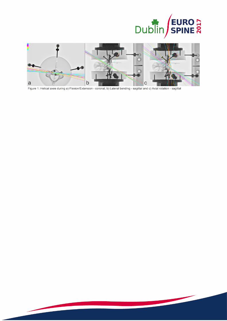
COMPARISON OF THREE-DIMENSIONAL HELICAL AXES OF THE CERVICAL SPINE BETWEEN IN VITRO AND IN VIVO TESTING René Jonas, Robert Demmelmaier, Steffen Paul Hacker, Hans-Joachim Wilke Institute of Orthopaedic Research and Biomechanics, Ulm, Germany The range of motion is a well-accepted kinematic parameter for the assessment and evaluation of spinal motion. Several studies have been performed on the lumbar spine but only few qualitative data of the kinematics of the cervical spine are available which are essential for the development and success of cervical disc arthroplasty [1]. The aim of this study was to provide basic information about helical axes (HA) of human cervical spine under in vitro conditions and compare them with published in vivo data. Six human cadaveric specimens (3 male and 3 female) with an average age of 48 years (range: 34 - 58 years) were carefully selected. After the preparation process only the vertebrae, the intervertebral discs and the ligaments remained intact. Each specimen was divided into three motion-segments: C2/3, C4/5 and C6/7. For three-dimensional motion recordings 3 markers were attached to each vertebra. Afterwards all motion segments were mounted in the three-dimensional spine tester. Then 3.5 full cycles of rotation about all axes: Flexion-Extension, Lateral Bending and Axial Rotation were performed by applying pure moments of 1.5 Nm without any preload. Following the in vitro tests the three-dimensional HA and the instantaneous centers of rotation were calculated and projected into the x-ray images. Rotation analysis of all three directions revealed similar results for all six specimens. All calculated HA and instantaneous centers of rotation were in agreement with published in vivo data (Fig. 1). During Flexion/Extension the angular orientation of the HA were almost constant (Fig. 1a). In case of lateral bending the alignment of the HA was perpendicular to the articular surfaces of the facet joints (Fig. 1b). For axial rotation similar orientation of the HA could be observed, except for the initial phase of rotation (Fig. 1c). The data gained from this study verifies cervical kinematics during in vitro testing using pure moments. Therefore in vitro experiments of cervical motion are capable of representing in vivo kinematics. As a consequence it may be assumed that the influence of muscle forces to cervical kinematics can be neglected. Only small variabilities of HA occurred due to mild degeneration artefacts within the specimens. REFERENCES: [1] Anderst, W.J. et al. (2015). Three-dimensional intervertebral kinematics in the healthy young adult cervical spine during dynamic functional loading. Journal of Biomechanics, 48(7), 1286-1293
Disclosures: Author 1: none; Author 2: none; Author 3: none; Author 4: none