Eukaryotic-type plastid nucleoid protein pTAC3 is essential … · essential for transcription by...
6
Eukaryotic-type plastid nucleoid protein pTAC3 is essential for transcription by the bacterial-type plastid RNA polymerase Yusuke Yagi a,b,c , Yoko Ishizaki c , Yoichi Nakahira c , Yuzuru Tozawa d , and Takashi Shiina c,1 a Faculty of Agriculture and b Institute of Advanced Study, Kyushu University, Fukuoka 812-8581, Japan; c Graduate School of Life and Environmental Science, Kyoto Prefectural University, Kyoto 606-8522, Japan; and d Cell-Free Science and Technology Research Center, Ehime University, Matsuyama 790-8577, Japan Edited by André T. Jagendorf, Cornell University, Ithaca, NY, and approved April 3, 2012 (received for review November 24, 2011) Plastid transcription is mediated by two distinct types of RNA pol- ymerases (RNAPs), bacterial-type RNAP (PEP) and phage-type RNAP (NEP). Recent genomic and proteomic studies revealed that higher plants have lost most prokaryotic transcription regulators and have acquired eukaryotic-type proteins during plant evolu- tion. However, in vivo dynamics of chloroplast RNA polymerases and eukaryotic-type plastid nucleoid proteins have not been di- rectly characterized experimentally. Here, we examine the associ- ation of the α-subunit of PEP and eukaryotic-type protein, plastid transcriptionally active chromosome 3 (pTAC3) with transcribed regions in vivo by using chloroplast chromatin immunoprecipita- tion (cpChIP) assays. PEP α-subunit preferentially associates with PEP promoters of photosynthesis and rRNA genes, but not with NEP promoter regions, suggesting selective and accurate recogni- tion of PEP promoters by PEP. The cpChIP assays further demon- strate that the peak of PEP association occurs at the promoter- proximal region and declines gradually along the transcribed region. pTAC3 is a putative DNA-binding protein that is localized to chloroplast nucleoids and is essential for PEP-dependent transcrip- tion. Density gradient and immunoprecipitation analyses of PEP revealed that pTAC3 is associated with the PEP complex. Interest- ingly, pTAC3 associates with the PEP complex not only during transcription initiation, but also during elongation and termina- tion. These results suggest that pTAC3 is an essential component of the chloroplast PEP complex. In addition, we demonstrate that light-dependent chloroplast transcription is mediated by light-in- duced association of the PEP–pTAC3 complex with promoters. This study illustrates unique dynamics of PEP and its associated protein pTAC3 during light-dependent transcription in chloroplasts. P lastids are DNA-containing organelles unique to plant cells and are thought to have originated from an ancestral cya- nobacterial endosymbiont. Whereas cyanobacteria contain over 3,000 genes, the plastid genome in higher plants consists of small, circular, double-stranded DNA (120–150 kbp) encoding ∼120 genes for photosynthesis and gene expression machineries (1, 2), indicating massive transfer of chloroplast genes to nuclear ge- nome during evolution (3). Vascular plants have evolved a com- plex transcriptional network that is mediated by two types of RNA polymerases (RNAPs): cyanobacterium-derived plastid- encoded plastid RNA polymerase (PEP) and nuclear-encoded phage-type RNA polymerase (NEP). PEP is composed of four catalytic subunits and a promoter recognition subunit, σ-factor (4). Genes for PEP core subunits, α, β, β′, and β′′ were retained in plastid genomes as rpoA, rpoB, rpoC1, and rpoC2 during plant evolution, but genes for σ-factors involved in transcription ini- tiation, have been transferred to the nuclear genome (5), which allows the nucleus to control PEP transcription initiation in response to developmental and environmental cues (recently reviewed in ref. 6). PEP is responsible for transcription of pho- tosynthesis genes in chloroplast in response to light. On the other hand, housekeeping genes encoding PEP core subunits and ri- bosomal proteins are transcribed by the phage-type NEP (7, 8). Thus, two types of RNAP have distinct roles in chloroplast transcription in higher plants. It has been proposed that light-dependent initiation of tran- scription by PEP is controlled by light-induced expression of nuclear-encoded plastid σ-factors (6). Contrarily, several evi- dences suggest that phosphorylated PEP may tightly bind to the promoter region to arrest transcription in the dark (9–11). Dark- induced phosphorylation of PEP and/or σ-factors by redox-reg- ulated plastid transcription kinase (PTK) may be a key step in light-dependent plastid gene transcription. Thus, molecular mechanism of light-dependent transcription in chloroplasts still remains controversial. Plastid DNAs are densely packed into protein–DNA com- plexes called “plastid nucleoids,” as well as bacterial nucleoids. However, higher plants and moss have lost prokaryotic major nucleoid proteins including Hu during evolution (12, 13), whereas several eukaryotic-type proteins such as PEND (14), MFP1 (15), SiR (16), and CND41 (17) have been identified as major components of nucleoids in higher plants. Furthermore, recent proteome analysis identified eukaryotic-type chloroplast nucleoid proteins including putative DNA/RNA-binding pro- teins as components of a chloroplast-derived DNA–protein complex termed pTAC (plastid transcriptionally active chromo- some) (18) and a blue native (BN)-PAGE separated basic PEP complex (19). These findings suggest that chloroplasts have lost most of prokaryotic nucleoid proteins involved in DNA packag- ing, replication, transcription, and translation and acquired eukaryotic-type chloroplast nucleoid proteins during evolution (20). Because vascular plants lack prokaryotic transcription reg- ulators such as DNA-binding proteins and transcription elonga- tion factors except for σ-factors, chloroplast transcription might be mediated by a unique hybrid system of prokaryotic-type RNA polymerase and eukaryotic-type accessory factors, However, the role of the nonprokryotic nucleoid proteins in plastid transcrip- tion remains largely unknown. Earlier studies on pTAC proteins and PEP-associated proteins (PAPs) have focused mainly on in planta analyses using mutant and transgenic plants [e.g., pTAC2, pTAC6, and pTAC12 (18); PAP3/pTAC10, PAP6/FLN1, and PAP7/pTAC14 (19); ET1 (21); Trx-z (22)]. However, mutations in plastid transcription-related genes occasionally give rise to drastic pleiotropic phenotypes such as albino or pale green plants due to reduced accumulation of plastid rRNA and tRNAs, which are mainly transcribed by PEP, and impaired translation of chloroplast-encoded essential proteins. Thus, it is sometimes difficult to characterize the Author contributions: Y.Y., Y.N., and T.S. designed research; Y.Y., Y.I., Y.N., and Y.T. performed research; Y.Y. analyzed data; and Y.Y. and T.S. wrote the paper. The authors declare no conflict of interest. This article is a PNAS Direct Submission. 1 To whom correspondence should be addressed. E-mail: [email protected]. This article contains supporting information online at www.pnas.org/lookup/suppl/doi:10. 1073/pnas.1119403109/-/DCSupplemental. www.pnas.org/cgi/doi/10.1073/pnas.1119403109 PNAS | May 8, 2012 | vol. 109 | no. 19 | 7541–7546 PLANT BIOLOGY
Transcript of Eukaryotic-type plastid nucleoid protein pTAC3 is essential … · essential for transcription by...

Eukaryotic-type plastid nucleoid protein pTAC3 isessential for transcription by the bacterial-typeplastid RNA polymeraseYusuke Yagia,b,c, Yoko Ishizakic, Yoichi Nakahirac, Yuzuru Tozawad, and Takashi Shiinac,1
aFaculty of Agriculture and bInstitute of Advanced Study, Kyushu University, Fukuoka 812-8581, Japan; cGraduate School of Life and Environmental Science,Kyoto Prefectural University, Kyoto 606-8522, Japan; and dCell-Free Science and Technology Research Center, Ehime University, Matsuyama 790-8577, Japan
Edited by André T. Jagendorf, Cornell University, Ithaca, NY, and approved April 3, 2012 (received for review November 24, 2011)
Plastid transcription is mediated by two distinct types of RNA pol-ymerases (RNAPs), bacterial-type RNAP (PEP) and phage-typeRNAP (NEP). Recent genomic and proteomic studies revealed thathigher plants have lost most prokaryotic transcription regulatorsand have acquired eukaryotic-type proteins during plant evolu-tion. However, in vivo dynamics of chloroplast RNA polymerasesand eukaryotic-type plastid nucleoid proteins have not been di-rectly characterized experimentally. Here, we examine the associ-ation of the α-subunit of PEP and eukaryotic-type protein, plastidtranscriptionally active chromosome 3 (pTAC3) with transcribedregions in vivo by using chloroplast chromatin immunoprecipita-tion (cpChIP) assays. PEP α-subunit preferentially associates withPEP promoters of photosynthesis and rRNA genes, but not withNEP promoter regions, suggesting selective and accurate recogni-tion of PEP promoters by PEP. The cpChIP assays further demon-strate that the peak of PEP association occurs at the promoter-proximal region and declines gradually along the transcribedregion. pTAC3 is a putative DNA-binding protein that is localized tochloroplast nucleoids and is essential for PEP-dependent transcrip-tion. Density gradient and immunoprecipitation analyses of PEPrevealed that pTAC3 is associated with the PEP complex. Interest-ingly, pTAC3 associates with the PEP complex not only duringtranscription initiation, but also during elongation and termina-tion. These results suggest that pTAC3 is an essential componentof the chloroplast PEP complex. In addition, we demonstrate thatlight-dependent chloroplast transcription is mediated by light-in-duced association of the PEP–pTAC3 complex with promoters. Thisstudy illustrates unique dynamics of PEP and its associated proteinpTAC3 during light-dependent transcription in chloroplasts.
Plastids are DNA-containing organelles unique to plant cellsand are thought to have originated from an ancestral cya-
nobacterial endosymbiont. Whereas cyanobacteria contain over3,000 genes, the plastid genome in higher plants consists of small,circular, double-stranded DNA (120–150 kbp) encoding ∼120genes for photosynthesis and gene expression machineries (1, 2),indicating massive transfer of chloroplast genes to nuclear ge-nome during evolution (3). Vascular plants have evolved a com-plex transcriptional network that is mediated by two types ofRNA polymerases (RNAPs): cyanobacterium-derived plastid-encoded plastid RNA polymerase (PEP) and nuclear-encodedphage-type RNA polymerase (NEP). PEP is composed of fourcatalytic subunits and a promoter recognition subunit, σ-factor(4). Genes for PEP core subunits, α, β, β′, and β′′ were retainedin plastid genomes as rpoA, rpoB, rpoC1, and rpoC2 during plantevolution, but genes for σ-factors involved in transcription ini-tiation, have been transferred to the nuclear genome (5), whichallows the nucleus to control PEP transcription initiation inresponse to developmental and environmental cues (recentlyreviewed in ref. 6). PEP is responsible for transcription of pho-tosynthesis genes in chloroplast in response to light. On the otherhand, housekeeping genes encoding PEP core subunits and ri-bosomal proteins are transcribed by the phage-type NEP (7, 8).
Thus, two types of RNAP have distinct roles in chloroplasttranscription in higher plants.It has been proposed that light-dependent initiation of tran-
scription by PEP is controlled by light-induced expression ofnuclear-encoded plastid σ-factors (6). Contrarily, several evi-dences suggest that phosphorylated PEP may tightly bind to thepromoter region to arrest transcription in the dark (9–11). Dark-induced phosphorylation of PEP and/or σ-factors by redox-reg-ulated plastid transcription kinase (PTK) may be a key step inlight-dependent plastid gene transcription. Thus, molecularmechanism of light-dependent transcription in chloroplasts stillremains controversial.Plastid DNAs are densely packed into protein–DNA com-
plexes called “plastid nucleoids,” as well as bacterial nucleoids.However, higher plants and moss have lost prokaryotic majornucleoid proteins including Hu during evolution (12, 13),whereas several eukaryotic-type proteins such as PEND (14),MFP1 (15), SiR (16), and CND41 (17) have been identified asmajor components of nucleoids in higher plants. Furthermore,recent proteome analysis identified eukaryotic-type chloroplastnucleoid proteins including putative DNA/RNA-binding pro-teins as components of a chloroplast-derived DNA–proteincomplex termed pTAC (plastid transcriptionally active chromo-some) (18) and a blue native (BN)-PAGE separated basic PEPcomplex (19). These findings suggest that chloroplasts have lostmost of prokaryotic nucleoid proteins involved in DNA packag-ing, replication, transcription, and translation and acquiredeukaryotic-type chloroplast nucleoid proteins during evolution(20). Because vascular plants lack prokaryotic transcription reg-ulators such as DNA-binding proteins and transcription elonga-tion factors except for σ-factors, chloroplast transcription mightbe mediated by a unique hybrid system of prokaryotic-type RNApolymerase and eukaryotic-type accessory factors, However, therole of the nonprokryotic nucleoid proteins in plastid transcrip-tion remains largely unknown.Earlier studies on pTAC proteins and PEP-associated proteins
(PAPs) have focused mainly on in planta analyses using mutantand transgenic plants [e.g., pTAC2, pTAC6, and pTAC12 (18);PAP3/pTAC10, PAP6/FLN1, and PAP7/pTAC14 (19); ET1 (21);Trx-z (22)]. However, mutations in plastid transcription-relatedgenes occasionally give rise to drastic pleiotropic phenotypessuch as albino or pale green plants due to reduced accumulationof plastid rRNA and tRNAs, which are mainly transcribed byPEP, and impaired translation of chloroplast-encoded essentialproteins. Thus, it is sometimes difficult to characterize the
Author contributions: Y.Y., Y.N., and T.S. designed research; Y.Y., Y.I., Y.N., and Y.T.performed research; Y.Y. analyzed data; and Y.Y. and T.S. wrote the paper.
The authors declare no conflict of interest.
This article is a PNAS Direct Submission.1To whom correspondence should be addressed. E-mail: [email protected].
This article contains supporting information online at www.pnas.org/lookup/suppl/doi:10.1073/pnas.1119403109/-/DCSupplemental.
www.pnas.org/cgi/doi/10.1073/pnas.1119403109 PNAS | May 8, 2012 | vol. 109 | no. 19 | 7541–7546
PLANTBIOLO
GY
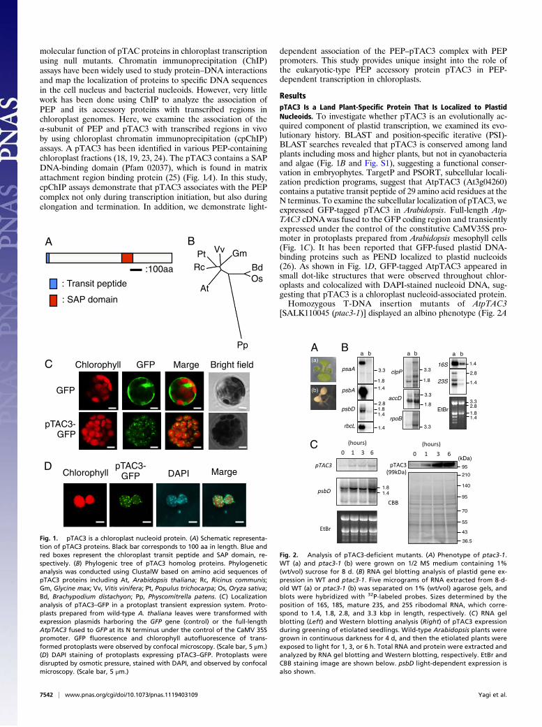
molecular function of pTAC proteins in chloroplast transcriptionusing null mutants. Chromatin immunoprecipitation (ChIP)assays have been widely used to study protein–DNA interactionsand map the localization of proteins to specific DNA sequencesin the cell nucleus and bacterial nucleoids. However, very littlework has been done using ChIP to analyze the association ofPEP and its accessory proteins with transcribed regions inchloroplast genomes. Here, we examine the association of theα-subunit of PEP and pTAC3 with transcribed regions in vivoby using chloroplast chromatin immunoprecipitation (cpChIP)assays. A pTAC3 has been identified in various PEP-containingchloroplast fractions (18, 19, 23, 24). The pTAC3 contains a SAPDNA-binding domain (Pfam 02037), which is found in matrixattachment region binding protein (25) (Fig. 1A). In this study,cpChIP assays demonstrate that pTAC3 associates with the PEPcomplex not only during transcription initiation, but also duringelongation and termination. In addition, we demonstrate light-
dependent association of the PEP–pTAC3 complex with PEPpromoters. This study provides unique insight into the role ofthe eukaryotic-type PEP accessory protein pTAC3 in PEP-dependent transcription in chloroplasts.
ResultspTAC3 Is a Land Plant-Specific Protein That Is Localized to PlastidNucleoids. To investigate whether pTAC3 is an evolutionally ac-quired component of plastid transcription, we examined its evo-lutionary history. BLAST and position-specific iterative (PSI)-BLAST searches revealed that pTAC3 is conserved among landplants including moss and higher plants, but not in cyanobacteriaand algae (Fig. 1B and Fig. S1), suggesting a functional conser-vation in embryophytes. TargetP and PSORT, subcellular locali-zation prediction programs, suggest that AtpTAC3 (At3g04260)contains a putative transit peptide of 29 amino acid residues at theN terminus. To examine the subcellular localization of pTAC3, weexpressed GFP-tagged pTAC3 in Arabidopsis. Full-length Atp-TAC3 cDNA was fused to the GFP coding region and transientlyexpressed under the control of the constitutive CaMV35S pro-moter in protoplasts prepared from Arabidopsis mesophyll cells(Fig. 1C). It has been reported that GFP-fused plastid DNA-binding proteins such as PEND localized to plastid nucleoids(26). As shown in Fig. 1D, GFP-tagged AtpTAC3 appeared insmall dot-like structures that were observed throughout chlor-oplasts and colocalized with DAPI-stained nucleoid DNA, sug-gesting that pTAC3 is a chloroplast nucleoid-associated protein.Homozygous T-DNA insertion mutants of AtpTAC3
[SALK110045 (ptac3-1)] displayed an albino phenotype (Fig. 2A
B
: Transit peptide
: SAP domain
A
:100aa
Chlorophyll GFP Marge
GFP
pTAC3-GFP
C
pTAC3-GFPChlorophyll MargeDAPI
D
Os
Pp
Bd
At
RcPt Vv Gm
Fig. 1. pTAC3 is a chloroplast nucleoid protein. (A) Schematic representa-tion of pTAC3 proteins. Black bar corresponds to 100 aa in length. Blue andred boxes represent the chloroplast transit peptide and SAP domain, re-spectively. (B) Phylogenic tree of pTAC3 homolog proteins. Phylogeneticanalysis was conducted using ClustalW based on amino acid sequences ofpTAC3 proteins including At, Arabidopsis thaliana; Rc, Ricinus communis;Gm, Glycine max; Vv, Vitis vinifera; Pt, Populus trichocarpa; Os, Oryza sativa;Bd, Brachypodium distachyon; Pp, Physcomitrella patens. (C) Localizationanalysis of pTAC3–GFP in a protoplast transient expression system. Proto-plasts prepared from wild-type A. thaliana leaves were transformed withexpression plasmids harboring the GFP gene (control) or the full-lengthAtpTAC3 fused to GFP at its N terminus under the control of the CaMV 35Spromoter. GFP fluorescence and chlorophyll autofluorescence of trans-formed protoplasts were observed by confocal microscopy. (Scale bar, 5 μm.)(D) DAPI staining of protoplasts expressing pTAC3–GFP. Protoplasts weredisrupted by osmotic pressure, stained with DAPI, and observed by confocalmicroscopy. (Scale bar, 5 μm.)
A B
C
Fig. 2. Analysis of pTAC3-deficient mutants. (A) Phenotype of ptac3-1.WT (a) and ptac3-1 (b) were grown on 1/2 MS medium containing 1%(wt/vol) sucrose for 8 d. (B) RNA gel blotting analysis of plastid gene ex-pression in WT and ptac3-1. Five micrograms of RNA extracted from 8-d-old WT (a) or ptac3-1 (b) was separated on 1% (wt/vol) agarose gels, andblots were hybridized with 32P-labeled probes. Sizes determined by theposition of 16S, 18S, mature 23S, and 25S ribodomal RNA, which corre-spond to 1.4, 1.8, 2.8, and 3.3 kbp in length, respectively. (C) RNA gelblotting (Left) and Western blotting analysis (Right) of pTAC3 expressionduring greening of etiolated seedlings. Wild-type Arabidopsis plants weregrown in continuous darkness for 4 d, and then the etiolated plants wereexposed to light for 1, 3, or 6 h. Total RNA and protein were extracted andanalyzed by RNA gel blotting and Western blotting, respectively. EtBr andCBB staining image are shown below. psbD light-dependent expression isalso shown.
7542 | www.pnas.org/cgi/doi/10.1073/pnas.1119403109 Yagi et al.

and Fig. S2 A and B). To analyze the role of pTAC3 in plastidtranscription, we examined global plastid gene expression pat-terns in ptac3-1 by using a plastid DNA macroarray. Expressionof PEP-dependent photosynthesis genes, which are activelytranscribed in chloroplasts, is significantly reduced in 8-d-oldptac3-1 mutant plants (Fig. S3). By contrast, knockout of pTAC3resulted in increased accumulation of several low abundancegene transcripts, including NEP-dependent genes such as accDand PEP core subunit genes (rpoA, and the rpoB-C1-C2 operon).RNA gel blot analysis confirmed the reduced accumulation ofPEP-dependent photosynthesis and rrn transcripts, and the up-regulation of NEP-dependent transcripts in ptac3-1 (Fig. 2B).This is a typical plastid gene expression pattern of mutant plantswith impaired PEP transcription. Furthermore, we analyzedthe developmental expression of AtpTAC3 during greening ofetiolated seedlings. Wild-type Arabidopsis plants were grown incontinuous darkness for 4 d and subsequently exposed to lightfor 1, 3, or 6 h. Both AtpTAC3 mRNA and AtpTAC3 protein areinduced by light within 1 h. (Fig. 2C). The transcript levels ofpsbD, which are a marker for light-dependent plastid transcrip-tion, are also increased by light. These data suggest that pTAC3may play a crucial role in PEP-dependent transcription and beessential for light-induced chloroplast development.
cpChIP Analysis of pTAC3 and PEP Association with Wheat ChloroplastDNA. To understand the role of pTAC3 in PEP-dependenttranscription in chloroplasts, we designed a modified cpChIPassay using wheat chloroplasts. First we evaluated the associationof PEP α-subunit with specific regions of plastid DNA, includingPEP promoters (psbA, rbcL, psaA, rrn16, psbDLRP, and trnEY),a NEP promoter (rpoB), the coding region of a NEP-dependentgene (rpoA), and the noncoding region between rps12 andrrn16 (Spacer) (Fig. S4 and Table S1). The immunoprecipitatedDNA was analyzed by quantitative PCR and quantified usingstandard curves based on a dilution series of input samples. Thedata were analyzed as a percentage of the input sample (detailsin Materials and Methods). The cpChIP assay showed binding ofPEP α-subunit to the promoter regions of PEP-dependent pho-tosynthesis genes and the rrn operon, but not to those of NEP-dependent genes or the spacer region in vivo (Fig. 3A). Bycontrast, the cpChIP assay using an antibody against the non–DNA-binding chloroplast protein, glutamime synthetase 2 (GS2)resulted in a lower cpChIP-QPCR signal at the psbA promotercomparable to that at the rpoB promoter (Fig. S5). These resultsindicate that the cpChIP analysis can be used to detect thespecific association of DNA-binding protein with chloroplastDNA. Furthermore, cpChIP signal of PEP α-subunit character-istics vary markedly among PEP promoters, suggesting that therelative association of PEP with promoters may be dependent onpromoter architecture. The highest signal was detected at thepsbA promoter, which is known as the most active promoter inthe chloroplast genome. We further examined the distribution ofPEP α-subunit along the psbA gene including the promoter,coding, and termination regions (Fig. 3B). The peak associationwith PEP α-subunit was localized in the promoter region, and thelevel decreased slightly toward the termination region of psbA,whereas the signal in the trnK-matK region, which is locatedupstream of psbA, was significantly lower. Less intensive ChIPsignals in the coding region compared with the promoter regionwere also observed in rbcL and the rrn operon (Fig. S6), sug-gesting that PEP density decreases gradually along the tran-scription unit. This cpChIP assay provides the first directevidence that PEP exclusively recognizes PEP promoters andinitiates transcription in vivo, suggesting that PEP–σ complexeshave high binding affinity to PEP promoters.We also examined the association of pTAC3 with chloroplast
DNA via cpChIP assays. As in the case of PEP α-subunit, pTAC3preferentially binds to promoter regions of PEP-dependent
genes, but not of NEP-dependent genes, suggesting a role forpTAC3 in the PEP complex (Fig. 3A and Fig. S3). We furtherexamined the local patterns of spatial association of pTAC3 withthe psbA transcription unit (Fig. 3B). We found that pTAC3binds not only to the promoter region but also the transcriptionelongation region during transcription, suggesting that pTAC3as well as PEP is associated with chloroplast DNA along thepsbA transcription unit. Thus, it is unlikely that pTAC3 binds tospecific sequences in the psbA promoter; rather it may be animportant component of the PEP complex in chloroplasts.To examine further the interaction between pTAC3 and PEP,
we isolated the PEP complex by glycerol density gradient cen-trifugation and probed for the presence of pTAC3 by Westernblotting. The molecular weight of the PEP complex from wheatchloroplasts was estimated as around 700 kDa, as reportedpreviously in mustard (27). pTAC3 was detected in two fractions,corresponding to a peak containing PEP α-subunit (fraction 8)and a lower molecular weight fraction (roughly estimated to 200–400 kDa; fractions 4 and 5) (Fig. 4A). These results suggest thatpTAC3 in wheat chloroplasts is associated mainly with the PEPcomplex, although a portion of the pTAC3 may form anothercomplex without PEP. Furthermore, immunoprecipitation assaysusing pTAC3 antibody with wheat chloroplast extracts demon-strated the presence of α-subunit in the pTAC3 immunopreci-pitated complex (Fig. 4B). These results demonstrate thatpTAC3 has a direct function in PEP transcription.
: NoAb
: PEP-
: pTAC3
A
% In
put
B
3.0
2.5
2.0
1.5
1.0
0.5
0.0
a b P C T
% In
put
: NoAb
: PEP-
: pTAC3
3.0
2.5
2.0
1.5
1.0
0.5
0.0
psbAtrnKmatK
: 200bpTCPba
Fig. 3. Association of PEP α-subunit and pTAC3 with chloroplast DNA. (A)Association of PEP α-subunit and pTAC3 with PEP promoter regions (PpsbA,PrbcL, PpsaA, Prrn16, psbDLRP, PtrnEY ), a NEP promoter region (PrpoB), thecoding region of the rpoA gene (rpoA), and a noncoding spacer region lo-cated between rps12 and rrn16 (Spacer) was analyzed by ChIP assay.Chloroplasts were prepared from wheat seedlings grown for 5 d in the lightand subjected to ChIP assays using antibodies against PEP α-subunit andpTAC3. NoAb, no antibody control. Enriched DNA was quantified by qPCR.The amount of immunoprecipitated DNA in each sample is presented asa percentage of the total input chromatin. Mean values and SDs of threeindependent experiments are shown. (B) Spatial association of α-subunit andpTAC3 along the psbA transcription unit. Data are shown as in A. Schematicgene map of the matK-psbA region is shown below. Horizontal black barrepresents 200 bp. Arrow indicates the transcription start site of the psbAgene and the direction of transcription. DNA regions corresponding to thepsbA promoter (P), coding region (C), terminator (T), and two units (a and b)in loci upstream of psbA are shown.
Yagi et al. PNAS | May 8, 2012 | vol. 109 | no. 19 | 7543
PLANTBIOLO
GY

Light-Dependent Association of the PEP–pTAC3 Complex with Chlo-roplast DNA. PEP transcription activity is greatly stimulated bylight in mature wheat chloroplasts (28). We therefore examinedthe light-dependent association of PEP with promoter regions ofseveral plastid genes in vivo. Chloroplasts were prepared fromwheat seedlings grown for 5 d in the light and then dark adaptedfor 24 h or seedlings reilluminated for 6 h after the 24-h darkadaptation. Immunoblot analysis revealed the constitutive accu-mulation of both PEP α-subunit and pTAC3 upon light illumi-nation in wheat seedlings (Fig. S7), suggesting that the expressionlevel of the PEP complex is not affected by light. On the otherhand, the cpChIP assays showed that the relative amount of PEPα-subunit associated with the promoter region of photosynthesisgenes including psbA, psbD, psaA, and rbcL, and ribosomal RNArrn16, was two- to fivefold higher in the illuminated chloroplaststhan in the dark-adapted chloroplasts (Fig. 5A). Amounts ofchloroplast DNA in input samples between both conditions werenot significantly different (Fig. S8). Thus, the light-dependentcpChIP signals would be indicative of light-dependent associationof PEP α-subunit and pTAC3 to chloroplast DNA. Moreover, wecould not detect tight binding of PEP α-subunit to the promoterregion of photosynthesis genes in the dark-adapted chloroplasts,suggesting a limited role for transcription arrest mediated bydark-induced phosphorylation of PEP subunits and σ-factors. Inaddition to promoter regions, we also detected light-dependentassociation of PEP α-subunit with coding and termination regionsof psbA (Fig. 5B). These results demonstrate that recruitment ofPEP to its target promoters is dependent on light. On the otherhand, light-dependent association of PEP with NEP-dependentgenes including rpoA and rpoB and the spacer region was notdetected. Furthermore, ChIP analysis of pTAC3 showed thatlight accelerated the association of pTAC3 with not only the PEPpromoter region but also the coding region in the psbA tran-scription unit (Fig. 5 A and B) similar to the distribution patternof PEP α-subunit, suggesting that pTAC3 associates with PEP-dependent transcribed regions as a component of a large PEPcomplex in a light-dependent manner.
DiscussionThis study reveals light-dependent associations of PEP andpTAC3 with chloroplast DNA in vivo using cpChIP assays. ChIPassays have been widely used to detect specific binding sitesfor transcription factors (29), distribution patterns of several
A1 2 3 4 5 6 7 8 9 10 11 12 13
pTAC3
PEP-
CBB
pTAC3
NoAb pTAC3
PEP-Antibody
IP NoAb pTAC3
B
Top Bottom
95
43
55
4336.5
95 43
(kDa)
(kDa) (kDa)
Fig. 4. Analysis of the pTAC3 protein complex. (A) Separation of the PEPcomplex by glycerol density gradient centrifugation. Total chloroplast pro-teins prepared from wheat grown under continuous light for 6 d wereloaded onto a 10–30% (vol/vol) glycerol density gradient and separatedby centrifugation. Thirteen fractions were collected from top to bottom andanalyzed by immunoblotting with anti-PEP α-subunit or pTAC3 antibodies.Gel staining with CBB is also shown. (B) Immunoprecipitation analysis of thepTAC3 complex. Total wheat chloroplast extracts were subjected to immu-noprecipitation with pTAC3 antibody or without (NoAb) and analyzed byimmunoblotting with anti-PEP α-subunit and pTAC3 antibodies.
% In
put
A
B
NoAb(Dark)RpoA(Dark)pTAC3(Dark)NoAb(Light)RpoA(Light)pTAC3(Light)
% In
put
NoAb(Dark)RpoA(Dark)pTAC3(Dark)NoAb(Light)RpoA(Light)pTAC3(Light)
a b P C T
NoAb (Dark)PEP- (Dark)pTAC3 (Dark)NoAb (Light)PEP- (Light)pTAC3 (Light)
NoAb (Dark)
PEP- (Dark)pTAC3 (Dark)NoAb (Light)PEP- (Light)pTAC3 (Light)
3.0
2.5
2.0
1.5
1.0
0.5
0.0
3.0
2.5
2.0
1.5
1.0
0.5
0.0
psbAtrnKmatK
: 200bpTCPba
Fig. 5. Analysis of light-dependent association of PEP α-subunit and pTAC3with chloroplast DNA. (A) Association of PEP α-subunit and pTAC3 withchloroplast DNA in response to light. Chloroplasts were prepared fromwheat seedlings grown for 5 d in the light and then dark adapted for 24 h(dark) or seedlings reilluminated for 6 h after the 24-h dark adaptation(light). ChIP was performed to determine the association level of PEPα-subunit and pTAC3 with PEP promoter regions (PpsbA, PrbcL, PpsaA,Prrn16, psbDLRP, and PtrnEY), a NEP promoter (PrpoB), the coding region ofthe rpoA gene (rpoA), and a noncoding region located between rps12 andrrn16 (Spacer), using anti-PEP α-subunit or pTAC3 antibodies or no antibody(NoAb). The immunoprecipitated DNA was analyzed by quantitative PCRand quantified via standard curves based on a dilution series of input sam-ples. The amount of immunoprecipitated DNA in each sample is presented asa percentage of the total input chromatin. Mean values and SDs of threeindependent experiments are shown. (B) Association level of PEP α-subunitand pTAC3 with regions of the psbA transcription unit in response to light.Data are presented as in A. Schematic gene map of the matK-psbA region isshown as in Fig. 3B.
7544 | www.pnas.org/cgi/doi/10.1073/pnas.1119403109 Yagi et al.

modified histones (30), and trafficking of RNAP and its associ-ated proteins on genomic DNA (31). In chloroplasts, a fewstudies have shown the association of endogenous (Whirly1) andrecombinant (LacI) transcription factors with chloroplast pro-moters in vivo by using ChIP assays (32, 33). However, in vivodynamics of chloroplast RNA polymerases and/or their associ-ated proteins have not been directly characterized experimen-tally. Unlike cyanobacteria, the chloroplasts of higher plantshave two types of RNA polymerase, PEP and NEP. Transcrip-tome analyses of PEP- or NEP-deficient mutants and in vitrotranscription analyses using isolated PEP and NEP have shownthat PEP and NEP preferentially initiate transcription frombacterial-type and phage-type promoters, respectively. Here, weprovide direct evidence that PEP exclusively associates in vivowith PEP promoters of photosynthesis genes and the rRNAoperon, but not with NEP promoters by using cpChIP assay (Fig.3), suggesting selective and accurate recognition of PEP pro-moters by PEP.It has been shown that several PEP promoters including psbA,
psbD LRP, rbcL, psaA, and rrn P1 are regulated by unique cis-elements, termed as the TATA-box, PGT-box, and AAG-box,CDF1-binding site, region U, and RUA, respectively, which arelocated upstream of or within the core promoter (28, 34–37) andrecognized by promoter-specific transcription factors. However,sequence specific DNA-binding proteins have not been identifiedand characterized in chloroplasts of higher plants. Chloroplastprotein pTAC3 contains a putative DNA-binding SAP domain.Here we show that pTAC3 is localized in chloroplast nucleoidsand is essential for PEP-dependent transcription, suggesting thatpTAC3 may be a chloroplast transcription factor that regulatesPEP-dependent transcription. Thus, we further searched forpTAC3-binding regions on plastid DNA by using cpChIP assays.We found that pTAC3 binds to the transcribed regions of allPEP-dependent genes examined (psbA, psaA, psbD, rbcL, andrrn promoters), but not to a specific cis-element in a particularpromoter, suggesting that pTAC3 is a PEP-associated general,rather than sequence-specific transcription factor. In contrast toPEP-dependent transcribed loci, pTAC3 associates weakly withNEP-dependent transcribed loci, suggesting that pTAC3 doesnot associate with the NEP transcription complex. However,chloroplasts exhibit very low NEP-dependent transcription ac-tivity. NEP proteins are thought to be present at very low levelsin leaf chloroplasts, because they were not detected in the pro-teomic analysis of whole chloroplast proteins and pTAC frac-tions. Thus, there remains a possibility that the cpChIP assaycould not detect the association of pTAC3 with NEP-dependenttranscribed loci, due to the low levels of accumulation of NEPin chloroplasts.In bacteria, it is proposed that RNAP accessory proteins mod-
ulate the promoter accessibility to RNAP by altering the 3Dstructure of the nucleoid. The nucleoid proteins factor for in-version stimulation (FIS) and histone-like nucleoid structuringprotein (H-NS) are associated with RNAP and bind chromosomalDNA at many regions (38). Furthermore, FIS and H-NS werecopurified with RNAP subunits and ribosomes in Escherichia coli(39). Similarly, cpChIP assays demonstrated that pTAC3 asso-ciates with PEP-dependent transcribed regions of photosynthesisand rRNA genes together with PEP α-subunit. Density gradientand immunoprecipitation analyses of PEP demonstrate thatpTAC3 is an essential component of PEP in chloroplasts. Fur-thermore, pTAC3 is crucial for PEP activity and its expressionincreases dramatically during chloroplast development, as PEPactivity increases. Taken together, these results suggest thatpTAC3 is an essential component of the PEP complex and criti-cally involved in chloroplast differentiation from immature plas-tids. Considering the eukaryotic origin of the SAP domain asa DNA-binding motif, our findings suggest that pTAC3 was
acquired from host cells during plant evolution to enhance and/ormaintain transcription of photosynthesis genes in higher plants.Our ChIP analysis of PEP α-subunit shows that PEP preferen-
tially associates with the promoter region rather than the codingregion (Fig. 3B and Fig. S3), suggesting that the PEP complexremains at the promoter-proximal region.E. coliChIP analysis usingspecific antibodies for RNAP core subunits revealed that there isalso preferential association of the RNAP core with the promoter-proximal region of transcribed genes and decreased RNAP-bindingdensity downstream from the promoter (31). The accumulation ofRNAP at the promoter-proximal region in E. coli is controlledmainly by interactions between the elongating complex of RNAPand elongation regulators, such as NusA, NusG, and Rho. TheArabidopsis genome encodes only one putative bacterial-type elon-gation factor, pTAC13 (At3g09210) containing a KOW domain,which is characteristic for members of the NusG protein family (40).Furthermore, maize ET1, which resembles the eukaryotic elonga-tion factor TFIIS, was identified as a component of the chloroplasttranscriptionally active fraction, and plants deficient in ET1 showedan aberrant chloroplast development phenotype and decreasedPEP-dependent transcription (21), suggesting that bacterial-typepTAC13 and eukaryotic-type ET1 may be involved in PEP elon-gation. In this study, we demonstrate that pTAC3 is associatedwith PEP through transcription initiation and elongation steps,suggesting a possible role for pTAC3 in the regulation of PEPtranscription elongation in chloroplasts.Light-dependent activation of PEP transcription may be con-
trolled by PTK and plastid redox signaling. It has been proposedthat phosphorylated PEP and/or σ-like factors tightly bind topromoters to arrest transcription under dark conditions (11). IfPEP is trapped at promoter regions in the dark, ChIP signals atPEP promoters would not decrease in dark-adapted leaves. Thepresent data, however, showed that ChIP signals at both pro-moters and coding regions of PEP-dependent photosynthesisand rRNA genes were reduced in dark-adapted wheat seedlings,suggesting that PEP dissociates from chloroplast genomic DNAin the dark. Thus, it is likely that light regulates the association ofthe PEP–pTAC3 complex with the promoter region possiblythrough the light-dependent expression of σ-factors, rather thanvia regulation of σ-factor phosphorylation by PTK.In conclusion, here we present a characterization of in vivo
dynamics of chloroplast RNA polymerase PEP and eukaryotic-type nucleoid protein pTAC3 with chloroplast DNA by usingcpChIP analysis. We show that PEP α-subunit preferentiallyassociates with the promoter-proximal regions of PEP-dependentphotosynthesis and rRNA genes in a light-dependent manner, butnot to NEP promoters, suggesting that light-dependent chloro-plast transcription is mediated by accurate recognition of PEPpromoters by PEP in response to light. Furthermore, we show thateukaryotic-type nucleoid protein pTAC3 is essential for PEPactivity and chloroplast development and associates with the PEPcomplex not only during transcription initiation, but also duringelongation and termination. As pTAC3 has been acquired earlyduring land plant evolution, understanding the molecular func-tion of pTAC3 should not only provide insights into the mecha-nisms enabling developmental regulation of plastid transcriptionbut also offer a unique way of understanding plant evolution.
Materials and MethodsPlant Material. The Arabidopsis ptac3-1 (SALK_110045) mutant (Columbiaecotype) was identified from the T-DNA insertion mutant line collectiongenerated at the Salk Institute. For expression analysis, wild-type (Columbia)and ptac3-1 were grown on half-strength MS medium containing 1%(wt/vol) sucrose under 16-h light and 8-h dark conditions (normal light; 50–60 μmol · m−2s−1). Wheat (Triticum aestivum) seeds were grown on vermic-ulite at 25 °C under continuous white light (50 μmol · m−2s−1) for 5–6 d.Details regarding the identification of the mutant allele of pTAC3 are in-cluded in SI Materials and Methods.
Yagi et al. PNAS | May 8, 2012 | vol. 109 | no. 19 | 7545
PLANTBIOLO
GY

Chloroplast ChIP Assay. Detailed procedures for cpChIP assays are included inSI Materials and Methods.
Plastid Gene Expression Analysis. Details for RNA extraction, RNA gel blottinganalysis, and construction and analysis of plastid DNA tiling macroarrays areincluded in SI Materials and Methods.
Localization Analysis by Transient Expression in Protoplasts. Protoplast tran-sient expression assays were performed as described previously (41). Detailsfor construction of pTAC3–GFP expression plasmids are included in SIMaterials and Methods.
Protein Analysis. Details regarding antibody materials, protein extractionfrom isolated chloroplasts, protein complex separation via glycerol densitygradient centrifugation, immunoprecipitation, and immunoblotting analysisare included in SI Materials and Methods.
ACKNOWLEDGMENTS. We thank Dr. M. H. Sato for helpful discussions, Mr.Y. Motomura for technical assistance, and the Salk Institute for providingArabidopsis insertion mutants. This work was supported by Ministry of Ed-ucation, Culture, Sports, Science and Technology Grants-in-Aid 21007485 (toY.Y.), 22004564 (to T.S.), and 2020060 (to Y.N.), and by the Private UniversityStrategic Research Foundation Support Program.
1. Sugiura M (1992) The chloroplast genome. Plant Mol Biol 19:149–168.2. Kaneko T, et al. (1996) Sequence analysis of the genome of the unicellular cyano-
bacterium Synechocystis sp. strain PCC6803. II. Sequence determination of the entiregenome and assignment of potential protein-coding regions (supplement). DNA Res3:185–209.
3. Martin W, Herrmann RG (1998) Gene transfer from organelles to the nucleus: Howmuch, what happens, and why? Plant Physiol 118:9–17.
4. Hu J, Bogorad L (1990) Maize chloroplast RNA polymerase: the 180-, 120-, and 38-kilodalton polypeptides are encoded in chloroplast genes. Proc Natl Acad Sci USA 87:1531–1535.
5. Allison LA (2000) The role of sigma factors in plastid transcription. Biochimie 82:537–548.
6. Lerbs-Mache S (2011) Function of plastid sigma factors in higher plants: Regulation ofgene expression or just preservation of constitutive transcription? Plant Mol Biol 76:235–249.
7. Hedtke B, Börner T, Weihe A (1997) Mitochondrial and chloroplast phage-type RNApolymerases in Arabidopsis. Science 277:809–811.
8. Hajdukiewicz PT, Allison LA, Maliga P (1997) The two RNA polymerases encoded bythe nuclear and the plastid compartments transcribe distinct groups of genes in to-bacco plastids. EMBO J 16:4041–4048.
9. Tiller K, Eisermann A, Link G (1991) The chloroplast transcription apparatus frommustard (Sinapis alba L.). Evidence for three different transcription factors whichresemble bacterial sigma factors. Eur J Biochem 198:93–99.
10. Tiller K, Link G (1993) Sigma-like transcription factors from mustard (Sinapis alba L.)etioplast are similar in size to, but functionally distinct from, their chloroplast coun-terparts. Plant Mol Biol 21:503–513.
11. Tiller K, Link G (1993) Phosphorylation and dephosphorylation affect functionalcharacteristics of chloroplast and etioplast transcription systems from mustard (Si-napis alba L.). EMBO J 12:1745–1753.
12. Kobayashi T, et al. (2002) Detection and localization of a chloroplast-encoded HU-likeprotein that organizes chloroplast nucleoids. Plant Cell 14:1579–1589.
13. Karcher D, Köster D, Schadach A, Klevesath A, Bock R (2009) The Chlamydomonaschloroplast HLP protein is required for nucleoid organization and genome mainte-nance. Mol Plant 2:1223–1232.
14. Sato N, Albrieux C, Joyard J, Douce R, Kuroiwa T (1993) Detection and characteriza-tion of a plastid envelope DNA-binding protein which may anchor plastid nucleoids.EMBO J 12:555–561.
15. Jeong SY, Rose A, Meier I (2003) MFP1 is a thylakoid-associated, nucleoid-bindingprotein with a coiled-coil structure. Nucleic Acids Res 31:5175–5185.
16. Chi-Ham CL, Keaton MA, Cannon GC, Heinhorst S (2002) The DNA-compacting proteinDCP68 from soybean chloroplasts is ferredoxin:sulfite reductase and co-localizes withthe organellar nucleoid. Plant Mol Biol 49:621–631.
17. Murakami S, Kondo Y, Nakano T, Sato F (2000) Protease activity of CND41, a chloro-plast nucleoid DNA-binding protein, isolated from cultured tobacco cells. FEBS Lett468:15–18.
18. Pfalz J, Liere K, Kandlbinder A, Dietz KJ, Oelmüller R (2006) pTAC2, -6, and -12 arecomponents of the transcriptionally active plastid chromosome that are required forplastid gene expression. Plant Cell 18:176–197.
19. Steiner S, Schröter Y, Pfalz J, Pfannschmidt T (2011) Identification of essential subunitsin the plastid-encoded RNA polymerase complex reveals building blocks for properplastid development. Plant Physiol 157:1043–1055.
20. Sato N (2001) Was the evolution of plastid genetic machinery discontinuous? TrendsPlant Sci 6:151–155.
21. da Costa e Silva O, et al. (2004) The Etched1 gene of Zea mays (L.) encodes a zincribbon protein that belongs to the transcriptionally active chromosome (TAC) ofplastids and is similar to the transcription factor TFIIS. Plant J 38:923–939.
22. Arsova B, et al. (2010) Plastidial thioredoxin z interacts with two fructokinase-likeproteins in a thiol-dependent manner: Evidence for an essential role in chloroplastdevelopment in Arabidopsis and Nicotiana benthamiana. Plant Cell 22:1498–1515.
23. Suzuki JY, et al. (2004) Affinity purification of the tobacco plastid RNA polymeraseand in vitro reconstitution of the holoenzyme. Plant J 40:164–172.
24. Schröter Y, Steiner S, Matthäi K, Pfannschmidt T (2010) Analysis of oligomeric proteincomplexes in the chloroplast sub-proteome of nucleic acid-binding proteins frommustard reveals potential redox regulators of plastid gene expression. Proteomics 10:2191–2204.
25. Aravind L, Koonin EV (2000) SAP: a putative DNA-binding motif involved in chro-mosomal organization. Trends Biochem Sci 25:112–114.
26. Terasawa K, Sato N (2005) Visualization of plastid nucleoids in situ using the PEND-GFP fusion protein. Plant Cell Physiol 46:649–660.
27. Pfannschmidt T, Link G (1994) Separation of two classes of plastid DNA-dependentRNA polymerases that are differentially expressed in mustard (Sinapis alba L.) seed-lings. Plant Mol Biol 25:69–81.
28. Satoh J, et al. (1999) Developmental stage-specific multi-subunit plastid RNA poly-merases (PEP) in wheat. Plant J 18:407–415.
29. Lee J, et al. (2007) Analysis of transcription factor HY5 genomic binding sites revealedits hierarchical role in light regulation of development. Plant Cell 19:731–749.
30. Johnson L, Cao X, Jacobsen S (2002) Interplay between two epigenetic marks. DNAmethylation and histone H3 lysine 9 methylation. Curr Biol 12:1360–1367.
31. Mooney RA, et al. (2009) Regulator trafficking on bacterial transcription units in vivo.Mol Cell 33:97–108.
32. Prikryl J, Watkins KP, Friso G, van Wijk KJ, Barkan A (2008) A member of the Whirlyfamily is a multifunctional RNA- and DNA-binding protein that is essential for chlo-roplast biogenesis. Nucleic Acids Res 36:5152–5165.
33. Newell CA, Gray JC (2010) Binding of lac repressor-GFP fusion protein to lac operatorsites inserted in the tobacco chloroplast genome examined by chromatin immuno-precipitation. Nucleic Acids Res 38:e145.
34. Satoh J, Baba K, Nakahira Y, Shiina T, Toyoshima Y (1997) Characterization of dy-namics of the psbD light-induced transcription in mature wheat chloroplasts. PlantMol Biol 33:267–278.
35. Lam E, Hanley-Bowdoin L, Chua NH (1988) Characterization of a chloroplast se-quence-specific DNA binding factor. J Biol Chem 263:8288–8293.
36. Cheng MC, Wu SP, Chen LF, Chen SC (1997) Identification and purification of a spin-ach chloroplast DNA-binding protein that interacts specifically with the plastid psaA-psaB-rps14 promoter region. Planta 203:373–380.
37. Suzuki JY, Sriraman P, Svab Z, Maliga P (2003) Unique architecture of the plastid ri-bosomal RNA operon promoter recognized by the multisubunit RNA polymerase intobacco and other higher plants. Plant Cell 15:195–205.
38. Grainger DC, Hurd D, Goldberg MD, Busby SJ (2006) Association of nucleoid proteinswith coding and non-coding segments of the Escherichia coli genome. Nucleic AcidsRes 34:4642–4652.
39. Butland G, et al. (2005) Interaction network containing conserved and essentialprotein complexes in Escherichia coli. Nature 433:531–537.
40. Steiner T, Kaiser JT, Marinkoviç S, Huber R, Wahl MC (2002) Crystal structures oftranscription factor NusG in light of its nucleic acid- and protein-binding activities.EMBO J 21:4641–4653.
41. Tsunoyama Y, et al. (2004) Blue light-induced transcription of plastid-encoded psbDgene is mediated by a nuclear-encoded transcription initiation factor, AtSig5. ProcNatl Acad Sci USA 101:3304–3309.
7546 | www.pnas.org/cgi/doi/10.1073/pnas.1119403109 Yagi et al.