Protists Eukaryotic Organisms. Protists Protists: Eukaryotic microorganisms in the Protist family.

EUKARYOTIC CELL STRUCTURE
Plant and animal cells have several differences and similarities (figure
below):
Structurally, plant and animal cells are very similar because they are both
eukaryotic cells. They both contain membrane-bound organelles such as
the nucleus, mitochondria, endoplasmic reticulum, Golgi apparatus,
lysosomes, and peroxisomes. Both also contain similar membranes,
cytosol, and cytoskeletal elements. The functions of these organelles are
extremely similar between the two classes of cells (peroxisomes perform
additional complex functions in plant cells having to do with cellular
respiration).
The few differences that exist between plant and animals are very
significant and reflect a difference in the functions of each cell. Plant
cells can be larger than animal cells. The normal range for an animal cell
varies from 10 to 30 micrometers while that for a plant cell stretches from
10 to 100 micrometers. Beyond size, animal cells do not have a cell wall
or chloroplasts but plant cells do. Animal cells are round and irregular in
shape while plant cells have fixed rectangular shapes.
In contrast to animal cells, plant cells often contain large
central vacuoles occupying up to 90% of the total cell volume, pushing
the nucleus against the cell wall.

Genetics (B 252) Lecture 1 part 2 2017-2018
2
Most of the cells have Cytoskeletal elements. They are group of
protein filaments that supports, maintains the shape of the cells and
controls the movement of the organelles. It is formed of a complex
system of microtubules, microfilaments and intermediate filaments.

Genetics (B 252) Lecture 1 part 2 2017-2018
3
1. Microtubule:
They are the dominant part of the cytoskeleton. It consists of straight
hollow cylinder. Its wall is formed of 13 protofilaments. Each
protofilament is a linear polymer of spherical alternating subunits of
globular protein of α- and β-tubulin. Microtubules involved in the
formation of spindle fibers, transport cellular materials and movements of
some microorganisms.
2. Microfilaments:
They are polymers of actin protein in which monomer Globular actin (G-
actin) polymerizes into long right-handed with double-helical strands of
polymer Fibrous actin (F-actin). They influence the cytoplasmic
streaming of the cell.
3. Intermediate filaments:
Most types of intermediate filaments are cytoplasmic, but one type,
the lamins, is nuclear in position. It has a structural role by providing
mechanical strength to cells and tissues.

Genetics (B 252) Lecture 1 part 2 2017-2018
4
TRANSMISSION AND INHERITANCE OF CHROMOSOMES
We have two types of gene transmission and inheritance:
nuclear (happen in nucleus) and cytoplasmic (happen in plastids and
mitochondria). Both plastids and mitochondria undergo self-replication.
We will focus in this lecture on the nucleus, nuclear transmission
and inheritance.
I. Nucleus:
The nucleus in the cell is analogous to the brain in the body. It is a
control center for a cell by maintaining the integrity of the genes and to
control the activities of the cell by regulating gene expression. The
nucleus stores all the information the cell needs to grow, reproduce, and
function. This information is contained in long but thin molecules of
deoxyribonucleic acid, or DNA. One of the functions of the nucleus is to
protect the cell’s DNA from damage, but that is not all that it does. It is
spherical membrane-bound organelle that contains the genetic
information of eukaryotic cell. It controls cellular, developmental and
genetic activities.
It consists of nuclear envelope, sap, pores, lamina, chromatin and
nucleolus.
1. Nuclear envelope:

Genetics (B 252) Lecture 1 part 2 2017-2018
5
It consists of 2 thin membranes separated by a perinuclear space
(10 - 50 nm). The outer membrane is continuous with rough
endoplasmic reticulum (rER).
2. Nuclear lamina:
It is a dense network of protein fibrous (lamins=intermediate
filaments) lining in the inner surface of the inner nuclear membrane
to support the nuclear envelope.
Note: Both Nuclear envelope and lamina enclose the nucleus entirely and
isolates its contents from the cellular cytoplasm. So, it serves as a barrier
to prevent macromolecules from diffusing freely between the
nucleoplasm and the cytoplasm.
3. Nuclear pores:
They are specialized opening in the nuclear envelope. In each pore,
the inner and outer membranes of the envelope fused together
forming a channel joining cytoplasm with nucleoplasm. This
channel is lined with proteins complex called nucleoporines, which
permit macromolecules to move across the nuclear envelope.
The nuclear membrane has pores through which the contents of the
nucleus communicate with the rest of the cell. Nucleoporins, a family of
50 to 100 proteins, are the main components of the nuclear pore complex
in eukaryotic cells. The nuclear membrane tightly controls what gets into
the nucleus and what gets out. Movement of large water-soluble
molecules such as proteins and RNA through the nuclear pores is
required for both gene expression and the maintenance of chromosomes.
Because the nuclear membrane is impermeable to large
molecules, nuclear pores are required that regulate nuclear transport of
molecules across this membrane. The pores cross both nuclear
membranes, providing a channel through which larger molecules must
be actively transported by carrier proteins while allowing free movement
Genetics (B 252) Lecture 1 part 2 2017-2018
6
of small molecules and ions. This regulation of communication by the
nuclear membrane has a great effect on what a cell looks like and what it
does.
4. Nuclear sap:
It is the inner clear viscous solution between chromatin granules
and also known as interchromatin substance or karyolymph.
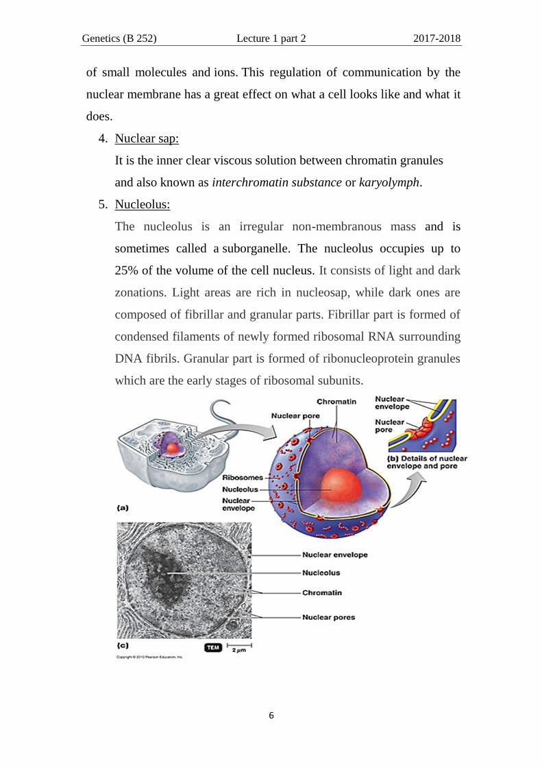
5. Nucleolus:
The nucleolus is an irregular non-membranous mass and is
sometimes called a suborganelle. The nucleolus occupies up to
25% of the volume of the cell nucleus. It consists of light and dark
zonations. Light areas are rich in nucleosap, while dark ones are
composed of fibrillar and granular parts. Fibrillar part is formed of
condensed filaments of newly formed ribosomal RNA surrounding
DNA fibrils. Granular part is formed of ribonucleoprotein granules
which are the early stages of ribosomal subunits.
Genetics (B 252) Lecture 1 part 2 2017-2018
7
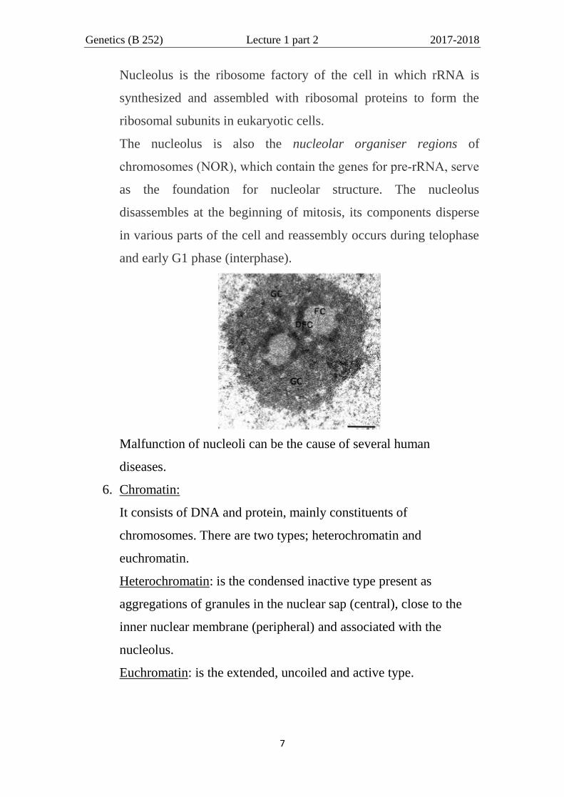
Nucleolus is the ribosome factory of the cell in which rRNA is
synthesized and assembled with ribosomal proteins to form the
ribosomal subunits in eukaryotic cells.
The nucleolus is also the nucleolar organiser regions of
chromosomes (NOR), which contain the genes for pre‐rRNA, serve
as the foundation for nucleolar structure. The nucleolus
disassembles at the beginning of mitosis, its components disperse
in various parts of the cell and reassembly occurs during telophase
and early G1 phase (interphase).
Malfunction of nucleoli can be the cause of several human
diseases.
6. Chromatin:
It consists of DNA and protein, mainly constituents of
chromosomes. There are two types; heterochromatin and
euchromatin.
Heterochromatin: is the condensed inactive type present as
aggregations of granules in the nuclear sap (central), close to the
inner nuclear membrane (peripheral) and associated with the
nucleolus.
Euchromatin: is the extended, uncoiled and active type.

Genetics (B 252) Lecture 1 part 2 2017-2018
8
CHROMOSOMES
Chromosomes are also located in the nucleus and are basically
organized from DNA and proteins. In eukaryotes, the chromosomal DNA
is packaged and organized into a non-condensed structure called
chromatin (figure below). How those happen? Double-
stranded DNA wraps twice around (loops) 8 special proteins called
histones, forming the nucleosome, which is the building block of
chromatin packaging. These nucleosomes coil and stack together to form
fibers called chromatin. Chromatin in turn forms larger loops and coils to
form chromosomes (figure below).

Genetics (B 252) Lecture 1 part 2 2017-2018
9
Chromosomes are single pieces of coiled double-stranded DNA
along with genes, proteins, and nucleotides, and chromatin is condensed
forming chromosomes that basically allow DNA to fit inside the nucleus,
so the genes within these chromosomes are known as the cell's nuclear
genome.
In eukaryotic organisms, the DNA inside the nucleus is also closely
associated with large protein complexes called histones. Along with the
nuclear membrane, histones help control which messages get sent from
the DNA to the rest of the cell. The information stored in DNA gets
transferred to the rest of the cell by a very elegant process—a process so
common and so important to life on Earth that it is called the central

Genetics (B 252) Lecture 1 part 2 2017-2018
10
dogma of biology (DNA → RNA → Protein). Chromosomal DNA
encodes most or all of an organism's genetic information.
The structure of chromosomes and chromatin varies through
the cell cycle. Chromosomes are even more condensed than chromatin
and are an essential unit for cellular division.
Chromosomes may be classified according to structure into
duplicated (dyad) or unduplicated (monad) in mitosis and as tetrad in
meiosis. Unduplicated chromosomes are single linear chromatid strands

Genetics (B 252) Lecture 1 part 2 2017-2018
11
contains one DNA molecule, which may be several inches long, whereas
duplicated chromosomes contain two identical copies (called arm or
chromatids or sister chromatids or 2 monads) joined by a centromere.
The centromere is a constricted region of the chromosome containing a
specific DNA sequence, to which is bound 2 discs of protein called
kinetochores. Kinetochores serve as points of attachment for
microtubules that move the chromosomes during cell division. The
regions at both ends of chromosome are the telomeres.
Note: Budding yeast has small highly complex kinetochores, which are
not visible by standard light or electron microscopy.
Chromosome Classification:
1. Each chromosome has two arms, p (the short one) and q (the
longer). The p arm is named for "petit" meaning 'small'; the q arm
is named q simply because it follows p in the alphabet.
Telomere
Genetics (B 252) Lecture 1 part 2 2017-2018
12
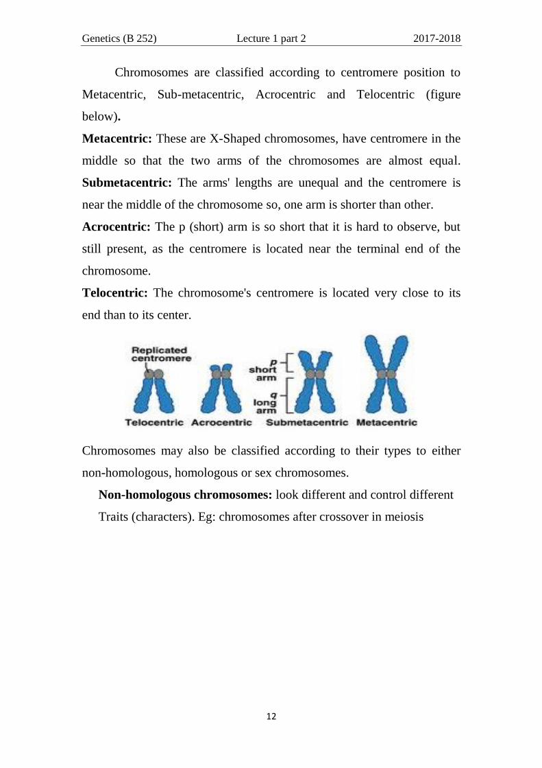
Chromosomes are classified according to centromere position to
Metacentric, Sub-metacentric, Acrocentric and Telocentric (figure
below).
Metacentric: These are X-Shaped chromosomes, have centromere in the
middle so that the two arms of the chromosomes are almost equal.
Submetacentric: The arms' lengths are unequal and the centromere is
near the middle of the chromosome so, one arm is shorter than other.
Acrocentric: The p (short) arm is so short that it is hard to observe, but
still present, as the centromere is located near the terminal end of the
chromosome.
Telocentric: The chromosome's centromere is located very close to its
end than to its center.
Chromosomes may also be classified according to their types to either
non-homologous, homologous or sex chromosomes.
Non-homologous chromosomes: look different and control different
Traits (characters). Eg: chromosomes after crossover in meiosis

Genetics (B 252) Lecture 1 part 2 2017-2018
13
Homologous chromosomes- are chromosome pairs of approximately the
same length, centromere position, and staining pattern, with genes for the
same characteristics at corresponding loci (i.e. control the same traits).
May code for different forms of each trait and have independent origin -
one homologous chromosome is inherited from the organism's mother;
the other from the organism's father. They are usually not identical, but
carry the same type of information i.e. similar but not identical. Eg: the
22 pairs of autosomes in human.
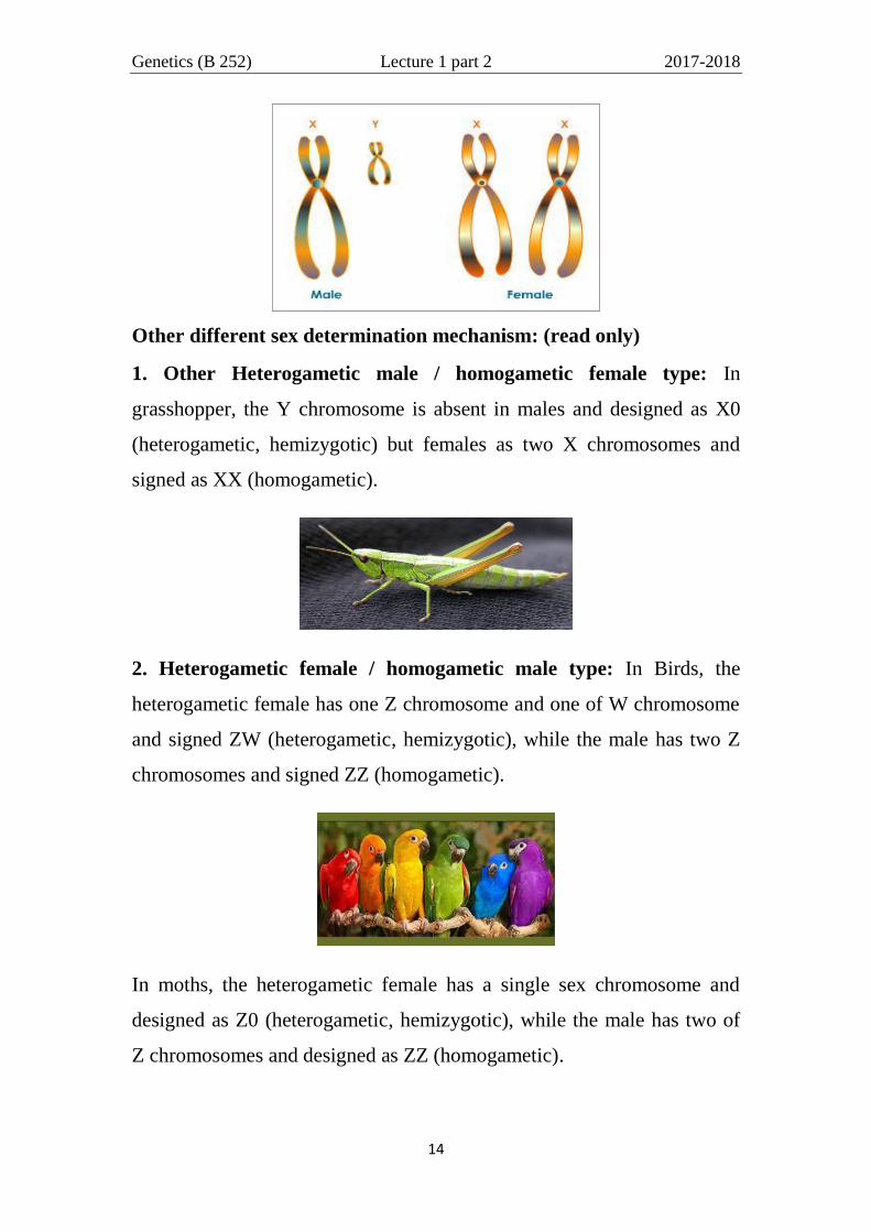
Sex chromosomes: Are distinct from each other in their characteristics
and are represented as X and Y to determine the sex of the individual, XX
being female (homogametic) and XY being male (Heterogametic,
hemizygotic) Eg as in Drosophila, plants and Human.
Genetics (B 252) Lecture 1 part 2 2017-2018
14
Other different sex determination mechanism: (read only)
1. Other Heterogametic male / homogametic female type: In
grasshopper, the Y chromosome is absent in males and designed as X0
(heterogametic, hemizygotic) but females as two X chromosomes and
signed as XX (homogametic).
2. Heterogametic female / homogametic male type: In Birds, the
heterogametic female has one Z chromosome and one of W chromosome
and signed ZW (heterogametic, hemizygotic), while the male has two Z
chromosomes and signed ZZ (homogametic).
In moths, the heterogametic female has a single sex chromosome and
designed as Z0 (heterogametic, hemizygotic), while the male has two of
Z chromosomes and designed as ZZ (homogametic).
Genetics (B 252) Lecture 1 part 2 2017-2018
15
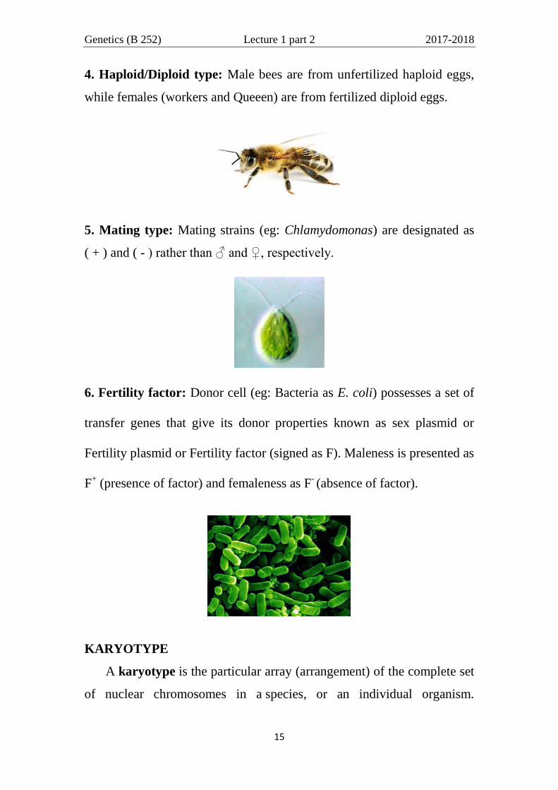
4. Haploid/Diploid type: Male bees are from unfertilized haploid eggs,
while females (workers and Queeen) are from fertilized diploid eggs.
5. Mating type: Mating strains (eg: Chlamydomonas) are designated as
( + ) and ( - ) rather than ♂ and ♀, respectively.
6. Fertility factor: Donor cell (eg: Bacteria as E. coli) possesses a set of
transfer genes that give its donor properties known as sex plasmid or
Fertility plasmid or Fertility factor (signed as F). Maleness is presented as
F+ (presence of factor) and femaleness as F
- (absence of factor).
KARYOTYPE
A karyotype is the particular array (arrangement) of the complete set
of nuclear chromosomes in a species, or an individual organism.

Genetics (B 252) Lecture 1 part 2 2017-2018
16
Karyotypes describe the number of chromosomes, and what they look
like under a light microscope. Chromosomes are arranged in a karyotype
for the purpose of analysis. This arrangement of the chromosomes is
based on their size, centromere position and banding patterns that are
specific for each chromosome (figure below).
The cell may be classified according to the number of
chromosomes copies (figure below) into either haploid (n) or diploid
(2n) or polyploidy (ns):
Haploid - A cell possessing a single copy of each chromosome
(human/plant sex cells).
Diploid - A cell possessing two copies of each chromosome
(human/plant body cells). Most eukaryotes have between 10 and 50

Genetics (B 252) Lecture 1 part 2 2017-2018
17
chromosomes in their body cells. Human cells have 46 chromosomes: 22
nearly-identical pairs (autosomes) and a pair of sex chromosome.
Polyploid- A cell possessing numerous copies of each chromosome, so it
may be triploid, tetraploid,…..