Eudistoma (Ascidiacea: Polycitoridae) from tropical Brazil · Eudistoma from tropical Brazil 197...
15
ZOOLOGIA 31 (2): 195–208, April, 2014 http://dx.doi.org/10.1590/S1984-46702014000200011 2014 Sociedade Brasileira de Zoologia | www.sbzoologia.org.br | www.scielo.br/zool All content of the journal, except where identified, is licensed under a Creative Commons attribution-type BY-NC. Eudistoma Caullery, 1909 is the most species-rich genus in Polycitoridae, with 124 valid species found in tropical and temperate waters, with a few records from Antarctica and the subtropics (MONNIOT 1978). Originally, Eudistoma was proposed as a subgenus of Distoma to include animals with few rows of stigmata in the pharynx (CAULLERY 1909). Eudistoma was treated as a genus by RITTER & FORSYTH (1917) who did not explain why. In subsequent studies, MICHAELSEN (1919), VAN NAME (1921) and TOKIOKA (1942) all considered Eudistoma as a subgenus of Polycitor. Later, VAN NAME (1945) considered Eudistoma a valid genus due to the three rows of pharyngeal slits, long esopha- gus, flat stomach in the posterior region of the abdomen, very conspicuous longitudinal muscles extending from the phar- ynx to the end of the abdomen and larvae that are incubated in the atrial cavity. While those characters are constant, color and shape are extremely variable and have caused confusion in species identification. Thus, species identification will be much more certain by using larval characters (MONNIOT 1983, KOTT 1990). Currently, 26 species of Eudistoma are known from the Atlantic Ocean, including seven in Brazil: Eudistoma carolinense Van Name, 1945, E. clavatum Rocha & Bonnet, 2009, E. recifense Millar, 1977, E. repens Millar, 1977, E. saldanhai Millar, 1977, E. spiculiferum Millar, 1977, and E. vannamei Millar, 1977. Most are tropical, but E. carolinense and E. clavatum are found in the subtropics (ROCHA & MORENO 2000, ROCHA & BONNET 2009). While few records of Eudistoma spp. have been published for the Brazilian coast, we suggest that diversity is greater than expected. Here we describe material collected in eastern Brazil and comment on the implications of species richness for the distribution of Eudistoma. MATERIAL AND METHODS We examined material from Paraíba, Pernambuco, Alagoas, Bahia and Espírito Santo collected from 1997 to 2013 (Figs 1-4). From Paraíba, we examined both the collections deposited in the Tunicata Collection of the Laboratory of In- vertebrates Paul Young of Departamento de Sistemática e Ecologia, Universidade Federal da Paraíba (LIPY/DSE/UFPB) and material recently collected (2009 and 2013) by the authors. Most of the surveys were done in the intertidal zone on many beaches along the whole coast. Samples from Pernambuco and Alagoas were very few and donated to us for identification and do not represent an extensive survey of those coasts. Samples from Bahia were collected by us using SCUBA diving at depths up to 11 m inside Baía de Todos os Santos and along the coast of Salvador. Main surveys occurred during one week in 1999, 2004, and 2007. In Espírito Santo, samples were also collected by us both in the intertidal and using SCUBA diving in the summer of 2011 and 2012. Specimens were anesthetized with menthol, fixed and preserved in formalin 4%. Dissection was with standard proce- dures following MONNIOT & MONNIOT (1972). Samples were stained with Harris hematoxylin and examined under a stereoscopic microscope. Holotypes were deposited in the Collection of the Museum of Zoology of São Paulo (MZUSP), while paratypes and additional material in the collection of ascidians from the Eudistoma (Ascidiacea: Polycitoridae) from tropical Brazil Livia de Moura Oliveira 1 , Gustavo Antunes Gamba 1 & Rosana Moreira da Rocha 1,2 1 Programa de Pós-graduação em Zoologia, Departamento de Zoologia, Universidade Federal do Paraná. Caixa Postal 19020, 81531-980 Curitiba, PR, Brazil. 2 Corresponding author: E-mail: [email protected] ABSTRACT. We studied material in collections from coastal intertidal and subtidal tropical waters of the Brazilian states of Paraíba, Pernambuco, Alagoas, Bahia, and Espírito Santo. We identified seven species of Eudistoma, of which two are new to science. Eudistoma alvearium sp. nov. colonies have fecal pellets around each zooid and zooids are 6-8 mm long with seven straight and parallel pyloric tubules; the larval trunk is 0.6 mm long with three adhesive papillae and ten ampullae. Eudistoma versicolor sp. nov. colonies are cushion-shaped, variable in color (blue, purple, brown, light green, gray or white) and zooids have six straight and parallel pyloric tubules; the larval trunk is 0.8 mm long with three adhesive papillae and six ampules. Three species – E. carolinense Van Name, 1945, E. recifense Millar, 1977, and E. vannamei Millar, 1977 – are known from northeastern Brazil. The identification of two additional species will require confirmation. We also propose a synonymy for E. carolinense with E. repens Millar, 1977, also previously described in Brazil. KEY WORDS. Atlantic; colonial ascidians; new species; taxonomy.
-
Upload
duongkhanh -
Category
Documents
-
view
219 -
download
0
Transcript of Eudistoma (Ascidiacea: Polycitoridae) from tropical Brazil · Eudistoma from tropical Brazil 197...
ZOOLOGIA 31 (2): 195–208, April, 2014http://dx.doi.org/10.1590/S1984-46702014000200011
2014 Sociedade Brasileira de Zoologia | www.sbzoologia.org.br | www.scielo.br/zoolAll content of the journal, except where identified, is licensed under a Creative Commons attribution-type BY-NC.
Eudistoma Caullery, 1909 is the most species-rich genusin Polycitoridae, with 124 valid species found in tropical andtemperate waters, with a few records from Antarctica and thesubtropics (MONNIOT 1978). Originally, Eudistoma was proposedas a subgenus of Distoma to include animals with few rows ofstigmata in the pharynx (CAULLERY 1909). Eudistoma was treatedas a genus by RITTER & FORSYTH (1917) who did not explain why.In subsequent studies, MICHAELSEN (1919), VAN NAME (1921) andTOKIOKA (1942) all considered Eudistoma as a subgenus ofPolycitor. Later, VAN NAME (1945) considered Eudistoma a validgenus due to the three rows of pharyngeal slits, long esopha-gus, flat stomach in the posterior region of the abdomen, veryconspicuous longitudinal muscles extending from the phar-ynx to the end of the abdomen and larvae that are incubatedin the atrial cavity. While those characters are constant, colorand shape are extremely variable and have caused confusionin species identification. Thus, species identification will bemuch more certain by using larval characters (MONNIOT 1983,KOTT 1990).
Currently, 26 species of Eudistoma are known from theAtlantic Ocean, including seven in Brazil: Eudistoma carolinenseVan Name, 1945, E. clavatum Rocha & Bonnet, 2009, E. recifenseMillar, 1977, E. repens Millar, 1977, E. saldanhai Millar, 1977, E.spiculiferum Millar, 1977, and E. vannamei Millar, 1977. Mostare tropical, but E. carolinense and E. clavatum are found in thesubtropics (ROCHA & MORENO 2000, ROCHA & BONNET 2009).
While few records of Eudistoma spp. have been publishedfor the Brazilian coast, we suggest that diversity is greater thanexpected. Here we describe material collected in eastern Brazil
and comment on the implications of species richness for thedistribution of Eudistoma.
MATERIAL AND METHODS
We examined material from Paraíba, Pernambuco,Alagoas, Bahia and Espírito Santo collected from 1997 to 2013(Figs 1-4). From Paraíba, we examined both the collectionsdeposited in the Tunicata Collection of the Laboratory of In-vertebrates Paul Young of Departamento de Sistemática eEcologia, Universidade Federal da Paraíba (LIPY/DSE/UFPB) andmaterial recently collected (2009 and 2013) by the authors.Most of the surveys were done in the intertidal zone on manybeaches along the whole coast. Samples from Pernambuco andAlagoas were very few and donated to us for identification anddo not represent an extensive survey of those coasts. Samplesfrom Bahia were collected by us using SCUBA diving at depthsup to 11 m inside Baía de Todos os Santos and along the coastof Salvador. Main surveys occurred during one week in 1999,2004, and 2007. In Espírito Santo, samples were also collectedby us both in the intertidal and using SCUBA diving in thesummer of 2011 and 2012.
Specimens were anesthetized with menthol, fixed andpreserved in formalin 4%. Dissection was with standard proce-dures following MONNIOT & MONNIOT (1972). Samples were stainedwith Harris hematoxylin and examined under a stereoscopicmicroscope. Holotypes were deposited in the Collection of theMuseum of Zoology of São Paulo (MZUSP), while paratypes andadditional material in the collection of ascidians from the
Eudistoma (Ascidiacea: Polycitoridae) from tropical Brazil
Livia de Moura Oliveira1, Gustavo Antunes Gamba1 & Rosana Moreira da Rocha1,2
1 Programa de Pós-graduação em Zoologia, Departamento de Zoologia, Universidade Federal do Paraná.Caixa Postal 19020, 81531-980 Curitiba, PR, Brazil.2 Corresponding author: E-mail: [email protected]
ABSTRACT. We studied material in collections from coastal intertidal and subtidal tropical waters of the Brazilian states of
Paraíba, Pernambuco, Alagoas, Bahia, and Espírito Santo. We identified seven species of Eudistoma, of which two are new
to science. Eudistoma alvearium sp. nov. colonies have fecal pellets around each zooid and zooids are 6-8 mm long with
seven straight and parallel pyloric tubules; the larval trunk is 0.6 mm long with three adhesive papillae and ten ampullae.
Eudistoma versicolor sp. nov. colonies are cushion-shaped, variable in color (blue, purple, brown, light green, gray or
white) and zooids have six straight and parallel pyloric tubules; the larval trunk is 0.8 mm long with three adhesive papillae
and six ampules. Three species – E. carolinense Van Name, 1945, E. recifense Millar, 1977, and E. vannamei Millar, 1977 –
are known from northeastern Brazil. The identification of two additional species will require confirmation. We also propose
a synonymy for E. carolinense with E. repens Millar, 1977, also previously described in Brazil.
KEY WORDS. Atlantic; colonial ascidians; new species; taxonomy.

196 L. de M. Oliveira et al.
ZOOLOGIA 31 (2): 195–208, April, 2014
Departamento de Zoologia, Universidade Federal do Paraná(DZUP) and in the Tunicata Collection of the Laboratory of In-vertebrates Paul Young of the Departamento de Sistemática eEcologia, Universidade Federal da Paraíba (LIPY UFPB).
TAXONOMY
We found seven species of Eudistoma, of which two arenew and three were already known in northeastern Brazil(Eudistoma carolinense, E. recifense, and E. vannamei). With theseseven species, Brazil has approximately 39% of the Eudistomaspecies found in the Atlantic and 9% found worldwide. Thefollowing identification key includes all species of the genusknown from Brazil. Characters used for identification are basedon our morphological analysis and the literature and are de-scribed in detail in Table I.
Eudistoma alvearium sp. nov. Rocha & OliveiraFigs 5-12
Description. The colony measures 2.2 x 1.6 x 1.2 cm and isencrusting and cushion-shaped with irregular outline. It is brownin life and light brown in formaldehyde. The tunic is transpar-ent, soft and gelatinous with a smooth surface. Fecal pellets aredistributed throughout the colony, but in greater quantity at thetop, arranged around each zooid and forming a beehive patternthat can be seen externally on the surface of the tunic.
Zooids are not arranged in systems, but are parallel toeach other, occupying the space from the surface to the baseof the colony, are approximately 6-8 mm in length when notcontracted and are colorless and transparent. Both siphonsopen at the surface of the colony and each has six roundedlobes. The tubular oral siphon is shorter than the lateral andhorizontally directed atrial siphon. Conspicuous circularmuscles form a sphincter at the base of each siphon.
The body wall is transparent with conspicuous longitu-dinal muscles. Ten parallel longitudinal muscular fibers are oneach side of the thorax. In the abdomen, muscles form twolateral-ventral bundles that extend to the posterior edge of thezooid. The transverse musculature is also conspicuous in thethorax with 16 fibers. A band of circular muscles is visible atthe beginning of the abdomen. Oral tentacles were not ob-served due to contraction of zooids. The first row of the phar-ynx has 16 stigmata on each side, while the second and thirdhave 14. The esophagus is long and straight. The stomach isspherical with smooth wall, posteriorly in the abdomen. Theanterior intestine is shorter than the stomach. The pyloric glandhas at least seven pyloric straight and parallel tubules, origi-nating in the posterior region of the stomach. The intestinalloop has two constrictions, one between the anterior intestineand the middle intestine and the other between the middleand ascending intestine. The anus is at the level of the secondrow of stigmata.
Gonads are in the intestinal loop, just below the stom-ach, reminiscent of a bunch of grapes. The testis has approxi-mately 16-20 follicles and a straight sperm duct, while the ovaryis central and has 2-3 oocytes. One to three, oval shaped, 0.6mm long trunk larvae are incubated in the oviduct. Larvae havethree adhesive papillae and ten ampullae, three pairs alongthe mid anterior line, one dorsal-lateral pair and two ventralampullae. The papillae have a wide peduncle, as long as theampullae. The dorsal pair of ampullae and the two pairs be-tween the adhesive papillae are elongated, the two ventral andthe dorsal-lateral pair of ampullae are shorter with roundedend. Larvae have three rows of pharyngeal slits. The larval ocel-lus and otolith are posterior and the tail makes a one-half turnaround the trunk on the left side of the adhesive papillae.
Type material. Holotype: BRAZIL, Bahia: Salvador(Naufrágio Boa Viagem, 12°56’6"S, 38°30’42"W, 7-8 m), 1colony, 3 slides, 03.viii.1999, Rocha, R.M. leg., (MZUSP 00035).
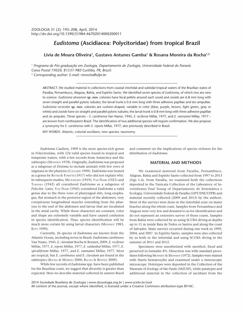
Figures 1-4. (1) Map of the Brazilian coast highlighting the study areas; (2) Paraíba and Pernambuco; (3) Bahia and Alagoas; (4) EspíritoSanto. 1) Barra de Camaratuba, 2) Baia da Traição, 3) Barra de Mamanguape, 4) Quebramar Cabedelo, 5) Pomar das Esponjas, Pontado Cabo Branco, Ponta do Seixas, Recife do Seixas, 6) Maceiozinho, Carapibus, 7) Coqueirinho, 8) Tabatinga, 9) Recife da Galé, 10)Ponta de Pedras, 11) Naufrágio do Areeiro, 12) Praia do Francês, 13) Boa Viagem, Quebramar Norte, Quebramar Sul, Farol Humaitá,Germânia e Porto da Barra, 14) Enseada das Garças, 15) Ilha dos Pacotes.
1 2 3 4

197Eudistoma from tropical Brazil
ZOOLOGIA 31 (2): 195–208, April, 2014
Etymology. The specific epithet is derived from latin andrefers to the beehive shape of the colony (alvearium = bee-hive).
Remarks. This species is unlike any other described inBrazil or elsewhere in the Atlantic Ocean. It is very distinctivein appearance with the tunic surface smooth and transparent,and zooids separated by fecal pellets and not arranged in cir-cular systems. In the Pacific, KOTT (1990) described E.constrictum, a very similar species (in the appearance of thecolony). However, E. constrictum has brownish pink zooids inethanol and the thorax contains 30 longitudinal fibers crossedby numerous transverse fibers in the middle of the thorax. Upto four embryos may be incubated in the atrial cavity. Larvaemeasure 1 mm long with the tail describing a three-quarterturn around the trunk. Larvae have three adhesive papillaeand six ampullae, one dorsal, one ventral and two betweeneach pair of papillae.
Eudistoma carolinense Van Name, 1945Figs 13-18
Eudistoma carolinense Van Name, 1945: 123-124, fig. 58; Pérès,1949: 170; Millar, 1977: 176-179, figs. 6, 7; Monniot, 1983:1011-1013, fig. 4 C-F; Rocha & Moreno, 2000: 10-11, fig. 1;Rocha & Faria, 2005: 5; Rocha & Kremer, 2005: 1173; Rochaet al., 2005: 463.
Eudistoma repens Millar, 1977: 184 (synonymy).
Description. Branching finger-like projections, approxi-mately 2.5 cm in length and variable width, project from anencrusting base. The ends of the projections are widened andsometimes flattened. The colony is brownish because of sandaccumulation in the tunic. Zooids occupy the distal third of
the colony projections, parallel to the major axis of the pro-jections, and reach a maximum size of around 4 mm in lengthwhen fully distended. The thorax and abdomen are 1.0 and3.0 mm in length, are opaque and so the internal organs aredifficult to see. Both siphons are tubular and have six slightlypointed lobes; the oral lobe is shorter than the atrial. Longitu-dinal musculature is formed by 5-6 fibers on each side of thethorax. The pharynx has three rows of stigmata, each with 15slits per half row.
The abdomen is long and slender and the stomach isposterior and laterally flattened. The intestine is long with aconstriction at the beginning of the ascending portion afterthe intestinal loop. The pyloric tubules have numerous globu-lar projections along the intestine wall, just above the stom-ach, and the tubules extends twice the length of the stomach,as depicted in figure 4D of MONNIOT (1983). A long and thincommon tube links the pyloric gland to the mid portion ofthe stomach.
The testis is posterior to the stomach and has 11-13 fol-licles. The sperm duct is elongated and is widened at the levelof the stomach. The ovary is the central to the testicular fol-licles and contains three oocytes. One or two larvae are incu-bated in the atrial cavity. The larvae are ovoid and the trunk is0.4 mm long, and have three adhesive papillae very close to-gether, supported by short peduncles. The ampullae are shortand rounded, 13 on each side plus a single one in the ventralregion. The tail wraps around three-quarters of the larva.
Examined material. BRAZIL, Paraíba: João Pessoa (Pontado Cabo Branco, 07°08’50"S, 34°47’51"W, intertidal), 1 colony,04.x.2007, Leonel, R.M.V. leg., (LIPY UFPB-Tun-358); João Pessoa(Quebramar Cabedelo, 6°57’44"S, 34° 50’35"W, intertidal), 1colony, 1 slide (EUD 1.11), 25.iii.2013, Oliveira, L.M. leg., (DZUP
Table I. Key to the identification of Eudistoma in the Brazilian coast. (1) Colony shape: C – cushion, F – fingerlike projections, H – headson cylindrical peduncles, S – spherical. (2) Colour of living colony: Bl = blue, B – brown, C – cream, G – grey, Gr – green, O – orange, P– purple, S – colour of the sand the covers the colony, U – uncolored, Y – yellow, W – white. (3) Zooid maximum reported size (mm).(4) Pigmentation of zooid: 0 – absent, P – present. (5) Organization of zooids in the colony: 0 – no systems, S – with systems. (6) Calcareousspicules in the tunic: 0 – absent, P – present. (7) Number of longitudinal muscle fibers in one side of the thorax. (8) Number of pharyngealstigmata per row on each side. (9) Maximum number of testicular follicles reported. (10) Number of incubated embryos or larvae. (11)Size of larval trunk (mm). (12) Total number of ectodermal ampullae in larvae.
1 2 3 4 5 6 7 8 9 10 11 12 Species
F S 4 0 0 0 5-6 11-15 13 1-2 0.4-0.54 26 Eudistoma carolinense1,2,4
H C 7 0 0 0 12-20 13-24 13 2-4 0.5 8 E. clavatum3
H B, G, Gr 8 0 S 0 ? 10 10 ? 0.7-0.75 6 E. saldanhai2
H Y, O 20 P 0 0 12-18 20-22 6 1 1.2-1.4 8 E. vannamei2,4
C B 8 0 0 0 10 14-16 20 1-3 0.6 10 E. alvearium sp. nov.
C S 11 0 S 0 28-32 24 13 ? ? ? Eudistoma sp. 1
C U 2.2 P 0 0 6-7 15 10 1-2 0.6-0.8 6 Eudistoma sp. 2
C, S P, Bl, B, W, Gr 10 0 S 0 18-25 14-16 25 1 0.6-0.7 6 E. versicolor sp. nov.
C, S G 10 P S 0 12-14 14-16 18 1 0.6 6 E. recifense2,4
C B, P 5 P ? P ? ? ? 1 1-1.15 ? E. spiculiferum2
1 VAN NAME (1945); 2 MILLAR (1977); 3 ROCHA & BONNET (2009); 4 Present study; ? No information.

198 L. de M. Oliveira et al.
ZOOLOGIA 31 (2): 195–208, April, 2014
EUD-48); Bahia: Salvador (Naufrágio Germânia, 13°00’34"S,38°31’59"W), 1 colony, 10.xii.2007, Rocha, R.M. leg., (DZUPEUD-73); Espírito Santo : Vila Velha (Ilha dos Pacotes,20°21’5.40"S, 40°15’3.79"W, 12 m), 01 colony, 13.ii. 2011.Rocha, R.M. leg., (DZUP EUD-29); Fundão (Enseada das Garças,20°01’57"S, 40°09’3"W, 0.3 m), 01 colony, 25.i.2012, Gamba,G.A. leg., (DZUP EUD-31).
Distribution. BRAZIL: Pará (MILLAR 1977); Ceará, Pernam-buco, Bahia, Espírito Santo (Tito Lotufo, pers. comm.); Paraíba(this study); Paraná (ROCHA & FARIA 2005, ROCHA & KREMER 2005);Santa Catarina (ROCHA & MORENO 2000, ROCHA et al. 2005). Glo-bal: United States: South Carolina, Florida (VAN NAME 1945);Guadalupe (MONNIOT 1983); Senegal (PÉRÈS 1949).
Remarks. Four of the six species of Eudistoma in Brazil
8
5
9
10
12
11
76
Figures 5-12. Eudistoma alvearium sp. nov.: (5) colony in formalin; (6) detail of the surface of the colony; (7) cross section of the colonyshowing the arrangement of zooids; (8) pyloric tubules; (9) larva; (10) zooid brooding embryos; (11) pyloric tubules; (12) larva. Scalebars: 5 = 1 cm; 8, 11 = 0.25 mm; 9, 10, 12 = 0.5 mm.

199Eudistoma from tropical Brazil
ZOOLOGIA 31 (2): 195–208, April, 2014
have lobed colonies: E. vannamei, E. saldanhai, E. repens and E.carolinense. The first two lack sand in the tunic and zooids andlarvae are sufficient different to be distinguished from E.carolinense. The description of E. repens does not say how it isdifferent from E. carolinense MILLAR (1977). We propose the syn-onymy of E. repens with E. carolinense, because of their mor-phological similarity, especially of the larvae. Some smalldifferences in colony size and number of pharyngeal stigmata
between the species was noted, but colony shape and larvalmorphology and size are very similar or identical and thus donot support them as separate species. MILLAR (1977) says thereare six embryos and larvae in the atrial cavity, but his illustra-tion shows only three. Our samples had two embryos in theatrial cavity, but we suggest that the number may vary. Colo-nies are quite similar and the small differences are probablydue to the different substrates where they were found.
Figures 13-18. Eudistoma carolinense: (13) the colony in situ; (14) colony in detail; (15) thorax with larvae; (16) abdomen; (17) thorax;(18) larva. Scale bars: 15-17 = 0.4 mm; 18 = 0.2 mm.
13
14
1815 17
16

200 L. de M. Oliveira et al.
ZOOLOGIA 31 (2): 195–208, April, 2014
Eudistoma carolinense has a disjunct distribution in tropi-cal and in colder waters, but no records from the geographi-cally intermediate waters of São Paulo and Rio de Janeiro. Itwas originally described from the southeastern United States(VAN NAME 1945) and, subsequently, in South (MILLAR 1977) andCentral America (MONNIOT 1983). The southernmost record inthe Atlantic is from Santa Catarina (ROCHA & MORENO 2000),where it was possibly introduced (ROCHA & FARIA 2005). This isthe first record for Paraíba. Northeastern populations are con-sidered cryptogenic because it remains unclear whether theybelong to the original tropical distribution or were transportedanthropogenically.
Eudistoma recifense Millar, 1977Figs 19-24
Eudistoma recifense Millar, 1977: 181-182 fig. 9.
Description. The colony is cushion-shaped, opaque andgrayish when preserved. The tunic is translucent, cartilaginous,and smooth. The colony from Paraíba was 4.0 x 3.0 cm longand had approximately 45 circular depressions that mark sys-tems, which were formed of 6-10 individuals, usually eight.
Zooids are transparent but with some black in the dorsalregion of the thorax and a few black spots in the abdomen. Inpreserved material the pigmentation is reduced and the abdo-men spots disappear over time. The most relaxed zooid was 10mm long. Oral and atrial siphons are 6-lobed with a black rim.The atrial siphon is longer than the oral, lateral and orientedhorizontally with pigment throughout.
Transverse musculature is conspicuous throughout thethorax, except at the base. Longitudinal musculature comprisesapproximately 12-14 fibers on each side of the thorax. Thereare 16 simple oral tentacles. The pharynx has 14-16 stigmataon each side of each row.
The abdomen is long and slender. The intestine is longand constricted at the beginning of the ascending portion, af-ter the intestinal loop. Three to five winding pyloric tubulesare on the ascending intestine, starting anterior to the stom-ach. The testis is in the intestinal loop just below the stomachand has approximately 18 follicles. The ovary is central to tes-ticular follicles and contains two eggs. The anus opens at thelevel of the second row of pharyngeal slits.
One embryo is incubated between the thorax and theabdomen. The ovoid larva has a 0.6 mm long trunk, three ad-hesive papillae with short, wide stalks, the ventral papilla far-ther from the rest. Five ampullae are in the mid anterior lineand one is dorso-lateral on the right side, for a total of six. Thepharynx has three rows of stigmata, with 14 slits per half row.The ocellus and otolith are in a posterior position of the larva.The tail makes three-quaters of a turn and passes on the leftside of the adhesive papillae.
Examined material. BRAZIL, Paraíba: João Pessoa (Pomardas Esponjas, 07°08’04"S, 34°46’20"W, sublittoral), 1 colony, 1slide (Eud 1.6), 12.i.2009, Projeto Biota Paraíba (LIPY UFPB-
Tun-206); Pernambuco: (Naufrágio do Areeiro 08°03’46"S,34°49’24"W, 12 m), 1 colony, 28.xi.1999, 1 slide (Poly1.59),Lotufo, T.M.C. leg. (DZUP POLY-52).
Distribution. BRAZIL: Ceará (MILLAR 1977), Paraíba andPernambuco (this study).
Remarks. This species was first described by MILLAR (1977)from Pernambuco and then it was found in Ceará. This is thefirst record for Paraíba. It is uncommon and all previous speci-mens found were collected in the subtidal zone. Characteris-tics of our specimen are in accordance with the originaldescription, with one exception in which the embryo in ourswas not incubated in the atrial cavity as described. but ratherbetween thorax and abdomen. Eudistoma saldanhai in Brazil isanother species with a firm cartilaginous colony but it is pe-dunculate, zooid systems are formed by 12 individuals, thethorax lacks pigment, has fewer pharyngeal slits (10) and thelarger larvae are incubated in the atrial cavity (MILLAR 1977).
Eudistoma sp. 1Figs 25-32
Description. The colony is cushion-shaped and rounded,with 5.5 cm in diameter and 2.6 cm thick, adherent to thesubstrate by the entire base. The tunic is fully encrusted withsand and so the colony is very firm. When alive, the color ofthe colony is grayish, but when fixed, tends to pale brown dueto the sand color on the surface.
Zooids are scattered throughout the colony formingbarely visible systems and arranged in circular groups of sixzooids. The zooids are perpendicular to the surface of thecolony, 11 mm long when fully stretched, but the thorax isalways small with only 2 mm. They are transparent but thedigestive system is yellowish with evident fecal pellets through-out. Both siphons are tubular with six rounded lobes at therim. The atrial siphon is long and opens on the surface of thecolony, close to the oral siphon. Thin and dense circular musclesappear along the whole extension of both siphons. The longi-tudinal musculature is conspicuous and consists of approxi-mately 28-32 fibers grouped in bundles of 5-6 fibers on eachside of the thorax and then extending to the posterior end ofthe abdomen. Thorax transverse fibers sum 42. Around 50 oraltentacles are arranged in three rows asymmetrically distributedwith longer tentacles in the last row. The pharynx has threerows of stigmata, each having 24 elongated slits.
The abdomen is over four times the length of the tho-rax. The stomach is posterior, elongated and has a depressionat both the anterior region (in connection with esophagus)and the posterior region (in connection with the anterior in-testine). The anterior intestine is narrow and as long as thestomach, followed by an ovoid post-stomach before the intes-tinal loop. The pyloric gland contains three parallel tubuleson each side of the ascending intestine, slightly swollen at thebase. The anterior portion of the pyloric tubules can windslightly.

201Eudistoma from tropical Brazil
ZOOLOGIA 31 (2): 195–208, April, 2014
The testis is located between the stomach and the post-stomach, in the intestinal loop. It is divided into 13 pyriformfollicles from which small tubules converge to a common straightsperm duct. The ovary was undeveloped and larvae were absent.
Examined material. BRAZIL, Espírito Santo: Vila Velha (Ilhados Pacotes 20°21’6"S, 40°15’4"W, 12 m), 01 colony, 13.ii.2011,Rocha, R.M. leg., (DZUP EUD-80).
Remarks. Several Eudistoma species have sand-encrustedcolonies. In the Atlantic, Eudistoma angolanum (Michaelsen,1914) from Togo most resembles our specimen (MICHAELSEN
1915). The species putatively has a wide geographical distribu-tion as reported from South Africa (MILLAR 1962), Japan (TOKIOKA
1954), West and East Australia (KOTT 1990), but Pacific colo-nies have a red or purplish-black tunic and pink zooids, evenwhen preserved (KOTT 1990), which suggests that this may be a
species complex. Additionally, other characters, such as thenumber of longitudinal and circular muscular fibers in the tho-rax and the number of stigmata in each half row, are also vari-able. Most descriptions do not show the morphology of thetubules of the pyloric gland which is unvariable in Eudistoma.Since we only have one colony, we cannot examine variabilityin the Brazilian population, and, due to the lack of larvae, weprefered to not yet name this species.
Eudistoma sp. 2
Figs 33-36
Description. The transparent colony is round, 2.0 cm indiameter and 2-3 mm thick, firmly adhered to the substrate,difficult to remove. Consistency is soft and gelatinous and the
Figures 19-24. Eudistoma recifense: (19) colony in formalin; (20) detail of the surface of the colony showing organization of zooids in systems;(21) zooid with larva; (22) pigmented thorax; (23) abdomen with pyloric tubules; (24) larva. Scale bars: 21-22 = 0.5 mm; 23-24 = 0.25 mm.
20
19
21 24
22 23

202 L. de M. Oliveira et al.
ZOOLOGIA 31 (2): 195–208, April, 2014
Figures 25-32. Eudistoma sp. 1: (25) colony fixed in formalin; (26) colony cross-section. (27) final portion of the abdomen; (28) abdo-men in detail, showing the pyloric tubules; (29) thorax and anterior abdomen; (30) musculature pattern on the thorax; (31) the middleportion of the abdomen; (32) posterior portion of the abdomen. Scale bars: 27 = 1.0 mm; 28-32 = 0.5 mm.
surface is smooth and shiny without sand. Zooids do not formsystems and are more concentrated in the central region ofthe colony.
Zooids are approximately 2.2 mm long, although verycontracted and the body was curved due to muscle contrac-tion. The thorax and abdomen are about 0.7 and 1.5 mm long,
27
2930
32
31
26
25
28

203Eudistoma from tropical Brazil
ZOOLOGIA 31 (2): 195–208, April, 2014
respectively. The zooids are perpendicular to the surface of thecolony and occupy the entire thickness. Surrounding the baseof the thorax there is a band of dark pigment wider in theregion close to endostyle and covering part of it. The oral si-phon is short and wide, lined with six rounded lobes. The atrialsiphon is tubular with six rounded lobes. The longitudinalmusculature has 6-7 fibers on each side of the thorax.
There are about 25 simple oral tentacles of three sizes.The pharynx has three rows of stigmata, each half row withabout 15 elongated slits. The elongated abdomen is almost threetimes the length of the thorax. The esophagus is long and nar-row and the stomach is ovoid and located in the posterior halfof the abdomen. The anterior intestine, narrower and shorter
than the stomach, is followed by an ovoid post-stomach. Thepyloric gland has 6 or 7 parallel tubules, anterior to the levelof the stomach. The testis is posterior to the intestinal loop, atthe end of the abdomen, possibly due to the state of contrac-tion of zooids; it presents 8-10 rounded follicles. The ovarywas not developed.
Larvae are ovoid, the trunk 0.6-0.8 mm long. Three largeand cup-shaped adhesive papillae are supported by very shortstalks. There are four central short and rounded ampullae andtwo additional narrow ampullae at each dorsal and ventral sideof the second ampulla. The tail makes three-quaters of a turnaround the larva. One or two larvae are in the anterior portionof the abdomen, just below the thorax.
Figures 33-36. Eudistoma sp. 2: (33) colonies on the substrate (white arrows); (34) colonies in formalin; (35) zooid; (36) larva. Scale bars= 0.3 mm.
35
34
36
33

204 L. de M. Oliveira et al.
ZOOLOGIA 31 (2): 195–208, April, 2014
Examined material. BRAZIL, Espírito Santo: Fundão (Enseadadas Garças 20°01’57"S, 40°09’32"W, 0,30 m), 01 colony25.i.2012, Gamba, G.A. leg. (DZUP-EUD 79).
Remarks. With one colony, character variation for thisspecies remains uncertain. Colonies and zooids fit the descrip-tion of Eudistoma planum Pérès, 1948 (further described in PÉRÈS
1949), but, PÉRÈS (1948, 1949) did not describe either larvae orpyloric tubules, important characters for species identification.In Brazil, our specimen most closely resembles E. recifense foundin Ceará and Pernambuco. But, colonies of the latter are thickerand zooids are arranged in round or oval systems (MILLAR 1977)and the larvae are different (Figs 7 and 11). Eudistoma alvearium,described herein, also has a gelatinous colony but zooids canbe individually recognized due to the fecal pellets, they incu-bate up to three embryos in the anterior abdomen and thelarva have more ampullae.
Eudistoma vannamei Millar, 1977Figs 37-45
Eudistoma vannamei Millar, 1977: 182-184, fig. 10.
Description. The colony is encrusting, formed by severalpedunculate heads, varying 1-2 cm in length with cylindricalstalks protruding from a sturdier and thicker base encrustedwith sand. The color is yellow or orange in life and light beigein formalin.
Zooids are parallel to each other without forming sys-tems and occupy the entire space from the surface to the baseof the colony. The most relaxed zooid was 20 mm long, butthey are usually only 10 mm. They have orange pigment inthe anterior region around the siphons, in the upper 1/3 ofthe abdomen and scattered spots in the intestinal loop, whenthe animal is recently preserved, turning white or beige overtime. Both siphons open at the surface of the colony, are 6-lobed, short, with strong circular muscles forming a sphincter.
The body wall is transparent with conspicuous longitu-dinal muscles in the thorax, having 12-14 parallel fibers ineach side. The transverse musculature is dense with 28-30muscle fibers between the first and third rows of stigmata. Insome zooids a muscular ring at the most anterior region of theabdomen, near the thorax was visible.
There are 32 oral tentacles. The pharynx has with threerows of stigmata, with 20-22 slits per row on each side.
The esophagus is long and straight. The anterior intes-tine is shorter than the stomach. The pyloric gland contains atleast seven straight and parallel pyloric tubules, starting in theposterior region of the stomach. The intestinal loop has twoconstrictions, one between the anterior intestine and themiddle intestine and followed by another prior to the ascend-ing intestine. The anus is bilobed and ends at the level of thesecond row of stigmata.
The gonads form a cluster inside the intestinal loop. Thetestis has approximately six follicles and the sperm duct isstraight; the ovary is surrounded by testicular follicles. Only one
oocyte was seen. Only one large larva is incubated in the atrialcavity. The larva is ovoid and orange, between 1.2 and 1.4 mmlong and has many brownish vesicles throughout the body wall,mainly in the anterior and posterior regions. It has three adhe-sive papillae and four pairs of elongated ampullae. The dorsalpapillae are closer to each other. Ocellus and otolith are in themost posterior region. The tail describes 3/4 turn around thelarva and passes through the left side of the adhesive papillae.
Examined material. BRAZIL, Paraíba: Mataraca (Barra deCamaratuba 6°36’06"S, 34° 57’57"W, intertidal), 1 colony,13.iii.2013, Oliveira, L.M. leg. (DZUP EUD-47); Baia da Traição,(6°41’19"S, 34° 55’60"W, intertidal), 1 colony, 12.iii.2013,Oliveira, L.M. leg. (DZUP EUD-46); Rio Tinto (Barra deMamanguape, 6°46’11"S, 34° 55’10"W, intertidal), 1 colony,11.ii.2009, Projeto Biota Paraíba, (LIPY UFPB-Tun-251); JoãoPessoa (Ponta do Cabo Branco, 07°08’50"S, 34°47’51"W, inter-tidal), 1 colony, 17.ii.1980, Christoffersen, M.L. leg. (LIPY UFPB-Tun-121); (Ponta do Cabo Branco, 07°08’50"S, 34°47’51"W,intertidal),1 colony, 28.iii.2013, Oliveira, L.M. leg. (DZUP EUD-45); João Pessoa (Ponta do Seixas, 7°09’21"S, 34°47’10"W,infralitoral), 1 colony, 10.xii.1984, Dijek, P.M. leg. (LIPY UFPB-Tun-83); Conde (Praia de Maceiozinho, 7°16’19"S, 34°48’07"W,intertidal), 1 colony, 06.v.2009, Projeto Biota Paraíba (LIPYUFPB-Tun-305); Conde (Carapibus, 7°16’19"S, 34°48’07"W, in-tertidal), 3 colonies, 1 slide (EUD 1.12), 26.iii.2013, Oliveira,L.M. leg. (DZUP EUD-42; 43; LIPY UFPB-Tun-375); Conde(Coqueirinho, 7°19’14"S, 34°47’40"W, intertidal), 2 colonies,27.iii.2013, Oliveira, L.M. leg. (DZUP EUD-44; LIPY UFPB-Tun-376); Conde (Praia de Tabatinga, 7°16’19"S, 34°48’07"W, inter-tidal), 1 colony, 10.ii.2009, Projeto Biota Paraíba (LIPY UFPB-Tun-202); Pitimbú (Recife da Galé, 07°28’01"S, 34°47’35"W,intertidal), 1 colony, 12.xii.2008, Projeto Biota Paraíba (LIPYUFPB-Tun-35); Alagoas, Marechal Deodoro (Praia do Francês,9°43’ S, 35°54’ W, intertidal), 1 colony, 26.xi.1999, Lotufo,T.M.C. leg. (DZUP POLY-51); Bahia: Salvador (NaufrágioGermânia, 13°00’34"S, 38°31’59"W, 8m), 1 colony, 1 slide (Eud1.16), 10.xii.2007, Rocha, R.M. leg. (DZUP EUD- 72).
Distribution. BRAZIL: Amapá and Bahia (MILLAR 1977);Paraíba, Alagoas (this study).
Remarks. The color, shape and size of the colony andsize of the larva make clear the identification of this species.However, small differences in larval morphology from the origi-nal description were found: vesicles were distributed through-out the body and not only in the anterior and posterior regions,and four pairs of ampullae were seen, instead of five (MILLAR
1977). Between the medial and ventral adhesive papillae, therewas one wide and long ampulla and not two (as in MILLAR 1977,fig. 10); this difference may indicate that the larvae were stilldeveloping. Eudistoma olivaceum (Van Name, 1902) coloniesare similar to E. vannamei, but beige and translucent. The zooidhas a black spot in the region of the dorsal ganglion and onthe most anterior region of the endostyle and more than 20testicular follicles. Eudistoma magalhaensis (Michaelsen, 1907)

205Eudistoma from tropical Brazil
ZOOLOGIA 31 (2): 195–208, April, 2014
from the Pacific also has colonies similar to E. vannamei, butthe larva differs in the number and arrangement of ampullae:alternating between the papillae with a pair on the side. Theanterior end of the ampullae between the dorsal and medialpapillae is divided (SANAMYAN & SCHORIES 2007).
Eudistoma vannamei is endemic to the northeastern coastof Brazil, with previous records for the states of Amapá, Cearáand Bahia (MILLAR 1977) and this is the first record for Paraíba.It is quite abundant in the intertidal coast of Paraíba, but rarein Bahia, which suggests that this is the southern limit of itsdistribution.
Eudistoma versicolor Rocha & Oliveira, sp. nov.Figs 46-55
Description. The colony is 2.5-6 cm long and 0.7-3.0 cmthick. It is massive, encrusting and may be cushion or roundedin shape. In some cases, more vertical growth leaves the base ofthe colony narrower than the surface. In life, colony color var-ies and can be purple, blue, brown, gray and white, while informalin they become dark purple, almost black, brown or green.The tunic is translucent with a smooth surface, but with encrus-tations (sand grains, bryozoans, shells of mollusks and calcare-
Figures 37-45. Eudistoma vannamei: (37) the colony in situ; (38) thorax; (39) thorax with larva (orange); (40) abdomen; (41) larva; (42)thorax; (43) thorax with larva; (44) abdomen; (45) larva showing the vesicles. Scale bars: 38, 40-45 = 0.5 mm; 39 = 1.0 mm.
38 39 40
41
37
45
42
43 44

206 L. de M. Oliveira et al.
ZOOLOGIA 31 (2): 195–208, April, 2014
ous algae) and fecal pellets at the base of the colony. The tunicalso has vacuole and pigment cells and siliceous spicules ofsponges in low density on the whole surface of the colony.
Zooids are completely embedded in the tunic in circularsystems of 4-9 individuals (most commonly 5-7) with atrialsiphons directed to the center of the circle. In most cases zoo-ids are contracted, about 7 mm in length, but some may reach10 mm, and are uncolored and transparent. Both siphons aretubular and lobed, with six rounded lobes, each opening onthe surface of the colony. The oral siphon is smaller than theatrial siphon which faces upwards. Circular musculature is con-spicuous at the base of the siphons, forming a band just belowthe lobes. Longitudinal musculature is conspicuous and formslateral fibers from the thorax to the ventral region of the abdo-men, where they unite. Longitudinal musculature has 18-25fibers on each side of the thorax, grouped in bundles of 8 atthe level of the first row of stigmata and extending to the endof the abdomen. The transverse musculature, with 17-20 fi-bers, is in the central region of the thorax between the firstand third row of stigmata; fibers are closer together in the an-terior region of the thorax.
There are about 12 simple oral tentacles. The pharynxhas 14-16 pharyngeal slits on each side of each row. The esopha-gus is long and straight. The stomach is globular and has adepression in the upper portion where it is penetrated by theesophagus. The anterior intestine and medium intestine areshort. The pyloric gland is formed by six straight and paralleltubules slightly sinuous at the level of the anterior region ofthe stomach. The intestinal loop has two constrictions, onebetween the anterior intestine and medium intestine and otherbetween the medium and ascendent intestine. The proximalportion of the rectum is wide. The anus is bilobed, openingbetween the second and third row of pharyngeal slits. One ortwo stoloniferous vessels can be short or long.
The gonads are in the intestinal loop, with the testis andovary posterior to the stomach, forming a cluster. The testis has12-18 follicles and the sperm duct is straight, while the ovary isin the central region with 1-2 oocytes. Usually one embryo isincubated at a time within the atrial cavity (but 1-3 have beenseen). The larva is oval, measuring 0.8 mm in trunk length. Thethree adhesive papillae have short and broad stalks, far apart,and of the six ectodermal ampullae, four are in the anterior mid-line between the adhesive papillae and two are lateral. The phar-ynx has three rows of stigmata, containing 10-12 slits on eachside of the thorax. The ocellus and otolith are in a central posi-tion. The tail describes 1/2 turn around the left side of the adhe-sive papillae. Vacuole cells are in the tunic of the larva.
Type material. Holotype: BRAZIL, Paraíba: Pitimbú (Recifeda Galé, 07°28’01"S, 34°47’35"W, intertidal) 1 colony, 1 slide(Eud 1.4), 12.xii.2008, Projeto Biota Paraíba (MZUSP 00038).Paratypes: BRAZIL, Paraíba: João Pessoa (Recife do Seixas,7°9’21"S, 34°47’10"W, infralitoral), 1 colony, 25.iii.2008, ProjetoBiota Paraíba (LIPY UFPB-Tun-308); Bahia: Salvador (Quebramar
Norte, 12°58’52"S, 38°30’57"W, 7-9 m), 1 colony, 06.viii.1999,Rocha, R.M. leg. (DZUP EUD-06); Salvador (Quebramar Sul,12°58’22"S, 38°31’09"W, 9-11 m), 1 colony, 06.viii.1999, Rocha,R.M. leg. (DZUP EUD 07).
Additional Material. BRAZIL, Paraíba: Baia da Traição(6°41’19"S, 34°55’60"W, intertidal), 1 colony, 1 slide (Eud 1.5),05.v.2008, Projeto Biota Paraíba (LIPY UFPB-Tun-187); Baia daTraição (6°41’19"S, 34°55’60"W, intertidal), 2 colonies,12.iii.2013, Oliveira, L.M. leg. (DZUP EUD-49, 50); Rio Tinto(Barra de Mamanguape, 6°46’11"S, 34°55’10"W, infralitoral), 1colony, 11.ii.2009, Projeto Biota Paraíba (LIPY UFPB-Tun-240);João Pessoa (Ponta do Cabo Branco, 07°08’50"S, 34°47’51"W,intertidal), 2 colonies, 28.iii.2013, Rocha, R.M. leg. (LIPY UFPB-Tun-383, 388); João Pessoa (Ponta do Cabo Branco, 07°08’50"S,34°47’51"W, intertidal), 2 colonies, 28.iii.2013, Rocha, R.M. leg.(DZUP EUD- 63, 64); João Pessoa (Recife do Seixas, 7°9’21"S,34°47’10"W), infralitoral, 2 colonies,1 slide (Eud 1.7), 23.xii.2008,Projeto Biota Paraíba (LIPY UFPB-Tun-301; 303); Conde (Praiade Carapibus, 7°16’19"S, 34°48’07"W, intertidal), 2 colonies,08.iii.2013, Projeto Biota Paraíba (LIPY UFPB-Tun-168; 182);Conde (Praia de Carapibus, 7°16’19"S, 34°48’07"W, intertidal),5 colonies, 1 slide (Eud 1.13), 26.iii.2013, Rocha, R.M. leg. (DZUPEUD- 51; 52; 53; 54; 55); Conde (Praia de Carapibus, 7°16’19"S,34°48’07"W, intertidal), 2 colonies, 26.iii.2013, Rocha, R.M. leg.(LIPY UFPB-Tun-380; 381); Conde (Praia de Coqueirinho,7°19’14"S, 34°47’40"W, intertidal), 2 colonies, 03.vi.2008, ProjetoBiota Paraíba (LIPY UFPB-Tun-152; 154); Conde (Praia deCoqueirinho, 7°19’14"S, 34°47’40"W, intertidal), 2 colonies,27.iii.2013, Rocha, R.M. leg. (LIPY UFPB-Tun-383; 384); Conde(Praia de Coqueirinho, 7°19’14"S, 34°47’40"W, intertidal), 2 colo-nies, 3 slides (Eud 1.1; 1.2, 1.14), 27.iii.2013, Rocha, R.M. leg.(DZUP EUD- 58, 59); Conde (Praia de Tabatinga, 7°16’19"S,34°48’07"W, intertidal), 1 colony, 10.ii.2009, Projeto BiotaParaíba (LIPY UFPB-Tun-235); Pitimbú (Recife da Galé,07°28’01"S, 34°47’35"W, intertidal), 2 colonies, 12.xii.2008,Projeto Biota Paraíba (LIPY UFPB-Tun-20; 189); Pernambuco:Goiana (Ponta de Pedras, 07°37’17"S, 34°48’15"W, intertidal), 1colony, 13.xii.2012, João Nogueira leg. (DZUP EUD-69); Bahia:Salvador (Naufrágio Boa Viagem, 12°56’6"S, 38°30’42"W, 7-8 m),1 colony, 3 slides (Eud 1.3; Poly.1.89; 1.99), 03.viii.1999, Rocha,R.M. leg. (DZUP EUD-01); Salvador (Quebramar Norte,12°58’52"S, 38°30’57"W, 7-9 m), 2 colonies, 06.viii.1999,01.viii.1999, Rocha, R.M. leg. (DZUP EUD-04; 05); Salvador(Quebramar Sul, 12°58’11"S, 38°31’14"W, 9-11 m), 2 colonies, 4slides (Poly.1.92; Poly.1.97; 1.98; Eud 1.10), 06.viii.1999, Rocha,R.M. leg. (DZUP EUD-02; 08); Salvador (Porto da Barra,13°00’37"S, 38°31’41"W, 5 m), 1 colony, 1 slide (Eud 1.4),09.xii.2007, Rocha, R.M. leg. (DZUP EUD- 74); Salvador (FarolHumaitá, 12°55’43"S, 38°31’7"W, 3-4 m), 1 colony, 2 slides (Poly1.90; 1.91), 05.viii.1999, Rocha, R.M. leg. (DZUP POLY-53); Sal-vador (Naufrágio Germânia, 13°00’34"S, 38°31’59"W, 8 m), 4colonies, 2 slides (Eud 1.11; 1.12), 10.xii.2007, 04.iii.2012, Rocha,R.M. leg. (DZUP EUD- 70, 71); Salvador (Naufrágio Blackadder,

207Eudistoma from tropical Brazil
ZOOLOGIA 31 (2): 195–208, April, 2014
Figures 46-55. Eudistoma versicolor sp. nov.: (46) and (47) colonies in situ with four different color morphs; (48) siliceous spicules ofsponges found in the tunic; (49) pigment cells in the tunic; (50) detail of the surface of the colony showing zooids arranged in systems;(51) pyloric tubules; (52) thorax and anterior abdomen; (53) longitudinal muscles grouped in bundles along the thorax; (54) pylorictubules; (55) larva. Scale bars: 51, 55 = 0.25 mm; 52, 54 = 0.5 mm; 53 = 0.2 mm.
12°56’06"S, 38°30’42"W, 7 m), 4 colonies, 4 slides (Eud 1.5; 1.6;1.13; 1.15), 14.xii.2007, Rocha, R.M. leg. (DZUP EUD-68).
Etymology. The species epithet is derived from latin andrefers to the variable colors of the colonies (versicolor = of vari-ous colors).
Remarks. Not to be confused with the purple coloniesthat become almost black in formaldehyde in Eudistomaalmadiense Pérès, 1953 which has colonies smaller than 1 cm
and 2 mm thick with 1-2 mm zooids and maximum of 12 tes-ticular follicles. Also different, Eudistoma hepaticum (Van Name,1921) has a slightly wrinkled colony surface with depressionsin the tunic, indicating the circular systems of zooids. Pylorictubules are parallel and very close together, with the end re-gion rounded, like a bulb. Larvae have many ectodermal am-pullae and the tail makes 3/4 of a turn. Eudistoma clivosumSanamyan et al., 2010 also resembles the purple E. versicolor
51 55
54
53
52
50
48 49
46 47

208 L. de M. Oliveira et al.
ZOOLOGIA 31 (2): 195–208, April, 2014
but was found in cold waters of the Pacific, in Chile, very dif-ferent from the warm waters of the tropical coast of Brazil, andthere are a few differences that distinguish the two species.Colonies of E. clivosum have small lobes on the surface and thecircular systems are sometimes indistinguishable, with onlyfour individuals. Zooids are not larger than 4.5 mm and haveonly 9-10 pharyngeal slits on each side of the pharynx. Thelarval adhesive papillae alternate with three ampullae.Eudistoma spiculiferum Millar, 1977 resembles brown colonies,but have calcareous spicules, zooids are only 5 mm long, usu-ally orange and may have dark pigmentation in the abdomen.The larva is orange and contains numerous small vesicles.
ACKNOWLEDGMENTS
We thank Martin L. Christoffersen for the loan of mate-rial from Tunicata Collection of the Laboratory of Marine In-vertebrates Paul Young of the Federal University of Paraíba.James J. Roper, Silvio Felipe, Roniere M. Oliveira, Rudá A.Lucena, and Ane I. Serrano for help during the collection ofspecimens, with permission ICMBio #36512. Thanks to JamesJ. Roper for his review of the English. The work was supportedby CAPES (scholarship to LMO and GAG) and CNPq (researchgrant to RMR). This is 1898 Contribution of the Departamentode Zoologia, Universidade Federal do Paraná.
LITERATURE CITED
CAULLERY, M. 1909. Recherches sur les synascidien du genre Colellaet considerations sur la famille des Distomidae. BulletinScientifiques de la France et de la Belgique 42: 7-59.
KOTT, P. 1990. The Australian Ascidiacea Part 2, Aplousobranchia(1). Memoirs of the Queensland Museum 29 (1): 1-266.
MICHAELSEN, W. 1915. Tunicata. Beitrage zur Kenntnis der Meeres-fauna Westafrikas 11: 312-518.
MICHAELSEN, W. 1919. Die Krikobranchen Ascidien des westlichenIndischen Ozeans: Claveliniden und Synoiciden. Jahrbuch derHamburgischen Wissenschaftlichen Anstalten 36: 71-102.
MILLAR, R.H. 1962. Further Descriptions of South African Ascidians.Annals of South African Museum 46 (7): 113-221.
MILLAR, R.H. 1977. Ascidians (Tunicata: Ascidiacea) from theNorthern and North-Eastern Brazilian shelf. Journal ofNatural History 11: 169-223.
MONNIOT, F. 1978. Quelques Didemnidae et Polycitoridae (Ascidiacea)de Kerguelen. Annales de l’Institut Océanographique 54: 163-170.
MONNIOT, F. 1983. Ascidies littorales de Guadeloupe V. Polyci-toridae. Bulletin du Muséum National D’HistoireNaturelle, 4 ser., 4 (5A): 999-1019.
MONNIOT, C. & F. MONNIOT. 1972. Clé mondiale des genresd’ascidies. Archives de Zoologie Expérimentable etGénérale 113: 311-367.
PÉRÈS, J.M. 1948. Sur une collection d’ascidies de la zoneintercotidale de Dakar. Bulletin du Muséum NationalD’Histoire Naturelle, 2 ser., 20 (1): 87-95.
PÉRÈS, J.M. 1949. Contribution à l’étude des Ascidies de la côteoccidentale d’Afrique. Bulletin de l’Institut françaisd’Afrique noire 11: 159-207.
RITTER, W.E. & R.H. FORSYTH. 1917. Ascidian of the littoral zone ofsouthern California. University of California Publicationsin Zoology 16: 439-512.
ROCHA, R.M. & N.Y.K. BONNET. 2009. Eudistoma clavatum sp. nov.(Tunicata: Ascidiacea: Polycitoridae) from Brazil. MarineBiodiversity Records 2: 1-4. doi:10.1017/S1755267208000031.
ROCHA, R.M. & S.B. FARIA. 2005. Ascidians at Currais Islands,Paraná, Brazil: taxonomy and distribution. Biota Neotropica5: 1-20. doi: 10.1590/S1676-06032005000300013.
ROCHA, R.M. & L.P. KREMER. 2005. Introduced Ascidians in ParanaguáBay, Paraná, southern Brazil. Revista Brasileira de Zoologia22: 1170-1184. doi: 10.1590/S0101-81752005000400052.
ROCHA, R.M. & T.R. MORENO. 2000. Ascidians associated withEudistoma carolinense Van Name, 1945 with description of anew species of Polycarpa. Ophelia 52: 916. doi: 10.1080/00785236.1999.10409415.
ROCHA, R.M.; T.R.MORENO & R. METRI. 2005. Ascídias (Tunicata,Ascidiacea) da Reserva Biológica Marinha do Arvoredo, SantaCatarina, Brasil. Revista Brasileira de Zoologia 22: 461-476. doi: 10.1590/S0101-81752005000200024.
SANAMYAN, K. & D. SCHORIES. 2007. Redescription of Eudistomamagalhaensis (Michaelsen, 1907) (Ascidiacea) from GuaitecasIslands, Chile. Zootaxa 1514: 65-68.
SHENKAR, N.; A. GITTENBERGER; G. LAMBERT; M. RIUS; R.M. ROCHA, R.;B.J. SWALLA & X. TURRON. 2013. Ascidiacea World Database.Available online at: http://www.marinespecies.org/ascidiacea/aphia.php?p=taxdetails&id=103465. [Accessed: 07/I/2014].
TOKIOKA, T. 1942. Ascidians found on the mangrove trees inIwayama Bay, Palao. Palao Tropical Biological StationStudies 2 (3): 499-507.
TOKIOKA, T. 1954. Contribution to Japanese ascidian fauna. 7.Invertebrate fauna of the intertidal zone of the TokaraIslands. 7. Ascidians. Publications of Seto Marine BiologyLaboratory 3 (3): 239-264.
VAN NAME, W.G. 1921. Ascidians of the West Indian region andSoutheastern United States. Bulletin American Museumof Natural History 44: 283-494.
VAN NAME, W.G. 1945. The North and South American Ascidians.Bulletin of the American Museum of Natural History 84:1-476.
Submitted: 04.XII.2013; Accepted: 17.I.2014.Editorial responsibility: Walter A.P. Boeger

Errata for ZOOLOGIA volume 31(2), page 201
The correct version for Figures 23 and 24 is presented below:
All changes are already incorporated in the online version of these articles available at http://www.scielo.br/zool
23 24