ESTIMATION OF ENZYMATIC ACTIVITIES OF DIFFERENT SPECIES … · This is to certify that the thesis...
69
ESTIMATION OF ENZYMATIC ACTIVITIES OF DIFFERENT SPECIES OF MUSHROOMS Thesis submiTTed To National Institute of Technology, Rourkela For the partial fulfilment of the Master degree in Life Science SUBMITTED BY SUPERVISED BY RAHUL CHANDRA MISHRA DR. BISMITA NAYAK ROLL NO:-409LS2046 ASST. PROFESSOR DEPARTMENT OF LIFE SCIENCE NATIONAL INSTITUTE OF TECHNOLOGY, ROURKELA -769008 2011
Transcript of ESTIMATION OF ENZYMATIC ACTIVITIES OF DIFFERENT SPECIES … · This is to certify that the thesis...

ESTIMATION OF ENZYMATIC ACTIVITIES OF DIFFERENT SPECIES OF MUSHROOMS
Thesis submiTTed To
National Institute of Technology, Rourkela
For the partial fulfilment of the Master degree in
Life Science
SUBMITTED BY SUPERVISED BY
RAHUL CHANDRA MISHRA DR. BISMITA NAYAK
ROLL NO:-409LS2046 ASST. PROFESSOR
DEPARTMENT OF LIFE SCIENCE NATIONAL INSTITUTE OF TECHNOLOGY,
ROURKELA -769008 2011

DEPARTMENT OF LIFE SCIENCE NATIONAL INSTITUTE OF TECHNOLOGY,
ROURKELA -769008
...............................................................................................................................
Dr. (Miss) Bismita Nayak, M.Sc., Ph.D., Ref. No.
Assistant Professor. Date: ............................
CERTIFICATE
This is to certify that the thesis entitled “ESTIMATION
OF ENZYMATIC ACTIVITIES OF DIFFERENT SPECIES OF
MUSHROOMS” submitted to National Institute of Technology;
Rourkela for the partial fulfilment of the Master degree in Life
science is a faithful record of bonafide and original research work
carried out by Rahul Chandra Mishra under my supervision and
guidance.
Dr. (Miss) Bismita Nayak
Advisor
.................................................................................................................
Phone no.: 0661-2462682 Email:[email protected]

DECLARATION
I hereby declare that the thesis entitled “Estimation of
Enzymatic Activities of Different Mushroom Species”, submitted to
the Department of Life Science, National Institute of Technology,
Rourkela for the partial fulfilment of the Master Degree in Life Science is
a faithful record of bonafide and original research work carried out by me
under the guidance and supervision of Dr. (Miss.) Bismita Nayak,
Assistant Professor, Department of Life Science , National Institute of
Technology, Rourkela. No part of this thesis has been submitted by any
other research persons or any students.
Date:
Place: RAHUL CHANDRA MISHRA

Acknowledgement I wish to express my sincere thanks and gratitude to my guide
Dr. Bismita Nayak, Assistant Professor, Dept. of Life Science, National
Institute of Technology, Rourkela, for her constant inspiration,
encouragement and guidance throughout my project. I consider myself
fortunate enough that she has given a decisive turn and boost to my
career.
I take this opportunity to express my indebtness to my
professors for their enthusiastic help, valuable suggestions and constant
encouragement throughout my work. I would also like to express my
whole hearted gratitude to the Head of the department of life-sciences
Dr. Samir Kumar Patra, and other faculty members, Dr. Surajit Das,
Dr. Sujit Kumar Bhutia and Dr. K. M. Purohit, National Institute of
Technology Rourkela, Orissa for their good wishes, inspiration and
unstinted support throughout my project.
I deeply acknowledge the constant support, encouragement, and
invaluable guidance at every step of my project by, Pradipta Ranjan
Rauta. PhD scholar, Dept. of life Science. I am obliged and thankful to
him for providing me the opportunity to gain knowledge and
understanding of working skills of the aspects of my work from him.
I express my special thanks to Dr. B. B. Mishra, Head, Dept. of
Microbiology and Mr. Ashok Kumar Mohanty, Ispat Mushroom House,
for their support and co-operation in various ways throughout my work.
I also thank Neelam Di, Moonmoon Di, Hirak Bhaiya, Nandini Di,
Dipta Bhaiya and the entire Department for their constant support
which provided me such an excellent working environment during my

Acknowledgement tenure. I take this opportunity to thank my friends Rohini, Pravat,
Kautilya, Amit and Priya, for their throughout co-operation.
Last but not the least I take this opportunity to thank my father
Mr. Rakesh Ch. Mishra and my mother Mrs. Vandana Mishra for
weathering my minor crises of confidence, for never doubting. Thank
you for everything Maa and Dad. I love you both.
Place: Rourkela Rahul Chandra Mishra
Date: 9th May 2011.
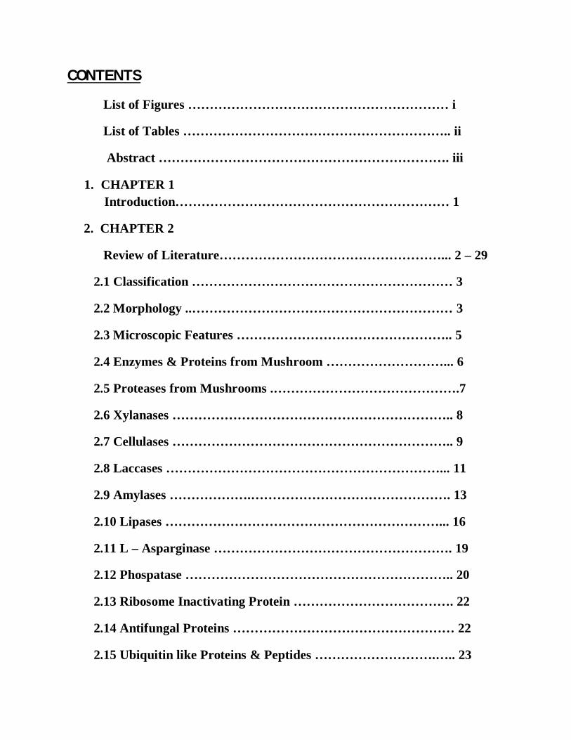
CONTENTS
List of Figures …………………………………………………… i
List of Tables …………………………………………………….. ii
Abstract …………………………………………………………. iii
1. CHAPTER 1 Introduction……………………………………………………… 1
2. CHAPTER 2
Review of Literature……………………………………………... 2 – 29
2.1 Classification …………………………………………………… 3
2.2 Morphology ..…………………………………………………… 3
2.3 Microscopic Features ………………………………………….. 5
2.4 Enzymes & Proteins from Mushroom ………………………... 6
2.5 Proteases from Mushrooms .…………………………………….7
2.6 Xylanases ……………………………………………………….. 8
2.7 Cellulases ……………………………………………………….. 9
2.8 Laccases ………………………………………………………... 11
2.9 Amylases ……………….………………………………………. 13
2.10 Lipases ………………………………………………………... 16
2.11 L – Asparginase ………………………………………………. 19
2.12 Phospatase …………………………………………………….. 20
2.13 Ribosome Inactivating Protein ………………………………. 22
2.14 Antifungal Proteins …………………………………………… 22
2.15 Ubiquitin like Proteins & Peptides ……………………….….. 23

2.16 Edible Mushrooms ……………………………………………. 25
2.17 Medicinal Mushrooms ………………………………………… 28
3. CHAPTER 3
Materials & Method ………………………………………….. 30 – 35
3.1 Mushroom Strains …………………………………………… 30
3.2 Preparation of Mycelial Cultures ………………………….... 30
3.3 Enzymatic Screening and Media Compositions ……………. 30
3.4 Estimation of Enzymes ……………………………………….. 33
4. CHAPTER 4
Results & Discussion …………………………………………… 36 – 46
4.1 Culture of Mushroom Mycelium ……………………………. 36
4.2 Screening of Potential Strain with Enzymatic Activity…….. 38
4.3 Estimation & Selection of Strain with Enzymatic Activity… 44
5. CHAPTER 5
Conclusion ………………………………………………………. 47
6. CHAPTER 6
References ……………………………………………………... 48 - 52

i
LIST OF FIGURES
1. The life cycle of a typical soil basidiomycete …………………………………………… 2
2. Asci – under Atomic Force Microscope ……………………………………………. 6
3. Spores – under Scanning Electron Microscope …………………………………….. 6
4. 3D – presentation of molecular structure of α – Amylases ………………………... 16
5. Pleurotus florida …………………………………………………………………………….. 25
6. Pleurotus sajor caju ………………………………………………………………………… 26
7. Volvariella volvaceae ………………………………………………………………………. 27
8. Ganoderma lucidium ………………………………………………………………. 28
9. Mycelial Culture of Volvariella volvaceae ……………………………………………… 36
10. Mycelial Culture of Ganoderma lucidium ………………………………………………. 36
11. Mycelial Cultures of Pleurotus sajor caju……………………………………………….. 37
12. Mycelial Cultures of Pleurotus florida ………………………………………………….. 37
13. Plate test for Amylase …………………………………………………………….. 39
14. Plate test for L – Asparginase …………………………………………………….. 40
15. Plate test for Xylanase ……………………………………………………………. 41
16. Plate test for Phosphate Solubalization …………………………………………… 42
17. Plate test for Lipase ……………………………………………………………….. 43

ii
LIST OF TABLES
1. Characteristics of Amylases produced by Fungi ………………………………… 14
2. Some commercially available microbial Lipases ………………………………... 18
3. Industrial application of microbial Lipases ………………………………………. 19
4. Extracellular enzymes produced from Mushrooms ………………………………. 24
5. Therapeutic Effects and Bioactive Compounds of Ganoderma lucidum ………….. 29

iii
ABSTRACT
Mushrooms are widely spread saprophytic macro-organisms, belong to class fungi growing on
dead organic matter of vegetative origin. They can utilize almost all agricultural wastes as their
substrates for their growth and metabolism. During the growth of mushroom mycelia and their
development into mature fruiting bodies (or sporophores), various biochemical changes are
known to occur, as a result of which enzymes are secreted extracellularly to degrade the
insoluble materials to simple and soluble molecules. Consequently, enzymes play a significant
role in mushroom development; in addition, they also affect the food and nutrient value, flavour
and shelf life of these fungi. Pleurotus spp, Volvariella volvaceae and Ganoderma lucidium are
edible tropical fast-growing, wood degrading macro fungi that are of economic importance in
India as both the sporophores and tuberous sclerotia are not only edible but also are used in
medicinal preparations by native doctors. This work was undertaken to evaluate the enzymatic
activities of Pleurotus florida, Pleurotus sajor caju, Vovariella volvaceae and Ganoderma
lucidium and the effects of these enzymes on shelf life and food nutrients of these mushrooms.

Chapter 1
INTRODUCTION

CHAPTER 1 INTRODUCTION
NIT-ROURKELA, 2011 Page 1
INTRODUCTION
There are at least 12, 000 species of fungi that can be considered as mushrooms, with at least
2000 species are edible. Over 200 species have been collected from the wild vegetation and used
for various traditional medicinal purposes, mostly in Far East. About 35 species have been
cultivated commercially and 20 are cultivated on an industrial scale. The most cultivated
mushroom worldwide is A. bisporus (button mushroom), followed by Lentinus edodes (shiitake),
Pleurotus spp (oyster mushrooms), Auricula auricula (wood ear mushroom), Flamulina
velutipes (winter mushroom) and Volvariella volvacea (straw mushroom). The edible
mushrooms are excellent foods that can be incorporated into well-balanced diets due to their low
content of fat and energy, and high content of dietary fiber and functional compounds. Their
benefit to health include; immunomodulatory, anti-tumoral, and hypo-cholesterolemic effects. In
numerous molecules synthesized by macrofungi are known to be bioactive compounds such as
polysaccharides, glycoproteins, terpenoids, lectins, among others.
The vegetative growth of mycelium is essential for the subsequent development of the
mushroom and this mycelial growth utilizes lignocellulosic materials, such as the
polysaccharides of straws and wood, for nutrient materials. Since such substrates are commonly
insoluble in water, they are broken down to smaller, soluble units through the activity of
enzymes excreted by the fungal cells. An active area of modern research involves the search in
mushroom species for compounds that can be used in the treatment of various cancers,
cardiovascular disease, viral diseases, etc. During the growth of mushroom mycelia and the
development to mature fruitbodies (or sporophores), biochemical changes are known to occur, as
a result of which enzymes are secreted extracellularly to degrade the insoluble materials in the
substrates into simple and soluble molecules which are subsequently utilized by intracellular
enzymes within the mushroom. Consequently, enzymes play significant role in mushroom
development, in addition, they also affect the food nutrient, flavor and shelf life of these fungi.
This work was done to evaluate the effect of substrates on the enzyme activities of mushrooms,
Pleurotus spp, Vovariella volvaceae and Ganoderma lucidium and the effects of these enzymes
on shelf life and food nutrients of this mushroom.

Chapter 2
REVIEW OF LITERATURE

CHAPTER 2 REVIEW OF LITERATURE
NIT – ROURKELA, 2011 Page 2
REVIEW OF LITERATURE
Mushrooms are the fleshy, spore-bearing fruiting body of a fungus, typically produced above
ground on soil or on its food source. Like all fungi, mushrooms are not plants and do not
undergo photosynthesis. The word "mushroom" is most often applied to
Basidiomycetes, Agaricomycetes that have a stem (stipe), a cap (pileus), and gills (lamellae) or
pores on the underside of the cap. Basidiomycetes are named for their characteristic structure or
cell, the basidium, which is involved in sexual reproduction (fig 1). A basidium [Greek basidion,
small base] is produced at the tip of hyphae and normally is club shaped. Two or more
basidiospores are produced by the basidium, and basidia may be held within fruiting bodies
called basidiocarps.
Figure 1. The life cycle of a typical soil basidiomycete

CHAPTER 2 REVIEW OF LITERATURE
NIT – ROURKELA, 2011 Page 3
Classification Typical mushrooms are the fruit bodies of members of the order Agaricales, whose type
genus is Agaricus and type species is the field mushroom, Agaricus campestris. However, in
modern molecularly-defined classifications, not all members of the order Agaricales produce
mushroom fruit bodies, and many other gilled fungi, collectively called mushrooms, occur in
other orders of the class Agaricomycetes. For example, chanterelles are in the Cantharellales,
false chanterelles like Gomphus are in the Gomphales, milk mushrooms (Lactarius) and russulas
(Russula) as well as Lentinellus are in the Russulales, while the tough leathery
genera Lentinus and Panus are among the Polyporales, but Neolentinus is in the Gloeophyllales,
and the little pin-mushroom genus, Rickenella, along with similar genera, are in
the Hymenochaetales. Within the main body of mushrooms, in the Agaricales, are common fungi
like the common fairy-ring mushroom (Marasmius oreades), shiitake, enoki, oyster
mushrooms, fly agarics, and other amanitas, magic mushrooms like species of Psilocybe, paddy
straw mushrooms, shaggy manes, etc. An atypical mushroom is the lobster mushroom, which is a
deformed, cooked-lobster-colored parasitized fruitbody of a Russula or Lactarius, colored and
deformed by the mycoparasitic Ascomycete Hypomyces lactifluorum. (Volk T., 2001)
Other mushrooms are not gilled and then the term "mushroom" is loosely used, so it is difficult
to give a full account of their classifications. Some have pores underneath (and are usually
called boletes), others have spines, such as the hedgehog mushroom and other tooth fungi, and so
on. "Mushroom" has been used for polypores, puffballs, jelly fungi, coral fungi, bracket
fungi, stinkhorns, and cup fungi. Thus, the term is more one of common application
tomacroscopic fungal fruiting bodies than one having precise taxonomic meaning. There are
approximately 14,000 described species of mushrooms. (Miles PG, Chang S-T, 2004).
Morphology A mushroom develops from a nodule, or pinhead, less than two millimeters in diameter, called
a primordium, which is typically found on or near the surface of the substrate. It is formed within

CHAPTER 2 REVIEW OF LITERATURE
NIT – ROURKELA, 2011 Page 4
the mycelium, the mass of thread like hyphae that make up the fungus. The primordium enlarges
into a roundish structure of interwoven hyphae roughly resembling an egg, called a "button". The
button has a cottony roll of mycelium, the universal veil that surrounds the developing fruit
body. As the egg expands, the universal veil ruptures and may remain as a cup, or volva, at the
base of the stalk, or as warts or volval patches on the cap. Many mushrooms lack a universal veil
and therefore do not have either a volva or volval patches. Often there is a second layer of tissue,
the partial veil, covering the bladelike gills that bear spores. As the cap expands, the veil breaks,
and remnants of the partial veil may remain as a ring, or annulus, around the middle of the stalk
or as fragments hanging from the margin of the cap. The ring may be skirt-like as in some
species of Amanita, collar-like as in many species of Lepiota, or merely the faint remnants of a
cortina (a partial veil composed of filaments resembling a spiderweb), which is typical of the
genus Cortinarius. Mushrooms that lack a partial veil do not form an annulus. (Stuntz et al.,
1978) The stalk (also called the stipe, or stem) may be central and support the cap in the middle,
or it may be off-center and/or lateral, as in species of Pleurotusand panus. In other mushrooms, a
stalk may be absent, as in the polypores that form shelf-like brackets. Puffballs lack a stalk but
may have a supporting base. Other mushrooms, like truffles, jellies, earthstars, bird's nests,
usually do not have stalks, and a specialized mycological vocabulary exists to describe their
parts. The way that gills attach to the top of the stalk is an important feature of mushroom
morphology. Mushrooms in the genera Agaricus, Amanita, Lepiota and Pluteus, among others,
have free gills that do not extend to the top of the stalk. Others have decurrent gills that extend
down the stalk, as in the genera Omphalotus and Pleurotus. There are a great number of
variations between the extremes of free and decurrent, collectively called attached gills. Finer
distinctions are often made to distinguish the types of attached gills: adnate gills, which adjoin
squarely to the stalk; notched gills, which are notched where they join the top of the stalk;
adnexed gills, which curve upward to meet the stalk, and so on. These distinctions between
attached gills are sometimes difficult to interpret, since gill attachment may change as the
mushroom matures, or with different environmental conditions. (Stuntz et al., 1978)
CHAPTER 2 REVIEW OF LITERATURE
NIT – ROURKELA, 2011 Page 5
Microscopic features
A hymenium is a layer of microscopic spore-bearing cells that covers the surface of gills. In the
non-gilled mushrooms, the hymenium lines the inner surfaces of the tubes of boletes and
polypores, or covers the teeth of spine fungi and the branches of corals. In the Ascomycota,
spores develop within a microscopic elongated, saclike cell called an ascus, which typically
contains eight spores. The Discomycetes—which contains the cup, sponge, brain, and some club-
like fungi—develop an exposed layer of asci, as on the inner surface of cup fungi or within the
pits of morels. The Pyrenomycetes, tiny dark-colored fungi that live on a wide range of
substrates including soil, dung, leaf litter, decaying wood, as well as other fungi, produce minute
flask-shaped structures called perithecia, within which the asci develop (Ammirati et al., 1985).
In the Basidiomycetes, usually four spores develop on the tips of thin projections
called sterigmata, which extend from a club-shaped cell called a basidium. The fertile portion of
the Gasteromycetes, called a gleba, may become powdery as in the puffballs or slimy as in
thestinkhorns. Interspersed among the asci are threadlike sterile cells called paraphyses. Similar
structures called cystidia often occur within the hymenium of the Basidiomycota. Many types of
cystidia exist and assessing their presence, shape, and size is often used to verify the
identification of a mushroom (Ammirati et al., 1985). The most important microscopic feature
for identification of mushrooms is the spores themselves. Their color, shape, size, attachment,
ornamentation, and reaction to chemical tests often can be the crux of identification. Spores often
have a protrusion at one end, called an apiculus, which is the point of attachment to the basidium,
termed the apical germ pore, from which the hypha emerges when the spore germinates.
(Ammirati et al., 1985)

CHAPTER 2 REVIEW OF LITERATURE
NIT – ROURKELA, 2011 Page 6
Figure 2. Figure 3. Figure No. 2. Asci as viewed under atomic force microscopy and 3. Spores as viewed under
Scanning electron microscope.
Mushrooms affect humans in many ways. Most are saprophytes that decompose plant debris,
especially cellulose and lignin. Many mushrooms are used as food throughout the world. Many
mushrooms produce specific alkaloids that act as either poisons or hallucinogens. One such
example is the “destroying angel” mushroom, Amanita phalloides. Two toxins isolated from this
species are phalloidin and amanitin. Phalloidin primarily attacks liver cells where it binds to
plasma membranes, causing them to rupture and leak their contents. Alpha-amanitin attacks the
cells lining the stomach and small intestine and is responsible for the severe gastrointestinal
symptoms associated with mushroom poisoning. Mushrooms can be used for dyeing wool and
other natural fibers. The chromophores of mushroom dyes are organic compounds and produce
strong and vivid colors, and all colors of the spectrum can be achieved with mushroom dyes.
Before the invention of synthetic dyes mushrooms were the source of many textile dyes.
(Mussak R, Bechtold T., 2009) Mushrooms and other fungi play a role in the development of
new biological remediation techniques (e.g., using mycorrhizae to spur plant growth) and
filtration technologies (e.g. using fungi to lower bacteria levels in contaminated water).
Enzymes and Proteins from Mushrooms
Mushrooms are saprophytic, growing on dead organic matter of vegetative origin. Therefore they
can utilize almost all agricultural wastes as substrates (Miles and Chang, 1997). During the

CHAPTER 2 REVIEW OF LITERATURE
NIT – ROURKELA, 2011 Page 7
growth of mushroom mycelia and the development to mature fruitbodies (or sporophores),
biochemical changes are known to occur, as a result of which enzymes are secreted
extracellularly to degrade the insoluble materials in the substrates into simple and soluble
molecules which are subsequently utilized by intracellular enzymes within the mushroom.
Consequently, enzymes play significant role in mushroom development; in addition, they also
affect the food nutrient, flavour and shelf life of these fungi (Baardseth, 1979; Paranjpe and
Chen, 1979; Wang, 1989; Zadrazil et al., 2004). A variety of proteins with interesting biological
actions is elaborated by mushrooms and many of these proteins have potentially applicable
activities. They include ribosome inactivating proteins, antifungal proteins, ribonucleases,
ubiquitin- like proteins and peptides, lectins, cellulases, xylanases, laccases, invertases and
phosphatases.
Proteases from Mushrooms Pleureryn is a small and novel aspartic protease with a molecular mass of 11.5 kDa from the
edible mushroom P. eryngii (Wang HX, Ng TB., 2001). However, its N-terminal sequence
resembles DNA replication licensing factor more than aspartic proteases. It also exhibits some
inhibitory activity against HIV-1 reverse transcriptase. This is reminiscent of a suppressive
action of HIV-1 protease, also an aspartic protease, on its homologous reverse transcriptase.
Pleureryn exhibits a pH optimum of 5 and a temperature optimum of 45o C, with considerable
activity remaining at high temperatures and at pH 4 and 12. Pleureryn is unique in that it is
relatively stable to changes in pH or temperature (Wang HX, Ng TB., 2001). Other mushroom
proteases tend to be less stable. The metalloproteases from Armillariella mellea and Tricholoma
saponaceum (Kim JH, Kim YS., 1999, 2001) are, by contrast, thermolabile. An aspartic protease
from another mushroom, I. lacteus, has a molecular mass of 35 kDa (Kobayashi H., et al 1989).
Metalloendopeptidases from A. mellea, P. ostreatus and T. saponaceum have a molecular mass
in the range 18.5 – 20 kDa (Nonaka T., et al 1995). A thermostable lysine-specific zinc-
metalloendopeptidase has been isolated from fruiting bodies of the mushroom Grifola frondosa
(Nonaka T., et al 1995). The protease has a molecular mass of 20 kDa, a pI of 7.46 and a pH
optimum of 9–10. It demonstrates high affinity for β -d-glucan and chitin. Prolylendopeptidases

CHAPTER 2 REVIEW OF LITERATURE
NIT – ROURKELA, 2011 Page 8
from A. bisporus and Lyophyllum cinerascens have molecular masses close to 78 kDa (Sattar
AKMA., et al 1990, 1990). An alkaline serine protease (Pen ch 13, also known as Renn 13) has
been identified as the major allergen from airborne Penicillium chrysogenum (P. notatum). It has
a molecular mass of 28 kDa. It exhibits 83 and 49% amino acid sequence identity with its
counterparts from P. citrinum and Aspergillus fumigatus, respectively (Chou H., et al 2001). An
alkaline serine proteinase with a molecular mass of 28.7 kDa has been isolated from Fusarium
culmorum (Pekkarinen AI, et al 2002). A pH of 8.3–9.6 and a temperature of 500 C are required
for its maximal activity. A subtilisin-like serine protease with a molecular mass of 36 kDa has
been purified from Podospora anserine (Paoletti M., 2001). An aspartic proteinase from A.
fumigatus demonstrates a molecular mass of 39 kDa and a broad range of activity from pH 2.0 to
7.0 (Reichard U., et al 2001). It exhibits 88% sequence identity with aspartic proteinase from A.
niger and 64% identity with the vacuolar proteinase A of S. cerevisiae. A protease with a
molecular mass of about 30 kDa has been isolated from Candida caseinolytica (Poza M., et al
2001). Its action is demonstrable over a broad pH range. In summary, proteases with different
molecular masses, optimum pH values and optimum temperatures are produced by different
fungal species.
Xylanases Xylanases (1,4-β-d-xylan xylanohydrolases) catalyze the random hydrolysis of the xylan
backbone of heteroxylans. As a result the cellulose fibrils are exposed and susceptible to attack
of side-chain cleaving enzymes such as α –arabinofuranosidases and acetylxylanases. Xylanases
occur in diverse organisms. Bacterial and fungal xylanases are produced inductively or
constitutively in response to the carbon source on which they are grown. A cellulose-free
xylanase has been purified from Aspergillus niger (Qy Y, Gao P, et al., 1996). Maximum
xylanase activity is induced by xylan, followed by lignocellulose. The enzyme exhibits the
highest activity at 45–500 C, but 70–95% of the activity disappears within 5 min. Cellulase free
xylan degrading enzymes from Acrophialophora nainiana, Humicola grisea var. thermoides and
two Trichoderma harzianum strains have been employed to bleach the pulp of Eucalyptus kraft
before a chlorine dioxide and alkaline bleaching sequence. The T. harzianum enzyme

CHAPTER 2 REVIEW OF LITERATURE
NIT – ROURKELA, 2011 Page 9
preparations are slightly more effective in decreasing pulp viscosity and chlorine chemical
consumption and enhancing the brightness of the kraft pulp. A. nainiana xylanase is the most
potent in reducing pulp viscosity (Medeiros RG, et al., 2002). The activities of some hydrolytic
enzymes (cellulase, endo-1,4-β-xylanase, β-glucosidase and amylase) and reductase enzymes
(monophenol monooxygenase and peroxidase) in strains of Fusarium oxysporum (Schlecht snyd.
and Hans) isolated from different habitats (plant substrates, cultivated soil and non-cultivated
soil) have been examined by Kurchenko et al. (Kruger et al., 2002). It is found that strains
isolated from plant substrates display the highest activity of hydrolytic enzymes, followed by
strains from cultivated soil, whereas strains isolated from non cultivated soil exhibit the lowest
activity. The reverse trend occurs regarding the redox enzymes. A purified xylanase from F.
verticillioides exhibits a molecular mass of 24 kDa, an optimum temperature of 500C, an
optimum pH of 5.5, a pH stability range of 4.0–9.5, and thermal stability up to 500C (Saha BC.,
2001). F. oxysporum f. sp. melonis produces an endo-1,4-β- xylanase with a molecular mass of
80 kDa. Its optimum pH and temperature are 5.0 and 500C, respectively (Alconada TM, Martinez
MJ, 1994). P. purpurogenum produces two immunologically distinct xylanases. Xylanase A
possesses a molecular mass of 33 kDa and an isoelectric point at pH 8.6. Xylanase B, the other
major form, manifests a molecular mass of 23 kDa and an isoelectric point at pH 5.9 (Trinci APJ
et al., 1994). The xylanase from the thermophilic fungus Humicola lanuginose is abundant in
acidic amino acids. It is unusual in that inactivation, probably due to aggregation, occurs after
storage at −200 C in the dry state for over 2 months (Anand L et al., 1990). From another
thermophilic fungus, Thermoascus aurantiacus, a xylanase, a glucosidase, an exocellulase and
an endocellulase have been purified. Two structurally similar xylanases from T. reesei and their
genes have been characterized (Torronen A et al., 1992). They have a molecular mass of 19 and
21 kDa and an isoelectric point of 5.2 and 9.0, respectively. A 66-kDa xylanase has been purified
from the rumen anaerobic fungus, Neocallimastix patriciarum. The large N-terminal reiterated
regions consisted of distinct catalytic domains which displayed similar substrate specificities to
the full-length enzyme (Gilbert HJ et al., 1992). Thus it appears that xylanes from different
species may differ in molecular mass, isoelectric point, optimum pH, pH stability range and
optimum temperature

CHAPTER 2 REVIEW OF LITERATURE
NIT – ROURKELA, 2011 Page 10
Cellulases
The cellulolytic enzymes of anaerobic fungi have been studied because of their potential value in
biotechnology (Gilbert HJ, 1992; Wallace RJ, 1994;) including use as enzyme supplements for
live stock, and in food and beverage, detergent, textile and pulp and paper industries (Campbell
GL, 1992). Their nutritional function is evident by the degradation of plant fiber serving as
carbon sources. In general, cellulolytic fungi produce a large number of cellulases (Slomczynsky
D et al., 1995); many use them for degradation of the plant cell wall polysaccharides (Akin DE
et al., 1990). Endoglucanases (endo-1,4-β-glucanases), cellobiohydrolases (CBH, exo-1,4-β-
glucanase), and β-glucosidases are three major types of cellulolytic enzymes. Endoglucanases
randomly hydrolyze 1,4-β- bonds along the interior of the cellulose chain. Cellobiohydrolases
cleave cellobiosyl units from non-reducing ends of the cellulose chains. Glucosidases cleave
glucosyl units from non-reducing ends of cello-oligosaccharides. Cellobiohydrolase Cel7A from
Trichoderma reesei has been expressed from Pichia pastoris. The thermostability, k(cat), Km
and pH optimum are not affected by heteroglycosylation (Boer H et al., 2000). A cellulase, with
a molecular mass of 58 kDa, a pH optimum of 5.5, and a temperature optimum at 400 C has been
purified from the ruminal fungus O. joyonii and cloned in Escherichia coli. The O. joyonii
cellulase exhibits strong activity on carboxy methyl cellulose (CMC), lichenan and barley β-
glucan. CMC is a water soluble long-chained cellulose with carboxymethyl substitutions. It is
commonly used as a model substrate for detecting β-1,4-endoglucanases. Digestion of lichenan
and barley β-glucans (mixtures of β-1,3- and β-1,4 linkages) may be mainly attributed to random
cleavage of β-1,4 linkages in the substrates because of the inability of the O. joyonii cellulase to
digest laminarin and pachyman which have β-1,3-glucans as the main components. The enzyme
has no activity on avicel (crystalline cellulose) and pullulan (α-1,6 glucan). O. joyonii cellulose is
able to cleave p-nitrophenyl-β-d-cellobioside but not glucopyranoside, suggesting that it
possesses cellobiohydrolase but not glucosidase activity. Its activity on p-nitrophenyl-β-d-
cellotrioside, -cellotraoside and -cellopentaoside indicates its cellodextrinase activity. The
enzyme has activity over a broad pH range (pH 5–7) with the highest activity at pH 5.5. It is
stable at temperatures up to 500 C with a temperature optimum of 400 C. However, it has to be
borne in mind that all of the enzyme activities were measured under anaerobic conditions in this

CHAPTER 2 REVIEW OF LITERATURE
NIT – ROURKELA, 2011 Page 11
investigation and the results might have been different had the experiments been conducted in an
anaerobic setting [200]. Despite the fact that many cellulases contain a cellulosebinding domain
and a catalytic domain, O. joyonii cellulose is unable to bind Avicel, a microcrystalline cellulose
and a search for cellulose-binding domain sequences has met with no success. The absence of
cellulose-binding domains and the presence of reiterated scaffold binding sequences in O. joyonii
CelB2 cellulase suggest the immobilization of the enzyme to cellulosome, a cellulose hydrolytic
complex. Cellulases, glucanases and xylanases from anaerobic rumen fungi have been cloned in
E. coli and expressed. These fungal enzymes increase the efficiency of feedstuff digestion in
monogastric animals by promoting breakdown of polymers in the plant cell wall and hence are
potential enzyme supplements for livestock. The enzymes are being used or considered for use
by pulp and paper, textile, detergent and food and beverage industries. Three exo-glucanases,
two endo-glucanases and two beta-glucosidases, have been isolated from the culture medium of
A. nidulans. The optimal pH for all forms of cellulase components ranges from pH 5.0 to 6.0 and
the optimum temperature is 50 and 650 C for exo-glucanases and endo-glucanases but 35 and 65 0 C for beta-glucosidases. All cellulase components are stable for 10 min at 40–500 C. Exo-II and
Exo-III exhibit a higher affinity for the substrate than Exo-1. The Km values of Endo-1 and
Endo-II and their maximum reaction velocities are comparable. The beta-glucosidases exhibit
Km values of 0.24 and 0.12 mmol and Vmax values of 8.00 and 0.67 IU/mg protein. The
molecular masses for various enzyme forms are: Exo-1, 29 kDa; Exo-II, 72.5 kDa; Exo-III, 138
kDa; Endo-1, 25 kDa; Endo-II, 32 kDa; beta-Gluco-1, 14 kDa and beta-Gluco-II, 26 kDa. Exo-
and endo-glucanases but not beta-glucosidases require metal ions as co-factors. Hg2+ ions inhibit
the activity of all cellulase components (Bagga PS et al., 1990). In summary, cellulases with a
variety of molecular masses, and temperature and pH optima are known.
Laccases
Lignin ranks second, after cellulose, in abundance in the biosphere as a renewable organic
compound. The biodegradation of lignin is a rate-limiting step in the carbon cycle. Ligninolytic
enzymes are highly non-specific on account of the complex structure of lignin, and can be used
in the degradation of structurally different environmental pollutants (Bumpus JA, Aust SD,
1987). Laccases (benzenediol: oxygen oxidoreductase) form a class of ligninolytic enzymes that
CHAPTER 2 REVIEW OF LITERATURE
NIT – ROURKELA, 2011 Page 12
are phenol oxidases capable of catalyzing one-electron oxidation of aromatic substrates and the
concomitant reduction of oxygen to water. Laccases can also act on non-phenolic lignin subunits
in the presence of readily oxidizable primary substrates which are electron-transfer mediators
(Bourbonnais R, Paice MG, 1990). Combinations of laccase/glucose oxidase and laccase/
manganese peroxidase, rather than laccase or peroxidase alone, have been considered to be the
minimal enzyme requirement for effective lignin degradation. The combined action of laccase
and FAD-dependent aryl alcohol oxidase significantly reduces the molecular mass of soluble
lignosulfonates. Laccases are multicopper blue oxidases, widely distributed in plants and fungi.
These enzymes are either monomeric or multimeric glycoproteins. Heterogeneity may be present
owing due to variable carbohydrate contents or differences in copper content. These enzymes
demonstrate a rather low degree of specificity with regard to the reducing substrate: they catalyze
the oxidation of orthoand para-diphenols and aromatic amines by removing an electron and a
proton from a hydroxyl group to generate a free radical. Structural information about the metal
sites of laccase has been gathered by spectroscopic studies. The biological function of laccase is
correlated to lignin biodegradation in combination with either manganese peroxidase and/or
lignin peroxidase. Laccase can also catalyze the oxidative polymerization of the phenolic
compounds originating from lignin, which are then easily eliminated. Laccases oxidize phenolic
units in lignin to phenoxy radicals, which can lead to the degradation of lignin-related structures.
In the presence of appropriate redox mediators, such as 2,2-azino-bis(3-ethylbenzothiazoline-6-
sulfonic acid) (ABTS) and 1 hydroxybenzotriazole (1-HBT), laccases catalyze the oxidation of
nonphenolic lignin model compounds, depolymerize kraft lignin, and degrade polycyclic
aromatic hydrocarbons, which are not substrates for laccase alone. Due to the aforementioned
characteristics, its efficacy as an agent for selected detoxification (Dec J et al., 2003), pollutant
degradation, and catalyst for regiospecific biotransformation, and its possible utilization as
electrode for organic phase enzymatic assay, laccase may play an important role for
biotechnological applications. Veratryl alcohol oxidase can act together with laccase to prevent
polymerization of phenolic compounds and reduce molecular mass of lignosulfates (Marzullo L
et al., 1995). Extracellular laccases have been purified from submerged cultures of Coriolus
versicolor, Panus tigrinus, Phlebia radiata and Phlebia tremellosa, and from cultures of P.

CHAPTER 2 REVIEW OF LITERATURE
NIT – ROURKELA, 2011 Page 13
tigrinus, P. radiata and A. bisporus grown on wheat straw (solid-state fermentation). A laccase
from Marasmius quercophilus has been characterized, in addition to laccases from A. bisporus,
Polyporus anceps, Pycnoporus cinnabarinus. Rigidoporus lignosus. Trametes trogii and C.
hirsutus. They are glycoproteins with molecular masses close to 60 kDa. However, Tricholoma
giganteum laccase has a molecular mass of only 43 kDa while C. cibarius laccase is composed
of two 46-kDa subunits. Laccases have also been identified and characterized from A. nidulans,
Botrytis cinerea, and N. crassa. A laccase gene homologous to the laccase gene of N. crassa has
been isolated and characterized from P. anserine. The promoter region of the laccase gene from
P. anserine contains two sequences identical to the eukaryotic xenobiotic responsive element and
another two sequences homologous to the eukaryotic antioxidant responsive element (Fernandez-
Larrea J et al., 1996). The activity and stability of laccase from Pleurotus ostreatus are enhanced
by copper but reduced by mercury. Laccases from different strains of P. ostreatus differ in their
Kcat and Km values for springaladazine, ABTS and guaiacol. Many fungal species examined
secrete more than one laccase isoenzyme. Different conditions of growth may produce different
patterns of isoenzymes.
Amylases
Enzymes that participate in the hydrolytic degradation of starch are collectively referred to as
amylolytic enzymes or amylases. Specific enzymes classified within this group include α
amylase, β-amylase, gluco-amylase (also known as amyloglucosidase), pullulanase and inso
amylase. Amylases are, classified into two categories, endoamylases and exoamylases (Gupta et
al., 2002). Endoamylases catalyse hydrolysis in a random manner in the interior of the starch
molecule. This action causes the formation of linear and branched oligosaccharides of various
chain lengths. Exoamylases hydrolyse from the non-reducing end, successfully resulting in short
end products. A large array of amylases, are involved in the complete breakdown of starch.
Enzymatic degradation of starch yields glucose, maltose and other low molecular weight sugars.
Also, enzymatically - mediated isomerisation of glucose yields high-fructose syrups. Abundant
supplies of starch may be obtained from seeds and tubers, such as corn, wheat, rice tapioca and

CHAPTER 2 REVIEW OF LITERATURE
NIT – ROURKELA, 2011 Page 14
potato. The widespread availability of starch from such inexpensive sources, coupled with large-
scale production of amylolytic enzymes, facilitates the production of syrups containing glucose,
fructose or maltose, which are of considerable importance in the food and confectionery
industry. Furthermore, they may be produced quite competitively when compared with the
production of sucrose, which is obtained directly from traditional sources such as sugar-beet or
sugar-cane (Gupta et al., 2002). Starch may be hydrolysed by chemical or enzymatic means.
Table 1. Characteristics of amylases produced by fungi

CHAPTER 2 REVIEW OF LITERATURE
NIT – ROURKELA, 2011 Page 15
α –Amylases α-Amylase activity is widely distributed in nature. α-Amylase is an endo-acting enzyme,
catalyzing the random hydrolysis of internal α-1,4 glycosidic linkages present in the starch
substrate. However, α - amylases which are in most demand hydrolyses the α- 1,4 glycosidic
bond in the interior of the molecule (Gupta et al., 2002). These enzymes are incapable of
hydrolyzing α-1,6 glycosidic linkages present at branch points of amylopectin chains. One
exception to this is the α-amylase produced by Thermactinomyces vulgaris, which can hydrolyse
both α-1-6 and α-1-4 glycosidic linkages. The α-amylase family consists of a large group of
starch hydrolases and related enzymes, currently known as glycosyl hydrolases family 13
(Henrissat, 1991). Thermostable α-amylases have been characterised from Pyrococcus woesei,
Pyrococcus furiosus (Koch et al., 1991) and Thermococcus profundus (Chung et al., 1995; Kwak
et al., 1998; and Lee et al., 1996). The optimum activity of these enzymes is 1000 C and 800 C
respectively. The gene encoding an extracellular α-amylase from P. Furiosus has been recently
cloned and the recombinant enzyme expressed in Bacillus subtilis and in E. coli (Dong et al.,
1997; Jorgenesn et al., 1997). The high thermostability of the pyrococcal extracellular α-amylase
(thermal activity) even at 1300 C in the absence of metal ions, together with its unique product
pattern and substrate specificity, makes this enzyme an interesting candidate for industrial
application (Niehaus et al., 1999).
Two of the more commonly used bacterial α-amylases are those isolated from Bacillus
amyloliquefaciens and Bacillus licheniformis. Bacillus amylases exhibit a pH optimum close to
neutrality and are stabilized by the presence of calcium ions. α-Amylase produced by Bacillus
licheniformis is particularly suited to industrial applications because of its thermal stability. This
enzyme consists of 483 amino acids and has a molecular weight of 55.2 kDa. Its pH optimum is
6.0 and its temperature optimum is 90oC. Most other α-amylases, including those produced by B.
amyloliquefaciens, are rapidly inactivated at temperatures above 40oC (Niehaus et al., 1999).
Several thermostable α-amylases have already been characterised (Koch et al., 1991). The most
thermostable α-amylase to date is from Pyrococcus woesei. It remained active after autoclaving
for 4 h at 1200 C (Antranikian, 1987).

CHAPTER 2 REVIEW OF LITERATURE
NIT – ROURKELA, 2011 Page 16
Figure 4. 3-Dimensional representation of the molecular structure of α-amylase
Lipases
Lipases (triacylglycerol acylhydrolases, E.C. 3.1.1.3) are ubiquitous enzymes of considerable
physiological significance and industrial potential. Lipases catalyze the hydrolysis of
triacylglycerols to glycerol and free fatty acids. In contrast to esterases, lipases are activated only
when adsorbed to an oil–water interface (Martinelle et al., 1995) and do not hydrolyze dissolved
substrates in the bulk fluid. A true lipase will split emulsified esters of glycerine and long-chain
fatty acids such as triolein and tripalmitin. Lipases are serine hydrolases. Lipases display little
activity in aqueous solutions containing soluble substrates. In contrast, esterases show normal
Michaelis–Menten kinetics in solution. In eukaryotes, lipases are involved in various stages of
lipid metabolism including fat digestion, absorption, reconstitution, and lipoprotein metabolism.
In plants, lipases are found in energy reserve tissues. How lipases and lipids interact at the
interface is still not entirely clear and is a subject of intense investigation (Balashev et al., 2001).
Because of their wide-ranging significance, lipases remain a subject of intensive study
(Alberghina et al., 1991; Bornscheuer, 2000). Research on lipases is focussed particularly on
structural characterization, elucidation of mechanism of action, kinetics, sequencing and cloning
of lipase genes, and general characterization of performance (Alberghina et al., 1991;
Bornscheuer, 2000). In comparison with this effort, relatively little work has been done on

CHAPTER 2 REVIEW OF LITERATURE
NIT – ROURKELA, 2011 Page 17
development of robust lipase bioreactor systems for commercial use. Commercially useful
lipases are usually obtained from microorganisms that produce a wide variety of extracellular
lipases. Many lipases are active in organic solvents where they catalyze a number of useful
reactions including esterification (Chowdary et al., 2001; Hamsaveni et al., 2001; Kiran et al.,
2001a; Kiyota et al., 2001; Krishna and Karanth, 2001; Krishna et al., 2001; Rao and Divakar,
2001), transesterification, regioselective acylation of glycols and menthols, and synthesis of
peptides (Ducret et al., 1998; Zhang et al., 2001) and other chemicals (Therisod and Klibanov,
1987; Weber et al., 1999; Bornscheuer, 2000; Berglund and Hutt, 2000; Liese et al., 2000; Azim
et al., 2001). The expectation is that lipases will be as important industrially in the future as the
proteases and carbohydrases are currently.
Lipases find promising applications in organic chemical processing, detergent formulations,
synthesis of biosurfactants, the oleochemical industry, the dairy industry, the agrochemical
industry, paper manufacture, nutrition, cosmetics, and pharmaceutical processing. Development
of lipase-based technologies for the synthesis of novel compounds is rapidly expanding the uses
of these enzymes (Liese et al., 2000). One limiting factor is a shortage of lipases having the
specific required processing characteristics. An increasing number of lipases with suitable
properties are becoming available and efforts are underway to commercialize biotransformation
and syntheses based on lipases (Liese et al., 2000). The major commercial application for
hydrolytic lipases is their use in laundry detergents. Detergent enzymes make up nearly 32% of
the total lipase sales. Lipase for use in detergents needs to be thermostable and remain active in
the alkaline environment of a typical machine wash. An estimated 1000 tons of lipases are added
to approximately 13 billion tons of detergents produced each year (Jaeger and Reetz, 1998).
Lesser amounts of lipases are used in oleochemical transformations (Bornscheuer, 2000).
Lipases can play an important role in the processing of g-linolenic acid, a polyunsaturated fatty
acid (PUFA); astaxanthine, a food colorant; methyl ketones, flavor molecules characteristic of
blue cheese; 4-hydroxydecanoic acid used as a precursor of g-decalactone, a fruit flavor;
dicarboxylic acids for use as prepolymers; interesterification of cheaper glycerides to more

CHAPTER 2 REVIEW OF LITERATURE
NIT – ROURKELA, 2011 Page 18
valuable forms (e.g., cocoa butter replacements for use in chocolate manufacture) (Undurraga et
al., 2001); modification of vegetable oils at position 2 of the triglyceride, to obtain fats similar to
human milkfat for use in baby feeds; lipid esters including isopropyl myristate, for use in
cosmetics; and monoglycerides for use as emulsifiers in food and pharmaceutical applications.
The increasing awareness of the importance of chirality in the context of biological activity has
stimulated a growing demand for efficient methods for industrial synthesis of pure enantiomers
including chiral anti - inflammatory drugs such as naproxen (Xin et al., 2001) and ibuprofen (Lee
et al., 1995; Ducret et al., 1998; Xie et al., 1998; Arroyo et al., 1999; Chen and Tsai, 2000);
antihypertensive agents such as angiotensin-converting enzyme (ACE) inhibitors (e.g., captopril,
enalapril, ceranopril, zofenapril, and lisinopril); and the calcium channel blocking drugs such as
diltiazem. Lipases are used in synthesis of these drugs (Berglund and Hutt, 2000).
Applications of Lipases Lipases are widely used in the processing of fats and oils, detergents and degreasing
formulations, food processing, the synthesis of fine chemicals and pharmaceuticals, paper
manufacture, and production of cosmetics, and pharmaceuticals (Rubin and Dennis, 1997a,b;
Kazlauskas and Bornscheuer, 1998). Lipase can be used to accelerate the degradation of fatty
waste (Masse et al., 2001) and polyurethane (Takamoto et al., 2001). Major applications of
lipases are summarized in Table 2. Most of the industrial microbial lipases are derived from
fungi and bacteria (Table 3).
Table 2: Some commercially available microbial lipases (Jaeger and Reetz, 1998)

CHAPTER 2 REVIEW OF LITERATURE
NIT – ROURKELA, 2011 Page 19
Table 3: Industrial applications of microbial Lipases (Vulfson, 1994)
L – Asparginase
L-Asparaginase (L-asparagine amidohydrolase, EC 3.5.1.1) is an enzyme that primarily catalyzes
the conversion of L-asparagine to L-aspartic acid and ammonia. Arima et al., showed that a
number of bacteria and fungi produced true extracellular asparaginase activity, i.e., the active
enzyme could be isolated from culture filtrates. A discovery that the anti-leukemic activity of
guinea pig serum is associated with its L-asparaginase activity (Broome JD; 1961, 1968) and
isolation of this enzyme from Escherichia coli (Mashburn, L. T., 1964) brought considerable
attention to bacterial L-asparaginases, leading to identification of related enzymes in several
other sources. The basis of their clinical activity is attributed to the reduction of circulating L-
asparagine in blood. Since some neoplastic cells depend on extracellular supplies of this amino
acid, they are selectively killed by L-asparagine deprivation. It was shown that some of the
CHAPTER 2 REVIEW OF LITERATURE
NIT – ROURKELA, 2011 Page 20
vaccine preparations had high L-asparaginase activity and this enzyme has been used for their
potency control during the production process (Beumer-Jachmans, M. P. 1973). The therapeutic
effect of vaccines used for immunotherapy of cancers such as lymphoblastic leukemia and breast
cancer (Hortobagyi, G. N, et al., 1980) was attributed to their asparaginase activity. L-
Asparaginases, includes the enzymes that primarily utilize L-asparagine as a substrate. The
enzymes belonging to the second class, also referred to as glutaminase asparaginases, catalyze
the hydrolysis of both L-asparagine and L-glutamine with comparable efficiency. Although the
anti-cancer properties were demonstrated for enzymes belonging to both classes, their practical
applications were highly restricted by the side effects associated with therapy. Thus, antitumor
activity of L - Asparginase demonstrated in studies with mice was accompanied by a variety of
side effects, linked (at least partially) to the L-glutaminase activity of this enzyme. L-
asparaginases, with their high specificity for L-asparagine and low-to-negligible activity against
L-glutamine, are reported to be less troublesome during the course of anti-cancer therapy.
Numerous studies of L asparaginases have been conducted in order to understand the catalytic
mechanism and the substrate specificity of these enzymes. Studies of the dependence of activity
on pH, conducted for various Lasparaginases, confirmed that the enzymes are stable and active
in the pH range of 4-9 (Wehner et al., 1992; Distasio J.A et al., 1976). It has been shown that the
enzymatic reaction proceeds according to a two step ping-pong mechanism similar to the
mechanism of serine proteases, except that the attacking nucleophile is a threonine. Two
threonine residues, Thr15ErA and Thr95ErA, are located in the active site of L-asparaginase.
The crystal structure of the acyl-enzyme intermediate of Thr89ValEcA mutant provided the first
experimental evidence that Thr15ErA is the nucleophile. The specificity toward a variety of
substrates of L-asparaginases was assessed in multiple kinetic studies.
Phosphatase Phosphate solubilizing microorganisms (PSMs) play an important role in supplementing
phosphorus to the plants, allowing a sustainable use of phosphate fertilizers. Application of
PSMs in the field has been reported to increase crop yield. Several mechanisms like lowering of
pH by acid production, ion chelation and exchange reactions in the growth environment have

CHAPTER 2 REVIEW OF LITERATURE
NIT – ROURKELA, 2011 Page 21
been reported to play a role in phosphate solubilization by PSMs (Abd-Alla, 1994; Whitelaw,
2000). Species of Aspergillus, Penicillium and yeast have been widely reported solubilizing
various forms of inorganic phosphates (Whitelaw, 2000). Fungi have been reported to possess
greater ability to solubilize insoluble phosphate than bacteria (Nahas, 1996). In the present study
fungal strains having potential to solubilize insoluble phosphates were isolated. The fungal
isolates were checked for the ability to solubilize different insoluble phosphates. The major part
of soil P (sometimes as much as 90%) is sequestered in the organic compounds
phosphomonoesters and phosphodiesters (Nygren 2008). The phosphatase enzyme capabilities of
ectomycorrhizal fungi are continuously distributed between species rather than restricted to a
particular taxonomic group (Nygren 2008). Sheathing mycorrhizal fungi have been shown to
possess phosphatase enzymes which can hydrolyze inositol hexaphosphate.
Phosphatase production by basidiomycete fungi in liquid culture is independent of P in the
medium. Saprophytic basidiomycetes tend to incorporate hydrolysed phosphate into their
biomass. In contrast mycorrhizal fungi release more hydrolysed phosphate into solution than
they absorb (Dighton 1983). Fungi are able to secrete hydrolytic enzymes involved in the
degradation of organic matter (Abuzinadah & Read 1986, Burns & Dick 2002, Lindahl et al.
2005). Acid phosphatases solubilize insoluble forms of P not readily available to uninfected plant
roots (Tibbett et al. 1998a). These enzymes are generally bound to the outer cell walls (Rast et
al. 2003, Alvarez et al. 2004). Phosphatase activities of ectomycorrhizal fungi can vary between
species, resulting in different efficiency of P utilization of host plant (Ho & Zak 1979, Dighton
1983). These enzymes are in direct contact with soil environment but are able to adapt to various
soil conditions and maintain activity. However, soil components, pH and trace elements can
modify the conformation of enzymes and affect their activities (Eivazi & Tabatabai 1977, Geiger
et al. 1998). Activities of acid phosphatase are found to differ significantly amongst
ectomycorrhiza synthesized with different fungi and among different species of the same fungi
(Antibus et al. 1986, Buée et al. 2005, 2007, Courty et al. 2006). Ectomycorrhizal phosphatases
generally have a pH and temperature optimum approaching that of their native soil.

CHAPTER 2 REVIEW OF LITERATURE
NIT – ROURKELA, 2011 Page 22
Ribosome Inactivating Proteins from Mushrooms Ribosome-inactivating proteins (RIPs) are a group of proteins that share the property of
damaging ribosomes in an irreversible manner, acting catalytically, i.e. enzymatically. RIPs were
initially detected in plants, mostly in Angiopermae, both mono- and dicotyledons, and also in
mushrooms (Yao et al., 1998; Lam and Ng, 2001), and in an alga, Laminaria japonica (Liu et al.,
2002). Lyophyllum shimeij fruiting bodies, (lyophyllin), Volvariella volvacea fruiting bodies (V.
volvacea RIP) Other proteins described as type 1 RIPs are not included because of some there is
only the deposited amino acid sequence (euserratin from Euphorbia serrata, a ‘PAP’ from
Phytolacca acinosa) and of others, from mushroom fruiting bodies, there is no stringent evidence
for their RIP activity (pleuturegin from Pleurotus tuberregium, velutin, flammulin from
Flammulina velutipes, hypsin from Hypsizygus marmoreus). The properties of RIPs raised hopes
of utilizing them for various purposes. Possible applications in medicine and in agriculture were
envisaged. In medicine, they have been studied as immunotoxins or as antiviral agents, mainly
against HIV, with the difficulties outlined above. Possibly, realistic hopes might be their use for
the ex vivo purging of bone marrow or other cell suspensions and for the therapy of topical
tumours, for instance of bladder cancer, as suggested by in vitro studies (Thiesen et al., 1987;
Battelli et al., 1996) and initial clinical trials (Yu et al., 1998; Zang et al.,2000). In agriculture,
RIPs are tested to increase resistance against viruses and possibly other parasites. Again, their
toxicity to transfected plants is a limit to their use. Immunotoxins could be used for experimental
purposes, but, surprisingly, their use is still scarce, with the noticeable exceptions of the
immunotoxins against various cells of the nervous system. Probably their use in this field has
been greatly facilitated by the availability of the appropriate immunotoxins. Hopefully,
immunotoxins against other cells types will be prepared and used in other fields.
Antifungal Proteins from Mushrooms Proteins with suppressive effects on fungal growth are produced by mushrooms and other fungi.
It has been demonstrated that both angiosperm ribosome inactivating proteins and antifungal
proteins exert anti-fungal activity. The same occurs in the mushroom L. shimeiji [88]. A 14-kDa
antifungal protein designated Lyophyllum antifungal protein (LAP) has been isolated from

CHAPTER 2 REVIEW OF LITERATURE
NIT – ROURKELA, 2011 Page 23
fruiting bodies of L. shimeji. Its antifungal potency is higher than that of lyophyllin, a ribosome
inactivating protein from the same mushroom. Lyophyllin is 30 times more potent than LAP
toward the fungus P. piricola. LAP suppresses cell-free translation with a low potency (IC50 =
70 µM) but inhibits HIV-1 reverse transcriptase with a high potency (IC50 = 5 nM). The
chromatographic behavior of LAP in general resembles that of lyophyllin. It is eluted from a
Mono S column slightly earlier than lyophyllin . Thaumatin-like proteins from the mushrooms
Lentinus edodes and Irpex lacteus and the fungus Rhizoctonia solani inhibit the growth of
Saccharomyces cerevisiae, and are capable of hydrolyzing polymeric carboxymethylated-
pachyman in an in-gel β-1,3-glucanase assay. An endo-1,3-β-glucanase from Agaricus bisporus
was characterized with regard to substrate specificity, optimum temperature and optimum pH.
Antifungal activity was not investigated, however. Erygin is an antifungal peptide from the
mushroom Pleurotus erygii with a molecular mass of 10 kDa and inhibitory activity toward
Fusarium oxysporum and Mycosphaerella arachidicola.
Ubiquitin-like Peptides and Proteins An 8-kDa ubiquitin-like peptide has been isolated from the mushroom Calvatia caelata
.Asimilar ubiquitin-like protein and a peptide have been purified from Pleurotus ostreatus and P.
sajor-caju cv hsiu tseng, respectively. All three of them inhibit cell-free translation, and
demonstrate ribonuclease and N-glycosidase activities, albeit with different potencies. A
ubiquitin-like peptide with ribonuclease activity against various polyhomoribonucleotides has
been purified from the yellow mushroom Cantharellus cibarius. Their sequences are distinct
from those of ribonucleases discussed in the following. The peptide from C. caelata
demonstrates antimitogenic activity toward mouse splenocytes and antiproliferative activity
toward human breast cancer cells. The ubiquitin-like peptide from Agrocybe cylindracea exerts
immunostimulating and antiproliferative activities. The ubiquitin-like peptides and proteins are
unadsorbed on DEAE-cellulose and adsorbed on Affi-gel blue gel and Mono S. Ubiquitin-
conjugated proteins including cell cycle regulatory proteins, p53 tumor suppressor, the
transcriptional regulator NF-kB and its inhibitor, many transcription factors, and the mos
protooncogene, are targets for degradation by the 26S proteasome. The ubiquitin-meidated

CHAPTER 2 REVIEW OF LITERATURE
NIT – ROURKELA, 2011 Page 24
pathway regulates cell-cycle progression, signal transcription regulation, receptor down-
regulation, endocytosis, immune response, development, and apoptosis. Defects in ubiquitin-
mediated events may be involved in the development of pathological conditions including
malignant transformation. Whether ubiquitins have similar significance in the life of mushrooms
remain to be elucidated, but it is likely in view of the conserved sequence exhibited by
ubiquitins.
Table 4: Extracellular enzymes produced from mushrooms
CHAPTER 2 REVIEW OF LITERATURE
NIT – ROURKELA, 2011 Page 25
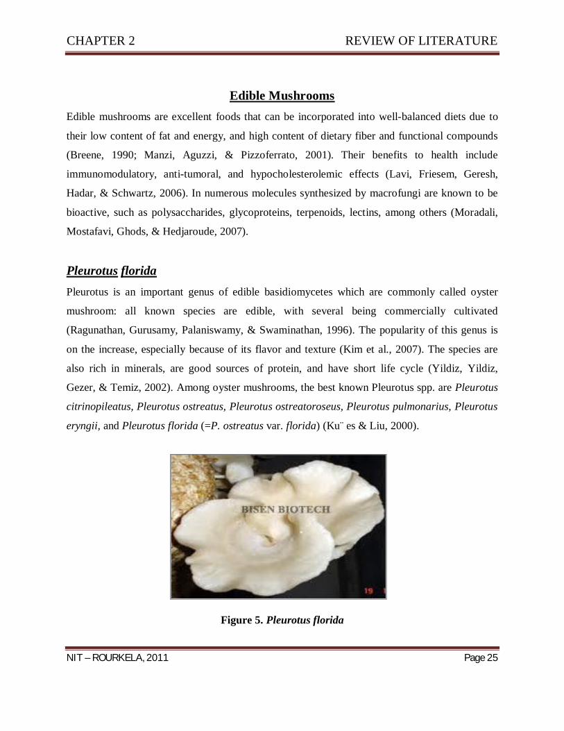
Edible Mushrooms Edible mushrooms are excellent foods that can be incorporated into well-balanced diets due to
their low content of fat and energy, and high content of dietary fiber and functional compounds
(Breene, 1990; Manzi, Aguzzi, & Pizzoferrato, 2001). Their benefits to health include
immunomodulatory, anti-tumoral, and hypocholesterolemic effects (Lavi, Friesem, Geresh,
Hadar, & Schwartz, 2006). In numerous molecules synthesized by macrofungi are known to be
bioactive, such as polysaccharides, glycoproteins, terpenoids, lectins, among others (Moradali,
Mostafavi, Ghods, & Hedjaroude, 2007).
Pleurotus florida Pleurotus is an important genus of edible basidiomycetes which are commonly called oyster
mushroom: all known species are edible, with several being commercially cultivated
(Ragunathan, Gurusamy, Palaniswamy, & Swaminathan, 1996). The popularity of this genus is
on the increase, especially because of its flavor and texture (Kim et al., 2007). The species are
also rich in minerals, are good sources of protein, and have short life cycle (Yildiz, Yildiz,
Gezer, & Temiz, 2002). Among oyster mushrooms, the best known Pleurotus spp. are Pleurotus
citrinopileatus, Pleurotus ostreatus, Pleurotus ostreatoroseus, Pleurotus pulmonarius, Pleurotus
eryngii, and Pleurotus florida (=P. ostreatus var. florida) (Ku¨ es & Liu, 2000).
Figure 5. Pleurotus florida

CHAPTER 2 REVIEW OF LITERATURE
NIT – ROURKELA, 2011 Page 26
Pleurotus sajor caju
The genus Pleurotus (Jacq: Fr.) Kumm. (Pleurotaceae, higher Basidiomycetes) comprises a
group of edible ligninolytic mushrooms with medicinal properties and important
biotechnological and environmental applications. The evolutionary connection among species in
the genus Pleurotus is still not clear and many taxonomic issues remain controversial. The
cultivation of Pleurotus spp is an economically important food industry worldwide which has
expanded in the past few years. P. sajor caju is the third most important cultivated mushroom for
food purposes. Nutritionally, it has unique flavor and aromatic properties; and it is considered to
be rich in protein, fiber, carbohydrates, vitamins and minerals. Pleurotus spp are promising as
medicinal mushrooms, exhibiting hematological, antiviral, antitumor, antibiotic, antibacterial,
hypocholesterolic and immunomodulation activities. The bioactive molecules isolated from the
different fungi are polysaccharides. One of the most important aspects of Pleurotus spp is related
to the use of their ligninolytic system for a variety of applications, such as the bioconversion of
agricultural wastes into valuable products for animal feed and other food products and the use of
their ligninolytic enzymes for the biodegradation of organopollutants, xenobiotics and industrial
contaminants.
Figure 6: Pleurotus sajor caju
CHAPTER 2 REVIEW OF LITERATURE
NIT – ROURKELA, 2011 Page 27
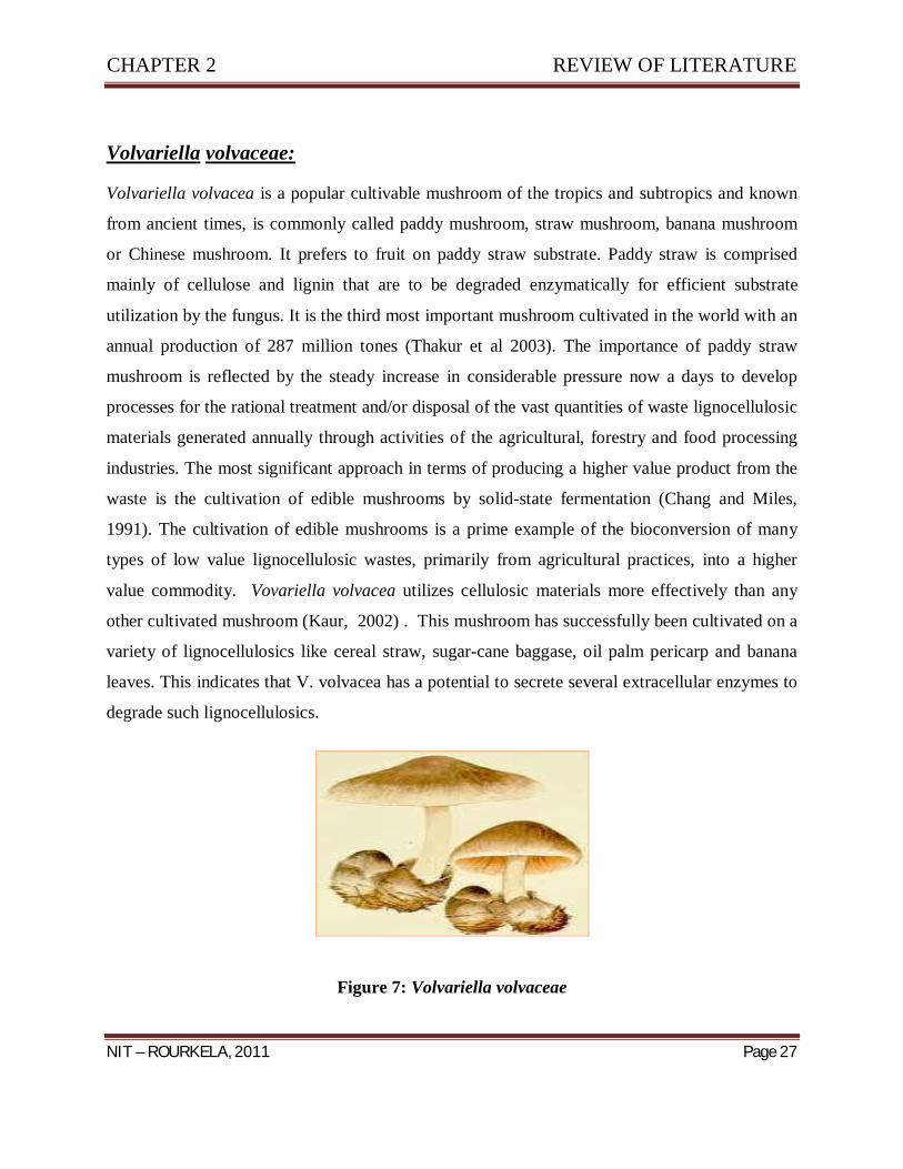
Volvariella volvaceae:
Volvariella volvacea is a popular cultivable mushroom of the tropics and subtropics and known
from ancient times, is commonly called paddy mushroom, straw mushroom, banana mushroom
or Chinese mushroom. It prefers to fruit on paddy straw substrate. Paddy straw is comprised
mainly of cellulose and lignin that are to be degraded enzymatically for efficient substrate
utilization by the fungus. It is the third most important mushroom cultivated in the world with an
annual production of 287 million tones (Thakur et al 2003). The importance of paddy straw
mushroom is reflected by the steady increase in considerable pressure now a days to develop
processes for the rational treatment and/or disposal of the vast quantities of waste lignocellulosic
materials generated annually through activities of the agricultural, forestry and food processing
industries. The most significant approach in terms of producing a higher value product from the
waste is the cultivation of edible mushrooms by solid-state fermentation (Chang and Miles,
1991). The cultivation of edible mushrooms is a prime example of the bioconversion of many
types of low value lignocellulosic wastes, primarily from agricultural practices, into a higher
value commodity. Vovariella volvacea utilizes cellulosic materials more effectively than any
other cultivated mushroom (Kaur, 2002) . This mushroom has successfully been cultivated on a
variety of lignocellulosics like cereal straw, sugar-cane baggase, oil palm pericarp and banana
leaves. This indicates that V. volvacea has a potential to secrete several extracellular enzymes to
degrade such lignocellulosics.
Figure 7: Volvariella volvaceae
CHAPTER 2 REVIEW OF LITERATURE
NIT – ROURKELA, 2011 Page 28
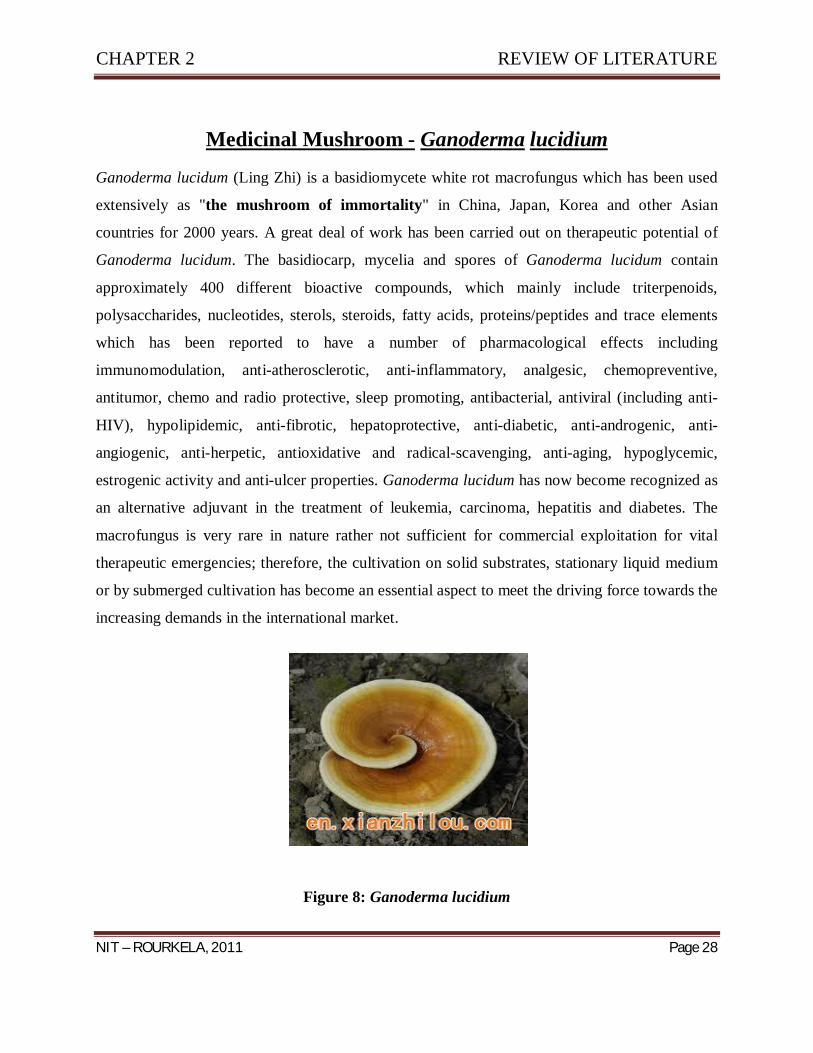
Medicinal Mushroom - Ganoderma lucidium
Ganoderma lucidum (Ling Zhi) is a basidiomycete white rot macrofungus which has been used
extensively as "the mushroom of immortality" in China, Japan, Korea and other Asian
countries for 2000 years. A great deal of work has been carried out on therapeutic potential of
Ganoderma lucidum. The basidiocarp, mycelia and spores of Ganoderma lucidum contain
approximately 400 different bioactive compounds, which mainly include triterpenoids,
polysaccharides, nucleotides, sterols, steroids, fatty acids, proteins/peptides and trace elements
which has been reported to have a number of pharmacological effects including
immunomodulation, anti-atherosclerotic, anti-inflammatory, analgesic, chemopreventive,
antitumor, chemo and radio protective, sleep promoting, antibacterial, antiviral (including anti-
HIV), hypolipidemic, anti-fibrotic, hepatoprotective, anti-diabetic, anti-androgenic, anti-
angiogenic, anti-herpetic, antioxidative and radical-scavenging, anti-aging, hypoglycemic,
estrogenic activity and anti-ulcer properties. Ganoderma lucidum has now become recognized as
an alternative adjuvant in the treatment of leukemia, carcinoma, hepatitis and diabetes. The
macrofungus is very rare in nature rather not sufficient for commercial exploitation for vital
therapeutic emergencies; therefore, the cultivation on solid substrates, stationary liquid medium
or by submerged cultivation has become an essential aspect to meet the driving force towards the
increasing demands in the international market.
Figure 8: Ganoderma lucidium

CHAPTER 2 REVIEW OF LITERATURE
NIT – ROURKELA, 2011 Page 29
Table 5. Therapeutic Effects and Bioactive Compounds of Ganoderma lucidum Reported in the Literature Until 2009

Chapter 3
MATERIALS & METHOD
CHAPTER 3 MATERIALS & METHOD
NIT – ROURKELA, 2011 Page 30
MATERIALS & METHOD
Mushroom strains:
Four Strains of Mushrooms, i.e. fruiting body of mushrooms were obtained from the following sources:
Volvoriella volvaceae – Sector 19 Market, Rourkela.
Pleurotus florida, - Ispat Mushroom House, Rourkela.
Pleurotus sajor caju – Ispat Mushroom House, Rourkela.
Ganoderma lucidium – Ispat Mushroom House, Rourkela.
Preparation of Malt Agar Spawn Culture Bottles:
Spawn Culture glass bottles of 250 ml capacity were used for culturing mycelia of the mushrooms.
Preparation of Mycelial Cultures of the Mushrooms:
Freshly obtained mushroom strains (Volvoriella volvaceae; Pleurotus florida, Pleurotus sajor
caju; and Ganoderma lucidium) were surface sterilized with distilled water and 0.2% HgCl2
solution (0.2 grams of HgCl2 in 100 ml distilled water). Then the mushrooms were cut into small
pieces and the transverse section of the untouched tissues were cut into 3×3 cm and were soak
dried using filter paper. These tissues were then centre inoculated in the pre prepared spawn
culture bottles. The spawn bottles containing the mycelial cultures were plugged tightly with
cotton and incubated at 25±3oC for 8 to 10 days.
Enzymatic Screening of Mushrooms using Agar Plates:
Mushrooms were screened for enzymes using agar plate method with the following compositions:
Starch Agar Medium (For Amylase Test) Ingredient gram/litre Starch 20.0 Peptone 5.0 Beef extract 3.0 Agar 15.0 pH 7.0 ± 0.2

CHAPTER 3 MATERIALS & METHOD
NIT – ROURKELA, 2011 Page 31
Gram’s iodine solution was used to detect the amylase production. A clear zone surrounding the
colony results in positive test for amylase.
Gelatin Agar Medium (For Protease Test)
Ingredient gram/litre Gelatin 20.0 Caesin 10.0 Sodium Chloride 10.0 Sodium taurocholate 5.0 Sodium bicarbonate 1.0 Agar 15.0 pH 8.5 ± 0.2 Mercuric chloride solution was used to detect the protease production resulted as clear zone
surrounding the colony.
Composition of the mercuric chloride reagent:
20% hydrochloric acid was mixed in 15% mercuric chloride solution.
Lipase Test Medium Ingredient gram/litre Peptone 10.0 Sodium chloride 5.0 Calcium chloride 0.1 Agar 20.0 Tween 20 10.0ml pH 6.0 ± 0.2 Tween 20 was sterilized separately and added to the medium at the time of pouring.
Fragmentation of the media resulted in the lipase production.
L- Asparaginase Test Medium (Modified Czapex Dox Medium) Ingredient gram/litre Glucose 2.0 L-asparagine 10.0 K2HPO4 1.52 Potassium chloride 0.52 Magnesium sulphate 0.52

CHAPTER 3 MATERIALS & METHOD
NIT – ROURKELA, 2011 Page 32
Cupric nitrate 0.001 Zinc sulphate 0.001 Ferrous sulphate 0.001 Phenol red 0.009 Agar 20.0 pH 7.2 ± 0.2
Nessler’s reagent was used as the reagent for L- Asparginase plate test to detect clear zone
surrounding the colonies
Phosphate Solubilizaton Test (Pickovaskaya Medium) Ingredient gram/litre Glucose 10.0 Tri-calcium phosphate 5.0 Ammonium sulphate 0.5 Sodium chloride 0.2 Magnesium sulphate 0.0001 Ferrous sulphate 0.001 Yeast Extract 0.5 Agar 15.0 pH 7.2 ± 0.2 Medium for Xylanase test Ingredient gram/litre Xylan 5.0 Peptone 5.0 Yeast Extract 5.0 K2HPO4 1.0 Magnesium sulphate 0.2 Agar 20.0 pH 7.0 ± 0.2 Reagent: 0.1% Congo red solution – 0.1 gm congo red powder in 100 ml distilled water. 1 molar NaCl solution The culture plates were flooded with Congo red solution. After 30 minutes they were washed with NaCl solution and observation was done for visualizing clear zone surrounding the colony.

CHAPTER 3 MATERIALS & METHOD
NIT – ROURKELA, 2011 Page 33
Estimation of Enzymes:
Estimation of L – Asparginase activity
Estimation of L – Asparginase activity was carried out using modified Czapex Dox’s media.
Conical flasks (150 ml) containing 50 ml each of the appropriate medium was inoculated with
each of the test organism. The flasks were incubated at 30oC at 240 rpm per minute in a
controlled environment incubator – shaker. Uninoculated media served as controls. The fungal
cultures were harvested by filtration through Whatman No. 1 filter paper. The enzyme activity
was estimated in culture filtrates by Nesselarization (Imada et al. 1973) and expressed as IU per
ml. One international unit of L – Asparginase activity is defined as that amount of enzyme which
catalyses the formation of 1 µmol of ammonia per unit under the conditions of assay.
Estimation of Available Phosphorous:
Composition of Reagents for Phosphate solubalization estimation
1. Extraction Solution: 0.55gm NH4F was mixed in 1.04ml conc. HCl, and volume was
made up to 500 ml with distilled water.
2. Standard stock solution: 0.109 gm KH2PO4 was mixed in 125 ml distilled water and 6.25
ml 7 Normal H2SO4. Then the volume was made up to 250 ml. 10 ml of this standard
Solution was added in 490ml distilled water to get a 10 ppm solution, i.e. 50 times
dilution.
3. REAGENT A: 6gm of Ammonium Molybdate was added in 125ml distilled water in one
flask, and 0.145gm Antimony potassium Tartarate was added in 50 ml distilled water in
another flask. These two solutions were then added to 500 ml of 2.5M H2SO4 solution,
mixed thoroughly and volume was made up to 1000ml.
4. REAGENT B: Dissolve 1.056gm ascorbic acid in 200ml reagent A and mix. Do not
keep for more than 24 hours.
While preparing curve for standard, the standard stock solution will be taken from 1 to 9 ml,
accordingly, the volume of water decreases and increases to keep the constant volume 24 ml.

CHAPTER 3 MATERIALS & METHOD
NIT – ROURKELA, 2011 Page 34
The volume of extraction solution and reagent B remains constant in all the tubes. After adding
reagent B the sample were left for 15 minutes incubation. The colour slowly develops. Then, the
readings were taken in UV Vis Spectrophotometer at 730 nm.
TABLE FOR STANDARD:
Stock Solution Extraction Solution Distilled Water Reagent B
Blank 5 ml 15 ml 4 ml
1 ml 5 ml 14 ml 4 ml
2 ml 5 ml 13 ml 4 ml
3 ml 5 ml 12 ml 4 ml
4 ml 5 ml 11 ml 4 ml
5 ml 5 ml 10 ml 4 ml
6 ml 5 ml 9 ml 4 ml
7 ml 5 ml 8 ml 4 ml
8 ml 5 ml 7 ml 4 ml
9 ml 5 ml 6 ml 4 ml
For estimating the concentration of phosphorous in sample broth, the broth was filtered
through Whattman Filter Paper No.1. The filtrate was kept in vials and the biomass was
weighed. Then 500 µl of sample was added in 5 ml of extraction solution and 14.5 ml of
distilled water with 4 ml of reagent B. After 15 minutes of incubation the readings were
taken. The concentration of phosphorous in 1 ml of stock solution was found out. Then
the values were put in X axis and OD in Y axis and the graph was plotted.
CHAPTER 3 MATERIALS & METHOD
NIT – ROURKELA, 2011 Page 35
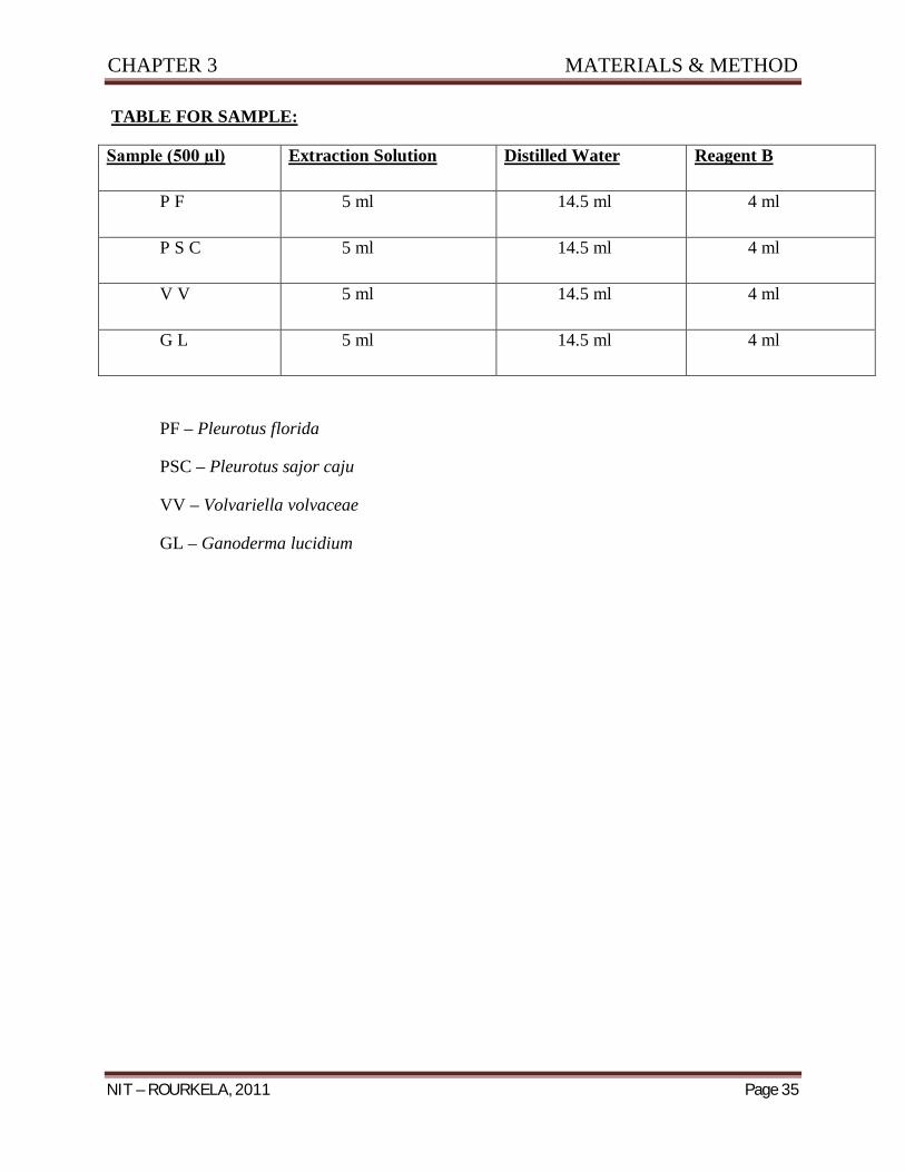
TABLE FOR SAMPLE:
Sample (500 µl) Extraction Solution Distilled Water Reagent B
P F 5 ml 14.5 ml 4 ml
P S C 5 ml 14.5 ml 4 ml
V V 5 ml 14.5 ml 4 ml
G L 5 ml 14.5 ml 4 ml
PF – Pleurotus florida
PSC – Pleurotus sajor caju
VV – Volvariella volvaceae
GL – Ganoderma lucidium

Chapter 4
RESULTS & DISCUSSION

CHAPTER 4 RESULTS & DISCUSSION
NIT – ROURKELA, 2011 Page 36
RESULTS & DISCUSSION
Culture of Mushroom Mycelium in Vitro conditions
Mycelial cultures of Mushrooms were obtained after 8 – 10 days of incubation at 25 ± 3 oC.
These mycelial cultures obtained were further used for the rest of the experiments.
Figure 9: Volvariella volvaceae
Figure 10: Ganoderma lucidium

CHAPTER 4 RESULTS & DISCUSSION
NIT – ROURKELA, 2011 Page 37
Figure 11: Pleurotus sajor caju
Figure 12: Pleurotus florida

CHAPTER 4 RESULTS & DISCUSSION
NIT – ROURKELA, 2011 Page 38
Screening of Potential Strain with Enzymatic Activitiy
After incubation of Agar plates for 6 – 7 days at 25 ± 3oC for the enzymatic tests the
following results were obtained:
Pleurotus
florida
Pleurotus sajor -
caju
Volvariella
volvaceae
Ganoderma
lucidium
Amylase + + + + + + + + + +
Protease + + + +
Lipase + + + + + + + +
L – Asparginase + + + + + + + + + +
Phosphate
Solubalization
+ + + + + + + + + +
Xylanases + + + + + + + +
It was seen that among the four different species i.e. Vovariella volvaceae, Pleurotus florida,
Pleurotus sajor caju and Ganoderma lucidium; Ganoderma lucidium was the most potent strain
which showed the best enzymatic activities among the four species. All the four species showed
very less protease activity.

CHAPTER 4 RESULTS & DISCUSSION
NIT – ROURKELA, 2011 Page 39
Figure 13: Plate Test for Amylase
Volvariella volvaceae
Pleurotus sajor caju
Pleurotus florida
Ganoderma lucidium
CONTROL RESULTS

CHAPTER 4 RESULTS & DISCUSSION
NIT – ROURKELA, 2011 Page 40
Figure 14: Plate Test for L – Asparginase
Volvariella volvaceae
Pleurotus sajor caju
Pleurotus florida
Ganoderma lucidium
CONTROL RESULTS
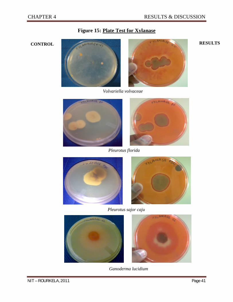
CHAPTER 4 RESULTS & DISCUSSION
NIT – ROURKELA, 2011 Page 41
Figure 15: Plate Test for Xylanase
Volvariella volvaceae
Pleurotus florida
Pleurotus sajor caju
Ganoderma lucidium
RESULTS CONTROL

CHAPTER 4 RESULTS & DISCUSSION
NIT – ROURKELA, 2011 Page 42
Figure 16: Plate Test for Phosphate Solubalization
Volvariella volvaceae Pleurotus florida
Pleurotus sajor caju Ganoderma lucidium

CHAPTER 4 RESULTS & DISCUSSION
NIT – ROURKELA, 2011 Page 43
Figure 17: Plate Test for Lipase
Volvariella volvaceae Pleurotus sajor caju
Pleurotus sajor caju Ganoderma lucidium

CHAPTER 4 RESULTS & DISCUSSION
NIT – ROURKELA, 2011 Page 44
Estimation and Selection of Strain with Enzymatic Activity
L – Asparginase Activity
The L – Asparginase activity of different mushroom species after 96 hrs of incubaton and
Nesslerization were found out spectrophotometrically at 450 nm in IU / ml and was found
out to be,
Mushroom Enzyme activity (IU / ml)
Pleurotus florida 0.95
Pleurotus sajor caju 0.73
Volvariella volvaceae 1.76
Ganoderma lucidium 2.15
Among the four mushroom species i.e. Vovariella volvaceae, Pleurotus florida, Pleurotus sajor
caju and Ganoderma lucidium; it was found out that Ganoderma lucidium is the most potent
strain showing L- Asparginase activity showing 2.15 IU/ml, whereas the other strains i.e.
Pleurotus florida, Pleurotus sajor caju and Volvariella volvaceae gave L – Asparginase activity
as 0.95, 0.73 and 1.73 IU / ml respecttievely.
Phosphate Solubilization Activity
The phosphate solubalization activity of different mushroom species were found out from the
culture filtrates after 6 – 7 days of incubation spectrophotometrically at 730 nm and the graph
was plotted by taking concentration on X – axis and OD on Y – axis. Among the four strains the
most potent strain showing phosphate solubilization activity was found out to be Volvariella
volvaceae at 0.4882 ppm concentration at 10% dilution. The other strains, i.e. Pleurotus sajor
caju, Pleurotus florida and Ganoderma lucidium showed phosphate solubilization activity at
0.082 ppm at 30 % dilution, 0.0435 ppm at 50% dilution and 0.0568 at 0% dilution.

CHAPTER 4 RESULTS & DISCUSSION
NIT – ROURKELA, 2011 Page 45
Table for Standard
OD OD’ Avg OD Concentraion (ppm)
Blank 1.032 1.037 0
1 0.048 0.082 0.065 0.416667
2 0.112 0.099 0.1055 0.833333
3 0.134 0.183 0.1585 1.25
4 0.218 0.243 0.2305 1.666667
5 0.271 0.279 0.275 2.083333
6 0.307 0.315 0.311 2.5
7 0.41 0.399 0.4045 2.916667
8 0.43 0.426 0.428 3.333333
9 0.466 0.476 0.471 3.75
Graph – Concentration vs OD

CHAPTER 4 RESULTS & DISCUSSION
NIT – ROURKELA, 2011 Page 46
Table for Samples
OD OD’ Avg. OD Conc (ppm) FinalConc (ppm)
PF 0.101 0.118 0.1095 0.0217 0.0435 PSC 0.126 0.14 0.133 0.0217 0.0824 VV 0.316 0.332 0.324 0.0488 0.4882 GL 0.402 0.373 0.3875 0.0568 0.0568
Table showing Biomass of the four Strains of Mushroom after Phosphate Solubilization Activity
Mushroom Strain Biomass ( in gms)
PF 0.9
PSC 0.9
VV 1
GL 4.1
PF – Pleurotus florida
PSC – Pleurotus sajor caju
VV – Volvariella volvaceae
GL – Ganoderma lucidium

Chapter 5
CONCLUSION

CHAPTER 5 CONCLUSION
NIT – ROURKELA, 2011 Page 47
CONCLUSION
In this work we studied, the enzyme activities of Pleurotus spp, Volvariella volvaceae and
Ganoderma lucidium grown on Malt Extract Agar media. Higher activities of Amylase, L –
Asparginase, Xylanase, Phosphate Solubalization, and Lipase were observed in Ganoderma
lucidium, where as very less Protease activity was observed in all the four strains of mushroom.
Phosphate solubilization and L – Asparginase values were higher in Volvariella volvaceae and
Ganoderma lucidium respectievely. Volvariella volvaceae showed phosphate solubilization
activity of 0.4882 ppm concentration at 10% dilution at 730 nm and Ganoderma lucidium
showed L- Asparginase activity of 2.15 IU/ml.
In summary in this work, we observed that during the growth of mushroom mycelia and the
development to mature fruitbodies (or sporophores), biochemical changes are known to occur, as
a result of which enzymes are secreted extracellularly to degrade the insoluble materials in the
substrates into simple and soluble molecules which are subsequently utilized by intracellular
enzymes within the mushroom. Consequently, enzymes play significant role in mushroom
development, in addition, they also affect the food nutrient, flavour and shelf life of these fungi.
Thus, Ganoderma lucidium which shows higher activity of L – Asparginase can be used for the
production of this enzyme or as a potent source therapeutic agent.

Chapter 6
REFERENCES
CHAPTER 6 REFRENCES
NIT – ROURKELA, 2011 Page 48
REFRENCES
1. Alconada TM, Martinez MJ., 1994, Purification and characterization of an extracellular
endo-1, 4-xylanase from Fusarium oxysporum f. sp. melonis. FEMS Microbiol Lett, 118,
305–10.
2. Akin DE, Borneman WS., 1990, Role of rumen fungi in fiber degradation. J Dairy Sci; 73,
3023–32.
3. Ammirati et al., 1985, 25–34.
4. Anand L, Krishnamurthy S, Vithayathil PJ., 1990, Purification and properties of xylanase
from the thermophilic fungus, Humicola lanuginose (Griffon and Maublanc) Bunce. Arch
Biochem Biophys; 276, 546–53.
5. Bagga PS, Sandhu DK, Sharma S., 1990, Purification and characterization of cellulolytic
enzymes produced by Aspergillus nidulans. J Appl Bacterial; 68, 61–8.
6. Beumer-Jachmans, M. P. (1973) Ann. Microbiol. 124, 289- 292
7. Boer H, Terri TT, Koivular A., 2000, Characterization of Trichoderma reesei
cellobiohydrolase Cel7A secreted from Pichia pastoris using two different promoters.
Biotechnol Bioeng; 69, 486–94.
8. Bourbonnais R, Paice MG., 1990, Oxidation of non-phenolic substrates: An expanded role
for laccase in lignin biodegradation. FEBS Lett; 267, 99–102.
9. Broome, J. D. (1961) Nature 191, 1114-1115.
10. Broome, J. D. (1968) Br. J. Cancer 22, 595-602.
11. Bumpus JA, Aust SD., 1987, Biodegradation of new environmental pollutants by the white
rot fungus Phanerochaete chrysoporium: involvement of the lignin degrading system.
BioEssays; 6, 166–70.
12. Campbell GL, Bedford MR., 1992, Enzyme application for monogastric feeds: a review.
Can J Anim Sci; 72:449–66.

CHAPTER 6 REFRENCES
NIT – ROURKELA, 2011 Page 49
13. Chou H, Lai HY, Tam MF, Chou MY, Wang SR, Hon SH, et al., 2001, cDNA cloning,
biological and immunological characterization of the alkaline serine protease major
allergen from Penicillium chrysogenum. Int Arch Allergy Immunol; 127, 15–26.
14. Dec J, Haider K, Bollag JM., 2003, Release of substitutents from phenolic compounds
during oxidative coupling reactions. Chemosphere; 52, 549–56.
15. Distasio, J. A., Niederman, R. A., Kafkewitz, D., and Goodman, D. (1976) J. Biol. Chem.
251, 6929-6933.
16. Fernandez-Larrea J, Stahl U., 1996, Isolation and characterization of a laccase gene from
Podospora anserine. Mol Gen Genet; 252, 539–51.
17. Gilbert HJ, Hazelwood GP, Laurie JI, Orpin CG, Xue GP., 1992, Homologous catalytic
domains in a rumen fungal xylanase: evidence for gene duplication and prokaryotic origin.
Mol Microbiol; 6, 2065.
18. Gomes J, Gomes I, Steiner W., 2000, Thermolabile xylanase of the Antartic yeast
Cryptococcus adeliae: production and properties. Extremophiles; 4:227–35.
19. Gupta R., Gigras P., Mohapatra H., Goswami V.K. and Chauhan B. 2003. Microbial α-
amylase: a b α-amylase biotechnological perspective. Journal of Process Biochemistry. 20,
1-18.
20. Hortobagyi, G. N., Yap, H. Y., Wiseman, C. L., Blumenschein, G. R., Buzdar, A. U.,
Legha, S. S., Gutterman, J. U., Hersh, E. M., and Bodey, G. P. (1980) Cancer Treat. Rep.
64, 157-159.
21. Kim JH, Kim YS., 1999, A fibrinolytic metalloprotease from the fruiting bodies of an
edible mushroom Armillariella mellea. Biosci Biotechnol Biochem; 63, 2130–6.
22. Kim JH, Kim YS., 2001, Characterization of a metal enzyme from a wild mushroom,
Tricholoma saponaceum. Biosci Biotechnol Biochem; 65, 356–62.

CHAPTER 6 REFRENCES
NIT – ROURKELA, 2011 Page 50
23. Kimura T, Ito J, Kawano A, Makino T, Kando H, Karita S, et al., 2000, Purification,
characterization, and molecular cloning of acidophilic xylanase from Penicillium sp. 40.
Biosci Biotechnol Biochem; 64, 1230–7.
24. Kobayashi H, Sokibata S, Shibuya H, Yoshida S, Kusakabe I, Murakami K., 1989,
Cloning and sequence analysis of cDNA for Irpex lacteus aspartic proteinase. Agric Biol
Chem; 53, 1927–33.
25. Kruger RP, Winter HC, Simonson- Leff N, Stuckey JA, Goldstein IJ, Dixon JE., 2002,
Cloning, expression and characterization of the gal-1, 3-gal high affinity lectin from the
mushroom Marasmius oreades. J Biol Chem; 277, 15002–5.
26. Marzullo L, Cannio R, Giardina R, Santini MT, Sannice G., 1995, Veratryl alcohol oxidase
from Pleurotus ostreatus participates in lignin biodegradation and prevents polymerization
of laccase-oxidized substrates. J Biol Chem; 270, 3823–7.
27. Mashburn, L. T., and Wriston, J. C. (1964) Arch. Biochem. Biophys. 105, 450-452.
28. Miles PG, Chang S-T. (2004). Mushrooms: Cultivation, Nutritional Value, Medicinal
Effect, and Environmental Impact. Boca Raton, Florida: CRC Press. ISBN 0-8493-10431
29. Mussak R, Bechtold T. (2009). Handbook of Natural Colorants. New York: Wiley.
pp. 183–200. ISBN 0-470-51199-0.
30. Nonaka T, Ishikawa H, Tumuraye Y, Hashimoto Y, Dohmae N., 1995, Characterisation of
a thermostable lysine-specific metalloendopeptidase from the fruiting bodies of a
basidiomycete Grifola frondosa. J Biochem (Tokyo); 118:1014–20.
31. Paoletti M, Castroviejo M, Begueret J, Clave C., 2001, Identification and characterization
of a gene encoding a subtilisin-like serine protease induced during the vegetative
incompatibility reaction in Podospora anserine. Curr Genet; 39, 244–52.
32. Pekkarinen AI, Jones BL, Niku-Paavola ML., 2002, Purification and properties of an
alkaline protease of Fusarium culmorum. Eur J Biochem; 269:798–807.

CHAPTER 6 REFRENCES
NIT – ROURKELA, 2011 Page 51
33. Poza M, ke Miyuel T, Sievro C, Villa TG., 2001, Characterization of a broad pH range
protease of Candida caseinolytica. J Appl Microbiol; 91:916–21.
34. Qy Y, Gao P, Wang D, Zhao X, Zhang X., 1996, Production, characterization and
application of the cellulase-free xylanase from Aspergillus niger. Appl Biochem
Biotechnol; 57, 375–81.
35. Reichard U, Cole GT, Ruchel R, Monod M., 2000; Molecular cloning and targeted deletion
of PEP2 which encodes a novel aspartic proteinase from Aspergillus fumigatus. Int J Med
Microbiol 290, 85–96.
36. Roberts, J., Holcenberg, J. S., and Dolowy, W. C. (1972) J. Biol. Chem. 247, 84-90.
37. Saha BC., 2001, Xylanase from a newly isolated Fusarium verticillioides capable of
utilizing cornfiber xylan. Appl Microbiol Biotechnol; 56:762–6.
38. Sattar AKMA, Yamamoto N, Yoshimoto T, Tsuru D., 1990, Purification and
characterization of an extracellular prolyl endopeptidase from Agaricus bisporus. J
Biochem (Tokyo); 107, 256–61.
39. Slomczynsky D, Nakas JP, Tanenbaum SW., 1995, Production and characterization of
laccase from Botrytis cinerea. Appl Environ Microbiol; 61, 907–12.
40. Stuntz et al., 1978, pp. 12–13.
41. Stuntz et al., 1978, 28–29
42. Torronen A, Mach RL, Messner R, Gonzalez R, Kalkkinen N, Harkk A, et al., 1992, The
two major xylanases from Trichoderma reesei: characterization of both enzymes and
genes. Biotechnol; 10, 1461–5.
43. Trinci APJ, Davies DR, Gull K, Lawrence MI, Nielsen BB, Richers A, et al., 1994,
Anaerobic fungi in herbivorous animals. Mycol Res; 98:129–52.
44. Volk T. (2001). "Hypomyces lactifluorum, the lobster mushroom". Fungus of the Month.
University of Wisconsin-La Crosse, Department of Biology. Retrieved 2008-10-13.
CHAPTER 6 REFRENCES
NIT – ROURKELA, 2011 Page 52
45. Wallace RJ., 1994, Ruminal microbiology, biotechnology and ruminant nutrition: progress
and problems. J Anim Sci; 72:2992–3003.
46. Wang HX, Ng TB., 2001, Pleureryn, a novel protease from fresh fruiting bodies of the
edible mushroom Pleurotus eryngii. Biochem Biophys Res Commun; 289, 750–5.
47. Wehner, A., Harms, E., Jennings, M. P., Beacham, I. R., Derst, C., Bast, P., and Ro¨hm, K.
H. (1992) Eur. J. Biochem. 208, 475-480.
48. Yoshimoto T, Sattar AKMA, Hirose W, Tsuru D., 1990, Purification and characterization
of an extracellular prolyl endopeptidase from Agaricus bisporus. J Biochem (Tokyo); 107,
256–61.