Establishment of HIV-1 resistance in CD4+ T cells by genome editing using zinc-finger nucleases
9
Establishment of HIV-1 resistance in CD4 + T cells by genome editing using zinc-finger nucleases Elena E Perez 1,2 , Jianbin Wang 3 , Jeffrey C Miller 3 , Yann Jouvenot 3,4 , Kenneth A Kim 3 , Olga Liu 1 , Nathaniel Wang 3 , Gary Lee 3 , Victor V Bartsevich 3 , Ya-Li Lee 3 , Dmitry Y Guschin 3 , Igor Rupniewski 3 , Adam J Waite 3 , Carmine Carpenito 1 , Richard G Carroll 1 , Jordan S Orange 2 , Fyodor D Urnov 3 , Edward J Rebar 3 , Dale Ando 3 , Philip D Gregory 3 , James L Riley 1 , Michael C Holmes 3 & Carl H June 1 Homozygosity for the naturally occurring D32 deletion in the HIV co-receptor CCR5 confers resistance to HIV-1 infection. We generated an HIV-resistant genotype de novo using engineered zinc-finger nucleases (ZFNs) to disrupt endogenous CCR5. Transient expression of CCR5 ZFNs permanently and specifically disrupted ~50% of CCR5 alleles in a pool of primary human CD4 + T cells. Genetic disruption of CCR5 provided robust, stable and heritable protection against HIV-1 infection in vitro and in vivo in a NOG model of HIV infection. HIV-1-infected mice engrafted with ZFN-modified CD4 + T cells had lower viral loads and higher CD4 + T-cell counts than mice engrafted with wild-type CD4 + T cells, consistent with the potential to reconstitute immune function in individuals with HIV/AIDS by maintenance of an HIV-resistant CD4 + T-cell population. Thus adoptive transfer of ex vivo expanded CCR5 ZFN–modified autologous CD4 + T cells in HIV patients is an attractive approach for the treatment of HIV-1 infection. CCR5, a seven-transmembrane chemokine receptor, is the major co- receptor for HIV-1 entry 1,2 . Since the discovery that the homozygous D32 deletion in CCR5 confers resistance to HIV-1 3–5 , CCR5 has been intensely studied and validated as a target for HIV therapy 6,7 . Recently, small-molecule approaches that block the CCR5-HIV interaction have shown promise in clinical trials 8 . However, the small-molecule approach has resulted in the development of resistance by selection for escape mutants, which continue to use CCR5 for viral entry 9 . These results, taken together with experience from individuals hetero- zygous for the D32 allele, point to the importance of a genetic knockout of CCR5 for phenotypic penetrance and long-term resis- tance to infection rather than its knock-down by approaches based on small molecules, intrabodies, antisense or RNA interference (RNAi) 10–15 . Therefore, we sought to permanently disrupt the endo- genous CCR5 and thus make a phenocopy of the D32CCR5 null genotype in primary human CD4 + T cells by the application of engineered ZFNs. Previously, we have shown that reconstituting CD4 + helper T-cell activity through adoptive transfer of costimulated CD4 + T cells may augment natural immunity to HIV-1 infection 13 . Here we show that engineered ZFNs targeting human CCR5 efficiently generate a double- strand break at a predetermined site in the CCR5 coding region upstream of the natural CCR5D32 mutation. The CCR5 ZFNs promote efficient and permanent disruption of CCR5 in primary human CD4 + T lymphocytes and confer robust protection against HIV-1 infection both in vitro and in an in vivo mouse model of HIV-1 infection. Combining the two approaches may provide further benefit to patients with HIV-1 in future clinical trials. RESULTS Design of ZFNs targeted against CCR5 (CCR5 ZFN) We designed and optimized a large series of ZFNs targeted to human CCR5 using a previously described approach 16 . For both target sites two zinc-finger protein (ZFP) DNA-binding domains, each containing four zinc-finger motifs (recognizing a total of 24 base pairs), were assembled from an archive of ZFP DNA-binding modules 17,18 . These ZFPs were coupled to the DNA cleavage domain of the type IIS restriction enzyme, FokI, to produce novel ZFNs in which the location of DNA cleavage is determined by the DNA-binding specificity of the engineered ZFP domains, as previously shown 16,17,19 . Targeting a double-strand break to a specific site in the genome with ZFNs has been used to disrupt permanently the genomic sequence surrounding the ZFN target site in a variety of eukaryotic organisms 20,21 via imperfect repair by nonhomologous end joining (NHEJ) 22,23 . To exploit this property of double-strand break repair, we elected to focus our ZFN designs upon the DNA sequence encoding the first transmembrane domain (TM1 spans residues Arg31 to Asn57) of the CCR5 co-receptor. We reasoned that this location, upstream of the Received 22 April; accepted 22 May; published online 29 June 2008; doi:10.1038/nbt1410 1 Abramson Family Cancer Research Institute, Department of Pathology and Laboratory Medicine, 421 Curie Blvd., Room 554, BRB II/III, Philadelphia, Pennsylvania 19104-6160, USA. 2 Children’s Hospital of Philadelphia, Division of Allergy and Immunology, Joseph Stokes, Jr. Research Institute, 3615 Civic Center Blvd., Philadelphia, Pennsylvania 19104-4318, USA. 3 Sangamo BioSciences, Inc., Point Richmond Tech Center II, 501 Canal Blvd., Suite A100, Richmond, California 94804, USA. 4 Present address: Process Science Department, Bayer Hematology/Cardiology, 800 Dwight Way, Berkeley, California 94701, USA. Correspondence should be addressed to C.H.J. ([email protected]). 808 VOLUME 26 NUMBER 7 JULY 2008 NATURE BIOTECHNOLOGY ARTICLES © 2008 Nature Publishing Group http://www.nature.com/naturebiotechnology
Transcript of Establishment of HIV-1 resistance in CD4+ T cells by genome editing using zinc-finger nucleases
Establishment of HIV-1 resistance in CD4+ T cells bygenome editing using zinc-finger nucleasesElena E Perez1,2, Jianbin Wang3, Jeffrey C Miller3, Yann Jouvenot3,4, Kenneth A Kim3, Olga Liu1,Nathaniel Wang3, Gary Lee3, Victor V Bartsevich3, Ya-Li Lee3, Dmitry Y Guschin3, Igor Rupniewski3,Adam J Waite3, Carmine Carpenito1, Richard G Carroll1, Jordan S Orange2, Fyodor D Urnov3,Edward J Rebar3, Dale Ando3, Philip D Gregory3, James L Riley1, Michael C Holmes3 & Carl H June1
Homozygosity for the naturally occurring D32 deletion in the HIV co-receptor CCR5 confers resistance to HIV-1 infection.
We generated an HIV-resistant genotype de novo using engineered zinc-finger nucleases (ZFNs) to disrupt endogenous CCR5.
Transient expression of CCR5 ZFNs permanently and specifically disrupted ~50% of CCR5 alleles in a pool of primary human
CD4+ T cells. Genetic disruption of CCR5 provided robust, stable and heritable protection against HIV-1 infection in vitro and
in vivo in a NOG model of HIV infection. HIV-1-infected mice engrafted with ZFN-modified CD4+ T cells had lower viral loads
and higher CD4+ T-cell counts than mice engrafted with wild-type CD4+ T cells, consistent with the potential to reconstitute
immune function in individuals with HIV/AIDS by maintenance of an HIV-resistant CD4+ T-cell population. Thus adoptive
transfer of ex vivo expanded CCR5 ZFN–modified autologous CD4+ T cells in HIV patients is an attractive approach for the
treatment of HIV-1 infection.
CCR5, a seven-transmembrane chemokine receptor, is the major co-receptor for HIV-1 entry1,2. Since the discovery that the homozygousD32 deletion in CCR5 confers resistance to HIV-13–5, CCR5 has beenintensely studied and validated as a target for HIV therapy6,7. Recently,small-molecule approaches that block the CCR5-HIV interaction haveshown promise in clinical trials8. However, the small-moleculeapproach has resulted in the development of resistance by selectionfor escape mutants, which continue to use CCR5 for viral entry9.These results, taken together with experience from individuals hetero-zygous for the D32 allele, point to the importance of a geneticknockout of CCR5 for phenotypic penetrance and long-term resis-tance to infection rather than its knock-down by approaches basedon small molecules, intrabodies, antisense or RNA interference(RNAi)10–15. Therefore, we sought to permanently disrupt the endo-genous CCR5 and thus make a phenocopy of the D32CCR5 nullgenotype in primary human CD4+ T cells by the application ofengineered ZFNs.
Previously, we have shown that reconstituting CD4+ helper T-cellactivity through adoptive transfer of costimulated CD4+ T cells mayaugment natural immunity to HIV-1 infection13. Here we show thatengineered ZFNs targeting human CCR5 efficiently generate a double-strand break at a predetermined site in the CCR5 coding regionupstream of the natural CCR5D32 mutation. The CCR5 ZFNspromote efficient and permanent disruption of CCR5 in primary
human CD4+ T lymphocytes and confer robust protection againstHIV-1 infection both in vitro and in an in vivo mouse model of HIV-1infection. Combining the two approaches may provide further benefitto patients with HIV-1 in future clinical trials.
RESULTS
Design of ZFNs targeted against CCR5 (CCR5 ZFN)
We designed and optimized a large series of ZFNs targeted to humanCCR5 using a previously described approach16. For both target sitestwo zinc-finger protein (ZFP) DNA-binding domains, each containingfour zinc-finger motifs (recognizing a total of 24 base pairs), wereassembled from an archive of ZFP DNA-binding modules17,18. TheseZFPs were coupled to the DNA cleavage domain of the type IISrestriction enzyme, FokI, to produce novel ZFNs in which the locationof DNA cleavage is determined by the DNA-binding specificity of theengineered ZFP domains, as previously shown16,17,19. Targeting adouble-strand break to a specific site in the genome with ZFNs hasbeen used to disrupt permanently the genomic sequence surroundingthe ZFN target site in a variety of eukaryotic organisms20,21 viaimperfect repair by nonhomologous end joining (NHEJ)22,23. Toexploit this property of double-strand break repair, we elected tofocus our ZFN designs upon the DNA sequence encoding the firsttransmembrane domain (TM1 spans residues Arg31 to Asn57) of theCCR5 co-receptor. We reasoned that this location, upstream of the
Received 22 April; accepted 22 May; published online 29 June 2008; doi:10.1038/nbt1410
1Abramson Family Cancer Research Institute, Department of Pathology and Laboratory Medicine, 421 Curie Blvd., Room 554, BRB II/III, Philadelphia, Pennsylvania19104-6160, USA. 2Children’s Hospital of Philadelphia, Division of Allergy and Immunology, Joseph Stokes, Jr. Research Institute, 3615 Civic Center Blvd.,Philadelphia, Pennsylvania 19104-4318, USA. 3Sangamo BioSciences, Inc., Point Richmond Tech Center II, 501 Canal Blvd., Suite A100, Richmond, California 94804,USA. 4Present address: Process Science Department, Bayer Hematology/Cardiology, 800 Dwight Way, Berkeley, California 94701, USA. Correspondence should beaddressed to C.H.J. ([email protected]).
808 VOLUME 26 NUMBER 7 JULY 2008 NATURE BIOTECHNOLOGY
A R T I C L E S©
2008
Nat
ure
Pub
lishi
ng G
roup
ht
tp://
ww
w.n
atur
e.co
m/n
atur
ebio
tech
nolo
gy

D32 mutation, would display substantial structural sensitivity in thecontext of the CCR5 protein. Thus mutations introduced duringrepair via NHEJ would be predicted to result in truncated ornonfunctional gene products that would fail to be expressed on thecell surface, in a manner analogous to the naturally occurring D32mutant allele3,4. The lead ZFN pair binds the sequence flanking thecodon for Leu55 (within TM1) of human CCR5 (Fig. 1a), and isreferred to throughout as ZFN-215. A variant of these ZFNs (ZFN-224) was generated that incorporates engineered FokI domains thatfunction as obligate heterodimers and thereby improve ZFN specifi-city. The complete sequence of the ZFN pair is shown in FASTAformat (Supplementary Fig. 1 online).
Entry inhibition of CCR5-tropic HIV-1 by ZFN-targeted disruption
To determine whether transient expression of the CCR5 ZFNs wouldalter CCR5 expression levels and HIV-1 entry, we transduced GHOST-CCR5 cells, a reporter cell line for HIV-1 infection containing multiple(approximately four) copies of an autologous CCR5 expressioncassette and an inducible green fluorescent protein (GFP) markergene under the control of the HIV-2 long terminal repeat (LTR)24,with CCR5 ZFNs and subjected them to CCR5-tropic HIV-1challenge. To achieve transient yet high-efficiency ZFN delivery, we
transduced GHOST-CCR5 cells with an ade-novirus (Ad5/35) vector25 encoding the leadCCR5 ZFNs. First, we confirmed and quanti-fied the generation of ZFN-induced muta-tions at the target site using an assay basedupon the mismatch-sensitive Surveyor nucle-ase (Supplementary Fig. 2 online). DNAanalysis using this assay revealed high-efficiency (50–80%) target gene mutation inthe population of GHOST-CCR5 cells trans-duced with CCR5 ZFNs (Fig. 1b). This result
was CCR5 ZFN dependent as neither nontransduced control cells, norcells transduced with an Ad5/35 vector encoding interleukin (IL)-2Rg–specific ZFNs16 showed detectable CCR5 modifications (Fig. 1b).
These transduced cell populations were maintained in culture and1 week later were infected with HIV-1BAL, a prototype CCR5-tropicHIV-1 isolate. Immediately before HIV-1 infection, CCR5 surfaceexpression was analyzed and found to be reduced by more thantenfold in the pool of CCR5 ZFN–transduced cells compared withcontrol IL-2Rg-ZFN–treated cells (Fig. 1c). Consistent with thisreduction in CCR5 expression, HIV-1BAL challenge demonstrated asubstantial decrease in HIV-1 infection in CCR5 ZFN–treated samples,as measured by loss of GFP induction 48 h after infection (Fig. 1d).Genetic modification at the intended target site within CCR5 wasconfirmed by sequencing of genomic DNA from ZFN-treatedGHOST-CCR5 cells (data not shown). CCR5 reconstitution experi-ments were carried out to confirm the mechanism of resistance toHIV-1 infection in the experiments described above. Single cell–derived clones from the ZFN-treated GHOST-CCR5 populationwere isolated and shown to be completely resistant to HIV-1 infectionusing CCR5-tropic HIV-1BAL in contrast to cloned GHOST-CCR5cells that retained unmodified CCR5 genes (Supplementary Fig. 3aonline). CCR5 reconstitution experiments demonstrated that
b
a
CCR5 GFP
0
50
100
150
200
250
NTD IL2Rγ 215 2240
50
100
NTD IL2Rγ 215 224
Per
cent
of M
ax
Per
cent
of M
ax
c
MF
I
GF
P (
%)
d
82 53
NTD21
5IL
2Rγ
224
Targeted disruption (%)<1<1
100 101 102 103 104 100 101 102 103 1040
20
40
60
80
100
0
20
40
60
80
100
ZFNbinding site
ZFN-R
Fokl
Fok l
ZFN-L
Chr3 (p21.31) 24 29
A T C T T T G G T T T T G T G G G C A A C A T G C T G G T C A T C C T C A T C C T G A T A A A C T G C A A A A G G C T G A A G A G C A T G A C T G A C
T A G A A A C C A A A A C A C C C G T T G T A C G A C C A G T A G G A G T A G G A C T A T T T G A C G T T T T C C G A C T T C T C G T A C T G A C T GValVal IleIle42 Met Met ThrIle IleLeu AsnAsn Asp66GlyGly PhePhe Cys Lys Lys SerArgLeu55 LeuLeu
Figure 1 ZFN-mediated disruption of CCR5 and
protection from HIV-1 infection in GHOST-CCR5
cells. (a) Schematic of the CCR5 coding region
showing the genomic DNA sequences targeted by
CCR5 ZFNs 215/224. (b) Level of targeted gene
disruption in GHOST-CCR5 cells transduced with
an Ad5/35 vector encoding ZFNs targeting either
CCR5 or IL-2Rg as assessed by the Surveyorassay (Supplementary Fig. 2). Lower migrating
products (arrows) are a direct measure of ZFN-
mediated gene disruption. NTD, nontransduced
cells. (c) Decreased CCR5 surface expression
measured by flow cytometry (light blue, IL2RgZFN; green CCR5 ZFN-224; orange, CCR5 ZFN-
215; dark blue, nontransduced cells; red,
unstained cells). (d) Protection from HIV-1BAL
measured by flow cytometry 48 h after HIV-1
challenge of CCR5 ZFN-215 and CCR5
ZFN-224–modified cells compared to IL2Rg ZFN
and control GHOST-CCR5 cells (red, IL2Rg ZFN;
green, CCR5 ZFN-224; blue, CCR5 ZFN-215).
GFP fluorescence indicates HIV-1 entry and is
plotted as average percent infected relative to
positive control. MFI, mean fluorescence intensity.
Histograms of CCR5 (c) and GFP expression
(d) show one replicate for each condition; bar
graphs below represent averages (±s.d.) oftriplicates. Expression of CCR5 and HIV-1
infection frequency of CCR5 ZFN–treated
cells is less than IL2Rg ZFN or nontransduced
cells (P o 0.001).
NATURE BIOTECHNOLOGY VOLUME 26 NUMBER 7 JULY 2008 809
A R T I C L E S©
2008
Nat
ure
Pub
lishi
ng G
roup
ht
tp://
ww
w.n
atur
e.co
m/n
atur
ebio
tech
nolo
gy
resistance to HIV-1 infection was mediated exclusively by a defect inviral entry through ZFN-mediated CCR5 disruption (SupplementaryFig. 3b). Taken together these results demonstrate that the CCR5ZFNs can efficiently cleave their DNA target site in CCR5 andconfirm that a high proportion of ZFN-induced mutations preventCCR5 cell-surface expression, resulting in resistance to CCR5-tropicHIV-1 infection.
Survival advantage of ZFN-modified CD4+ T cells in vitro
Next, we evaluated whether ZFN-mediated disruption of CCR5 wouldconfer the long-term resistance to HIV-1 expected from a permanentgenetic change. PM1 cells, a CD4+ T-cell line with similar levels ofCCR5 expression to primary CD4+ T cells (data not shown), wereelectroporated with sub-optimal amounts of CCR5 ZFN expression
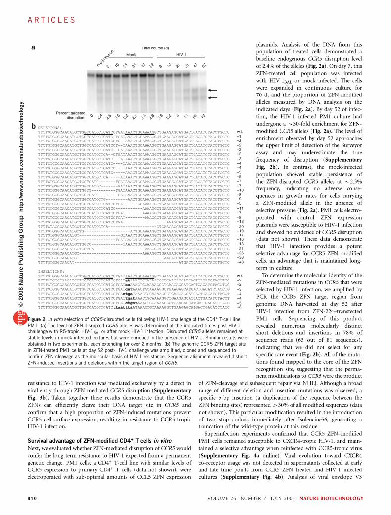
plasmids. Analysis of the DNA from thispopulation of treated cells demonstrated abaseline endogenous CCR5 disruption levelof 2.4% of the alleles (Fig. 2a). On day 7, thisZFN-treated cell population was infectedwith HIV-1BAL or mock infected. The cellswere expanded in continuous culture for70 d, and the proportion of ZFN-modifiedalleles measured by DNA analysis on theindicated days (Fig. 2a). By day 52 of infec-tion, the HIV-1–infected PM1 culture hadundergone a B30-fold enrichment for ZFN-modified CCR5 alleles (Fig. 2a). The level ofenrichment observed by day 52 approachesthe upper limit of detection of the Surveyorassay and may underestimate the truefrequency of disruption (SupplementaryFig. 2b). In contrast, the mock-infectedpopulation showed stable persistence ofthe ZFN-disrupted CCR5 alleles at B2.3%frequency, indicating no adverse conse-quences in growth rates for cells carryinga ZFN-modified allele in the absence ofselective pressure (Fig. 2a). PM1 cells electro-porated with control ZFN expressionplasmids were susceptible to HIV-1 infectionand showed no evidence of CCR5 disruption(data not shown). These data demonstratethat HIV-1 infection provides a potentselective advantage for CCR5 ZFN–modifiedcells, an advantage that is maintained long-term in culture.
To determine the molecular identity of theZFN-mediated mutations in CCR5 that wereselected by HIV-1 infection, we amplified byPCR the CCR5 ZFN target region fromgenomic DNA harvested at day 52 afterHIV-1 infection from ZFN-224–transfectedPM1 cells. Sequencing of this productrevealed numerous molecularly distinctshort deletions and insertions in 78% ofsequence reads (63 out of 81 sequences),indicating that we did not select for anyspecific rare event (Fig. 2b). All of the muta-tions found mapped to the core of the ZFNrecognition site, suggesting that the perma-nent modifications to CCR5 were the product
of ZFN-cleavage and subsequent repair via NHEJ. Although a broadrange of different deletion and insertion mutations was observed, aspecific 5-bp insertion (a duplication of the sequence between theZFN binding sites) represented 430% of all modified sequences (datanot shown). This particular modification resulted in the introductionof two stop codons immediately after Isoleucine56, generating atruncation of the wild-type protein at this residue.
Superinfection experiments confirmed that CCR5 ZFN–modifiedPM1 cells remained susceptible to CXCR4-tropic HIV-1, and main-tained a selective advantage when reinfected with CCR5-tropic virus(Supplementary Fig. 4a online). Viral evolution toward CXCR4co-receptor usage was not detected in supernatants collected at earlyand late time points from CCR5 ZFN–treated and HIV-1–infectedcultures (Supplementary Fig. 4b). Analysis of viral envelope V3
a
Pre-in
fectio
n
3 310 21 31 42 52
Mock HIV-1
10 21 31 42 52
2.5
2.4
2.6
2.6
2.1
2.4
2.3
2.8
2.6 4 11 58 73
Time course (d)
Percent targeted disruption: 0
bw.t.–1–2–2–2–2–3–4–4–5–5–7–7–10–8–9–5–11–7–8–18–20–19–17–16–13–21–26–36–43
w.t.+2+2+3+4+4+5+8
Figure 2 In vitro selection of CCR5-disrupted cells following HIV-1 challenge of the CD4+ T-cell line,
PM1. (a) The level of ZFN-disrupted CCR5 alleles was determined at the indicated times post-HIV-1challenge with R5-tropic HIV-1BAL or after mock HIV-1 infection. Disrupted CCR5 alleles remained at
stable levels in mock-infected cultures but were enriched in the presence of HIV-1. Similar results were
obtained in two experiments, each extending for over 2 months. (b) The genomic CCR5 ZFN target site
in ZFN-treated PM1 cells at day 52 post-HIV-1 challenge was amplified, cloned and sequenced to
confirm ZFN cleavage as the molecular basis of HIV-1 resistance. Sequence alignment revealed distinct
ZFN-induced insertions and deletions within the target region of CCR5.
810 VOLUME 26 NUMBER 7 JULY 2008 NATURE BIOTECHNOLOGY
A R T I C L E S©
2008
Nat
ure
Pub
lishi
ng G
roup
ht
tp://
ww
w.n
atur
e.co
m/n
atur
ebio
tech
nolo
gy

sequences isolated from the cultures supports this finding (Supple-mentary Fig. 4c,d), as viruses isolated from both GFP- and CCR5ZFN–treated cells matched most closely to the CCR5-tropic consensusenvelope sequence. Together, these results demonstrate that transientexpression of CCR5 ZFNs can establish stable and selective resistanceto CCR5-tropic HIV-1, consistent with expectations based on indivi-duals carrying the naturally occurring CCR5D32 mutation3–5,26.
Selective advantage of CCR5 ZFN–modified primary CD4+ T cells
during HIV-1 infection
To determine the efficacy of CCR5 ZFNs in primary human cells, wetransduced CD4+ T cells from healthy wild-type CCR5 donors with anAd5/35 vector encoding CCR5 ZFNs to provide transient yet high-efficiency ZFN delivery. Multiplicities of infection (MOI)-dependentlevels of ZFN-mediated CCR5 disruption (reaching 40–60% of theCCR5 alleles) were observed in multiple experiments using cellsisolated from different donors (Fig. 3a and data not shown). Thepopulation-doubling rate of the modified primary CD4+ T cells wasindistinguishable from nontransduced cells, with the proportion ofCCR5-modified alleles remaining stable for at least 1 month duringin vitro culture (Fig. 3b and data not shown).
Infection of a bulk CCR5 ZFN–transduced CD4+ T-cell populationwith the CCR5-tropic HIV-1US1 resulted in a twofold enrichment ofgene-edited cells with ZFN-disrupted CCR5 alleles over 17 d ofculture, whereas mock-infected control populations maintained astable level of ZFN-disrupted CCR5 alleles (Fig. 3c). CD4+ T cellstransduced with an Ad5/35 GFP control vector showed no detectabledisruption of CCR5 (data not shown).
After demonstrating a high frequency of mutagenesis at the CCR5locus, we determined what percentage of CD4+ T cells have bothalleles modified. CD4+ T cells were infected with the Ad5/35 CCR5ZFN vector and cloned by flow sorting. Fifty-two T-cell clones wereobtained, and 12/52 (23%) had CCR5 modification as judged by theSurveyor nuclease assay, confirming the results found within the bulkpopulation (28–30%) before cloning. The 12 clones with disruptedCCR5 loci were then genotyped by directly sequencing the CCR5
alleles. Four out of the 12 (33%) mutant clones were homozygous forCCR5 disruption. This experiment was done under nonselectingconditions; we expect the frequency of homozygous disruption tobe higher in the presence of HIV-1 challenge.
Specificity of CCR5 ZFNs in primary CD4+ T cells
The efficacy and tolerability of CCR5 ZFN–driven genome editing inprimary human cells supports the potential clinical application ofautologous ZFN-modified CD4+ T cells in patients with HIV. How-ever, in developing such an approach it is critical to verify thespecificity of ZFN action. Cleavage at a given target site requiresbinding of two ZFNs in a specific spatial orientation relative to eachother, that is, on opposite strands and separated by 5 or 6 bp tofacilitate the dimerization of the FokI domains necessary for DNAcleavage27,28 (Fig. 1a). This serves to restrict the induction of a double-strand break to those positions in the genome where binding sites fortwo ZFNs are found in the required juxtaposition. To quantify thenumber of double-strand breaks generated after ZFN expression, weconducted intranuclear staining for genome-wide double-strandbreaks via immunodetection of p53 binding protein 1 (53BP1) focias an unbiased measure of ZFN action throughout the nucleus. 53BP1is recruited to the sites of double-strand breaks early in the repairresponse and is required for NHEJ29. The genomic integrity of CD4+
T cells was assessed at several time points after transduction withAd5/35 expressing the indicated CCR5 ZFN by enumeration of thenumber of 53BP1 foci per nucleus (Fig. 3d). We observed a transient1.4–1.6-fold increase on days 2 and 3 of culture in the mean numberof intranuclear 53BP1 foci when comparing ZFN-224 transduced tonontransduced or GFP-transduced CD4+ T cells (Fig. 3e). In contrast,etoposide-treated positive control cells had a 4.2-fold increase in53BP1 staining over control cells that persisted for at least a week(data not shown). No significant difference in the mean perimeter of53BP1 foci was observed among all conditions (data not shown).
To confirm the specificity of ZFN-224 action, we experimentallydetermined the consensus ZFN binding site by SELEX (Supplemen-tary Fig. 5a online). The experimentally derived consensus matches
a db10ZFN ZFN
Pop
ulat
ion
doub
lings
GFP0
54
10 mm
Nontransduced CCR5 ZFN 224 Etoposide
No virus
Days
53B
P1
foci
/nuc
leus
(av
erag
e) GFP-100 MOICCR5 ZFN 224-100 MOI
44 44 30%
MOI
215 224
301003010030100
00 5 10 15 20
Days after transduction
c
e
25
20
15
10
5
0CCR5
-mod
ified
T c
ells
(%
)
Day 0
Day 5
Day 1
1
Day 1
7
Days post infection
10 mm 10 mm
4.0
3.0
2.0
1.0
0.00 2 4 6 8
Figure 3 Enrichment of CCR5 ZFN–modified primary CD4+ T cells during in vitro HIV-1 challenge. (a) Primary
CD4+ T cells from CCR5 wild-type anonymous healthy donor were transduced with Ad5/35 vector expressingCCR5 ZFN-215, ZFN-224 or GFP at MOI of 30 and 100; percentage of total alleles are indicated below each
lane. (b) Population doubling rate for CCR5 ZFN– and GFP control–transduced CD4+ T cells (triangle,
nontransduced; square, CCR5 ZFN-224; diamond, GFP transduced). (c) Enrichment of ZFN-215–transduced
CD4+ T cells over time following in vitro challenge with CCR5-tropic HIV-1US1 compared to mock (square, HIV-1
infected; triangle, mock infected). An B10% starting level of ZFN-disrupted CCR5 alleles was obtained by
mixing Ad5/35-transduced CD4+ T cells from a 1 in 3 with unmodified CD4+ T cells. (d) Intranuclear 53BP1
immunostaining and epifluorescence microscopy 2 d after CD4+ T cells were transduced with Ad5/35 vectors expressing CCR5 ZFN pair 224, nontransduced
(negative control) or 1 mM etoposide (positive control). Representative images are shown in the panels. (e) The mean (± s.d.) numbers of foci over time is
shown following Ad5/35 vector transduction with CCR5 ZFN-224, GFP and nontransduced CD4+ T cells. Significant elevation in the number of foci was
observed for CCR5 ZFN-224 treated cells on days 2 (P ¼ 0.03) to 3 post treatment (P ¼ 0.004, unpaired t-test, n ¼ 4), whereas GFP-transduced cells
were statistically indistinguishable from nontransduced control lymphocytes.
NATURE BIOTECHNOLOGY VOLUME 26 NUMBER 7 JULY 2008 811
A R T I C L E S©
2008
Nat
ure
Pub
lishi
ng G
roup
ht
tp://
ww
w.n
atur
e.co
m/n
atur
ebio
tech
nolo
gy

the unique intended target sequence in CCR5. To expand this analysis,we extended the consensus to allow up to two mismatches per ZFNbinding site and identified the top 15 putative alternate cleavage sitesthroughout the genome with the highest similarity (SupplementaryFig. 5b). Surveyor nuclease assays revealed no detectable ZFN activity(1% limit of detection) at any of these sites with the exception ofCCR2, the closest relative of CCR5 in the human genome. Weobserved 4.1% modification of CCR2 alleles in the populationunder conditions that revealed 35.6% ZFN-modified CCR5 alleles(Supplementary Fig. 5c). Note that CCR5 and CCR2 are foundjuxtaposed to one another on the same chromosome, a fact thatmay have rendered CCR2 more susceptible to cleavage30,31. The closeproximity of these two genes eliminates the possibility of visualizingtwo independent 53BP1 foci by intranuclear immunodetection(Fig. 3d). Loss of CCR2 in CD4+ T cells is predicted to be welltolerated as CCR2�/� mice display phenotypes that are not dis-abling32. Mutant alleles of CCR2 have been correlated with delayedprogression to AIDS in HIV-infected individuals, although no influ-ence on the incidence of HIV-1 infection was observed33. Thus,parallel mutation of a small proportion of CCR2 in CD4+ T cellsex vivo is unlikely to be deleterious and may increase protection ofmodified CD4+ T cells to HIV infection. The combination of ZFPconsensus-binding-site-directed analysis of the most similar off-targetsites in the genome together with the unbiased intranuclear staining
for genome-wide double-strand break generation suggests thatZFN-224 is a specific engineered nuclease with measurable activityat only CCR5 and, to an approximately tenfold lesser extent, the CCR5homolog CCR2.
To increase the probability of detecting rare off-target events atpotential genome-wide alternative targets, we performed ultra-deeppyrosequencing34 on bulk CD4+ T cells after ZFN-224 treatment. Thisapproach permits targeted deep sequencing of heterogeneous DNAmaterial. PCR probes for the top 15 sites identified by SELEX (shownin Supplementary Fig. 5b) were designed, and a multiplex PCR assaycombined with subsequent 454 pyrosequencing was carried out.Approximately 40,000 sequences were recovered from each site.Under conditions where CCR5 was modified at 36% efficiency, therewere 1,995 probable NHEJ events of 37,028 sequences at the CCR2locus that were read, so that measured disruption frequency by 454pyrosequencing was 5.39%. Of the other 13 sites, the only additionaloff-target site found was in an intron of ABLIM2 on chromosome 4,which had a frequency of two mutations in 38,023 sequences (Sup-plementary Fig. 5d). Low-frequency intronic mutations in ABLIM2would be expected to have minimal effect on ABLIM2 expression orfunction. Thus, except for CCR2 (5.39%), and rare (B1/20,000)events at ABLIM2, all the remaining sites showed no evidence ofNHEJ, given a threshold level of detection of B1 in 10,000 sequencesor better, dependent on the number of reads in a given sample. Taken
dCCR5 ZFN treated -> HIV infectedGFP treated -> HIV infected
62 55 76 50 65 56 53 0 0 62 %
f
Days post infection
10 20 30 40 50
10
100
1,000
Group
Vira
l loa
d (c
opie
s/m
l)
CD
+ T
-cel
l cou
nt (
cell/
µl)105
104
103
102
101
P < 0.001
Mock (GFP) CCR5 ZFN
e
1 2 3 4 5 6 7 8 9 10 1 2 3 4 5 6 7 8 9 10
CC
R5
dis
rup
tion
(%
)
a
b
c
Mock
7.0
7.3
15.7
N.D
.6.
2
6.0
2.2
9.2
31.9
43.0
22.2
40.9
20.2
N.D
.
6.3
23.1
HIV
Ad5/35 CCR5 ZFN
HIV infect half PBMC
Day: 0 1 3 7 27 35 40
CD4+ cell isolation
stimulation
Sample cell mix for pre-injection
disruption frequency
Mix cells: 7.5e6 ZFN-disrupted CD4+ T cells + 1e6 PHA-blasted PBMC + 1e6 resting PBMC
In vitro In vivo
Diagnostic bleeds for engraftment
Euthanizationand analysis
Inject 8 mice/group with cell mix, including
HIV or mock PBMC
0
10
20
30
40
50
Mock HIV
P = 0.008
8014
8015
8016
8022
8029
8030
8031
8033
8002
8003
8004
8005
8036
8039
8040
8041
Figure 4 Reduction in viremia and selection for CCR5 ZFN–modified CD4+ T cells in the presence of HIV-1 challenge in vivo. (a) Experimental outline for
adoptive transfer of modified CD4+ T cells and in vivo HIV-1 challenge in NOG mice. CCR5 ZFN–transduced CD4+ T-cell population pre-mix (day 3); CCR5
disruption level, 33%. Injected mixed samples baseline CCR5 disruption level 15% in control (mock infected) and 14% in HIV-infected group (day 7).
(b) Level of ZFN-disrupted CCR5 alleles in CD4+ T cells isolated on day 40 from spleens of control or HIV-infected mice. Percent disruption indicated at
base of each lane. One mouse from each group (HIV-infected mouse no. 8039 and control mouse no. 8022) excluded for later analysis due to inadequate
CD4+ T-cell DNA recovery and purification. N.D., not determined. (c) Plot of in vivo disruption frequencies in spleens on day 40. Results for each group
(n ¼ 7) averaged and analyzed using an unpaired t-test with mean ± 95% confidence intervals indicated. (d–f) In an independent experiment, mice were
engrafted with CCR5 ZFN–transduced CD4+ T cells (51% disruption) or GFP-transduced cells (mock) and followed for 50 d post HIV-1 infection. Enrichment
for CCR5-disrupted CD4+ T cells in peripheral blood on day 50 post infection (Surveyor assay); lower migrating products (arrows) are a direct measure of ZFN-
mediated gene disruption (d). Plasma viremia in mice day 10 post infection. HIV-1 viral RNA (copies/ml) is plotted for the individual mice; the mean ± 95%
confidence interval is shown (e). The CCR5 ZFN–treated mice had a significantly lower viral load (P o 0.001; Mann Whitney test). Engraftment of CD4+
T cells in peripheral blood from days 10 to 50 post infection. The CD4+ T-cell counts (Trucount assay) for the mice engrafted with CCR5 ZFN– (solid symbols)
and GFP-modified cells (open symbols) are plotted (f). Mice engrafted with CCR5 ZFN–treated cells had higher CD4+ T-cell counts on days 30–50 postinfection (P ¼ 0.04).
812 VOLUME 26 NUMBER 7 JULY 2008 NATURE BIOTECHNOLOGY
A R T I C L E S©
2008
Nat
ure
Pub
lishi
ng G
roup
ht
tp://
ww
w.n
atur
e.co
m/n
atur
ebio
tech
nolo
gy
together, the 454 pyrosequencing data, the Surveyor nuclease data, the53BP1 immunostaining and the preservation of biologic and replica-tive properties of the cells after transduction support the conclusionthat the CCR5 ZFNs are specific in CD4+ T cells.
Reduced viremia and selection of CCR5 ZFN–modified primary
CD4+ T cells during HIV-1 infection in vivo
To explore the feasibility, safety and therapeutic potential of thisapproach, we used a NOG mouse35 model of HIV-1 infection totest adoptive transfer and protection from HIV-1 infection of theZFN-modified CD4+ T cells in vivo. Primary CD4+ T cells weretransduced with Ad5/35 vectors expressing the CCR5 ZFNs or GFP,and expanded in culture using anti-CD3/anti-CD28–coated magneticbeads in the presence of IL-2. NOG mice were randomly assigned totwo treatment groups, which received CCR5 ZFN–transduced ex-vivoexpanded primary human CD4+ T cells and either noninfected orHIV-1–infected phytohemagglutinin A (PHA)-blasted peripheralblood mononuclear cells (PBMC) (Fig. 4a). Peripheral bloodsampling was performed on the indicated days after adoptivetransfer (Fig. 4a) and analyzed for engraftment by flow cytometryfor human CD45, CD4 and CD8 expression. All groups showed equalengraftment, although in the HIV-infected groups, but not in mock-infected controls, we noted a reduced CD4+ to CD8+ T-cell ratioin vivo relative to that infused, consistent with HIV-induced CD4+
T-cell depletion (Supplementary Fig. 6a online).After a month of HIV-1 infection in vivo, mice were killed and
genomic DNA from human CD4+ T lymphocytes purified from thespleen was used for analysis of ZFN-mediated CCR5 disruption withthe Surveyor nuclease assay (Fig. 4b). Of the CD4+ T-cell DNApreparations that passed quality control (Supplementary Fig. 6b),we found an approximately threefold enrichment for ZFN-disruptedCCR5 alleles in the HIV-infected group (27.5% average CCR5 disrup-tion), compared with animals receiving the identical starting popula-tion of ZFN-treated CD4+ T cells in the absence of HIV infection(mock group 8.5%, P ¼ 0.008) (Fig. 4c). An independent experimentwas carried out to further determine whether CCR5-modified cellshave a protective effect on CD4+ T-cell depletion and on viremia(Fig. 4d–f). Mice were engrafted and infected as in Figure 4a, andfollowed for 50 d after infection. By day 50 after infection, 8 of 10HIV-infected mice had 450% CCR5-disrupted CD4+ T cells inperipheral blood (Fig. 4d). The HIV-infected mice had increasednumbers of CD4+ T cells in peripheral blood on days 30 to 50 afterinfection; however, the early engraftment was not different (Fig. 4f).In addition, mice given CCR5 ZFN–treated cells had substantiallylower plasma viremia (mean viral load 8,300 copies/ml) than micepopulated with the mock CD4+ T cells (mean viral load 60,100); thisdemonstrates highly significant protection (P o 0.001, n ¼ 10 miceper group; Fig. 4e). Thus, the modified cells confer resistance to HIV-1infection in vivo as measured by preferential expansion, viral load andCD4+ T-cell counts. Furthermore, these results demonstrate normalengraftment and growth of these same ZFN-transduced cells even inthe absence of this selective pressure.
The above studies indicate that the function of CCR5 is abrogatedby ZFN treatment as expected. One issue regarding the possibleimmunogenicity of autologous CCR5-modified CD4+ T cells iswhether there is expression of any type of CCR5 fragment aftermodification. From the DNA sequence data shown in Figure 2b, wedetermined the predicted protein sequence up to the first stop codonfor the insertions and deletions identified in PM-1 cells (Supplemen-tary Fig. 7 online). To further address this concern, we usedwestern blot analysis of CD4+ T-cell lysates following modification
by adenoviral infection with the CCR5 ZFNs (Supplementary Fig. 8online). Nontransduced CD4+ T cells and naturally occurringCCR5D32 null mutation CD4+ T cells were used as negative andpositive controls, respectively. Using antibodies that bind to the N andC termini of the unmutagenized CCR5 product, we observed a dose-dependent decrease in wild-type CCR5 expression. Notably, noproducts appeared that were not present in wild-type CD4+ T cells.Together, these data indicate that the transient delivery of engineeredZFNs succeeded in mimicking the selective advantage of the naturallyoccurring CCR5D32 null mutation. Furthermore, the in vivo datademonstrates genome editing to introduce a disease-resistance geno-type at therapeutic levels of efficiency.
DISCUSSION
To our knowledge, genome editing that is sufficiently robust tosupport therapy in an animal model has not been shown previously.The ZFN-guided genomic editing was highly specific and welltolerated, as revealed by examination of the stability, growth andengraftment characteristics of the genome-modified sub-populationeven in the absence of selection. The fidelity of ZFN action wasfurther supported by direct staining for intranuclear double-strandbreak–induced 53BP1 foci, testing for cleavage at the most similarputative off-target genomic sites and deep pyrosequencing. Moreover,in the presence of a selective pressure in the form of active HIV-1infection, ZFN modification conferred a significant survival advantageduring CCR5-tropic, but not CXCR4-tropic, HIV-1 challenge assays invitro to levels comparable to those obtained with naturally occurringhomozygous CCR5D32 cells. We also observed a threefold enrichmentof the ZFN-modified primary human CD4+ T cells and protectionfrom viremia in a NOG mouse model of active HIV-1 infection. Aspredicted for a genetically determined trait, the ZFN-modified cellsdemonstrated stable and heritable resistance in progeny cells to HIV-1infection both in vitro and in vivo. These results demonstrate thatZFN-mediated genome editing can be used to reproduce a CCR5 nullgenotype in primary human cells.
The selection of CCR5 as the focus of this work stemmed from theearlier discovery of healthy individuals naturally homozygous for theCCR5D32 allele and thus possessing a CCR5 null genotype withconsequent resistance to CCR5-tropic HIV infection3–5. This findingestablished CCR5 as a promising target for HIV treatment. Strategiesbased on small molecules, intrabodies, antisense or RNAi involvepartial subtraction or blockade of CCR5 at an mRNA or protein level.For example, gene therapy approaches using RNAi and/or ribozymestargeting CCR5 have been used to prevent HIV-1 infection in vitro11,14
and have now progressed to nonhuman primate models of HIVinfection36. The advantage of the present approach is that one cangenetically replicate the CCR5 null cell and avoid the use of integratingviral vectors, which have been associated with insertional mutagenesis.It should be noted, however, that the random nature of the ZFN-induced repair process results in a broad range of insertions anddeletions. It remains formally possible that some of these mutatedalleles encode novel CCR5 epitopes that could be recognized as foreignand eliminated by the host immune system. We note, however, that aspecific 5-bp insertion (a duplication of the sequence between theZFN binding sites) represented 430% of all modified sequences andresults in the introduction of two stop codons immediately afterIsoleucine56, thus generating a simple truncation of the wild-typeprotein at this residue.
One limitation of the present work is the availability and validity ofanimal models to predict the impact of novel biotherapeutics inpatients. We have used an acute HIV infection in the NOG mouse,
NATURE BIOTECHNOLOGY VOLUME 26 NUMBER 7 JULY 2008 813
A R T I C L E S©
2008
Nat
ure
Pub
lishi
ng G
roup
ht
tp://
ww
w.n
atur
e.co
m/n
atur
ebio
tech
nolo
gy

which has the advantage of directly testing the modified human CD4+
T cells and the Ad5/35 CCR5 ZFN virus that will be used clinically. Alimitation of this model is that it is an assay of the resistance toinfection, in that it demonstrates that the ZFN-modified CD4+ T cellshave been made HIV resistant in vivo, but does not extend tomodeling the chronic phase of infection or to issues pertaining tothe remaining T-cell repertoire in immunodepleted patients. A com-plementary approach would be to test ZFN-modified CD4+ T cells inthe SHIV and SIV nonhuman primate models. However, the sequenceof CCR5 within the ZFN binding site in macaques is not conservedwith humans and, thus, this experiment would require the design andassembly of a distinct ZFN binding set for testing in SIV infection.
ZFN-modified cells are permanently CCR5 negative, preferentiallysurvive HIV-1 infection and give rise to daughter cells resistant toHIV-1 infection. In practice, such an approach could theoreticallycomplement the use of small-molecule CCR5 inhibitors, which maylead to the emergence of escape variants that retain tropism forCCR537. A number of gene transfer studies in HIV infection havebeen conducted, demonstrating safety and some evidence of antiviralefficacy38. Autologous CD4+ T cells transduced with a lentiviral vectorexpressing an anti-HIV antisense molecule have already shownpromise in clinical trials of HIV patients failing antiretroviral therapy,despite the limitations of post-entry blockade and the needfor random integration of the viral vector into the genome39.ZFN-mediated permanent genetic modification eliminates viralentry without the requirement for the integration of any foreignDNA into the genome, as all of the results obtained here used onlytransient delivery and expression of the CCR5 ZFNs. Such a selectiveadvantage in vivo could augment the enhancement of CD4+ T-cellcounts and possibly anti-HIV immune effects already observed inphase 1/2 clinical trials of adoptive transfer of ex vivo–expanded,costimulated CD4+ T cells in HIV patients.
In summary, the present results support the clinical development ofadoptive immunotherapy in the setting of HIV-1 infection to recon-stitute or preserve the memory cell pool of HIV-infected patients withZFN-modified ex vivo expanded, polyclonal CD4+ T cells that areintrinsically resistant to HIV infection. In our recent preclinical studieswe have successfully adapted this process to large scale, yielding1 � 1010 ZFN-modified CD4+ T cells (data not shown), a numbersufficient in principle to support clinical trials. The existence ofmemory T cells with stem cell–like qualities and the capability forextensive self-renewal40,41 further supports the rationale for thisapproach to replenish the memory T-cell pool. Finally, althoughthere would be additional safety considerations in extending thiswork to stem cells, recent work indicates that it is possible to applyZFN-based approaches to stem cells42, so that it is conceivable that theframework presented here could be applied to a number of monogeniccongenital and acquired diseases.
METHODSCCR5 ZFN construct assembly. We designed a series of ZFNs targeted to
human CCR5 using a previously described approach16. ZFPs were optimized
against the coding sequence of CCR5 and were assembled from an archive of
in-vitro-selected modules18,43, assembled as described44, and after a-helix
optimization, yielded the following ZFP moieties (target gene; ZFP name;
target sequence; recognition a-helices): CCR5; ZFN-R; AAACTGCAAAAG;
RSDNLSV, QKINLQV, RSDVLSE, QRNHRTT and CCR5; ZFN-L;GATGAG
GATGAC; DRSNLSR, ISSNLNS, RSDNLAR, TSGNLTR. Assembled ZFPs were
cloned in-frame as NH2-terminal fusions to the catalytic domain of FokI45–47,
and cloned into pVax1 (Invitrogen). The ZFN-224 pair was generated by
incorporating engineered FokI domains that function as obligate heterodimers
shown previously to improve ZFN specificity48. The ZFNs were cloned into the
pAdEasy-1/F35 vector using a 2A sequence and a cytomegalovirus internal
promoter, and the Ad5/35 virus was generated as described49.
Surveyor nuclease assay. Genomic DNA was extracted from modified and
control cells using the MasturePureTM DNA purification kit (Epicentre
Biotechnologies). After radioactive PCR amplification of the CCR5 ZFN
binding site, the Surveyor nuclease (Surveyor mutation detection kit; Trans-
genomic) was used according to the manufacturer. Products were resolved by
PAGE and bands quantified by phosphorimager. Ratio of cleaved to uncleaved
products was calculated (Supplementary Fig. 2) as a measure of frequency of
gene disruption. The assay is sensitive enough to detect single-nucleotide
changes induced by NHEJ and has a detection limit of B1%.
Cell culture. PM150, CXCR4- and CCR5-GHOST24 cells were obtained from
the National Institutes of Health (NIH) AIDS Research and Reference Reagent
Program; CCR5 GHOST cells were originally produced by KewalRamani and
Littman51. Anonymous healthy donors donated lymphocytes at the University
of Pennsylvania Apheresis Unit after informed consent under an Institutional
Review Board–approved protocol, and cells were processed at the Center for
AIDS Research Immunology Core of the University of Pennsylvania. CD4+
T cells were purified from the PBMC using the Miltenyi column bead
purification system. CD4+ T cells were maintained at a density of 0.8-1e6
cells/ml in X-Vivo medium with 10% FCS, 1% penicillin-streptomycin and
0.9% N-acetylcysteine and IL-2 at 300 IU/ml after bead stimulation.
Ex vivo targeted gene disruption. PM1 cells were grown according to the
suppliers’ instructions and transfected by Nucleofection (Solution V, Program
T16, Amaxa Biosystems) according to the manufacturer’s protocol. Cell lines or
Miltenyi column-purified CD4+ T cells from healthy donors were activated on
day 0 and transduced 24 h later in 12-well plates by the addition of Ad5/35
vector at the specified MOI. GFP control vector routinely resulted in a
transduction efficiency of Z50% at an MOI of 30.
In vitro HIV-1 infection challenges. CCR5 tropic strains, US-1 (gift from
J. Mascola) and Bal-1 (gift from S. Gartner), of HIV-1 were used for in vitro
challenge infections. CXCR4 tropic HIV-1BK132 was from J. Mascola and used
as an X4 control where appropriate. Infections were initiated with MOI from
0.01 (BAL-1) to 0.1 (US-1). Viruses were obtained from the NIH AIDS
Research and Reference Reagent Program and propagated in CD8-depleted
PBMC to generate working stocks. CCR5 detection was done by flow cytometry
using anti-CCR5 monoclonal antibodies 2D7 and 3A9 (Becton Dickinson).
In vivo HIV-1 infection challenges. Primary CD4+ T cells were transduced
with the Ad5/35 vectors and expanded in culture using anti-CD3/anti-CD28–
coated magnetic beads in the presence of IL-2. NOG mice (7–9 weeks old) were
randomly assigned to two treatment groups (n ¼ 8 to 10 mice per group) with
an equal mix of males and females in each group. These mice were maintained
in a defined flora animal facility at the University of Pennsylvania with approval
of our institutional animal care and use committee. Both groups received an
intraperitoneal injection of 100 ml of PBS containing 7.5 million CCR5 ZFN ex-
vivo expanded primary human CD4+ T cells and 1 million of resting,
autologous PBMCs to promote engraftment in combination. In the experiment
shown in Figure 4a–c, the mock-treated animals received 1 million noninfected
PHA-blasted autologous PBMCs whereas the infected group of animals
received 1 million CCR5-tropic HIV-1US1 infected PHA-blasted PBMCs. In
the experiment shown in Figure 4d–f, the mice were injected either with CCR5
ZFN–treated CD4 cells or with GFP-transduced CD4 cells, and all mice were
also injected with HIV-1US1–infected PHA-blasted PBMCs.
To assess engraftment, peripheral blood sampling was performed at 10-d
intervals after adoptive transfer and analyzed for engraftment by flow cyto-
metry for human CD45, CD4 and CD8. After 4.5 to 7 weeks, mice were killed
and CD4+ T lymphocytes from peripheral blood and spleen were purified using
the Miltenyi MACS separation kit. Only samples with 475% purity were used
for the final analysis. HIV-1 RNA viral loads were determined in mouse plasma
at the Contra Costa Public Health Lab using the COBAS Ampliprep/COBAS
Taqman HIV-1 test (Roche Diagnostics).
To determine CCR5 disruption frequency, a modified Surveyor nuclease assay
was performed by performing a nested PCR approach to fully remove
814 VOLUME 26 NUMBER 7 JULY 2008 NATURE BIOTECHNOLOGY
A R T I C L E S©
2008
Nat
ure
Pub
lishi
ng G
roup
ht
tp://
ww
w.n
atur
e.co
m/n
atur
ebio
tech
nolo
gy

contaminating mouse genomic DNA. The DNA from purified splenic CD4+ T
cells was amplified first using 50 pmols of outside primers (R5-det-out-F1:
CTGCCTCATAAGGTTGCCCTAAG; C5_HDR_R: CCAGCAATAGATGATC
CAACTCAAATTCC) for 25 cycles (95 1C 30 s, 58 1C 30 s and 68 1C 3 min),
the resulting material was gel purified, and the Surveyor nuclease assay was
performed on the purified product as per the manufacturers’ recommendations.
Microscopy. Intranuclear stain for 53BP1 was performed by collecting CD4+
T cells at the indicated times post-transduction. Slides were prepared by
attaching the cells using a cytospin (Thermo Scientific), and fixing the cells
with methanol. The cells were then permeabilized by treatment with 0.5%
Triton X-100 buffer (0.5% Triton X-100, 1% BSA, 0.02% NaN3, PBS) at 25 1C
for 5 min. Cells were then incubated with anti-53BP1 rabbit polyclonal
antibodies (Bethyl Laboratories) in the presence of 5% goat serum to block
nonspecific staining followed by incubation with Alexa Fluor 594-conjugated
secondary antibodies (Invitrogen-Molecular Probes). Slides were mounted in
the presence of DAPI (Invitrogen-Molecular Probes) to counterstain cell nuclei
and examined under an immunofluorescence microscope (Nikon Eclipse 80i).
Images were acquired with a CCD camera, and the data was analyzed using the
SimplePCI software (Compix). Analysis of discrete regions of 53BP1 fluores-
cence was performed by adjusting exposure time and thresholds to minimize
autofluorescence. Individual regions identified were then enumerated and
measured. Only green fluorescent regions that colocalized with DAPI fluores-
cence were included in the final analyses.
Pyrosequencing. Pyrosequencing-based technology34 was used to measure low
frequency DNA mutational events (454 Sequencing). This technology permits
rapid (4 h) sequencing of a large number (250,000) of distinct DNA molecules
permitting the ultra-deep sequencing of the bulk CD4+ T-cell population
modified by ZFN treatment. The 454 pyrosequencing technology operates to
provide sequence information from no fewer than 400 K DNA molecules with
an average read length of 100 bp in a single run. To multiplex this assay and
monitor ZFN at any of the predicted top 15 off-target sites, we made use of the
fact that the 454 process uses an initial PCR step—the target locus is amplified
before sequencing. Therefore, the designed PCR primer pairs for each locus
serve as distinct sequence tags to uniquely identify PCR products originating
from each potential off-target site. For comparison, we sequenced the region of
the CCR5 locus targeted by CCR5 ZFN-224. PCR products from these different
samples were pooled and analyzed simultaneously in the context of a single
454 sequencing run. This strategy for multiplexing the results from a single
sequencing run has been validated in several cellular proof-of-concept studies,
and the sensitivity of the data indicates that this method detects a mutation
frequency of 41:10,000 cells.
Statistical analysis. Data from at least three sets of samples were used for
statistical analysis. Mean ± s.d. are shown. Statistical significance was calculated
by Student’s t-test, or Mann-Whitney U test if indicated. P-values o0.05 were
considered significant.
Requests for reagents. [email protected].
Note: Supplementary information is available on the Nature Biotechnology website.
ACKNOWLEDGMENTSResearch supported in part by National Institutes of Health, a grant from ATPNIST and the Abramson Family Cancer Research Institute. Elena Perez wassupported by K08AI062468 for this work. The authors are grateful forconstructive comments from Frederick Bushman, for help by Anthony Secretoand other lab members, for support from the Center for AIDS Research Cores,for advice from Bruce Levine and Gwen Binder, for bioinformatics supportfrom Beilin Zhang, for analysis of the V3 loop data by Toby Dylan Hocking,and for experimental assistance from Gwenn-ael H. Danet-Desnoyers and theXenograft Core Facility at the University of Pennsylvania School of Medicine,Erica Moehle, Jeremy Rock, Lei Zhang, Shuyuan Yao, Nhu Tran, MatthewMendel, Deng Xia and Sarah Hinkley and members of the Sangamo productiongroup, Melody Hung-Fan and the Contra Costa Public Health Lab for HIVRNA analyses, for pAdEasy-1/F35 vector provided by Xiaolong Fong, and atBioqual Inc., Mark Lewis and Jake Yalley-Ogunro. CXCR4 tropic HIV-1BK132and CCR5 tropic strains, US-1 were from John Mascola (Vaccine ResearchCenter, NIH, Bethesda, Maryland), and Bal-1 was from Suzanne Gartner (Johns
Hopkins, Baltimore). The following reagent was obtained through the NIHAIDS Research and Reference Reagent Program, Division of AIDS, NIAID, NIH:(GHOST (3) Hi-5 and GHOST (3) CXCR4) from Vineet N. KewalRamani andDan R. Littman.
AUTHOR CONTRIBUTIONSE.E.P., Y.J., J.W., O.L., C.C., K.A.K., J.S.O., J.C.M., V.V.B., D.Y.G., I.R., A.J.W.,Y.-L.L., N.W., G.L., F.D.U. and E.J.R. designed and performed experiments;R.G.C., D.A. and P.D.G. assisted with experimental design; J.L.R., M.C.H.,P.D.G. and C.H.J. are co-senior authors; E.E.P., M.C.H., P.D.G. and C.H.J.wrote the manuscript.
COMPETING INTERESTS STATEMENTThe authors declare competing financial interests: details accompany the full-textHTML version of the paper at http://www.nature.com/naturebiotechnology/.
Published online at http://www.nature.com/naturebiotechnology/
Reprints and permissions information is available online at http://npg.nature.com/
reprintsandpermissions/
1. Deng, H.K. et al. Identification of a major co-receptor for primary isolates of HIV-1.Nature 381, 661–666 (1996).
2. Alkhatib, G. et al. Cc Ckrs: A Rantes, Mip-1 Alpha, Mip-1 Beta Receptor As A FusionCofactor for Macrophage-Tropic HIV-1. Science 272, 1955–1958 (1996).
3. Liu, R. et al. Homozygous defect in HIV-1 coreceptor accounts for resistance of somemultiply-exposed individuals to HIV-1 infection. Cell 86, 367–377 (1996).
4. Samson, M. et al. Resistance to HIV-1 infection in Caucasian individuals bearingmutant alleles of the CCR-5 chemokine receptor gene. Nature 382, 722–725(1996).
5. Huang, Y.X. et al. The role of a mutant CCR5 allele in HIV-1 transmission and diseaseprogression. Nat. Med. 2, 1240–1243 (1996).
6. Lederman, M.M. et al. Prevention of vaginal SHIV transmission in rhesus macaquesthrough inhibition of CCR5. Science 306, 485–487 (2004).
7. Mosier, D.E. et al. Highly potent RANTES analogues either prevent CCR5-using humanimmunodeficiency virus type 1 infection in vivo or rapidly select for CXCR4-usingvariants. J. Virol. 73, 3544–3550 (1999).
8. Fatkenheuer, G. et al. Efficacy of short-term monotherapy with maraviroc, a new CCR5antagonist, in patients infected with HIV-1. Nat. Med. 11, 1170–1172 (2005).
9. Kuhmann, S.E. et al. Genetic and phenotypic analyses of human immunodeficiencyvirus type 1 escape from a small-molecule CCR5 inhibitor. J. Virol. 78, 2790–2807(2004).
10. Abad, J.L. et al. Novel interfering bifunctional molecules against the CCR5 coreceptorare efficient inhibitors of HIV-1 infection. Mol. Ther. 8, 475–484 (2003).
11. Bai, J.R. et al. Characterization of anti-CCR5 ribozyme-transduced CD34(+) hemato-poietic progenitor cells in vitro and in a SCID-hu mouse model in vivo. Mol. Ther. 1,244–254 (2000).
12. Barassi, C. et al. Induction of murine mucosal CCR5-reactive antibodies as an anti-human immunodeficiency virus strategy. J. Virol. 79, 6848–6858 (2005).
13. Levine, B.L. et al. Adoptive transfer of costimulated CD4(+) T cells induces expansionof peripheral T cells and decreased CCR5 expression in HIV infection. Nat. Med. 8,47–53 (2002).
14. Qin, X.F., An, D.S., Chen, I.S.Y. & Baltimore, D. Inhibiting HIV-1 infection in humanT cells by lentiviral-mediated delivery of small interfering RNA against CCR5. Proc.Natl. Acad. Sci. USA 100, 183–188 (2003).
15. Steinberger, P., Andris-Widhopf, J., Buhler, B., Torbett, B.E. & Barbas, C.F. Functionaldeletion of the CCR5 receptor by intracellular immunization produces cells that arerefractory to CCR5-dependent HIV-1 infection and cell fusion. Proc. Natl. Acad. Sci.USA 97, 805–810 (2000).
16. Urnov, F.D. et al. Highly efficient endogenous human gene correction using designedzinc-finger nucleases. Nature 435, 646–651 (2005).
17. Moore, M., Choo, Y. & Klug, A. Design of polyzinc finger peptides with structuredlinkers. Proc. Natl. Acad. Sci. USA 98, 1432–1436 (2001).
18. Jamieson, A.C., Miller, J.C. & Pabo, C.O. Drug discovery with engineered zinc-fingerproteins. Nat. Rev. Drug Discov. 2, 361–368 (2003).
19. Smith, J. et al. Requirements for double-strand cleavage by chimeric restrictionenzymes with zinc finger DNA-recognition domains. Nucleic Acids Res. 28,3361–3369 (2000).
20. Bibikova, M., Golic, M., Golic, K.G. & Carroll, D. Targeted chromosomal cleavage andmutagenesis in Drosophila using zinc-finger nucleases. Genetics 161, 1169–1175(2002).
21. Lloyd, A., Plaisier, C.L., Carroll, D. & Drews, G.N. Targeted mutagenesis using zinc-finger nucleases in Arabidopsis. Proc. Natl. Acad. Sci. USA 102, 2232–2237 (2005).
22. Jasin, M. Genetic manipulation of genomes with rare-cutting endonucleases. TrendsGenet. 12, 224–228 (1996).
23. Valerie, K. & Povirk, L.F. Regulation and mechanisms of mammalian double-strandbreak repair. Oncogene 22, 5792–5812 (2003).
24. Morner, A. et al. Primary human immunodeficiency virus type 2 (HIV-2) isolates, likeHIV-1 isolates, frequently use CCR5 but show promiscuity in coreceptor usage. J. Virol.73, 2343–2349 (1999).
NATURE BIOTECHNOLOGY VOLUME 26 NUMBER 7 JULY 2008 815
A R T I C L E S©
2008
Nat
ure
Pub
lishi
ng G
roup
ht
tp://
ww
w.n
atur
e.co
m/n
atur
ebio
tech
nolo
gy
25. Schroers, R. et al. Gene transfer into human T lymphocytes and natural killercells by Ad5/F35 chimeric adenoviral vectors. Exp. Hematol. 32, 536–546(2004).
26. Hung, C.S., Vander Heyden, N. & Ratner, L. Analysis of the critical domain in the V3loop of human immunodeficiency virus type 1 gp120 involved in CCR5 utilization.J. Virol. 73, 8216–8226 (1999).
27. Bibikova, M. et al. Stimulation of homologous recombination through targeted cleavageby chimeric nucleases. Mol. Cell. Biol. 21, 289–297 (2001).
28. Bitinaite, J., Wah, D.A., Aggarwal, A.K. & Schildkraut, I. FokI dimerization is requiredfor DNA cleavage. Proc. Natl. Acad. Sci. USA 95, 10570–10575 (1998).
29. Schultz, L.B., Chehab, N.H., Malikzay, A. & Halazonetis, T.D. p53 Binding protein 1(53BP1) is an early participant in the cellular response to DNA double-strand breaks.J. Cell Biol. 151, 1381–1390 (2000).
30. Thiriet, C. & Hayes, J.J. Chromatin in need of a fix: Phosphorylation of H2AX connectschromatin to DNA repair. Mol. Cell 18, 617–622 (2005).
31. Tsukuda, T., Fleming, A.B., Nickoloff, J.A. & Osley, M.A. Chromatin remodelling at aDNA double-strand break site in Saccharomyces cerevisiae. Nature 438, 379–383(2005).
32. Peters, W., Dupuis, M. & Charo, I.F. A mechanism for the impaired IFN-gammaproduction in C–C chemokine receptor 2 (CCR2) knockout mice: Role of CCR2 inlinking the innate and adaptive immune responses. J. Immunol. 165, 7072–7077(2000).
33. Smith, M.W. et al. CCR2 chemokine receptor and AIDS progression. Nat. Med. 3,1052–1053 (1997).
34. Margulies, M. et al. Genome sequencing in microfabricated high-density picolitrereactors. Nature 437, 376–380 (2005).
35. Watanabe, S. et al. Hematopoietic stem cell-engrafted NOD/SCID/IL2Rgamma nullmice develop human lymphoid systems and induce long-lasting HIV-1 infection withspecific humoral immune responses. Blood 109, 212–218 (2007).
36. An, D.S. et al. Stable reduction of CCR5 by RNAi through hematopoietic stem celltransplant in non-human primates. Proc. Natl. Acad. Sci. USA 104, 13110–13115(2007).
37. Trkola, A. et al. HIV-1 escape from a small molecule, CCR5-specific entry inhibitor doesnot involve CXCR4 use. Proc. Natl. Acad. Sci. USA 99, 395–400 (2002).
38. Rossi, J.J., June, C.H. & Kohn, D.B. Genetic therapies against HIV. Nat. Biotechnol.25, 1444–1454 (2007).
39. Levine, B.L. et al. Gene transfer in humans using a conditionally replicating lentiviralvector. Proc. Natl. Acad. Sci. USA 103, 17372–17377 (2006).
40. Sallusto, F., Lenig, D., Forster, R., Lipp, M. & Lanzavecchia, A. Two subsets of memoryT lymphocytes with distinct homing potentials and effector functions. Nature 401,708–712 (1999).
41. Zhang, Y., Joe, G., Hexner, E., Zhu, J. & Emerson, S.G. Host-reactive CD8(+) memorystem cells in graft-versus-host disease. Nat. Med. 11, 1299–1305 (2005).
42. Lombardo, A. et al. Gene editing in human stem cells using zinc finger nucleases andintegrase-defective lentiviral vector delivery. Nat. Biotechnol. 25, 1298–1306 (2007).
43. Isalan, M., Klug, A. & Choo, Y. A rapid, generally applicable method to engineer zincfingers illustrated by targeting the HIV-1 promoter. Nat. Biotechnol. 19, 656–660(2001).
44. Isalan, M. & Choo, Y. Rapid, high-throughput engineering of sequence-specific zincfinger DNA-binding proteins. Methods Enzymol. 340, 593–609 (2001).
45. Bibikova, M., Beumer, K., Trautman, J.K. & Carroll, D. Enhancing gene targeting withdesigned zinc finger nucleases. Science 300, 764 (2003).
46. Porteus, M.H. & Baltimore, D. Chimeric nucleases stimulate gene targeting in humancells. Science 300, 763 (2003).
47. Smith, J., Berg, J.M. & Chandrasegaran, S. A detailed study of the substratespecificity of a chimeric restriction enzyme. Nucleic Acids Res. 27, 674–681(1999).
48. Miller, J.C. et al. An improved zinc-finger nuclease architecture for highly specificgenome editing. Nat. Biotechnol. 25, 778–785 (2007).
49. Nilsson, M. et al. Development of an adenoviral vector system with adenovirus serotype35 tropism; efficient transient gene transfer into primary malignant hematopoieticcells. J. Gene Med. 6, 631–641 (2004).
50. Lusso, P. et al. Growth of macrophage-tropic and primary human-immunodeficiencyvirus type 1 (HIV-1) isolates in a unique CD4+ T-cell clone (PM1): failure to down-regulate CD4 and to interfere with cell-line-tropic HIV-1. J. Virol. 69, 3712–3720(1995).
51. Morner, A. et al. Primary human immunodeficiency virus type 2 (HIV-2) isolates, likeHIV-1 isolates, frequently use CCR5 but show promiscuity in coreceptor usage. J. Virol.73, 2343–2349 (1999).
816 VOLUME 26 NUMBER 7 JULY 2008 NATURE BIOTECHNOLOGY
A R T I C L E S©
2008
Nat
ure
Pub
lishi
ng G
roup
ht
tp://
ww
w.n
atur
e.co
m/n
atur
ebio
tech
nolo
gy