ESTABLISHMENT OF AN EFFICIENT PROTOCOL …5)/40.pdf · establishment of an efficient protocol for...
7
Pak. J. Bot., 47(5): 1921-1927, 2015. ESTABLISHMENT OF AN EFFICIENT PROTOCOL FOR MICROPROPAGATION OF SOME PAKISTANI CULTIVARS OF DATE PALM (PHOENIX DACTYLIFERA L.) USING NOVEL INFLORESCENCE EXPLANTS MUSHTAQUE AHMED JATOI 1 * , ADEL AHMED ABUL-SOAD 2 , GHULAM SARWAR MARKHAND 1 AND NAJAMUDDIN SOLANGI 1 1 Date Palm Research Institute, Shah Abdul Latif University, Khairpur, Sindh, Pakistan. 2 Horticulture Research Institute, Agricultural Research Center, Cairo, Egypt. * Corresponding author: [email protected] Abstract An efficient protocol for rapid and large scale In vitro propagation of some Pakistani cultivars of date palm has been established using inflorescence explants at Date Palm Research Institute (DPRI), Shah Abdul Latif University (SALU), Khairpur, Pakistan. Immature inflorescences of desired cultivars of date palm detached from mother palms followed by surface sterilization with low torrent of current tap water and then 30% NaOCl 2 solution, the outer cover were removed in order to get spike explants and cut into the 2-3 cm small pieces and cultured on modified MS medium supplemented with 0.1 mg l -1 2, 4-D + 0.1 mg l -1 IAA + 5.0 mg l -1 NAA for initiation and establishment of cultures. The obtained somatic embryos were subjected to multiplication medium involved 0.1 mg l -1 NAA + 0.05 mg l -1 BA. Rooting was achieved using quarter strength MS medium containing 0.1 mg l -1 NAA without activated charcoal (AC) initially and then with 3 g l -1 AC. Strong rooted plantlets with 2-3 leaves were transferred to pots contained sand and peat moss mixture (1:1 v/v) with more than 95% success in acclimatization. The acclimatized plants with at least one compound leaf were shifted to the open field conditions at SALU campus for further studying morphological and fruit characterization to ensure the true-to-type nature of tissue culture derived plantlets. High multiplication efficiency and survival percentage with no any somaclonal variation ensured the efficacy of the protocol developed for the production of elite cultivars of date palm of Pakistan and can be used to optimize production of other cultivars of date palm worldwide. Key words: Acclimatization, Date palm, Inflorescence explants, Micropropagation, Multiplication, Rooting. Introduction The Date palm (Phoenix dactylifera L.) can be propagated naturally through seeds or offshoots and by Plant tissue culture artificially. Using seeds always brings heterozygosity due to its dioecious nature while offshoots usage for commercial propagation usually facing limitation of offshoot availability and often a source of spreading diseases in case if the offshoots taken from infected trees thus making tissue culture as only method enabling mega production with uniformity, round the year availability and transportation without a fear of spreading diseases. The earliest attempts of tissue culture of date palm reported since 1970s but limited to callus & somatic embryos production and only few succeeded to produce plantlets (Schroeder, 1970; Smith, 1975; Tisserat, 1979; El-Hennawy et al., 1980). At present, several methods for commercial production through micropropagation of date palm have been reported by different researchers (Omar et al., 1992; Zaid et al., 2007; Abul-Soad & Mahdi, 2010). The work on tissue culture of date palm is being done since long back using various explants like shoot tips (Veramendi & Navarro, 1996), immature zygotic embryos (Reynolds & Murashigue, 1979) lateral buds (Drira, 1983), lateral leaves of shoot tip (Bhaskaran & Smith, 1992; Fki et al., 2003) and immature inflorescences (Bhaskaran & Smith, 1992; Abul-Soad, 2003; Abul-Soad et al., 2004, 05, 07; Abul Soad & Mahdi, 2010). A huge number of individual efforts for In vitro propagation of date palm from both dates producing & non dates producing countries have been reported but are limited to callus, somatic embryogenesis, multiplication, rooting, acclimatization while only few succeeded to transfer the plants into field conditions but with small scale. At present, a number of public and private sector laboratories concerned with date palm micropropagation on commercial scale such as; Date palm Developments (UK), Al-Rajhi tissue culture laboratory (Saudi Arabia), Al-Ain University date palm tissue culture laboratory (UAE), Marrionet G.F.A (France), Rahan Meristem (Israel), Sapad tissue culture date palm company (Saudi Arabia), Domaine Agricole el bassatine (Morocco), date palm research center (Oman), Green Cost nurseries, Fujairah (UAE), Al-Wathba Marrionet (UAE) producing thousands of tissue cultured plants annually (Zaid et al., 2011; Rajmohan, 2011; Jatoi, 2013). The usage of the offshoots derived explants in tissue culture of date palm has been practicing since decades while the potential of inflorescence explants have been tested to develop direct (Abul-Soad et al., 2004) and indirect somatic embryos (Drira & Al-Sha’ary, 1993; Abul-Soad et al., 2005) of date palm later on. Inflorescence explants exhibited numerous advantages over worldwide frequently used offshoot explants for date palm micropropagation such as: no or less browning& bacterial contamination, short production cycle and possibility to produce rare male and elite female cultivars of date palm in case of no offshoots availability (Bhaskaran & Smith, 1992; Abahmane et al., 1999; Zaid et al., 2007; Abul-Soad, 2011; Jatoi, 2013). Pakistan always ranked among top 6 dates producing countries in the world having very rich date palm varietal structure and one of the strongest contenders among the countries claiming place of the date palm origin (Jatoi et al ., 2009; Jatoi, 2013; Mirbahar et al ., 2014; Haider et al ., 2015; Abul-Soad et al., 2015). The growers of Pakistan still practicing centuries old date palm propagation method using offshoot transplantation for local cultivars and as well importing some international cultivars offshoots that often
-
Upload
vuongthien -
Category
Documents
-
view
218 -
download
0
Transcript of ESTABLISHMENT OF AN EFFICIENT PROTOCOL …5)/40.pdf · establishment of an efficient protocol for...
Pak. J. Bot., 47(5): 1921-1927, 2015.
ESTABLISHMENT OF AN EFFICIENT PROTOCOL FOR MICROPROPAGATION OF
SOME PAKISTANI CULTIVARS OF DATE PALM (PHOENIX DACTYLIFERA L.)
USING NOVEL INFLORESCENCE EXPLANTS
MUSHTAQUE AHMED JATOI1 *, ADEL AHMED ABUL-SOAD
2, GHULAM SARWAR MARKHAND
1
AND NAJAMUDDIN SOLANGI1
1Date Palm Research Institute, Shah Abdul Latif University, Khairpur, Sindh, Pakistan.
2Horticulture Research Institute, Agricultural Research Center, Cairo, Egypt.
*Corresponding author: [email protected]
Abstract
An efficient protocol for rapid and large scale In vitro propagation of some Pakistani cultivars of date palm has been
established using inflorescence explants at Date Palm Research Institute (DPRI), Shah Abdul Latif University (SALU),
Khairpur, Pakistan. Immature inflorescences of desired cultivars of date palm detached from mother palms followed by
surface sterilization with low torrent of current tap water and then 30% NaOCl2 solution, the outer cover were removed in
order to get spike explants and cut into the 2-3 cm small pieces and cultured on modified MS medium supplemented with
0.1 mg l-1 2, 4-D + 0.1 mg l-1 IAA + 5.0 mg l-1 NAA for initiation and establishment of cultures. The obtained somatic
embryos were subjected to multiplication medium involved 0.1 mg l-1 NAA + 0.05 mg l-1BA. Rooting was achieved using
quarter strength MS medium containing 0.1 mg l-1NAA without activated charcoal (AC) initially and then with 3 g l-1 AC.
Strong rooted plantlets with 2-3 leaves were transferred to pots contained sand and peat moss mixture (1:1 v/v) with more
than 95% success in acclimatization. The acclimatized plants with at least one compound leaf were shifted to the open field
conditions at SALU campus for further studying morphological and fruit characterization to ensure the true-to-type nature of
tissue culture derived plantlets. High multiplication efficiency and survival percentage with no any somaclonal variation
ensured the efficacy of the protocol developed for the production of elite cultivars of date palm of Pakistan and can be used
to optimize production of other cultivars of date palm worldwide.
Key words: Acclimatization, Date palm, Inflorescence explants, Micropropagation, Multiplication, Rooting.
Introduction
The Date palm (Phoenix dactylifera L.) can be
propagated naturally through seeds or offshoots and by
Plant tissue culture artificially. Using seeds always brings
heterozygosity due to its dioecious nature while offshoots
usage for commercial propagation usually facing limitation
of offshoot availability and often a source of spreading
diseases in case if the offshoots taken from infected trees
thus making tissue culture as only method enabling mega
production with uniformity, round the year availability and
transportation without a fear of spreading diseases. The earliest attempts of tissue culture of date palm
reported since 1970s but limited to callus & somatic embryos production and only few succeeded to produce plantlets (Schroeder, 1970; Smith, 1975; Tisserat, 1979; El-Hennawy et al., 1980). At present, several methods for commercial production through micropropagation of date palm have been reported by different researchers (Omar et al., 1992; Zaid et al., 2007; Abul-Soad & Mahdi, 2010).
The work on tissue culture of date palm is being done since long back using various explants like shoot tips (Veramendi & Navarro, 1996), immature zygotic embryos (Reynolds & Murashigue, 1979) lateral buds (Drira, 1983), lateral leaves of shoot tip (Bhaskaran & Smith, 1992; Fki et al., 2003) and immature inflorescences (Bhaskaran & Smith, 1992; Abul-Soad, 2003; Abul-Soad et al., 2004, 05, 07; Abul Soad & Mahdi, 2010).
A huge number of individual efforts for In vitro propagation of date palm from both dates producing & non dates producing countries have been reported but are limited to callus, somatic embryogenesis, multiplication, rooting, acclimatization while only few succeeded to transfer the plants into field conditions but with small
scale. At present, a number of public and private sector laboratories concerned with date palm micropropagation on commercial scale such as; Date palm Developments (UK), Al-Rajhi tissue culture laboratory (Saudi Arabia), Al-Ain University date palm tissue culture laboratory (UAE), Marrionet G.F.A (France), Rahan Meristem (Israel), Sapad tissue culture date palm company (Saudi Arabia), Domaine Agricole el bassatine (Morocco), date palm research center (Oman), Green Cost nurseries, Fujairah (UAE), Al-Wathba Marrionet (UAE) producing thousands of tissue cultured plants annually (Zaid et al., 2011; Rajmohan, 2011; Jatoi, 2013).
The usage of the offshoots derived explants in tissue culture of date palm has been practicing since decades while the potential of inflorescence explants have been tested to develop direct (Abul-Soad et al., 2004) and indirect somatic embryos (Drira & Al-Sha’ary, 1993; Abul-Soad et al., 2005) of date palm later on. Inflorescence explants exhibited numerous advantages over worldwide frequently used offshoot explants for date palm micropropagation such as: no or less browning& bacterial contamination, short production cycle and possibility to produce rare male and elite female cultivars of date palm in case of no offshoots availability (Bhaskaran & Smith, 1992; Abahmane et al., 1999; Zaid et al., 2007; Abul-Soad, 2011; Jatoi, 2013).
Pakistan always ranked among top 6 dates producing
countries in the world having very rich date palm varietal
structure and one of the strongest contenders among the
countries claiming place of the date palm origin (Jatoi et al.,
2009; Jatoi, 2013; Mirbahar et al., 2014; Haider et al., 2015;
Abul-Soad et al., 2015). The growers of Pakistan still
practicing centuries old date palm propagation method using
offshoot transplantation for local cultivars and as well
importing some international cultivars offshoots that often

MUSHTAQUE AHMED JATOI ET AL., 1922
incurred with some deadly pest and disease problem and thus,
making the micropropagation of elite local and exotic
commercial cultivars in Pakistan as need of the day. The
efforts have been made for few decades through dispersed
trials in the country to produce date palm plants by tissue
culture technology. However, limited success has been
achieved and trials weren’t fruitful on large scale (Qureshi &
Rashid, 1993; Rashid & Quraishi, 1994; Hussain et al., 1995;
Quraishi et al., 1997; Hussain et al., 2001; Khan & Bibi,
2012).
On the other hand, Date Palm Research Institute
(DPRI), SALU, Khairpur, Pakistan not only succeeded to
established cultures of many elite local and exotic cultivars
of date palm in the laboratory for commercial production
range from juvenile to rooting stage and shifted few
thousand tissue culture derived plantlets to Glass house
(Jatoi, 2013) by using inflorescence explants within short
period of time but, also shifted the tissue culture derived
date palm plants into field conditions for field and fruit
evaluation (Abul-Soad, 2011; Jatoi, 2013; Abul-Soad et al.,
2015) and distributed a large number of plantlets among the
growers of the region and other parts of Pakistan.
Material and Methods
This work was carried out in the biotechnology and
tissue culture laboratory of DPRI, SALU, Pakistan during
2007 - 2013. The protocol was done as under:
Explant source: The immature inflorescences were
excised from the mother trees of different date palm
cultivars namely Gajar, Kashoo wari and Dedhi (Fig. 1)
from Khairpur, Sindh, Pakistanin early spring. The excised
inflorescences were kept in clean plastic cover and handled
carefully from an open field to the laboratory.
Surface sterilization& explant preparation: The intact
spathes were dipped into fungicide solution (2 gl-1
Topsin M 70) for30 seconds only without shaking
followed by washing under current tap water for 30-60
seconds only. 30% sodium hypochlorite (NaOCl)
solution was used as surfactant for 5 minutes and
washed three times with sterilized distilled water for 30-
60 seconds without shaking.
After sterilization, the outer protective sheath or
cover was removed carefully without any damage to the
spikes inside. The spikes were cut from their bases and
cultured directly if 3-4 cm in length (Fig. 2A) while
longer spikelet were cut and divided in to 2-3 cm each of
which possessed 2-4 immature florets and laid in such a
way that the entire explant is in contact with the surface
of nutrient medium (Fig. 2B).
Cultural conditions: All cultured explants were
incubated in a controlled growth room at 25 ± 2ºC under
full darkness and re-cultured about 3-4weeks on same
initiation medium as mentioned in Table 1. Well-
responded explants were transferred on to maturation
medium for 1-2 re-cultures. Matured and early
differentiated explants under darkness were shifted onto
differentiation medium under light conditions for 1-2 re-
cultures. Subsequently the differentiated cultures were
shifted to the multiplication medium to acquire desired
number of shoots and then the elongated shoots were
detached from multiplication stage and subjected to
rooting medium. The individual plantlet with 2-3 leaves
and thickened adventitious roots were selected and shifted
to the glass house for acclimatization.
Acclimatization & field transference: The
acclimatization protocol of date palm was followed as
described by Abul-Soad (2011). Plantlets were taken out
from test tubes and the roots were gently washed in luke-
warm distilled water to remove any residual gel or
medium. Before planting, plantlets were immersed in
0.5% (w/v) fungicide solution for 5 minutes. The plants
were placed into 250 mm plastic pots containing soil
mixture 1:1 of wash sand: peat moss (v/v) with little
amount of perlite. Plants kept under natural day light and
high relative humidity (90-95%) using a cover of white
polyethylene sheet for one week and removed gradually
to develop the plants under greenhouse conditions. The
plants were watered once a week and sprayed with the
fungicide if needed. The plants with at least one
compound leaf were shifted to the field conditions at
SALU campus and kept under observation for fruit and
field evaluation to ensure the true-to-type nature of tissue
culture derived date palm plants.
Gajar
Kashoo wari
Dedhi
Fig. 1. The fruit of studied cultivars used for micropropagation.
EFFECTIVE PROTOCOL FOR MICROPROPAGATION OF SOME PAKISTANI CULTIVARS OF DATE PALM 1923
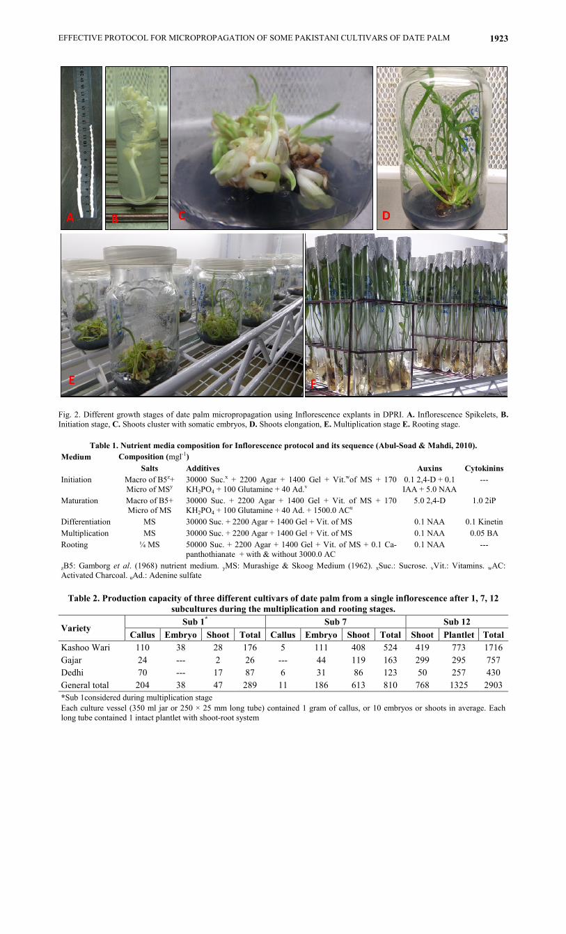
Fig. 2. Different growth stages of date palm micropropagation using Inflorescence explants in DPRI. A. Inflorescence Spikelets, B.
Initiation stage, C. Shoots cluster with somatic embryos, D. Shoots elongation, E. Multiplication stage E. Rooting stage.
Table 1. Nutrient media composition for Inflorescence protocol and its sequence (Abul-Soad & Mahdi, 2010).
Medium Composition (mgl-1)
Salts Additives Auxins Cytokinins
Initiation Macro of B5z+
Micro of MSy
30000 Suc.x + 2200 Agar + 1400 Gel + Vit.wof MS + 170
KH2PO4 + 100 Glutamine + 40 Ad.v
0.1 2,4-D + 0.1
IAA + 5.0 NAA
---
Maturation Macro of B5+
Micro of MS
30000 Suc. + 2200 Agar + 1400 Gel + Vit. of MS + 170
KH2PO4 + 100 Glutamine + 40 Ad. + 1500.0 ACu
5.0 2,4-D 1.0 2iP
Differentiation MS 30000 Suc. + 2200 Agar + 1400 Gel + Vit. of MS 0.1 NAA 0.1 Kinetin
Multiplication MS 30000 Suc. + 2200 Agar + 1400 Gel + Vit. of MS 0.1 NAA 0.05 BA
Rooting ¼ MS 50000 Suc. + 2200 Agar + 1400 Gel + Vit. of MS + 0.1 Ca-
panthothianate + with & without 3000.0 AC
0.1 NAA ---
zB5: Gamborg et al. (1968) nutrient medium. yMS: Murashige & Skoog Medium (1962). xSuc.: Sucrose. vVit.: Vitamins. wAC:
Activated Charcoal. uAd.: Adenine sulfate
Table 2. Production capacity of three different cultivars of date palm from a single inflorescence after 1, 7, 12
subcultures during the multiplication and rooting stages.
Variety Sub 1
* Sub 7 Sub 12
Callus Embryo Shoot Total Callus Embryo Shoot Total Shoot Plantlet Total
Kashoo Wari 110 38 28 176 5 111 408 524 419 773 1716
Gajar 24 --- 2 26 --- 44 119 163 299 295 757
Dedhi 70 --- 17 87 6 31 86 123 50 257 430
General total 204 38 47 289 11 186 613 810 768 1325 2903
*Sub 1considered during multiplication stage
Each culture vessel (350 ml jar or 250 × 25 mm long tube) contained 1 gram of callus, or 10 embryos or shoots in average. Each
long tube contained 1 intact plantlet with shoot-root system

MUSHTAQUE AHMED JATOI ET AL., 1924
Results and Discussion
The experiments were resulted in successful large
scale micropropagation protocol of date palm using
inflorescence explants. Date palm cultures during
initiation process have been commonly observed to
release phenolic compounds into the nutrient medium,
which inhibit and often cease their own growth (Reuveni
& Kipins, 1974). No browning and bacterial
contamination observed during initiation phase which is
commonly found using offshoot shoot tip explants. After
initiation process, the nutrient medium protocol decides
that whether the explants turned into callus or somatic
embryos categorized into indirect and direct somatic
embryogenesis respectively. All inflorescence spike
explants responded well to the starting nutrient medium.
Shining globular creamy structures formation was
obtained within 2 months through 1-2 re-cultures.
Maturation of initial structures occurred within 2-3
months through2-3 re-cultures. After the differentiation
process, three types of cultures were obtained e.g.,
embryogenic callus, somatic embryos and green shoots.
Somatic embryos can be generally divided into two
categories. First category is the individual somatic
embryos and second is a cluster of embryos (multiple
embryos) (Fig. 2C). The growth behavior of the
individual embryo is to grow vertically to produce more
leaves and roots (Fig. 2D) while the multiple embryosis
usually proliferating to additional shoots and somatic
embryos which suits the multiplication stage (Fig. 2E).
Hussain et al. (2001) reported while working on
micropropagation of three Pakistani date palm varieties
using shoot tip explants that multiplication rate is variety
dependent response. Al-Khateeb (2006) while working on
role of auxin and cytokinin concentrations on shoot
proliferation of date palm var. Sukary found that lower
concentrations of PGRs enhanced bud and shoot
formation whereas higher concentrations inhibited buds
and shoots production and resulted in abnormal
development. Zaid et al. (2006) also obtained a high rate
of multiplication when the cultures were transferred to
media supplement with low levels of hormones.
In the first subculture of multiplication stage, 110, 24
& 70jars were having embryogenic callus in cvs. Kashoo
wari, Gajar & Dedhi, respectively. While, with 38 jars
having multiple embryos cv. Kashoo wari appeared the
only cultivar produced embryos. However, 28, 2 & 17 jars
of cvs. Kashoo wari, Gajar & Dedhi were appeared with
shoots respectively (Table 2). All of these cultures were
transferred onto the proliferation medium (Table 1). The
embryogenic callus exposed high morphogenetic
potentiality to differentiate to intact somatic embryos.
During this process very little callus formation was
occurred till subculture 7 of multiplication stage where
the callus jars decreased to 5, 0 & 6 jars in cvs. Kashoo
wari, Gajar & Dedhi respectively. While, with 111 and
408 jars of embryo and shoot cv. Kashoo wari produced
prominent number of cultures as compared to cv. Gajar
(44 embryo and 119 shoot jars) and cv. Dedhi (31 embryo
and 86 shoots jars). During multiplication stage some
shoots were growing up and reached to an appropriate
height for rooting stage and subsequently subjected to the
rooting medium (Table 1) (Fig. 2F). It was observed that
removing the initial roots completely or trimming to 1-2
mm enhanced thicker-white adventitious root formation.
Leaving the primary roots without trimming during
rooting stage inhibited the adventitious roots formation
which is important for the further growth in the
acclimatization stage (Abul-Soad & Jatoi, 2014). Tisserat
(1982) found the optimum adventitious rooting and
subsequent plant survival using medium supplemented
with 0.1 mg/l auxin for 8 to 16 weeks before transfer of
plantlets to greenhouse for acclimatization. Hassan et al.
(2008) studied the interaction between sucrose
concentrations and MS strength during In vitro rooting
stage of date palm var. Bartamouda. They observed that
sucrose plays a significant role in root initiation. They
found that ¾ MS salt strength in combination with 45 g/l
sucrose enhanced number and length of roots, number and
length of shoots, and thickness of roots and leaves. Finally, all callus and embryos differentiated into
shoots and rooted plantlets on rooting medium. Where number of shoots and plantlets were reached at 419 and 773 in cv. Kashoo wari, 299 and 295 in cv. Gajar, and 50 and 257 in cv. Dedhi, respectively. The shoot jars increased from 28, 2 & 17 in subculture 1, then 408, 119 & 86 in subculture 7, and 419, 299 & 50 in subculture 12. Each jar maintained 20-30 shoots, 5 of the mat least in the size of rooting stage while 1325 plantlets were in rooting stage in sub culture 12.
Rooting quality of the ex vitro plantlets of date palm
was the vital factor increased the survival percentage in
the greenhouse. Most of the reports indicated low survival
percentage 25-35% during acclimatization stage rather
than it used to be a big obstacle in the whole
micropropagation protocol (Abul-Soad et al., 1999;
Hegazy et al., 2006; Taha et al., 2007). Date palm tissue
culture derived plants do not establish easily when
transferred to ex vitro conditions (Abul-Soad et al., 1999).
Miller (1983) and Ziv (1986) suggested that removal of
lids or caps from culture vessels and placing in the area
where they will be acclimatized like in greenhouse for
few days may have positive effects when the plantlets are
transplanted into the soil medium and kept in green house
for acclimatization. On the contrary, Preece & Sutter
(1991) recommended that uncapping of culture vessels
should be done in stages, first by loosening the lids for a
day or two, then by partially removing the cap for another
day or two followed by complete removal. But the caps of
larger vessels such as mouth mason jars or aluminum foil
trays should be removed more slowly than narrow
mouthed vessels such as culture tubes. Gabr & Abd-Alla
(2010) reported that pre-acclimatization is an important
step to complete micropropagation process. Plantlets
grown in lab under optimum conditions (moisture, salts,
sucrose and water) lack cuticle layer in leaves with high
transpiration rate. They observed that the presence of
PEG in MS medium increased the cuticle formation in
leaves & root thickness and decreased transpiration rate
that made a balance between transpiration and salts
uptake from nutrient media. They established pre-
acclimatization stage by gradual removing caps of culture
vessels that resulted in high survival rates after five weeks
of transplanting the plantlets.
EFFECTIVE PROTOCOL FOR MICROPROPAGATION OF SOME PAKISTANI CULTIVARS OF DATE PALM 1925
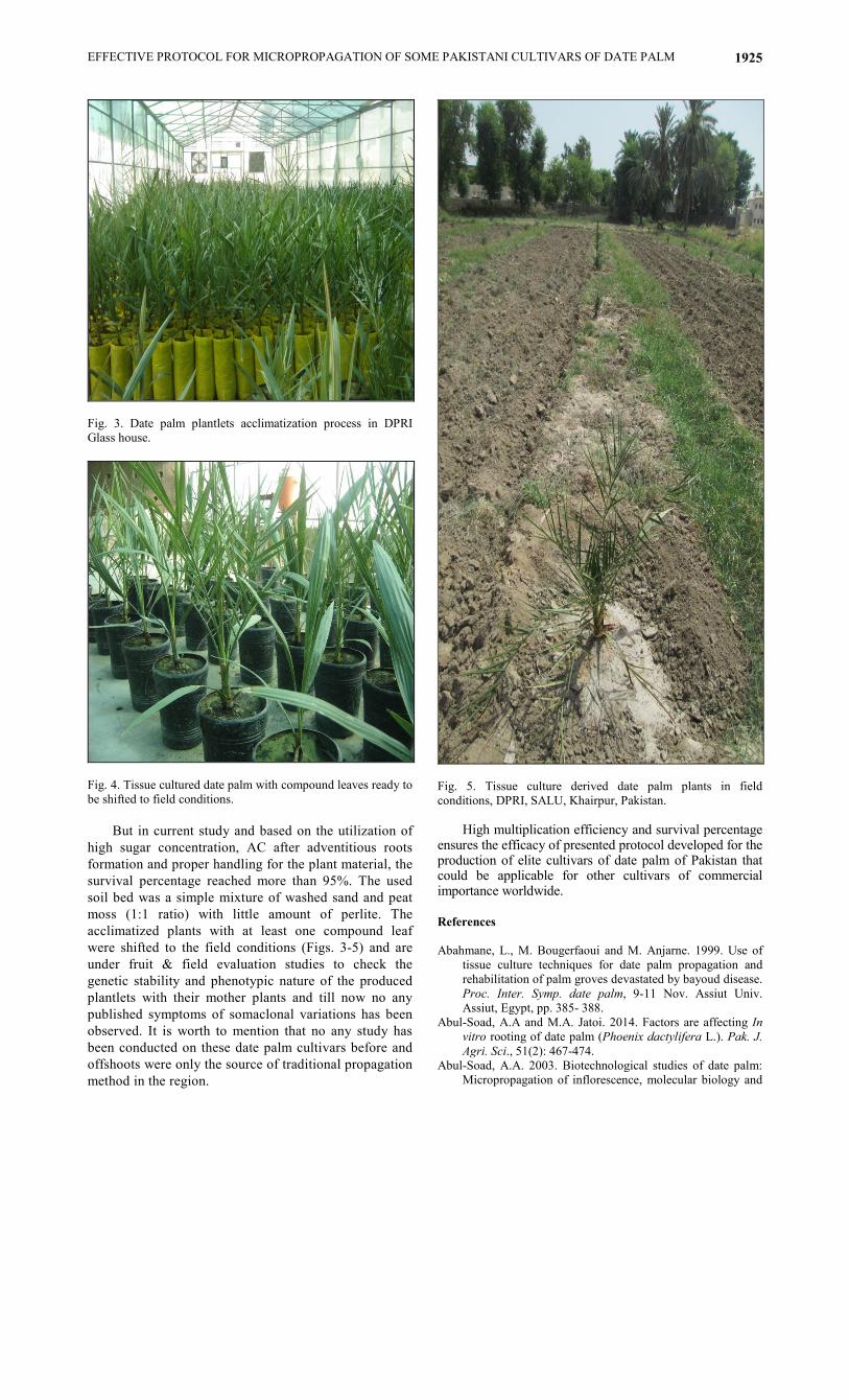
Fig. 3. Date palm plantlets acclimatization process in DPRI
Glass house.
Fig. 4. Tissue cultured date palm with compound leaves ready to
be shifted to field conditions.
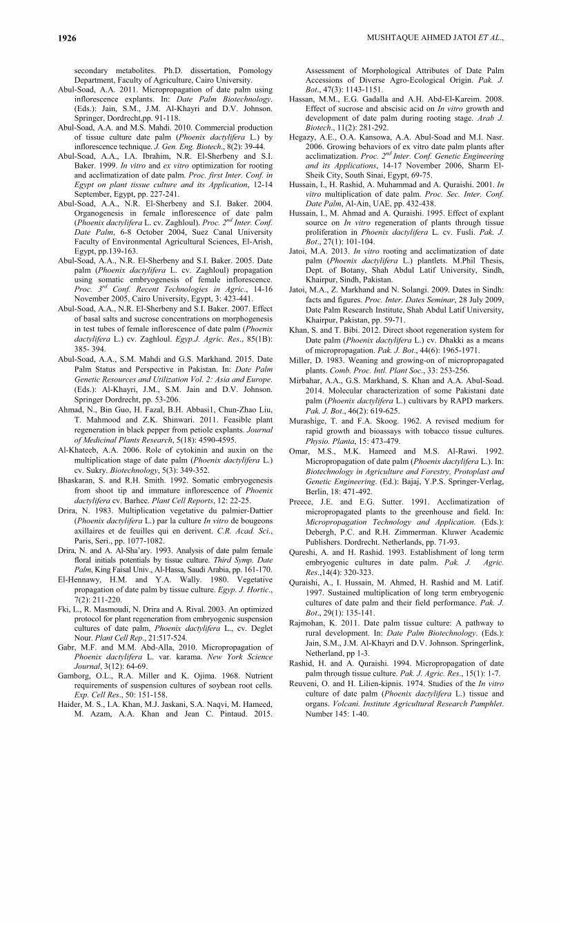
Fig. 5. Tissue culture derived date palm plants in field
conditions, DPRI, SALU, Khairpur, Pakistan.
But in current study and based on the utilization of
high sugar concentration, AC after adventitious roots
formation and proper handling for the plant material, the
survival percentage reached more than 95%. The used
soil bed was a simple mixture of washed sand and peat
moss (1:1 ratio) with little amount of perlite. The
acclimatized plants with at least one compound leaf
were shifted to the field conditions (Figs. 3-5) and are
under fruit & field evaluation studies to check the
genetic stability and phenotypic nature of the produced
plantlets with their mother plants and till now no any
published symptoms of somaclonal variations has been
observed. It is worth to mention that no any study has
been conducted on these date palm cultivars before and
offshoots were only the source of traditional propagation
method in the region.
High multiplication efficiency and survival percentage ensures the efficacy of presented protocol developed for the production of elite cultivars of date palm of Pakistan that could be applicable for other cultivars of commercial importance worldwide. References
Abahmane, L., M. Bougerfaoui and M. Anjarne. 1999. Use of
tissue culture techniques for date palm propagation and
rehabilitation of palm groves devastated by bayoud disease.
Proc. Inter. Symp. date palm, 9-11 Nov. Assiut Univ.
Assiut, Egypt, pp. 385- 388.
Abul-Soad, A.A and M.A. Jatoi. 2014. Factors are affecting In
vitro rooting of date palm (Phoenix dactylifera L.). Pak. J.
Agri. Sci., 51(2): 467-474.
Abul-Soad, A.A. 2003. Biotechnological studies of date palm:
Micropropagation of inflorescence, molecular biology and
MUSHTAQUE AHMED JATOI ET AL., 1926
secondary metabolites. Ph.D. dissertation, Pomology
Department, Faculty of Agriculture, Cairo University.
Abul-Soad, A.A. 2011. Micropropagation of date palm using
inflorescence explants. In: Date Palm Biotechnology.
(Eds.): Jain, S.M., J.M. Al-Khayri and D.V. Johnson.
Springer, Dordrecht,pp. 91-118.
Abul-Soad, A.A. and M.S. Mahdi. 2010. Commercial production
of tissue culture date palm (Phoenix dactylifera L.) by
inflorescence technique. J. Gen. Eng. Biotech., 8(2): 39-44.
Abul-Soad, A.A., I.A. Ibrahim, N.R. El-Sherbeny and S.I.
Baker. 1999. In vitro and ex vitro optimization for rooting
and acclimatization of date palm. Proc. first Inter. Conf. in
Egypt on plant tissue culture and its Application, 12-14
September, Egypt, pp. 227-241.
Abul-Soad, A.A., N.R. El-Sherbeny and S.I. Baker. 2004.
Organogenesis in female inflorescence of date palm
(Phoenix dactylifera L. cv. Zaghloul). Proc. 2nd Inter. Conf.
Date Palm, 6-8 October 2004, Suez Canal University
Faculty of Environmental Agricultural Sciences, El-Arish,
Egypt, pp.139-163.
Abul-Soad, A.A., N.R. El-Sherbeny and S.I. Baker. 2005. Date
palm (Phoenix dactylifera L. cv. Zaghloul) propagation
using somatic embryogenesis of female inflorescence.
Proc. 3rd Conf. Recent Technologies in Agric., 14-16
November 2005, Cairo University, Egypt, 3: 423-441.
Abul-Soad, A.A., N.R. El-Sherbeny and S.I. Baker. 2007. Effect
of basal salts and sucrose concentrations on morphogenesis
in test tubes of female inflorescence of date palm (Phoenix
dactylifera L.) cv. Zaghloul. Egyp.J. Agric. Res., 85(1B):
385- 394.
Abul-Soad, A.A., S.M. Mahdi and G.S. Markhand. 2015. Date
Palm Status and Perspective in Pakistan. In: Date Palm
Genetic Resources and Utilization Vol. 2: Asia and Europe.
(Eds.): Al-Khayri, J.M., S.M. Jain and D.V. Johnson.
Springer Dordrecht, pp. 53-206.
Ahmad, N., Bin Guo, H. Fazal, B.H. Abbasi1, Chun-Zhao Liu,
T. Mahmood and Z.K. Shinwari. 2011. Feasible plant
regeneration in black pepper from petiole explants. Journal
of Medicinal Plants Research, 5(18): 4590-4595.
Al-Khateeb, A.A. 2006. Role of cytokinin and auxin on the
multiplication stage of date palm (Phoenix dactylifera L.)
cv. Sukry. Biotechnology, 5(3): 349-352.
Bhaskaran, S. and R.H. Smith. 1992. Somatic embryogenesis
from shoot tip and immature inflorescence of Phoenix
dactylifera cv. Barhee. Plant Cell Reports, 12: 22-25.
Drira, N. 1983. Multiplication vegetative du palmier-Dattier
(Phoenix dactylifera L.) par la culture In vitro de bougeons
axillaires et de feuilles qui en derivent. C.R. Acad. Sci.,
Paris, Seri., pp. 1077-1082.
Drira, N. and A. Al-Sha’ary. 1993. Analysis of date palm female
floral initials potentials by tissue culture. Third Symp. Date
Palm, King Faisal Univ., Al-Hassa, Saudi Arabia, pp. 161-170.
El-Hennawy, H.M. and Y.A. Wally. 1980. Vegetative
propagation of date palm by tissue culture. Egyp. J. Hortic.,
7(2): 211-220.
Fki, L., R. Masmoudi, N. Drira and A. Rival. 2003. An optimized
protocol for plant regeneration from embryogenic suspension
cultures of date palm, Phoenix dactylifera L., cv. Deglet
Nour. Plant Cell Rep., 21:517-524.
Gabr, M.F. and M.M. Abd-Alla, 2010. Micropropagation of
Phoenix dactylifera L. var. karama. New York Science
Journal, 3(12): 64-69.
Gamborg, O.L., R.A. Miller and K. Ojima. 1968. Nutrient
requirements of suspension cultures of soybean root cells.
Exp. Cell Res., 50: 151-158.
Haider, M. S., I.A. Khan, M.J. Jaskani, S.A. Naqvi, M. Hameed,
M. Azam, A.A. Khan and Jean C. Pintaud. 2015.
Assessment of Morphological Attributes of Date Palm
Accessions of Diverse Agro-Ecological Origin. Pak. J.
Bot., 47(3): 1143-1151.
Hassan, M.M., E.G. Gadalla and A.H. Abd-El-Kareim. 2008.
Effect of sucrose and abscisic acid on In vitro growth and
development of date palm during rooting stage. Arab J.
Biotech., 11(2): 281-292.
Hegazy, A.E., O.A. Kansowa, A.A. Abul-Soad and M.I. Nasr.
2006. Growing behaviors of ex vitro date palm plants after
acclimatization. Proc. 2nd Inter. Conf. Genetic Engineering
and its Applications, 14-17 November 2006, Sharm El-
Sheik City, South Sinai, Egypt, 69-75.
Hussain, I., H. Rashid, A. Muhammad and A. Quraishi. 2001. In
vitro multiplication of date palm. Proc. Sec. Inter. Conf.
Date Palm, Al-Ain, UAE, pp. 432-438.
Hussain, I., M. Ahmad and A. Quraishi. 1995. Effect of explant
source on In vitro regeneration of plants through tissue
proliferation in Phoenix dactylifera L. cv. Fusli. Pak. J.
Bot., 27(1): 101-104.
Jatoi, M.A. 2013. In vitro rooting and acclimatization of date
palm (Phoenix dactylifera L.) plantlets. M.Phil Thesis,
Dept. of Botany, Shah Abdul Latif University, Sindh,
Khairpur, Sindh, Pakistan.
Jatoi, M.A., Z. Markhand and N. Solangi. 2009. Dates in Sindh:
facts and figures. Proc. Inter. Dates Seminar, 28 July 2009,
Date Palm Research Institute, Shah Abdul Latif University,
Khairpur, Pakistan, pp. 59-71.
Khan, S. and T. Bibi. 2012. Direct shoot regeneration system for
Date palm (Phoenix dactylifera L.) cv. Dhakki as a means
of micropropagation. Pak. J. Bot., 44(6): 1965-1971.
Miller, D. 1983. Weaning and growing-on of micropropagated
plants. Comb. Proc. Intl. Plant Soc., 33: 253-256.
Mirbahar, A.A., G.S. Markhand, S. Khan and A.A. Abul-Soad.
2014. Molecular characterization of some Pakistani date
palm (Phoenix dactylifera L.) cultivars by RAPD markers.
Pak. J. Bot., 46(2): 619-625.
Murashige, T. and F.A. Skoog. 1962. A revised medium for
rapid growth and bioassays with tobacco tissue cultures.
Physio. Planta, 15: 473-479.
Omar, M.S., M.K. Hameed and M.S. Al-Rawi. 1992.
Micropropagation of date palm (Phoenix dactylifera L.). In:
Biotechnology in Agriculture and Forestry, Protoplast and
Genetic Engineering. (Ed.): Bajaj, Y.P.S. Springer-Verlag,
Berlin, 18: 471-492.
Preece, J.E. and E.G. Sutter. 1991. Acclimatization of
micropropagated plants to the greenhouse and field. In:
Micropropagation Technology and Application. (Eds.):
Debergh, P.C. and R.H. Zimmerman. Kluwer Academic
Publishers. Dordrecht. Netherlands, pp. 71-93.
Qureshi, A. and H. Rashid. 1993. Establishment of long term
embryogenic cultures in date palm. Pak. J. Agric.
Res.,14(4): 320-323.
Quraishi, A., I. Hussain, M. Ahmed, H. Rashid and M. Latif.
1997. Sustained multiplication of long term embryogenic
cultures of date palm and their field performance. Pak. J.
Bot., 29(1): 135-141.
Rajmohan, K. 2011. Date palm tissue culture: A pathway to
rural development. In: Date Palm Biotechnology. (Eds.):
Jain, S.M., J.M. Al-Khayri and D.V. Johnson. Springerlink,
Netherland, pp 1-3.
Rashid, H. and A. Quraishi. 1994. Micropropagation of date
palm through tissue culture. Pak. J. Agric. Res., 15(1): 1-7.
Reuveni, O. and H. Lilien-kipnis. 1974. Studies of the In vitro
culture of date palm (Phoenix dactylifera L.) tissue and
organs. Volcani. Institute Agricultural Research Pamphlet.
Number 145: 1-40.

EFFECTIVE PROTOCOL FOR MICROPROPAGATION OF SOME PAKISTANI CULTIVARS OF DATE PALM 1927
Reynolds, J.F. and T. Murashige. 1979. Asexual embryogenesis
in callus culture of palms. In vitro, 15: 383-387.
Schroeder, C.A. 1970. Tissue culture of date shoots and
seedlings. Date Grower’s Inst. Rept., 49: 25-27.
Smith, S.N. 1975. Vegetative propagation of the date palm by
root tip culture. Bull. D’Agronomie Saharienne, 1: 67-77.
Taha, H.S., M.M. Hassan and M.K. El-Bahr. 2007.
Micropropagation of some Egyptian date palm dry
cultivars, 1- Maturation of somatic embryos. Arab J.
Biotech., 10(2):333-340.
Tisserat, B. 1979. Propagation of date palm (Phoenix dactylifera
L.) In vitro.J. Exp. Bot., 30(6): 1275-1283.
Tisserat, B. 1982. Factors involved in the production of plantlets
from date palm callus cultures. Euphytica, 31(1): 201-214.
Veramendi, J. and L. Navarro. 1996. Influence of physical
conditions of nutrient medium and sucrose on somatic
embryogenesis of date palm. Plant Cell Tiss. Org. Cult.,
45(2): 159-164.
Zaid, A., B. El-Korchi and H.J. Visser. 2011. Commercial date
palm tissue culture procedures and facility establishment. In:
Jain. S.M., J. M. Al-Khayri and D.V. Johnson (Eds,) Date
Palm Biotechnology, Springer, Dordrecht. pp. 137-180.
Zaid, A., H.H. Al Kaabi and B. El Korchi. 2007. Large scale In
vitro propagation of a rare and unique male date palm
(Phoenix dactylifera) using inflorescences technique. Acta
Hort., 736: 243-254.
Zaid, A., H.H. Al-Kaabi and B. El-Korchi. 2006. Impact of
lower concentration of growth regulators on the
multiplication stage of date palm organogenesis. 3rd Intern.
Date Palm Conf., Abu Dhabi, UAE. pp. 19-21.
Ziv, M. 1986. In vitro hardening and acclimatization of tissue
culture plants. In: Withers, L.A. and P.G. Alderson (Eds)
Plant Tissue Culture and its Applications. Butterworths,
London, UK. pp. 187-196.
(Received for publication 16 August 2014)