
ESICM obstetric critical care module - PACTpact.esicm.org/media/Obstetric critical care 30 April...
79
AN ESICM MULTIDISCIPLINARY DISTANCE LEARNING PROGRAMME FOR INTENSIVE CARE TRAINING Obstetric critical care Clinical problems 2013 Module Authors Eoin Casey The Mercy Hospital for Women and The Royal Melbourne Hospital, Melbourne, Australia Niamh Hayes Mater University and Rotunda Hospitals, Dublin, Ireland Andrew Ross The Mercy Hospital for Women and the Alfred Hospital, Melbourne, Australia Module Reviewers Liz Connolly and Daryl Dob Section Editor Janice Zimmerman
Transcript of ESICM obstetric critical care module - PACTpact.esicm.org/media/Obstetric critical care 30 April...

AN ESICM MULTIDISCIPLINARY DISTANCE LEARNING PROGRAMME FOR INTENSIVE CARE TRAINING
Obstetric critical care
Clinical problems
2013 Module Authors Eoin Casey The Mercy Hospital for Women and The Royal
Melbourne Hospital, Melbourne, Australia
Niamh Hayes Mater University and Rotunda Hospitals, Dublin, Ireland
Andrew Ross The Mercy Hospital for Women and the Alfred Hospital, Melbourne, Australia
Module Reviewers Liz Connolly and Daryl Dob Section Editor Janice Zimmerman

Learning Objectives:
After studying this module on Obstetric critical care, you should be able to:
1. Recognise critical illness and the indications for critical care admission in the obstetric population.
2. Institute immediate and appropriate resuscitative measures with due consideration for both mother and fetus.
3. Understand the physiological adaptations of pregnancy and appreciate how they impact on critical illness.
4. Clinically evaluate and manage both pregnancy-specific and other conditions associated with pregnancy requiring critical care admission.
Copyright©2013. European Society of Intensive Care Medicine. All rights reserved.
Contents
Introduction ............................................................................................... 1
1/ Initial assessment and stabilisation ............................................................... 2
General principles ..................................................................................... 2
Respiratory distress ................................................................................... 3
Causes of respiratory distress in the obstetric patient ....................................... 4
Initial evaluation and management .............................................................. 4
Airway management of the obstetric patient requiring intubation ........................ 5
Haemodynamic compromise ......................................................................... 5
Assessment of the patient with haemodynamic compromise ................................ 6
Resuscitation of the haemodynamically compromised patient ............................. 7
Altered mental status/neurological abnormalities ............................................. 10
Radiological imaging ................................................................................. 12
Medication use ........................................................................................ 13
Fetal considerations ................................................................................. 14
Prediction of maternal prognosis .................................................................. 16
2/ Cardiovascular disorders of pregnancy ........................................................... 17
Hypertensive disorders .............................................................................. 17
Pre-eclampsia ...................................................................................... 18
Eclampsia ........................................................................................... 25
HELLP syndrome ................................................................................... 25
Peripartum cardiomyopathy ........................................................................ 27
Diagnosis and clinical evaluation ................................................................ 28
Management ........................................................................................ 29
Venous thromboembolism ........................................................................... 30

Diagnosis and clinical evaluation ................................................................ 30
Management ........................................................................................ 31
Amniotic fluid embolism ............................................................................ 32
Diagnosis and clinical evaluation ................................................................ 33
Management ........................................................................................ 34
Cardiac arrest in pregnancy ........................................................................ 35
Management ........................................................................................ 35
3/ Haemorrhage during pregnancy ................................................................... 37
Evaluation of post-partum haemorrhage ...................................................... 39
Management of PPH ............................................................................... 40
Trauma in pregnancy ................................................................................ 44
Management ........................................................................................ 45
Blunt abdominal trauma ............................................................................. 46
4/ Respiratory disorders during pregnancy ......................................................... 49
Asthma ................................................................................................. 49
Clinical evaluation ................................................................................. 49
Management ........................................................................................ 49
Acute respiratory distress syndrome (ARDS) ..................................................... 50
Management ........................................................................................ 51
Tocolytic-induced pulmonary oedema ............................................................ 51
Management ........................................................................................ 52
Mechanical ventilation .............................................................................. 53
5/ Infection in Pregnancy .............................................................................. 55
Clinical evaluation ................................................................................... 55
Management ........................................................................................... 56
6/ Hepatic disorders in pregnancy ................................................................... 59
Clinical evaluation ................................................................................... 59
Acute fatty liver of pregnancy ..................................................................... 60
Diagnosis and clinical evaluation ................................................................ 60
Management ........................................................................................ 63
Intrahepatic cholestasis of pregnancy (IHCP) .................................................... 63
Conclusion ................................................................................................ 64
Patient Challenges ...................................................................................... 65

1
INTRODUCTION The critically ill obstetric patient presents a unique clinical challenge to the intensivist because of maternal physiological adaptations to pregnancy, pregnancy-specific conditions which may require critical care management and also the presence of a fetus whose well-being is linked to the mother. Successful maternal and neonatal outcomes for patients admitted to a critical care facility are largely dependent on a multidisciplinary approach to management requiring input from critical care personnel, obstetricians, anaesthetists, neonatologists and midwives. The obstetric patient may be afflicted with any surgical/medical condition necessitating intensive care unit (ICU) admission. There are however a number of pregnancy-specific conditions which account for the majority of critical care admissions. This module will focus, for the most part, on these particular conditions. Critical care admission of the obstetric patient is relatively infrequent; data from the UK and USA reveal admission rates of up to 0.9% of all mothers during their pregnancy or puerperium. Although the obstetric population is generally young and healthier, maternal mortality for those admitted to an ICU ranges from 5-20%.
Maternal mortality is rare in the developed world. The Eighth Report of the Confidential Enquiries into Maternal Deaths in the United Kingdom revealed a reduction in the overall maternal death rate from 13.95 per 100,000 maternities in the previous triennium to 11.39 in the 2006-08 triennium.
Cantwell R, Clutton-Brock T, Cooper G, Dawson A, Drife J, Garrod D, et al. Saving Mothers’ Lives: Reviewing maternal deaths to make motherhood safer: 2006-2008. The Eighth Report of the Confidential Enquiries into Maternal Deaths in the United Kingdom. BJOG 2011; 118 Suppl 1: 1-203. PMID 21356004. http://www.hqip.org.uk/cmace-reports/
Baskett TF. Epidemiology of obstetric critical care. Best Pract Res Clin Obstet Gynaecol 2008; 22(5): 763-774. PMID 18667364
Pollock W, Rose L, Dennis CL. Pregnant and postpartum admissions to the intensive care unit: a systematic review. Intensive Care Med 2010; 36(9): 1465–1474. PMID 20631987
Providing equity of critical and maternity care for the critically ill pregnant or recently pregnant woman, July 2011 http://www.rcog.org.uk/files/rcog-corp/Prov_Eq_MatandCritCare.pdf
Female admissions (aged 16-50 years) to adult, general critical care units in England, Wales and Northern Ireland, reported as “currently pregnant” or “recently pregnant” 1 January 2007 to 31 December 2007 www.oaa-anaes.ac.uk/assets/_managed/editor/File/Reports/ICNARC_obs_report_Oct2009.pdf

2
1/ INITIAL ASSESSMENT AND STABILISATION General principles As in the critically ill non-pregnant patient, initial evaluation and resuscitation of the obstetric patient should focus on airway, breathing and circulation. The clinician should have an appreciation of the physiological and anatomical adaptations to pregnancy in order to optimally evaluate and manage the critically ill obstetric patient. For more information on the conduct of a primary survey, review the PACT module on Clinical examination.
In the obstetric patient, uterine displacement should be considered part of the initial ABC evaluation in the haemodynamically unstable obstetric patient. Immediate assessment of gestational age is necessary because of potential aorto-caval compression from the pregnant uterus if >20 weeks gestation. If there is any doubt about gestational age, the clinician should proceed to perform a uterine displacement manoeuvre as soon as possible in the setting of haemodynamic instability. Senior obstetric, anaesthetic and midwifery staff need to be notified early of the critically ill obstetric patient as delivery of the fetus may be required to rescue a deteriorating situation. Ultimately, maternal well-being is the priority.
When treating a critically ill obstetric patient, remember that what benefits the mother is (in general) good for the fetus. Q. How do you clinically evaluate gestational age (GA)? A. The three basic methods used to help estimate gestational age (GA) are menstrual history, clinical examination, and ultrasonography. The first day of the last menstrual period (LMP) is required for calculation of an expected date of delivery (EDD) according to Naegele’s rule; Add 9 months + 7 days to LMP. The size of the uterus can be assessed clinically by measuring the symphysis to fundal height which is at best a crude estimate of gestation. At 20 weeks the fundal height should be 20cm +/- 2cm above the symphysis (approximately at the level of the umbilicus). Obstetric ultrasonography is the most accurate method of estimating GA. Multiple pregnancies, fetal macrosomia, polyhydramnios, or uterine fibroids can make GA assessment more difficult. Q. What methods are available to facilitate uterine displacement? A. In the operating room, a lateral tilt can be applied to the table, aiming for approximately 15-30 of tilt to the left. A wedge/doubled-up pillow can be placed under the patient’s right hip to facilitate inclination to her left.

3
Manual uterine displacement by lifting the uterus with two hands cephalad and to the left can also be performed. See images: http://circ.ahajournals.org/content/122/18_suppl_3/S829. figures-only Fetal monitoring is an essential aspect of the management of the critically ill obstetric patient and should be performed by an obstetric nurse in the ICU at least every 4 to 8 hours while the patient is critically ill and more frequently should their condition deteriorate. Continuous fetal monitoring is appropriate in the most serious situations. Urgent Caesarean delivery may be required at short notice and as such, all necessary staff and equipment should be readily available. Respiratory distress Assessment, resuscitation and management of the critically ill obstetric patient with respiratory compromise use the same principles as for the non-obstetric patient. Anatomical and physiological maternal adaptations to pregnancy may affect the approach to resuscitation and management and are summarised in the table below (Adapted from the Royal College of Obstetricians and Gynaecology Green-top guideline No.56, http://www.rcog.org.uk/files/rcog-corp/GTG56.pdf). Physiological and physical changes of pregnancy
Parameter Change Impact on resuscitative care
Anatomical Upper
airway/Larynx/Trachea Mucosal oedema & hypervascularity
Difficult airway management; use of smaller
endotracheal tube Diaphragm Splinted secondary to gravid
uterus Decreased functional
residual capacity; earlier hypoxia during apnoeic
periods Stomach Reduced motility and
increased intragastric pressure
Increased likelihood of aspiration
Physiological Minute ventilation 45% / ↓PCO2 Decreased buffering
capacity; acidosis more likely
Oxygen consumption ↑20% Hypoxia more likely Functional residual capacity ↓25% Develop hypoxia earlier
Chest wall compliance ↓ Secondary to gravid uterus displacing diaphragm
cephalad
↓ Transalveolar pressures
Maternal acid-base physiology also changes significantly through pregnancy as detailed in the table below:
4
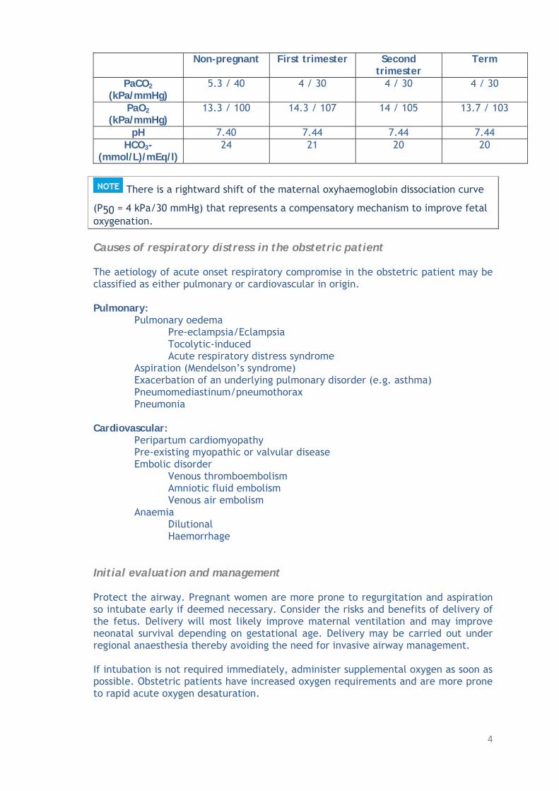
Non-pregnant First trimester Second trimester
Term
PaCO2
(kPa/mmHg) 5.3 / 40 4 / 30 4 / 30 4 / 30
PaO2
(kPa/mmHg) 13.3 / 100 14.3 / 107 14 / 105 13.7 / 103
pH 7.40 7.44 7.44 7.44 HCO3-
(mmol/L)/mEq/l) 24 21 20 20
There is a rightward shift of the maternal oxyhaemoglobin dissociation curve
(P50 = 4 kPa/30 mmHg) that represents a compensatory mechanism to improve fetal oxygenation. Causes of respiratory distress in the obstetric patient The aetiology of acute onset respiratory compromise in the obstetric patient may be classified as either pulmonary or cardiovascular in origin. Pulmonary:
Pulmonary oedema Pre-eclampsia/Eclampsia Tocolytic-induced Acute respiratory distress syndrome Aspiration (Mendelson’s syndrome) Exacerbation of an underlying pulmonary disorder (e.g. asthma) Pneumomediastinum/pneumothorax Pneumonia
Cardiovascular:
Peripartum cardiomyopathy Pre-existing myopathic or valvular disease Embolic disorder Venous thromboembolism Amniotic fluid embolism Venous air embolism Anaemia Dilutional Haemorrhage
Initial evaluation and management Protect the airway. Pregnant women are more prone to regurgitation and aspiration so intubate early if deemed necessary. Consider the risks and benefits of delivery of the fetus. Delivery will most likely improve maternal ventilation and may improve neonatal survival depending on gestational age. Delivery may be carried out under regional anaesthesia thereby avoiding the need for invasive airway management. If intubation is not required immediately, administer supplemental oxygen as soon as possible. Obstetric patients have increased oxygen requirements and are more prone to rapid acute oxygen desaturation.

5
Airway management of the obstetric patient requiring intubation The incidence of a difficult airway is approximately four times more likely in the obstetric population while the incidence of failed intubation is approximately ten times more likely. All obstetric patients should be considered as having potential difficult airways until proven otherwise. If intubation is deemed necessary in a critically ill obstetric patient, adhering to a few basic principles will optimise conditions and increase the likelihood of success.
1. Determine that intubation is clinically necessary. 2. Perform a rapid and complete airway assessment. Have a low threshold for
calling for help if a difficult airway is suspected. 3. Neutralise gastric acid should time allow. 4. Optimise patient positioning. Allow for uterine displacement. 5. Ensure presence of adequately trained assistants. 6. Choose the most appropriate laryngoscopic device, usually that which the
operator is most familiar with. 7. Ensure all airway equipment is checked and operational. Have difficult airway
equipment readily available. 8. Consider smaller diameter endotracheal tube given the potential for airway
oedema. Think: Why might a nasopharyngeal airway be relatively contraindicated in the obstetric patient.
9. Allow time for pre-oxygenation: at least 3 minutes of breathing 100% oxygen with a closed circuit and tightly fitted facemask or if time is limited, instruct the patient to take five vital capacity breaths of 100% oxygen.
10. Have a back-up plan should intubation with standard laryngoscopic technique prove difficult or impossible.
11. Once the patient has been intubated, consider spontaneous ventilation modes. Positive pressure ventilation may worsen the effects of aorto-caval compression on venous return.
For more information on management of the difficult airway please review the PACT module on Airway management and the following reference.
Vasdev GM, Harrison BA, Keegan MT, Burkle CM. Management of the difficult
and failed airway in obstetric anesthesia. J Anesth 2008; 22(1): 38–48. PMID 18306012
For information on mechanical ventilation in the critically ill obstetric patient refer to the section on Respiratory disorders in pregnancy. Haemodynamic compromise Haemodynamic compromise is a common indication for ICU admission in the obstetric population manifesting as hypotension, hypertension or more rarely as a cardiac dysrhythmia.

6
Causes of maternal hypotension include: Obstetric haemorrhage (particularly post-partum) Sepsis Peripartum cardiomyopathy Amniotic fluid embolism Pulmonary embolism Uterine rupture Epidural/spinal anaesthetic
The most common causes of hypotension in the obstetric population are haemorrhage and sepsis. For more information on hypotension and shock please review the PACT module on Hypotension.
Hypertension in the obstetric patient must be considered a sign of pre-eclampsia until proven otherwise. For more information on hypertensive disorders of pregnancy please review the task on cardiovascular disorders of pregnancy. Cardiac dysrhythmias are a rare cause of haemodynamic compromise in the obstetric population. Increased oestrogen production and significant changes to cardiovascular physiology are felt to make pregnancy a more pro-arrhythmic state. The incidence of paroxysmal supraventricular tachycardia is increased in pregnancy whereas atrial fibrillation and ventricular tachycardia are rare. Treatment of cardiac dysrhythmias in the obstetric patient is similar to the non-obstetric patient. For more information on the management of cardiac arrhythmias, review the PACT module on Arrhythmia.
Amiodarone should be avoided as it may inhibit fetal thyroid function. The remainder of this task will focus on the assessment, resuscitation and management of the obstetric patient presenting with shock. Assessment of the patient with haemodynamic compromise Having ensured a patent airway and adequate ventilatory status, the clinical focus should turn to evaluation of the patient’s haemodynamic state. A focused history, physical examination and application of basic cardiorespiratory monitors (heart rate, blood pressure and pulse oximetry) should occur early but should not distract from life-threatening resuscitative measures. Similar to the non-pregnant patient, clinical features in the obstetric patient with circulatory shock may include hypotension, tachycardia, tachypnoea, oliguria, and altered mental status.
Clinical evidence of shock may occur late or abnormal clinical features may be interpreted as normal for pregnancy. Clinical interpretation of physiological parameters in the patient with haemodynamic compromise must take into account the anatomical and physiological cardiovascular maternal adaptations that are described in the table below. (Adapted from the Royal

7
College of Obstetricians and Gynaecology Green-top guideline No.56, http://www.rcog.org.uk/files/rcog-corp/GTG56.pdf). Physiological and physical changes of pregnancy
Parameter Change Impact on resuscitative care
Plasma volume ↑ 50% Dilutional anaemia, ↓ O2 carrying capacity, Enhanced physiological
reserve against haemorrhage Heart rate ↑ 15-20 bpm Chest compressions during
CPR likely to be less effective as demand is
higher
Cardiac output ↑40%
Arterial blood pressure ↓ 10-15% ↓ Physiological reserve Uterine blood flow Accounts for 10% of cardiac
output at term Potential for massive blood
loss Cardiac anatomy Heart rotated cephalad and
to the left ↑ Chamber size, particularly
the left atrium
Predisposition to cardiac dysrhythmias, especially
supraventricular tachycardia
Think: What changes might you expect on examination of the praecordium?
The reduction in blood pressure in pregnancy is predominantly secondary to a decrease in the diastolic component which is reflective of the progesterone-stimulated reduction in systemic vascular resistance and the development of the placenta, a low resistance vascular bed. The increased cardiac output that develops in pregnancy is further augmented during the third stage of labour (delivery of the placenta) as a result of auto-transfusion of blood from the utero-placental to maternal circulation as the uterus contracts. Relief of aorto-caval compression also increases preload.
The utero-placental vascular bed under normal circumstances is maximally dilated. Resuscitation of the haemodynamically compromised patient The priority in resuscitation is to optimise and maintain maternal cardiac output and preserve adequate tissue and placental perfusion. Establish large bore intravenous access and send blood for
Complete blood count Urea, creatinine & electrolytes Liver function tests Acid-base analysis Coagulation screen Group/Type and crossmatch
Q. What is the supine hypotension syndrome? A. This syndrome is a physiological phenomenon (also referred to as aorto-caval compression) occurring after 20 weeks gestation when the supine position results in

8
compression of the inferior vena cava and abdominal aorta by the gravid uterus. This results in a reduction in venous return (preload) and in severe cases afterload can increase with a secondary reduction in cardiac output and hence blood pressure. Patients may present with pallor, nausea, sweating, dizziness, hypotension and bradycardia. It is particularly prominent in patients whose cardiovascular system has already been compromised (e.g. those under anaesthesia, significant blood loss). Interpretation of laboratory results in haemodynamic compromise requires a knowledge of expected values in a normal pregnancy at a particular gestation. The table below details the common changes in haematological and biochemical parameters that occur with pregnancy and how they impact on maternal/fetal resuscitation.
Parameter Non-pregnant Term pregnancy Impact on resuscitative care PaO2 (kPa / mmHg)
13.3 / 100 13.7 / 103 A rightward shift of the maternal oxyhaemoglobin dissociation curve is a compensatory mechanism to improve fetal oxygenation
PaCO2 (kPa / mmHg)
5.3 / 40 4 / 30 Maintenance of materno-fetal CO2 gradient is important for ongoing fetal CO2 excretion
HCO3- (mmol/L/mEq/L)
24 20 ↓ Buffering capacity, acidosis more likely
Haematocrit (%) 37-39 33-35 ↓ Oxygen carrying capacity White cell count (n × 109/l)
4 – 11 6 - 16 Interpretation of trends in infection more difficult
Platelet count (n × 109 /l
150-400 150-400 Gestational thrombocytopaenia is common, a level <100 × 109/l warrants investigation
Coagulation screen
Fibrinogen levels may increase up to 50% at term
PT (Prothrombin time) / aPTT are unchanged Predominant ↑ in clotting factors and ↓ in fibrinolytic activity Generalised hypercoagulable state
Urea 2.5–7.5 (mmol/L) (7.0–21.0 mg/dL)
2.4–3.8 (mmol/L) (6.7–10.6 mg/dL)
Seemingly normal renal indices may indicate renal dysfunction in the parturient
Creatinine 65-101(mol/l) (0.7–1.14 mg/dL)
55-73 (mol/l) (0.6–0.8 mg/dL)
Liver function tests
Transaminase levels - unchanged. Alkaline phosphatase markedly elevated
Total protein 64-86 (g/l) (6.4–8.6 g/dL)
48-64 (g/l) (4.8–6.4 g/dL)
Reduction in albumin:globulin ratio, ↑free fraction of albumin-bound medications ↓ Colloid oncotic pressure
9
Fluid therapy in combination with vasoactive and/or inotropic agents are central to the initial correction of haemodynamic compromise in the critically ill obstetric patient.
The colloid oncotic pressure in the obstetric patient is reduced approximately 14% from the non-pregnant state. Overzealous fluid loading in obstetric patients, particularly those with pre-eclampsia can precipitate pulmonary oedema due to leaky capillaries. The best fluid for resuscitation will depend on the cause of haemodynamic instability. Major haemorrhage generally requires replacement with blood products while other causes of shock will require judicious use of either a crystalloid or colloid solution or a combination of both. In general, critically ill obstetric patients are probably better off with a slightly negative volume status given the potential deleterious effects of fluid overload and noncardiogenic pulmonary oedema. They may tolerate a negative volume status better given their lack of significant co-morbidities. Vasopressors are commonly used in obstetrics particularly following spinal or epidural anaesthesia for Caesarean section. Ephedrine and phenylephrine are the most commonly used agents to offset the effects of sympathetic blockade. Phenylephrine causes less fetal acidosis than ephedrine. If vasopressors are required in a critical care setting, the choice of agent should be determined by maternal mean arterial pressure, systemic vascular resistance and cardiac output and should follow appropriate fluid loading.
All vasopressor agents may have deleterious effects on utero-placental perfusion. As in all patients with haemodynamic compromise, maternal response to therapy and clinical status in terms of tissue perfusion should be continuously re-evaluated. Basic indicators of tissue perfusion include:
Level of consciousness (Glasgow coma score) Vital signs Urine output Acid-base status and lactate concentration
Other monitors which may be considered include:
Minimally invasive devices (e.g. trans-oesophageal echocardiography, oesophageal Doppler or pulse contour analysis)
Invasive monitoring (e.g. central or mixed venous oxygen saturation) Noninvasive assessments (e.g. transthoracic echocardiography which is not
appropriate for continuous monitoring but valuable for the assessment of global cardiac function).
Assessment of the utero-placental-fetal unit (fetal well-being) is an important guide to adequacy of tissue perfusion and resuscitation. For further information on how to choose the appropriate haemodynamic monitor,

10
refer to the PACT module on Haemodynamic Monitoring and Management. Other useful references include:
Fujitani S, Baldisseri MR. Hemodynamic assessment in a pregnant and
peripartum patient. Crit Care Med 2005; 33(10 Suppl): S354-361. PMID 16215359
Dennis AT. Transthoracic echocardiography in obstetric anaesthesia and obstetric critical illness. Int J Obstet Anesth 2011; 20(2): 160–168. PMID 21315578
Altered mental status/neurological abnormalities Up to 50% of obstetric critical care patients have some form of neurological compromise. In most circumstances this occurs as a consequence of their admission diagnosis (i.e. pre-eclampsia or obstetric haemorrhage), rather than as the precipitant of their ICU admission.
Female admissions (aged 16-50 years) to adult, general critical care units in
England, Wales and Northern Ireland, reported as “currently pregnant” or “recently pregnant” 1 January 2007 to 31 December 2007 www.oaa-anaes.ac.uk/assets/_managed/editor/File/Reports/ICNARC_obs_report_Oct2009.pdf
Munnur U, Karnad DR, Bandi VD, Lapsia V, Suresh MS, Ramshesh P, et al. Critically ill obstetric patients in an American and an Indian public hospital: comparison of case-mix, organ dysfunction, intensive care requirements, and outcomes. Intensive Care Med 2005; 31(8): 1087-1094. PMID 16012807
Neurological diagnoses occurring as a result of or secondary to obstetric critical illness may be classified as: Complication of a pregnancy-specific illness:
Eclampsia Acute fatty liver of pregnancy Amniotic fluid embolism
Pre-existing medical condition that deteriorates during pregnancy:
Hypertension (encephalopathy) Intracranial neoplasm Epilepsy
Obstetric predisposition to particular medical conditions:
Cerebral venous sinus thrombosis Hepatitis E infection Subarachnoid haemorrhage

11
For a comprehensive review of neurological disorders in pregnancy, refer to:
Karnad DR, Guntupalli KK. Neurologic disorders in pregnancy. Crit Care Med
2005; 33(10 Suppl): S362-371. PMID 16215360
The principal aims in the neurological support of the critically ill obstetric patient are to relieve pain and anxiety and also to prevent secondary cerebral injury in those with a primary cerebral insult. All commonly prescribed sedative/anxiolytic and analgesic agents used in critical care are considered safe for both mother and fetus. Patients requiring longer term sedation and/or analgesia in critical care are probably best managed with benzodiazepines (e.g. midazolam or lorazepam) and/or opioids (e.g. fentanyl or morphine). If delivery is imminent and there is concern about fetal accumulation of longer-acting agents such as morphine, consider using shorter-acting agents. (e.g. remifentanil, propofol).
Long-term use of propofol as a sedative agent for obstetric patients in ICU raises concern about the potential for development of propofol-related infusion syndrome in the neonate. For more information on the use of sedatives and analgesics in the obstetric critical care patient please review: Pregnancy in the ICU: Drug implications http://www.sccm.org/Publications/Critical_Connections/Archives/August2010/Pages/PregnancyintheICU.aspx ABCD stabilisation with concomitant consideration of emergency fetal delivery if indicated are the initial priorities in preventing secondary neurological injury. The next step in prevention of further cerebral insult is maintenance of the cerebral perfusion pressure to ensure adequate cerebral blood flow with the following measures:
Ensure an adequate perfusing mean arterial pressure Nurse patient at 30 head up position (c.f. uterine displacement) Maintain normoglycaemia/normoxia/relative normocarbia of pregnancy Avoidance of pyrexia and institution of infection prophylaxis measures
Cerebral blood flow increases in pregnancy such that at term there is a 15% increase compared with the non-pregnant state. Cerebral autoregulation is preserved in healthy pregnancies. For more information on the prevention of secondary brain injury review the PACT module on Traumatic brain injury. Appropriate investigations as determined by the clinical context may include routine bloodwork, radiological investigations such as computerised tomography and/or magnetic resonance imaging. Electroencephalography and diagnostic lumbar puncture may also be useful and can be performed in the ICU.

12
Think: What contraindications to lumbar puncture do you know? For more information on the management, investigation and monitoring of patients requiring neuro-critical care please review PACT modules on Coma and altered consciousness, Sedation and analgesia and Traumatic brain injury. Criteria for diagnosis of brain death in the obstetric patient are the same as for the non-obstetric population. The presence of a pre-viable fetus in the event of maternal brain death raises complex legal and ethical issues regarding ongoing maternal support such that fetal gestational viability can be attained. For more information on this sensitive topic review the following reference.
Lane A, Westbrook A, Grady D, O’Connor R, Counihan TJ, Marsh B, et al.
Maternal brain death: medical, ethical and legal issues. Intensive Care Med 2004; 30(7): 1484-1486. PMID 15107974
Radiological imaging Maternal antenatal exposure to ionising radiation raises concerns about potentially harmful fetal effects which may occur secondary to teratogenic, carcinogenic or genetic effects. The risk of teratogenesis is greatest from week 1-15 of gestation. Exposure to ionising radiation is expressed in terms of the rad and fetal exposure to <5 rad is considered safe. The exposure dose to the fetus of most common radiological procedures performed on critical care patients is <1 rad. (see table below).
Procedure Fetal dose (millirad) Chest X-ray <1 Cervical spine plain film <1 CT Thorax 30-1300 (mean 600) CT Abdomen 250 CT Head <1000 Helical CT pulmonary angiogram (CTPA) <50 V/Q scan <100 Barium enema 700 -1600
CTPA was previously preferred to V/Q scan as the imaging of choice in the antenatal population for diagnosis of pulmonary embolism due to lower fetal radiation exposure. More recently, perfusion scanning is often preferred as the initial investigation due to concern about increased susceptibility of maternal breast tissue to the carcinogenic effects of ionising radiation in pregnancy. Think: What simple protective strategy can you use to protect the fetus from unnecessary exposure. When considering radiological investigation in the obstetric critical care patient, adhering to a few basic principles will lessen fetal exposure to radiation.

13
1. Consider whether maternal benefit of the investigation outweighs potential risk to the fetus. Do not withhold any investigation that may have maternal benefit because of concern about potential harmful fetal effects. 2. Avoid unnecessary routine imaging such as a daily chest X-ray unless clinically indicated. 3. Attempt to limit fetal radiation exposure during diagnostic testing by applying a lead apron to the maternal abdomen. 4. Consider if the maternal illness can be investigated by other imaging techniques such as ultrasound or MRI which do not require exposure to ionising radiation. For more information on radiological investigation of the obstetric patient, review
ACOG Committee on Obstetric Practice. ACOG Committee Opinion. Number
299, September 2004 (replaces No. 158, September 1995). Guidelines for diagnostic imaging during pregnancy. Obstet Gynecol 2004; 104(3): 647-651. PMID 15339791
http://www.acog.org/~/media/Committee%20Opinions/Committee%20on%20Obstetric%20Practice/co299.pdf?dmc=1&ts=20121107T2158533065
Medication use Invariably the critically ill obstetric patient will require medication both to optimise her physiological state and/or treat her underlying condition. Common critical care medications such as sedative agents, cardiovascular drugs and anticoagulant medication will often be required as well as obstetric-specific drugs such as magnesium sulphate, and uterotonic (oxytocin and ergometrine) and tocolytic (salbutamol and glyceryl trinitrate) agents. Maternal physiological changes significantly alter the absorption, distribution, metabolism and excretion aspects of drug pharmacokinetics which makes optimal dosing for prevention and therapy difficult (e.g. low molecular weight heparins). All clinicians should be familiar with the pharmacokinetic/pharmacodynamic profile particular to the obstetric patient and the medication prescribed as well as the potential adverse fetal effects. Input of a clinical pharmacist may be helpful in determining optimal dosing as well as safety.
Necessary medications should not be withheld because of fetal concerns. Medications used in obstetrics are classified according to risk to the fetus. Several countries have developed risk category systems and the table below lists the US Food and Drug administration (FDA) categories for some of the drugs that may be used in the critically ill patient. For more information on drugs commonly used in the obstetric critical care patient see the following reference.
Santillan M, Yankowitz J. Fetal Effects of Drugs Commonly Used in Critical
Care. In: Belfort MA, Saade GR, Foley MR, Phelan JP, Dildy GA III, editors. Critical Care Obstetrics. 5th ed. Wiley-Blackwell; 2010. ISBN-13: 978-1405152730. pp. 626–638

14
Drug Category A & B C D X
No fetal risk Evidence inconclusive: Use if maternal benefits outweigh fetal
risks
Fetal risk but potential for
maternal benefit may necessitate
use
Contraindicated as definite fetal
risk and no potential maternal benefit
Cardiovascular Dobutamine Methyldopa
Norepinephrine/epinephrine Milrinone
Glyceryl trinitrate / Hydralazine Digoxin /Adenosine
ACE (Angiotensin-converting
enzyme) inhibitors (>14 weeks gestation)
Amiodarone B-blockers (<14
weeks gestation)
Analgesics & Sedatives
Propofol Paracetamol NSAIDs (< 28
weeks gestation)
Fentanyl, Morphine, Haloperidol
Diazepam, Midazolam Lorazepam
NSAIDs >28 weeks gestation
Antimicrobial agents
Penicillins Cephalosporins
Macrolides Acyclovir
Aminoglycosides Vancomycin Quinolones
Amphotericin B
Fluconazole Ketoconazole
Anticoagulants Heparins & Low molecular weight heparins
Warfarin
Other Magnesium sulphate Insulin
Salbutamol
Glucocorticoids Neuromuscular relaxants
Fetal considerations Fetal well-being is inextricably linked to that of their mothers. Optimal maternal resuscitation and critical care management will improve utero-placental perfusion, feto-maternal gas exchange and acid-base equilibrium. The fetus survives in a relatively hypoxic environment despite high fetal oxygen extraction that is facilitated by increased utero-placental perfusion, increased fetal haematocrit, and fetal haemoglobin which shifts the oxyhaemoglobin curve to the left. Should early fetal delivery be required and time permits, administration of systemic corticosteroids (e.g. betamethasone 24 mg intramuscularly) in 2 divided doses 12 hours apart has been shown to improve fetal lung maturity and reduce the incidence of respiratory distress syndrome in premature neonates. Many neonatologists consider a fetus to be viable at a gestation of >24 weeks or estimated fetal weight of >500 g (although some have survived following delivery as early as 21 weeks gestation).

15
Clinicians should offer a single course of antenatal corticosteroids to women between 24 and 35 weeks gestation at risk of preterm birth. More information on the use of antenatal corticosteroids to prevent respiratory distress syndrome can be reviewed in the following reference.
Royal College of Obstetricians and Gynaecologists. Antenatal corticosteroids to reduce neonatal morbidity and mortality: Green-top guideline No. 7, 2010. http://www.rcog.org.uk/files/rcog-corp/GTG%207.pdf
Adequate fetal monitoring in critical care requires a trained obstetric nurse or midwife skilled in the application and interpretation of fetal heart rate monitors such as:
Doppler assessment of fetal heart rate Cardiotocograph assessment of fetal heart rate variability and reactivity to
uterine activity, present after 28 weeks gestation. Fetal monitor profiles can also be used as surrogate maternal monitors because deterioration in fetal monitor profiles tends to reflect deterioration in maternal status. Ultrasound assessment of the fetus allows calculation of a biophysical profile/score which assesses the fetus in terms of:
Breathing Tone Movement Amniotic fluid volume Non-stress test
Each parameter is assigned a score of 2 when present and 0 if absent. Scores >8 imply fetal well-being, 6 is equivocal, and <4 should prompt delivery provided delivery does not pose a serious maternal risk. More information on the biophysical profile can be found at this website. http://emedicine.medscape.com/article/405454-overview#aw2aab6b4
Ultrasound may also be used to assess flow in the umbilical vessels. Absent end-diastolic flow in the umbilical artery is strongly suggestive of impending fetal demise. Maternal admission to critical care is associated with significant fetal mortality risk, up to 50% in one study. Risk factors for fetal loss include low gestational age, maternal shock, and maternal blood transfusion.
Hazelgrove JF, Price C, Pappachan VJ, Smith GB. Multicenter study of obstetric
admissions to 14 intensive care units in southern England. Crit Care Med 2001; 29(4): 770–775. PMID 11373467

16
Cartin-Ceba R, Gajic O, Iyer VN, Vlahakis NE. Fetal outcomes of critically ill pregnant women admitted to the intensive care unit for nonobstetric causes. Crit Care Med 2008; 36(10): 2746-2751. PMID 18828192
Prediction of maternal prognosis The mortality rate for the critically ill pregnant patient ranges from 12-20%. Patients with an obstetric precipitant for their critical care admission tend to have a better prognosis because delivery will often improve their physiological status. Predicting prognosis in this population is difficult as no specific scoring system takes into account the maternal physiological adaptations as well as the rapid resolution of these parameters that tend to occur with delivery.
Standard critical care scoring systems tend to overestimate maternal mortality rates. Studies have revealed the APACHE II (Acute Physiology and Chronic Health Evaluation) and SAPS II (Simplified Acute Physiology Score) scoring systems to be more accurate in predicting mortality in obstetric patients admitted to critical care with non-obstetric illnesses. The Glasgow Coma Scale score does have predictive outcome value in patients admitted to critical care with eclampsia.
Naylor DF Jr, Olson MM. Critical care obstetrics and gynecology. Crit Care Clin
2003; 19(1): 127–149. PMID 12688581
Karnad DR, Lapsia V, Krishnan A, Salvi VS. Prognostic factors in obstetric patients admitted to an Indian intensive care unit. Crit Care Med 2004; 32(6): 1294–1299. PMID 15187509
Gilbert TT, Smulian JC, Martin AA, Ananth CV, Scorza W, Scardella AT; Critical Care Obstetric Team. Obstetric admissions to the intensive care unit: outcomes and severity of illness. Obstet Gynecol 2003; 102(5 Pt 1): 897-903. PMID 14672460
Bhagwanjee S, Paruk F, Moodley J, Muckart DJ. Intensive care unit morbidity and mortality from eclampsia: an evaluation of the Acute Physiology and Chronic Health Evaluation II score and the Glasgow Coma Scale score. Crit Care Med 2000; 28(1): 120-124. PMID 10667510

17
2/ CARDIOVASCULAR DISORDERS OF PREGNANCY Hypertensive disorders Hypertension remains a relatively common medical complication of pregnancy with related disorders affecting up to 12% of cases. A recent review revealed that hypertensive related disorders were the most prevalent indication for ICU admission [median: 0.9/1000 deliveries (range 0.2-6.7)]. In the most recent Centre for Maternal and Child Enquiries (CMACE 2011) report, pre-eclampsia/eclampsia persist as a leading cause of maternal mortality with a rate of 0.83/100,000 maternities.
Cantwell R, Clutton-Brock T, Cooper G, Dawson A, Drife J, Garrod D, et al.
Saving Mothers’ Lives: Reviewing maternal deaths to make motherhood safer: 2006-2008. The Eighth Report of the Confidential Enquiries into Maternal Deaths in the United Kingdom. BJOG 2011; 118 Suppl 1: 1-203. PMID 21356004. http://www.hqip.org.uk/cmace-reports/
Pollock W, Rose L, Dennis CL. Pregnant and postpartum admissions to the intensive care unit: a systematic review. Intensive Care Med 2010; 36(9): 1465-1474. PMID 20631987
Classification of hypertensive disorders in pregnancy
Chronic Hypertension (i) increased BP before week 20 (or known to exist prior to pregnancy) (ii) hypertension persistent for more than 12 weeks after pregnancy
Pre-eclampsia/Eclampsia (i) de novo appearance of hypertension after mid-pregnancy and proteinuria
at least 300 mg/24 hr Pre-eclampsia superimposed on Chronic Hypertension
(i) new onset proteinuria Gestational Hypertension
(i) transient hypertension appearing after mid-pregnancy confirmed by return to normal BP post-partum and the absence of proteinuria
From Working Group Report on High Blood Pressure in Pregnancy. National Heart, Lung, and Blood Institute. NIH Publication No. 00-3029, July 2000 http://www.nhlbi.nih.gov/guidelines/archives/hbp_preg/ For the remainder of this section we will focus on the pregnancy-specific condition, pre-eclampsia. For further information on other hypertensive disorders of pregnancy, refer to:
Mustafa R, Ahmed S, Gupta A, Venuto RC. A comprehensive review of
hypertension in pregnancy. J Pregnancy 2012; 2012: 105918. doi: 10.1155/2012/105918. Epub 2012 May 23. PMID 22685661
Vidaeff AC, Carroll MA, Ramin SM. Acute hypertensive emergencies in pregnancy. Crit Care Med 2005; 33(10 Suppl): S307-312. PMID 16215352

18
Pre-eclampsia Pre-eclampsia is characterised by the onset of hypertension and proteinuria after 20 weeks gestation in a previously normotensive patient. Abnormal placentation releases circulating factors that cause vascular endothelial dysfunction in the mother, manifesting as hypertension and renal dysfunction. Risk factors are listed below:
Primigravidae Multiple gestation Obesity Age >40 years Previous history of pre-eclampsia Family history of pre-eclampsia Increased time between pregnancies Chronic hypertension Chronic renal disease Diabetes mellitus Black race Antiphospholipid syndrome Autoimmune disease
For more information on the risk factors of pre-eclampsia please review the following article.
Duckitt K, Harrington D. Risk factors for pre-eclampsia at antenatal booking:
systematic review of controlled studies. BMJ 2005; 330(7491): 565. PMID 15743856
Diagnosis and clinical evaluation Pre-eclampsia is a multisystem disease process. Two cardinal features must exist in order to make the diagnosis:
Presence of sustained hypertension o Systolic blood pressure >140 mmHg o Diastolic blood pressure >90 mmHg
Proteinuria o >300 mg protein in a 24-hour urine collection
Usually such changes only occur after 20 weeks gestation. An exception is a molar pregnancy when they may occur at an earlier gestation. Peripheral oedema was previously considered a necessary component for diagnosis of pre-eclampsia but because of its prevalence with all pregnancies it has been removed as a diagnostic criterion.
Severe pre-eclampsia is more likely to require critical care and is diagnosed by the presence of more severe blood pressure elevation and significant organ dysfunction as noted below.
Systolic blood pressure >160 mmHg or diastolic blood pressure >110 mmHg on two occasions at least 2 hours apart
Proteinuria: >5 g in a 24hr collection Oliguria: <500 mL in 24hr
19
Elevated serum creatinine Pulmonary oedema or cyanosis Persistent headaches Visual disturbances Seizures Epigastric or right upper quadrant pain and/or elevated liver function tests Thrombocytopaenia and/or deranged coagulation tests Oligohydramnios, decreased fetal growth or placental abruption
Pre-eclampsia predominantly affects the maternal cardiovascular, neurological and renal systems, but all systems are affected to a greater or lesser degree. Tailor your clinical evaluation to focus on the more susceptible systems.
The systemic pathophysiological insult of pre-eclampsia also affects the feto-placental unit causing impaired utero-placental perfusion which manifests as intra-uterine growth restriction and oligohydramnios with an increased risk of fetal demise. Cardiovascular findings in pre-eclamptic patients tend to be complex and vary with gestation and disease severity. In addition to hypertension, there is usually increased systemic vascular resistance and reduced intravascular volume with a resultant reduction in cardiac output as disease severity progresses. Left ventricular function tends to deteriorate with worsening preload and afterload such that overzealous fluid resuscitation can precipitate pulmonary oedema. This is further exacerbated by the reduction in colloid oncotic pressure and increased capillary permeability that occurs with pre-eclampsia.
Vasoactive agents should be titrated with caution in pre-eclamptic patients secondary to their associated vascular hyper-reactivity. Think: Which monitors, laboratory and/or radiological investigations may be beneficial in the haemodynamic assessment of the pre-eclamptic patient? The presence of neurological signs and symptoms tend to be indicative of severe pre-eclampsia. The more common manifestations include:
Persistent headache Visual disturbances Hyperreflexia Seizures (indicative of eclampsia until proven otherwise)
Worsening neurological symptoms/signs may herald impending eclampsia or presence of intracerebral haemorrhage. Q. What are the potential causes of convulsions in the third trimester or puerperium? A. Convulsions in the third trimester or early puerperium should be considered eclampsia until proven otherwise

20
Other potential causes in the obstetric patient include: Hypoglycaemia Intracranial haemorrhage Cerebral venous sinus thrombosis Epilepsy
For a more complete list of the causes of seizures review the PACT module on Coma and altered consciousness and the following review article.
Mirski MA, Varelas PN. Seizures and status epilepticus in the critically ill. Crit
Care Clin 2008; 24(1): 115-147. PMID 18241782 Renal system The characteristic renal lesion of pre-eclampsia is caused by glomeruloendotheliosis. This lesion leads to a glomerulopathy, resultant reduced glomerular filtration rate, and increased glomerular permeability to serum proteins causing proteinuria. Generally, the worse the proteinuria the more severe the pre-eclampsia. Similarly, oliguria (<500 mL/24hr) is reflective of severe pre-eclampsia. Increasing serum uric acid levels also tend to reflect increasing severity of pre-eclampsia. Uric acid levels >5.5 mg/dL have been associated with an increase in perinatal morbidity and mortality.
The serum creatinine level tends to remain below normal non-pregnant levels unless there is severe pre-eclampsia with associated renal failure. Pre-eclampsia may worsen the already pre-existing pharyngolaryngeal oedema of pregnancy putting patients at even greater risk of airway compromise and difficulty with ventilation and oxygenation in an emergency. Pulmonary oedema is also a potential complication of severe pre-eclampsia. See PACT module on Airway management Laboratory investigations are necessary to assess the severity of pre-eclampsia. Appropriate investigations include:
Complete blood count (to include platelet count) Blood urea nitrogen/serum creatinine/serum urate Liver function tests (bilirubin/transaminases) Coagulation profile Blood type (group) and antibody screen 24 hour urine collection for protein if time permits
Other investigations should be guided by the patient’s clinical evaluation. Other evaluations that may be helpful include:
Chest X-ray if respiratory compromise is present, may identify pulmonary oedema
Non-contrast CT brain or MRI brain if seizures occur, rule out intracranial pathology

21
Ultrasound for evidence of intra-uterine growth restriction or oligohydramnios. Doppler estimation of umbilical artery flow may identify reverse end-diastolic flow indicative of fetal compromise and impending demise.
Management The only known cure for pre-eclampsia is delivery of the fetus and placenta. Optimal timing of delivery is a significant factor in materno-fetal prognosis. The main determinant of when to deliver is the assessment of disease severity. Evidence of severe pre-eclampsia mandates emergent delivery regardless of gestation whereas timing of delivery may be determined by gestation in mild cases, assuming the disease state remains stable with regular assessment. Delivery should not be delayed in pre-eclamptic mothers after 37 weeks because continuing the pregnancy puts both mother and fetus at risk from potential complications. Other immediate treatment priorities in managing pre-eclampsia are blood pressure control and seizure prophylaxis.
At term (at/after 37weeks gestation), it is deemed safe to deliver a fetus as fetal morbidity secondary to prematurity is very unlikely. Access to tertiary level critical care for both mother and baby should be readily available if required. For the most part patients with pre-eclampsia can be monitored and successfully managed in an obstetric unit with intermediate level care or a high dependency unit. Occasionally some patients may require management in an intensive care facility as a result of associated respiratory compromise and/or the need for invasive haemodynamic monitoring or support. Route of delivery is generally by Caesarean section for severe pre-eclamptics while those with mild or moderate disease and without evidence of significant fetal compromise may be offered a trial of labour and vaginal delivery. For more information: See Fetal considerations in Task 1. Serial measurement of haematological and biochemical parameters such as haemoglobin, platelet count, coagulation profile, electrolytes (serum magnesium), serum urate, liver function tests and urinalysis should be performed to aid assessment of disease progress. Although delivery of fetus and placenta is curative, post-partum patients are still at risk of pre-eclamptic complications particularly in the first 24-48 hours post delivery. For this reason, they should be closely monitored during this period with regular cardiovascular, respiratory and neurological assessments. Antepartum antihypertensive medications and seizure prophylaxis should be continued for at least the first 24 hours post-partum at which point patient re-evaluation should allow a decrease in therapies.
Eclamptic seizures have been reported up to 4 weeks post-partum.

22
Zeeman GG. Obstetric critical care: a blueprint for improved outcomes. Crit
Care Med 2006; 34(9 Suppl): S208-214. PMID 16917425 Fluid balance Intravenous fluid therapy in pre-eclamptic patients is controversial secondary to a potential delicate balance between the reduced intravascular volume associated with pre-eclampsia and the increased risk of fluid overload and pulmonary oedema that exists secondary to reduced colloid oncotic pressure and increased capillary permeability. Prior to commencement of intravenous fluid therapy, each patient’s hydration status should be assessed, a urinary catheter inserted and accurate hourly urine output recorded. Think: What clinical and biochemical markers of dehydration do you know? There is no evidence that colloid replacement in pre-eclampsia is superior to crystalloid except perhaps in cases where there is renal or cardiopulmonary compromise. Some authorities, however, advocate use of colloid replacement in patients with a significant reduction in colloid oncotic pressure (<12 mmHg). For more information on fluid management and other therapeutic principles in pre-eclampsia please review the following references.
Steegers EA, von Dadelszen P, Duvekot JJ, Pijnenborg R. Pre-eclampsia. Lancet
2010; 376(9741): 631-644. PMID 20598363
Engelhardt T, MacLennan FM. Fluid management in pre-eclampsia. Int J Obstet Anesth 1999; 8(4): 253-259. PMID 15321120
Invasive monitoring is usually only required in severe cases and the relative merits and potential complications of such monitors should be assessed on a case by case basis. For more information on invasive haemodynamic monitoring review the PACT module on Haemodynamic Monitoring and Management.
Fujitani S, Baldisseri MR. Hemodynamic assessment in a pregnant and
peripartum patient. Crit Care Med 2005; 33(10 Suppl): S354-361. PMID 16215359
Blood pressure control Treatment of hypertension in pre-eclampsia does not alter the underlying pathophysiological process. As such, the goal of antihypertensive treatment is
23
prevention of potential complications such as stroke (intracerebral haemorrhage), cardiac failure and placental abruption while maintaining adequate utero-placental perfusion. The threshold for treatment is a diastolic blood pressure (DBP) >110 mmHg and/or systolic blood pressure (SBP) >160 mmHg. Many patients with mild pre-eclampsia will be commenced on methyldopa as a first line agent which cannot be used in an acute situation due to its slow onset of action. Slow but steady reduction of SBP to 140-160 mmHg and DBP to 80-110 mmHg with constant fetal monitoring (fetal heart rate) is required.
Avoid large precipitous drops in blood pressure as this may compromise utero-placental perfusion.
The aim is not normalisation of blood pressure in pre-eclampsia but to reduce it to a safer level. Drugs commonly used in the acute setting for blood pressure control include hydralazine, labetalol, nicardipine and nifedipine. In severe hypertension, labetalol may be administered in bolus form (20-40 mg intravenous aliquots to a maximum of 220 mg) with or without a continuous infusion (1-2 mg/min) to achieve blood pressure control. Hydralazine tends to be used for its additive arteriolar vasodilatory effects in resistant cases. It can be given in 5mg aliquots every 20 minutes to a maximum of 40 mg. Nicardipine should be administered by infusion commenced at 5 mg/hr and titrated by 2.5 mg/hr every 5 mins to a maximum rate of 15 mg/hr or until a 15% reduction in mean arterial pressure is achieved. For the parturient in labour, continuous epidural analgesia (provided no evidence of thrombocytopaenia or coagulopathy) may be useful for blood pressure control secondary to the associated reduction in systemic vascular resistance. If more potent agents such as sodium nitroprusside or high dose nitroglycerine are required, the patient will require ICU admission and monitoring.
Beware of use of nifedipine in conjunction with magnesium sulphate. Combined administration may result in an exaggerated hypotensive response. For more detail on the pharmacology of these agents refer to PACT module on Hypertension. Q. What agents (and why) should be avoided in the management of severe hypertension in the pre-eclamptic patient? A. Angiotensin-converting enzyme (ACE) inhibitors and angiotensin -2 receptor blockers are both contraindicated in the acute management of pre-eclampsia secondary to their potential to cause neonatal renal failure and also secondary to their relatively delayed onset of action (1–4 hours). Further information on management of hypertension in pregnancy and pre-eclampsia can be found in the following reference.

24
Vidaeff AC, Carroll MA, Ramin SM. Acute hypertensive emergencies in
pregnancy. Crit Care Med 2005; 33(10 Suppl): S307-312. PMID 16215352 Seizure prophylaxis Magnesium sulphate (MgSO4) is the anticonvulsant of choice in obstetrics for prevention and treatment of eclamptic seizures, although its mechanism of action is unknown. Whether or not all peripartum pre-eclamptic patients require prophylactic MgSO4 infusions is much debated. Most obstetricians and obstetric anaesthetists, however, feel it prudent to commence MgSO4 prophylactic therapy in cases of severe pre-eclampsia. The Magpie Trial Collaborative group published a landmark randomised trial in 2002 that found that women allocated to MgSO4 had 58% less risk of an eclamptic seizure than those randomised to placebo.
Altman D, Carroli G, Duley L, Farrell B, Moodley J, Neilson J, et al; Magpie Trial
Collaboration Group. Do women with pre-eclampsia, and their babies, benefit from magnesium sulphate? The Magpie Trial: a randomised placebo-controlled trial. Lancet 2002; 359(9321): 1877–1890. PMID 12057549
A standard prophylactic and therapeutic MgSO4 regime includes:
Loading dose of 4-6 g over 15 min intravenously Maintenance infusion of 1-2 g/hr Target serum concentration of magnesium: 2-3.5 mmol/L (4.8–8.4 mg/dL) Monitoring of magnesium levels
Most centres continue MgSO4 therapy for at least 24 hours post-partum. Q. What are the clinical features of magnesium toxicity and its management? A. At plasma magnesium concentration of 2 to 3.5 mmol/L (4.8–8.4 mg/dL), nausea, flushing, headache, lethargy, drowsiness, and diminished deep tendon reflexes develop. When plasma magnesium concentration reaches 3.5 to 5 mmol/L (8.4–12 mg/dL), somnolence, hypocalcaemia, absent deep tendon reflexes, hypotension, bradycardia, and ECG changes may occur. At plasma magnesium concentration above 5 mmol/L (12 mg/dL), the patient is at risk for muscle paralysis, respiratory paralysis, complete heart block, and cardiac arrest. In most cases, respiratory failure precedes cardiac collapse. Specific treatment modalities for hypermagnesaemia include:
Administration of calcium e.g. 10 mL of 10% calcium gluconate in the patient with cardiorespiratory compromise
Aggressive fluid therapy and loop diuretics Haemodialysis

25
Eclampsia Eclamptic seizures may occur antenatally (38%), intra-partum (18%) or post-partum (44%). They may occur in parturients without any prodromal evidence of pre-eclampsia. The aetiology is uncertain but is most likely related to hypertensive encephalopathy. Vasospasm, haemorrhage, ischaemia and cerebral oedema have also been identified as features predisposing to eclampsia. Clinical evaluation Severe headache and/or visual symptoms are often present before the onset of seizures, although some women experience no prodromal symptoms. On clinical examination there may be brisk deep tendon reflexes and clonus as evidence of neurological excitability. Typically, there is an associated fetal bradycardia during the seizure that normalises on cessation of the convulsion. This likely represents a reduction in utero-placental perfusion/oxygenation while the seizure is ongoing and need not provoke emergency delivery if it resolves afterwards. Management Magnesium sulphate is the mainstay of treatment once a patient’s airway is protected (place patient in left lateral position initially) and oxygenation maintained. MgSO4 should be administered as per the pre-eclamptic regimen outlined above. If seizures persist, diazepam or lorazepam may be used to stop convulsions. Occasionally patients may require intubation and ventilation for airway protection and oxygenation if convulsions are prolonged.
With prolonged seizures neuro-imaging is warranted to diagnose intracranial pathology which may influence management decisions. If the seizure occurs antenatally or intra-partum, fetal delivery should be expedited by Caesarean section once the patient is stabilised. For further information regarding usefulness of MgSO4
please review this important paper.
[No authors listed]. Which anticonvulsant for women with eclampsia? Evidence
from the Collaborative Eclampsia Trial. Lancet 1995; 345(8963): 1455-1463. PMID 7769899
HELLP syndrome The HELLP syndrome, an acronym for Haemolysis (microangiopathic), Elevated Liver enzyme levels and Low Platelet count is often considered to be a variant of severe pre-eclampsia. However, hypertension may be absent in up to 20% of cases. It may arise antenatally or early post-partum and in its severe form is associated with a high rate of feto-maternal morbidity and mortality. The pathogenesis of HELLP syndrome is unclear. It may occur without typical signs and symptoms of pre-eclampsia and can occur up to 7 days post-partum. Early diagnosis is critical as morbidity and mortality is high. HELLP may be difficult to distinguish from haemolytic-uraemic syndrome or thrombotic thrombocytopenic purpura (TTP), and these alternative diagnoses and/or treatment with plasmapheresis should be considered in severe, atypical cases that don’t improve rapidly post-partum.

26
Diagnosis and clinical evaluation Clinical features of HELLP such as abdominal pain and nausea occur as a result of vasospasm of the maternal hepatic vasculature. There may be no premorbid clinical evidence of pre-eclampsia so a high degree of clinical suspicion is required to make the diagnosis. Laboratory investigations and results consistent with HELLP include:
Peripheral blood smear o Presence of burr cells and/or schistocytes indicates microangiopathic
haemolytic anaemia Reduced serum haptoglobin levels, elevated serum bilirubin and LDH >600
IU/L are consistent with haemolysis Presence of thrombocytopaenia (Platelet count <100,000/L) Elevated liver function tests
o Aspartate aminotransferase (AST) and alanine aminotransferase (ALT) > 70 IU/L
AST/ALT levels >1000 IU/L are unlikely to be secondary to HELLP syndrome and are suggestive of other hepatic disorders such as acute hepatitis. A differential diagnosis for HELLP syndrome should include:
Acute fatty liver of pregnancy Acute hepatitis Autoimmune thrombocytopaenic purpura Thrombotic thrombocytopaenic purpura Haemolytic-uraemic syndrome
Think: How would you differentiate between the diagnoses listed above. Complications of HELLP syndrome include:
Disseminated intravascular coagulation Pulmonary oedema/pleural effusions Acute renal failure Hepatic rupture, hepatic infarction and periportal liver dysfunction Acute respiratory distress syndrome Placental abruption
Management Delivery of the fetus and placenta is the mainstay of treatment. Timing of delivery is dependent on severity of the syndrome, maternal and fetal stability, and gestational age. Treatment should be supportive up to the point of delivery and managed in an appropriately monitored setting i.e. HDU/ICU. Consider delaying delivery for a period if there is significant fetal immaturity. Administration of systemic steroids has been shown to reduce the risk of neonatal respiratory distress syndrome.

27
The use of systemic corticosteroids in HELLP syndrome may increase maternal platelet count but has not been shown to improve the progression of disease or maternal or infant mortality. For more information on the use of systemic corticosteroids in HELLP syndrome please review the references below.
Fonseca JE, Méndez F, Cataño C, Arias F. Dexamethasone treatment does not
improve the outcome of women with HELLP syndrome: a double-blind, placebo-controlled, randomized clinical trial. Am J Obstet Gynecol 2005; 193(5): 1591-1598. PMID 16260197
Woudstra DM, Chandra S, Hofmeyr GJ, Dowswell T. Corticosteroids for HELLP (hemolysis, elevated liver enzymes, low platelets) syndrome in pregnancy. Cochrane Database Syst Rev 2010; (9): CD008148. PMID 20824872
Peripartum cardiomyopathy Peripartum cardiomyopathy is a rare pregnancy-specific condition of uncertain aetiology which accounts for less than 1% of all cardiovascular events related to pregnancy. The incidence ranges from 1 in 3000 to 4000 pregnancies. Critical care input is generally only needed in moderate to severe cases where ventilatory and/or haemodynamic support is required.
Neligan PJ, Laffey JG. Clinical review: Special populations–critical illness and
pregnancy. Crit Care 2011; 15(4): 227. PMID 21888683
de Beus E, van Mook WN, Ramsay G, Stappers JL, van der Putten HW. Peripartum cardiomyopathy: a condition intensivists should be aware of. Intensive Care Med 2003; 29(2): 167-174. PMID 12594581
Cruz MO, Briller J, Hibbard JU. Update on peripartum cardiomyopathy. Obstet Gynecol Clin North Am 2010; 37(2): 283-303. PMID 20685554
Blauwet LA, Cooper LT. Diagnosis and management of peripartum cardiomyopathy. Heart 2011; 97(23): 1970-1981. PMID 22058286
Although no definitive aetiology exists, a number of associated/risk factors have been identified. These include:
Older maternal age (>30 years) Multiparity Twin pregnancy Pre-eclampsia/gestational hypertension Previous history of peripartum cardiomyopathy Black race Genetic Malnutrition Maternal cocaine use Chronic tocolytic use Chlamydia/enterovirus infection Selenium deficiency

28
Diagnosis and clinical evaluation A diagnosis of peripartum cardiomyopathy requires the presence of the following four criteria:
Development of cardiac failure in the last month of pregnancy or within 5 months of delivery (~78% of cases)
Absence of any other identifiable cause for the cardiac failure Absence of heart disease prior to last month of pregnancy Echocardiographic evidence of reduced left ventricular function
o Ejection fraction <45% and/or o Fractional shortening <30% o End-diastolic dimension >2.7 cm/m2
The clinical features of peripartum cardiomyopathy are similar to those of a non-obstetric patient with heart failure. Common features include fatigue, dyspnoea, orthopnoea, elevated jugular venous distension, S3, rales, lower extremity oedema, and hypoxia. Peripartum cardiomyopathy is a diagnosis of exclusion.
The parturient in her last month of pregnancy may display some of the clinical features of heart failure as a result of the physiologic adaptations of pregnancy. A rare but significant presentation of peripartum cardiomyopathy is pulmonary embolism. The combination of the hypercoagulable state of pregnancy with cardiomyopathy places these patients at much higher risk of venous thromboembolism. For more information on assessment and management of patients with heart failure please review the PACT module on Heart failure. Appropriate investigation of patients with suspected heart failure will be determined following a full history and physical examination. The focus is to rule out other causes of the patient’s presentation. If the patient is antepartum, remember to check on the well-being of the fetus. Basic investigations include:
Electrocardiogram Chest X-ray Laboratory Investigations:
o Full blood count o Renal profile o Liver function tests o Coagulation profile o Cardiac enzymes/troponin o Arterial blood gas o B-type natriuretic peptide
Transthoracic echocardiogram Think: What would be your differential diagnosis in a parturient at 37 weeks gestation displaying features of heart failure.

29
Management For the antenatal patient, early fetal monitoring should be instituted and senior obstetric assistance requested. Establish adequate intravenous access and consider invasive haemodynamic monitoring as determined by the patient’s condition. Management principles for the critically ill patient with peripartum cardiomyopathy are similar to those for the non-obstetric population presenting with acute heart failure. Timing and route of delivery are important considerations that require the close collaboration of cardiologists, obstetricians and anaesthetists. In severe progressive cases, early delivery may be required usually by Caesarean section with invasive haemodynamic monitoring and critical care back-up. Treatment options include:
Optimisation of preload o Salt and fluid restriction +/- diuretics o Continuous venovenous haemofiltration may be required in cases
refractory to more conservative measures.
Reduction in afterload o Vasodilators such as nitroglycerine and/or hydralazine o ACE inhibitors are contraindicated antepartum due to potential
teratogenic effects but are safe in post-partum and in breast feeding mothers.
Vasodilators may compromise utero-placental perfusion.
Haemodynamic support o Determined by severity of presentation and response to initial
interventions. Inotropic and/or vasopressor support may occasionally be required.
Antiarrhythmic therapy should follow normal protocols. Occasionally AICD
(automated implantable cardioverter-defibrillator) devices are inserted.
Anticoagulation should be considered in all patients with peripartum cardiomyopathy because of increased risk of venous thrombo-embolism.
Immunosuppressive (if myocarditis proven on biopsy) or immunomodulatory
therapy may be beneficial in patients when standard treatment has not yielded an adequate response.
Heart transplantation is reserved for the most severe cases unresponsive to
other treatments. Many of these patients may need to be bridged to transplant with either an intra-aortic balloon pump or a left ventricular assist device.
In a recent pilot study, the addition of bromocriptine to standard therapy
has shown improvement in left ventricular function in patients with peripartum cardiomyopathy. These results need to be confirmed by randomised controlled trials.
For more information on this development, please read the following references.
30
Sliwa K, Blauwet L, Tibazarwa K, Libhaber E, Smedema JP, Becker A, et al.
Evaluation of bromocriptine in the treatment of acute severe peripartum cardiomyopathy: a proof-of-concept pilot study. Circulation 2010; 121(13): 1465-1473. PMID 20308616
Habedank D, Kühnle Y, Elgeti T, Dudenhausen JW, Haverkamp W, Dietz R. Recovery from peripartum cardiomyopathy after treatment with bromocriptine. Eur J Heart Fail 2008; 10(11): 1149-1151. PMID 18926768
Jahns BG, Stein W, Hilfiker-Kleiner D, Pieske B, Emons G. Peripartum cardiomyopathy–a new treatment option by inhibition of prolactin secretion. Am J Obstet Gynecol 2008; 199(4): e5-6. PMID 18928969
Unfortunately peripartum cardiomyopathy is associated with a relatively poor prognosis with mortality ranging from 15-50%. Death tends to result from cardiac failure, malignant arrhythmias or thromboembolism. The prognosis will be worse if cardiac function has not returned to normal by six months post-partum. Venous thromboembolism Venous thromboembolism (VTE) remains a leading cause of maternal mortality despite a marked reduction (>50%) in directly attributable maternal deaths reported in the most recent Confidential Enquiry into Maternal Deaths in the UK. VTE is 10 times more common in the pregnant population and the greatest risk occurs during the puerperium. Pregnancy potentiates all three components of Virchow’s triad:
Stasis: Progesterone-induced venodilation and obstruction to venous flow by the gravid uterus
Hypercoagulability: Increase in coagulation factors I, II, VII, VIII, IX, X and reduction in Protein S
Endothelial injury: Occurs at delivery – utero-placental vascular injury Diagnosis and clinical evaluation Clinical features consistent with VTE in the obstetric patient include leg pain and swelling (deep vein thrombosis), low grade pyrexia, dyspnoea, tachypnoea, tachycardia, chest pain, haemoptysis and cardiovascular collapse (pulmonary embolism).
Leg swelling, dyspnoea and tachycardia are common in pregnancy.
Any obstetric patient with clinical features suggestive of VTE requires diagnostic confirmation and anticoagulation therapy should be commenced until investigation rules out VTE For more information on the clinical features of VTE please review the PACT module on Bleeding and thrombosis. Many clinical investigations for VTE may be less helpful in the pregnant patient. The ECG is neither sensitive nor specific for pulmonary embolism (PE) because sinus tachycardia is common in pregnancy and PE. Arterial blood gas analysis may reveal

31
respiratory alkalosis in PE but this is an expected finding in pregnancy. A normal D-dimer level is reassuring but levels increase as pregnancy progresses so this test is not helpful in screening patients for VTE. The investigation of choice for deep vein thrombosis (DVT) in the obstetric patient is compression duplex ultrasound. Approximately 90% of DVTs in pregnancy occur in the left leg and involve pelvic, iliofemoral and popliteal veins. This imaging study is also helpful in making treatment decisions when PE is suspected. Specific imaging of the lungs and radiation exposure is not needed if the compression ultrasound study is positive for DVT. If the clinical presentation is suspicious of PE, a chest X-ray may be helpful to rule out other potential causes of respiratory compromise. Investigations of choice for diagnosis of PE in the obstetric patient are ventilation-perfusion (V/Q) lung scan and CT pulmonary angiogram. If the initial chest X-ray is clear and clinical suspicion remains high, a V/Q scan is the investigation of choice. V/Q scans are more accurate in pregnant women than the general population as they tend not to have concomitant lung or heart disease. Think: What are the classical radiological features of pulmonary embolism on chest X-ray?
In cases of suspected massive PE, bedside echocardiography may reveal changes consistent with acute right heart pressure overload. For more information on evaluation of suspected PE in the pregnant patient review the following clinical guideline issued jointly by the American Thoracic Society and the Society of Thoracic Radiology.
Leung AN, Bull TM, Jaeschke R, Lockwood CJ, Boiselle PM, Hurwitz LM, et al;
ATS/STR Committee on Pulmonary Embolism in Pregnancy. An official American Thoracic Society/Society of Thoracic Radiology clinical practice guideline: evaluation of suspected pulmonary embolism in pregnancy. Am J Respir Crit Care Med 2011; 184(10): 1200–1208. PMID 22086989
Management Anticoagulation is the cornerstone of management of patients with diagnosed VTE in pregnancy and the puerperium. Low molecular weight heparins (LMWHs) are the anticoagulant of choice in suspected and diagnosed VTE. Recommendations on dosing and duration of therapy are derived from guidelines developed for non-pregnant patients. Enoxaparin is a commonly used LMWH which is recommended in a dose of 1 mg/kg twice daily to achieve a therapeutic anti-Xa level. LMWH is usually continued for 6 months post diagnosis of VTE and for at least 6 weeks post-partum.
There is a scarcity of data regarding appropriate dosing of LMWHs in the pregnant population. Anti-Xa levels should be monitored aiming for a therapeutic level of 0.6 -1.0IU/mL. Acute PE can present with maternal collapse. In such cases evaluation and management should follow the ABCD approach for resuscitation. Thrombolysis may be considered, but there are potential significant complications for both mother and
32
fetus. It is usually reserved for cases in which there is shock and life-threatening persistent cardiorespiratory compromise. Think: What are the potential maternal and fetal complications associated with thrombolytic therapy? For more information on VTE in pregnancy please review the following references.
Royal College of Obstetricians and Gynaecologists. Reducing the risk of
thrombosis and embolism during pregnancy and the puerperium. Green-top guideline No. 37a. http://www.rcog.org.uk/files/rcog-corp/GTG37aReducingRiskThrombosis.pdf
Royal College of Obstetricians and Gynaecologists. The acute management of thrombosis and embolism during pregnancy and the puerperium. Green-top guideline No. 37b. http://www.rcog.org.uk/files/rcog-corp/GTG37b_230611.pdf
Bates SM, Greer IA, Middeldorp S, Veenstra DL, Prabulos AM, Vandvik PO; American College of Chest Physicians. VTE, thrombophilia, antithrombotic therapy, and pregnancy: Antithrombotic Therapy and Prevention of Thrombosis, 9th ed: American College of Chest Physicians Evidence-Based Clinical Practice Guidelines. Chest 2012; 141(2 Suppl): e691S-736S. PMID 22315276
Marik PE. Venous thromboembolism in pregnancy. Clin Chest Med 2010; 31(4): 731-740. PMID 21047579
Bourjeily G, Paidas M, Khalil H, Rosene-Montella K, Rodger M. Pulmonary embolism in pregnancy. Lancet 2010; 375(9713):500-512. PMID 19889451
Amniotic fluid embolism Amniotic fluid embolism (AFE) is an uncommon, potentially catastrophic and unpredictable condition unique to pregnancy. The most recent Confidential Enquiry into Maternal Death in the UK for the triennium 2006-2008 recorded AFE as the fourth most common cause of maternal death with a rate of 0.57 per 100,000. Incidences vary worldwide and range from 1 in 8000 to 80,000 deliveries. A recent study in the US revealed an incidence of approximately 1 in 20,000.
Gilbert WM, Danielsen B. Amniotic fluid embolism: decreased mortality in a
population-based study. Obstet Gynecol 1999; 93(6): 973–977. PMID 10362165
The aetiology of AFE is unclear although it is felt that the exposure of amniotic fluid and/or fetal material to the maternal circulation is the primary catalytic event. Clark et al suggested a more descriptive term for AFE would be anaphylactoid syndrome of pregnancy in view of the clinical similarities it shares with anaphylaxis and septic shock.

33
Clark SL, Hankins GD, Dudley DA, Dildy GA, Porter TF. Amniotic fluid embolism:
analysis of the national registry. Am J Obstet Gynecol 1995; 172(4 Pt 1): 1158-1167; discussion 1167-1169. PMID 7726251
Risk factors for AFE include:
Induction of labour Advanced maternal age Forceful uterine contractions Caesarean section/instrumental delivery Grand multiparity Placenta praevia and abruptio placentae Cervical lacerations Fetal distress Eclampsia
Although these factors are associated with AFE, a causative factor does not appear to exist as AFE can occur in any parturient at any stage of their pregnancy. For more information on risk factors of AFE, please review the reference below.
Knight M, Tuffnell D, Brocklehurst P, Spark P, Kurinczuk JJ; UK Obstetric
Surveillance System. Incidence and risk factors for amniotic-fluid embolism. Obstet Gynecol 2010; 115(5): 910-917. PMID 20410762
Diagnosis and clinical evaluation AFE predominantly occurs during labour. The classical clinical picture of AFE involves acute development of severe hypoxia, cardiovascular collapse, and disseminated intravascular coagulation. Other features may include sweating, shivering, dyspnoea, cyanosis, bronchospasm, and fetal compromise (bradycardia).
A significant proportion of patients with AFE (20-30%) will expire within the first hour of presentation secondary to overwhelming cardiorespiratory compromise. Q. What potential pathophysiological processes contribute to the hypoxia of AFE? A. The hypoxia of AFE is felt to be secondary to one or a combination of significant ventilation:perfusion mismatching, pulmonary vasopasm, pulmonary oedema as a result of overwhelming acute left ventricular dysfunction, and bronchospasm.
Initially both cardiogenic and distributive shock contribute to the presentation of AFE. If the patient survives long enough, hypovolaemic shock may contribute as a result of haemorrhage and DIC (Disseminated intravascular coagulation).

34
Q. What other diagnoses would you consider if presented with a parturient suffering profound cardiorespiratory compromise? A. The dramatic presentation and profound cardiorespiratory collapse associated with AFE may also occur with: Pulmonary embolism / air embolism Anaphylaxis Pulmonary aspiration Septic shock Myocardial infarction and acute left ventricular failure
AFE is a diagnosis of exclusion based on the dramatic acute classical presentation of profound cardiorespiratory compromise and associated coagulopathy. Management Management of AFE is predominantly supportive. As AFE presents predominantly intra-partum or early post-partum, most patients will be assessed and managed initially in the delivery suite or operating theatre (if Caesarean section) by the obstetric and anaesthetic teams in attendance but early involvement of critical care staff will be required. The primary focus in the initial management is to stabilise the patient’s cardiorespiratory status.
Clinicians should be up to date with BLS/ALS algorithms and resuscitation for cardiac arrest in pregnancy. www.resus.org.uk Early intubation and administration of 100% oxygen via positive pressure ventilation are appropriate initial measures. Large bore intravenous access and invasive haemodynamic monitoring should be established early (invasive blood pressure and central venous pressure monitoring) to assist resuscitation and administration of vasopressor/inotropic agents as required. If AFE presents antenatally, following initial maternal stabilisation, the treating team should consider emergency delivery which will potentially improve both neonatal prognosis and maternal cardiorespiratory status. Associated coagulopathy (DIC) should be anticipated and the blood bank informed of the potential requirement for transfusion of large volumes of blood, fresh frozen plasma, platelets and fibrinogen concentrate or cryoprecipitate. Management of DIC involves regular clinical assessment and frequent measurement of serum haemoglobin, platelet count and coagulation profile including fibrinogen level to guide transfusion. For further information on management of coagulopathy see PACT module on Bleeding and thrombosis. Upon stabilisation and transfer to the ICU, ongoing care is supportive and following resolution of the coagulopathy, thromboprophylaxis should be strongly considered as these patients are at increased risk of thromboembolic complications. In the past, maternal mortality figures from AFE were as high as 80%. Early recognition and prompt resuscitative measures have improved outcomes and admission to ICU is associated with long-term survival. Maternal morbidity rates are high, particularly neurological impairment. Neonatal outcomes are poor with

35
mortality rates as high as 20-25% and up to 50% of survivors may have a poor neurological outcome. More information on AFE can be reviewed in the following references.
Clark SL. Amniotic fluid embolism. Clin Obstet Gynecol 2010; 53(2): 322-328.
PMID 20436307
Conde-Agudelo A, Romero R. Amniotic fluid embolism: an evidence-based review. Am J Obstet Gynecol 2009; 201(5): 445. e1-13. PMID 19879393
Cardiac arrest in pregnancy Cardiac arrest in pregnancy is a rare occurrence with an estimated incidence of approximately 1 in 30,000 pregnancies. Maternal collapse tends to occur in the setting of a pregnancy–specific condition, most commonly obstetric haemorrhage, but may also result from severe pre-eclampsia, amniotic fluid embolism, pulmonary embolism or pre-existing or acquired cardiac disease. The 6 Hs (hypoxia, hyovolaemia, hyper/hypokalaemia, hyper/hypothermia, hydrogen ions [acidosis], hypoglycaemia) and 4 Ts (tension pneumothorax, cardiac tamponade, thromboembolism, toxicity) should also be considered as potential causes of maternal arrest.
Early compressions and early defibrillation are as important in the obstetric population as the non-obstetric population. Management The principles of resuscitation are the same as for the non-obstetric patient but incorporate additional issues outlined below.
Summon help immediately; call for an obstetrician, anaesthesiologist and neonatologist
Commence cardiopulmonary resuscitation according to advanced life support algorithms
If gestation >20 weeks, use a left lateral tilt to avoid aorto-caval compression
A definitive airway should be secured as early as possible given the increased risk of aspiration
Establish large bore IV access; initiate aggressive volume resuscitation unless suspicious of pre-eclampsia/eclampsia
Defibrillation and resuscitation drugs should be administered according to established algorithms
Prepare for perimortem Caesarean section
Difficult laryngoscopy may necessitate insertion of a supraglottic airway device to facilitate ventilation in the short-term.

36
Although there is a small risk of inducing fetal arrhythmias, cardioversion and defibrillation are considered safe at all stages of pregnancy. If there has not been a return of spontaneous circulation (ROSC) as the resuscitation effort approaches 4 minutes and the gestational age is >20 weeks, emergency Caesarean section should be performed if resources and expertise are available. The best survival rates for infants over 24 weeks gestation occur when delivery is achieved within 5 minutes of the mother’s cardiac arrest.
A perimortem Caesarean section tray should be available in all places where maternal collapse may occur. Delivery relieves aorto-caval compression facilitating more efficient cardiopulmonary resuscitation and thereby increasing the likelihood of successful maternal resuscitation. If gestation is >23 weeks, delivery also provides access to the infant enabling neonatal resuscitation and potential survival. Think: How many parturients have been admitted after cardiac arrest to your ICU? What were their subsequent management issues? For more information on perimortem Caesarean section and associated outcomes please review the following article.
Katz V, Balderston K, DeFreest M. Perimortem cesarean delivery: were our
assumptions correct? Am J Obstet Gynecol 2005; 192(6): 1916-1920; discussion 1920-1921. PMID 15970850
For more information on management of maternal collapse please review the following guidelines.
Soar J, Perkins GD, Abbas G, Alfonzo A, Barelli A, Bierens JJ, et al. European
Resuscitation Council Guidelines for Resuscitation 2010 Section 8. Cardiac arrest in special circumstances: Electrolyte abnormalities, poisoning, drowning, accidental hypothermia, hyperthermia, asthma, anaphylaxis, cardiac surgery, trauma, pregnancy, electrocution. Resuscitation 2010; 81(10): 1400–1433. PMID 20956045
Royal College of Obstetricians and Gynaecologists. Maternal collapse in pregnancy and the puerperium: Green-top guideline No. 56 http://www.rcog.org.uk/files/rcog-corp/GTG56.pdf

37
3/ HAEMORRHAGE DURING PREGNANCY Obstetric haemorrhage is the leading cause of maternal mortality worldwide. In the most recent report of the Confidential Enquiries into Maternal Death in the UK for the triennium 2006-2008, there was a decrease in the haemorrhage-related mortality rate to 0.39/100,000 from 0.66/100,000 maternities in the previous triennium. Obstetric haemorrhage and its associated complications are also a leading obstetric-specific cause for maternal admission to ICU with a rate of approximately 0.7/1000 deliveries (range: 0.1-2.3).
Pollock W, Rose L, Dennis CL. Pregnant and postpartum admissions to the
intensive care unit: a systematic review. Intensive Care Med 2010; 36(9): 1465-1474. PMID 20631987
Obstetric blood loss is defined as significant if greater than 1000 mL and major if greater than 2500 mL and/or the transfusion of five or more units of blood and/or requiring treatment for coagulopathy. Obstetric bleeding tends to be very difficult to estimate particularly at delivery given the mixing of blood with amniotic fluid and also the potential for concealed haemorrhage into the myometrium and/or broad ligament.
At term, utero-placental blood flow ranges from 600–900 mL/min, necessary in terms of maintaining a viable fetus but with significant potential for rapid catastrophic haemorrhage. Q. What are the physiologic adaptations of pregnancy that protect mothers against excessive blood loss at delivery? A. Gradual increase in blood volume during pregnancy resulting in a 1000-2000 mL increase at term. Hypercoagulable state Immediate post delivery uterine involution which essentially tourniquets bleeding spiral arteries and provides auto-transfusion to the maternal circulation. Obstetric haemorrhage may be classified as antepartum or post-partum. Antepartum haemorrhage (APH) is caused by the following conditions:
Placenta praevia (placenta accreta) Placental abruption/abruptio placentae Vasa praevia Uterine rupture Ectopic (tubal or abdominal) pregnancy
Placenta praevia and abruptio placentae account for the majority of significant cases of APH. Placenta praevia refers to placental development in the lower uterine segment resulting in complete or partial placental covering of the cervical os. Normal vaginal delivery in such circumstances could result in catastrophic haemorrhage. Abruptio placentae is defined as the partial or complete separation of the placenta

38
from the uterine wall prior to delivery. Both placenta praevia and abruptio placentae may result in significant haemorrhage necessitating emergency delivery, interventions to control bleeding, massive transfusion and its associated complications. For more information on these conditions please read the following reference.
Ngeh N, Bhide A. Antepartum haemorrhage. Curr Obstet Gynaecol 1996; 16(2):
79–83 Placenta accreta is a rare condition associated with placenta praevia and encompasses a spectrum of pathologies characterised by abnormal placental invasion into the uterine wall.
Placenta accreta - placental villi embed into the myometrium Placenta increta - placental villi embed through the depth of the
myometrium Placenta percreta - placental invasion through the myometrium and uterine
serosa, occasionally invading neighbouring viscera (e.g. bladder) Management of placenta increta and percreta is multidisciplinary and may require interventional radiology, and/or general and vascular surgical input. Ideally patients are delivered by elective Caesarean section with the above support and critical care available. Although these conditions are more frequently diagnosed antenatally using ultrasound and/or other radiology techniques such as MRI, occasionally they are only diagnosed at the time of delivery. For more information on placenta accreta please read the following review.
Jauniaux E, Jurkovic D. Placenta accreta: pathogenesis of a 20th century
iatrogenic uterine disease. Placenta 2012; 33(4): 244-251. PMID 22284667
There is overlap between these conditions and post-partum haemorrhage as their occurrence may necessitate delivery and complicate or exacerbate post-partum haemostasis. Post-partum haemorrhage (PPH) accounts for the majority of morbidity and mortality associated with obstetric haemorrhage. PPH may be primary, if occurring within the first 24 hours of delivery or secondary if it occurs more than 24 hours after and within 6 weeks of delivery. For the purposes of this module we will focus on primary PPH. Risk factors for PPH include the following:
Placenta praevia Abruptio placentae Placenta accreta Maternal obesity Macrosomia/multiple pregnancy Advanced maternal age Previous post-partum haemorrhage Operative delivery

39
The aetiology of PPH involves the following categories (The 4 Ts):
Tone – uterine atony Tissue – retained placenta/placenta accreta Trauma – genital tract trauma Thrombosis - coagulopathy
Uterine atony accounts for approximately 80% of cases of PPH. Risk factors include:
High parity Prolonged labour Use of agents that cause uterine relaxation (nitroglycerine, magnesium
sulphate, B2-agonists) Prolonged use of oxytocin during labour Chorioamnionitis Over-stretched uterus (uterine fibroids, macrosomia/polyhydramnios,
multiple pregnancy)
An atonic uterus will not involute effectively and compress the bleeding spiral arteries of the uterus. Evaluation of post-partum haemorrhage An organised approach to evaluating the patient with PPH is critical to making treatment decisions. Key features of your initial assessment should be:
Focused medical/obstetric history to rule out any potential causes of PPH Examination to assess uterine tone and to rule out genital tract trauma or
retained placenta.
Careful examination of placenta (when delivered) will help determine if there are retained products. Continual re-evaluation of the patient’s haemodynamic state includes assessment of:
Blood loss (notoriously difficult post delivery) Heart rate Blood pressure (invasive/noninvasive) Mentation Capillary refill/tissue turgor Urine output Acid-base status/lactate concentration
Early and repeated laboratory investigations are essential to help guide the maternal resuscitative effort:
Full blood count Urea/electrolytes and creatinine concentration Coagulation profile Type and crossmatch ABG (arterial blood gas): Acid-base status and gas exchange

40
Management of PPH Organisation and early intervention are key to a successful outcome in the management of a patient with significant PPH. Initial patient evaluation and management should follow the ‘ABCDE’ approach with early establishment of large bore intravenous access and immediate commencement of fluid resuscitation.
Early communication with senior obstetric, anaesthetic, haematology (and blood bank), intensive care personnel, midwives and operating theatre staff is essential. Every obstetric department/delivery suite should have a standard protocol for dealing with major obstetric haemorrhage. It is imperative that everybody working in these units is familiar with the protocol and how to initiate its implementation. Definitive management of PPH involves identifying and treating the cause. As uterine atony is the predominant cause of PPH we will focus on its management. Conservative measures such as fundal massage (‘rubbing up a contraction’) and/or bimanual compression (with the other hand in the vagina) to compress the uterus may slow down blood loss while pharmacological therapies are considered and/or the patient is transferred to the operating suite for definitive management. See the Bimanual uterine compression figure on the following website http://helid.digicollection.org/fr/d/Jwho43e/5.4.4.html Synthetic forms of oxytocin are the first line pharmacological agents in prevention and treatment of the atonic uterus. Oxytocin is administered in bolus form, up to 10 IU and also as an infusion usually at a rate of 10 IU/hour. Q. What potential adverse effects are associated with use of synthetic oxytocin? Mention two in particular. A. 1. Rapid administration of synthetic oxytocin may result in acute hypotension secondary to smooth muscle relaxation with a reflex tachycardia. In addition, the synthetic form of oxytocin is mixed with the preservative chlorobutanol which has direct negative inotropic effects and may contribute to hypotension with reflex tachycardia when the drug is administered rapidly. 2. Exogenous oxytocin has antidiuretic effects, hence co-administration with large volumes of hypotonic solutions may result in water intoxication and systemic effects of hyponatraemia.
Avoid rapid administration of oxytocin in patients with congenital or acquired heart disease as these patients tend to tolerate tachycardia and/or hypotension poorly. Parenteral administration of ergometrine (0.2 mg) results in alpha-adrenergic stimulated contraction of uterine smooth muscle. Its uterotonic action appears as effective as that of oxytocin and concomitant use will result in synergistically increased uterine tone. It is more likely to cause side effects, particularly nausea and vomiting.

41
Avoid ergometrine in pre-eclamptic/hypertensive patients as it has potential to cause malignant hypertension and stroke. 15-methyl prostaglandin F-2α (carboprost) may be given intramuscularly or intra-myometrially and may be administered in cases unresponsive to oxytocin and ergometrine. It should be administered in doses of 0.25 mg intermittently up to a maximum dose of 2.0 mg. This drug may cause significant side effects secondary to global smooth muscle contraction. It may precipitate severe bronchoconstriction in asthmatics. In cases of uterine atony where blood loss is massive, early consideration of surgical management is important while resuscitation and pharmacological management is ongoing.
Make sure to rule out concomitant genital tract trauma or retained placenta as the cause of uterine atony. Uterine balloon tamponade is an effective first line surgical approach to intractable PPH which works by way of compressing bleeding spiral arteries against the uterine wall (see Fig. below). This approach has evolved from a similar effect caused by uterine packing with thrombin-soaked gauze packs. There is potential for infection with this approach and prophylactic antibiotics are administered.
Fig. Correct intra-uterine position of an inflated Bakri-balloon The uterus may be compressed externally by way of loop sutures applied vertically (original B-lynch sutures – see Fig. below). This approach is indicated in cases which show a haemostatic response to bimanual compression. Other variants on this method have been developed with similar effects (i.e. external tamponade of bleeding uterine vessels). Potential complications include intra-uterine bands, pyometra and uterine necrosis.

42
Fig. B-Lynch sutures (arrows) compressing atonic uterus. If bleeding persists despite application of the above techniques, more advanced interventions should be considered early. Proximal arterial ligation (uterine or anterior branch of the internal iliac artery) is possible but may prove difficult to identify in cases of massive bleeding. Embolisation of bleeding vessels can be successful but requires considerable organisation including rapid access to a 24 hour interventional radiology suite. In cases where massive haemorrhage and transfusion are ongoing, embolisation may prove logistically very difficult. For more information on this topic please review the following reference.
Bloom AI, Verstandig A, Gielchinsky Y, Nadiari M, Elchalal U. Arterial
embolisation for persistent primary postpartum haemorrhage: before or after hysterectomy? BJOG 2004; 111(8): 880–884. PMID 15270943
In cases of extreme haemodynamic instability, the aorta may be compressed by the application of external epigastric pressure applied until patient transfer to the operating theatre. Intra-operatively the aorta may be clamped as a temporising measure in order to optimise fluid and blood product resuscitation. Hysterectomy is usually a last resort to control life-threatening haemorrhage that cannot be managed with other pharmacological and surgical approaches. It may be considered earlier in those patients who have completed their family or where transfusion options are limited (e.g. presence of antibodies/patient refusal of transfusion). For more information on pharmacologic and surgical management of obstetric haemorrhage please review the following articles.
Snegovskikh D, Clebone A, Norwitz E. Anesthetic management of patients with
placenta accreta and resuscitation strategies for associated massive

43
hemorrhage. Curr Opin Anaesthesiol 2011; 24(3): 274-281. PMID 21494133
Wise A, Clark V. Challenges of major obstetric haemorrhage. Best Pract Res Clin Obstet Gynaecol 2010; 24(3): 353–365. PMID 20110196
Concomitant to stopping the bleeding, it is necessary to restore and maintain an adequate circulatory volume in order to maintain perfusion and oxygen carrying capacity to vital organs. Initial resuscitation should be with either crystalloid, colloid or blood product.
Avoid dextrans as they may interfere with platelet function and cross-matching. In cases where bleeding is massive, restoration of circulating volume with universal donor (O negative) blood may be required while waiting for cross-matched blood to become available. All blood products should be administered via a blood-warmer and appropriate filter. Blood products such as fresh frozen plasma (FFP), platelets and cryoprecipitate or fibrinogen concentrate should be administered as required. Many institutions will have a major haemorrhage protocol. An example of such a protocol is available (interactive version of module).
Think: Does your institution have a protocol for major obstetrical haemorrhage? For more information on management of massive transfusion and associated potential complications please review the PACT module on Bleeding and thrombosis. Continual re-evaluation of patients’ haemodynamic, biochemical, and acid-base status is necessary. Appropriate monitoring of patients requiring massive transfusion includes:
Invasive blood pressure Central venous pressure Urinary output Body temperature Periodic arterial blood gas analysis Periodic coagulation assessment Electrolyte and lactate analysis
Tranexamic acid, an anti-fibrinolytic agent, is recommended in cases unresponsive to uterotonic agents or in cases where bleeding is related to genital tract trauma. These recommendations are based on use in the surgical patient where it has been shown to reduce volume of blood transfusion. The recommended dose in the setting of PPH is 1g IV repeated 4 hours later.
Henry DA, Carless PA, Moxey AJ, O’Connell D, Stokes BJ, Fergusson DA, et al.
Anti-fibrinolytic use for minimising perioperative allogeneic blood transfusion. Cochrane Database Syst Rev 2011; (3): CD001886. PMID 21412876
Ducloy-Bouthors AS, Jude B, Duhamel A, Broisin F, Huissoud C, Keita-Meyer H, et al; EXADELI Study Group, Susen S. High-dose tranexamic acid reduces

44
blood loss in postpartum haemorrhage. Crit Care 2011; 15(2): R117. PMID 21496253
Recombinant factor VIIa is not recommended by the World Health Organization as a validated therapeutic strategy for PPH.
Recombinant factor VIIa will not fix a surgical bleed or a hypotonic uterus. It is ineffective in acidaemic and hypothermic patients.
Pregnancy is a hypercoagulable state and administration of recombinant factor VIIa may increase the risk of a thromboembolic event. For more information on the management of obstetric/post-partum haemorrhage please review the following references.
WHO guidelines for the management of postpartum haemorrhage and retained
placenta. 2009. ISBN 978 92 4 159851 4 whqlibdoc.who.int/publications/2009/9789241598514_eng.pdf
Wise A, Clark V. Strategies to manage major obstetric haemorrhage. Curr Opin Anaesthesiol 2008; 21(3): 281-287. PMID 18458542
Mercier FJ, Van de Velde M. Major obstetric hemorrhage. Anesthesiol Clin 2008; 26(1):53-66, vi. PMID 18319179
Trauma in pregnancy Trauma is a leading cause of death in all age groups, including those of child-bearing age. Maternal trauma complicates up to 7% of pregnancies, in the form of motor vehicle crashes (the majority), falls, assaults and burns. The severity, pattern and mechanisms of resultant injuries vary widely. Proper seat-belt use in pregnancy is associated with primary prevention of maternal trauma and is supported by evidence. The National Highway Transportation Safety Administration (USA) recommends that pregnant women wear their seatbelts with the shoulder harness portion positioned over the collar-bone between the woman’s breasts, and the lap belt portion under the pregnant abdomen as low as possible on the hips and across the upper thighs and not above or over the abdomen. Correct and incorrect seat-belt positioning in the obstetric patient. See image on the following website: http://www.smartmotorist.com/motorist-news/seat-belt-and-airbags-are-recommended-during-pregnancy-and-its-ideal-position.html
Trauma in pregnancy (both minor and major) is associated with unfavourable maternal outcomes.

45
Cheng HT, Wang YC, Lo HC, Su LT, Lin CH, Sung FC, Hsieh CH. Trauma during
pregnancy: a population-based analysis of maternal outcome. World J Surg 2012; 36(12): 2767-2775. PMID 22941234
Management The approach to management of the maternal trauma patient should follow the usual approach to trauma victims, keeping in mind that initial evaluation and resuscitation should be directed to the mother. Pregnancy produces multisystem anatomic and physiological changes which may alter the evaluation and interpretation of clinical signs in the injured pregnant patient. Physiological maternal tachycardia can obscure the utility of tachycardia in diagnosing occult traumatic haemorrhage.
The fetus has low/no chance of survival if the mother is not appropriately managed. The anatomical and physiological changes in pregnancy also influence the pattern of traumatic injury and the approach to resuscitation. The uterus remains an intrapelvic organ in the first trimester of pregnancy, where it is protected by the bony pelvis from blunt and some penetrating abdominal injuries. After this stage (approximately 13 weeks gestation), it enlarges into the abdominal cavity where it becomes more vulnerable to injury. By 20 weeks gestation, it will have reached the umbilicus, and can potentially cause aorto-caval compression when the patient is supine. All pregnant trauma patients should have left lateral tilt or left uterine displacement after 20 weeks gestation during the initial evaluation and management to maximise cardiac output. See section on Initial assessment and stabilisation. In later pregnancy, clinical signs from abdominal examination are unreliable because of displacement of intra-abdominal organs by the enlarging uterus. Upper abdominal penetrating trauma is more likely to cause multiple gastrointestinal injuries, whereas lower abdominal injuries usually involve the uterus and contents (which offers some protection to other lower abdominal structures). The fetus is usually considered viable when it is more than 24 weeks gestation, and/or more than 500 g estimated fetal weight. Reliable assessment of either gestational age or fetal weight may be difficult in the emergency setting but a useful consideration is that if the uterus is above the umbilicus, the fetus may be viable. Consideration may need to be given to emergent delivery of the fetus in the setting of unsurvivable maternal injury or maternal cardiac arrest. See task on perimortem Caesarean section As in the non-obstetric patient, a rapid but thorough primary survey for identification and initial management of acute life-threatening injuries is required and should follow an ABCDE approach (as detailed in section on Initial assessment and stabilisation). The resuscitation/trauma team should include an obstetrician and neonatologist.

46
If chest tube placement is indicated, consideration should be given to placing the tube one or two interspaces higher than normal because of diaphragmatic displacement in later pregnancy. The focus in a secondary survey is a thorough examination to identify all injuries:
Head to toe Front and back Surface – orifice – cavity – skeleton
The clinical examination is supplemented by radiological imaging and secondary interventions are performed based on the findings. No diagnostic imaging should be withheld from the mother if it is deemed necessary for her evaluation and subsequent management. Q. What acute maternal conditions relating to the fetus can ultrasound examination diagnose in the setting of trauma? A. Ultrasound can rapidly diagnose: Fetal demise Fetal heart rate Fetal movement Multiple pregnancy Fetal extrusion into the abdominal cavity (uterine rupture) Direct fetal injury In addition, a FAST examination can be performed to examine for other intra-abdominal pathology. Ultrasound is useful to diagnose placental abruption only 50% of the time because fresh retroplacental haemorrhage has the same echo density as placental tissue itself. Other modalities that may be used in trauma patients include plain radiographs, open periumbilical diagnostic peritoneal lavage, CT and MRI scanning. For more information on the conduct of a primary and secondary survey please review the PACT module on Multiple trauma. Blunt abdominal trauma Clinical examination of the abdomen is unreliable in pregnancy, partly as a result of displaced abdominal structures by the enlarging uterus. However, an abdominal examination can give useful information about significant injury to the uterus and its contents. Vaginal examination is best completed by an obstetrician to assess for vaginal injury and ruptured membranes. Significant vaginal injury may be associated with open pelvic fracture in the setting of trauma. Q. What is the differential diagnosis of uterine tenderness in obstetrical trauma? A. Significant uterine trauma may present as: Placental abruption Uterine rupture

47
Placental abruption is relatively common in blunt abdominal trauma from shearing forces at the utero-placental interface. It is often associated with significant consumptive coagulopathy requiring antifibrinolytics and blood product support with fibrinogen concentrate. Q. Why is it difficult to differentiate between placental abruption and uterine rupture? A. Differentiating major placental abruption from uterine rupture in the acute setting is difficult due to the relatively insensitive and non-specific clinical features that accompany each condition. Classic features of a major placental abruption include vaginal bleeding, ‘woody hard’ uterus with impalpable fetal parts (the Couvelaire uterus), painful tetanic uterine contractions and a non-reassuring cardiotocograph (CTG). Haemodynamic instability and a significant coagulopathy may develop within minutes to hours, particularly profound hypofibrinogenaemia. Ultrasound is notoriously unreliable in identifying placental abruption and the Kleihauer–Betke test to identify feto-maternal haemorrhage is relatively insensitive. Uterine rupture in the obstetric patient may be spontaneous or occur as a result of external trauma. It is more common in the ‘scarred uterus’ (previous Caesarean section/uterine surgery). A non-reassuring CTG trace is the most common clinical sign. Abdominal pain and cessation of uterine contractions occur less frequently. Intra-peritoneal bleeding is more common than vaginal bleeding and the patient may in fact present with severe haemodynamic compromise and coagulopathy as a result of concealed haemorrhage. Feto-maternal haemorrhage occurs with transplacental spill of fetal blood into the maternal circulation. If significant, it can result in fetal exsanguination and subsequent demise. It has a reported incidence of up to 30% in maternal trauma (often associated with placental abruption). Initial laboratory testing for the maternal trauma patient should always include Rhesus(Rh) D status of the mother. If the mother is Rh D negative, sensitisation (with subsequent development of anti-D antibodies) from feto-maternal haemorrhage can result in severe fetal hydrops and death of the fetus in utero in subsequent pregnancies.
Anti-D immune globulin should be given to all Rh D negative women of child-bearing age following abdominal trauma.
Some obstetrical services conduct Kleihauer-Betke testing to quantify the feto-maternal haemorrhage and guide additional doses of anti-D immune globulin in cases of large haemorrhage.
Moise KJ Jr, Argoti PS. Management and prevention of red cell
alloimmunization in pregnancy: a systematic review. Obstet Gynecol 2012; 120(5): 1132-1139. PMID 23090532

48
In the setting of other types of trauma such as penetrating trauma, thermal and blast injuries, appropriate and directed management strategies need to be considered. Appropriate thromboprophylaxis is particularly important when pregnant or recently pregnant patients are immobilised. For more information on these specific types of trauma please review the PACT modules on Multiple trauma and Environmental hazards. For more information on trauma in pregnancy please review the references below.
Raja AS, Zabbo CP. Trauma in pregnancy. Emerg Med Clin North Am 2012;
30(4): 937-948. PMID 23137404
Brown HL. Trauma in pregnancy. Obstet Gynecol 2009; 114(1):147-160. PMID 19546773

49
4/ RESPIRATORY DISORDERS DURING PREGNANCY Acute respiratory failure during pregnancy or the peripartum period is a rare precipitant of critical care admission. Acute respiratory illness may arise de novo (pneumonia, ARDS, pulmonary embolism) or occur secondary to an exacerbation of a pre-existing disease (asthma, cystic fibrosis). A subjective feeling of breathlessness is common in pregnancy. For a good review of respiratory disease in pregnancy consider the references below.
Stone S, Nelson-Piercy C. Respiratory disease in pregnancy. Obstetrics,
Gynaecology and Reproductive Medicine 2010; 20(1): 14–21. doi:10.1016/j.ogrm.2009.10.003
Budev MM, Arroliga AC, Emery S. Exacerbation of underlying pulmonary disease in pregnancy. Crit Care Med 2005; 33(10 Suppl): S313-318. PMID 16215353
Asthma Asthma is the most common respiratory condition to affect pregnant women with a prevalence of 4-7%. An interrelationship exists such that pregnancy can affect the pathophysiology of asthma; equally, asthma may affect the clinical course of a pregnancy. Approximately one third of pregnant asthmatics will deteriorate, one third will improve and one third will remain unchanged. In general the more brittle an asthmatic is at baseline the worse they will be with pregnancy. Asthma has been associated with an increased risk of perinatal mortality, preterm labour, pre-eclampsia and obstetric haemorrhage.
The physiological adaptations and resultant biochemical changes are likely contributors to the unpredictable pathophysiological course of asthma in pregnancy. Clinical evaluation Clinical features of asthma in pregnancy are similar to those found in the non-pregnant population and include dyspnoea, tachypnoea, expiratory wheeze and tachycardia. Bedside tests such as the peak expiratory flow rate remain useful despite the marked reduction in functional residual capacity. The forced expiratory volume in 1 second / forced vital capacity ratio remains unchanged.
Remember the physiological respiratory alkalosis of pregnancy and the potential for misinterpreting the severity of an acute asthmatic episode. For a more detailed review of the clinical evaluation and management (particularly the ventilator management) of the patient with asthma review the PACT module on COPD and Asthma. Management The acute management of asthma is the same as for the non-pregnant patient. The primary concerns are:
Prevention of maternal hypoxia

50
Prevention of maternal hypercarbia Reversal of bronchoconstriction Avoiding dehydration Avoiding maternal exhaustion Avoiding intubation and mechanical ventilation if possible
Medical management, as for the non-pregnant patient involves a combination of O2 therapy, inhaled beta-agonists and anticholinergic agents, systemic glucocorticoids and potentially magnesium sulphate.
Magnesium sulphate may be particularly beneficial in patients with concomitant pre-eclampsia.
Beware of tocolytic-induced pulmonary oedema associated with beta-agonists in pregnancy. As with the non-pregnant asthmatic patient, intubation and mechanical ventilation is necessary when respiratory effort cannot meet demand. A strategy for securing the airway has already been discussed. Points to remember when ventilating the pregnant obstetric patient with an acute exacerbation of asthma include:
Avoid hyperventilation (pCO2 < 3.7 kPa, 28 mmHg) as this causes reduced uterine blood flow and potential fetal hypoxia
Employ strategies to limit dynamic hyperinflation: o Ventilate with small tidal volumes (6-8 mL/kg) o High peak inspiratory flow rates (100-120 L/min) o Low respiratory rates (8-10 breaths/minute)
Avoid hypotension (remember aorto-caval compression) In cases refractory to standard medical therapies and ventilation strategies, fetal delivery may be considered to improve maternal respiratory status. For more information on asthma and pregnancy review the following articles.
McCallister JW. Asthma in pregnancy: management strategies. Curr Opin Pulm
Med 2013; 19(1): 13-17. PMID 23154712
Namazy JA, Schatz M. Pregnancy and asthma: recent developments. Curr Opin Pulm Med 2005; 11(1): 56-60. PMID 15591889
Hardy-Fairbanks AJ, Baker ER. Asthma in pregnancy: pathophysiology, diagnosis and management. Obstet Gynecol Clin North Am 2010; 37(2): 159-172. PMID 20685546
Acute respiratory distress syndrome (ARDS) ARDS occurs rarely during pregnancy. Its occurrence is a poor prognostic indicator for both mother and fetus. Diagnostic criteria in the obstetric population are the same as in non-obstetric patients. For more information on evaluation and management of ARDS review the PACT module on Acute respiratory failure.

51
The table below details the most common causes of ARDS in the obstetric population.
Obstetric population Infection
Pre-eclampsia Aspiration
Haemorrhage Tocolytic-induced pulmonary oedema
Amniotic fluid embolism Think: What predisposes patients with pre-eclampsia to ARDS? Management Therapeutic goals in the management of the pregnant patient with ARDS are the same as for the non-pregnant patient and include:
Initial maternal stabilisation Confirming the diagnosis and identifying its aetiology Ongoing fetal assessment and making a plan for delivery to optimise both
maternal and fetal prognosis. Lung-protective ventilatory strategies employed for non-pregnant patients with ARDS are appropriate for use in the obstetric population. Features particular to the pregnant patient that may influence ventilatory strategy include:
A maternal PaO2 >9 kPa (67.5 mmHg) is necessary to avoid fetal hypoxia o Lower PaO2/FiO2 limits that may be acceptable in other patients with
ARDS are likely not advisable in obstetric patient with ARDS Avoid maternal hypercarbia due to the potential for fetal acidosis and
hypoxia For more information on the pregnant patient with respiratory disease and/or ARDS please review the following references.
Cole DE, Taylor TL, McCullough DM, Shoff CT, Derdak S. Acute respiratory
distress syndrome in pregnancy. Crit Care Med 2005; 33(10 Suppl): S269-278. PMID 16215347
Bandi VD, Munnur U, Matthay MA. Acute lung injury and acute respiratory distress syndrome in pregnancy. Crit Care Clin 2004; 20(4): 577-607. PMID 15388190
Tocolytic-induced pulmonary oedema The use of tocolytic agents varies, the most commonly used tocolytic agents in the UK being beta-agonists e.g. salbutamol, terbutaline, and ritodrine while, in the USA, magnesium sulphate predominates. Other tocolytic agents include:
Nitroglycerine Calcium channel blockers e.g. nifedipine Cyclo-oxygenase enzyme inhibitors e.g. indomethacin Oxytocin hormone inhibitors e.g. atosiban

52
Q. What adverse effects are associated with the use of these agents? A. 1. Beta-agonists:hyperglycaemia, hypokalaemia, tachyarrhythmias, salt and water retention, and pulmonary oedema. 2. Nitroglycerine: Headache. 3. Magnesium sulphate: See effects of hypermagnesaemia in hypertensive disorders. 4. Cyclo-oxygenase inhibitors: Premature closure of ductus arteriosus. 5. Calcium channel blockers: Flushing, palpitations, hypotension, pulmonary oedema. 6. Atosiban: Nausea/vomiting, chest pain, dyspnoea.
Pulmonary oedema has been associated with use of magnesium sulphate; whether the agent itself or the associated fluid therapy is responsible is not clear. Tocolytic-induced pulmonary oedema is most commonly associated with beta-agonists. Its aetiology is uncertain but associated volume loading is a likely contributor. Common clinical features of tocolytic-induced pulmonary oedema include:
Shortness of breath Chest pain Cough Tachpnoea Tachycardia Fever Rales on auscultation of lung fields Diffuse bilateral air-space opacification on chest X-ray
Q. You are asked to review an obstetric patient at 34 weeks gestation, 2 hours after tocolytic therapy for premature labour. She has presented with shortness of breath, fever and rales on auscultation? What is your most likely diagnosis? A. Presence of the above features in a parturient receiving tocolytic therapy should prompt the diagnosis if there is no other obvious cause for respiratory compromise. Management Discontinuation of the tocolytic agent, in discussion with the obstetric team, is a key intervention. Symptoms tend to resolve within 12 hours of its cessation. Other supportive measures include supplemental oxygen, fluid restriction and diuresis. Intubation and mechanical ventilation is occasionally required. More information on tocolytic-induced pulmonary oedema can be found in the following references.
Pisani RJ, Rosenow EC 3rd. Pulmonary edema associated with tocolytic therapy.
Ann Intern Med 1989; 110(9): 714-718. PMID 2648928
Lamont RF. The pathophysiology of pulmonary oedema with the use of beta-agonists. BJOG 2000; 107(4): 439-444. Review. PMID 10759259

53
Mechanical ventilation All potential ventilatory strategies may be considered in the critically ill obstetric patient. The most appropriate strategy for an individual patient should be determined on clinical grounds, available expertise, and local resources. For more information on ventilatory strategies, review the PACT module on Acute respiratory failure. Noninvasive ventilatory strategies have not been extensively studied in the obstetric population and there is a potentially greater risk of gastric aspiration. In obstetric patients with a reduced level of consciousness, a lack of respiratory drive or a severe acidosis, noninvasive ventilation is unsuitable. These patients will require invasive ventilation for optimal management. Invasive ventilatory strategies in the obstetric population are similar to the non-obstetric population.
Due to reduced transalveolar pressures occurring as a result of reduced total lung compliance (predominantly reduced chest wall compliance), higher plateau pressures may be required to achieve acceptable PaO2 and PaCO2 levels. Avoid such a strategy in patients with ARDS. Important considerations when caring for the ventilated antenatal obstetric patient include:
Aim for a higher PaO2 or SpO2 than normal to reduce the risk of fetal hypoxia in a potentially compromised fetoplacental circulation.
Maintain normal gestational PaCO2 levels. Maintenance of the feto-maternal CO2 gradient is important for fetal CO2 excretion.
Avoid high levels of positive end-expiratory pressure in the parturient greater than 20 weeks gestation as it may further impair venous return and cardiac output.
Initiate a trial of spontaneous ventilation as early as possible to avoid positive intra-thoracic pressure.
Delivery may improve the mechanics of ventilation. Each case should be considered individually along with maternal and fetal concerns in the decision-making process.
A guide to ventilation in the obstetric critical care patient can be found in the references below.
Lapinsky SE, Posadas-Calleja JG, McCullagh I. Clinical review: Ventilatory
strategies for obstetric, brain-injured and obese patients. Crit Care 2009; 13(2): 206. PMID 19291279
Soubra SH, Guntupalli KK. Critical illness in pregnancy: an overview. Crit Care Med 2005; 33(10 Suppl): S248-255. PMID 16215344
Cole DE, Taylor TL, McCullough DM, Shoff CT, Derdak S. Acute respiratory distress syndrome in pregnancy. Crit Care Med 2005; 33(10 Suppl): S269-278. PMID 16215347
The issue of timing of delivery in the ventilated obstetric patient is discussed in this reference.

54
Hollingsworth HM, Irwin RS. Acute respiratory failure in pregnancy. Clin Chest
Med 1992; 13(4): 723-740. PMID 1362144 Extracorporeal membrane oxygenation (ECMO) may be used in patients whose disease process is refractory to standard ventilatory support measures. The H1N1 pandemic in 2009 resulted in a number of such interventions in the pregnant population.
Nair P, Davies AR, Beca J, Bellomo R, Ellwood D, Forrest P, et al.
Extracorporeal membrane oxygenation for severe ARDS in pregnant and postpartum women during the 2009 H1N1 pandemic. Intensive Care Med 2011; 37(4): 648-654. PMID 21318437

55
5/ INFECTION IN PREGNANCY Although rare, sepsis is now the leading cause of direct maternal mortality in the UK. The most recent (eighth) Report of the Confidential Enquiries into Maternal Deaths in the UK revealed an increase in genital tract sepsis related mortality from 0.85 per 100,000 deliveries in the 2003-05 triennium to 1.13 per 100,000 for the 2006-08 period.
Substandard evaluation and management of the septic parturient have contributed significantly to this increased mortality rate. Obstetric sepsis may be specific to pregnancy but may also occur as a result of any of the common community acquired pathogens (e.g. pneumococcal pneumonia). In the non-pregnant population there are well recognised definitions for bacteraemia, infection, sepsis, systemic inflammatory response syndrome, severe sepsis and septic shock. These definitions are applicable to the obstetric population but the physiological adaptations to pregnancy make interpretation of physiological derangements less clear-cut. Physiological adaptations to pregnancy predispose parturients to infection as well as limit their physiological reserve in the event of sepsis. The markedly increased cardiac output, blood volume and associated reduction in systemic vascular resistance at term leave an already stressed cardiovascular system at risk of precipitous decompensation in the event of sepsis-induced vasodilation and myocardial depression. The relative hypoalbuminemia of pregnancy puts parturients at greater risk of developing pulmonary oedema in the event of sepsis and its associated leaky capillaries. The mild respiratory alkalosis (and associated compensatory mild metabolic acidosis) that develops as a result of the increased minute ventilation makes parturients less capable of compensating for the metabolic acidosis associated with sepsis/septic shock. The most common sources of obstetric infection include:
Urinary tract infection / pyelonephritis Post-partum endometritis (Caesarean > vaginal delivery) Chorioamnionitis Septic abortion
Pyelonephritis is a leading cause of maternal sepsis. The genitourinary physiological adaptations to pregnancy which put mothers at risk include the reduced renal concentrating ability and urinary stasis secondary to ureteric dilation and bladder flaccidity. The relatively acidotic vaginal wall epithelium along with the presence of greater glycogen stores predisposes the parturient to chorioamnionitis and septic abortion. The gravid uterus, increased intragastric pressure as well as reduced gastro-oesophageal sphincter tone puts parturients at increased risk of aspiration and subsequent pneumonitis. Clinical evaluation Early recognition and stabilisation of the physiological abnormalities of sepsis/septic shock are vital to ensure a favourable maternal and fetal outcome.

56
Pregnant women with clinical features of sepsis can deteriorate rapidly if appropriate resuscitative measures are not instituted early. Clinical features consistent with maternal sepsis include:
Pyrexia or Hypothermia Persistent tachycardia (>100 bpm) Tachypnoea (RR > 20 breaths/minute) Leukopaenia (<4 x10⁹/L) or leukocytosis (but be aware of physiological
leukocytosis associated with pregnancy – see note below) In addition to the standard indices of sepsis, the following strengthen the
suspicion of sepsis o Diarrhoea and/or vomiting o Lower abdominal pain o Abnormal or absent fetal heart beat
The white cell count in pregnancy normally ranges from 6-16 × 109/L Similar to medical and surgical patients, early warning of impending critical illness is central to a favourable outcome. Specific early warning monitors for the obstetric patient have been developed for use. These monitors require regular evaluation of 5/6 physiological parameters:
Mental function Heart rate Systolic and diastolic blood pressure Respiratory rate Temperature +/- urine output
These are examples of ‘track and trigger’ observation monitors. If the measured parameters exceed certain predetermined values, this should trigger an escalation in medical attention for that patient. An example of one such observation chart is the MEOWS (Modified Early Obstetric Warning System) as recommended by The Confidential Enquiry into Maternal and Child Health (CEMACH) (interactive version only). Management The primary goals in treating the septic obstetric patient are no different than other patients, namely aggressive, early resuscitation in combination with infective source control. Early goal directed therapy and treatment bundles as described by the Surviving Sepsis Campaign are appropriate in the obstetric patient. Potential resuscitation goals include:
Central venous pressure (CVP): 8-12 mmHg Mean arterial pressure (MAP) >65 mmHg Urine output >0.5 mL/kg/hr Central venous or mixed venous oxygen saturation >70% or 65% respectively.
Early antimicrobial therapy (within the first hour of recognition of consistent clinical features of sepsis) is important. Initially broad spectrum therapy should be administered and a more rational approach can be undertaken once results of cultures are available. Two sets of blood cultures should be taken prior to antimicrobial administration. Other appropriate culture sites in the obstetric patient,

57
depending on the clinical information, may include swabs from vagina/throat, urine, sputum, stool, and breast milk. If a central venous catheter is in situ and it is suspected as a possible source of infection, it should be removed and the tip cultured or alternatively, if the patient is not unstable, have blood drawn from the CVC for culture with a paired ‘clean-stab’ blood culture to make a diagnosis of catheter-related Infection (CRI). See PACT module on Pyrexia. Commonly implicated organisms in obstetric sepsis include:
E. coli Group A, (beta-haemolytic) streptococci Klebsiella species Staphylococcus aureus
As a result of the increasing prevalence of sepsis related maternal mortality, CMACE have now issued guidelines on appropriate antibiotic strategies for the septic obstetric patient detailed in the table below. Table: Empiric antibiotic therapy for the septic obstetric patient. Adapted from CMACE recommendations p.93.
Cantwell R, Clutton-Brock T, Cooper G, Dawson A, Drife J, Garrod D, et al. Saving Mothers’ Lives: Reviewing maternal deaths to make motherhood safer: 2006-2008. The Eighth Report of the Confidential Enquiries into Maternal Deaths in the United Kingdom. BJOG 2011; 118 Suppl 1: 1-203. PMID 21356004. http://www.hqip.org.uk/cmace-reports/
Initial empiric therapy in the obstetric patient with sepsis not requiring critical care admission
Broad spectrum penicillin/cephalosporin + metronidazole for anaerobic cover*. e.g. Amoxicillin-clavulanic acid 1.2g q 8h or Cefotoxime 1-2g q 6-12h + Metronidazole 500mg q 8h
(As above) in cases of penicillin or cephalosporin allergy
Macrolide (e.g. clarithromycin 500mg q 12h) + aminoglycoside* (e.g. gentamicin 5-7mg/kg/day) or Fluroquinolone (e.g. clindamycin 600mg to 1.2g q 6-8h + aminoglycoside* (e.g. gentamicin 5-7mg/kg/day)
Empiric therapy for the obstetric patient with severe sepsis or septic shock
Piperacillin-tazobactam 4.5g q 6h + aminoglycoside* (e.g. gentamicin 5-7mg/kg/day) or a fluoroquionolone(e.g. ciprofloxacin 600 mg q 12h) + aminoglycoside* (e.g. gentamicin 5-7mg/kg/day) If after 48-72h there is no evidence of clinical improvement a carbopenem such as meropenem 500 mg - 1g q 8h may be added to increase antimicrobial coverage. Metronidazole 500 mg q 8h may be considered to increase anaerobic coverage
If Group A streptococcal infection is suspected
Fluroquinolones (e.g. clindamycin (600 mg to 1.2 mg q 6-8h) are more effective than penicillins as they inhibit exotoxin production
If there are risks for MRSA Add Vancomycin** 1.5 g q12h or teicoplanin 10 mg/kg q 12h for 3 doses then 10 mg/kg daily or linezolid 600 mg q 12h

58
* Aminoglycoside dosing: adjust the dosage interval for renal function, and test trough levels to ensure adequate elimination before next therapeutic dose. ** Vancomycin dosing: adjust dosage interval for renal function, and test trough levels to ensure adequate elimination before next therapeutic dose.
Early identification of the infective source is key to a successful outcome. Surgery should be considered early if the uterus is the potential source (e.g. retained placental tissue identified clinically or on ultrasound). Rarely, hysterectomy may be necessary. For more information on assessment, resuscitation and management of the septic patient, review PACT modules on Sepsis and MODS and Severe infection.
Dellinger RP, Levy MM, Rhodes A, Annane D, Gerlach H, Opal SM, et al;
Surviving Sepsis Campaign Guidelines Committee including The Pediatric Subgroup. Intensive Care Med 2013; 39(2): 165-228. PMID 23361625
Also, guidelines particular to management of sepsis in the obstetric patient can be found in the following reference.
Cantwell R, Clutton-Brock T, Cooper G, Dawson A, Drife J, Garrod D, et al.
Saving Mothers’ Lives: Reviewing maternal deaths to make motherhood safer: 2006-2008. The Eighth Report of the Confidential Enquiries into Maternal Deaths in the United Kingdom. BJOG 2011; 118 Suppl 1: 1-203. PMID 21356004
Additional reviews of infection in obstetrical patients can be found in the following references.
Paruk F. Infection in obstetric critical care. Best Pract Res Clin Obstet Gynaecol
2008; 22(5): 865-883. PMID 18693141
van Dillen J, Zwart J, Schutte J, van Roosmalen J. Maternal sepsis: epidemiology, etiology and outcome. Curr Opin Infect Dis 2010; 23(3): 249-254. PMID 20375891
Outcomes for obstetric patients with sepsis requiring critical care support appear significantly better (mortality rate 0-20%) when compared with the non-obstetric population (mortality rate 30-60%). Youth, lack of associated co-morbidities and a focused site of infection (e.g. pelvis) which is amenable to source control are the major contributors to this improved mortality rate.
Fernández-Pérez ER, Salman S, Pendem S, Farmer JC. Sepsis during pregnancy.
Crit Care Med 2005; 33(10 Suppl): S286-293. PMID 16215349
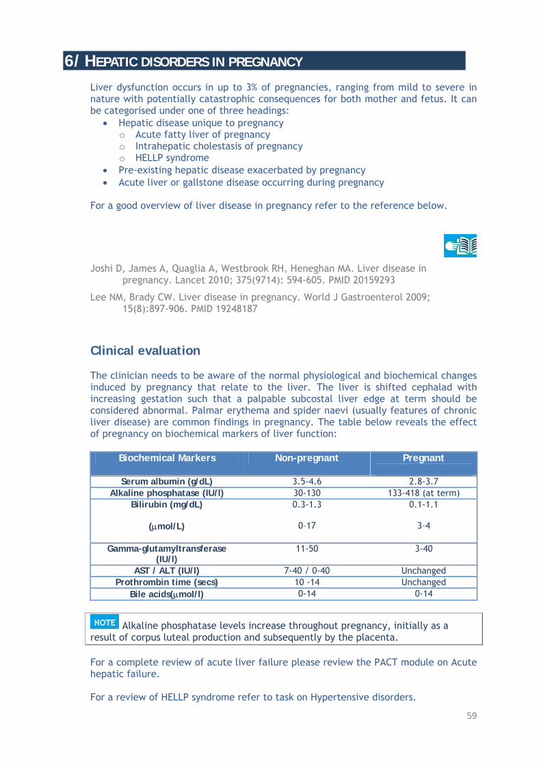
59
6/ HEPATIC DISORDERS IN PREGNANCY Liver dysfunction occurs in up to 3% of pregnancies, ranging from mild to severe in nature with potentially catastrophic consequences for both mother and fetus. It can be categorised under one of three headings:
Hepatic disease unique to pregnancy o Acute fatty liver of pregnancy o Intrahepatic cholestasis of pregnancy o HELLP syndrome
Pre-existing hepatic disease exacerbated by pregnancy Acute liver or gallstone disease occurring during pregnancy
For a good overview of liver disease in pregnancy refer to the reference below.
Joshi D, James A, Quaglia A, Westbrook RH, Heneghan MA. Liver disease in
pregnancy. Lancet 2010; 375(9714): 594-605. PMID 20159293
Lee NM, Brady CW. Liver disease in pregnancy. World J Gastroenterol 2009; 15(8):897-906. PMID 19248187
Clinical evaluation The clinician needs to be aware of the normal physiological and biochemical changes induced by pregnancy that relate to the liver. The liver is shifted cephalad with increasing gestation such that a palpable subcostal liver edge at term should be considered abnormal. Palmar erythema and spider naevi (usually features of chronic liver disease) are common findings in pregnancy. The table below reveals the effect of pregnancy on biochemical markers of liver function:
Biochemical Markers Non-pregnant Pregnant
Serum albumin (g/dL) 3.5-4.6 2.8-3.7 Alkaline phosphatase (IU/l) 30-130 133-418 (at term)
Bilirubin (mg/dL)
(mol/L)
0.3-1.3
0–17
0.1-1.1
3–4
Gamma-glutamyltransferase (IU/l)
11-50 3-40
AST / ALT (IU/l) 7-40 / 0-40 Unchanged Prothrombin time (secs) 10 -14 Unchanged
Bile acids(mol/l) 0-14 0-14
Alkaline phosphatase levels increase throughout pregnancy, initially as a result of corpus luteal production and subsequently by the placenta. For a complete review of acute liver failure please review the PACT module on Acute hepatic failure. For a review of HELLP syndrome refer to task on Hypertensive disorders.

60
For the remainder of this task the focus will be on hepatic disorders unique to pregnancy, namely acute fatty liver of pregnancy and intrahepatic cholestasis of pregnancy. Acute fatty liver of pregnancy Acute fatty liver of pregnancy (AFLP) is a rare condition with an incidence ranging from 1 in 7000 to 15,000 deliveries. Diagnosis of AFLP should prompt patient transfer to a critical care environment due to its potential for significant morbidity and/or mortality. One study revealed AFLP to account for 2-4.5% of obstetric conditions necessitating ICU admission.
Munnur U, Karnad DR, Bandi VD, Lapsia V, Suresh MS, Ramshesh P, et al.
Critically ill obstetric patients in an American and an Indian public hospital: comparison of case-mix, organ dysfunction, intensive care requirements, and outcomes. Intensive Care Med 2005; 31(8): 1087-1094. PMID 16012807
No direct cause of AFLP has been elucidated but there appears to be a strong link with abnormalities in fatty acid oxidation. It occurs more commonly in primiparous women and there appears to be an association with the presence of a male fetus and multiple gestations. AFLP may also arise as part of the spectrum of pre-eclampsia/eclampsia/HELLP syndrome. For more information on the pathogenesis of AFLP, review the following references.
Ibdah JA. Acute fatty liver of pregnancy: an update on pathogenesis and
clinical implications. World J Gastroenterol 2006; 12(46): 7397-7404. PMID 17167825
Wilcken B, Leung KC, Hammond J, Kamath R, Leonard JV. Pregnancy and fetal long-chain 3-hydroxyacyl coenzyme A dehydrogenase deficiency. Lancet 1993; 341(8842):407-408.PMID 8094173
Diagnosis and clinical evaluation AFLP tends to present during the third trimester (>28 weeks gestation), generally between gestational weeks 30 and 38. Clinical features are relatively non-specific. Symptoms:
Nausea and vomiting General malaise Abdominal pain Headache and fatigue Pruritus
Signs:
Fever Hypertension

61
Generalised oedema Proteinuria Jaundice (50% of patients) Encephalopathy Coagulopathy Polyuria/diabetes insipidus
Q. What are the potential causes of jaundice in the critically ill obstetric patient? A.
AFLP Obstetric cholestasis Viral hepatitis Pre-eclampsia/HELLP Sepsis Acute cholecystitis Ascending cholangitis Puerperal sepsis Drug-induced Methyldopa Pre-existing liver disease Primary biliary cirrhosis Sclerosing cholangitis
Diagnosis of AFLP is primarily clinical with supportive evidence gleaned from appropriate laboratory investigations as listed below.
Blood glucose: o Decreasing levels are indicative of deteriorating liver function.
Liver function:
o Elevated serum transaminases (AST > ALT) and alkaline phosphatase
o Serum bilirubin and ammonia levels increase with worsening hepatic function
Renal function:
o Serum urea and creatinine tend to increase as the clinical picture deteriorates secondary to the development of hepatorenal syndrome.
Increased serum creatinine may be the earliest biochemical indicator of AFLP. Some form of renal impairment is present in all cases.
Coagulation profile: o Prolonged prothrombin (PT) and activated partial thromboplastin
(aPTT) times are common. As liver function deteriorates PT and aPTT prolong further while serum fibrinogen levels drop and thrombocytopenia develops, indicative of disseminated intravascular coagulation.
Decreased levels of antithrombin III are a common finding in AFLP.

62
Miscellaneous:
o Elevated serum amylase/lipase – suggestive of concomitant acute pancreatitis.
o Serum albumin tends to be low. CT and MRI imaging may reveal a hypodense liver. This is not diagnostic of AFLP but may serve to complement clinical and laboratory findings. Definitive diagnosis can be made by liver biopsy but this is rarely performed given the risks associated with the concomitant coagulopathy and the lack of impact on treatment. Ch’ng et al proposed the Swansea criteria as a method of diagnosing AFLP. Six or more criteria are required for diagnosis in the absence of another cause. The Swansea criteria were validated by Knight et al in their prospective study focusing on AFLP in the UK.
The Swansea Criteria Vomiting Abdominal pain Polydipsia/ polyuria Encephalopathy Elevated bilirubin > 14 μmol/L (239 mg/dL) Hypoglycaemia < 4 mmol/L (72 mg/dL) Elevated urea > 34 mmol/L (204 mg/dL) Leukocytosis > 11 × 10⁹ /L Ascites or bright liver on ultrasound scan Elevated transaminases (AST or ALT) 42 > IU/l Elevated ammonia > 47 μmol/l (65.8 μg/dL) Renal impairment; creatinine > 150 μmol/l (1.95 mg/dL) Coagulopathy; prothrombin time > 14 seconds or APPT > 34 seconds Microvesicular steatosis on liver biopsy
Ch’ng CL, Morgan M, Hainsworth I, Kingham JG. Prospective study of liver
dysfunction in pregnancy in Southwest Wales. Gut 2002; 51(6): 876-880. PMID 12427793
Knight M, Nelson-Piercy C, Kurinczuk JJ, Spark P, Brocklehurst P; UK Obstetric Surveillance System. A prospective national study of acute fatty liver of pregnancy in the UK. Gut 2008; 57(7): 951-956. PMID 18332072
Q. Which laboratory markers help to differentiate between AFLP and HELLP syndrome? A. AFLP tends to produce a greater increase in bilirubin and is associated with jaundice in 50% of cases. Significant hypoglycaemia, severe hyperuricaemia, haematological features of DIC and elevated serum amylase, lipase and ammonia levels are more suggestive of a diagnosis of AFLP.

63
Management After maternal stabilisation, the focus should turn to expeditious safe delivery of the fetus in the patient with AFLP. The longer the pregnancy lasts after the diagnosis, the greater the risk of developing fulminant hepatic failure. Expectant management is not an option. Delivery is required as soon as the mother’s condition is stable.
There are no reports of AFLP resolving prior to delivery. Pre- and post-partum treatment should be supportive in a critical care environment with appropriate monitoring and treatment of complications such as hypoglycaemia, coagulopathy, renal failure, encephalopathy, diabetes insipidus, and acute pancreatitis. Plasmapheresis may be considered in cases where liver failure continues to worsen despite delivery. In extremis, orthotopic liver transplant may be required.
Jin F, Cao M, Bai Y, Zhang Y, Yang Y, Zhang B. Therapeutic effects of plasma
exchange for the treatment of 39 patients with acute fatty liver of pregnancy. Discov Med 2012; 13(72): 369-373. PMID 22642918
Although most patients will recover fully following delivery, AFLP has a mortality rate of 0-12.5%. Fetal mortality is worse ranging from 15-66%. There is potential for recurrence of AFLP in subsequent pregnancies and patients should be counselled prior to embarking on subsequent pregnancies. For more information on AFLP please review the following references.
Guntupalli SR, Steingrub J. Hepatic disease and pregnancy: an overview of
diagnosis and management. Crit Care Med 2005; 33(10 Suppl): S332-339. PMID 16215356
Mjahed K, Charra B, Hamoudi D, Noun M, Barrou L. Acute fatty liver of pregnancy. Arch Gynecol Obstet 2006; 274(6): 349-353. PMID 16868757
Intrahepatic cholestasis of pregnancy (IHCP) IHCP ranges in incidence from 0.1–15.6% of pregnancies. Its aetiology is unknown but genetic, hormonal and environmental influences are thought to contribute significantly. It is diagnosed clinically and is characterised by pruritus, elevated bile acids and/or serum transaminases in the absence of other potential causes. Pruritus may be intolerable, classically develops on the soles of the feet, and is worse at night.
Maternal prognosis is good in IHCP with disease progression limited by delivery.

64
IHCP is associated with fetal morbidity/mortality secondary to the occasional necessity for premature delivery and associated neonatal respiratory distress syndrome and rarely intra-uterine death. There may be an association with post-partum haemorrhage in severe cases with maternal hypo-prothrombinaemia resulting from malabsorption of vitamin K (fat-soluble vitamin). For more information on intrahepatic cholestasis of pregnancy review the following reference.
Royal College of Obstetricians and Gynaecologists. Obstetric cholestasis. Green-
top guideline No. 43, 2011. http://www.rcog.org.uk/womens-health/clinical-guidance/obstetric-cholestasis-green-top-43
CONCLUSION Optimal management of the critically ill obstetric patient necessitates a multidisciplinary approach with close collaboration of the critical care team with obstetricians, midwives, anaesthesiologists and neonatologists. A thorough understanding of diseases particular to pregnancy as well as the maternal and fetal physiological adaptations that occur will help guide the initial resuscitation effort as well as ongoing critical care management. Maternal stabilisation should be the initial priority in treatment because what is good for the mother is good for the fetus. With this in mind, neither therapy nor investigation should be withheld because of fetal concerns. For the antenatal critically ill patient, optimal timing of delivery determined by maternal well-being and fetal gestational age, should be considered early. Delivery is of particular importance for patients suffering a pregnancy-specific disease as delivery tends to abrogate or limit the disease process.

65
PATIENT CHALLENGES Patient 1 You are called to the antenatal ward of your hospital to review a 36-year-old primiparous woman at 33 weeks gestation. The obstetric resident is requesting critical care admission for invasive haemodynamic monitoring. The patient was admitted 48 hours previously with a suspected new diagnosis of pre-eclampsia, confirmed by 24 hour urine collection of protein (4g) and sustained hypertension. She was commenced on oral labetalol 200 mg three times daily.
Diagnosis of pre-eclampsia Her initial blood tests revealed: FBC: Hb: 13.6 g/L, WCC: 10.5 × 10⁹/L, Plt: 140 × 109/L Urea: 4.6 mmol/L(27.6 mg/dL), Creat: 67 mol/L (0.87 mg/dL), Uric acid: 4.8 mg/dL LFTs: ALT: 40 IU/L, AST: 36 IU/L, Bili: 14 mol/L (0.83 mg/dL) Coagulation screen: PT: 13 sec, aPTT: 32 sec, INR: 1.0 Her blood pressure responded initially to oral labetalol but over the last 6 hours, has deteriorated with her systolic blood pressure (SBP) ranging from 170-190 mmHg and diastolic blood pressure (DBP) ranging from 110–130 mmHg despite the addition of intermittent intravenous boluses of hydralazine (5 mg) while monitored on the ward. She has also been administered 12 mg betamethasone intramuscularly to aid fetal lung maturation in case emergency delivery is required.
Blood pressure control in pre-eclampsia Fetal considerations in the critically ill obstetric patient Q. Presuming you agree with the opinion of the resident critical care admission, give two reasons for your viewpoint.
A. First, her blood pressure is dangerously high putting her at risk of a (possibly catastrophic) intracranial haemorrhage if it persists or trends higher. Second, pre-eclampsia is a multisystem disease and although she is not exhibiting any other systemic manifestations as yet, she needs close monitoring in case these develop.
Q. What are your initial patient management priorities?
A. Initial priorities include: -Invasive blood pressure monitoring and gradual reduction in mean arterial pressure to <125mmHg -Continuous fetal heart rate monitoring -Optimisation of hydration; input/output monitoring -Eclampsia (seizure) prophylaxis (with a magnesium infusion) -Regular monitoring of haemoglobin, platelet count, urea, creatinine, uric acid, liver function tests, coagulation.

66
Fluid balance in pre-eclampsia Clinical features of severe pre-eclampsia ‘Eclampsia prophylaxis’ is primarily seizure prophylaxis Subsequent to critical care admission, her hypertension is treated with titrated boluses and an infusion of labetalol, regular intravenous hydralazine and magnesium sulphate infusion. Blood pressure control gradually improves to SBP 150-170 mmHg and DBP 95-110 mmHg. However, two hours later she starts to complain of a severe frontal headache, blurred vision and generalised abdominal pain. Her repeat blood tests reveal: FBC: Hb: 14.6 g/dL, WCC: 15.5 × 10⁹/L, Plt: 60 × 109/L Urea: 8.6 mmol/L (51.6 mg/dL), Creat: 104 mol/L (1.35 mg/dL), Uric acid: 7.5 mg/dL LFTs: ALT: 450 IU/L, AST: 360 IU/L, Bili: 24 mol/L (1.42 mg/dL) Coagulation screen: PT: 14 sec, aPTT: 48 sec, INR: 1.1 Serum LDH: 620 IU/L Serum haptogobin: 200 mg/dL Continuous cardiotocograph (CTG) monitoring reveals loss of baseline variability. Q. What is your assessment of the patient’s clinical status and of the most appropriate course of management at this point?
A. This patient’s condition has deteriorated. Given her worsening clinical picture and CTG evidence of fetal compromise, urgent delivery is warranted.
Q. Her elevated LDH and decreased serum haptoglobin level are indicative of haemolysis; her platelet count has dropped and liver function tests are markedly increased. What is your diagnosis?
A. She now meets criteria for HELLP syndrome.
HELLP syndrome Q. List a differential diagnosis for HELLP syndrome.
A. Acute fatty liver of pregnancy Acute hepatitis Autoimmune thrombocytopaenic purpura Thrombotic thrombocytopaenic purpura Haemolytic-uraemic syndrome

67
Following patient review by the consultant obstetrician and anaesthesiologist, urgent Caesarean section is deemed necessary. The anaesthesiologist advises that the low platelet count precludes neuraxial anaesthesia and the patient is advised that Caesarean section will be carried out under general anaesthesia.
The anaesthesiologist has considered the potential complications of a central neuraxial block (CNB), particularly vertebral canal haematoma and secondary spinal cord injury, against the potential complications of emergent general anaesthesia. A threshold platelet count at which CNB is safe does not exist but many will use (for obstetric patients) a level of 100 × 109/L for epidural anaesthesia and 70 × 109/L for spinal anaesthesia as a reasonable threshold, assuming there is no other evidence of coagulopathy. In this case, the patient’s platelet count is 60 × 109/L in the setting of HELLP syndrome with hepatic dysfunction and renal dysfunction both of which could further impair platelet function.
Q. Name the most pressing concerns regarding tracheal intubation (and the use of general anaesthesia) in this patient? Four potential concerns are listed here.
A.
1. Laryngoscopy and intubation may be difficult given physiologic adaptations of pregnancy.
2. Clinically significant pharyngolaryngeal oedema in severe pre-eclampsia/HELLP syndrome.
3. Aspiration pneumonitis and development of Mendelson’s syndrome.
4. The most worrying is the risk of a hypertensive crisis associated with laryngoscopy and intubation which may cause intracranial haemorrhage.
Physiologic adaptations of pregnancy – Airway Airway management See PACT module on Airway management Her Caesarean section is successful and a 2.2 kg baby boy was delivered, but intubated shortly after birth due to increased work of breathing. Her intra-operative course was relatively stable with an estimated blood loss of 1.2 L; she was not transfused but received 1.5 L of Ringer’s lactate and 500 mL of a colloid (gelatin) solution. The obstetrician appeared satisfied with haemostasis and remarked that the uterus was well contracted at the end of the case. The anaesthesiologist inserted a triple lumen central venous catheter in her right internal jugular vein. She was extubated and monitored in the post-anaesthetic care unit for one hour postoperatively at which time she was returned to your care in the ICU. As she appeared lucid postoperatively she was given a morphine PCA (patient controlled analgesia). Other than the PCA, the labetalol and magnesium sulphate infusion were still running and she had also been commenced on an oxytocin infusion (40 IU in 1 L of 0.9% NaCl) to run over the first four hours postoperatively.

68
Q. Does removal of the cause of pre-eclampsia eliminate the risk of eclampsia-related complication?
A. No
Q. List clinically significant complications to which she remains at risk?
A. She remains at risk of eclampsia, intracerebral haemorrhage, cerebral oedema, pulmonary oedema, oliguria and renal failure, coagulopathy, hepatic infarction and liver rupture.
Q. What therefore are your management priorities for the first 24 hours post-partum?
A. This patient requires intensive monitoring post-partum with particular attention to control of blood pressure and of fluid balance, seizure prophylaxis (with magnesium) and monitoring/control of haematology/coagulation, biochemistry and of renal indices. Standard perioperative antimicrobial prophylaxis is completed.
Management of pre-eclampsia Complications of pre-eclampsia/HELLP syndrome One hour following her return from the operating theatre, the nurse and midwife tending to her request an immediate review as there has been significant post-partum haemorrhage (PPH) (estimated 600 mL). She remains awake and alert but her blood pressure has dropped to 100/75 mmHg (150/95 mmHg) and heart rate increased to 105 bpm/regular (85 bpm/regular). Q. What are the potential causes of her PPH?
A. Potential causes include: Uterine atony Retained placenta Genital tract trauma Coagulopathy Uterine rupture Q. How would you proceed?
A. This is an obstetric emergency requiring immediate assessment, resuscitation and definitive intervention. Senior obstetric assistance is required immediately. Meanwhile assess and treat the patient from the point of view of ‘ABCD’ and perform a focused assessment for potential causes of the bleeding (e.g. uterine atony, obvious genital tract trauma, major coagulopathy) while waiting for obstetric assessment.

69
Q. What resuscitative measures, further tests and communication would you undertake?
A. Ensure large bore intravenous access and commence fluid resuscitation/O negative blood resuscitation if massive haemorrhage is present. Send blood for FBC, urea, electrolytes, ABG (arterial blood gas), coagulation screen, group and crossmatch, and notify blood bank and haematologist of potential for massive transfusion. Notify operating theatre staff including a senior anaesthesiologist of the likelihood of an emergency transfer from the ICU.
Aetiology of post-partum haemorrhage Management of post-partum haemorrhage On abdominal examination you notice a palpable soft, non-contracted uterus. This is confirmed by the obstetrician who has just arrived in the ICU. Q. What basic manoeuvre can be performed to improve uterine tone?
A. External uterine massage and bimanual uterine compression
Q. Which uterotonic agent should you avoid and why?
A. Ergometrine (methyergonovine) causes hypertension and has been associated with maternal mortality secondary to intracerebral haemorrhage.
Management of post-partum haemorrhage Your patient stabilises, uterine tone improves with bimanual compression and a bolus of oxytocin (5 IU). The remainder of her ICU course is unremarkable. The labetalol infusion is weaned to the oral form 36 hours post-partum and blood pressure control remained stable thereafter. The magnesium sulphate infusion is discontinued on day 2 post-partum. While she was oliguric initially post-partum, urine output normalised on day 2 post-partum as did her renal indices. Her platelet count rose to 90 × 109/L on day 2 post-partum and there was no further evidence of coagulopathy. Her liver function tests began to normalise on day 3 post-partum at which time she was discharged to the postnatal ward. The remainder of her inpatient stay was unremarkable.
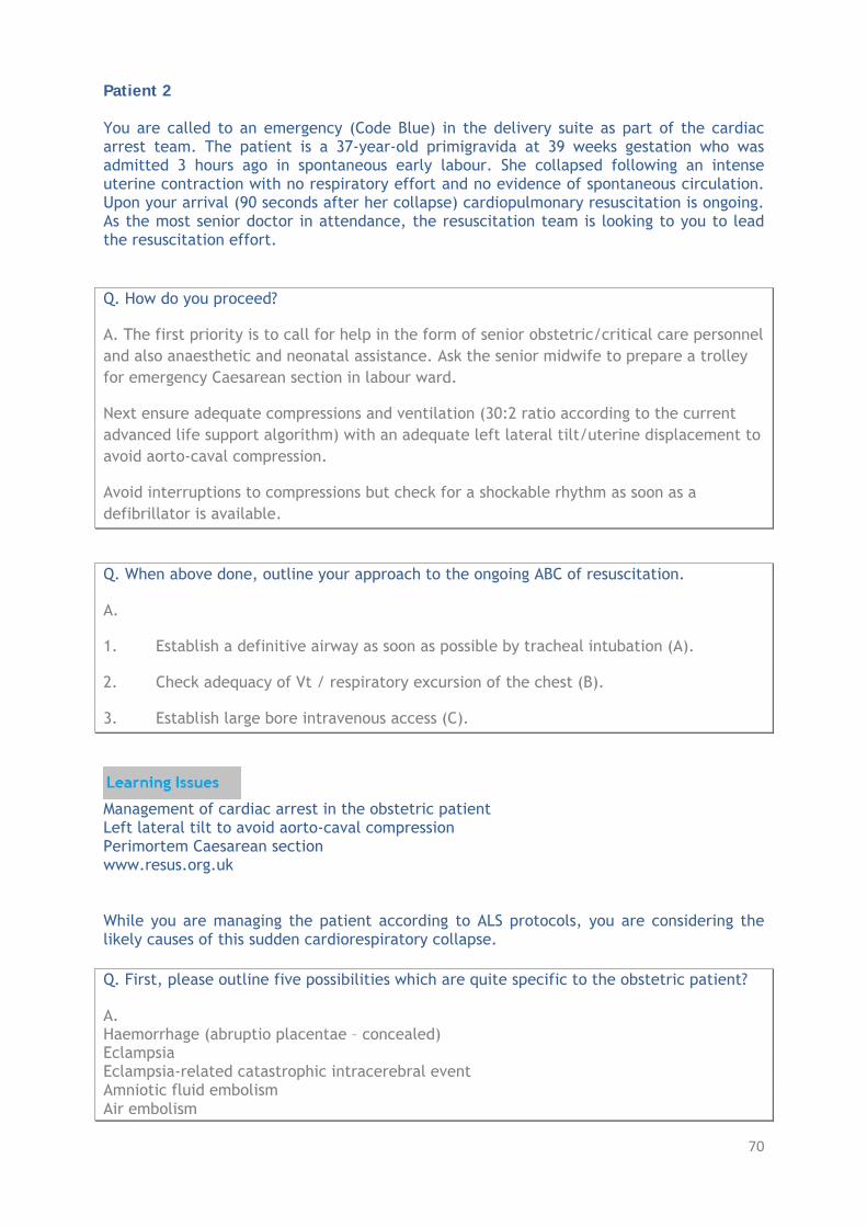
70
Patient 2 You are called to an emergency (Code Blue) in the delivery suite as part of the cardiac arrest team. The patient is a 37-year-old primigravida at 39 weeks gestation who was admitted 3 hours ago in spontaneous early labour. She collapsed following an intense uterine contraction with no respiratory effort and no evidence of spontaneous circulation. Upon your arrival (90 seconds after her collapse) cardiopulmonary resuscitation is ongoing. As the most senior doctor in attendance, the resuscitation team is looking to you to lead the resuscitation effort. Q. How do you proceed?
A. The first priority is to call for help in the form of senior obstetric/critical care personnel and also anaesthetic and neonatal assistance. Ask the senior midwife to prepare a trolley for emergency Caesarean section in labour ward.
Next ensure adequate compressions and ventilation (30:2 ratio according to the current advanced life support algorithm) with an adequate left lateral tilt/uterine displacement to avoid aorto-caval compression.
Avoid interruptions to compressions but check for a shockable rhythm as soon as a defibrillator is available.
Q. When above done, outline your approach to the ongoing ABC of resuscitation.
A.
1. Establish a definitive airway as soon as possible by tracheal intubation (A).
2. Check adequacy of Vt / respiratory excursion of the chest (B).
3. Establish large bore intravenous access (C).
Management of cardiac arrest in the obstetric patient Left lateral tilt to avoid aorto-caval compression Perimortem Caesarean section www.resus.org.uk While you are managing the patient according to ALS protocols, you are considering the likely causes of this sudden cardiorespiratory collapse. Q. First, please outline five possibilities which are quite specific to the obstetric patient?
A. Haemorrhage (abruptio placentae – concealed) Eclampsia Eclampsia-related catastrophic intracerebral event Amniotic fluid embolism Air embolism

71
Q. Please outline five further general possibilities that are not specific to the obstetric patient? A. Anaphylaxis Pulmonary embolism Septic shock Myocardial infarction Malignant arrhythmia There is return of spontaneous circulation following defibrillation of ventricular fibrillation approximately four minutes after the initial collapse. Initial post-resuscitation blood pressure is 90/60 mmHg, heart rate is 90 bpm/regular but the patient is not making any respiratory effort and her Glasgow Coma Scale score is 5/15. The cardiotocograph reveals evidence of fetal distress with sustained fetal bradycardia (see Fig. below).
Sustained fetal bradycardia with no reactivity. Q. At this point what is the most appropriate management of this patient?
A. This patient requires an emergent Caesarean section to optimise both patient and fetal prognosis.
Q. Presuming the possibility of the need for massive transfusion, who would you contact?
A. Blood bank and the haematology consultant should be notified of this potential.
The patient is transferred to the operating theatre immediately and emergency Caesarean section is performed under general anaesthesia. A 3.1 kg neonate is delivered with an APGAR score of 2 at 1 minute with significant bradycardia (58 bpm). The neonate is successfully resuscitated and transferred intubated to the neonatal intensive care unit.

72
Intra-operatively, a right radial arterial catheter and a left internal jugular large bore catheter are inserted. Following delivery the obstetrician remarks on excessive oozing of blood from the suture lines in the uterus and the anaesthesiologist notices bleeding at all the venipuncture sites and the presence of blood stained urine. Q. What is the likely diagnosis underlying these findings?
A. Disseminated intravascular coagulation (DIC)
Q. What laboratory assays might you request to support this clinical diagnosis?
A. Laboratory findings to support the diagnosis of DIC include: Prolonged prothrombin time Prolonged activated partial thromboplastin time Prolonged thrombin time Thrombocytopaenia Hypofibrinogenaemia Elevated D-dimer Elevated levels of fibrin degradation products Reduced antithrombin times.
You should not delay in treating a patient suspected of DIC while awaiting laboratory confirmation.
The normal physiological alterations of pregnancy affect the r common measurements of coagulation e.g. there is a 50% (approximately) increase in fibrinogen levels and an increase in D-dimer levels. PT/aPTT/thrombin time and FDPs remain at normal levels. Thrombocytopaenia also occurs. Q. What level of thrombocytopaenia should be considered abnormal? A. Levels less than 100 × 109/L
An intra-operative ABG while patient was being ventilated with an FiO2 of 0.70 revealed: pH: 7.11 - PaO2: 95 mmHg (12.7 kPa) HCO3-: 13 mmol/L (13 mEq/L) - PaCO2: 38 mmHg (5 kPa) BE: - 12 - Lactate: 9 mmol/L (81 mg/dL) Hb: 6.4 g/L Q. How do you interpret the acidaemia?
A. The patient is profoundly acidaemic as evidenced by pH, HCO3-, base deficit. The underlying process is metabolic in origin and tissue hypoperfusion (and anaerobic metabolism) is likely to be a major contributor as evidenced by the significantly elevated serum lactate.

73
Q. How do you interpret the indices of gas exchange?
A. There is also a significant issue with gas exchange. Under normal circumstances one would expect a much higher PaO2 with a FiO2 of 0.7. Furthermore the ‘normal’ PaCO2 reflects an inadequate compensatory response as a low PaCO2 (due to hyperventilation in a spontaneously breathing patient) would be the expected ‘normal’ response to this acidaemia.
See PACT module on Electrolytes and Homeostasis
Q. How do interpret and respond to the Hb of 6.4 g/L?
A. The patient is anaemic. Given that there is ongoing blood loss and clinical evidence of coagulopathy, blood and blood product transfusion is mandated in this patient.
Q. What is the patient’s A-a gradient?
A. Given her FiO2 is 0.70, using the alveolar gas equation, her PaO2 should be approximately 451 mmHg (60 kPa). Normal A-a gradient for this patient if breathing room air (FiO2: 0.21) would be [Age + 10/4, 47/4 ~ 12 mmHg]. Given her FiO2 of 0.7, the upper limit of normal would be 47 mmHg (6.3 kPa) (allowing approximately 7 mmHg (1 kPa) increase for every 10% increase in FiO2). Her A-a gradient is 404 mmHg (53.8 kPa). Such a severe A-a gradient is suggestive of either a severe ventilation/perfusion mismatch or a significant shunt.
Q. Given her diagnosis of severe pre-eclampsia, what is the likely pathophysiologic process accounting for this gradient?
A. Acute pulmonary oedema secondary to ‘leaky capillaries’ is the most likely. However, given the acuity of her presentation and also considering that she presented in labour, amniotic fluid embolism should also be considered.
The intra-operative team commence blood and blood product transfusion which include 9 units of red cell concentrate, 6 units of fresh frozen plasma (FFP), 10 units of cryoprecipitate and 2 pools of platelets. During this resuscitation effort, the obstetric team make the decision to perform a hysterectomy to help control bleeding. During the resuscitation effort the initial coagulation screen results reveal:
Prothrombin time (PT): 21 sec INR: 1.8 Activated partial thromboplastin time (aPTT): 49 sec Fibrinogen level: 0.085 g/dL Platelet count: 65 × 109/L
Following the resuscitation effort and hysterectomy, adequate haemostasis is achieved and at the end of the case when she is ready to return to ICU, the patient’s laboratory parameters have improved somewhat.
Hb: 10.2 g/L

74
Platelets: 95 × 109/L PT: 16 sec INR: 1.2 aPTT: 39 sec Fibringen: 0.26 g/dL
Also her acid-base status shows improvement and she has started to produce urine.
pH: 7.31 PO2: 110 mmHg (14.7 kPa) HCO3
-: 19 mmol/L (19 mEq/L) PCO2: 40 mmHg (5.3 kPa) BE: - 5 Lactate: 3.9 mmol/L (35 mg/dL)
On arrival in the ICU, she is ventilated, a noradrenaline infusion, to supplement the volume resuscitation in achieving an acceptable MAP, has been commenced and the anaesthesiologist has placed a pulmonary artery catheter post op. Her initial chest X-ray reveals significant bilateral infiltrates in her lung fields and shows the PAC to be in reasonable position (Fig. below)
Q. Given the circumstances and the acuity of her initial clinical presentation and the severity of the findings on chest X-ray, is your second diagnostic option (mentioned above) now, in retrospect, more likely?
A. Yes, this is likely to reflect amniotic fluid embolism.
Q. Is there any test or investigation you can perform to confirm your diagnosis?
A. No, as this is now considered a clinical diagnosis.
Previously the presence of fetal squames or debris from blood aspirated from the distal port of a pulmonary artery catheter were considered diagnostic. In recent years however it has become clear that fetal squamous cells are a common finding in the circulation of pregnant women who do not develop the syndrome.

75
Q. Mention some other diagnoses that could account for these changes on chest X-ray? Four diagnoses are provided here.
A. Severe pre-eclampsia/HELLP syndrome Transfusion related acute lung injury (TRALI) Aspiration pneumonitis Severe sepsis with related ARDS
Severe pre-eclampsia See PACT module on Sepsis and MODS Three hours after admission to ICU her condition is once again deteriorating. Volume resuscitative and noradrenaline requirements are escalating and there is continuous bleeding from her abdominal wound and venipuncture sites despite further red cell, platelets, FFP and cryoprecipitate therapy. Notable laboratory results include: Hb: 7.8 g/L pH: 7.18 Plt: 85 × 109/L HCO3
-: 16 PT: 17 sec BE: -8 INR: 1.5 Fibrinogen: 0.1 g/dL The nurse in attendance has noted that the abdomen has become increasingly distended and a focused abdominal ultrasound, performed at the bedside, by the ICM consultant reveals free fluid surrounding the liver. Q. What is the likely diagnosis?
A. This patient appears to be bleeding internally.
Q. What is required?
A. A laparotomy is likely the best option to reveal the source and attempt haemostasis as soon as possible.
Q. Who needs to be notified?
A. The obstetrician and on-call surgeon as well as the consultant anaesthesiologist and haematologist.
Laparotomy reveals no major bleeding vessel but continuous oozing from operative beds and areas of previous tissue damage.

76
Q. What other options might you consider to aid haemostasis?
A. Tranexamic acid (an anti-fibrinolytic agent) and/or recombinant factor VIIa (rFVIIa) might be considered.
Q. Are these drugs licensed in this setting?
A. No, they are not licensed for use in this setting but there are anecdotal reports of haemostasis being achieved when used in post-partum haemorrhage refractory to standard methods of haemostasis.
At this point, following consultation with the consultant haematologist on-duty, further resuscitation and factor replacement with FFP (4 u), cryoprecipitate (10 u) and 2 pools of platelets are administered. Treatment with recombinant factor VIIa was not considered given her ongoing acidaemia. Approximately 20 minutes after transfusion the obstetrician and surgeon noted a significant improvement in the bleeding sites, surgery was completed and the patient was transferred back to the ICU for ongoing management of her ARDS, haemodynamic, homeostatic/renal and coagulation status.
Use of rFVIIa has been associated with greater morbidity/mortality when used in the treatment of AFE (Amniotic Fluid Embolism)-associated DIC (see reference below).
Leighton BL, Wall MH, Lockhart EM, Phillips LE, Zatta AJ. Use of recombinant
factor VIIa in patients with amniotic fluid embolism: a systematic review of case reports. Anesthesiology 2011; 115(6): 1201-1208. PMID 21720243
Repeat coagulation screen in ICU revealed an aPTT and PT of 45 and 11 seconds respectively, a fibrinogen level of 0.09 g/dL and a platelet count of 65 × 109/L. She remained stable in ICU although she did require further blood product transfusion (cryoprecipitate and 1 pool of platelets) for a slow ooze from her abdominal wound; it stopped within 24 hours. Postoperatively she required haemodialysis starting on day 3 for persistent oligo-anuria and renal failure and which also allowed, when haemodynamically stable, the active removal of fluid to treat the ARDS. The period of renal failure was relatively short and her gas exchange and ventilatory parameters also improved over the first 3 days postoperatively and she was extubated successfully on day 4. She was discharged home with her new baby on day 12 post delivery. On reflection: These patient descriptions highlight the relative acuity of the presentation of pregnancy-specific diseases and their potentially fulminant multisystem involvement. Successful maternal and fetal outcomes are dependent on prompt responsiveness and the close collaboration between obstetricians, midwives and anaesthesiologists together with the critical care clinicians and other general hospital multidisciplinary personnel and facilities.