Equine placenta marvelous organ and a lethal weapon ...
16
1 Equine placenta – marvelous organ and a lethal weapon Malgorzata Pozor, DVM, PhD, Diplomate ACT Introduction Placenta has been defined as: „an apposition between parent (usually maternal) and fetal tissue in order to establish physiological exchange” (1). Another definition of this important organ was proposed by Steven and Morris: „a device consisting of one or more transport epithelia located between fetal and maternal blood supply” (2). The main function of placenta is to provide an interface between the dam and the the fetus and to allow the metabolic exchange of the the nutrients, oxygen and waste material. The maternal circulation is brought into a close apposition to the fetal circulation, while a separation of these two circulatory systems remain separated (3). A degree and complexity of this „intimate relationship” varies greately between species mostly due to the structural diversity of the extraembryonic membranes of the vertebrates. The early feto-maternal exchange in the equine pregnancy is established as early as on day 22 after fertilization. The fetal and choriovitellin circulations are already present, the capsule ruptures and the allantois is already visible (4). The allantois starts expanding by day 32 and vascularizes approximately 90% of the chorion and fuses with it to form chorioallantois by day 38 of gestation (5). The equine placenta continues increasing its complexity till approximately day 150 of gestation. Equids have epitheliochorial placenta, there are six leyers separating maternal and fetal circulation, and there are no erosion of the luminal, maternal epithelium, like in ruminants (6). Thousands of small chorionic microvilli develop and penetrate into endometrial invaginations. There are secondary and teriary branching, forming unique for equine placenta microcotyledons. Furthermore, glandular histotroph is continuously produced by the endometrial glands and uptaken by the trophoblast cells of the areolae between the microcotyledons. Despite the complexity of this interface the transfer of nutrients through all six leyers separating fetal and maternal circulation is relatively poor, and therefore only one fetus can be supported. The stereological measurements reveal that the total microscopic area of the feto-maternal contact at the chorioallantois–endometrium interface in an average Thoroughbred mare at term is 50–60 m 2 (7). This entire surface has to be in close approximation at all times to support equine pregnancy. Any alteration of endometrial or chorioallantoic surfaces results in fetal loss or severe compromise. The equine placenta has another unique function. The strip of specialized trophoblastic cells appear at the margin of of the yolk sac and the chorioallantois forming so-called chorionic girdle. These cells are already visible on day 25, and begin to invade the uterine epithelium on day 34. Only cells that penetrate into maternal stroma and separate from the basal leyers survive and enlarge to form endometrial cups. The cups start synthesising equine chorionic gonadotropin (eCG), which is a placental form of luteinizing hormone (LH) and is responsible for ovulations and/or luteinization of ovarian follicles and formation of secondary or accessory corpora lutea (8). This hormon is produced from approximately 40 to 100 day of gestation. The cups begin degenerating on day 80. Some of them may become enclosed in a fold of chorioallantoic tissue, called an chorioallantoic pouch (9).
Transcript of Equine placenta marvelous organ and a lethal weapon ...

1
Equine placenta – marvelous organ and a lethal weapon
Malgorzata Pozor, DVM, PhD, Diplomate ACT
Introduction
Placenta has been defined as: „an apposition between parent (usually maternal) and fetal
tissue in order to establish physiological exchange” (1). Another definition of this
important organ was proposed by Steven and Morris: „a device consisting of one or more
transport epithelia located between fetal and maternal blood supply” (2). The main
function of placenta is to provide an interface between the dam and the the fetus and to
allow the metabolic exchange of the the nutrients, oxygen and waste material. The
maternal circulation is brought into a close apposition to the fetal circulation, while a
separation of these two circulatory systems remain separated (3). A degree and
complexity of this „intimate relationship” varies greately between species mostly due to
the structural diversity of the extraembryonic membranes of the vertebrates.
The early feto-maternal exchange in the equine pregnancy is established as early as on
day 22 after fertilization. The fetal and choriovitellin circulations are already present, the
capsule ruptures and the allantois is already visible (4). The allantois starts expanding by
day 32 and vascularizes approximately 90% of the chorion and fuses with it to form
chorioallantois by day 38 of gestation (5). The equine placenta continues increasing its
complexity till approximately day 150 of gestation. Equids have epitheliochorial
placenta, there are six leyers separating maternal and fetal circulation, and there are no
erosion of the luminal, maternal epithelium, like in ruminants (6). Thousands of small
chorionic microvilli develop and penetrate into endometrial invaginations. There are
secondary and teriary branching, forming unique for equine placenta microcotyledons.
Furthermore, glandular histotroph is continuously produced by the endometrial glands
and uptaken by the trophoblast cells of the areolae between the microcotyledons. Despite
the complexity of this interface the transfer of nutrients through all six leyers separating
fetal and maternal circulation is relatively poor, and therefore only one fetus can be
supported. The stereological measurements reveal that the total microscopic area of the
feto-maternal contact at the chorioallantois–endometrium interface in an average
Thoroughbred mare at term is 50–60 m2 (7). This entire surface has to be in close
approximation at all times to support equine pregnancy. Any alteration of endometrial or
chorioallantoic surfaces results in fetal loss or severe compromise.
The equine placenta has another unique function. The strip of specialized trophoblastic
cells appear at the margin of of the yolk sac and the chorioallantois forming so-called
chorionic girdle. These cells are already visible on day 25, and begin to invade the uterine
epithelium on day 34. Only cells that penetrate into maternal stroma and separate from
the basal leyers survive and enlarge to form endometrial cups. The cups start synthesising
equine chorionic gonadotropin (eCG), which is a placental form of luteinizing hormone
(LH) and is responsible for ovulations and/or luteinization of ovarian follicles and
formation of secondary or accessory corpora lutea (8). This hormon is produced from
approximately 40 to 100 day of gestation. The cups begin degenerating on day 80. Some
of them may become enclosed in a fold of chorioallantoic tissue, called an chorioallantoic
pouch (9).
2
And finally, the utero-placental tissues play a major role in producting and transporting
various hormones. Some steroid precursors, such as cholesterol, are transported from
maternal to fetal circulation and metabolized within the fetal and placental tissues. The
major products of these chemical reactions are progestagens and estrogens, which are
secreted back to the maternal circulation (10). The feto-placental unit is essentiall in
supporting pregnancy by production of progestagens, such as 5αDHP (5α-pregnan-3,20-
dione). This hormon circulate in maternal plasma during the second half of the
pregnancy, but it is also ruturned to the fetus, especially near term. The total progestagen
concentration in maternal plasma increases rapidly during the last two weeks of
pregnancy thanks to the increased level of delivery of the substrates, doubling of the fetal
adrenal glands, and increased expression of the progestagenic enzymes in the fetal
adrenals and placenta.The final decline in progestagen concentration occurs usually soon
before parturition. Other hormons produced by the equine placenta are: estrogens and
relaxin. The precursor for estrogens, DHEA (dehydroepiandrostenone), is produced by
enlarging fetal gonads, regardles of fetal sex. DHEA is excreted to the umbilical artery,
transported into the placenta and aromatased into estrogens – estrone, estradiol 17-α and
estradiol 17-β. Relaxin is produced mainly by the placenta. This hormon reaches its high
concentration in maternal circulation by day 80 and remains high until it increases again
during parurition, as a response to rising concentrations of oxytocin.
Summarizing, the equine placenta is a very complex, marvelous organ which acts in a
synchrony with fetal development and its needs, as well as with a dam and her ability to
support her pregnancy. Unfortunately, things not always go, as planned, and equine
placenta may become a deadly trap for the fetus and/or a silent killer for the mare.
Evaluation of equine placenta post partum
The fetal part of the equine placenta (fetal membranes) is usually expelled within 30
minutes after delivery of the foal. A delay in expulsion of the fetal membranes beyond 3
hours is considered abnormal in the mare and may have serious consequences, such as
metritis/laminitis/septicemia complex. It is important to remember that even a very small
piece of chorioallantois retained in mare’s uterus is just as lethal as fetal membranes
retained as a whole. Therefore, a thorough examination of the fetal membranes that were
passed post partum is so crucial for planning both foal’s and mare’s care. Placenta
consists of maternal and fetal part, and only fetal part is expelled in the horse. However,
many horsemen, farmers, animal scientists, as well as veterinarians, refer to this tissues as
just the placenta. Therefore, we will use this term from now on in this paper as the
equivalent to the fetal membranes.
The best time to evaluate equine placenta is immidiately after foaling, and placenta
expulsion. Placenta should be protected from tearing and excessive contamination with
feces, shavings, straw or sand. Therefore, any freely hanging from the mare’s vulva
fragment of the placenta should be tied immediately after foaling. Farm crew should be
trained to collect expelled placenta and to place it in a clean backet or trash bag for
evaluation. Preparing an outline of the examination and a form, which has to be filled out
is very helpful in collecting and saving good records.
Equine placenta is usually expelled intact, inside out – with the chorionic surface inside
and the allantoic surface facing outside. The exception to this rule is a premature
placental separation, when the entire chorioallantoic sac comes out with the chorionic

3
surface outermost. The chorioallantois has a similar shape as the gravid uterus, with the
gravid horn significantly bigger than another, non-gravid one. Normal placental weight at
term is approximately 11% of the weight of the foal, which translates into approximately
2.2-6.4 kg (11, 12). The best way of checking for a completeness is arranging the entire
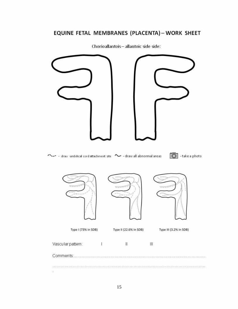
placenta into so-called „lazy F position” (Fig. 1).
Figure 1.
Placenta in „lazy F position”; left - chorionic side out; right – allantoic side out
Completeness of chorioallantois is determined first, with a special attention to the tips of
both horns. The most common site of placental tears is a tip of the non-gravid horn due to
much thinner wall than the gravid horn. This horn is expelled last which makes it even
more prone for being torn and retained. The tip of the non-gravid horn may be shredded
but complete. This poses a challenge for a person examining the placenta. One should put
all pieces together, like a puzzle, to make sure that, in fact, the entire chorioallantois was
passed. After checking for the completeness, both sides of the chorioallantois are
examined. The chorionic side should be red, and has a velvet-like surface. The gravid
horn may have a brighter color that the non-gravid horn, which may appear darker than
the gravid horn, or even brownish color. The gravid horn is usually larger, smoother and
thicker than the non-gravid horn. Its tip has an obvious edema (Fig. 2). In contrast, the
non-gravid horn is often, but not always, shorter, much thinner, and appears wrinkled due
to the numerous allantoic vessels, which hold this horn’s shape in place (13).
Figure 2
Chorioallantois - tips of horns; edematous gravid horn and thin non-gravid horn: left –
chorionic side out; right – allantoic side out

4
One has be really careful in interpreting color changes, since even a short storage or
transportation may cause dramatic changes in color patterns on the chorionic side. Patchy
brownish to tan discoloration is often present in normal equine placentas (Fig. 3).
Figure 3
Normal chorioallantois with patchy discoloration due to short storage
There are small avillous areas on the tips of both horns, which are facing the oviductal
papillae in the uterus (Fig. 4). The body of the uterus is relatively thin. There are always
at least two avillous areas on the chorionic surface of this part of the equine placenta. One
is located around the attachement of the umbilical cord and another one is at the internal
cervical os and is called cervical star (Fig. 4). The cervical star brakes during parturition
initiating the second stage labor. This area is also affected first in a process of ascending
placentitis.
Figure 4
Avillous areas: left – oviductal papilla; middle - cervical star; right – fold on the large
allantoic vessel
Small avillous areas are also present at the previous location of the endometrial cups.
Occassionally, we can see the longitudinal avillous areas, which are overlying large
branches of the umbilical vessels (Fig. 4) (14). These vessels have limited elasticity and
cause “in utero” folding of the chorioallantoic membrane (15).The fetal foot PAD
5
(placental area of degeneration) can be also found on the chorionic surface of the gravid
horn, close to the tip, as a result of chronic pressing of the fetal foot against the placenta
during the last trimester of the pregnancy (14). Any other areas with no villi or with
significant discoloration should be a reason for concern.
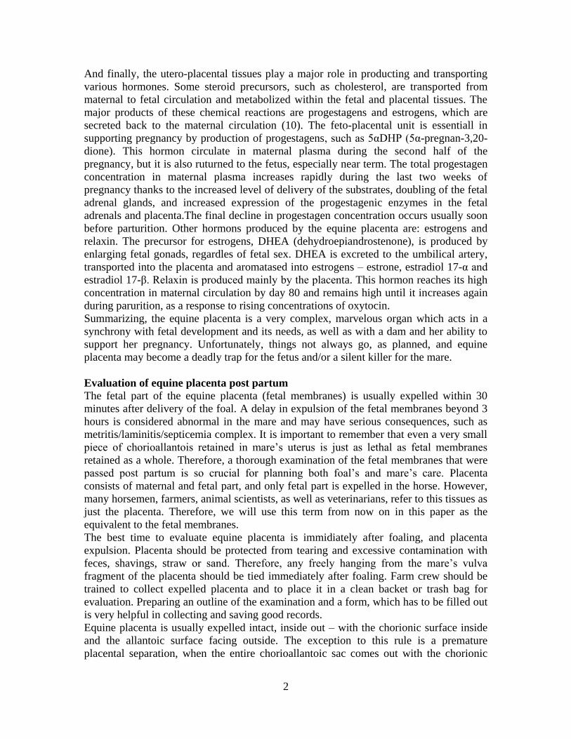
Figure 5
Chorioallantois – allantoic side: left – allantoic vesicles; middle – allantoic pouches; right
- hippomane
Allantoic side of the equine placenta should be smooth and transparent. Accumulation of
fluid in allantoic stroma may lead to the formation of allantoic vesicles, which may be
found around the blood vessels. (Fig. 5) Allantoic pouches have different origin. They
contain necrotic remains of the endometrial cups which slough from the endometrial
surface. They are usually present near the attachment of the umbilical cord. Characteristic
features that are often found in the allantoic cavity are hippomanes (Fig. 5). There are
also called “allantoic calculi”. They contain lipids, cellular debris, degenerated blood
cells and irregular mineralized material (11). Their significance is unknown.
There are many big arteries and veins on this side that provide the blood supply to the
chorion and the chorionic villi. There are three vascular patterns that can be identified on
the allantoic side, with type I being the most commonly seen among the Standardbred
(STD) mares (16). Pattern II is consistent with changing location of the fetus from one
horn where implantation took place to another one, where the fetus is present at term.
Pattern III is considered abnormal and is associated with having a co-twin, which died
spontaneously or was reduced early. The umbilical cord should be attached on the dorsal
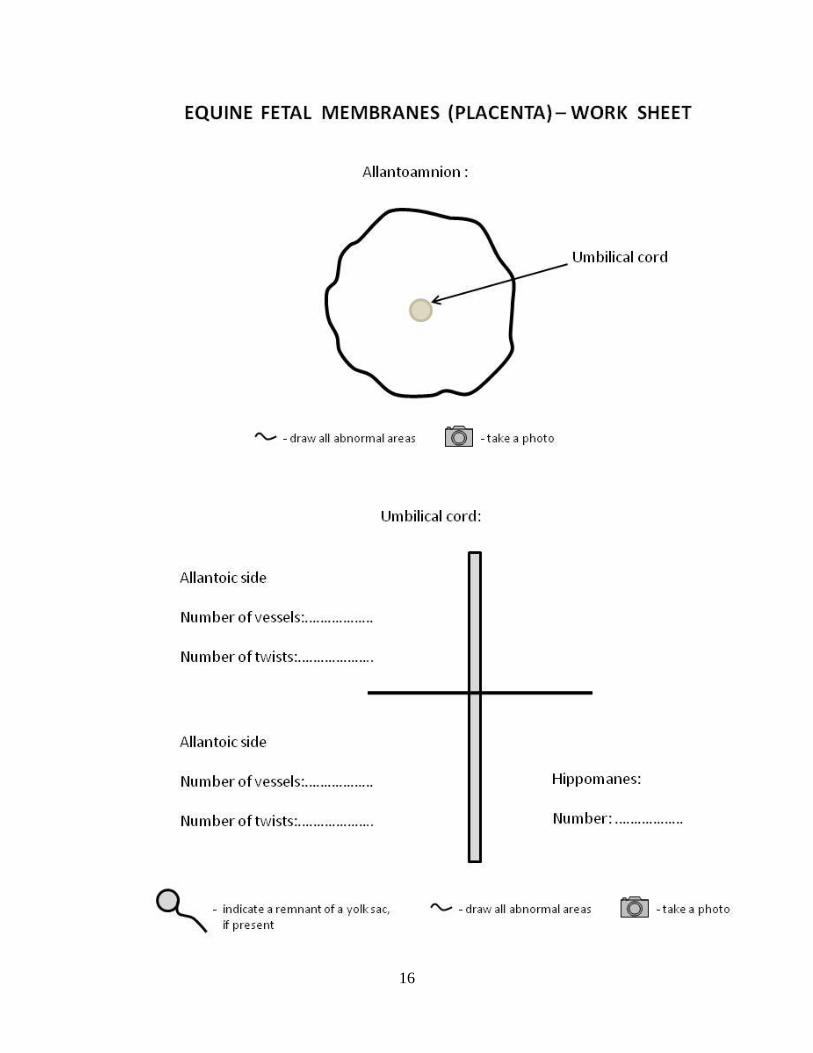
side at the base of one of the horns. It consists of one vein and two arteries on the fetal
side and two arteries and two veins on the maternal side (Fig. 6). The length of the cord
varies, but the large study on 143 Thoroughbred mares showed that the mean length is 55
cm, with a broad range from 32 - 90 cm (14). The urachus connects the fetal urinary
bladder and the allantoic cavity. This structure runs within loose stromal tissue of the
amniotic section of the cord (14). A remnant of the yolk sac is a normal feature of the
equine placenta and is often present along the allantoic portion of the cord (Fig. 6).

6
Figure 6
Umbilical cord: left – normal architecture; right – remnant of the yolk sac
The allantoamnion is a large, translucent sac attached to the umbilical cord (Fig. 7). This
membrane contains numerous blood vessels, which are tortuous and prominent (14,15).
The amnion itself is not vascular, but the allantoic side provides necessary
vascularization to the allantoamnion.
Figure 7
Normal allantoamnion: left – overview; right – amniotic plagues
There may be amniotic plagues present on the amniotic surface of the allantoamnion and
on the amniotic part of the umbilical cord. These features are focal areas of squamous
metaplasia (Fig. 8). Their surface may become keratinized and they look like small horn-
like rugose growths (14).
Placental abnormalities
A variety of placental abnormalities can be identified during thorough examination.
Areas of placental separation are initially bright red or bruise-like as a result of congested
vessels in microcodyledons (14). This is especially obvious when a so-called red-bag
delivery occurs and the entire fetus is born within the intact chorioallantoic sac. The
cervical star does not rupture and may be very thick. One should consider fescue
toxicosis as a cause of this scenario. However, a much more common reason for placental
7
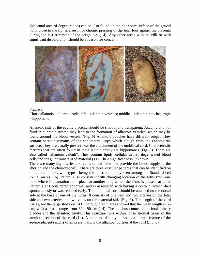
separation in mares is ascending bacterial placentitis. It starts from thickened, congested
chorioallantois at the cervical star, which appears bright red on evaluation, but later in the
process this area become necrotic, avillous, pale to cream color (Fig. 8). This disease
affects mostly the cervical star area and the uterine body. An obvious line of demarcation
between the affected and healthy tissues is often seen.
Figure 8
Ascending placentitis: left – bright-red changes at cervical star; middle : very thick, red
changes at cervical star; right – pale/cream color changes at cervical star
Occasionally, the process may extend to the allantoic side, where neovascularisation and
chorionic adenomatous hyperplasia occurs, which can be identified as raised, ward-like
lesions (17). The fetus is severely compromised by decreased nourishment and
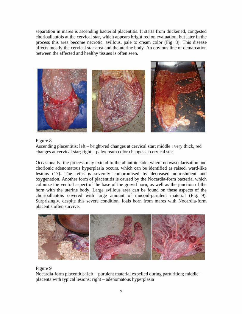
oxygenation. Another form of placentitis is caused by the Nocardia-form bacteria, which
colonize the ventral aspect of the base of the gravid horn, as well as the junction of the
horn with the uterine body. Large avillous area can be found on these aspects of the
chorioallantois covered with large amount of mucoid-purulent material (Fig. 9).
Surprisingly, despite this severe condition, foals born from mares with Nocardia-form
placentis often survive.
Figure 9
Nocardia-form placentitis: left – purulent material expelled during parturition; middle –
placenta with typical lesions; right – adenomatous hyperplasia
8
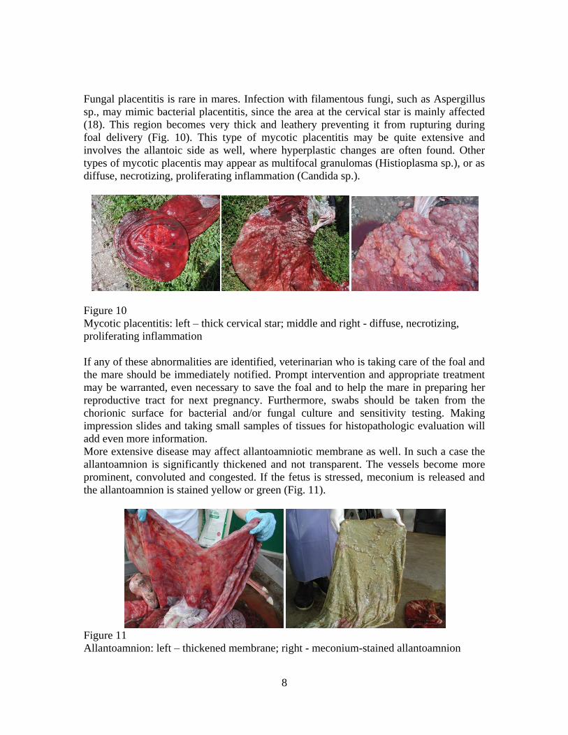
Fungal placentitis is rare in mares. Infection with filamentous fungi, such as Aspergillus
sp., may mimic bacterial placentitis, since the area at the cervical star is mainly affected
(18). This region becomes very thick and leathery preventing it from rupturing during
foal delivery (Fig. 10). This type of mycotic placentitis may be quite extensive and
involves the allantoic side as well, where hyperplastic changes are often found. Other
types of mycotic placentis may appear as multifocal granulomas (Histioplasma sp.), or as
diffuse, necrotizing, proliferating inflammation (Candida sp.).
Figure 10
Mycotic placentitis: left – thick cervical star; middle and right - diffuse, necrotizing,
proliferating inflammation
If any of these abnormalities are identified, veterinarian who is taking care of the foal and
the mare should be immediately notified. Prompt intervention and appropriate treatment
may be warranted, even necessary to save the foal and to help the mare in preparing her
reproductive tract for next pregnancy. Furthermore, swabs should be taken from the
chorionic surface for bacterial and/or fungal culture and sensitivity testing. Making
impression slides and taking small samples of tissues for histopathologic evaluation will
add even more information.
More extensive disease may affect allantoamniotic membrane as well. In such a case the
allantoamnion is significantly thickened and not transparent. The vessels become more
prominent, convoluted and congested. If the fetus is stressed, meconium is released and
the allantoamnion is stained yellow or green (Fig. 11).
Figure 11
Allantoamnion: left – thickened membrane; right - meconium-stained allantoamnion
9
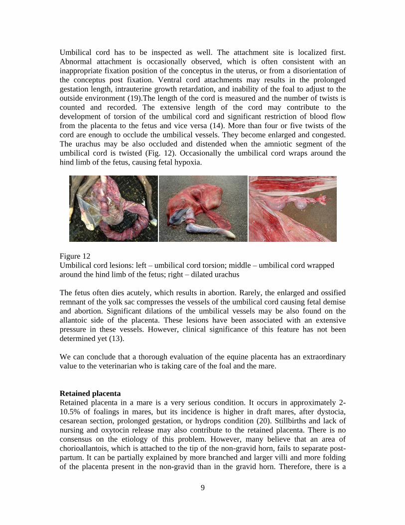
Umbilical cord has to be inspected as well. The attachment site is localized first.
Abnormal attachment is occasionally observed, which is often consistent with an
inappropriate fixation position of the conceptus in the uterus, or from a disorientation of
the conceptus post fixation. Ventral cord attachments may results in the prolonged
gestation length, intrauterine growth retardation, and inability of the foal to adjust to the
outside environment (19).The length of the cord is measured and the number of twists is
counted and recorded. The extensive length of the cord may contribute to the
development of torsion of the umbilical cord and significant restriction of blood flow
from the placenta to the fetus and vice versa (14). More than four or five twists of the
cord are enough to occlude the umbilical vessels. They become enlarged and congested.
The urachus may be also occluded and distended when the amniotic segment of the
umbilical cord is twisted (Fig. 12). Occasionally the umbilical cord wraps around the
hind limb of the fetus, causing fetal hypoxia.
Figure 12
Umbilical cord lesions: left – umbilical cord torsion; middle – umbilical cord wrapped
around the hind limb of the fetus; right – dilated urachus
The fetus often dies acutely, which results in abortion. Rarely, the enlarged and ossified
remnant of the yolk sac compresses the vessels of the umbilical cord causing fetal demise
and abortion. Significant dilations of the umbilical vessels may be also found on the
allantoic side of the placenta. These lesions have been associated with an extensive
pressure in these vessels. However, clinical significance of this feature has not been
determined yet (13).
We can conclude that a thorough evaluation of the equine placenta has an extraordinary
value to the veterinarian who is taking care of the foal and the mare.
Retained placenta
Retained placenta in a mare is a very serious condition. It occurs in approximately 2-
10.5% of foalings in mares, but its incidence is higher in draft mares, after dystocia,
cesarean section, prolonged gestation, or hydrops condition (20). Stillbirths and lack of
nursing and oxytocin release may also contribute to the retained placenta. There is no
consensus on the etiology of this problem. However, many believe that an area of
chorioallantois, which is attached to the tip of the non-gravid horn, fails to separate post-
partum. It can be partially explained by more branched and larger villi and more folding
of the placenta present in the non-gravid than in the gravid horn. Therefore, there is a

10
tendency for this part of the chorioallantois to be retained more often than any other
fragments of the equine placenta.
Figure 13
Retained non-gravid horn of chorioallantois: first left – chorioallantois with missing tip of
the non-gravid horn; second left – small weight attached to the placental tag; first right –
large-volume lavage of mare’s uterus; second right – retrieved placental tag
Unfortunately, retention of even a very small fragment of the chorioallantois is just as
deadly as retention of the entire equine placenta (Fig. 13). The retained tissues undergo
quick autolysis, followed by a massive bacterial growth, severe inflammation, and
absorption of toxins, leading to endotoxemia and laminitis. The consequences of the
untreated retained placenta or its fragment in the horse often include metritis, laminitis,
septicemia or death (Fig.14).
Figure 14
Mare with metritis/laminitis/septicemia complex due to retained placenta

11
Regardless of the amount of retained tissue, treatment has to be implemented
immediately. Administration of oxytocin is a treatment of choice for retained placenta
and should be initiated as early as three hours post partum. Injection of oxytocin
promotes uterine contractions and facilitates a release of microvilli from endometrial
crypts. There are several methods to administer oxytocin. Frequent boluses of 10-20 IU
IV or 5-40 IU IM, every 2-4 hrs, are usually given in the first 3-6 hours. Administration
of higher doses of oxytocin is contraindicated, as it leads to a development of a strong
crump of the uterus, which can hold retained tissue in place preventing expulsion. If this
method is not effective, a slow infusion of 30-100 IU in 1 or 3 L saline over 30 to 60
minutes, followed by 5-10 minutes of walking, can be implemented (21). Mares usually
tolerate this procedure quite well. However, some may show signs of significant
discomfort. If the mare is hospitalized, 40-60 IU in 5 L of lactated Ringer's solution can
be given as a slow IV drip.
Re-distention of the chorioallantois or Burn’s technique is also a valid option if
chorioallantois is intact (no tears). During this method the chorioallantois is distended
with large amounts (up to 12 L) of a weak (<2%) povidone-iodine solution in water or
saline through the sterile stomach tube and tied with an umbilical tape or held closed with
the operator’s hand. Fluid is maintained in the uterus for several minutes prior to
expulsion. It is believed that endogenous oxytocin is released and fluid-filled fetal
membranes are often successfully expelled.
Some practitioners attempt to remove equine placenta manually by twisting a fragment of
fetal membranes, which is hanging freely from the mare’s vulva. A caution has to be
excercised when this technique is utilized. Numerous villi can be broken off during this
procedure, which will provide material for further bacterial growth and endotoxemia. The
author of this paper does not recommend this technique. However, manual manipulations
to localize a small fragment of the retained chorioallantois are encouraged. This may be
facilitated by infusing large amount of fluid (saline or Ringer’s) into the uterus.
Operator’s hand reaches far into the expanded by fluid non-gravid horn and gently grabs
the retained tissues. The tissues are not pulled, but a long piece of the umbilical tape or
suture material is attached to them and exteriorized at the vulvar lips. A small plastic
bottle with approximately 250 ml of fluid or a plastic rectal sleeve with a handful of
rectal lube is tied on the end to provide gentle tension and to promote expulsion.
If placenta is retained as a result of dystocia, is retained for more than 6 hrs, or the mare
shows any signs of a systemic disease, systemic antibiotics have to be given for at least 5-
7 days to prevent bacterial growth. Anti-inflammatory drugs are also added to prevent
endotoxemia and laminitis (Flunixine meglumine, Pentoxifylline). It is highly
recommended that IV fluids spiked with 125 ml of calcium borogluconate (23%) are
administered as well. Uterus is lavaged with of 0.9% saline or lactated Ringer’s solution
once or twice a day. Home-made (non-sterile) saline can be made in large batches by
mixing 35 grams of table salt (NaCl) with 4 L of tap water resulting in approximately a
0.95 NaCl solution. Stomach tube and a stomach pump are used to infuse a large volume
of this fluid (up to 10L). The tip of the tube is protected by the operator’s hand to prevent
uterine wall and fetal membranes from being sucked into the tube. Complete retrieval of
infused fluid may be difficult in mares with retained placenta. Many clinicians implement
preventive treatment for laminitis. Cold water hosing or icing of all feet are often used.
Others prefer supportive footing and special pads on front feet.
12
Affected mares have to be monitored closely for any signs of systemic disease or
laminitis through daily observation and a routine blood work. Foal heat breeding after
retained placenta is not recommended.
Equine placenta is a marvelous organ, which nourish equine fetus throughout the entire
pregnancy. Furthermore, it is an open book when evaluated post partum. It conveys
priceless information to a clinician who is taking care of the foal and the mare. However,
it can also be a deadly weapon if it does not function properly. The fetus is trapped “in
utero” and often dies as a result. When retained in the uterus post partum, placenta is a
silent killer of the mare. It works like a Petri dish for deadly bacteria, which do the dirty
work causing metritis/laminitis/septicemia complex.
References:
1. Mossman HW. Comparative morphogenesis of the fetal membranes and
accessory uterine structures Contrib Embryol Carnegie Inst1937;26:129–246. 2. Steven DH and Morris G Development of the foetal membranes. In: Steven DH
(ed) Comparative Placentation, New York: Academic Press, 1975, pp.214-67.
3. Enders AC and Blankership T. Comparative placental structure. Advanced Drug
Delivery Review 1999;38:3-15.
4. Enders AC and Liu IKM. Lodgment of the equine blastocyst in the uterus from
fixation through endometrial cup formation. J Reprod Fertil Suppl 1991;44:427-
38.
5. Steven DH. Placentation in the mare. J Reprod Fertil 1982; 31:41-5.
6. Mossman HW. Vertebrate fetal membranes: comparative ontogeny and
morphology; evolution; phylogenetic significance; basic functions; research
opportunities. Macmillan, Houndmills, Basingstoke, Hampshire and London
1987.
7. Wilsher S and Allen WR. The effects of maternal age and parity on placental and
fetal development in the mare. Equine Vet J 2003;35:476-83.
8. Urwin VE and Allen WR. Pituitary and chorionic gonadotrophic control of
ovarian function during early pregnancy in equids. J Reprod Fertil Suppl
1982;32:371-81.
9. Clegg MT, Boda JM, Cole HH. The endometrial cups and allantochorionic
pouches in the mare with emphasis on the source of equine gonadotrophin.
Endocrinology. 1954 Apr;54:448-63.
10. Ousey J. Endocrinology of Pregnancy. In: Equine Reproduction. Second Edition.
McKinnon AO, EL Squires, WE Vaala, DD Varner (Eds). Wiley-Blackwell,
Ames, Iowa. 2011, pp 2222-2233.
11. Schlafer DH. Postmortem examination of the equine placenta, fetus and neonate,
methods and interpretation of findings. AAEP Proceedings 2004
12. Knottenbelt DC. Equine stud farm medicine and surgery. Elsevier 2003. 13. Wester N. Associations between placental parameters, mare and foal body
condition score at birth, and foal health in the first month of life. Doctoral thesis,
Faculty of Veterinary Medicine, University of Utrecht, 2010.
13
14. Schlafer DH. Examination of the placenta. In: Equine Reproduction. Second
Edition. McKinnon AO, EL Squires, WE Vaala, DD Varner (Eds). Wiley-
Blackwell, Ames, Iowa. 2011, pp 99-110.
15. Rossdale PD and Ricketts SW. Evaluation of the fetal membranes at foaling.
Equine vet Educ Manual 2002;5:78-82.
16. Whitehead AE, Chenier TS, Foster RA. Placentation characteristics of
Standardbred mares. AAEP Proceedings 2005;51:215-20.
17. Christensen BW. Parturition. In: Equine Reproduction. Second Edition.
McKinnon AO, EL Squires, WE Vaala, DD Varner (Eds). Wiley-Blackwell,
Ames, Iowa. 2011, pp 2272-5.
18. Hong CB, Donahue JM, Giles RC Jr, Petrites-Murphy MB, Poonacha KB,
Roberts AW, Smith BJ, Tramontin RR, Tuttle PA, Swerczek TW. Etiology and
pathology of equine placentitis. J Vet Diagn Invest 1993;5:56-63.
19. Wilsher S, Ousey J, Allen WR Abnormal umbilical cord attachment sites in the
mare: a review illustrated by three case reports. Equine Vet J. 2009;41:930-9.
20. Threlfall WR. Retained fetal membranes. In: Equine Reproduction. Second
Edition. McKinnon AO, EL Squires, WE Vaala, DD Varner (Eds). Wiley-
Blackwell, Ames, Iowa. 2011, pp 2520-9.
21. Troedsson M, Spensley M, Fahning M. Retained fetal membranes. In: Current
Therapy in Equine Medicine 4, Ed. Robinson NE, W.B. Saunders Comp.,
Philadelphia 1997, 560-1.
14
15
16