Epilepsy and Anesthesia · anesthesia; the presence of ketogenic diet and stimulatory of the vagus...
23
Rev Bras Anestesiol REVIEW ARTICLE 2011; 61: 2: 232-254 232 Revista Brasileira de Anestesiologia Vol. 61, N o 2, March-April, 2011 Received from the Discipline of Pharmacology, Instituto de Ciências Biológicas (IBC), Uni- versidade de Pernambuco (UPE) - Anesthesiology Service of the Hospital Universitário Oswaldo Cruz - CET/SBA do Hospital da Restauração, Hospital Getulio Vargas and Hospital Universitário Oswaldo Cruz. 1. Pharmacolgy Professor of the ICB of UPE; Anesthesiologist of the Hospital Universitário Oswaldo Cruz; Cor-responsable for CET/SBA of Hospitals Restauração, Getúlio Vargas and Universitário Oswaldo Cruz; Teaching and Training Commission Member/SBA 2. Undergraduate Medicine Student UPE Submitted on June 28, 2010. Approved on October 20, 2010. Correspondence to: Dr. Marcius Vinícius M. Maranhão Rua Manuel Bernardes 134/702 Magdalene 50710-350 – Recife, Brazil E-mail: [email protected] Epilepsy and Anesthesia Marcius Vinícius Mulatinho Maranhão, TSA 1 , Eni Araújo Gomes 2 , Priscila Evaristo de Carvalho 2 Summary: Maranhão MVM, Gomes EA, Carvalho EP - Epilepsy and Anesthesia. Background and objectives: Epilepsy is one of the most frequent chronic neurological diseases. Although anesthesia for epilepsy patients is more common in neurosurgery, this group of patients needs, just as the general population, anesthesia for different diagnostic and therapeutic pro- cedures. This article aims to address the issues of greatest interest to the anesthesiologist in the perioperative management of epileptic patients undergoing anesthesia for non-neurosurgical procedures. Content: We discuss relevant aspects of pathophysiology, classification and diagnosis of epilepsy; anticonvulsant therapy and interactions with anesthetic drugs; surgery and the ketogenic diet; pro-and anticonvulsant effects of drugs used in anesthesia; preoperative evaluation, intra- and postoperative conduct in epileptic patients, as well as the diagnosis and treatment of perioperative seizures. Conclusions: In the perioperative management of epileptic patients is important for anesthesiologists to identify the type of epilepsy, the fre- quency, severity and the factors triggering the epileptogenic crises; the use of anticonvulsant drugs and possible interactions with drugs used in anesthesia; the presence of ketogenic diet and stimulatory of the vagus nerve, and its implications in anesthetic techniques. It is essential the understanding of pro- and anticonvulsant properties of drugs used in anesthesia, minimizing the risk of seizure activity in the intra- and postopera- tive. Finally, it is important to outline the diagnosis and initiate treatment of seizures, perioperative, which offers lower both morbidity and mortality. Keywords: Epilepsy; Anesthesia; Perioperative care; Seizures. [Rev Bras Anestesiol 2011;61(2): 232-254] ©Elsevier Editora Ltda. INTRODUCTION Epilepsy is a chronic neurological disorder and may be pro- gressive in relation to cognitive impairment, frequency and se- verity of critical events characterized by recurrent seizures 1 . Epilepsy is as old as mankind. It was first described about 3,000 years ago in Akkadian, Mesopotamia (Iraq). The sei- zures were attributed to the god of the moon. In the early 17 th century, William Gilbert described the electrical phenomena responsible for epilepsy, discarding the mystical and super- natural theory. The word epilepsy is derived from the Greek verb “ëpilamvanein” (attack, seizure) 2 . Epilepsy is one of the most frequent neurological diseases, only surpassed by strokes 3 . It affects about 1% of world popu- lation. The incidence of this disease varies with age, sex, race, type of epilepsy syndrome and socioeconomic conditions. In developed countries, the prevalence of epilepsy is around 0.5% of the population. In developing countries, it ranges from 1.5% to 2% of the population. In the U.S. there are two million people with epilepsy and 300,000 are children under the age of 14 years 4 . There are few studies on the prevalence and there are no published studies on the incidence of epilepsy in Brazil. It is estimated that in our country 340,000 new cases of epilepsy are diagnosed each year with 1.8 million patients with active epilepsy, and that at least 9 million people have had at seizure some time in life 5,6 . In developed countries, the curve of epilepsy by age shows higher incidence in children and the elderly, and it shows higher incidence in young adults in developing countries, which probably reflects different eti- ologies. While in developed countries there is a predominance of developmental disorders and idiopathic epilepsy in children (i.e. epilepsy resulting from genetic susceptibility), and the vascular and degenerative processes of aging, in developing countries infectious and parasitic causes, and head trauma contribute to a significant percentage of cases 1,4 . It is more common in males and in lower socioeconomic classes 7 . It is important to emphasize that the presence of epilepsy is high in children with mental retardation, cerebral palsy, autism, psychiatric behavior disorders. Approximately 30% of children with autism may have different types of seizures during ado- lescence 4 . The epileptic patient has a higher mortality from unex- pected sudden death, status epilepticus and a high suicide rate 4 . The disease is characterized by a state of hyperactiv- ity of neurons and brain circuits capable of generating syn- chronous electrical discharges, which may manifest itself in various forms, since interictal encephalographic discharges to prolonged seizures with epileptic crises or, in more severe cases, assuming the form of status epilepticus, a condition characterized by isolated prolonged seizures or by repeat- ed seizures in short intervals. The interictal discharge cor- responds, at the cellular level, to synchronized paroxysmal discharges of certain neuronal population, represented by action potential bursts 1 . Este é um artigo Open Access sob a licença de CC BY-NC-ND
Transcript of Epilepsy and Anesthesia · anesthesia; the presence of ketogenic diet and stimulatory of the vagus...

Rev Bras Anestesiol REVIEW ARTICLE 2011; 61: 2: 232-254
232 Revista Brasileira de Anestesiologia Vol. 61, No 2, March-April, 2011
Received from the Discipline of Pharmacology, Instituto de Ciências Biológicas (IBC), Uni-versidade de Pernambuco (UPE) - Anesthesiology Service of the Hospital Universitário Oswaldo Cruz - CET/SBA do Hospital da Restauração, Hospital Getulio Vargas and Hospital Universitário Oswaldo Cruz.
1. Pharmacolgy Professor of the ICB of UPE; Anesthesiologist of the Hospital Universitário Oswaldo Cruz; Cor-responsable for CET/SBA of Hospitals Restauração, Getúlio Vargas and Universitário Oswaldo Cruz; Teaching and Training Commission Member/SBA2. Undergraduate Medicine Student UPE
Submitted on June 28, 2010.Approved on October 20, 2010.
Correspondence to:Dr. Marcius Vinícius M. MaranhãoRua Manuel Bernardes 134/702Magdalene50710-350 – Recife, BrazilE-mail: [email protected]
Epilepsy and Anesthesia
Marcius Vinícius Mulatinho Maranhão, TSA 1, Eni Araújo Gomes 2, Priscila Evaristo de Carvalho 2
Summary: Maranhão MVM, Gomes EA, Carvalho EP - Epilepsy and Anesthesia.
Background and objectives: Epilepsy is one of the most frequent chronic neurological diseases. Although anesthesia for epilepsy patients is more common in neurosurgery, this group of patients needs, just as the general population, anesthesia for different diagnostic and therapeutic pro-cedures. This article aims to address the issues of greatest interest to the anesthesiologist in the perioperative management of epileptic patients undergoing anesthesia for non-neurosurgical procedures.
Content: We discuss relevant aspects of pathophysiology, classification and diagnosis of epilepsy; anticonvulsant therapy and interactions with anesthetic drugs; surgery and the ketogenic diet; pro-and anticonvulsant effects of drugs used in anesthesia; preoperative evaluation, intra- and postoperative conduct in epileptic patients, as well as the diagnosis and treatment of perioperative seizures.
Conclusions: In the perioperative management of epileptic patients is important for anesthesiologists to identify the type of epilepsy, the fre-quency, severity and the factors triggering the epileptogenic crises; the use of anticonvulsant drugs and possible interactions with drugs used in anesthesia; the presence of ketogenic diet and stimulatory of the vagus nerve, and its implications in anesthetic techniques. It is essential the understanding of pro- and anticonvulsant properties of drugs used in anesthesia, minimizing the risk of seizure activity in the intra- and postopera-tive. Finally, it is important to outline the diagnosis and initiate treatment of seizures, perioperative, which offers lower both morbidity and mortality.
Keywords: Epilepsy; Anesthesia; Perioperative care; Seizures.[Rev Bras Anestesiol 2011;61(2): 232-254] ©Elsevier Editora Ltda.
INTRODUCTION
Epilepsy is a chronic neurological disorder and may be pro-gressive in relation to cognitive impairment, frequency and se-verity of critical events characterized by recurrent seizures 1.
Epilepsy is as old as mankind. It was first described about 3,000 years ago in Akkadian, Mesopotamia (Iraq). The sei-zures were attributed to the god of the moon. In the early 17th century, William Gilbert described the electrical phenomena responsible for epilepsy, discarding the mystical and super-natural theory. The word epilepsy is derived from the Greek verb “ëpilamvanein” (attack, seizure) 2.
Epilepsy is one of the most frequent neurological diseases, only surpassed by strokes 3. It affects about 1% of world popu-lation. The incidence of this disease varies with age, sex, race, type of epilepsy syndrome and socioeconomic conditions. In developed countries, the prevalence of epilepsy is around 0.5% of the population. In developing countries, it ranges from 1.5% to 2% of the population. In the U.S. there are two million people with epilepsy and 300,000 are children under the age
of 14 years 4. There are few studies on the prevalence and there are no published studies on the incidence of epilepsy in Brazil. It is estimated that in our country 340,000 new cases of epilepsy are diagnosed each year with 1.8 million patients with active epilepsy, and that at least 9 million people have had at seizure some time in life 5,6. In developed countries, the curve of epilepsy by age shows higher incidence in children and the elderly, and it shows higher incidence in young adults in developing countries, which probably reflects different eti-ologies. While in developed countries there is a predominance of developmental disorders and idiopathic epilepsy in children (i.e. epilepsy resulting from genetic susceptibility), and the vascular and degenerative processes of aging, in developing countries infectious and parasitic causes, and head trauma contribute to a significant percentage of cases 1,4. It is more common in males and in lower socioeconomic classes 7. It is important to emphasize that the presence of epilepsy is high in children with mental retardation, cerebral palsy, autism, psychiatric behavior disorders. Approximately 30% of children with autism may have different types of seizures during ado-lescence 4.
The epileptic patient has a higher mortality from unex-pected sudden death, status epilepticus and a high suicide rate 4. The disease is characterized by a state of hyperactiv-ity of neurons and brain circuits capable of generating syn-chronous electrical discharges, which may manifest itself in various forms, since interictal encephalographic discharges to prolonged seizures with epileptic crises or, in more severe cases, assuming the form of status epilepticus, a condition characterized by isolated prolonged seizures or by repeat-ed seizures in short intervals. The interictal discharge cor-responds, at the cellular level, to synchronized paroxysmal discharges of certain neuronal population, represented by action potential bursts 1.
Este é um artigo Open Access sob a licença de CC BY-NC-ND
Revista Brasileira de Anestesiologia 233Vol. 61, No 2, March-April, 2011
EPILEPSY AND ANESTHESIA
PATHOPHySIOLOGy
Excessive and synchronous neuronal discharges that charac-terize epileptic phenomenon may originate from one point of the cerebral hemisphere (focal seizures) or a more extensive area involving the two hemispheres (generalized seizures). The focal seizures may become secondarily generalized sei-zures with the spread of the discharge. These excessive and synchronous neuronal discharges are provoked by excitatory stimuli, mediated mainly by glutamate (the major excitatory neurotransmitter) or the lack of inhibition mediated by GABA (gamma aminobutyric acid), an inhibitory neurotransmit-ter. The generalized seizures involve thalamic circuits in the generation of diffuse, bilateral and synchronous discharges, whereas focal seizures involve part of one or both brain hemi-spheres. The cell damage and the harmful consequences of generalized seizures arise from the influx of calcium ions during the depolarization and activation of excitatory amino acid receptors, promoting acute cell necrosis and apoptotic cell death in the long term, then confirming the excitotoxic cell damage.
CLASSIFICATION
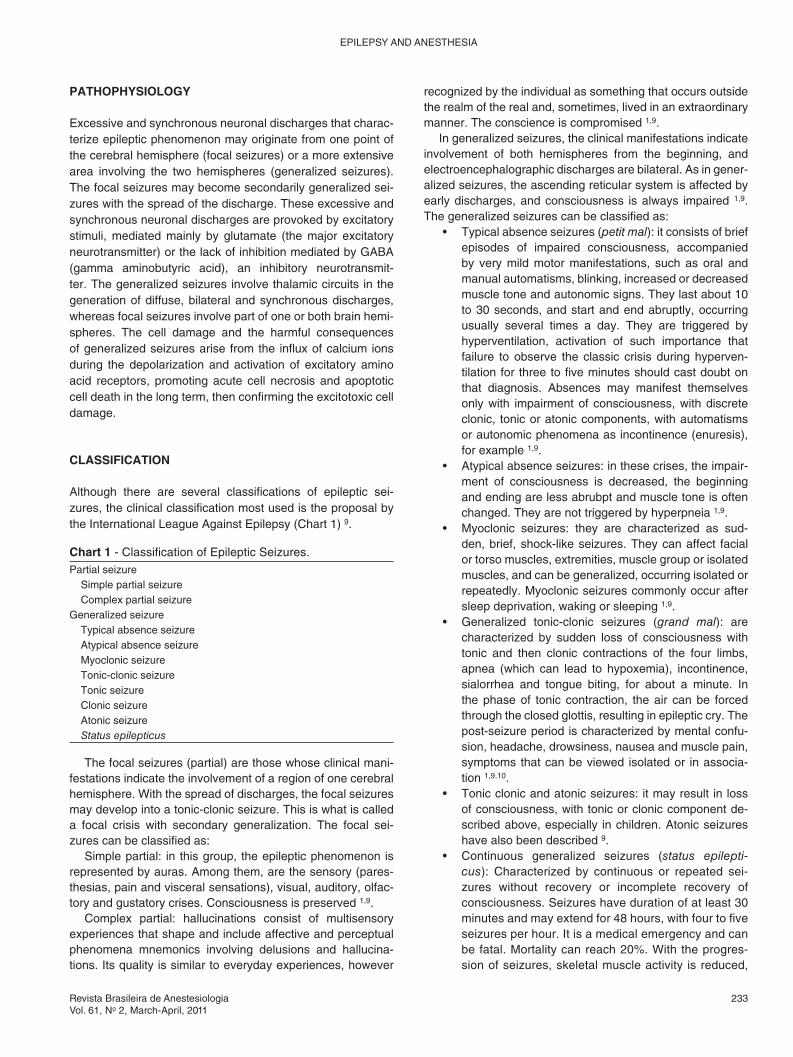
Although there are several classifications of epileptic sei-zures, the clinical classification most used is the proposal by the International League Against Epilepsy (Chart 1) 9.
recognized by the individual as something that occurs outside the realm of the real and, sometimes, lived in an extraordinary manner. The conscience is compromised 1,9.
In generalized seizures, the clinical manifestations indicate involvement of both hemispheres from the beginning, and electroencephalographic discharges are bilateral. As in gener-alized seizures, the ascending reticular system is affected by early discharges, and consciousness is always impaired 1,9. The generalized seizures can be classified as:
• Typical absence seizures (petit mal): it consists of brief episodes of impaired consciousness, accompanied by very mild motor manifestations, such as oral and manual automatisms, blinking, increased or decreased muscle tone and autonomic signs. They last about 10 to 30 seconds, and start and end abruptly, occurring usually several times a day. They are triggered by hyperventilation, activation of such importance that failure to observe the classic crisis during hyperven-tilation for three to five minutes should cast doubt on that diagnosis. Absences may manifest themselves only with impairment of consciousness, with discrete clonic, tonic or atonic components, with automatisms or autonomic phenomena as incontinence (enuresis), for example 1,9.
• Atypical absence seizures: in these crises, the impair-ment of consciousness is decreased, the beginning and ending are less abrubpt and muscle tone is often changed. They are not triggered by hyperpneia 1,9.
• Myoclonic seizures: they are characterized as sud-den, brief, shock-like seizures. They can affect facial or torso muscles, extremities, muscle group or isolated muscles, and can be generalized, occurring isolated or repeatedly. Myoclonic seizures commonly occur after sleep deprivation, waking or sleeping 1,9.
• Generalized tonic-clonic seizures (grand mal): are characterized by sudden loss of consciousness with tonic and then clonic contractions of the four limbs, apnea (which can lead to hypoxemia), incontinence, sialorrhea and tongue biting, for about a minute. In the phase of tonic contraction, the air can be forced through the closed glottis, resulting in epileptic cry. The post-seizure period is characterized by mental confu-sion, headache, drowsiness, nausea and muscle pain, symptoms that can be viewed isolated or in associa-tion 1,9,10.
• Tonic clonic and atonic seizures: it may result in loss of consciousness, with tonic or clonic component de-scribed above, especially in children. Atonic seizures have also been described 9.
• Continuous generalized seizures (status epilepti-cus): Characterized by continuous or repeated sei-zures without recovery or incomplete recovery of consciousness. Seizures have duration of at least 30 minutes and may extend for 48 hours, with four to five seizures per hour. It is a medical emergency and can be fatal. Mortality can reach 20%. With the progres-sion of seizures, skeletal muscle activity is reduced,
Chart 1 - Classification of Epileptic Seizures.Partial seizure
Simple partial seizureComplex partial seizure
Generalized seizureTypical absence seizureAtypical absence seizureMyoclonic seizureTonic-clonic seizureTonic seizure Clonic seizureAtonic seizureStatus epilepticus
The focal seizures (partial) are those whose clinical mani-festations indicate the involvement of a region of one cerebral hemisphere. With the spread of discharges, the focal seizures may develop into a tonic-clonic seizure. This is what is called a focal crisis with secondary generalization. The focal sei-zures can be classified as:
Simple partial: in this group, the epileptic phenomenon is represented by auras. Among them, are the sensory (pares-thesias, pain and visceral sensations), visual, auditory, olfac-tory and gustatory crises. Consciousness is preserved 1,9.
Complex partial: hallucinations consist of multisensory experiences that shape and include affective and perceptual phenomena mnemonics involving delusions and hallucina-tions. Its quality is similar to everyday experiences, however
MARANHÃO, GOMES, CARVALHO
234 Revista Brasileira de Anestesiologia Vol. 61, No 2, March-April, 2011
Epileptiform EEG changes are essential in the diagnosis and therapeutic management in the epileptic patient 8. This is a simple and inexpensive diagnostic method to determine the physiological manifestations of abnormal cortical excitability associated with epilepsy. The EEG shows three types of infor-mation: confirmation of abnormal electrical activity, the type of seizures and localization of the convulsive focus. Specificity is high, 78-98%, but sensitivity is low, around 25-26%. This is due to the fact that the EEG analyzes only the superficial lay-ers of the cerebral cortex. The short duration of routine EEG is one of the reasons why only 50% of the initial EEG shows epileptiform discharges 4. In general, the EEG is performed af-ter 48 hours or more of the seizure, although more recent evi-dence reveals that investigations within the first 24 hours may show interictal discharges 13. Preferably, the EEG should be performed during sleep, light stimulation and hyperventilation, because certain types of seizures occur in these conditions 14.
Other tests used in diagnosis of epilepsy include video EEG, functional magnetic resonance imaging, positron emis-sion tomography (PET) and single photon emission computed tomography (SPECT).
TREATMENT
Approximately 10% of the population has a single convulsive episode during their lifetime. In this situation, the use of anti-convulsants is not indicated. After the occurrence of a second seizure, the diagnosis of epilepsy is confirmed, initiating then the routine treatment with antiepileptic drugs 4.
The choice of the anticonvulsant should be made accord-ing to the type of crisis, the effectiveness and side effects, and used as monotherapy whenever possible, i.e. the goal of pharmacological treatment is to control seizures without side effects 1,4,15. It is important to emphasize that most of the adverse effects of antiepileptic drugs is dose-dependent, i.e. decreasing the dose of the drug reduces the severity of side effects, with no need to discontinue the drug 4. The appropri-ate prescription of anticonvulsants requires knowledge of the pharmacokinetics of the drug. One must consider that, once the pharmacological treatment is established, the anticonvul-sant can only achieve metabolic balance after a period of at least four half-lives, the time required to obtain tissue satu-ration. Monitoring blood levels of antiepileptic drugs is rarely necessary, because blood levels of drugs do not consistently correlate with clinical toxicity or clinical control of seizures. The suspension of antiepileptic drugs can be considered in the absence of seizures after two years of initiation of drug therapy, except for neonates in whom therapy can be discon-tinued 2 weeks after the last seizure. The risk of recurrence of seizures is high in patients with structural lesion, electro-encephalographic abnormalities, seizures onset in adoles-cence, neurological disorders, severe epilepsy, when they require multiple medications or when the drug is discontinued abruptly. In general, the prognosis for treatment of epilepsy is good. About 70% of patients’ present seizure control for ap-proximately five years, and 50% manage to stay medication
and seizure activity may be evident only by EEG. Re-spiratory effects of status epilepticus include inhibi-tion of the respiratory centers, uncoordinated skeletal muscle activity with ventilation impairment. The ab-normal autonomic activity can cause bronchospasm. It requires immediate treatment to avoid the risk of brain injury 1,9-12.
DIAGNOSIS
The diagnosis of epilepsy is based on clinical history, descrip-tion of seizure activity by those who witnessed, physical ex-amination (looking for focal findings) and encephalographic findings 4.
Nonspecific changes such as headache, mood changes, lethargy and myoclonic contraction warn some patients to an impending seizure, within hours of its occurrence. These pro-dromal symptoms are different from aura, which may precede the generalized seizure by a few seconds or minutes, and is part of epileptic seizure. In most patients, seizures occur un-predictably, without any regard to the posture or activity. Oc-casionally, however, crises occur in certain situations, such as sleep, or related to external predisposing factors such as sleep deprivation, poor diet, stress, menstruation, ingestion or withdrawal of alcohol and drug use. Fever and nonspecific infectious process can trigger seizures in epileptic patients. In a few patients, the seizures can be provoked by specific stimuli such as television flashing (photosensitive epilepsy), music or reading 9.
Physical examination between seizures in idiopathic epi-lepsy shows no abnormalities, but in the immediate posictal period, extensor plantar response can be observed 9.
In partial seizures, differential diagnosis should be per-formed with transient ischemic attack, aggressiveness and panic attacks 9. In generalized seizures, differential diagnosis should be done with syncope, cardiac arrhythmias, brainstem ischemia and pseudoepilepsia (due to psychiatric illness) 9.
Blood tests and lumbar puncture are rarely necessary for the diagnosis of epilepsy, except in the presence of suspect-ed electrolyte abnormalities, liver diseases, cancer, infectious diseases or exposure to toxins, althouhg that conduct is not yet consensus 4,9. Some authors consider it important, in the initial evaluation, the hemogram, glucose, liver function tests, kidney function and serological tests for syphilis. Serum pro-lactin and creatinine significantly increased after the tonic-clonic seizures in most patients 9.
A CT scan or MRI is indicated for patients with focal neuro-logical signs and symptoms, focal seizures or EEG findings of a focal seizure; some neurologists routinely indicate imaging for all patients in the initial evaluation of a seizure 9.
In children, emergency neuroimaging studies are per-formed when post convulsive deficit or altered mental status occurs that do not disappear quickly. Neuroimaging studies can be performed electively in patients where there is no de-termined cause for the focal seizure or infants who have a non febrile seizure 4.

Revista Brasileira de Anestesiologia 235Vol. 61, No 2, March-April, 2011
EPILEPSY AND ANESTHESIA
free without recurrence of seizures 4. Following is a summary of the pharmacology of the major anticonvulsants, with em-phasis on the indication, mechanism of action, adverse effects and drug interactions.
Phenobarbital: long-term effect barbiturate, effective against all types of epilepsy, but not the primary generalized non-convulsive type. Despite the low cost, presents cognitive and behavioral effects that limit the use of this drug to treat epilepsy; considered second-line treatment of epilepsy 1,10,15. Phenobarbital exerts its antiepileptic effect by modulating the postsynaptic action of gamma aminobutyric acid (GABA), an inhibitory neurotransmitter, and also by the block of postsyn-aptic excitatory effect induced by glutamate, especially those mediated by the activation of the AMPA (α-amino-3-hydroxy-5-methyl-4-isoxazolepropionic acid) receptor. . The GABAer-gic action causes increased duration of chloride channels opening, promoting neuronal hyperpolarization, limiting activ-ity and increasing the seizure threshold 1,15,16. The main ad-verse effects include sedation, depression, hyperactivity (in children), confusion (in elderly), skin changes, megaloblastic anemia, osteomalacia, nystagmus, ataxia, abnormal collagen deposition (Dupuytren’s contracture), dependence, tolerance and low therapeutic index 4-5,8,10,16,25. The drug, like other bar-biturates, may precipitate attacks of porphyria 10. Congenital defects can occur when phenobarbital is chronically adminis-tered during pregnancy. Changes in coagulation and bleed-ing in the neonate can occur. Interaction of phenobarbital with other drugs arises from the hepatic microsomal enzyme in-duction by phenobarbital, which may persist for 30 days after discontinuation of the drug. This can exacerbate the hepatic metabolism of many liposoluble drugs, such as anticonvul-sants, oral contraceptives, beta-blockers, corticosteroids, di-goxin, anti-inflammatory drugs and anticoagulants 10,15,17. The enzyme induction caused by phenobarbital may increase the biotransformation and toxicity of inhaled anesthetics. There-fore isoflurane should be the drug of choice 17.
Valproic acid: it is a carboxylic acid (2-propylpentanoic acid) effective in all generalized primary epilepsy and all convulsive epilepsy types. It is less effective in the treatment of partial non convulsive epilepsy 1,15. The valproic acid is a weak inhibitor of two enzymatic systems that inactivate GABA: the GABA trans-aminase and succinate semialdehyde dehydrogenase. There is some evidence that the drug may potentiate the action of GABA by a postsynaptic action 10. This drug also works by sus-tained and repetitively limiting the neuronal discharge through voltage-dependent sodium channels, that is, it prevents the generation of action potential bursts that constitute the micro-physiological substrate of the epileptic neuron 1,15. Drug ad-verse effects include tremors, weight gain, dyspepsia, nausea and vomiting, anorexia, alopecia, edema, encephalopathy (due to increased levels of ammonia), teratogenicity, agranulo-cytosis, aplastic anemia, allergic dermatitis, Stevens-Johnson syndrome, hepatotoxicity (especially in children younger than 2 years of age), platelet changes that can lead to bleeding, and pancreatitis 4,8,10,15. Valproic acid and phenytoin may displace diazepam from its protein binding sites, resulting in increased pharmacological activity of these drugs. Furthermore, it is an
enzyme inhibitor, leading to a decrease in metabolic rate of phenytoin. With chronic use of this drug, plasma concentration of phenobarbital increases by approximately 50%, probably due to inhibition of liver microsome enzymes 15.
Felbamate: normally used to treat adults presenting iso-lated partial epilepsy or secondary generalized epilepsy. In children, it is used in the treatment of partial or generalized epilepsy associated with Lennox-Gastaut syndrome 10,15. The mechanism of action, although not fully explained, appears to be related to the decrease of sodium ion current (block of voltage-dependent sodium channels), increased the inhibitory effects of GABA, and blockage of the N-methyl-D-aspartate receptors 15. The adverse effects described are insomnia, anorexia, nausea, headache, irritability, aplastic anemia and hepatotoxic effects 4,15,10. Monitoring through blood and liver function tests is indicated 15. Felbamate interacts with other anticonvulsants 10. Should the drug be used in combination with phenytoin, carbamazepine or valproic acid, the drug dos-age should be reduced between 20% and 30% in order to prevent toxic effects 15.
Phenytoin: it is effective in the treatment of partial and gen-eralized epilepsies 10,15. It has a high therapeutic index. Phe-nytoin regulates neuronal excitability and thus the spread of seizure activity from the seizure focus by blocking voltage-de-pendent sodium channels, possibly in the transport of calcium through the neuronal membrane 10,15,16. The stabilizing effect on the cell membrane is relatively selective for the cerebral cortex, but also extends to peripheral nerves. Additionally to the effect on the ion flow, phenytoin acts on the second mes-senger systems as calmodulin and cyclic nucleotides 15. Side effects of phenytoin include nystagmus, diplopia, dizziness (vestibular cerebellar dysfunction), ataxia, nausea and vomit-ing, gingival hyperplasia, depression, megaloblastic anemia, drowsiness, agranulocytosis, aplastic anemia, allergic derma-titis, Stevens-Johnson syndrome, hyperglycemia, hepatotox-icity, pancreatitis, acne, rough skin, hirsutism, teratogenicity and Dupuytren’s contracture 4,8,10,15,16. Chronically adminis-tered during pregnancy, the drug can cause congenital mal-formations 15. Phenytoin can induce the oxidative metabolism of several liposoluble drugs such as carbamazepine, valproic acid, ethosuximide, corticosteroids and anticoagulants 15,16. Patients treated with this drug have decreased plasma con-centrations of thiopental, propofol, midazolam, opioids and neuromuscular non depolarizing blockers 8,17.
Phosphofenitoin: it is a prodrug of phenytoin, presenting the same pharmacological properties. Its main indication is the replacement of intravenous phenytoin in the treatment of status epilepticus 18.
Carbamazepine: is used in the treatment of convulsive and nonconvulsive partial epilepsy, as well as trigeminal and glos-sopharyngeal nerves neuralgia, bipolar disorders and alco-hol withdrawal syndrome 10,15. Carbamazepine changes the ionic conductance to sodium with a membrane stabilizing ef-fect 15,16. Its side effects can be sedation, diplopia, dizziness, neutropenia, nausea, drowsiness, diarrhea, jaundice, oliguria, hypertension, cardiac arrhythmias, hyponatremia, agranulo-cytosis, aplastic anemia, allergic dermatitis, Stevens-Johnson

MARANHÃO, GOMES, CARVALHO
236 Revista Brasileira de Anestesiologia Vol. 61, No 2, March-April, 2011
syndrome and systemic lupus erythematosus (SLE) 4,8,10,15,16. In addition to accelerating its own metabolism, carbamaze-pine exacerbates hepatic oxidation and conjugation of other liposoluble drugs. The most common interactions occur with oral contraceptives, haloperidol, valproic acid, theophylline, ethosuximide, clonazepam, phenobarbital, steroids, antico-agulants and antipsychotic drugs 10,15,16. Drugs that inhibit the metabolism of carbamazepine may increase its toxicity and include cimetidine, propoxyphene, diltiazem, verapamil, erythromycin and isoniazid 10,15,16. Due to its potent enzyme inducer, carbamazepine reduces the plasma concentrations of thiopental, propofol, midazolam, opioids and neuromuscu-lar non depolarizing blockers 10,17. There is risk of hepatotox-icity after anesthesia with halothane, enflurane, and possibly with sevoflurane 17.
Oxcarbazepine: it was designed to prevent the formation of the epoxide metabolite of carbamazepine, responsible for many toxic effects of the drug 10. It has blocking action of the voltage-dependent sodium channels. It is generally better tolerated than carbamazepine. The most common adverse effects are sedation, dizziness, headache, amnesia, ataxia, diplopia, depression, insomnia, anxiety, nausea and vomiting, diarrhea, hyponatremia, acne, alopecia and urticaria 8,10. It shows less pharmacokinetic changes, not inducing the me-tabolism of other antiepileptic drugs 10.
Lamotrigine: it acts by stabilizing the voltage-sensitive so-dium channels and preventing the release of glutamate and aspartate 10,15,16. This drug has a broad spectrum of activity and it is effective when used alone or in combination in adult patients with partial or generalized epilepsy, and in children with Lennox-Gastaut syndrome 10,15. It shows, as side effects, dizziness, diplopia, tremor, insomnia, aggression, ataxia, al-lergic dermatitis, Stevens-Johnson syndrome, headache, nausea, vomiting and blood dyscrasias 4,8,10,15,16. Lamotrigine induces the hepatic microsomal system, speeding up their metabolism. The valproic acid slows the metabolism of la-motrigine, increasing its elimination half-life 15.
Gabapentin: it is used in the treatment of partial epilepsy and generalized partial seizures and in treating various pain syndromes, such as erythromelalgia, reflex sympathetic dys-trophy and neuropathic pain 15. The mechanism of action is unclear, appearing to act by binding to specific receptors in the brain, inhibiting the voltage-dependent sodium current, apart from also increasing GABA action 4,15. The main adverse drug effects are sedation, diplopia, dizziness, ataxia, skin changes, weight gain, nausea and leucopenia 8,10,15.
Primidone: it is metabolized in phenobarbital and an-other active metabolite, phenylethylmalonamide. The ef-fectiveness of the drug is similar to phenobarbital, but less tolerated 4,10,15,16. Adverse reactions include fatigue, depres-sion, psychosis, decreased libido, skin changes, leukopenia, thrombocytopenia and SLE 8,10. Chronic use of the drug in-creases the metabolism of enflurane, halothane, and probably isoflurane 17.
Clonazepam: in general, it is associated with other drugs. It is the drug of choice only in myoclonic seizures 15. It binds to GABAA in the brain, enhancing the inhibitory transmission
mediated by GABA, which increases the conductance to chlo-ride ion, due to an increase in frequency of chloride channels’ opening, leading to cell hyperpolarization and inhibition of neuronal trigger 4,15,16. The adverse effects of the drug include personality changes manifested as behavioral disorders, in-cluding hyperactivity, irritability and difficulty concentrating, especially in children. Depression can occur in the elderly. Si-alorrhea and increased bronchial secretion may be common in children. Generalized convulsive activity may occur if the drug is abruptly discontinued 8,15,16.
Ethosuximide: it is the drug of choice in treating absence seizures (petit mal). It acts by decreasing conductance in volt-age-dependent calcium channels in thalamic neurons 10,15,16. Adverse effects of the drug include nausea, anorexia, vom-iting, lethargy, agitation, headache, drowsiness, dizziness, ataxia, photophobia, agranulocytosis, aplastic anemia, aller-gic dermatitis, Stevens-Johnson syndrome, SLE and, rarely, bone marrow depression 8,10,15,16.
Topiramate: it is an antiepileptic drug used initially as an ad-juvant to partial seizures 10. Additional studies suggest its effica-cy as monotherapy and also in the tonic-clonic seizures and in Lennox-Gastaut syndrome 10. The drug acts by multiple mech-anisms, such as blocking a subtype of non-NMDA glutamate receptor, potentiating the action of GABA and blocking sodium channels 4,10,16. Side effects include dizziness, nervousness, cognitive disturbances, ataxia, depression, diarrhea, glaucoma, loss of appetite and weight loss, paresthesias and development of urolithiasis 4,8,10,16. Patients treated with topiramate should be evaluated under clinical and laboratory aspects, preoperative assessment to detect asymptomatic metabolic acidosis, which can occur with chronic use of the drug 19.
Levetiracetam: it is used in the treatment of partial seizures as adjunctive or monotherapy 10. It is generally well tolerated. The mechanism of action of the drug has not been determined, although recent studies show clearly that the drug binds to protein A2 (SV2A), related to the release of glutamate in the synaptic vesicle 16,20. Common side effects include drowsi-ness, weakness, dizziness, ataxia, amnesia, depression, anxiety, anorexia, diarrhea, dyspepsia, skin changes and pancytopenia 8,10. It presents little risk of drug interactions 10.
Tiagabine: it is indicated in adjuvant therapy of patients with partial seizures. Clinical trials indicate drug efficacy in tonic-clonic seizure when combined with other anticonvul-sants 10. The action mechanism of the drug is the blockage of GABA reuptake 10,16. The most frequent side effects are diz-ziness, asthenia, somnolence, anxiety, nausea, nervousness, tremors, abdominal pain and cognitive disorders 8,10,16.
Zonisamide: it is an antiepileptic drug used in the broad spectrum of adjuvant therapy of partial seizures and second-arily generalized seizures. It is also shown to be effective in controlling the generalized tonic-clonic epilepsy, the myo-clonic epilepsy and Lennox-Gastaut syndrome. It has multiple mechanisms of action. It has its pharmacological effect by blocking voltage-dependent sodium channels, also inhibiting the calcium current through T-type channels. The drug has an effect on the synthesis, degradation and release of several neurotransmitters such as glutamate, GABA, dopamine, sero-

Revista Brasileira de Anestesiologia 237Vol. 61, No 2, March-April, 2011
EPILEPSY AND ANESTHESIA
tonin and acetylcholine, which increases synaptic inhibition. It is a carbonic anhydrase inhibitor, although this effect is not correlated with epileptic activity. Adverse effects include seda-tion, asthenia, diplopia, hypohidrosis, skin changes, Stevens-Johnson syndrome, dizziness, headache, ataxia, anorexia, agitation, irritability, and nephrolithiasis 21. It may cause meta-bolic acidosis. Serum bicarbonate levels should be evaluated before treatment and periodically after it, even in the absence of symptoms 22.
OTHER THERAPIES
ACTH and corticosteroids: in children with West syndrome, the use of ACTH and corticosteroids is well established. The mechanism of action is uncertain, postulating effects on cholinergic modulation or serotoninergic 23. It is impor-tant that the anesthesiologist knows the adverse effects of corticosteroids that can influence the conduct of anesthesia and involve multiple organ systems. These effects include irritability, Cushing syndrome, electrolyte disturbances (hy-pokalemia), impaired glucose tolerance, dilation of cerebral ventricles, increased cerebrospinal fluid, hypertension, car-diomyopathy, and sepsis 4,23.
Ketogenic diet: an alternative therapy for refractory epi-lepsy in children and adolescents is the ketogenic diet, which consists of a diet rich in fats and low in carbohydrates and proteins, leading to ketosis and production of beta-hydroxybu-tyrate, which could have an antiepileptic effect, probably due to the increase in brain energy reserves 4,23,24. This diet seems to work better in generalized seizures (absence seizures and myoclonus), but any type of crisis may benefit from this kind of therapy 23. Ketosis leads to metabolic acidosis, which can be exacerbated by products that contain carbohydrates. It is important to avoid solutions in pre-medication containing glu-cose, and dextrose-containing intravenous solutions, which can cause a decrease in plasma levels of ketones and in-creased risk of seizures 4. Children with ketogenic diet can safely undergo general anesthesia. Although serum glucose levels generally do not change, the pH and serum bicarbonate levels should be monitored every 2 or 3 hours, even on short procedures to avoid severe metabolic acidosis 4,25.
SURGICAL TREATMENT
Surgical treatment is indicated in cases of refractory epilepsy with drug treatment. Surgical treatment includes frontal lo-bectomy, focal cortical resection, Calostoma (section on the bridge level through which the fibers that connect one brain hemisphere to the other), hemispherectomy and placement of the vagus nerve stimulator 26.
The vagus nerve stimulator consists of a bipolar wire with two electrodes involving the left vagus nerve at cervical level, and a generator implanted subcutaneously in the infraclavicu-lar region 26,27. Laboratory studies suggest that this device in-creases the release of noradrenaline in the locus coeruleus,
increasing the seizure threshold. The most common adverse effects with the use of such equipment are voice changes and pain in the pharynx. In patients with the vagus nerve stimulator undergoing surgery, the equipment must be eval-uated before and after surgery. It is important to emphasize that these patients are at risk of aspiration because of the possibility of laryngeal-pharyngeal dysfunction during vagus nerve stimulation. Although there are no reports of electro-magnetic interference with the use of electrocautery and ex-ternal defibrillator, these devices can damage the electrodes and the generator of the vagus nerve stimulator. Similar care should be taken with patients with cardiac pacemakers. Dur-ing activation of the vagus nerve stimulator in patients with obstructive sleep apnea, airway obstruction can occur. Con-sidering the association of the residual effect of anesthesia and the activation of the vagus nerve stimulator, airway ob-struction in the postoperative period in patients with obstruc-tive sleep apnea may accur. Central venous access on the side of stimulation of the vagus nerve should be avoided. The neck should be placed in neutral position in order to avoid stretching or displacement of the electrodes 28.
ANESTHETIC APPROACHES IN EPILEPTIC PATIENT
Anesthetic drugs and epilepsy
Chloral hydrate: this drug is used to induce sleep during the EEG, and to promote sedation in preanesthetic medication in children. It has shown anticonvulsant properties 4,29. Through oral administration, the drug latency to induce sleep is 40 minutes in average, while for sedation, the onset is unpre-dictable, ranging from 30 to 60 minutes. It is considered a safe drug, although reports of deaths and severe neurologi-cal injury have been reported 30. There is a risk of respiratory depression and hypoxemia, especially in patients at risk for airway obstruction, for example, Down syndrome, or obstruc-tive sleep apnea after tonsillectomy. Other adverse effects include nausea, vomiting, restlessness, sweating, dizziness and sedation failure 4,29,31,32.
Benzodiazepines: they have anticonvulsant activity. How-ever, benzodiazepines can cause short periods of seizure ac-tivity, and in EEG in patients with Lennox-Gastaut syndrome, a form of epilepsy of difficult treatment in children 8,33,34. Ben-zodiazepines are commonly used to treat acute seizures 4. The antiepileptic effect of benzodiazepines is due to its po-tentiating action of the inhibitory transmission mediated by GABAA, increasing the opening frequency of chloride chan-nels and leading to neuronal hyperpolarization 8,33. Midazolam has potent anticonvulsant activity and is commonly used in the treatment of status epilepticus mainly in children 8,35,36. As noted, clonazepam is effective in the prevention and treat-ment of seizures, especially in myoclonic seizures in children. Some antiepileptic drugs have significant sedative effects, such as phenobarbital and carbamazepine, which have the ability to potentiate the effects of anesthetics and other central nervous system depressants 8,37.

MARANHÃO, GOMES, CARVALHO
238 Revista Brasileira de Anestesiologia Vol. 61, No 2, March-April, 2011
Flumazenil may precipitate seizures in patients treated with benzodiazepines or who received this drug as part of premedication due to its competitive antagonism 38,39. It has been reported that the use of flumazenil to reverse benzodi-azepine sedation caused generalized convulsions and coma in epileptic patients who underwent the procedure under lo-cal anesthesia and sedation with diazepam 40. Given its pro-convulsant effects, flumazenil should be used with caution in patients with epilepsy 41.
Propofol: in spite of the conflicting results in the literature, there is clinical evidence that propofol has anticonvulsant effect and is considered a safe drug for sedation, induc-tion and maintenance of general anesthesia in children and adults 4,8,33,34,42. In patients with epilepsy, the occurrence of seizures of epileptic origin is extremely rare with the use of propofol, and occurs frequently in the recovery from anesthe-sia 42. The antiepileptic activity of propofol is probably related to inhibition of pre-and postsynaptic chloride channels medi-ated by GABA 8. Propofol can cause abnormal movements, as opisthotonus and myoclonia in both epileptic and healthy patients, but these changes do not seem to relate to epilep-togenic activity 43-45. Propofol is an effective alternative in the treatment of refractory epileptic seizures to usualantiepileptic drugs and in cases of status epilepticus 46-48.
Etomidate: etomidate appears to have anticonvulsant ac-tivity at high doses 33.49 and pro-convulsant characteristics in usual clinical doses 33,50-54. Threfore, it is recommended to avoid its use in epileptic patients 4,8,34.
Ketamine: although there are reports of anticonvulsant property of ketamine, especially when associated with drugs with GABAergic activity (benzodiazepines and propofol) for cases refractory to usual therapy 33,55-59, most of the clinical evidence shows that ketamine, at usual doses, has significant epileptogenic potential 33,54,60, and should be, therefore, avoid-ed in epileptic patients 4,8,34,61.
Droperidol: it does not produce neuron excitability nor induce seizures in epileptics; it can be used safely in these patients 4,54.
Dexmedetomidine: sedation with dexmedetomidine ob-served in the EEG shows a pattern similar to stage II sleep. It presents no pro- or anticonvulsant effects and has been used successfully in children with autism undergoing EEG 4,62. This drug has been safely used in adult patients for epilepsy sur-gery, allowing an adequate mapping of epileptic foci and sub-sequent resection 63,64. Thus, dexmedetomidine can be used safely in the epileptic patient.
Clonidine: the effect of clonidine in epileptic patients is con-troversial. There are reports that the drug lowers the seizure threshold in epileptic patients (pro-convulsant effect) 65-67. Other authors have used the drug in autistic patients (epilepsy in 50% of patients), with the purpose of sedation for the per-formance of EEG, and concluded that there was no effect on the EEG. Moreover, it is easy to be administered, the seda-tion time is short and it promotes good quality sedation 68. In animal experiments, clonidine presented no pro- or anticon-vulsant activity in the lidocaine-induced seizures 69. So there is not enough clinical evidence at present that the use of cloni-dine is contraindicated in epilepsy patients.
Barbiturates: barbiturates, except for the methohexital, present a significant anticonvulsant activity in the EEG 4, and its use is considered safe for induction of anesthesia in epi-leptic patients, as well as treatment of status epilepticus 8,34.
Opioids: meperidine has neuroexcitatory effect attrib-uted to its metabolite normeperidine. Clinically, it is char-acterized as tremors, myoclonus and seizures 8,34,70. At the doses commonly used as adjuvant drug in general or re-gional anesthesia, it appears safe in the epileptic patient. However, in situations where there may be accumulation of normeperidine, such as prolonged use (patient-controlled analgesia), anticonvulsants (phenytoin and phenobarbital) and phenothiazines (chlorpromazine), patients with renal failure, advanced malignancy and sickle cell disease, the drug can cause seizures; it shall then not be used in epi-leptic patients 70.
Morphine has no pro-convulsant effect when used intrave-nously and can be used safely in epileptic patients. However, tonic-clonic seizures were observed when employing epidural drug in epileptic patients 8,70.
Regarding phenylpiperidine derivatives (fentanyl, alfent-anil, remifentanil and sufentanil), there are several reports that the administration of these drugs cause tonic-clonic movements, although in many of these articles there have been no EEG monitoring, making it difficult to confirm whether these movements are the result of seizure activ-ity or whether, in fact, represent myoclonus or stiffness of skeletal muscles 57,70. In other studies with EEG monitoring, there was no correlation between these movements and tonic-clonic seizure activity 70. However, there are reports of epileptogenic EEG activity with the use of fentanyl, alfentanil and remifentanil - these drugs can be helpful in localizing the epileptic focus 71-73. Therefore, we recommend avoiding the use of high doses or rapid administration of opioids of the phenylpiperidine group in epileptic patients 8. It is important to emphasize that there may be pharmacokinetic interaction between the phenylpiperidine opioids (mostly fentanyl) and anticonvulsant drugs, leading to a greater need to administer opioids during anesthesia 74.
Nitrous oxide: although nitrous oxide has excitatory ef-fects on central nervous system, the epileptogenic potential of the drug is very low and can be used safely in epileptic patients 4,8,34,70.
Halothane: most published studies show that halothane has potent anticonvulsant effect, and can be used safely in epileptic patients. However, there are reports of seizures as-sociated with halothane 34,75,76.
Enflurane: it may produce epileptiform EEG activity and seizures, especially at concentrations above 2 MAC and hypocapnia in the presence of stimulation, tactile, visual or hearing impairment; then it should be avoided in epileptic pa-tients 8,34,70,75,77,78.
Isoflurane: although there are reports of potential epi-leptogenic with isoflurane 79, most studies show potent anticonvulsant effect of the drug, that is also used in the treat-ment of status epilepticus 80, and certainly in epileptic pa-tients 4,8,34,70,75.
Revista Brasileira de Anestesiologia 239Vol. 61, No 2, March-April, 2011
EPILEPSY AND ANESTHESIA
Sevoflurane: the epileptiform activity of sevoflurane has been extensively studied 57. A review of 30 studies evaluated the correlation between the use of sevoflurane and epilep-tiform EEG changes and/or presence of convulsive move-ments in patients with or without epilepsy. The incidence of these changes ranged from 0-100%. The depth of anesthe-sia with sevoflurane and/or the presence of hyperventilation (hypocapnia) were risk factors for the emergence of excit-atory activity of the central nervous system. The authors of this review recommend avoiding this drug at concentrations above 1.5 MAC in epileptic patients and in the presence of hypocapnia 81.
Desflurane: although there are few studies, there is no evidence that desflurane has epileptiform activity, even when used in high concentrations or in the presence of hypocapnia, and has even been administered in the treatment of status asthmaticus and safely in epileptic patients 8,82,83.
Neuromuscular blockers
Some antiepileptic drugs have a depressant action on ace-tylcholine release at the neuromuscular junction 8. Phenytoin and carbamazebina, when used chronically, decrease the duration of action of some neuromuscular blockers such as rocuronium 84,85, pancuronium 86, vecuronium 87, cisatracu-rium 88, due to increased hepatic metabolism of these drugs. In contrast, atracurium 89,90 and mivacurium 91 that are not dependent on the hepatic metabolism for their elimination, have not presented reduction in the time of action. Acute administration of phenytoin potentiates the neuromuscular blockade of rocuronium, by an unknown mechanism 92. Al-though laudanosine, an atracurium metabolite, may present epileptiform EEG activity and seizures when administered in higher concentrations 93 in experimental animals, it is unlikely that those effects can be observed with the usual doses used in anesthesia. With regard to succinylcholine, there is small increase in duration of action in patients chronically treated with antiepileptic drugs, factor which is irrelevant under the clinical point of view 94. Because of drug interactions between antiepileptic drugs and neuromuscular blockers, we recom-mend intraoperative monitoring of neuromuscular blockade 8.
Anticholinesterase agents: it has not been observed epi-leptiform activity on EEG or seizures after administration of cholinesterase inhibitors during anesthesia in patients with epilepsy 8,34.
Anticholinergics: atropine, scopolamine and glycopyrrolate can be used safely in the epileptic patient 8.
Local Anesthetics: local anesthetics have pro-convulsant and anticonvulsant properties due to the stabilizing effect of the membrane. In small doses, local anesthetics reduce ce-rebral blood flow and metabolism, as well as brain electrical activity, and act as anticonvulsants, sedatives and analgesics, while at high doses it act as pro-convulsant drug, lowering the seizure threshold in the cerebral cortex, amygdala and hippocampus, leading to generalized convulsions 34,95. The systemic toxicity associated with regional anesthesia is a
cause of seizures in approximately 5/10.000 patients, which can be found even with local anesthetics for use later. It is more frequent with bupivacaine and those regional anesthe-sia techniques in which large doses of local anesthetics such as epidural and caudal are employed 8,95-97.
A retrospective study on 335 patients with different types of epilepsy undergoing regional anesthesia showed that 24 patients (6%) had seizures in the perioperative period. In 19 patients, seizures were the result of epileptic disease and not related to the use of local anesthetic. In the 5 remaining pa-tients, systemic toxicity resulting from the use of local anes-thetic cannot be ruled out. An interesting aspect of the study is that the seizures were more frequent in those patients who had presented with epileptic seizures less than a week prior to surgery and used phenytoin as an anticonvulsant drug. As the seizure threshold appears to be decreased in patients with recent seizures, it is important to be prepared to treat any seizure in these patients when using local anesthetics in the perioperative period 98.
ANESTHETIC MANAGEMENT IN THE EPILEPTIC PATIENT
Preoperative assessment and premedication
In the perioperative management of epileptic patients, it is important, whenever possible, an adequate control of the disease, being essential a careful review of medical history, especially in regard to the evolution of the disease, factors triggering the seizures (fasting, stress, sleep deprivation, alco-hol and drugs), and comorbidities and their treatment 4,8. The presence of mental retardation, hypotonia, and risk factors for aspiration and airway obstruction should be examined 4.
A preoperative evaluation of the neurologist responsible for the patient is recommended, especially in the case of recent changes in disease evolution 8,57.
Anticonvulsants should be used until the day of surgery, including in children and pregnant women 8.
It is important to know the adverse effects of anticonvul-sants, as well as the possibility of drug interactions with anes-thetic agents 4.
The plasma dosing of antiepileptic drugs should not be done in a systematic way, unless there have been recent changes in drug dosage or in the case of onset of kidney or liver failure, arrhythmias and electrolyte changes 8.
The premedication is usually carried out with the use of a benzodiazepine, midazolam being the most widely used due to its potent anticonvulsant and anxiolytic effects. It is important to emphasize that some anticonvulsants, and the ketogenic diet can cause sedation and interact with benzodiazepines.
CONDUCT DURING SURGERy
The monitoring of epileptic patients is based on the type of surgical procedure being performed and the patient’s clinical

MARANHÃO, GOMES, CARVALHO
240 Revista Brasileira de Anestesiologia Vol. 61, No 2, March-April, 2011
condition. Patients undergoing major surgical procedures or severe clinical conditions require invasive monitoring. In most procedures, the basic monitoring is adequate.
The literature shows that most beta-lactam antibiotics showed some degree of pro-convulsant activity in laboratory animals and in clinical trials, probably due to inhibition of inhib-itory postsynaptic response mediated by GABAA
99. However, the widespread use of these drugs in epileptic patients in the perioperative period, with no reports of epileptogenic activity, seems to show that these antibiotics can be safely used in patients with epilepsy.
On inducing anesthesia in epileptic patients, the most com-monly used drugs are thiopental, benzodiazepines and, pro-pofol despite its pro-and anticonvulsant effects. The use of ketamine and etomidate should be avoided 8,57.
During maintenance of anesthesia, the inhalational agent most often used due to its potent anticonvulsant effect is iso-flurane. Sevoflurane administered at concentrations of less than 1.5 MAC (avoiding hyperventilation) seems safe in the epileptic patient. Halothane and desflurane may also be used safely. The use of nitrous oxide is controversial, and enflurane is contraindicated in those patients 8,57.
Although there are reports of convulsive movements with the use of opioids, these drugs have low seizure frequency when used in low doses and slow intravenous administration. Meperidine can cause seizures through its metabolite norme-peridine, and should be avoided in epileptic patients. The use of neuraxial opioids rarely cause seizures, which leads to no contraindication to its use in the epileptic patient 8.
In the intraoperative handling of the epileptic patient, it is im-portant to avoid changes that reduce the seizure threshold, such as hypoxia, hypotension, hypocapnia, and hyponatremia 28.
The regional anesthesia techniques can be used safely in the epileptic patient. However, coagulation changes that may occur with the administration of most antiepileptic drugs should be evaluated 8.
POSTOPERATIVE MANAGEMENT
Anticonvulsant drugs should be restarted as soon as possible after surgery, according to the time of fasting. If the fasting time is 12-24 hours, there is no need to replace the oral by the parenteral route, and treatment should be restarted when the oral route is cleared. In the case of fasting over 24 hours and in the chronic use of the orally administered anticonvul-sant drugs phenytoin or phenobarbital, we replace the enteral route for venous 2-3 times a day 8. When there is no presen-tation for parenteral use with anticonvulsant drugs used orally by the patient, it is possible to replace these drugs by tempo-rarily 3 mg.kg-1.dia-1 phenytoin or intravenous phenobarbital 2.3 mg.kg-1.dia-1 in 2-3 doses 8.
After surgery, whenever possible, the monitoring of plasma levels of antiepileptic drugs for at least 48 hours should be performed because there are significant variations in the ap-parent volume of distribution, linked to plasma proteins, he-patic metabolism and renal elimination 8,100.
PERIOPERATIVE SEIZURES IN EPILEPTIC PATIENTS
The seizures of epileptic origin are rare in the perioperative peri-od, usually occurring in the induction and recovery from anesthe-sia. However, they can happen up to 72 hours after surgery 57.
It is important to remember that the anesthetic drugs which commonly induce epileptogenic activity rarely progress to sei-zures in the perioperative period 57, although there is greater propensity to occur more frequently in epileptic patients than in non-epileptic patients 57.
The effects of general anesthesia were assessed in 236 epileptic patients of both sexes with an average age of 21 years (range 1 month to 84 years) undergoing diagnostic and therapeutic procedures, excluding neurosurgical procedures and invasive neurological examinations. The procedures per-formed included magnetic resonance imaging (35.7%), surgi-cal procedures (32.1%), other imaging studies of non-nuclear magnetic resonance (16.9%), biopsies (12.1%), and dental procedures (3%). Partial epilepsy was present in 70.7% of patients, generalized epilepsy in 27.1%, and epilepsy of un-known cause in 2.1%. A total of 297 procedures were evaluat-ed under anesthesia. Ten procedures were performed under local or regional anesthesia, a procedure under spinal anes-thesia and the other under general anesthesia. Seizures were observed in 6 (2%) patients, and therapy with intravenous benzodiazepine was only necessary in one patient.
The seizure activity was observed in the induction of an-esthesia (n = 1) or after the procedure before recovery from general anesthesia (n = 5). Five of the patients who had sei-zures were younger than 18 years (mean = 8 years ranging from 1 to 13 years). Five of the patients who had had epileptic seizures had refractory epilepsy, requiring multiple anticon-vulsant drugs. The drugs used in general anesthesia of pa-tients included sevoflurane, isoflurane, nitrous oxide, propofol, thiopental, ketamine, fentanyl, atracurium, cisatracurium and vecuronium. None of the patients who received local anes-thesia, regional or spinal anesthesia showed seizure activity. This article shows that the incidence of perioperative seizures in epileptic patients is low and does not require anticonvulsant therapy in most patients. It is most common in children and in patients with refractory epilepsy that require multiple anticon-vulsant drugs 101.
Although the occurrence of seizures in the perioperative period is low, they can be serious, if prolonged, leading to brain damage from hypoxia, apnea, prolonged postoperative mechanical ventilation, and delayed awakening from anesthe-sia. The seizure activity may impair the physiological regula-tion of cardiac and respiratory activities. Although the seizures are associated with tachycardia and tachypnea, there are reports of apnea and bradycardia following the seizure, and leading to sudden death in epileptic patients, probably due to autonomic instability, which causes cardiac arrhythmias or neurogenic pulmonary edema 101,102.
In the case of the emergence of seizures in the periopera-tory period, the initial approach is to maintain a patent airway with adequate ventilation and protect the patient from injuries resulting from seizures. Monitoring should be performed us-

Revista Brasileira de Anestesiologia 241Vol. 61, No 2, March-April, 2011
EPILEPSY AND ANESTHESIA
ing cardioscope, blood pressure and pulse oximetry. Seizures lasting less than five minutes do not usually require treat-ment 4,8.
Should convulsions persist for more than five minutes, intravenous benzodiazepine should be used. The preferred drug is lorazepam, which, however, is not available for paren-teral use in Brazil. In our country, the best alternative is diaz-epam (5-20 mg). Should convulsions persist, a second dose of benzodiazepine associated with phenytoin (20 mg.kg-1, 30 minutes) should be used. In the case of refractory seizures, phenobarbital (1.5 mg.kg-1.min-1 or 100 mg/70 kg.min-1 can be used with a maximum dose of 15 mg.kg-1 or 1,000 mg/70 kg), midazolam (0.1-0.3 mg.kg-1 in 2 to 5 minutes, followed by in-fusion of 0.05 to 0.4 mg.kg-1.hour-1), propofol (1-2 mg.kg-1 fol-lowed infusion of 2-10 mg.kg-1.h-1), thiopental (5-10 mg.kg-1 in 10 minutes, followed by infusion of 100-400 mg.h-1), lido-caine (1.5-2 mg.kg-1 in 2-5 minutes, followed by infusion of 2-3
mg.kg-1.h-1 for 12 hours), isoflurane (0.5 to 1.5%) and ketamine (50-100 mg followed by infusion of 50-100 mg.h-1) 8,57,103.
In summary, in the perioperative management of epileptic patients, it is important for the anesthesiologist in the preop-erative evaluation to identify the type of epilepsy, frequency, intensity and the triggers to the epileptogenic episodes, the use of anticonvulsant drugs and possible interactions with drugs used in anesthesia as well as the presence of other treatments such as vagus nerve stimulator and ketogenic diet and its implications in anesthetic techniques. The an-esthesiologist must know the properties of pro- and anti-convulsant drugs used in anesthesia, minimizing the risk of seizure activity during surgery. Finally, although an uncom-mon event, it is important to outline the diagnosis and, where necessary, establish the treatment of perioperative seizures, which would allow lower morbidity and mortality in patients with epilepsy.

Rev Bras Anestesiol ARTIGO DE REVISÃO 2011; 61: 2: 232-254
242 Revista Brasileira de Anestesiologia Vol. 61, No 2, Março-Abril, 2011
Recebido da Disciplina de Farmacologia do Instituto de Ciências Biológicas (ICB) da Uni-versidade de Pernambuco (UPE) – Serviço de Anestesiologia do Hospital Universitário Oswaldo Cruz – CET/SBA do Hospital da Restauração, Hospital Getúlio Vargas e Hospital Universitário Oswaldo Cruz.
1. Professor de Farmacologia do ICB da UPE; Anestesiologista do Hospital Universitário Oswaldo Cruz; Corresponsável pelo CET/SBA dos Hospitais da Restauração, Getúlio Var-gas e Universitário Oswaldo Cruz; Membro da Comissão de Ensino e Treinamento/SBA2. Graduanda do Curso de Medicina da UPE
Submetido em 28 de junho de 2010.Aprovado para publicação em 20 de outubro de 2010.
Correspodência para:Dr. Marcius Vinícius M. Maranhão Rua Manuel Bernardes 134/702Madalena 50710-350 – Recife, PE, Brasil E-mail: [email protected]
Artigo DE REVISÃO
Epilepsia e Anestesia
Marcius Vinícius Mulatinho Maranhão, TSA 1, Eni Araújo Gomes 2, Priscila Evaristo de Carvalho 2
Resumo: Maranhão MVM, Gomes EA, Carvalho PE – Epilepsia e Anestesia.
Justificativa e objetivos: A epilepsia é uma doença neurológica crônica das mais frequentes. Embora a anestesia para portadores de epilepsia seja mais frequente em neurocirurgia, esse grupo de pacientes necessita, da mesma maneira que a população geral, de anestesia para dife-rentes procedimentos diagnósticos e terapêuticos. Este artigo visou abordar os aspectos de maior interesse para o anestesiologista na conduta perioperatória do paciente epiléptico submetido à anestesia para procedimentos não neurocirúrgicos.
Conteúdo: Abordam-se aspectos relevantes de fisiopatologia, classificação e diagnóstico da epilepsia; terapêutica anticonvulsivante e interações com drogas anestésicas; tratamento cirúrgico e dieta cetogênica; efeitos pró- e anticonvulsivante das drogas utilizadas em anestesia; avaliação pré-operatória, conduta intra- e pós-operatória no paciente epiléptico, bem como o diagnóstico e a terapêutica das convulsões perioperatórias.
Conclusões: No manuseio perioperatório do paciente epiléptico é importante para o anestesiologista identificar o tipo de epilepsia; a frequên-cia, a gravidade e os fatores desencadeantes das crises epileptogênicas; o uso de drogas anticonvulsivantes e as eventuais interações com as drogas utilizadas em anestesia; a presença de dieta cetogênica e de estimulador do nervo vago e suas implicações na técnica anestésica. É essencial o conhecimento das propriedades pró- e anticonvulsivantes dos fármacos utilizados na anestesia, minimizando o risco de atividade convulsiva no intra- e no pós-operatório. Por fim, é importante traçar o diagnóstico e realizar o tratamento das crises convulsivas perioperatórias, o que possibilita menor morbimortalidade.
Unitermos: DOENÇAS, Neurológicas: epilepsia.[Rev Bras Anestesiol 2011;61(2): 232-254] ©Elsevier Editora Ltda.
INTRODUçãO
A epilepsia é uma doença neurológica crônica, podendo ser progressiva em relação a distúrbios cognitivos, frequência e gravidade dos eventos críticos, caracterizada por crises epi-lépticas recorrentes 1.
A epilepsia é tão antiga quanto a humanidade. Foi descrita pela primeira vez há cerca de 3 mil anos em acadiano, na Mesopotâmia (Iraque). As crises convulsivas eram atribuídas ao deus da Lua. No início de século XVII, William Gilbert des-creveu o fenômeno elétrico responsável pela epilepsia, des-cartando a teoria mística e sobrenatural. A palavra epilepsia deriva do verbo grego ëpilamvanein (ataque, convulsão) 2.
A epilepsia é uma das doenças neurológicas mais frequen-tes, sendo superada apenas pelo acidente vascular cerebral 3. Afeta aproximadamente 1% da população mundial. A incidên-cia dessa patologia varia de acordo com idade, sexo, raça, tipo de síndrome epiléptica e condições socioeconômicas. Nos paí-ses desenvolvidos, a prevalência da epilepsia está em torno de 0,5% da população. Nos países em desenvolvimento, em tor-
no de 1,5% a 2% da população. Nos Estados Unidos existem dois milhões de pessoas com epilepsia e 300 mil são crianças com idade inferior a 14 anos 4. Há poucos estudos sobre a prevalência e não há estudos publicados sobre a incidência de epilepsia no Brasil. Estima-se que, em nosso país, sejam diag-nosticados, a cada ano, 340 mil novos casos de epilepsia, com a existência de 1,8 milhão de pacientes com epilepsia ativa, e que pelo menos 9 milhões de pessoas já apresentaram crise epiléptica alguma vez na vida 5-6. Nos países desenvolvidos, a curva de incidência da epilepsia por idade mostra predomínio em crianças e idosos e, nos países em desenvolvimento, reve-la maior incidência em adultos jovens, fato que provavelmente reflete diferenças etiológicas. Enquanto nos países desenvol-vidos predominam os distúrbios do desenvolvimento e as epi-lepsias idiopáticas na infância (ou seja, epilepsias decorrentes de susceptibilidade genética) e os processos degenerativos e vasculares da terceira idade, nos países em desenvolvimen-to as causas infecciosas, parasitárias e os traumatismos cra-nioencefálicos contribuem com percentual significativo dos casos 1,4. É mais frequente em pacientes do sexo masculino e nas classes socioeconômicas mais baixas 7. É importante enfatizar que a presença de epilepsia é elevada em crianças portadoras de retardo mental, paralisia cerebral, autismo, alte-rações psiquiátricas ou de comportamento. Aproximadamente 30% das crianças com autismo podem apresentar diferentes tipos de crises epilépticas durante a adolescência 4.
O paciente epiléptico apresenta maior mortalidade decor-rente de morte súbita inesperada, estado de mal epiléptico e elevado índice de suicídio 4. A doença é caracterizada por um estado de hiperatividade dos neurônios e circuitos cerebrais, capazes de gerar descargas elétricas sincrônicas, podendo manifestar-se de formas diversas, desde descargas interic-tais eletroencefalográficas até surtos prolongados cursando
Este é um artigo Open Access sob a licença de CC BY-NC-ND

Revista Brasileira de Anestesiologia 243Vol. 61, No 2, Março-Abril, 2011
EPILEPSIA E ANESTESIA
com crises epilépticas ou, em casos mais graves, assumindo a forma de estado de mal epiléptico, condição caracterizada por crises epilépticas isoladas prolongadas ou por crises re-petidas em intervalos curtos. A descarga interictal correspon-de, no nível celular, a descargas paroxísticas sincronizadas de determinada população neuronal, representadas por sur-tos de potenciais de ação 1.
FISIOPATOLOGIA
Descargas neuronais excessivas e sincrônicas que caracterizam o fenômeno epiléptico podem ter origem em apenas um ponto do hemisfério cerebral (crises focais) ou uma área mais exten-sa envolvendo os dois hemisférios cerebrais (crises generaliza-das). As crises focais podem, com a propagação da descarga, transformar-se em crises secundariamente generalizadas. Es-sas descargas neuronais excessivas e sincrônicas são provo-cadas por estímulo excitatório, mediado principalmente pelo glutamato (principal neurotransmissor excitatório) ou pela falta da inibição mediada pelo GABA (ácido gamaaminobutírico), um neurotransmissor inibitório. As crises generalizadas envolvem circuitos talâmicos na geração de descargas difusas, bilaterais e sincrônicas, enquanto as crises focais envolvem parte de um ou de ambos os hemisférios cerebrais. A lesão celular e as con-sequências deletérias das crises generalizadas decorrem do influxo de íons cálcio durante a fase de despolarização e ati-vação dos receptores de aminoácidos excitatórios, promovendo necrose celular aguda e morte celular apoptótica no longo prazo confirmando o dano celular excitotóxico 1.
CLASSIFICAçãO
Embora existam diversas classificações das crises epilépti-cas, a classificação clínica mais utilizada é a proposta pela Liga Internacional Contra a Epilepsia (Quadro 1) 9.
generalização secundária 1. As crises focais podem ser classificadas em:
Parciais simples: neste grupo, o fenômeno epiléptico é re-presentado por auras. Entre elas, figuram crises sensitivas (parestesias, dor e sensações viscerais), visuais, auditivas, olfatórias e gustativas. A consciência é preservada 1,9.
Parciais complexas: consistem em alucinações multissen-soriais que configuram experiências e incluem fenômenos perceptuais afetivos e mneumônicos envolvendo ilusões e alucinações, cuja qualidade é similar à das experiências co-tidianas, porém reconhecidas pelo indivíduo como algo que ocorre fora do contexto real e, às vezes, extraordinariamente vividas. A consciência é comprometida 1,9.
Nas crises generalizadas, as manifestações clínicas indi-cam envolvimento de ambos os hemisférios cerebrais desde o início, e as descargas eletroencefalográficas são bilaterais. Como nas crises generalizadas, o sistema reticular ascen-dente é precocemente acometido pelas descargas e a cons-ciência é sempre comprometida 1,9. As crises generalizadas podem ser classificadas em:
• Crises de ausência típica (pequeno mal): consistem em breves episódios de comprometimento da cons-ciência, acompanhados de manifestações motoras muito discretas, como automatismos orais e manuais, piscamento, aumento ou diminuição do tono muscular e sinais autonômicos. Duram cerca de 10 a 30 segun-dos e apresentam início e término abrupto, ocorrendo, em geral, várias vezes ao dia. São desencadeadas por hiperventilação, ativação de tal forma importante que a não observação da crise clássica durante a hiperventi-lação por três a cinco minutos deve colocar em dúvida esse diagnóstico. As ausências podem manifestar-se apenas com comprometimento da consciência, com discretos componentes clônicos, tônicos ou atônicos, com automatismos ou com fenômenos autonômicos, como, por exemplo, liberação esfincteriana (ausências enuréticas) 1,9.
• Crises de ausências atípicas: nessas crises, o com-prometimento da consciência é menor, o início e o término são menos abruptos e o tono muscular mos-tra-se frequentemente alterado. Não são desencadea-das pela hiperpneia 1,9.
• Crises mioclônicas: são caracterizadas por contra-ções musculares súbitas, breves, que se assemelham a choques. Podem afetar musculatura facial, tronco, extremidade, músculo ou grupo muscular e podem ser generalizadas, ocorrendo de forma isolada ou repe-tida. As crises mioclônicas frequentemente ocorrem após privação de sono, despertar ou adormecer 1,9.
• Crises tônico-clônicas (grande mal): caracterizam-se pela perda súbita da consciência, com contração tô-nica e depois clônica dos quatro membros, apneia (podendo levar à hipoxemia), liberação esfincteriana, sialorreia e mordedura da língua, durante cerca de um minuto. Na fase de contração tônica, o ar pode ser ex-pulso através da glote fechada, o que resulta no grito epiléptico. O período pós-convulsivo é caracterizado
Quadro 1 – Classificação das Crises Epilépticas.Crises parciais
Parcial simples Parcial complexa
Crises generalizadas Crises de ausência típica Crises de ausência atípica Crise mioclônica Crise tônico-clônicaCrise tônicaCrise clônicaCrise atônica Estado de mal epiléptico
As crises focais (parciais) são aquelas cujas manifes-tações clínicas indicam o envolvimento de uma região do hemisfério cerebral 1. As crises focais podem, com a propagação das descargas, evoluir para uma crise tônico- clônica generalizada. É o que se chama crise focal com
244 Revista Brasileira de Anestesiologia Vol. 61, No 2, Março-Abril, 2011
MARANHÃO, GOMES, CARVALHO
por confusão mental, cefaleia, sonolência, náuseas e dor muscular, sintomas que podem ser vistos isolada-mente ou em associação 1,9,10.
• Crises tônica, clônica e atônica: pode ocorrer perda da consciência, com o componente tônico ou clônico como descrito acima, principalmente em crianças. Cri-ses atônicas também têm sido descritas 9.
• Crises contínuas generalizadas (estado de mal epi-léptico): caracterizadas por convulsões contínuas ou repetidas sem recuperação ou com recuperação in-completa da consciência. As crises convulsivas apre-sentam duração de pelo menos 30 minutos, podendo estender-se por 48 horas, apresentando quatro a cin-co convulsões por hora. Trata-se de uma emergência médica, podendo ser fatal. A mortalidade pode atingir 20%. Com a progressão das convulsões, a atividade muscular esquelética fica reduzida e a atividade con-vulsiva poderá ser evidente apenas através de ele-troencefalograma. Os efeitos respiratórios do estado de mal epiléptico incluem inibição dos centros respi-ratórios, atividade da musculatura esquelética não coordenada com comprometimento da ventilação. A atividade autonômica anormal pode causar bronco-constrição. Exige tratamento imediato, para evitar o risco de lesão cerebral 1,9-12.
DIAGNóSTICO
O diagnóstico de epilepsia se baseia em história clínica, descrição da atividade convulsiva por quem a presenciou, exame físico (procura de achados focais) e achados ence-falográficos 4.
Alterações não específicas como cefaleia, alterações do humor, letargia e contratura mioclonica alertam alguns pa-cientes para uma crise convulsiva iminente, horas antes de sua ocorrência. Esses sintomas prodrômicos são diferentes da aura, que pode preceder por poucos segundos ou minu-tos a convulsão generalizada e faz parte da crise epiléptica. Na maioria dos pacientes, as crises ocorrem de forma im-previsível, sem qualquer relação com a postura ou atividade. Ocasionalmente, contudo, as crises ocorrem em determi-nadas situações, como o sono, ou relacionadas a fatores predisponentes externos, como privação do sono, falta de alimentação, estresse, menstruação, ingestão ou abstinên-cia ao álcool e uso de drogas. Febre e processos infecciosos não específicos podem desencadear crises convulsivas em pacientes epilépticos. Em poucos pacientes, as convulsões podem ser provocadas por estímulos específicos, como tele-visão piscando (epilepsia fotossensível), música ou leitura 9.
O exame físico entre as crises convulsivas, na epilepsia idiopática, não mostra anormalidades, porém, no período posictal imediato, pode ser observada resposta do extensor plantar 9.
Nas crises parciais, o diagnóstico diferencial deverá ser realizado com ataque isquêmico transitório, ataque de agres-sividade e ataque de pânico 9. Nas crises generalizadas, o
diagnóstico diferencial deverá ser feito com síncope, arritmias cardíacas, isquemia do tronco cerebral e pseudoepilepsia (decorrentes de doenças psiquiátricas) 9.
Dosagens sanguíneas e punção liquórica raramente são necessárias para o diagnóstico de epilepsia, exceto na pre-sença de suspeita de alterações eletrolíticas, hepatopatias, neoplasias malignas, doenças infecciosas ou exposição a toxinas, embora essa conduta não seja consenso 4,9. Alguns autores consideram importante, na avaliação inicial, a reali-zação de hemograma, glicemia, testes de função hepática e renal e sorologia para sífilis. Os níveis séricos de prolactina e creatinoquinase aumentam significativamente após as crises tônico-clônicas na maioria dos pacientes 9.
A tomografia computadorizada ou a ressonância nuclear magnética está indicada para pacientes com sinais e sinto-mas neurológicos focais, crises focais ou achados eletroence-falográficos de uma crise focal; alguns neurologistas indicam rotineiramente exames de imagem para todos os pacientes na avaliação inicial de uma crise convulsiva 9.
Em crianças, os exames de neuroimagem de emergência são realizados quando ocorre déficit pós-convulsivo ou alte-ração do estado mental que não desaparece rapidamente. Os exames de neuroimagem podem ser realizados eletivamente naqueles pacientes em que não há causa determinada para a crise focal ou em lactentes que apresentem uma crise con-vulsiva não febril 4.
As alterações epileptiformes eletroencefalográficas são essenciais no diagnóstico e na conduta terapêutica no pa-ciente epiléptico 8. Trata-se de um método diagnóstico sim-ples e barato que determina as manifestações fisiológicas da excitabilidade cortical anormal associadas à epilepsia. O EEG mostra três tipos de informação: confirmação da ativida-de elétrica anormal, o tipo de crise epiléptica e a localização do foco convulsivo. A especificidade é alta, 78-98%, porém a sensibilidade é baixa, em torno de 25-26%. Isso decorre do fato de que o EEG analisa, apenas, as camadas superficiais do córtex cerebral. A curta duração do EEG de rotina é uma das razões pelas quais apenas 50% do EEG inicial mostra descargas epileptiformes 4. Em geral, o EEG é realizado após 48 horas ou mais da crise convulsiva, embora evidências mais recentes revelem que investigações dentro das primeiras 24 horas podem mostrar mais descargas interictais 13. Preferen-cialmente, o EEG deverá ser realizado durante o sono, es-timulação luminosa e hiperventilação, porque determinados tipos de convulsão ocorrem nessas condições 14.
Outros exames utilizados no diagnóstico da epilepsia in-cluem vídeo EEG, ressonância magnética funcional, tomogra-fia por emissão de pósitrons (pósitron emission tomography – PET) e tomografia por emissão de fóton único (single pho-ton emission computed tomography – SPECT).
TRATAMENTO
Aproximadamente 10% da população apresenta um único episódio convulsivo durante a vida. Nessa situação, a utili-zação de anticonvulsivantes não está indicada. Após a ocor-

Revista Brasileira de Anestesiologia 245Vol. 61, No 2, Março-Abril, 2011
EPILEPSIA E ANESTESIA
rência de uma segunda crise convulsiva, o diagnóstico de epilepsia é confirmado, dando-se início rotineiro ao tratamen-to com drogas antiepilépticas 4.
A escolha do anticonvulsivante deve ser feita de acor-do com o tipo de crise, a eficácia e os efeitos colaterais e, sempre que possível, utilizada em monoterapia, ou seja, o objetivo do tratamento farmacológico é o controle das convul-sões sem efeitos colaterais 1,4,15. É importante enfatizar que a maioria dos efeitos adversos dos anticonvulsivantes é dose- dependente, ou seja, a diminuição da dose do fármaco reduz a intensidade dos efeitos colaterais, sem haver necessidade de suspensão da droga 4. A prescrição adequada do anticon-vulsivante requer o conhecimento da farmacocinética da dro-ga. Deve-se considerar que, uma vez instituída a terapêutica farmacológica, o anticonvulsivante só atingirá equilíbrio me-tabólico após um período de pelo menos quatro meias vidas, tempo necessário para obter a saturação tissular 1. A monito-ração dos níveis sanguíneos dos antiepilépticos raramente é necessária, porque os níveis sanguíneos das drogas não se correlacionam consistentemente com a toxicidade clínica ou com o controle clínico das convulsões.
A suspensão dos antiepilépticos pode ser considerada na ausência de convulsões após dois anos de início da terapêu-tica farmacológica, à exceção dos pacientes neonatos, nos quais a terapêutica poderá ser suspensa após 2 semanas da última crise convulsiva. O risco de recorrência das crises convulsivas é elevado em pacientes com lesão estrutural, anormalidades eletroencefalográficas, com início das crises convulsivas na adolescência, alterações neurológicas, na epilepsia severa, necessitando de múltiplas medicações ou quando a droga é suspensa de maneira abrupta. De maneira geral, o prognóstico no tratamento da epilepsia é bom. Cer-ca de 70% dos pacientes apresentam controle das crises por aproximadamente cinco anos e 50% conseguem suspender à medicação anticonvulsivante sem recidiva das crises 4. Fa-remos, a seguir, um resumo da farmacologia dos principais anticonvulsivantes, com ênfase na indicação, no mecanismo de ação, nos efeitos adversos e nas interações medicamen-tosas.
Fenobarbital: é um barbitúrico de longa duração, efetivo contra todos os tipos de epilepsias, exceto a generalizada primária não convulsiva. Apesar do baixo custo, apresenta efeitos cognitivos e comportamentais que limitam o uso dessa droga no tratamento da epilepsia, considerada de segunda linha no tratamento da epilepsia 1,10,15. O fenobarbital exerce seu efeito antiepiléptico ao modular a ação pós-sináptica do ácido gamaminobutírico (GABAA), um neurotransmissor inibi-tório, e também pelo bloqueio do efeito pós-sináptico excitató-rio induzido pelo glutamato, principalmente aqueles mediados pela ativação do receptor AMPA (alfa-amino-3-hidroxi-5-me-til-4-isoxazol-propiônico). A ação gabaérgica provoca aumen-to da duração de abertura dos canais de cloro, promovendo hiperpolarização neuronal, limitando a atividade e aumentan-do o limiar convulsivo 1, 15,16. Os principais efeitos adversos incluem sedação, depressão, hiperatividade (em crianças), confusão (em idosos), alterações cutâneas, anemia megalo-bástica, osteomalácia, nistagmo, ataxia, deposição anormal
de colágeno (contratura de Dupuytren), dependência, tolerân-cia e pequeno índice terapêutico 4-5,8,10,16,25. A droga, assim como outros barbitúricos, pode precipitar crises de porfiria 10. Malformações congênitas podem ocorrer quando o fenobarbi-tal é administrado cronicamente na gravidez. Podem ocorrer alteração na coagulação e hemorragia no neonato. Intera-ção do fenobarbital com outras drogas decorre da indução das enzimas microssomais hepáticas pelo fenobarbital, que pode persistir por 30 dias após a suspensão da droga, o que exacerba o metabolismo hepático de muitas drogas lipossolú-veis, tais como anticonvulsivantes, contraceptivos orais, beta--bloqueadores, corticosteroides, digoxina, anti-inflamatórios não hormonais e anticoagulantes 10,15,17. A indução enzimá-tica provocada pelo fenobarbital pode aumentar a biotrans-formação e a toxicidade dos anestésicos inalatórios, sendo preferível utilizar o isoflurano 17.
ácido valproico: é um ácido carboxílico (ácido 2-propilpen-tanoico) efetivo em todas as epilepsias primárias generaliza-das e todas as epilepsias convulsivas. É menos efetivo no tratamento da epilepsia parcial não convulsiva 1,15. O ácido valproico é um inibidor fraco de dois sistemas enzimáticos que inativam o GABA: a GABA transaminase e a succino semialdeído desidrogenase. Há algumas evidências de que a droga pode potencializar a ação do GABA por uma ação pós-sinaptica 10. Essa droga também atua limitando, de ma-neira sustentada e repetitiva, o disparo neuronal, através dos canais de sódio voltagem-dependente, ou seja, impede a ge-ração de surtos do potencial de ação que constitui o subs-trato microfisiológico do neurônio epiléptico 1,15. Os efeitos adversos da droga incluem tremor, ganho de peso, dispep-sia, náuseas e vômitos, anorexia, alopecia, edema periféri-co, encefalopatia (devido ao aumento dos níveis séricos de amônia), teratogenicidade, agranulocitose, anemia aplástica, dermatite alérgica, síndrome de Stevens-Johnson, hepato-xicidade (principalmente em crianças com idade inferior a 2 anos), alterações plaquetárias que podem levar a sangra-mento e pancreatite 4,8,10,15. O ácido valpróico pode deslocar a fenitoína e o diazepam de seus locais de ligação proteica, resultando no aumento da atividade farmacológica dessas drogas. Além disso, é um inibidor enzimático, levando a uma lentificação no metabolismo da fenitoína. Com a utilização crônica dessa droga, a concentração plasmática do fenobar-bital aumenta em torno de 50%, provavelmente devido à inibi-ção das enzimas do microssoma hepático 15.
Felbamato: normalmente utilizado no tratamento de adul-tos que apresentem epilepsia parcial isolada ou epilepsia generalizada secundária. Em crianças, é utilizado na tera-pêutica da epilepsia parcial ou generalizada, associado à síndrome de Lennox-Gastaut 10,15. O mecanismo de ação, embora não esteja completamente elucidado, parece estar relacionado com diminuição na corrente do íon sódio (blo-queio dos canais de sódio voltagem dependente), aumento dos efeitos inibitórios do GABA e bloqueio dos receptores N-metil-D-aspartato 15. Apresentam como efeitos adversos: insônia, anorexia, náusea, cefaleia, irritabilidade, anemia aplástica e efeitos hepatotóxicos 4,15,10. A monitoração atra-vés do hemograma e dos testes de função hepática está
246 Revista Brasileira de Anestesiologia Vol. 61, No 2, Março-Abril, 2011
MARANHÃO, GOMES, CARVALHO
indicada 15. O felbamato interage com outros anticonvulsi-vantes 10. No caso de a droga ser utilizada em associação com a fenitoína, a carbamazepina ou o ácido valproico, a dosagem dessas drogas deverá ser reduzida entre 20% a 30%, a fim de prevenir efeitos tóxicos 15.
Fenitoína: droga efetiva no tratamento das epilepsias par-cial e generalizada 10,15. Apresenta elevado índice terapêuti-co. A fenitoína regula a excitabilidade neuronal e, portanto, a propagação da atividade convulsiva a partir do foco convulsi-vo pelo bloqueio dos canais de sódio voltagem-dependente e, eventualmente, no transporte de cálcio através da membrana neuronal 10,15,16. O efeito estabilizante na membrana celular é relativamente seletivo para o córtex cerebral, embora também se estenda aos nervos periféricos. Além do efeito sobre o flu-xo de íons, a fenitoína atua sobre os sistemas de segundo mensageiro calmodulina e nucleotídeos cíclicos 15. Os efeitos colaterais da fenitoína incluem nistagmo, diplopia, tonturas (disfunção vestíbulo cerebelar), ataxia, náuseas e vômitos, hi-perplasia gengival, depressão, anemia megaloblástica, sono-lência, agranulocitose, anemia aplástica, dermatite alérgica, síndrome de Stevens-Johnson, hiperglicemia, hepatoxici-dade, pancreatite, acne, pele áspera, hirsutismo, teratoge-nicidade e contratura de Dupuytren 4,8,10,15,16,. Administrada cronicamente durante a gravidez, pode causar malformações congênitas 15. A fenitoína pode induzir o metabolismo oxida-tivo de várias drogas lipossolúveis, como carbamazepina, ácido valproico, etossuximida, anticoagulantes e corticoste-roides 15,16. Pacientes tratados com essa droga apresentam concentrações plasmáticas diminuídas de tiopental, propofol, midazolam, opioides e bloqueadores neuromusculares ades-polarizantes 8,17.
Fosfofenitoína: trata-se de um pró-fármaco da fenitoína, apresentando as mesmas propriedades farmacológicas. Sua principal indicação é a substituição da fenitoína venosa no tratamento do estado de mal epiléptico 18.
Carbamazepina: é utilizada no tratamento da epilepsia parcial convulsiva e não convulsiva, bem como na neuralgia dos nervos trigêmio e glossofaríngeo, distúrbios de humor bipolar e síndrome de abstinência alcoólica 10,15. A carba-mazepina altera a condutância iônica ao sódio, tendo efei-to estabilizante de membrana 15,16. Apresenta como efeitos adversos: sedação, diplopia, tonturas, neutropenia. Também náuseas, sonolência, diarreia, icterícia, oligúria, hipertensão, arritmias cardíacas, hiponatremia, agranulocitose, anemia aplástica, dermatite alérgica, síndrome de Stevens-Johnson e lúpus eritematoso sistêmico 4,8,10,15,16. Além de acelerar seu próprio metabolismo, a carbamazepina exacerba a oxida-ção hepática e a conjugação de outras drogas lipossolúveis. As interações mais comuns ocorrem com os contraceptivos orais, haloperidol, ácido valproico, teofilina, etossuximida, clonazepam, fenobarbital, corticosteróides, anticoagulantes e fármacos antipsicóticos 10,15,16,. Drogas que inibem o metabo-lismo da carbamazepina podem aumentar sua toxicidade e incluem cimetidina, propoxifeno, diltiazem, verapamil, isonia-zida e eritromicina 10,15,16. Devido ao seu potente efeito indutor enzimático, a carbamazepina reduz as concentrações plas-máticas de tiopental, propofol, midazolam, opioides e bloque-
adores neuromusculares não despolarizantes 10,17. Há risco de hepatoxicidade após anestesia com halotano, enflurano e, possivelmente, com sevoflurano 17.
Oxcarbazepina: foi desenvolvida para evitar a formação do metabólito epóxido da carbamazepina, responsável por mui-tos efeitos tóxicos da droga 10. Tem ação bloqueadora dos canais de sódio voltagem-dependente. Em geral, é mais bem tolerada do que a carbamazepina. Os efeitos adversos mais frequentes são sedação, tontura, cefaleia, amnésia, ataxia, diplopia, depressão, insônia, ansiedade, náuseas e vômitos, diarréia, hiponatremia, acne, alopecia e urticária 8,10. Exibe menos alterações farmacocinéticas, não induzindo ao meta-bolismo de outras drogas antiepilépticas 10.
Lamotrigina: atua estabilizando os canais de sódio vol-tagem-sensíveis e prevenindo a liberação de glutamato e aspartato 10,15,16. Essa droga tem um amplo espectro de ati-vidade e se mostra efetiva quando utilizada isoladamente ou em combinação em pacientes adultos que apresentam epi-lepsia generalizada ou parcial e em crianças com síndrome de Lennox-Gastaut 10,15. Apresenta, como efeitos adversos, tonturas, diplopia, tremores, insônia, agressividade, ataxia, dermatite alérgica, síndrome de Stevens-Johnson, cefaleia, náuseas, vômitos e discrasias sanguíneas 4,8,10,15,16. A lamotri-gina induz o sistema microssomal hepático, acelerando o pró-prio metabolismo. O ácido valproico lentifica o metabolismo da lamotrigina, aumentando sua meia vida de eliminação 15.
Gabapentina: é usada no tratamento da epilepsia parcial e na crise parcial generalizada, bem como no tratamento de várias síndromes dolorosas, como eritromelalgia, distrofia simpática reflexa e dor neuropática 15. O mecanismo de ação é incerto, parecendo atuar ao se ligar a receptores específicos no cérebro, inibindo a corrente de sódio voltagem-dependente, além de au-mentar a ação do GABA 4,15. Os principais efeitos adversos da droga são: sedação, diplopia, tonturas, ataxia, alterações cutâ-neas, ganho de peso, náuseas e leucopenia 8,10,15.
Primidona: é metabolizada em fenobarbital e em outro me-tabólito ativo – a feniletilmalonamida. A eficácia da droga é semelhante ao fenobarbital, porém menos tolerada 4,10,15,16. As reações adversas incluem fadiga, depressão, psicose, diminuição da libido, alterações cutâneas, leucopenia, trom-bocitopenia e lúpus eritematoso sistêmico 8,10. O uso crônico da droga aumenta o metabolismo do enflurano, halotano e, provavelmente, isoflurano 17.
Clonazepam: em geral, é utilizada de forma associada a outras drogas. É droga de escolha somente na crise mioclô-nica 15. Liga-se aos receptores GABAA no cérebro, potencia-lizando a transmissão inibitória mediada pelo GABA, a qual aumenta a condutância ao íon-cloro, devido a um aumento na frequência de abertura dos canais de cloro, levando a hi-perpolarização celular e inibição do gatilho neuronal 4,15-16. Os efeitos adversos da droga incluem alterações da personalida-de manifestada como distúrbios do comportamento, incluindo hiperatividade, irritabilidade e dificuldade de concentração, es-pecialmente em crianças. Depressão pode ocorrer em idosos. Sialorreia e aumento da secreção brônquica podem ser fre-quentes em crianças. Atividade convulsiva generalizada pode ocorrer se a droga for suspensa de maneira abrupta 8,15-16.

Revista Brasileira de Anestesiologia 247Vol. 61, No 2, Março-Abril, 2011
EPILEPSIA E ANESTESIA
Etossuximida: é a droga de escolha no tratamento das crises de ausência (pequeno mal). Atua por diminuir a con-dutância nos canais de cálcio voltagem-dependente em neu-rônios talâmicos 10,15-16. Os efeitos adversos da droga incluem náusea, anorexia, vomito, letargia, agitação, cefaleia, sono-lência, tonturas, ataxia, fotofobia, agranulocitose, anemia aplástica, dermatite alérgica, síndrome de Stevens-Johnson, lúpus eritematoso sistêmico e, raramente, depressão da me-dula óssea 8,10,15-16.
Topiramato: é uma droga antiepiléptica utilizada inicial-mente como fármaco adjuvante para crises parciais 10. Estu-dos adicionais sugerem sua eficácia também em monoterapia e nas crises tônico-clônicas generalizadas e na síndrome de Lennox-Gastaut 10. A droga age por múltiplos mecanismos, tais como bloqueio de um subtipo de receptor do glutama-to não NMDA, potencialização da ação do GABA e bloqueio dos canais de sódio 4,10,16. Os efeitos colaterais são tontura, nervosismo, distúrbios cognitivos, ataxia, depressão, diarreia, glaucoma, perda de apetite e peso, parestesias e desenvol-vimento de litíase renal 4,8,10,16. Pacientes tratados com to-piramato deverão ser avaliados do ponto de vista clínico e laboratorial, no pré-operatório, para a detecção de acidose metabólica assintomática, que pode ocorrer com o uso crô-nico da droga 19.
Levetiracetam: é utilizado no tratamento das crises par-ciais, como adjuvante ou em monoterapia 10. Em geral, é bem tolerado. O mecanismo de ação da droga não foi determina-do, embora estudos recentes evidenciem que a droga se liga à proteína A2(SV2A), relacionada à liberação de glutamato, na vesícula sináptica 16,20. Efeitos colaterais mais comuns incluem sonolência, astenia, tonturas, ataxia, amnésia, de-pressão, ansiedade, anorexia, diarreia, dispepsia, alterações cutâneas e pancitopenia 8,10. Apresenta pouco risco de intera-ções medicamentosas 10.
Tiagabine: é indicada na terapia adjuvante em pacientes com crises parciais. Ensaios clínicos apontam para eficácia da droga na crise tônico-clônica generalizada quando asso-ciada a outros anticonvulsivantes 10. O mecanismo de ação da droga consiste no bloqueio da recaptação do GABA 10,16. Os efeitos colaterais mais frequentes são tonturas, astenia, sonolência, ansiedade, náuseas, nervosismo, tremores, dor abdominal e alterações cognitivas 8,10,16.
Zonisamida: droga antiepiléptica de amplo espectro utiliza-da na terapia adjuvante das crises parciais e secundariamente generalizada. Mostra-se também eficaz no controle das cri-ses tônico-clônicas generalizadas, mioclônicas e síndrome de Lennox-Gastaut. Apresenta múltiplos mecanismos de ação. Exerce seu efeito farmacológico pelo bloqueio dos canais de sódio voltagem-dependente, inibindo também a corrente de cálcio através dos canais tipo T. A droga apresenta efeito na síntese, liberação e degradação de diversos neurotransmis-sores, como glutamato, GABA, dopamina, serotonina e ace-tilcolina, o que aumenta a inibição sináptica. É um inibidor da anidrase carbônica, embora esse efeito não esteja correla-cionado com a atividade antiepiléptica. Os efeitos adversos incluem sedação, astenia, diplopia, hipo-hidrose, alterações cutâneas, síndrome de Stevens-Johnson, tonturas, cefaleia,
ataxia, anorexia, agitação, irritabilidade e nefrolitíase 21. Pode causar acidose metabólica. Avaliar os níveis séricos de bicar-bonato antes e, periodicamente, durante o tratamento, mes-mo na ausência de sintomas 22.
OUTRAS TERAPÊUTICAS
ACTH e corticosteroides: em crianças com síndrome de West, o uso do ACTH e corticosteroides está bem estabelecido. O mecanismo de ação é incerto, postulando-se efeito sobre a modulação colinérgica ou serotominérgica 23. É importan-te que o anestesiologista conheça os efeitos adversos dos corticosteroides que podem influenciar na condução da anes-tesia e envolver múltiplos sistemas orgânicos. Esses efeitos incluem irritabilidade, síndrome de Cushing, distúrbios hidroe-letrolíticos (hipocalemia), intolerância à glicose, dilatação dos ventrículos cerebrais, aumento do líquido céfalo- raquidiano, hipertensão, cardiomiopatia e sepse 4,23.
Dieta cetogênica: uma terapia alternativa para a epilep-sia refratária em crianças e adolescentes é a dieta cetogê-nica, que consiste de uma dieta rica em gorduras e pobre em carboidratos e proteínas, levando à cetose e produção de beta-hidroxibutirato, que teria um efeito antiepiléptico, provavelmente devido ao aumento das reservas cerebrais de energia 4,23,24. Essa dieta parece atuar melhor nas crises generalizadas (crises de ausência e mioclonicas), porém qualquer tipo de crise pode beneficiar-se dessa espécie de terapêutica 23. A cetose leva à acidose metabólica, a qual pode ser exacerbada por produtos que contêm carboidratos. É importante evitar soluções na pré-medicação que conte-nham glicose, bem como soluções intravenosas que con-tenham dextrose, que podem causar diminuição nos níveis plasmáticos de cetonas e aumento no risco de convulsões 4. Crianças com dieta cetogênica poderão ser submetidas à anestesia geral com segurança. Embora os níveis séricos de glicose, de maneira geral, não se alterem, o pH e os níveis séricos de bicarbonato deverão ser monitorados a cada 2 ou 3 horas, mesmo em procedimentos de curta duração, para evitar acidose metabólica grave 4,25.
TRATAMENTO CIRúRGICO
O tratamento cirúrgico está indicado no caso de epilepsia re-fratária ao tratamento farmacológico. O tratamento cirúrgico inclui lobectomia frontal, ressecção cortical focal, calostomia (secção no nível da ponte por onde passam as fibras que conectam um hemisfério cerebral ao outro), hemisferectomia e colocação de estimulador do nervo vago 26.
O estimulador do nervo vago é constituído de um fio bipo-lar de dois eletrodos que envolvem o nervo vago esquerdo, em nível cervical, e de um gerador implantado no subcutâneo da região infraclavicular 26-27. Estudos laboratoriais sugerem que esse aparelho aumenta a liberação de noradrenalina no locus ceruleus, ampliando o limiar convulsivo. Os efeitos adversos mais comuns com o uso desse equipamento são

248 Revista Brasileira de Anestesiologia Vol. 61, No 2, Março-Abril, 2011
MARANHÃO, GOMES, CARVALHO
alterações na voz e dor na faringe. No paciente portador de estimulador do nervo vago submetido a procedimento ci-rúrgico, o equipamento deverá ser avaliado antes e após a cirurgia. É importante enfatizar que esses pacientes correm risco de aspiração, devido à possibilidade de disfunção la-ringo-faríngea durante a estimulação do nervo vago. Embora não existam relatos de interferências eletromagnéticas com a utilização do bisturi elétrico e do desfibrilador externo, es-ses aparelhos podem danificar os eletrodos e o gerador do estimulador do nervo vago. Deverão ser tomados cuidados semelhantes aos portadores de marca-passo cardíaco. Du-rante ativação do estimulador do nervo vago em pacientes com apneia obstrutiva do sono, pode ocorrer obstrução das vias aéreas. Consideradas a associação do efeito residual da anestesia e a ativação do estimulador do nervo vago, poderá ocorrer obstrução das vias aéreas no pós-operatório em pa-cientes portadores de apneia obstrutiva do sono. O acesso venoso central do lado do estímulo do nervo vago deverá ser evitado. O pescoço deverá ser colocado em posição neutra, a fim de evitar o estiramento ou deslocamento dos eletrodos 28.
CONDUTAS ANESTÉSICAS NO PACIENTE EPILÉPTICO
Drogas anestésicas e epilepsia
Hidrato de cloral: essa droga é utilizada para induzir o sono durante a realização do EEG, bem como promover sedação na medicação pré-anestésica em crianças. Tem mostrado propriedades anticonvulsivantes 4,29. Por via oral, a latência da droga para induzir o sono é, em média, de 40 minutos, enquanto, para promover sedação, o início de ação é imprevi-sível, variando de 30 a 60 minutos. É considerada uma droga segura, embora relatos de óbitos e lesão neurológica grave tenham sido relatados 30. Há risco de depressão respiratória e hipoxemia, principalmente em pacientes de risco para obstru-ção das vias aéreas como, por exemplo, síndrome de Down, apneia obstrutiva do sono ou após amidalectomia. Outros efeitos adversos incluem náusea, vômito, agitação, sudorese, tonturas e falha na sedação 4,29,31,32.
Benzodiazepínicos: apresentam atividade anticonvulsivan-te. No entanto, os benzodiazepínicos podem causar períodos curtos de atividade convulsivas ao EEG em pacientes com sín-drome de Lennox-Gastaut, uma forma de epilepsia de difícil tratamento nas crianças 8,33-34. Os benzodiazepínicos são utili-zados habitualmente no tratamento de crises convulsivas agu-das 4. O efeito antiepiléptico dos benzodiazepínicos decorre de sua ação potencializadora da transmissão inibitória mediada pelo GABAA, aumentando a frequência de abertura dos canais de cloro e levando à hiperpolarização neuronal 8,33. O mida-zolam tem potente atividade anticonvulsivante, sendo utilizado habitualmente no tratamento do estado de mal epiléptico prin-cipalmente em crianças 8,35-36. Como já vimos, o clonazepam é efetivo na prevenção e no tratamento das crises convulsivas, principalmente nas crises mioclônicas em crianças. Algumas drogas antiepilépticas têm importante efeito sedativo, como o fenobarbital e a carbamazepina, que têm a capacidade de po-
tencializar os efeitos dos agentes anestésicos e outros depres-sores do sistema nervoso central 8,37.
O flumazenil pode desencadear crise convulsiva em pa-cientes em tratamento com benzodiazepínicos ou que rece-beram essa droga como parte da medicação pré-anestésica, devido ao seu antagonismo competitivo 38-39. Tem sido rela-tado, em pacientes epilépticos submetidos a procedimento sob anestesia local e sedação com diazepam, que o uso de flumazenil para reverter a sedação do benzodiazepínico pro-vocou convulsões generalizadas e coma 40. Em virtude de seu efeito pró-convulsivante, o flumazenil deverá ser utilizado com cautela em pacientes epilépticos 41.
Propofol: em que pesem os resultados conflitantes na li-teratura, existem evidências clínicas de que o propofol tem efeito anticonvulsivante, sendo considerada uma droga segu-ra para sedação, indução e manutenção da anestesia geral em crianças e adultos 4,833-34,42. Em pacientes com epilepsia, a ocorrência de convulsões de origem epiléptica, com o uso do propofol, é extremamente rara e ocorre geralmente na recu-peração da anestesia 42. A atividade antiepiléptica do propofol provavelmente está relacionada à inibição pré- e pós-sináp-tica dos canais de cloro mediados pelo GABA 8. O propofol pode causar movimentos anormais, como opistótonos, mio-clonias tanto em pacientes epilépticos quanto em pacientes sadios, mas essas alterações parecem não se relacionar com atividade epileptogênica 43-45. O propofol é uma alternativa eficaz no tratamento das crises convulsivas refratárias aos antiepilépticos habituais e nos casos de estado de mal epi-léptico 46-48.
Etomidato: o etomidato parece apresentar atividade an-ticonvulsivante em doses elevadas 33,49 e característica pró- convulsivante em doses clínicas usuais 33,50-54, pelo que se recomenda evitar seu uso em pacientes epilépticos 4,8,34.
Cetamina: embora existam relatos de propriedade anticonvulsivante da cetamina, principalmente quando associada a drogas com atividade GABAérgicas (benzo-diazepínicos e propofol), em casos refratários a terapêutica usual 33,55,56,57,58,59, na maioria das evidências clínicas, mos-tra que a cetamina, em doses usuais, apresenta importante potencial epileptogênico 33,54,60, devendo ser evitada em pa-cientes epilépticos 4,834,61.
Droperidol: não produz neuroexcitabilidade nem induz convulsões em epilépticos, podendo ser utilizado com segu-rança nesses pacientes 4,54.
Dexmedetomidina: a sedação observada com a dexmede-tomidina ao EEG mostra um padrão semelhante ao estágio II do sono. Não apresenta efeitos pró- ou anticonvulsivantes, tendo sido utilizada com sucesso em crianças autistas para a realização de EEG 4,62. A dexmedetomidina tem sido uti-lizada com segurança em pacientes adultos para a cirurgia de epilepsia, permitindo um adequado mapeamento do foco epiléptico e subsequente ressecção 63-64. Portanto, a dexme-detomidina pode ser utilizada com segurança no paciente epiléptico.
Clonidina: A ação da clonidina em pacientes epilépticos é controversa. Existem relatos de que a droga diminui o limiar convulsivo em pacientes epilépticos (efeito pró-convulsi-

Revista Brasileira de Anestesiologia 249Vol. 61, No 2, Março-Abril, 2011
EPILEPSIA E ANESTESIA
vante) 65-67. Outros autores utilizaram a droga em pacientes autistas (epilepsia presente em 50% dos pacientes), com a finalidade de sedação para a realização de EEG, e concluí-ram que não houve efeito sobre o EEG. Ademais, é fácil de ser administrada, o tempo de sedação é curto e promove se-dação de boa qualidade 68. Em animais de experimentação, a clonidina não apresentou atividade pró- ou anticonvulsi-vante nas convulsões induzidas por lidocaína 69. Portanto, não existem evidências clínicas suficientes, no momento, que contraindiquem o uso de clonidina em pacientes epi-lépticos.
Barbitúricos: os barbitúricos, à exceção do meto-hexital, apresentam importante atividade anticonvulsivante ao EEG 4, sendo sua utilização considerada segura na indução da anes-tesia em pacientes epilépticos, bem como no tratamento do estado de mal epiléptico 8,34.
Opioides: a meperidina apresenta efeito neuroexcitatório, atribuído a seu metabólito normeperidina. Clinicamente, ca-racteriza-se como tremores, mioclonias e convulsões 8,34,70. Nas doses habitualmente utilizadas como droga coadjuvan-te da anestesia geral ou regional, parece segura no paciente epiléptico. Entretanto, em situações nas quais pode haver acúmulo de normeperidina, como uso prolongado (analge-sia controlada pelo paciente), uso de anticonvulsivantes (fenitoína e fenobarbital) e fenotiazinicos (clorpromazina), pacientes com insuficiência renal, neoplasia maligna avan-çada e anemia falciforme, a droga pode causar convulsões, não devendo ser utilizada em pacientes epilépticos 70.
A morfina não apresenta efeito pró-convulsivante quando utilizada por via venosa, podendo ser utilizada com seguran-ça em pacientes epilépticos. Entretanto, convulsões tônico- clônicas foram observadas quando do emprego da droga por via peridural em pacientes epilépticos 8,70.
Com relação aos derivados fenilpiperidínicos (fentanil, al-fentanil, remifentanil e sulfentanil), existem diversos relatos de que a administração dessas drogas causa movimentos tônico-clônicos, embora, em muitos desses artigos, não te-nha sido realizada monitoração pelo EEG, não se podendo confirmar se esses movimentos decorrem de atividade con-vulsiva ou se, na realidade, representam mioclonias ou rigi-dez da musculatura esquelética 57,70. Em outros estudos com monitoração do EEG, não houve correlação entre esses mo-vimentos tônico-clônicos e atividade convulsiva 70. Entretanto, existem relatos de atividade epileptogenica ao EEG com o uso de fentanil, alfentanil e remifentanil – essas drogas po-dem ser úteis na localização do foco epiléptico 71-73. Portanto, é recomendável evitar a utilização de doses elevadas ou a administração rápida de opioides do grupo fenilpiperidínicos em pacientes epilépticos 8. É importante enfatizar que pode haver interação farmacocinética entre os opioides fenilpipe-ridínicos (principalmente o fentanil) e as drogas anticonvul-sivantes, levando a maior necessidade de se administrarem opioides durante a anestesia 74.
óxido nitroso: embora o óxido nitroso tenha efeitos excita-tórios no sistema nervoso central, o potencial epileptogênico do fármaco é muito baixo, podendo ser utilizado com segu-rança em pacientes epilépticos 4,8,34,70.
Halotano: a maioria dos trabalhos publicados mostra que o halotano apresenta potente efeito anticonvulsivante, podendo ser utilizado com segurança em pacientes epilépticos. Entre-tanto, existem relatos de convulsões associadas ao uso do halotano 34,75-76,.
Enflurano: pode desencadear atividade epileptiforme ao EEG e crises convulsivas, especialmente em concentrações superiores a 2 CAM e na presença de hipocapnia, estimula-ção táctil, visual ou auditiva, devendo ser evitado em pacien-tes epilépticos 8,34,70,75,77-78.
Isoflurano: embora existam relatos de potencial epilep-togênico com o uso de isoflurano 79, a maioria dos estudos mostra potente efeito anticonvulsivante da droga, que é utili-zada, inclusive, no tratamento do estado de mal epiléptico 80 e, seguramente, em pacientes epilépticos 4,8,34,70,75.
Sevoflurano: a atividade epileptiforme do sevoflurano tem sido exaustivamente estudada 57. Uma revisão de 30 estudos avaliou a correlação entre o uso do sevoflurano e as altera-ções epileptiformes no EEG e/ou presença de movimentos convulsivos em pacientes portadores ou não de epilepsia. A incidência dessas alterações variou de 0-100%. A profun-didade da anestesia com sevoflurano e ou a presença de hiperventilação (hipocapnia) foram fatores de risco para o surgimento dessa atividade excitatória do sistema nervoso central. Os autores desta revisão recomendam evitar essa droga em concentrações superiores a 1,5 CAM em pacientes epilépticos, bem como na presença de hipocapnia 81.
Desflurano: embora existam poucos estudos, não existem evidências de que o desflurano apresente atividade epilepti-forme, mesmo quando se utilizam altas concentrações ou na presença de hipocapnia, tendo sido, inclusive, administrado no tratamento do estado de mal epitléptico e, com segurança, em pacientes epilépticos 8,82-83.
Bloqueadores neuromusculares
Alguns fármacos antiepilépticos têm ação depressora na li-beração de acetilcolina na junção mioneural 8. A fenitoína e a carbamazebina, quando utilizadas cronicamente, diminuem a duração de ação de alguns bloqueadores neuromusculares, como rocurônio 84-85, pancurônio 86, vecurônio 87, cisatracú-rio 88, devido ao aumento do metabolismo hepático dessas drogas. Diferentemente, o atracúrio 89-90 e o mivacúrio 91, que não dependem da metabolização hepática para sua eliminação, não apresentam redução no tempo de ação. A administração aguda de fenitoína potencializa o bloqueio neuromuscular produzido pelo rocurônio, por um mecanismo desconhecido 92. Embora a laudenosina, um metabólito do atracúio, possa, em animais de experimentação, apresentar atividade epileptiforme ao EEG e em concentrações mais elevadas crises convulsivas 93, é pouco provável que, nas doses usuais utilizadas em anestesia, esses efeitos sejam observados. Com relação à succinilcolina, verifica-se peque-no aumento na sua duração de ação, em pacientes tratados cronicamente com drogas antiepilépticas, fator irrelevante do ponto de vista clínico 94. Em virtude das interações medica-

250 Revista Brasileira de Anestesiologia Vol. 61, No 2, Março-Abril, 2011
MARANHÃO, GOMES, CARVALHO
mentosas entre as drogas antiepilépticas e os bloqueadores neuromusculares, é recomendável a monitoração intraopera-tória do bloqueio neuromuscular 8.
Anticolinesterásicos: não se tem observado atividade epi-leptiforme no EEG ou crises convulsivas após a administra-ção de inibidores da colinesterase, durante a anestesia, em pacientes portadores de epilepsia 8,34.
Anticolinérgicos: a atropina, a escopolamina e o glicopir-rolato podem ser utilizados com segurança no paciente epi-léptico 8.
Anestésicos Locais: os anestésicos locais têm proprieda-des pró-convulsivante e anticonvulsivante devido ao efeito estabilizador de membrana. Em doses pequenas, os anes-tésicos locais reduzem o fluxo sanguíneo e o metabolismo cerebral, bem como a atividade elétrica cerebral, e atuam como anticonvulsivantes, sedativos e analgésicos, enquan-to em doses elevadas atuam como droga pró-convulsivante, diminuindo o limiar convulsivo no córtex cerebral, amídala e hipocampo, o que leva a convulsões generalizadas 34,95. A toxicidade sistêmica relacionada com a anestesia regional é causa de crise convulsiva em aproximadamente 5/10.000 pa-cientes, podendo ser observada inclusive com os anestésicos locais de uso mais recente. É mais frequente com o emprego da bupivacaína e naquelas técnicas de anestesia regional em que são empregadas grandes doses de anestésicos locais, como a anestesia peridural e caudal 8,95-97 .
Estudo retrospectivo realizado em 335 pacientes portado-res de diferentes tipos de epilepsia, submetidos à anestesia regional, mostrou que 24 pacientes (6%) apresentaram cri-ses convulsivas no período perioperatório. Em 19 pacientes, as crises convulsivas decorreram de doença epiléptica, sem relação com o uso do anestésico local. Nos cinco pacientes restantes, não pode ser descartado efeito tóxico sistêmico de-corrente do uso de anestésico local. Um aspecto interessante do estudo é que as crises convulsivas foram mais frequentes naqueles pacientes que haviam apresentado crise epileptifor-me com menos de uma semana do procedimento cirúrgico e que utilizavam como droga anticonvulsivante a fenitoína. Como o limiar convulsivo parece estar diminuído no paciente com crise convulsiva recente, é importante estar preparado para tratar eventual crise convulsiva nesses pacientes quan-do se utilizam anestésicos locais no perioperatório 98.
MANUSEIO ANESTÉSICO NO PACIENTE EPILÉPTICO
Avaliação pré-operatoria e medicação pré-anestésica
No manuseio perioperatório do paciente epiléptico, é impor-tante, sempre que possível, um controle adequado da doen-ça, sendo imprescindível uma revisão cuidadosa da história clínica, principalmente no que se refere a evolução da do-ença, fatores desencadeantes das crises convulsivas (jejum, estresse, privação do sono, alcoolismo e drogas), bem como presença de comorbidades e seu tratamento 4,8. Devem ser pesquisada a presença de retardo mental, hipotonia e fatores de risco para aspiração e obstrução das vias aéreas 4.
Uma avaliação pré-operatória do neurologista responsável pelo paciente é recomendável, principalmente no caso de ha-ver alterações recentes na evolução da doença 8,57.
Os anticonvulsivantes devem ser utilizadas até o dia da cirurgia, inclusive nas gestantes e crianças 8.
É importante conhecer os efeitos adversos dos anticonvul-sivantes, bem como a possibilidade de interações farmacoló-gicas com os agentes anestésicos 4.
A dosagem plasmática das drogas antiepilépticas não deve ser realizada de maneira sistemática, a não ser quan-do tenham ocorrido modificações recentes na posologia da droga ou no caso do aparecimento de insuficiência renal ou hepática, arritmias e alterações eletrolíticas 8.
A medicação pré-anestésica é realizada usualmente com o emprego de um benzodiazepínico, sendo o midazolam o mais utilizado, devido a seu potente efeito ansiolítico e anti-convulsivante. É importante enfatizar que alguns anticonvulsi-vantes, bem como a dieta cetogênica, podem causar sedação e interagir com os benzodiazepínicos.
CONDUTA NO INTRAOPERATóRIO
A monitoração do paciente epiléptico se baseia no tipo de procedimento cirúrgico a ser realizado, bem como nas con-dições clínicas do paciente. Pacientes submetidos a grandes procedimentos cirúrgicos ou em condições clínicas graves necessitam de monitoração invasiva. Na maioria dos procedi-mentos, a monitoração básica é adequada.
A literatura evidencia que a maioria dos antibióticos beta-lactamicos apresenta algum grau de atividade pró-convul-sivante em animais de laboratório e em ensaios clínicos, provavelmente devido à inibição da resposta inibitória pós- sináptica mediada pelos receptores GABAA
99. Entretanto, a utilização ampla dessas drogas em pacientes epilépticos, no perioperatório, sem relatos de atividade epileptogênica, pare-ce mostrar que esses antibióticos podem ser utilizados com segurança em pacientes portadores de epilepsia.
Na indução da anestesia do paciente epiléptico, as drogas mais utilizadas são tiopental, benzodiazepínicos e, apesar de seus efeitos pró- e anticonvulsivantes, o propofol. Deve-se evitar o uso de cetamina e etomidato 8,57.
Na manutenção da anestesia, o agente inalatório mais utili-zado, pelo seu potente efeito anticonvulsivante, é o isoflurano. O sevoflurano administrado em concentrações inferiores a 1,5 CAM, evitando-se a hiperventilação, parece seguro no pacien-te epiléptico. O halotano e o desflurano podem também são uti-lizados com segurança. O uso do óxido nitroso é controverso, e o enflurano está contraindicado nesse tipo de paciente 8,57.
Embora haja relatos de movimentos convulsivos com o emprego dos opioides, essas drogas, quando utilizadas em doses baixas e em administração venosa lenta, apresentam frequência de crises convulsivas baixa. A meperidina, através de seu metabólito normeperidina, pode causar convulsões, devendo ser evitada em pacientes epilépticos. O uso de opioi-des no neuroeixo raramente pode causar crises convulsivas, o que não contraindica seu emprego no paciente epiléptico 8.

Revista Brasileira de Anestesiologia 251Vol. 61, No 2, Março-Abril, 2011
EPILEPSIA E ANESTESIA
No manuseio intraoperatório do paciente epiléptico, é im-portante evitar alterações que reduzam o limiar convulsivo, como hipóxia, hipotensão, hipocapnia e hiponatremia 28.
As técnicas de anestesia regional podem ser utilizadas com segurança no paciente epiléptico, devendo, entretanto, ser avaliadas alterações da coagulação que eventualmente ocorram com a administração da maioria das drogas antie-pilépticas 8.
CONDUTA PóS-OPERATóRIA
As drogas anticonvulsivantes deverão ser reiniciadas o mais precocemente possível no pós-operatório, de acordo com o tempo de jejum. Se o tempo de jejum for de 12-24 horas, não é necessário substituir a via oral pela parenteral, devendo o tratamento ser reiniciado quando a via oral for liberada. No caso de jejum superior a 24 horas e de as drogas anticonvul-sivantes de uso crônico, utilizadas por via oral, forem fenitoí-na ou fenobarbital, substitui-se a via enteral pela venosa, 2-3 vezes ao dia 8. Quando não houver apresentação para uso parenteral com as drogas anticonvulsivantes, utilizadas por via oral pelo paciente, é possível substituir momentaneamen-te essas drogas pela fenitoína 3 mg.kg-1.dia-1 ou fenobarbital 2-3 mg.kg-1.dia-1em 2-3 doses, por via venosa 8.
Após a cirurgia, sempre que possível, deve-se realizar a monitoração dos níveis plasmáticos das drogas antiepilép-ticas durante pelo menos 48 horas, pois existem importan-tes variações no volume aparente de distribuição, ligação a proteínas plasmáticas, metabolismo hepático e eliminação renal 8,100.
CONVULSõES PERIOPERATóRIAS NO PACIENTE EPILÉPTICO
As convulsões de origem epiléptica são raras no periopera-tório, ocorrendo geralmente na indução e na recuperação da anestesia. No entanto, podem ocorrer até 72 horas do pós- operatório 57.
É importante lembrar que as drogas anestésicas que co-mumente induzem atividade epileptogênica raramente evo-luem para convulsões no perioperatório 57, embora exista maior propensão de que ocorram com mais frequência em pacientes epilépticos do que em pacientes não epilépticos 57.
As repercussões da anestesia geral foram avaliadas em 236 pacientes epilépticos, de ambos os sexos, com idade mé-dia de 21 anos (variação de 1 mês a 84 anos), submetidos a procedimentos diagnósticos e terapêuticos, excluídos os procedimentos neurocirúrgicos e exames neurológicos inva-sivos. Os procedimentos realizados incluíam ressonância nu-clear magnética (35,7%), procedimentos cirúrgicos (32,1%), outros exames de imagem que não ressonância nuclear mag-nética (16,9%), biópsias (12,1%) e procedimentos odontoló-gicos (3%). Epilepsia parcial esteve presente em 70,7% dos pacientes, epilepsia generalizada em 27,1% e epilepsia de causa desconhecida em 2,1%. Foram avaliados 297 procedi-
mentos sob anestesia. Dez procedimentos foram realizados sob anestesia local ou regional, um procedimento sob raquia-nestesia e os demais sob anestesia geral. Convulsões foram observadas em 6 (2%) dos pacientes, e a terapêutica venosa com benzodiazepínico só foi necessária em um paciente.
A atividade convulsiva foi observada na indução da anes-tesia (n = 1) ou após o procedimento antes da recupera-ção da anestesia geral (n = 5). Cinco dos pacientes que apresentaram crises convulsivas tinham idade inferior a 18 anos (média de 8 anos = variação de 1 a 13 anos). Cinco dos pacientes que apresentaram crises convulsivas tinham epilepsia refratária, necessitando de várias drogas anticon-vulsivantes. As drogas utilizadas na anestesia geral desses pacientes incluíam sevoflurano, isoflurano, óxido nitroso, propofol, tiopental, cetamina, fentanil, atracúrio, cisatracúrio e vecurônio. Nenhum dos pacientes que receberam anes-tesia local, regional ou raquianestesia apresentou atividade convulsiva. Este artigo mostra que a incidência de crise con-vulsiva perioperatória em paciente epiléptico é baixa, não necessitando de terapêutica anticonvulsivante na maioria dos pacientes. É mais frequente na população pediátrica e em pacientes com epilepsia refratária que necessitam de vá-rias drogas anticonvulsivantes 101.
Embora a ocorrência de crises convulsivas no perioperató-rio seja baixa, elas podem ser graves, se prolongadas, levando a danos cerebrais por hipóxia, apneia pós-operatória prolonga-da com necessidade de ventilação mecânica e retardo no des-pertar da anestesia. A atividade convulsiva pode prejudicar a regulação fisiológica da atividade cardíaca e respiratória. Em-bora as convulsões estejam associadas a taquicardia e taquip-neia, existem relatos de apneia e bradicardia acompanhando a convulsão e levando à morte súbita em pacientes epilépticos, provavelmente devido à instabilidade autonômica, que causa arritmias cardíacas ou edema pulmonar neurogênico 101-102.
No caso do surgimento de crise convulsiva no periopera-tório, a conduta inicial consiste em manter a permeabilida-de das vias aéreas com adequada ventilação e proteger o paciente contra traumatismos decorrentes das convulsões. Deve-se realizar monitoração com o uso de cardioscópio, pressão arterial e oxímetro de pulso. As convulsões com du-ração inferior a cinco minutos normalmente não demandam tratamento 4,8.
Caso as convulsões persistam por mais de 5 minutos, deve-se utilizar um benzodiazepínico, via venosa. A droga preferencial é o lorazepam, o qual, contudo, não está dispo-nível para uso parenteral no Brasil. No nosso meio, a me-lhor alternativa é o diazepam (5-20 mg). Caso as convulsões persistam, utiliza-se uma segunda dose do benzodiazepínico associado à fenitoína (20 mg.kg-1, em 30 minutos). No caso de convulsões refratárias, podem-se utilizar fenobarbital (1,5 mg.kg-1.minuto-1 ou 100 mg / 70 kg.minuto-1 com dose má-xima de 15 mg.kg-1 ou 1.000 mg//70 kg), midazolam (0,1-0,3 mg.kg-1 em 2 a 5 minutos, seguido de infusão de 0,05-0,4 mg.kg-1.hora-1), propofol (1-2 mg.kg-1 seguido de infusão de 2-10 mg.kg-1.h-1), tiopental (5-10 mg.kg-1 em 10 minutos, seguido por infusão de 100-400 mg.h-1), lidocaína (1,5-2 mg.kg-1 em 2-5 mi-nutos, seguido de infusão de 2-3 mg.kg-1.h-1 por 12 horas), iso-

252 Revista Brasileira de Anestesiologia Vol. 61, No 2, Março-Abril, 2011
MARANHÃO, GOMES, CARVALHO
flurano (concentrações de 0,5 - 1,5%) e cetamina (50-100 mg seguido de infusão de 50-100 mg.hora-1) 8,57,103.
Em resumo, no manuseio perioperatório do paciente epi-léptico, é importante para o anestesiologista na avaliação pré-operatoria identificar o tipo de epilepsia, a frequência, a intensidade e os fatores desencadeantes das crises epilepto-gênicas; o uso de fármacos anticonvulsivantes e as eventuais interações com as drogas utilizadas na anestesia, bem como a presença de outros tipos de tratamento, como o estimula-dor de nervo vago e a dieta cetogênica e suas implicações na técnica anestésica. O anestesiologista deverá conhecer as propriedades pró- e anticonvulsivantes das drogas utili-zadas na anestesia, minimizando o risco de atividade con-vulsiva no intraoperatório. Por fim, embora seja um evento pouco frequente, é importante traçar o diagnóstico e, quando necessário, estabelecer o tratamento das crises convulsivas perioperatórias, o que possibilitaria menor morbimortalidade nos pacientes epilépticos.
REFERÊNCIAS / REFERENCES
01. Yacubian EMT – Epilepsias, em: Nitrine R, Bacheschi LA – A Neu-rologia que Todo Médico Deve Saber. 2a Ed, São Paulo, Atheneu, 2008;235-256.
02. Goldensohn ES – Historical Perspective, em: Engel Jr J, Pedley TA - Epilepsy: a Comprehensive Textbook, Philadelphia, Lippincott-Ra-ven, 1997;15-16.
03. Porter RJ – Classification of Epileptic Seizures and Epileptic Syndro-mes, em: Engel Jr J, Pedley TA - Epilepsy: a Comprehensive Text-book. Philadelphia, Lippincott-Raven, 1997;47-57.
04 Ren WHP – Anesthetic management of epileptic pediatric patients. Int Anesthesiol Clin, 2009:47:101-116.
05. Gallucci Neto J, Marchetti RL – Aspectos epidemiológicos e relevân-cia dos transtornos mentais associados à epilepsia. Rev Bras Psi-quiatr, 2005;27:323-328.
06. Instituto Brasileiro de Geografia e Estatística – Censo Demográfico. Sinopse preliminar, 2003. Disponível em: <http://www.ibge.gov.br/ibge/estatistica/ populaçao/censo2000/default.shtm>.
07. Hauser WA – Incidence and Prevalence, em: Engel Jr J, Pedley TA – Epilepsy: a Comprehensive Textbook, Philadelphia, Lippincott-Ra-ven,1997;47-57.
08. Pedroviejo V, Ayuso M, Jiménez A – Tratamiento anestésico del pa-ciente adulto epiléptico no neuroquirúrgico. Rev Esp Anestesiol Rea-nim, 2009;56:425-435.
09. Aminof MJ – Nervous System Disorders, em: McPhee SJ, Papa-dakis MA – Current Medical Diagnosis and Treatment, 49th Ed, New York, McGraw Hill Lange, 2009;878-889.
10. Arandas FS, Sena EP – Drogas Antiepilépticas, em: Silva P – Farma-cologia, 7a Ed, Rio de Janeiro, Guanabara Koogan, 2006;416-423.
11. Kliemann FAD, Monte TL – Antiepilépticos, em: Fuchs FD,Wannmacher L – Farmacologia Clínica, 2a Ed, Rio de Janeiro, Guanabara Koogan, 1998;332-347.
12. Dierdorf SF, Walton JS - Rare and Coexisting Diseases, em Barash PG, Cullen BF, Stoelting RK – Clinical Anesthesia, 6th, Philadelphia, Lippincott Williams Wilkins, 2009;623-643.
13. Smith SJ – EEG in the diagnosis, classification, and management of patients with epilepsy. J Neurol Neurosurg Psychiatry, 2005;76(suppl 2):ii2-ii7.
14. Brown TR, Holmes GL – Epilepsy. N Engl J Med, 2001;344:1145-1151.
15. Stoelting RK – Antiepileptic Drugs, em: Stoelting RK – Pharmacology and Physiology in Anesthetic Practice, 4th Ed, Philadelphia, Lippincott Williams & Wilkins, 2006;569-579.
16. Tripathi KD – Fármacos Antiepilépticos em: Tripathi KD – Far-
macologia Médica, 5a Ed, Rio de Janeiro, Guanabara Koogan, 2006;323-332.
17. Ortenzi AV – Avaliação Pré- Anestésica, em: Cangiani LM, Posso IP, Potério GM et al.. – Tratado de Anestesiologia SAESP, 6a Ed, São Paulo, Atheneu, 2006;1015-1039.
18. Hernandez AO, Lagunes JLV – Antiepilepticos - Farmacologiaclinica’s Blog.2008. Disponível em: <http//www.farmacologiaclinica.word-press.com>
19. Groeper K, McCann MC – Topiramate and metabolic acidosis: a case series and review of the literature. Pediatr Anesth, 2005;15:167-170.
20. Valencia I – Monoterapia con los nuevos anticonvulsivantes. Acta Neurol Colomb, 2006;22:134-140.
21. Zaccara G, Specchio LM – Long-term safety and effectiveness of zo-nisamide in the treatment of epilepsy: a review of the literature. Neu-ropsychiatr Dis Treat, 2009;5:249-259.
22. Food and Drug Administration – Information for healthcare professio-nal: zonisamide (FDA alert - february 23, 2009). Disponível em: <http:/www.fda.gov>
23. Rizzutti S, Nishiyama AN, Muszkat M et al. – Epilepsias. Tratamentos alternativos. Rev Neurociências, 1999;7:32-38.
24. Vasconcelos MM, Azevedo PMC,Esteves L et al. – Dieta cetogênica para epilepsia intratável em crianças e adolescentes: relato de seis casos. Rev Assoc Med Bras, 2004;50:380-385.
25. Valencia I, Pfeifer H, Thiele EA – General anesthesia and the ketogenic diet: clinical experience in nine patients. Epilepsia, 2002;43:525-529.
26. Vuksanaj D, Deshpande JK – Anesthesia for neurosurgery in infants and children. ASA Refresher Courses Anesthesiol, 2008;36:215-226.
27. Van de Wiele B– Anesthesia for neurosurgery in infants and children. ASA Refresher Courses Anesthesiol, 2006;34:181-193.
28. Ramani R – Vagus nerve stimulation therapy for seizures. J Neuro-surg Anesthesiol, 2008;20:29-35.
29. Pranzatelli MR, Tate ED – Chloral hydrate for progressive myoclonus epilepsy: a new look at an old drug. Pediatr Neurol, 2001;25:385-389.
30. Coté CJ, Karl HW, Notterman DA et al.. – Adverse sedation events in pediatrics: analysis of medications used for sedation. Pediatrics, 2000;106:633-644.
31. Olson DM, Sheehan MG, Thompson W et al. – Sedation of children for electroencephalograms. Pediatrics, 2001;108:163-165.
32. Cortellazi P, Lamperti M, Minati L et al. – Sedation of neurologically impaired children undergoing MRI: a sequential approach. Pediatr Anesth, 2007;17:630-636.
33. Modica PA, Tempelhoff R, White PF – Pro and anticonvulsante effects of anesthetics (Part 2). Anesth Analg, 1990;70:433-444.
34. Bhagat H, Dash HH – Anesthesiologist’s role in the management of an epileptic patient. Indian J Anaesth, 2006;50:20-26.
35. Foutain NB, Adams RE – Midazolam treatment of acute and refractory status epilepticus. Clin Neurophamacol, 1999;22:261-267.
36. Morrison G, Gibbons E, Whitehouse WP – High-dose midazolam the-rapy for refractory status epilepticus in children. Intensive Care Med, 2006;32:2070-2076.
37. Brodie MJ – Drug interactions in epilepsy. Epilepsia, 1992;33(Suppl 1):S13-22.
38. Watanabe S, Satumae T, Takeshima R et al. – Opisthotonos after flumazenil administered to antagonize midazolam previously admi-nistered to treat developing local anesthetic toxicity. Anesth Analg, 1998;86:677-678.
39. Schulze-Bonhage A, Elger CE – Induction of parcial epileptic seizures by flumazenil. Epilepsia, 2000;41;186-192.
40. Taki H, Shinomura T, Shirakami G – A case of convulsion induced by flumazenil. Masui, 1996;45:1247-1251.
41. Ng KO, Tang GJ, Hseu SS et al. – Seizures after reversal of benzo-diazepine treatment with flumazenil: a report f two cases. Zhonghua Yi Xue Za Zhi, 1994;53:383-387.
42. Meyer S, Grundmann U, Kegel B et al. – Propofol: pro or anticonvul-sant drug. Anesth Analg, 2009;108:1993-1994.
43. Islander G, Vinge E – Severe neuroexcitatory symptoms after anaes-thesia - with focus on propofol anaesthesia. Acta Anaesthesiol Scand, 2000;44:144-149.
44. Walder B, Tramer MR, Seeck M – Seizure-like phenomena and pro-pofol: a systematic review. Neurology, 2002;58:1327-1332.

Revista Brasileira de Anestesiologia 253Vol. 61, No 2, Março-Abril, 2011
EPILEPSIA E ANESTESIA
45. Sneyd JR – Propofol and epilepsy. Br J Anaesth, 1999;82:168-169.46. Pitt-Miller PL, Elcock BJ, Maharaj M – The management of sta-
tus epilepticus with a continuous propofol infusion. Anesth Analg, 1994;78:1193-1194.
47. Parviainen I, Kalviainen R, Ruokonen E – Propofol and barbiturates for the anesthesia of refractory convulsive status epilepticus: pros and cons. Neurol Res, 2007;29:667-671.
48. De Riu PL, Petruzzi V, Testa C et al. - Propofol anticonvulsant activity in experimental epileptic status. Br J Anaesth, 1992;69:178-181.
49. Kofke WA, Bloom MJ, Van Cott A et al. – Electrographic tachyphylaxis to etomidate and ketamine used for refractory status epilepticus con-trolled with isoflurane. J Neurosurg Anesthesiol, 1997;9:269-272.
50. Avramov MN, Husain MM, White PF – The comparative effects of methohexital, propofol, and etomidate for electroconvulsive therapy. Anesth Analg, 1995;81:596-602.
51. Ebrahim ZY, DeBoer GE, Luders H et al. – Effect of etomidate on the electroencephalogram of patients with epilepsy. Anesth Analg, 1986;65:1004-1006.
52. Reddy RV, Moorthy SS, Dierdorf SF – Excitatory effects and electro-encephalographic correlation of etomidate, thiopental, methohexital, and propofol. Anesth Analg, 1993;77:1008-1011.
53. Balakrishnan G, Grover KM, Mason K et al. – A retrospective analysis of the effect of general anesthetics on the successful detection of inte-rictal epileptiform activity in magnetoencephalography. Anesth Analg, 2007;104:1493-1497.
54. Sloan TB – Anesthetic effects on electrophysiologic recordings. J Clin Neurophysiol, 1998;15:217-226.
55. Szmuk P, Kee S, Pivalizza EG et al. – Anaesthesia for magnetoence-phalography in children with intractable seizures. Paediatr Anaesth, 2003;13:811-817.
56. Borris DJ, Bertram EH, Kapur J – Ketamine controls prolonged status epilepticus. Epilepsy Res, 2000;42:117-122.
57. Voss LJ, Sleigh JW, Barnard JP et al. – The howling cortex: seizures and general anesthetic drugs. Anesth Analg, 2008;107:1689-1703.
58. Martin BS, Kapur J – A combination of ketamine and diazepam syner-gistically controls refractory status epilepticus induced by cholinergic stimulation. Epilepsia, 2008;49:248-255.
59. Pruss H, Holtkamp M – Ketamine successfully terminates malignant status epilepticus. Epilepsy Res, 2008;82:219-222.
60. Burmeister-Rother R, Streatfeild KA, Yoo MC – Convulsions following ketamine and atropine. Anaesthesia, 1993;48:82.
61. Bruder N, Bonnet M – Agents pharmacologiques épileptogènes en anesthésie. Ann Fr Anesth Reanim, 2001;20:171-179.
62. Mason KP, O’Mahony E, Zurakowsk D et al. – Effects of dexmedetomi-dine sedation on the EEG in children. Pediatr Anesth, 2009;19:1175-1183.
63. Oda Y, Toriyama S, Tanaka K et al. – The effect of dexmedetomidine on electrocorticography in patients with temporal lobe epilepsy under sevoflurane anesthesia. Anesth Analg, 2007;105:1272-1277.
64. Souter MJ, Rozet I, Ojemann JG et al. – Dexmedetomidine sedation during awake craniotomy for seizures resection: effects on electrocor-ticography. J Neurosurg Anesthesiol, 2007;19:38-44.
65. Ahmed SU, Vallejo R, Hord ED – Seizures after a Bier block with clo-nidine and lidocaine. Anesth Analg, 2004;99:593-594.
66. Schmitt H, Druschky K, Hummel C et al. – Detection of an epileptic mir-ror focus after oral aplication of clonidine. Br J Anaesth, 1999;83:349-351.
67. Kirchberger K, Schmitt H, Hummel C et al. – Clonidine and metho-hexital - induced epileptic magnetoencephalographic discharges in patients with focal epilepsies. Epilepsia, 1998;39:841-849.
68. Mehta UC, Patel I, Castello FV – EEG sedation for children with au-tism. J Dev Behav Pediatr, 2004;25:102-104
69. Yokoyana M, Hirakawa M, Goto H – Clonidine does not affect lidocai-ne seizure threshold in rats. Can J Anaesth, 1993;40:1205-1209.
70. Modica PA, Tempelhoff R, White PF – Pro and anticonvulsant effects of anesthetics (Part 1). Anesth Analg, 1990;70:303-315.
71. Manninen PH, Burk SJ, Wennberg R et al. – Intraoperative locali-zation of an epileptogenic focus with alfentanil and fentanyl. Anesth Analg, 1999;88:1101- 1106.
72. Keene DL, Roberts D, Splinter WM et al. – Alfentanil mediated activa-
tion of epileptiform activity in the electrocorticogram during resection of epileptogenic foci. Can J Neurol Sci, 1997;24:37-39.
73. McGuire G, El-Beheiry H, Manninen P et al. – Activation of electrocor-ticographic activity with remifentanil and alfentanil during neurosurgi-cal excision of epileptogenic focus. Br J Anaesth, 2003;91:651-655.
74. Tempelhoff R, Modica PA, Spitznagel EL Jr – Anticonvulsant therapy increases fentanyl requirements during anaesthesia for craniotomy. Can J Anaesth, 1990;37:327-332.
75. Kofke WA, Tempelhoff R, Dasheiff RM – Anesthesia for Epileptic Pa-tients and for Epilepsy Surgery, em: Cottrell JE, Smith DS - Anesthe-sia and Neurosurgery. 3rd Ed, St.Louis, Mosby, 1994:495-520.
76. Smith PA, McDonald TR, Jones CS – Convulsions associated with halothane anaesthesia. Two case reports. Anaesthesia, 1966;21:229-233.
77. Lebowitz MH, Blitt CD, Dillon JB – Enflurane induced central nervous system excitation and its relation to carbon dioxide tension. Anesth Analg, 1972;51:355-363.
78. Burchiel KJ, Stockard JJ, Myers RR – Metabolic and electrophysiolo-gic mechanisms in the initiation and termination of enflurane - indu-ced seizures in man and cats. Electroencephalogr Clin Neurophysiol, 1975;38:555.
79. Iijina T, Nakamura Z, Iwao Y et al. – The epileptogenic properties of the volatile anesthetics sevoflurane and isoflurane in patients with epilepsy. Anesth Analg, 2000;91:989-995.
80. Mirsattari SM, Sharpe MD, Young GB – Treatment of refractory status epilepticus with inhalational anasthetic agents isoflurane and desflu-rane. Arch Neurol, 2004;61:1254-1259.
81. Constant I, Seeman R, Murat I – Sevoflurane and epileptiform EEG changes. Pediatr Anesth, 2005;15:266-274.
82. Vakkuri AP, Seitsonen ER, Jantti VH et al. – A rapid increase in the ins-pired concentration of desflurane is not associated with epileptiform encephalogram. Anesth Analg, 2005;101:396-400.
83. Sharpe MD, Young GB, Mirsattari S et al. – Prolonged desflura-ne administration for refractory status epilepticus. Anesthesiology, 2002;97:261-264.
84. Spacek A, Neiger FX, Krenn CG et al. – Rocuronium-induced neuro-muscular block is affected by chronic carbamazepine therapy. Anes-thesiology, 1999;90:109-112.
85. Hernández-Palazón J, Tortosa JA, Martínez-Lage JF et al. – Ro-curonium-induced neuromuscular blockade is affected by chronic phenytoin therapy. J Neurosurg Anesthesiol, 2001;13:79-82.
86. Roth S, Ebrahim ZY – Resistence to pancuronium in patients recei-ving carbamazepine. Anesthesiology, 1987;66:691-693.
87. Wright PM, McCarthy G, Szenohradszky J et al. – Influence of chronic phenytoin administration on the pharmacokinetics and pharmacody-namics of vecuronium. Anesthesiology, 2004;100:626-633.
88. Richard A, Girard F, Girard DC et al. – Cisatracurium-induced neuro-muscular blockade is affected by chronic phenytoin or carbamazepine treatment in neurosurgical patients. Anesth Analg, 2005;100:538-544.
89. Spacek A, Neiger FX, Spiss CK et al. – Atracurium-induced neuro-muscular block is not affected by chronic anticonvulsant therapy with carbamazepine. Acta Anaesthesiol Scand, 1997;41:1308-1311.
90. Ornstein E, Matteo RS, Schwartz AE et al. – The effect of phenytoin on the magnitude and duration of neuromuscular block following atra-curium or vecuronium. Anesthesiology, 1987;67:191-196.
91. Spacek A, Neiger FX, Spiss CK et al. – Chronic carbamazepine thera-py does not influence mivacurium-induced neuromuscular block. Br J Anaesth, 1996;77:500-502.
92. Spacek A, Nickl S, Neiger FX et al. – Augmentation of the rocuronium--induced neuromuscular block by the acutely administered phenytoin. Anesthesiology, 1999;90:1551-1555.
93. Fodale V, Santamaria LB – Laudanosine, an atracurium and cisatra-curium metabolite. Eur J Anaesthesiol, 2002;19:466-473.
94. Melton AT, Antognini JF, Gronert GA – Prolonged duration of succinyl-choline in patients receiving anticonvulsivants: evidence for mild up--regulation of acetylcholine receptors? Can J Anaesth, 1993;40:939-942.
95. Moore DC, Bridenbaugh LD, Brindenbaugh PO et al. – Does com-pounding of local anesthetic agents increase their toxicity in humans? Anesth Analg, 1972;51:579-585.

254 Revista Brasileira de Anestesiologia Vol. 61, No 2, Março-Abril, 2011
MARANHÃO, GOMES, CARVALHO
96. Abouleish EI, Elias M, Nelson C – Ropivacaíne-induced seizure after extradural anaesthesia. Br J Anaesth, 1998;80:843-844.
97. Moore DC, Brindenbaugh LD, Thompson GE et al. – Bupivacaine: a review of 11080 cases. Anesth Analg, 1978;57:42-53.
98. Kopp SL, Wynd KP, Horlocker TT et al. – Regional blockade in pa-tients with a history of seizure disorder. Anesth Analg, 2009;109:272-278.
99. Sugimoto M, Uchida I, Mashimo T et al. – Evidence of the involve-ment of GABA(A) receptor blockade in convulsions induced by ce-phalosporins. Neuropharmacology, 2003;45:304-314.
100. Cheng MA, Tempelhoff R – Anesthesia and epilepsy. Curr Opin Ana-esthesiol, 1999;12:523-528.
101. Berish SM, Cascino GD, Warner ME et al. – Effect of general anes-thesia in patients with epilepsy: a population-based study. Epilepsy Behav, 2010;17:87-89.
102. Sethi D, Chhabra A – Seizure disorder leading to apnea and brady-cardia in a 9-year-old child in immediate postoperative period. Pedia-tr Anesth, 2008;18:1211-1212.
103. Durham D – Management of status epilepticus. Crit Care Resusc, 1999;1:344-353.
Resumen: Maranhão MVM, Gomes EA, Carvalho PE – Epilepsia y Anestesia.
Justificativa y objetivos: La epilepsia es una enfermedad neuroló-gica crónica de las más frecuentes. Aunque la anestesia para porta-dores de epilepsia sea más frecuente en neurocirugía, ese grupo de pacientes necesita la anestesia de la misma forma que la población
en general, para diferentes procedimientos diagnósticos y terapéuti-cos. Este artículo pretendió abordar los aspectos de más interés para el anestesiólogo sobre la conducta perioperatoria a ser seguida con el paciente epiléptico sometido a la anestesia para procedimientos no neuroquirúrgicos.
Contenido: Abordamos aspectos relevantes de fisiopatología, clasi-ficación y diagnóstico de la epilepsia; terapéutica anticonvulsiva e in-teracciones con fármacos anestésicos; tratamiento quirúrgico y dieta cetogénica; efectos pro y anticonvulsivos de los fármacos utilizados en anestesia; evaluación preoperatoria, conducta intra y postopera-toria en el paciente epiléptico, como también el diagnóstico y la tera-péutica de las convulsiones perioperatorias.
Conclusiones: En el manejo perioperatorio del paciente epiléptico, es importante para el anestesiólogo identificar el tipo de epilepsia; la frecuencia, la gravedad y los factores desencadenantes de las crisis epilépticas; el uso de fármacos anticonvulsivos y las eventuales inte-racciones con los fármacos utilizados en anestesia; la presencia de una dieta cetogénica y estimuladora del nervio vago y sus implicacio-nes en la técnica anestésica. Es esencial también, el conocimiento de las propiedades pro y anticonvulsivas de los fármacos utilizados en la anestesia, minimizando así el riesgo de la actividad convulsiva en el intra y en el postoperatorio. En resumen, es importante trazar el diagnóstico y realizar el tratamiento de las crisis convulsivas periope-ratorias, minimizando el nivel de morbimortalidad.
Descriptores: ENFERMEDADES, Neurologicas: epilepsia.