Epidemiological Analysis of Displaced Supracondylar Fractures
12
Received 04/06/2020 Review began 04/07/2020 Review ended 04/13/2020 Published 04/19/2020 © Copyright 2020 Pilla et al. This is an open access article distributed under the terms of the Creative Commons Attribution License CC-BY 4.0., which permits unrestricted use, distribution, and reproduction in any medium, provided the original author and source are credited. Epidemiological Analysis of Displaced Supracondylar Fractures Nick I. Pilla , John Rinaldi , Mark Hatch , William Hennrikus 1. Orthopaedics, Penn State College of Medicine, Penn State Milton S. Hershey Medical Center, Hershey, USA 2. Orthopaedics, Allegheny General Hospital, Pittsburgh, USA 3. Orthopaedics, Rosenberg Cooley Metcalf Orthopedic Clinic, Park City, USA 4. Orthopaedic Surgery, Penn State Health Milton S. Hershey Medical Center, Hershey, USA Corresponding author: Nick I. Pilla, [email protected] Abstract Introduction: Supracondylar fractures are one of the most common fracture patterns sustained by children, and one of the most common injuries requiring operative fixation. Understanding the complications associated with supracondylar fractures is vital for the practicing orthopedic surgeon. This analysis of supracondylar fractures examined the clinically important aspects including vascular injury, compartment syndrome, neurological injury, brachialis entrapment, associated injuries, and etiologies of injury. Recent advances in technology have resulted in a myriad of new forms of recreational equipment for children to play with. The purpose of this study is to compare the historical literature, the current literature, and a single surgeon’s sample of supracondylar fractures. In addition, this study aims to evaluate if any changes in epidemiology or etiology have occurred due to the development of new recreational equipment. Objective: The purpose of this study is to evaluate and provide a qualitative overview of the epidemiology of displaced supracondylar fractures, to compare historically reported numbers to more recent literature as well as a single surgeon sample, and to evaluate if changes in epidemiology or etiology have occurred due to the new recreational equipment that children use. Methods: Some 75 displaced supracondylar elbow fractures were reviewed. Data elements recorded from the electronic medical record (EMR) included patient age, gender, height, weight, handedness, date, time, location, mechanism, Gartland classification, concurrent injuries, and neurovascular status. Results: In this study, there were 42 males and 33 females. The average age was six years. Some 70 of the 75 patients were older than the age three. One fracture was open, nine fractures had a pucker sign, seven presented with a nerve palsy, four presented without a pulse, and seven patients presented with an additional ipsilateral distal radius fracture. All fractures were the result of a fall. Falls from playground equipment resulted in 29 fractures. There were 10 from falls off of furniture, six from falls during sports, three from falls on the stairs, and three from fall off of bikes. The remaining fractures resulted from running, tripping, falling from a toy ball, sled, tree, wagon, fence, bounce house, van, deck, power wheels car, ATV, and a go-cart. Some 64 fractures were transferred from 27 different outside hospitals. Eleven fractures presented directly to the ED. Twenty-six fractures occurred during the summer, 20 occurred in the autumn, 6 occurred in the winter, and 23 occurred during the spring. Some 35 fractures occurred at home, 30 on the school grounds, four in a gymnasium, four in a park, one at a farm show, and one in a parking lot. Some 25 fractures were treated between midnight and 8 am, 16 were treated between 8 am and 5 pm, and 34 were treated between 5 pm and midnight. Conclusion: Pediatric supracondylar fractures are common in children, and many of them require operative intervention. This study examined the most important aspects of supracondylar fractures. This update provides a look at the clinically important aspects of supracondylar fractures and compares them to previous teachings and canon. Despite the advancement and changes in recreational equipment that children are using, children are still sustaining supracondylar fractures in the most common ways including falls from playground equipment and falls from standing. Categories: Pediatrics, Orthopedics, Trauma Keywords: supracondylar, pediatric fractures, paediatric orthopedics, pediatric trauma, upper extremity Introduction Supracondylar fractures are the most common elbow fracture in the pediatric population. Supracondylar fractures comprise 60% of all elbow fractures [1]. This qualitative analysis of supracondylar fractures examined the clinically important aspects of supracondylar fractures including vascular injury, compartment syndrome, neurological injury, brachialis entrapment, associated injuries, etiologies of injury, and indications for transfer. There have been many new forms of recreational equipment developed for children 1 2 3 4 Open Access Original Article DOI: 10.7759/cureus.7734 How to cite this article Pilla N I, Rinaldi J, Hatch M, et al. (April 19, 2020) Epidemiological Analysis of Displaced Supracondylar Fractures. Cureus 12(4): e7734. DOI 10.7759/cureus.7734
Transcript of Epidemiological Analysis of Displaced Supracondylar Fractures
Received 04/06/2020 Review began 04/07/2020 Review ended 04/13/2020 Published 04/19/2020
© Copyright 2020Pilla et al. This is an open access articledistributed under the terms of theCreative Commons Attribution LicenseCC-BY 4.0., which permits unrestricteduse, distribution, and reproduction in anymedium, provided the original author andsource are credited.
Epidemiological Analysis of DisplacedSupracondylar FracturesNick I. Pilla , John Rinaldi , Mark Hatch , William Hennrikus
1. Orthopaedics, Penn State College of Medicine, Penn State Milton S. Hershey Medical Center, Hershey, USA 2.Orthopaedics, Allegheny General Hospital, Pittsburgh, USA 3. Orthopaedics, Rosenberg Cooley Metcalf OrthopedicClinic, Park City, USA 4. Orthopaedic Surgery, Penn State Health Milton S. Hershey Medical Center, Hershey, USA
Corresponding author: Nick I. Pilla, [email protected]
AbstractIntroduction: Supracondylar fractures are one of the most common fracture patterns sustained by children,and one of the most common injuries requiring operative fixation. Understanding the complicationsassociated with supracondylar fractures is vital for the practicing orthopedic surgeon. This analysis ofsupracondylar fractures examined the clinically important aspects including vascular injury, compartmentsyndrome, neurological injury, brachialis entrapment, associated injuries, and etiologies of injury. Recentadvances in technology have resulted in a myriad of new forms of recreational equipment for children toplay with. The purpose of this study is to compare the historical literature, the current literature, and asingle surgeon’s sample of supracondylar fractures. In addition, this study aims to evaluate if any changes inepidemiology or etiology have occurred due to the development of new recreational equipment.
Objective: The purpose of this study is to evaluate and provide a qualitative overview of the epidemiology ofdisplaced supracondylar fractures, to compare historically reported numbers to more recent literature aswell as a single surgeon sample, and to evaluate if changes in epidemiology or etiology have occurred due tothe new recreational equipment that children use.
Methods: Some 75 displaced supracondylar elbow fractures were reviewed. Data elements recorded from theelectronic medical record (EMR) included patient age, gender, height, weight, handedness, date, time,location, mechanism, Gartland classification, concurrent injuries, and neurovascular status.
Results: In this study, there were 42 males and 33 females. The average age was six years. Some 70 of the 75patients were older than the age three. One fracture was open, nine fractures had a pucker sign,seven presented with a nerve palsy, four presented without a pulse, and seven patients presented with anadditional ipsilateral distal radius fracture. All fractures were the result of a fall. Falls from playgroundequipment resulted in 29 fractures. There were 10 from falls off of furniture, six from falls during sports,three from falls on the stairs, and three from fall off of bikes. The remaining fractures resulted from running,tripping, falling from a toy ball, sled, tree, wagon, fence, bounce house, van, deck, power wheels car, ATV,and a go-cart.
Some 64 fractures were transferred from 27 different outside hospitals. Eleven fractures presented directly tothe ED. Twenty-six fractures occurred during the summer, 20 occurred in the autumn, 6 occurred in thewinter, and 23 occurred during the spring. Some 35 fractures occurred at home, 30 on the school grounds,four in a gymnasium, four in a park, one at a farm show, and one in a parking lot. Some 25 fractures weretreated between midnight and 8 am, 16 were treated between 8 am and 5 pm, and 34 were treated between 5pm and midnight.
Conclusion: Pediatric supracondylar fractures are common in children, and many of them require operativeintervention. This study examined the most important aspects of supracondylar fractures. This updateprovides a look at the clinically important aspects of supracondylar fractures and compares them to previousteachings and canon. Despite the advancement and changes in recreational equipment that children areusing, children are still sustaining supracondylar fractures in the most common ways including falls fromplayground equipment and falls from standing.
Categories: Pediatrics, Orthopedics, TraumaKeywords: supracondylar, pediatric fractures, paediatric orthopedics, pediatric trauma, upper extremity
IntroductionSupracondylar fractures are the most common elbow fracture in the pediatric population. Supracondylarfractures comprise 60% of all elbow fractures [1]. This qualitative analysis of supracondylar fracturesexamined the clinically important aspects of supracondylar fractures including vascular injury, compartmentsyndrome, neurological injury, brachialis entrapment, associated injuries, etiologies of injury, andindications for transfer. There have been many new forms of recreational equipment developed for children
1 2 3 4
Open Access OriginalArticle DOI: 10.7759/cureus.7734
How to cite this articlePilla N I, Rinaldi J, Hatch M, et al. (April 19, 2020) Epidemiological Analysis of Displaced Supracondylar Fractures. Cureus 12(4): e7734. DOI10.7759/cureus.7734
to play with over the years. Some of these new forms of recreational equipment include hoverboards, electricscooters, and motorized toy vehicles for children to ride in. The purpose of this study is to compare thehistorical literature, the current literature, and a single surgeon’s sample of supracondylar fractures, as wellas to evaluate if any changes in epidemiology or etiology have occurred due to the new recreationalequipment that children use.
Materials And MethodsThe College of Medicine Institutional Review Board (IRB) has approved this study. The electronic medicalrecords (EMRs) and radiographs for all patients who underwent operative fixation of supracondylar humeralfractures by the senior author (W.H.) from 2010 to 2014 were analyzed. Seventy-five consecutive displacedsupracondylar elbow fractures were reviewed.
Data elements recorded from the EMR included patient age, gender, height, weight, handedness, date, time,location, mechanism, Gartland classification of presenting fracture, date and time of transfer and/or EDarrival, and physical exam findings including associated fractures, concurrent injuries, and neurovascularstatus. Results of the current study were compared to the historical canon.
ResultsForty-two males (56%) and 33 females (44%) were studied. The average age was six years (range: one yearfour months to 12 years four months). Two patients were one-two years of age, three were two-three yearsof age, and 70 were older than three. The left elbow was fractured in 45 cases (60%). Some 87% were righthand dominant, 9% were left hand dominant, and 4% were too young to determine handedness. Some 58%injured the nondominant arm. Twenty-three (31%) were Gartland Type 2 and 52 (69%) were Gartland Type 3fractures.
One fracture was open. Nine (12%) fractures had a pucker sign. Seven (9%) presented with a nerve palsy.Four (5%) presented without a pulse. Seven patients (9%) presented with an additional ipsilateral distalradius fracture. Sixty-four fractures (85%) were transferred from 27 different outside hospitals in 17counties, while 11 fractures (15%) presented directly to the ED. Twenty-six (35%) fractures occurred duringthe summer, 20 (27%) in the autumn, 6 (8%) in the winter, and 23 (31%) during the spring season. Thirty-fivefractures (47%) occurred at home, 30 (40%) on the school grounds, four (5%) in a gymnasium, four (5%) in apark, one at a farm show, and one in a parking lot. Twenty-five of the fractures were treated betweenmidnight and 8 am, 16 were treated between 8 am and 5 pm, and 34 were treated between 5 pm andmidnight.
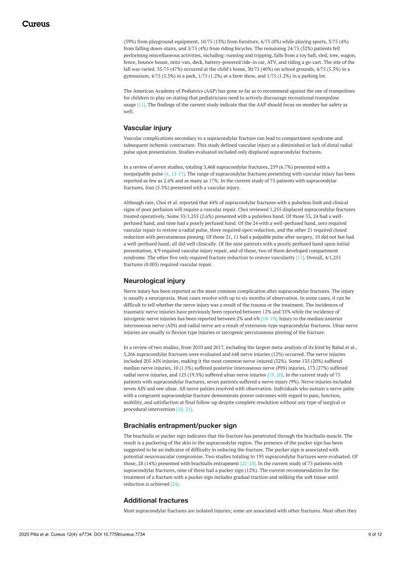
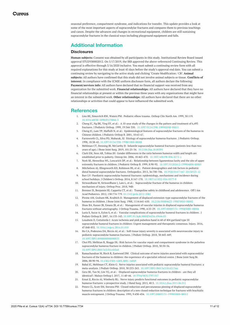
All fractures stemmed from a reported accidental fall, including: 29 (39%) from playground equipment, 10(13%) from furniture, six (8%) while playing sports, three (4%) from falling down stairs, and three (4%) fromriding bicycles. The remaining 24 (32%) patients fell performing miscellaneous activities including: runningand tripping, falls from a toy ball, sled, tree, wagon, fence, bounce house, mini-van, deck, battery-poweredride-in car, all-terrain vehicle (ATV), and a go-cart. Tables 1-5 demonstrate the demographic data, fracturebreakdown, mode of arrival vs. fracture type, and mode of arrival vs. transfer type. Figures 1-4 representexamples of two children included in the study that sustained a Gartland Type 2 and Gartland Type 3supracondylar fractures respectively.
2020 Pilla et al. Cureus 12(4): e7734. DOI 10.7759/cureus.7734 2 of 12
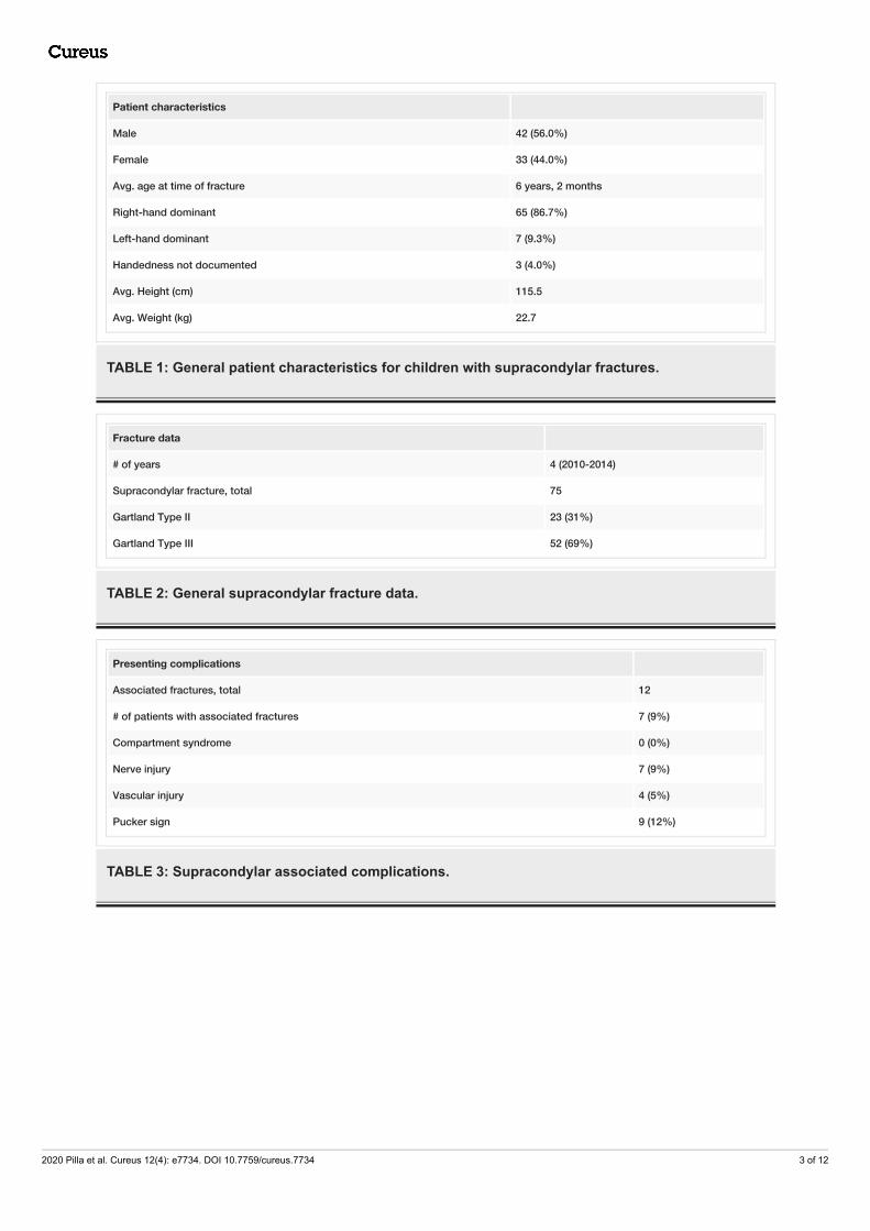
Patient characteristics
Male 42 (56.0%)
Female 33 (44.0%)
Avg. age at time of fracture 6 years, 2 months
Right-hand dominant 65 (86.7%)
Left-hand dominant 7 (9.3%)
Handedness not documented 3 (4.0%)
Avg. Height (cm) 115.5
Avg. Weight (kg) 22.7
TABLE 1: General patient characteristics for children with supracondylar fractures.
Fracture data
# of years 4 (2010-2014)
Supracondylar fracture, total 75
Gartland Type II 23 (31%)
Gartland Type III 52 (69%)
TABLE 2: General supracondylar fracture data.
Presenting complications
Associated fractures, total 12
# of patients with associated fractures 7 (9%)
Compartment syndrome 0 (0%)
Nerve injury 7 (9%)
Vascular injury 4 (5%)
Pucker sign 9 (12%)
TABLE 3: Supracondylar associated complications.
2020 Pilla et al. Cureus 12(4): e7734. DOI 10.7759/cureus.7734 3 of 12
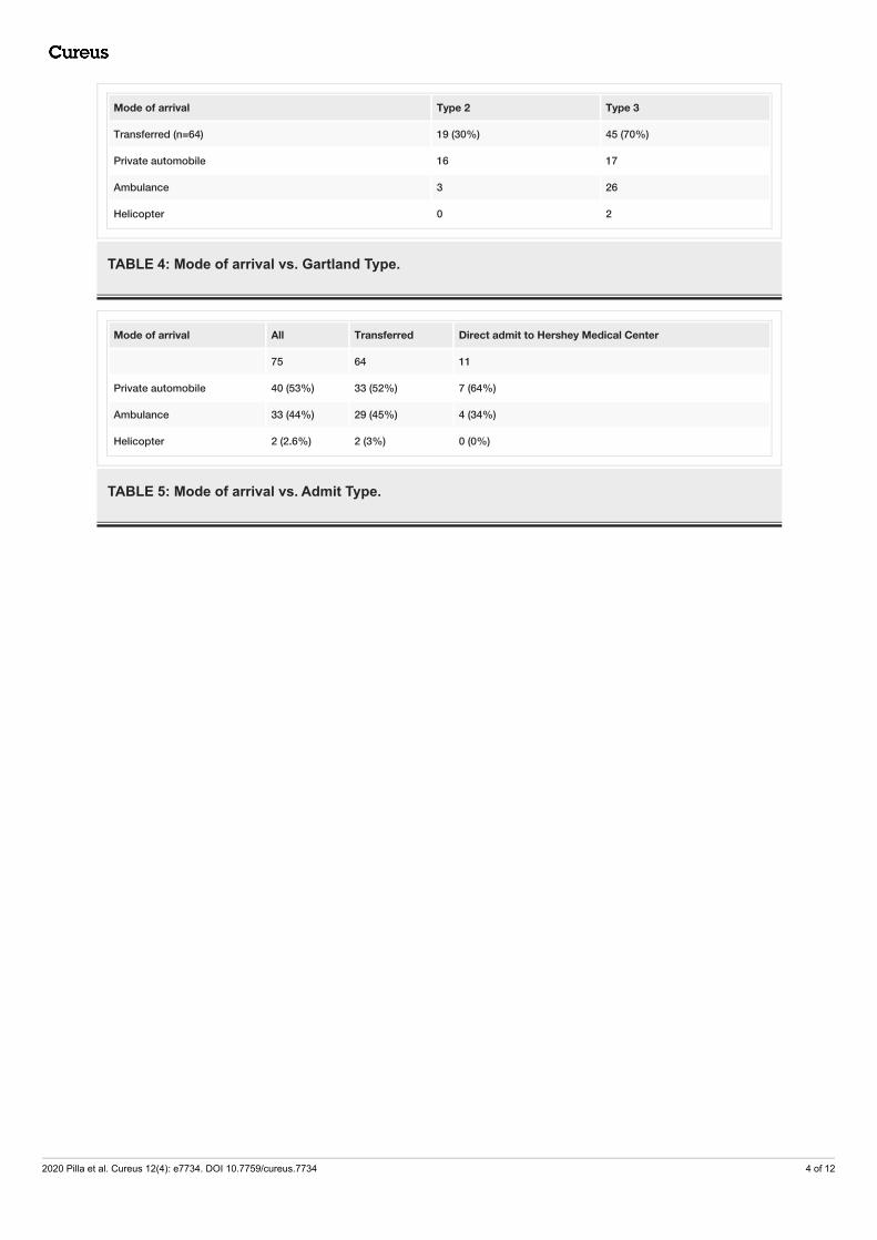
Mode of arrival Type 2 Type 3
Transferred (n=64) 19 (30%) 45 (70%)
Private automobile 16 17
Ambulance 3 26
Helicopter 0 2
TABLE 4: Mode of arrival vs. Gartland Type.
Mode of arrival All Transferred Direct admit to Hershey Medical Center
75 64 11
Private automobile 40 (53%) 33 (52%) 7 (64%)
Ambulance 33 (44%) 29 (45%) 4 (34%)
Helicopter 2 (2.6%) 2 (3%) 0 (0%)
TABLE 5: Mode of arrival vs. Admit Type.
2020 Pilla et al. Cureus 12(4): e7734. DOI 10.7759/cureus.7734 4 of 12

FIGURE 1: AP view of a Type 2 supracondylar fracture in seven-year-oldgirl who fell off of a trampoline.AP, anteroposterior
2020 Pilla et al. Cureus 12(4): e7734. DOI 10.7759/cureus.7734 5 of 12

FIGURE 2: Lateral view of a Type 2 supracondylar fracture in a seven-year-old girl who fell off of a trampoline.
2020 Pilla et al. Cureus 12(4): e7734. DOI 10.7759/cureus.7734 6 of 12

FIGURE 3: AP view of a Type 3 supracondylar fracture in a six-year-oldboy after a fall from the monkey bars.AP, anteroposterior
2020 Pilla et al. Cureus 12(4): e7734. DOI 10.7759/cureus.7734 7 of 12
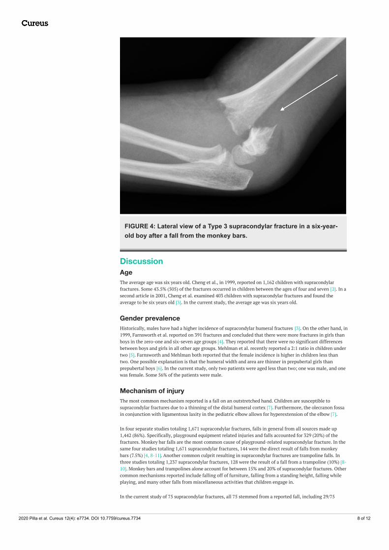
FIGURE 4: Lateral view of a Type 3 supracondylar fracture in a six-year-old boy after a fall from the monkey bars.
DiscussionAgeThe average age was six years old. Cheng et al., in 1999, reported on 1,162 children with supracondylarfractures. Some 43.5% (505) of the fractures occurred in children between the ages of four and seven [2]. In asecond article in 2001, Cheng et al. examined 403 children with supracondylar fractures and found theaverage to be six years old [3]. In the current study, the average age was six years old.
Gender prevalence Historically, males have had a higher incidence of supracondylar humeral fractures [3]. On the other hand, in1999, Farnsworth et al. reported on 391 fractures and concluded that there were more fractures in girls thanboys in the zero-one and six-seven age groups [4]. They reported that there were no significant differencesbetween boys and girls in all other age groups. Mehlman et al. recently reported a 2:1 ratio in children undertwo [5]. Farnsworth and Mehlman both reported that the female incidence is higher in children less thantwo. One possible explanation is that the humeral width and area are thinner in prepubertal girls thanprepubertal boys [6]. In the current study, only two patients were aged less than two; one was male, and onewas female. Some 56% of the patients were male.
Mechanism of injury The most common mechanism reported is a fall on an outstretched hand. Children are susceptible tosupracondylar fractures due to a thinning of the distal humeral cortex [7]. Furthermore, the olecranon fossain conjunction with ligamentous laxity in the pediatric elbow allows for hyperextension of the elbow [7].
In four separate studies totaling 1,671 supracondylar fractures, falls in general from all sources made up1,442 (86%). Specifically, playground equipment related injuries and falls accounted for 329 (20%) of thefractures. Monkey bar falls are the most common cause of playground-related supracondylar fracture. In thesame four studies totaling 1,671 supracondylar fractures, 144 were the direct result of falls from monkeybars (7.5%) [4, 8-11]. Another common culprit resulting in supracondylar fractures are trampoline falls. Inthree studies totaling 1,237 supracondylar fractures, 128 were the result of a fall from a trampoline (10%) [8-10]. Monkey bars and trampolines alone account for between 15% and 20% of supracondylar fractures. Othercommon mechanisms reported include falling off of furniture, falling from a standing height, falling whileplaying, and many other falls from miscellaneous activities that children engage in.
In the current study of 75 supracondylar fractures, all 75 stemmed from a reported fall, including 29/75
2020 Pilla et al. Cureus 12(4): e7734. DOI 10.7759/cureus.7734 8 of 12
(39%) from playground equipment, 10/75 (13%) from furniture, 6/75 (8%) while playing sports, 3/75 (4%)from falling down-stairs, and 3/75 (4%) from riding bicycles. The remaining 24/75 (32%) patients fellperforming miscellaneous activities, including: running and tripping, falls from a toy ball, sled, tree, wagon,fence, bounce house, mini-van, deck, battery-powered ride-in car, ATV, and riding a go-cart. The site of thefall was varied. 35/75 (47%) occurred at the child’s home, 30/75 (40%) on school grounds, 4/75 (5.3%) in agymnasium, 4/75 (5.3%) in a park, 1/75 (1.2%) at a farm show, and 1/75 (1.2%) in a parking lot.
The American Academy of Pediatrics (AAP) has gone so far as to recommend against the use of trampolinesfor children to play on stating that pediatricians need to actively discourage recreational trampolineusage [11]. The findings of the current study indicate that the AAP should focus on monkey bar safety aswell.
Vascular injury Vascular complications secondary to a supracondylar fracture can lead to compartment syndrome andsubsequent ischemic contracture. This study defined vascular injury as a diminished or lack of distal radialpulse upon presentation. Studies evaluated included only displaced supracondylar fractures.
In a review of seven studies, totaling 3,468 supracondylar fractures, 259 (6.7%) presented with anonpalpable pulse [4, 12-17]. The range of supracondylar fractures presenting with vascular injury has beenreported as few as 2.6% and as many as 17%. In the current study of 75 patients with supracondylarfractures, four (5.3%) presented with a vascular injury.
Although rare, Choi et al. reported that 44% of supracondylar fractures with a pulseless limb and clinicalsigns of poor perfusion will require a vascular repair. Choi reviewed 1,255 displaced supracondylar fracturestreated operatively. Some 33/1,255 (2.6%) presented with a pulseless hand. Of those 33, 24 had a well-perfused hand, and nine had a poorly perfused hand. Of the 24 with a well-perfused hand, zero requiredvascular repair to restore a radial pulse, three required open reduction, and the other 21 required closedreduction with percutaneous pinning. Of those 21, 11 had a palpable pulse after surgery, 10 did not but hada well-perfused hand; all did well clinically. Of the nine patients with a poorly perfused hand upon initialpresentation, 4/9 required vascular injury repair, and of those, two of them developed compartmentsyndrome. The other five only required fracture reduction to restore vascularity [17]. Overall, 4/1,255fractures (0.003) required vascular repair.
Neurological injury Nerve injury has been reported as the most common complication after supracondylar fractures. The injuryis usually a neuropraxia. Most cases resolve with up to six months of observation. In some cases, it can bedifficult to tell whether the nerve injury was a result of the trauma or the treatment. The incidences oftraumatic nerve injuries have previously been reported between 12% and 35% while the incidence ofiatrogenic nerve injuries has been reported between 2% and 6% [18-19]. Injury to the median/anteriorinterosseous nerve (AIN) and radial nerve are a result of extension-type supracondylar fractures. Ulnar nerveinjuries are usually to flexion type injuries or iatrogenic percutaneous pinning of the fracture.
In a review of two studies, from 2010 and 2017, including the largest meta-analysis of its kind by Babal et al.,5,266 supracondylar fractures were evaluated and 648 nerve injuries (12%) occurred. The nerve injuriesincluded 205 AIN injuries, making it the most common nerve-injured (32%). Some 135 (20%) sufferedmedian nerve injuries, 10 (1.5%) suffered posterior interosseous nerve (PIN) injuries, 173 (27%) sufferedradial nerve injuries, and 125 (19.5%) suffered ulnar nerve injuries [19, 20]. In the current study of 75patients with supracondylar fractures, seven patients suffered a nerve injury (9%). Nerve injuries includedseven AIN and one ulnar. All nerve palsies resolved with observation. Individuals who sustain a nerve palsywith a congruent supracondylar fracture demonstrate poorer outcomes with regard to pain, function,mobility, and satisfaction at final follow-up despite complete resolution without any type of surgical orprocedural intervention [18, 21].
Brachialis entrapment/pucker sign The brachialis or pucker sign indicates that the fracture has penetrated through the brachialis muscle. Theresult is a puckering of the skin in the supracondylar region. The presence of the pucker sign has beensuggested to be an indicator of difficulty in reducing the fracture. The pucker sign is associated withpotential neurovascular compromise. Two studies totaling to 195 supracondylar fractures were evaluated. Ofthose, 28 (14%) presented with brachialis entrapment [22-23]. In the current study of 75 patients withsupracondylar fractures, nine of them had a pucker sign (12%). The current recommendation for thetreatment of a fracture with a pucker sign includes gradual traction and milking the soft tissue untilreduction is achieved [24].
Additional fractures Most supracondylar fractures are isolated injuries; some are associated with other fractures. Most often they
2020 Pilla et al. Cureus 12(4): e7734. DOI 10.7759/cureus.7734 9 of 12

are the result of low impact activities such as a fall from height. In a review of three studies totaling 1,071supracondylar fractures, a total of 97 additional fractures (9%) were reported [3- 4, 12]. In these threestudies, the most common additional fracture was an ipsilateral forearm fracture which was reported as theadditional fracture in 52 of the 97 additional fractures (53%). In the current study of 75 patients, there were87 total fractures. Of the 75 children with supracondylar fractures, 12/75 (16%) had an additional fracture.The most common additional fracture sustained was an ipsilateral distal radius or ulna fracture reported in12/12 fractures (100%).
Season A review of two studies in addition to this study was evaluated with regard to the effect of seasons onsupracondylar fractures. Six-hundred-ninety-six fractures from three different climates were evaluatedincluding Toronto, San Diego, and Pennsylvania [4, 12]. Sixty-six percent occurred in the spring and summer(March-September). Supracondylar fractures occur more frequently in the spring and summer monthsregardless of geographic location.
Compartment syndrome Compartment syndrome of the forearm is a rare, but potentially devastating complication associated withsupracondylar fractures. Muscle tissue and nerve ischemia are the major risk risks of missing compartmentsyndrome, resulting in tissue death and compartment contracture [25].
In a review of two studies totaling 2,069 supracondylar fractures, there were five (0.2%) cases ofcompartment syndrome in isolated supracondylar fractures [17, 26]. This is consistent with the national dataprovided by the Robertson et al.: 67 isolated supracondylar fractures resulted in compartment syndrome outof the national data of 31,167 supracondylar fractures (0.2%) [26]. Of note, several studies including thestudy by Blakemore et al. examined an additional 43 children with supracondylar fracture and ipsilateralforearm fractures. Of those, three developed compartment syndromes. This study indicates that additionalfractures associated with a supracondylar fracture increase the risk for compartment syndrome [27]. In thecurrent study of 75 patients with supracondylar fractures, there were no reported cases of compartmentsyndrome.
In addition to the fracture the treatment of the fracture can lead to compartment syndrome. Preventativemeasures such as avoiding more than 90 degrees of flexion, uni-valving, or bi-valving the cast may releaseupwards of 77% of the pressure within a cast [28].
Indications for transferAccording to the ACGME requirements, diagnosis and treatment of a pediatric supracondylar fracture is anorthopedic residency core competency. A list of these requirements can be found on the ACGMEwebsite [29]. Every graduating orthopedic resident has met the supracondylar fracture requirementscompetency, thereby demonstrating expertise in managing this injury. Despite meeting competencyrequirements, the majority of supracondylar fractures continued to be transferred. In the current study asmany as 85% of supracondylar fractures were transferred to our center.
One reason for which transfer of a supracondylar fracture should occur is if no orthopedic surgeon isavailable to care for fracture. A second reason is if a hand remains pulseless or dysvascular followingreduction, and the center does not have a plastic or vascular surgeon available. This scenario occurs in lessthan 1% of cases.
Delays in treatment due to transfers are common. Delays in treatment can lead to an increase in morbidity topatients and their families including compartment syndrome requiring fasciotomy [30]. In this singlesurgeon study of 75 supracondylar fractures, 85% of them were transferred from outside facilities. In thecurrent study, no compartment syndromes occurred.
About 12% of children with supracondylar fractures may present with a neurological palsy. Almost all ofthese nerve injuries recover with six months of observation. A nerve palsy on its own is not an indication fortransfer.
LimitationsLimitations of the study include use of a single surgeon’s sample as a reference, a homogenous cohort ofsubjects (80% Caucasian), the retrospective design, and analysis of all the studies involved.
ConclusionsPediatric supracondylar fractures are the most common surgically treated fracture in children. This studyexamined the most important facets pertaining to supracondylar fractures including age, gender prevalence,mechanism of injury, vascular injury, neurological injury, brachialis sign, presence of additional fractures,
2020 Pilla et al. Cureus 12(4): e7734. DOI 10.7759/cureus.7734 10 of 12
seasonal preference, compartment syndrome, and indications for transfer. This update provides a look atsome of the most important aspects of supracondylar fractures and compares them to previous teachingsand canon. Despite the advances and changes in recreational equipment, children are still sustainingsupracondylar fractures in the classical ways including playground equipment and falls.
Additional InformationDisclosuresHuman subjects: Consent was obtained by all participants in this study. Institutional Review Board issuedapproval STUDY00002012. On 5/17/2019, the IRB approved the above-referenced Continuing Review. Thisapproval is effective through 5/16/2020 inclusive. You must submit a continuing review form with allrequired explanations for this study at least 45 days before the study’s approval end date. You can submit acontinuing review by navigating to the active study and clicking ‘Create Modification / CR’. Animalsubjects: All authors have confirmed that this study did not involve animal subjects or tissue. Conflicts ofinterest: In compliance with the ICMJE uniform disclosure form, all authors declare the following:Payment/services info: All authors have declared that no financial support was received from anyorganization for the submitted work. Financial relationships: All authors have declared that they have nofinancial relationships at present or within the previous three years with any organizations that might havean interest in the submitted work. Other relationships: All authors have declared that there are no otherrelationships or activities that could appear to have influenced the submitted work.
References1. Lins RE, Simovitch RW, Waters PM: Pediatric elbow trauma. Orthop Clin North Am. 1999, 30:119.
10.1016/s0030-5898(05)70066-32. Cheng JC, Ng BK, Ying SY, et al.: A 10-year study of the changes in the pattern and treatment of 6,493
fractures. J Pediatric Orthop. 1999, 19:344-350. 10.1097/01241398-199905000-000113. Cheng JC, Lam TP, Maffulli N, et al.: Epidemiological features of supracondylar fractures of the humerus in
Chinese children. J Pediatric Orthop B. 2001, 10:63-67.4. Farnsworth CL, Silva PD, Mubarak, SJ: Etiology of supracondylar humerus fractures . J Pediatric Orthop.
1998, 18:38-42. 10.1097/01241398-199801000-000085. Mehlman CT, Denning JR, McCarthy JJ: Infantile supracondylar humeral fractures (patients less than two
years of age). J Bone Joint Surg. 2019, 101:25-34. 10.2106/jbjs.18.003916. Clark EM, Ness AR, Tobias JH: Gender differences in the ratio between humerus width and length are
established prior to puberty. Osteop Int. 2006, 18:463-470. 10.1007/s00198-006-0275-y7. Nork SE, Hennrikus WL, Loncarich DP, et al.: Relationship between ligamentous laxity and the site of upper
extremity fractures in children. J Pediatric Orthop B. 1999, 8:90-92. 10.1097/01202412-199904000-000058. Mitchelson AJ, Illingoworth KD, Robinson BS, et al.: Patient demographics and risk factors in pediatric
distal humeral supracondylar fractures. Orthopedics. 2013, 36:700-706. 10.3928/01477447-20130523-129. Barr LV: Paediatric supracondylar humeral fractures: epidemiology, mechanisms and incidence during
school holidays. J Children’s Orthop. 2014, 8:167-170. 10.1007/s11832-014-0577-010. Siriwardhane M, Siriwardhane J, Lam L, et al.: Supracondylar fracture of the humerus in children:
mechanism of injury. Orthop Proc. 2018, 94B:11. Brenner JS, Benjamin HJ, Cappetta CT, et al.: Trampoline safety in childhood and adolescence . Off J Am
Acad Pediatrics. 2012, 130:774-779. 10.1542/peds.2012-208212. Pirone AM, Graham HK, Krajbich JI: Management of displaced extension-type supracondylar fractures of the
humerus in children. J Bone Joint Surg. 1988, 1114:641-650. 10.2106/00004623-198870050-0000213. Shaw BA, Kasser JR, Emans JB, et al.: Management of vascular injuries in displaced supracondylar humerus
fractures without arteriography. J Orthop Trauma. 1990, 4:25-29. 10.1097/00005131-199003000-0000414. Luria S, Sucar A, Eylon S, et al.: Vascular complications of supracondylar humeral fractures in children . J
Pediatr Orthop B. 2007, 16:133-143. 10.1097/01.bpb.0000236236.49646.0315. Louahem D, Cottalorda J: Acute ischemia and pink pulseless hand in 68 of 404 gartland type III
supracondylar humeral fractures in children: Urgent management and therapeutic consensus. Injury. 2016,47:848-852. 10.1016/j.injury.2016.01.010
16. Ho CA, Podeszwa DA, Riccio AI, et al.: Soft tissue injury severity is associated with neurovascular injury inpediatric supracondylar humerus fractures. J Pediatr Orthop. 2018, 38:443-449.10.1097/BPO.0000000000000855
17. Choi PD, Melikian R, Skaggs DL: Risk factors for vascular repair and compartment syndrome in the pulselesssupracondylar humerus fracture in children. J Pediatr Orthop. 2010, 30:50-56.10.1097/BPO.0b013e3181c6b3a8
18. Ramachandran M, Birch R, Eastwood DM: Clinical outcome of nerve injuries associated with supracondylarfractures of the humerus in children: the experience of a specialist referral centre. J Bone Joint Surg Br.2006, 88:90-94. 10.1302/0301-620X.88B1.16869
19. Babal JC, Mehlman CT, Klein G: Nerve injuries associated with pediatric supracondylar humeral fractures: ameta-analysis. J Pediatr Orthop. 2010, 30:253-263. 10.1097/BPO.0b013e3181d213a6
20. Gera SK, Tan M, Lim YG, et al.: Displaced supracondylar humerus fractures in children - are they allidentical?. Malays Orthop J. 2017, 11:40-44. 10.5704/MOJ.1707.017
21. Ernat JJ, Riccio AI, Wimberly RL: Nerve injury predicts functional outcomes in pediatric supracondylarhumerus fractures: a prospective study. J Hand Surg. 2015, 40:3. 10.1016/j.jhsa.2015.06.012
22. Peters CL, Scott SM, Stevens PM: Closed reduction and percutaneous pinning of displaced supracondylarhumerus fractures in children: description of a new closed reduction technique for fractures with brachialismuscle entrapment. J Orthop Trauma. 1995, 9:430-434. 10.1097/00005131-199505000-00012
2020 Pilla et al. Cureus 12(4): e7734. DOI 10.7759/cureus.7734 11 of 12

23. Archibeck MJ, Scott SM, Peters CL: Brachialis muscle entrapment in displaced supracondylar humerusfractures: a technique of closed reduction and report of initial results. J Pediatr Orthop. 1997, 17:298-302.10.1097/01241398-199705000-00006
24. Smuin DM, Hennrikus WL: The effect of the pucker sign on outcomes of type III extension supracondylarfractures in children. J Pediatr Orthop. 2017, 37:229-232. 10.1097/BPO.0000000000000893
25. Bae DS, Kadiyala RK, Waters PM: Acute compartment syndrome in children: contemporary diagnosis,treatment, and outcome. J Pediatr Orthop. 2001, 21:680-688. 10.1097/01241398-200109000-00025
26. Robertson AK, Snow E, Browne TS, et al.: Who gets compartment syndrome?: a retrospective analysis of thenational and local incidence of compartment syndrome in patients with supracondylar humerus fractures. JPediatr Orthop. 2018, 38:252-256. 10.1097/BPO.0000000000001144
27. Blakemore LC, Cooperman DR, Thompson GH, et al.: Compartment syndrome in ipsilateral humerus andforearm fractures in children. Clin Orthop Relat Res. 2000, 32:38. 10.1097/00003086-200007000-00006
28. Zaino CJ, Patel MR, Arief MS, et al.: The effectiveness of bivalving, cast spreading, and webril cutting toreduce cast pressure in a fiberglass short arm cast. J Bone Joint Surg Am. 2015, 97:374-380.10.2106/JBJS.N.00579
29. Accreditation Council for Graduate Medical Education . (2015). Accessed: March 3, 2020:https://www.acgme.org/Specialties/Milestones/pfcatid/14/Orthopaedic.
30. Ramachandran M, Skaggs DL, Crawford HA, et al.: Delaying treatment of supracondylar fractures inchildren: has the pendulum swung too far?. J Bone Joint Surg Br. 2008, 90:1228-1233. 10.1302/0301-620X.90B9.20728
2020 Pilla et al. Cureus 12(4): e7734. DOI 10.7759/cureus.7734 12 of 12