EPI-200 Phototoxicity Protocol - zebet - mattek.com · final version Standard Operation Procedure...
22
Phototoxicity Protocol For Use with EpiDerm™ Model (EPI-200) This protocol was developed by Dr. Manfred Liebsch of ZEBET (Berlin, Germany) under a grant from FFVFF (Zurich, Switzerland). ZEBET (Centre for Documentation and Evaluation of Alternative Methods to Animal Ex- periments) is a division of The German Federal Institute for Health Protection of Consumers and Veterinary Medicine (BgVV). The protocol was refined in co-operation with Dr. Frank Gerberick of Procter & Gamble (Cincinnati, USA) and Dr. Uwe Pfannenbecker (Beiersdorf, Hamburg, Germany) and successfully used in a prevalidation study under grant from ECVAM (lspra, Italy).
Transcript of EPI-200 Phototoxicity Protocol - zebet - mattek.com · final version Standard Operation Procedure...

Phototoxicity Protocol For Use with EpiDerm™ Model (EPI-200)
This protocol was developed by Dr. Manfred Liebsch of ZEBET (Berlin, Germany) under a grant from FFVFF (Zurich, Switzerland). ZEBET (Centre for Documentation and Evaluation of Alternative Methods to Animal Ex-periments) is a division of The German Federal Institute for Health Protection of Consumers and Veterinary Medicine (BgVV). The protocol was refined in co-operation with Dr. Frank Gerberick of Procter & Gamble (Cincinnati, USA) and Dr. Uwe Pfannenbecker (Beiersdorf, Hamburg, Germany) and successfully used in a prevalidation study under grant from ECVAM (lspra, Italy).

final version Standard Operation Procedure page 2
5 Novemb. 97 EpiDerm™ Phototoxicity Assay (model: Epi-200) of 22
The following SOP is the final version used within the scope of the ECVAM project "Evaluation of the Prevalidation Process"
subproject:
"Prevalidation of the EpiDerm™ Skin Phototoxicity Test" - Phase III (blind trial) -
The first draft SOP based upon the Skin² test developed by ZEBET and Advanced Tissue Sciences for the full skin model in the EU/COLIPA joint project "In vitro Photoirritation". During phase I of the ECVAM prevalidation study ZEBET adopted the method to the EpiDerm™ technology and established a data base of 39 tests performed on 12 chemicals. Op-timisation experiments revealed overnight exposure (18-24 hrs) with the test chemical and use of a light dose of 6 J/cm² the best design. Tests with chlorpromazine applied in H20, as well as in oil, and in a H20/oil emulsion showed that the assay is capable to handle formulations. A refined SOP (version 2, 30 May 1997) was distributed for comments to the labs participating in the ECVAM prevaildation study. In a meeting in Berlin (4 September 1997) P&G, Beiersdorf and ZEBET agreed on several modifications of the SOP (ver-sion 2). The major modification was a new technique of topical application using paper pads. Since there was no experi-ence with the pad technique additional identical experiments were performed in all of the 3 labs and in a phone conference held on 1 October 1997, P&G, BDF and ZEBET agreed on the final SOP to be used in phase III. Apart from minor changes in details and wording, compared to the 1st draft SOP, the main amendments of the final SOP comprise
• optionally, UV irradiation can be performed in 24 well plates on 0.3 mL medium instead of 6-well plates on 0.9 mL medium.
This change was made, since 0.3 mL medium is sufficient for supply of the tissues during 60 min-utes irradiation. Thus, the weekly testing throughput can be increased.
• wherever possible, chemicals shall be applied as solutions, either in oil or in H20. If chemi-cals cannot be dissolved either in H20 or in oil, they shall be applied as suspensions in oil.
• chemicals dissolved in H20 are applied at 50 µL without using a pad. • chemicals dissolved (or suspended) in oil are applied at 20 µL using a pad (Finn chamber
disk, 8 mm Ø). • reading of optical densities of formazan extracts is done with 570 nm (or equivalently 540
nm) without using a reference filter. • a simplified Methods Documentation Sheet (MDS) is used • a modified MS Excel data spreadsheet (P-SPREAD.XLS) is used
The following three laboratories have approved the final SOP and will perform testing in phase III according to the SOP:
• Dr Manfred Liebsch, ZEBET at the BgVV, Diedersdorfer Weg 1, D-12277 Berlin Germany +49-30-8412-2275 Fax +49-30-8412-2958 e-mail [email protected]
• Dr. Frank Gerberick / Lynn Cruse, Procter & Gamble, 11810 East River Road ROSS, Ohio 45061, USA +1-513-627-2909 Fax +1-513-627-0400 e-mail [email protected] +1-513-627-2909 Fax +1-513-627-0400 e-mail [email protected]
• Uwe Pfannenbecker, Beiersdorf AG, KSt 4232, Unnastraße 48, D-20245 Hamburg, Germany +49-40-4909-3916 Fax +49-40-4909-3589 e-mail [email protected]
final version Standard Operation Procedure page 3
5 Novemb. 97 EpiDerm™ Phototoxicity Assay (model: Epi-200) of 22
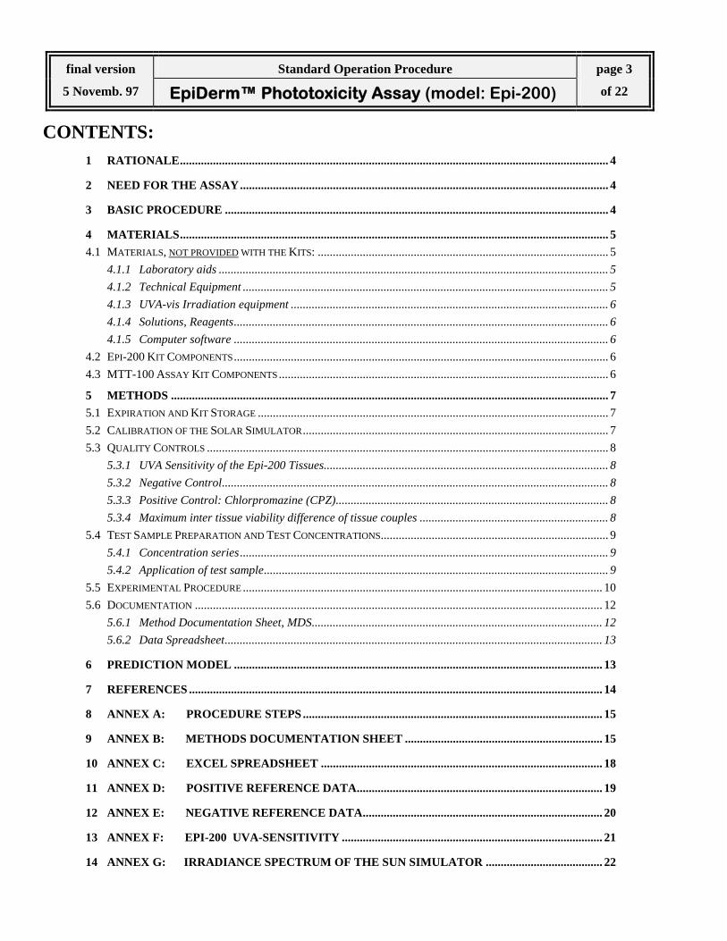
CONTENTS: 1 RATIONALE............................................................................................................................................... 4
2 NEED FOR THE ASSAY........................................................................................................................... 4
3 BASIC PROCEDURE ................................................................................................................................ 4
4 MATERIALS............................................................................................................................................... 5 4.1 MATERIALS, NOT PROVIDED WITH THE KITS: ................................................................................................. 5
4.1.1 Laboratory aids .................................................................................................................................. 5 4.1.2 Technical Equipment .......................................................................................................................... 5 4.1.3 UVA-vis Irradiation equipment .......................................................................................................... 6 4.1.4 Solutions, Reagents............................................................................................................................. 6 4.1.5 Computer software ............................................................................................................................. 6
4.2 EPI-200 KIT COMPONENTS............................................................................................................................. 6 4.3 MTT-100 ASSAY KIT COMPONENTS.............................................................................................................. 6
5 METHODS .................................................................................................................................................. 7 5.1 EXPIRATION AND KIT STORAGE ..................................................................................................................... 7 5.2 CALIBRATION OF THE SOLAR SIMULATOR...................................................................................................... 7 5.3 QUALITY CONTROLS ...................................................................................................................................... 8
5.3.1 UVA Sensitivity of the Epi-200 Tissues............................................................................................... 8 5.3.2 Negative Control................................................................................................................................. 8 5.3.3 Positive Control: Chlorpromazine (CPZ)........................................................................................... 8 5.3.4 Maximum inter tissue viability difference of tissue couples ............................................................... 8
5.4 TEST SAMPLE PREPARATION AND TEST CONCENTRATIONS............................................................................ 9 5.4.1 Concentration series........................................................................................................................... 9 5.4.2 Application of test sample................................................................................................................... 9
5.5 EXPERIMENTAL PROCEDURE ........................................................................................................................ 10 5.6 DOCUMENTATION ........................................................................................................................................ 12
5.6.1 Method Documentation Sheet, MDS................................................................................................. 12 5.6.2 Data Spreadsheet.............................................................................................................................. 13
6 PREDICTION MODEL ........................................................................................................................... 13
7 REFERENCES .......................................................................................................................................... 14
8 ANNEX A: PROCEDURE STEPS .................................................................................................... 15
9 ANNEX B: METHODS DOCUMENTATION SHEET .................................................................. 15
10 ANNEX C: EXCEL SPREADSHEET .............................................................................................. 18
11 ANNEX D: POSITIVE REFERENCE DATA.................................................................................. 19
12 ANNEX E: NEGATIVE REFERENCE DATA................................................................................ 20
13 ANNEX F: EPI-200 UVA-SENSITIVITY ....................................................................................... 21
14 ANNEX G: IRRADIANCE SPECTRUM OF THE SUN SIMULATOR ....................................... 22

final version Standard Operation Procedure page 4
5 Novemb. 97 EpiDerm™ Phototoxicity Assay (model: Epi-200) of 22
1 RATIONALE Phototoxicity (photoirritation) is here defined as acute toxic response that is elicited after the first exposure of skin to cer-tain chemicals and subsequent exposure to light, or that is induced similarly by skin irradiation after systemic administra-tion of a chemical substance. The present assay is designed to detect the phototoxic potential of a chemical by using a three dimensional human epi-dermis model*. Since the assay allows application of test materials to the air exposed surface (stratum corneum), it mimics the in vivo situation and thus may allow to predict phototoxic potency of test materials applied in usage concentrations. The test is based upon a comparison of the cytotoxicity of a chemical when tested with and without additional exposure to a non toxic dose of UVA+visible light. Cytotoxicity is expressed as reduction of mitochondrial conversion of MTT to formazan 1, determined one day after chemical treatment and UVA exposure. * MatTek´s EpiDerm System 2, 3 consists of normal, human-derived epidermal keratinocytes which have been cultured to form a multilayered, highly differentiated model of the human epidermis. It consists of organised basal, spinous and granular layers, and a multi-layered stratum corneum containing intercellular lamellar lipid layers arranged in patterns analogous to those found in vivo. The EpiDerm tissues (surface 0.6 cm²) are cultured on specially prepared cell culture inserts (Millicells®, 10 mm ∅) and shipped world-wide as kits, containing 24 tissues on shipping agarose.
2 NEED FOR THE ASSAY It has been shown in a joint EU/COLIPA validation project 6, 7, that the phototoxic potential of chemicals can be correctly predicted by using cell culture monolayers in a specially designed cytotoxicity assay, the 3T3-NRU-phototoxicity test. Since the phototoxic potential of a chemical predicted using a cellular system may not be relevant when topically applied to the skin at low concentrations (e.g. in a formulation) there is a need for adjunct tests, which allow for the assessment of safe usage concentrations on a dose per area basis before testing them in humans. Reconstituted skin models and epider-mis models have shown to be able to predict both, photoirritancy 4, 5, 8, as well as the photoprotective action of sunscreens 5. In addition, skin models can handle formulations (e.g. emulsions, suspensions) which the 3T3 test cannot handle. Thus, in a testing strategy which is based purely on in vitro tests, there is a need to combine the basic 3T3 NRU PT with other in vitro tests, which may allow to assess safety or phototoxic potency of formulations. In addition, a phototoxicity test in-volving a human skin model may be useful for risk benefit analysis of dermal pharmaceuticals.
3 BASIC PROCEDURE On day of receipt (e.g. Tuesday afternoon) EpiDerm™ tissues are stored over night in a refrigerator. Next day, at least one hour before starting the assay, tissues are transferred to 6-well plates with assay medium and the medium is ex-changed. Then, 5 concentrations of the test material (dissolved in H20 or oil or suspended in oil) are topically applied onto 2 tissues per concentration (i.e. 1 vehicle control + 5 concentrations = 12 tissues). A second set of 12 tissues is treated identically. Plates are incubated over night. Next day, one set of tissues is exposed to 6 J/cm² UVA (+UVA part of the test) and the other set is kept in the dark for the same period (-UVA part of the test). Tissues are then rinsed with PBS to remove test material, transferred to new 6 well plates with fresh medium and incubated over night. Next day, assay me-dium is replaced by MTT-medium and tissues are incubated for 3 hours with MTT. Tissues are then rinsed with PBS, and the formazan is extracted with Isopropanol. Optical density is determined at 540/570 nm in a plate spectrophotometer and cell viability is calculated for each tissue as % of the corresponding vehicle control either irradiated or unirradiated.

final version Standard Operation Procedure page 5
5 Novemb. 97 EpiDerm™ Phototoxicity Assay (model: Epi-200) of 22
4 MATERIALS
4.1 Materials, not provided with the Kits:
4.1.1 Laboratory aids Sterile, blunt-edged forceps for transferring inserts 6-well tissue culture plates (in addition to those provided)
If, instead of replacing media inserts are trans-ferred to new plates with media
24-well culture plates (in addition to those provided)
for UVA irradiation, if not performed in 6-well plates
96-well plates (flat-bottom) for OD reading in plate spectrophotometer Sterile disposable pipette tips 5 Beakers á 50 ml sterile, capped glass or plastic test tubes for preparing the concentration series Finn chamber filter pads, Ø 8 mm, sterilised HERMAL, Scholtzstr. 3, D-21465 Reinbek, Purchase Order No.: D 9503
for application of test materials dissolved or sus-pended in oil
Repeat pipetter (2 ml) for adding the extractant solution Positive displacement pipettes (20 µL, 50µl) for application of viscous test materials adjustable Pipet (100 µl) for pipetting the concentration series adjustable Pipet (200 µl) for pipetting the concentration series adjustable Pipet (1000 uL) for medium change
4.1.2 Technical Equipment Bunsen burner or autoclave for sterilising forceps
37 °C humidified incubator with 5% CO2, for incubating tissues prior to and during assays
vacuum source/trap for aspirating solutions
laminar flow hood for transferring tissues under sterile conditions and for application of test materials.
37 °C water bath for warming up Assay Medium, PBS etc.
Laboratory balance for preparing concentration series
96-well Spectrophotometer (Plate-Reader) equipped with filter 570 nm or 540 nm
for reading optical density at 570 nm,
Shaker for cell culture plates for extraction of formazan
Laboratory centrifuge 1500 x g for centrifugation of MTT medium
Vortex mixer for keeping test suspensions homogeneous during preparation of the concentration series
electric homogeniser for preparing test chemical suspensions in oil

final version Standard Operation Procedure page 6
5 Novemb. 97 EpiDerm™ Phototoxicity Assay (model: Epi-200) of 22
4.1.3 UVA-vis Irradiation equipment UV-sun simulator type SOL 500 or SOL 3
Dr. K. Hönle GmbH, Frauenhoferstr. 5, D-82152 Martinsried, Germany Contact: Dr. G. Schmid
: +49-89-856 08-0 Fax: +49-89-856 08-48
Any appropriate, adjustable and stable tripod For the fixation of the SOL 500 UVA-meter, type No. 37, Dr. Hönle For everyday check of calibration UVA-meter, type No. 37, Dr. Hönle Use only as a reference in case of unexpected
readings with the everyday radiometer Filter, type H1, Dr. Hönle Use to cut-off emitted UVB
4.1.4 Solutions, Reagents Sesame oil (USP/EP/DAB grade) source: pharmacy
Vehicle for test materials
H2O Aqua Pur (Millipore®), or distilled H20 Vehicle for test materials PBS with Ca ++ and Mg++: ~ 500ml per test (e.g. Gibco # 14040)
For rinsing-off test materials after irradiation
4.1.5 Computer software (MS Windows) software for Plate spec-trophotometer
software must be able to export data
MS Excel 5.0 For and data analysis in the Data Spreadsheet
4.2 Epi-200 Kit Components Examine all kit components for integrity. If there is a concern call MatTek Corporation immediately (Mitch Klaus-ner, +1-508-881-6771, Fax +1-508-879-1532).
1 Sealed 24-well plate Contains 24 inserts with tissues on agarose
2 24-well plates Use for MTT assay and formazan extraction
4 6-well plates Use for pre-incubation (and assay)
1 bottle Serum-Free Assay Medium DMEM-based medium
1 bottle Maintenance Medium Do not use in the present assay
1 bottle PBS Rinse Solution (100 mL) Use for rinsing the inserts in MTT assay
1 vial 1% Triton X-100 Solution (10 mL) Skin irritant reference chemical Do not use in present assay
1 MTT Assay Protocol steps are included in the present SOP
4.3 MTT-100 Assay Kit Components 1 vial, 2 ml MTT concentrate 1 vial, 8 ml MTT diluent For diluting MTT concentrate 1 bottle, 60 mL Extractant Solution (Isopropanol) For extraction of formazan crystals

final version Standard Operation Procedure page 7
5 Novemb. 97 EpiDerm™ Phototoxicity Assay (model: Epi-200) of 22
5 METHODS
5.1 Expiration and Kit Storage Epi-200 kits are shipped from Boston on Monday; make sure that they are arriving in the laboratory on Tuesday. Upon receipt of the EpiDerm tissues, place the sealed 24 well plates and the assay medium into the refrigerator (4°C). Place the MTT concentrate containing vial in the freezer (- 20°C) and the MTT diluent in the refrigerator (4°C).
part # description conditions shelf life EPI-200 EpiDerm™ cultures refrigerate (4°C) until Friday EPI-100 assay medium refrigerate (4°C) 7 days MTT-099 MTT diluent refrigerate (4°C) 7 days MTT-100 MTT concentrate freeze (- 20°C) 2 month
Record lot numbers of all components and transfer lot/production label on sealed tray onto the Methods Documen-tation Sheet (MDS see ANNEX B).
Note: Since testing starts on Wednesday, irradiation on Thursday, MTT assay on Friday, do not order more Epi-200 kits per week than can be dosed or irradiated on one day, respectively. This does not hold for US labs: if they receive kits on Tuesday before 12:00 the test can be started.
5.2 Calibration of the Solar Simulator Note: New metal halide burners should be burned for ~100 hrs prior to first use to achieve a stable emittance.
According to Dr Hoenle the burner has a shelflife (in which the spectrum is stable) of at least 800 hrs. Re-cording of lamp usage hours is, therefore, recommended. Extended use is only acceptable if the emitted en-ergy spectrum can be checked. 1. Mount the SOL 500 / SOL 3 lamp, equipped with a H1-filter, on any appropriate stable tripod allowing
fine-adjustment of the exposure distance. 2. Adjust SOL 500 / SOL 3 to a distance of about 60 cm. 3. Switch the Lamp on, wait at least 15 minutes and measure irradiance through the lid of a cell culture
plate using the calibrated UV radiometer (type 37, Dr. Hönle), equipped with an UVA-sensor of the same serial number.
4. Adjust distance of SOL 500 / SOL 3 to achieve a UVA irradiance of 1.7 mW/cm² (The resulting dose will be 1 J/cm² per 10 min. exposure time)
5. According to the number of plates to be exposed concurrently, check the exposure area for equal distri-bution of irradiance: A range of 1.6 - 1.8 mW/cm2 is acceptable. Important: A maximum difference of 1.5 and 1.9 mW/cm2 can be accepted, if positions of the plates with low and high irradiance are changed after half time of the irradiation (30 minutes) is reached (like chess castling).
Calibration of the SOL 500 / SOL 3 shall be checked as described above each time before performing a phototox-icity assay. In case measurements with the UV radiometer reveal unexpected results, either the metal halide burner may have reached the end of it's shelflife, or the radiometer is de-calibrated due to various reasons. In this case, a second reference radiometer of the same type and calibration, which is has not been handled every day and kept in the dark shall be used for cross check.

final version Standard Operation Procedure page 8
5 Novemb. 97 EpiDerm™ Phototoxicity Assay (model: Epi-200) of 22
5.3 Quality Controls
5.3.1 UVA Sensitivity of the Epi-200 Tissues Note: A UVA sensitivity experiment should be performed once the test is newly set up in a laboratory. If UVA sen-sitivity of the tissues is within the acceptance range this type of experiment should be repeated in greater intervals (e.g. once every 6 months). 1. Incubate 24 tissues (37°C, 5 % CO2) for at least 1 hr in 6-well plates with 0.9 ml assay medium /well to allow
release of metabolites and debris accumulated during the shipment. 2. Adjust irradiance of the SOL 500 / SOL 3 to 1.7 mW/cm2 (measure through plate lid !) 3. For UV irradiation, transfer 21 tissues to a 24-well plate filled with 0.3 mL assay medium per well. 4. Prepare a 24 well plate with 0.3 ml assay medium per well and transfer the 3 tissues serving as non-irradiated
control. Place this plate in a dark box at room temperature. 5. Start irradiation of the 21 tissues through the lid of the plate. Use a fan to prevent H20 condensation under
the lid. Every 30 minutes (= 3 J/cm2) transfer 3 tissues from the irradiation site to the dark box. The resulting dose series is 3, 6, 9, 12, 15, 18, 21 J/cm2.
6. Incubate tissues over night (18 - 24 hrs.) at 37°C, 5 % CO2, 90 % humidity 7. Determine tissue viability according to 5.5. Compared to the non irradiated tissues (100 % viability) up to 6
J/cm2 (= 60 minutes) there shall be no reduction of viability exceeding 20%. The historical ID50 UVA is in the range of ~12 - 18 J/cm2 (see ANNEX F).
5.3.2 Negative Control The absolute OD of the negative control tissues in the MTT-test (see 5.5) is an indicator of tissue viability ob-tained in the testing laboratory after shipping procedure and under specific conditions of the assay. Tissue viability is meeting the acceptance criterion if the mean OD of the two negative control tissues (determined without reference filter) is OD ≥ 0.8.
5.3.3 Positive Control: Chlorpromazine (CPZ) For the present study, it is not necessary to include a positive control into each phototoxicity test as this reduces the number of concentrations of the test chemical. When the assay is newly established perform a full experiment with five concentrations of Chlorpromazine (dissolved in H20) ranging from 0.001% up to 0.1%. Repeat this test on a regular basis. A dose dependent reduction of cell viability occurring only in the UVA-irradiated tissues, shall be observed be-tween 0.00316% and 0.0316% (see ANNEX D). Note: If, in other studies, CPZ shall be included in each assay, use 0.316%.
5.3.4 Maximum inter tissue viability difference of tissue couples The new spreadsheet calculates differences in viability between tissue couples that are treated identically. Accord-ing to the historical data base of ZEBET the mean difference between untreated tissue duplicates is 9% ± 7% (S.D.). A difference > 30% (i.e. exceeding the 99% confidence interval) between two tissues treated identically should be regarded as a rejection criterion, and re-testing of the chemical is recommended if the resulting viability is near to the classification cut-off.
final version Standard Operation Procedure page 9
5 Novemb. 97 EpiDerm™ Phototoxicity Assay (model: Epi-200) of 22
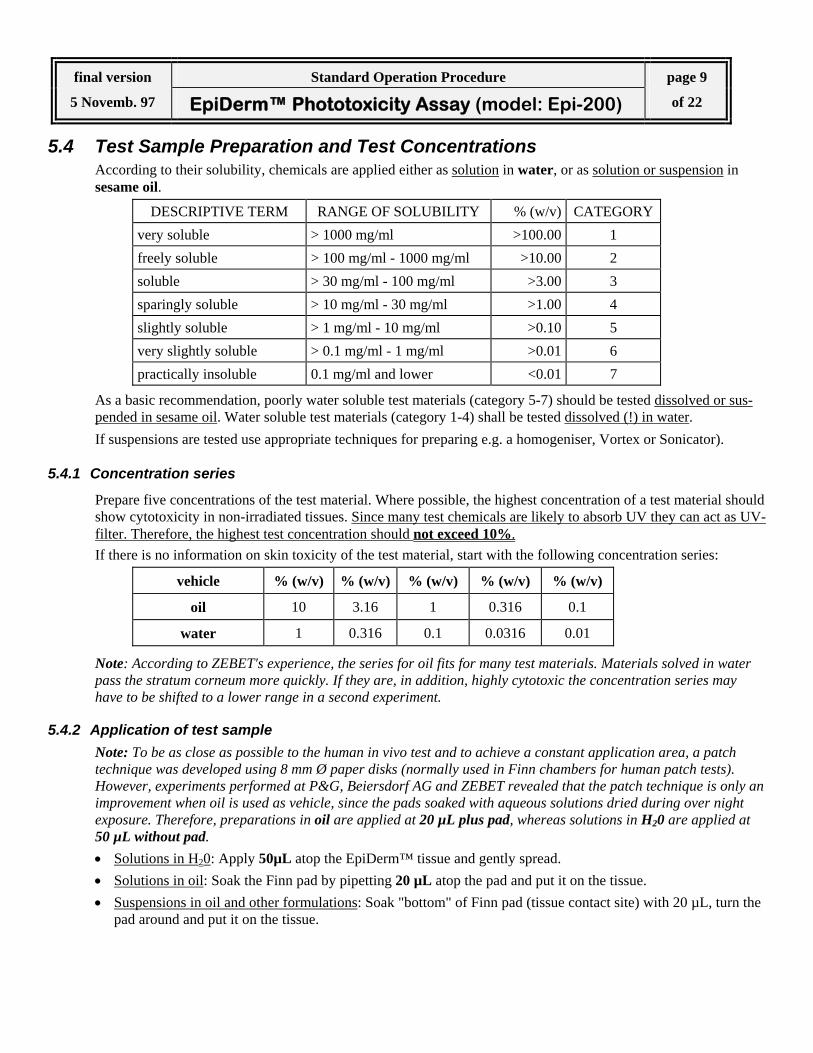
5.4 Test Sample Preparation and Test Concentrations According to their solubility, chemicals are applied either as solution in water, or as solution or suspension in sesame oil.
DESCRIPTIVE TERM RANGE OF SOLUBILITY % (w/v) CATEGORY very soluble > 1000 mg/ml >100.00 1 freely soluble > 100 mg/ml - 1000 mg/ml >10.00 2 soluble > 30 mg/ml - 100 mg/ml >3.00 3 sparingly soluble > 10 mg/ml - 30 mg/ml >1.00 4 slightly soluble > 1 mg/ml - 10 mg/ml >0.10 5 very slightly soluble > 0.1 mg/ml - 1 mg/ml >0.01 6 practically insoluble 0.1 mg/ml and lower <0.01 7
As a basic recommendation, poorly water soluble test materials (category 5-7) should be tested dissolved or sus-pended in sesame oil. Water soluble test materials (category 1-4) shall be tested dissolved (!) in water. If suspensions are tested use appropriate techniques for preparing e.g. a homogeniser, Vortex or Sonicator).
5.4.1 Concentration series
Prepare five concentrations of the test material. Where possible, the highest concentration of a test material should show cytotoxicity in non-irradiated tissues. Since many test chemicals are likely to absorb UV they can act as UV-filter. Therefore, the highest test concentration should not exceed 10%. If there is no information on skin toxicity of the test material, start with the following concentration series:
vehicle % (w/v) % (w/v) % (w/v) % (w/v) % (w/v)
oil 10 3.16 1 0.316 0.1
water 1 0.316 0.1 0.0316 0.01
Note: According to ZEBET's experience, the series for oil fits for many test materials. Materials solved in water pass the stratum corneum more quickly. If they are, in addition, highly cytotoxic the concentration series may have to be shifted to a lower range in a second experiment.
5.4.2 Application of test sample Note: To be as close as possible to the human in vivo test and to achieve a constant application area, a patch technique was developed using 8 mm Ø paper disks (normally used in Finn chambers for human patch tests). However, experiments performed at P&G, Beiersdorf AG and ZEBET revealed that the patch technique is only an improvement when oil is used as vehicle, since the pads soaked with aqueous solutions dried during over night exposure. Therefore, preparations in oil are applied at 20 µL plus pad, whereas solutions in H20 are applied at 50 µL without pad. • Solutions in H20: Apply 50µL atop the EpiDerm™ tissue and gently spread. • Solutions in oil: Soak the Finn pad by pipetting 20 µL atop the pad and put it on the tissue. • Suspensions in oil and other formulations: Soak "bottom" of Finn pad (tissue contact site) with 20 µL, turn the
pad around and put it on the tissue.
final version Standard Operation Procedure page 10
5 Novemb. 97 EpiDerm™ Phototoxicity Assay (model: Epi-200) of 22
5.5 Experimental Procedure
Day before testing Upon receipt of EpiDerm™ kits (Europe: Tuesday afternoon), place assay medium and sealed 24-well plates containing tissues on agarose into refrigerator (4°C ± 2°C). Place the vial containing the MTT con-centrate in the freezer (-20°C ± 5°C ).
First day of testing Note: It is essential that, before the test is started, tissues are incubated for at least 1 hr in assay medium into which they can release metabolites and debris accumulated during the shipment. This medium has to be replaced before the assay is started. For this important incubation 6-well plates have to be used with 0.9 mL medium per well. Alternative techniques (e.g. use of 24 well plates or incubation of all insets in a petri dish) had to be disapproved during this study). 1. Prewarm assay medium in a 37°C waterbath 2. Pipet 0.9 ml of assay medium into each well of sterile 6-well plates 3. Using sterile techniques transfer the inserts (be sure to remove all transport agar) into 6-well plates
containing prewarmed assay medium. Any air bubbles trapped underneath the inserts should be re-leased. Incubate for a minimum of 1 hr at 37°C, 5% CO2 Record incubation time in the MDS
4. While tissues are in the incubator, for each test chemical, prepare a series of five concentrations ac-cording to 5.4.1. Record preparations in the MDS
5. After (at least) 1 hr incubation transfer inserts into new 6-well plates prepared with new 0.9 mL as-say medium per well and prewarmed in the incubator.
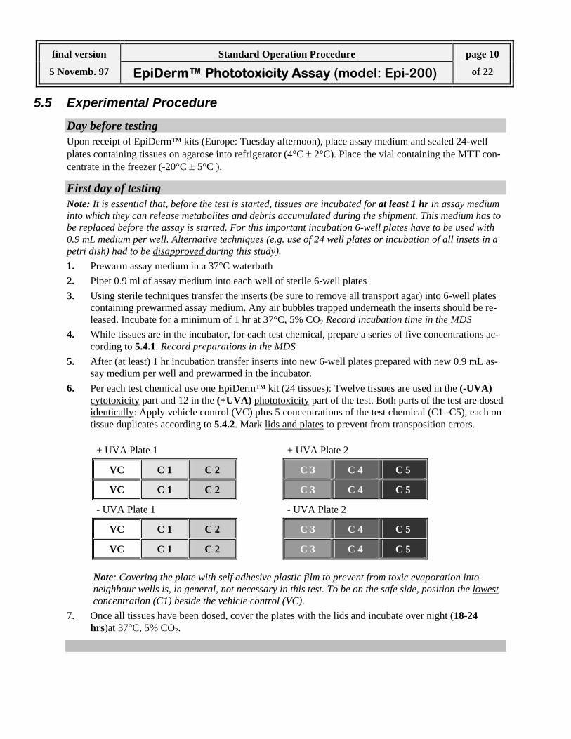
6. Per each test chemical use one EpiDerm™ kit (24 tissues): Twelve tissues are used in the (-UVA) cytotoxicity part and 12 in the (+UVA) phototoxicity part of the test. Both parts of the test are dosed identically: Apply vehicle control (VC) plus 5 concentrations of the test chemical (C1 -C5), each on tissue duplicates according to 5.4.2. Mark lids and plates to prevent from transposition errors.
+ UVA Plate 1 + UVA Plate 2
VC C 1 C 2 C 3 C 4 C 5
VC C 1 C 2 C 3 C 4 C 5
- UVA Plate 1 - UVA Plate 2
VC C 1 C 2 C 3 C 4 C 5
VC C 1 C 2 C 3 C 4 C 5
Note: Covering the plate with self adhesive plastic film to prevent from toxic evaporation into neighbour wells is, in general, not necessary in this test. To be on the safe side, position the lowest concentration (C1) beside the vehicle control (VC).
7. Once all tissues have been dosed, cover the plates with the lids and incubate over night (18-24 hrs)at 37°C, 5% CO2.

final version Standard Operation Procedure page 11
5 Novemb. 97 EpiDerm™ Phototoxicity Assay (model: Epi-200) of 22
Second day of testing 1. Remove 6-well plates from the incubator. Remove application pads. Irradiate (+UVA)-plates (cov-
ered with lids) for 60 min with 1.7 mW/cm2 (= 6 J/cm2) at room temperature. Ventilate with fan to prevent condensation under the lid. Place (-UVA)-plates in the dark at room temperature.
2. While tissues are irradiated, prepare appropriate amount of new 6-well plates with 0.9 mL of fresh assay medium per well and prewarm in the incubator.
3. After UVA irradiation is completed, use wash bottle with sterile PBS and rinse each insert of the (+UVA) plates and (-UVA) plates. Then transfer all inserts to the new plates prepared in 2.
4. Incubate (+UVA)- and (UVA) plates over night (18-24 hrs) at 37°C, 5 % CO2
Third day of testing 1. Prepare MTT medium: thaw MTT concentrate in a water bath and dilute with MTT diluent. Spin
down (300×g, 5 min) to remove any precipitate. Prewarm MTT medium (water bath) to 37°C.
2. For each test material prepare one 24-well plate with 300 µL prewarmed MTT medium per well. Label plates (lid and bottom) and transfer tissue inserts according to the plate design given below. Any air bubbles trapped underneath the inserts should be released.
VC C 1 C 2 C 3 C 4 C 5 +UVA
VC C 1 C 2 C 3 C 4 C 5 +UVA
VC C 1 C 2 C 3 C 4 C 5 -UVA
VC C 1 C 2 C 3 C 4 C 5 -UVA
4. Incubate 24 well plate 3 hours (37°C, 5 % CO2). Record start and stop time for MTT incubation in the MDS.
Note: Deviations from 3 hour time for MTT incubation will result in different MTT readings. For consis-tency it is recommended that 3 hour MTT incubation time be adhered very strictly.
5. After incubation aspirate MTT medium (gently using a suction pump), refill wells with PBS and as-pirate PBS. Repeat the procedure twice and make sure tissues are dry after the last aspiration. Trans-fer inserts to new 24 well plates.
6. For formazan extraction immerse the inserts by gently pipetting 2 mL extractant solution (isopropa-nol) into each insert. The level will rise above the upper edge of the insert, thus completely covering the tissue from both sides.
7. Seal the 24 well plate (e.g. with a zip bag) to inhibit isopropanol evaporation. Record start time of ex-traction in the MDS. Extract 2 hrs with shaking (~120 rpm) at room temperature.
8. After formazan extraction period is complete pierce the inserts with an injection needle (~ gauge 20 / 0.9 mm Ø) and allow the extract to run into the well from which the insert was taken. Afterwards the insert can be discarded. Place the 24-well plates on a shaker for 15 minutes until solution is homogeneous in colour.
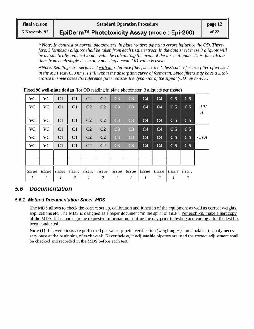
9. Per each tissue transfer 3 × 200µL aliquots* of the blue formazan solution into a 96-well flat bottom mi-crotiter plate. For the 96 well plate, use exactly the plate design given below as this configuration is used in the EXCEL data spreadsheet. Read OD in a plate spectrophotometer at 570 nm, without refer-ence filter.# Alternatively, ODs can be read at 540 nm.
final version Standard Operation Procedure page 12
5 Novemb. 97 EpiDerm™ Phototoxicity Assay (model: Epi-200) of 22
* Note: In contrast to normal photometers, in plate readers pipetting errors influence the OD. There-fore, 3 formazan aliquots shall be taken from each tissue extract. In the data sheet these 3 aliquots will be automatically reduced to one value by calculating the mean of the three aliquots. Thus, for calcula-tions from each single tissue only one single mean OD-value is used. # Note: Readings are performed without reference filter, since the "classical" reference filter often used in the MTT test (630 nm) is still within the absorption curve of formazan. Since filters may have a ± tol-erance in some cases the reference filter reduces the dynamics of the signal (OD) up to 40%.
Fixed 96 well-plate design (for OD reading in plate photometer, 3 aliquots per tissue)
VC VC C1 C1 C2 C2 C3 C3 C4 C4 C 5 C 5
VC VC C1 C1 C2 C2 C3 C3 C4 C4 C 5 C 5 +UVA
VC VC C1 C1 C2 C2 C3 C3 C4 C4 C 5 C 5
VC VC C1 C1 C2 C2 C3 C3 C4 C4 C 5 C 5
VC VC C1 C1 C2 C2 C3 C3 C4 C4 C 5 C 5 -UVA
VC VC C1 C1 C2 C2 C3 C3 C4 C4 C 5 C 5
tissue tissue tissue tissue tissue tissue tissue tissue tissue tissue tissue tissue 1 2 1 2 1 2 1 2 1 2 1 2
5.6 Documentation
5.6.1 Method Documentation Sheet, MDS
The MDS allows to check the correct set up, calibration and function of the equipment as well as correct weights, applications etc. The MDS is designed as a paper document "in the spirit of GLP". Per each kit, make a hardcopy of the MDS, fill in and sign the requested information, starting the day prior to testing and ending after the test has been conducted. Note (1): If several tests are performed per week, pipette verification (weighing H20 on a balance) is only neces-sary once at the beginning of each week. Nevertheless, if adjustable pipettes are used the correct adjustment shall be checked and recorded in the MDS before each test.

final version Standard Operation Procedure page 13
5 Novemb. 97 EpiDerm™ Phototoxicity Assay (model: Epi-200) of 22
5.6.2 Data Spreadsheet
The MS EXCEL 5.0 spreadsheet P-SPREAD.XLS is provided by ZEBET. Data files of optical densities (ODs) generated by the microplate reader are copied from the reader software to the Windows Clipboard and then pasted into the first map of the EXCEL spreadsheet in the fixed 96-well format given above (Note: Only 72 wells of the 96 wells are used!). P-SPREAD.XLS consists of two maps, IMPORT, and SPREAD. The first map (Import) is used for pasting OD values (cursor position: A20!). The second map (Spread) does the calculations and provides a column graph of the results. In addition, entry all information requested (tissue lot-no., test material codes, date...) into this map. In Phase III of the prevalidation study test chemicals are coded by BIBRA with a four digit code. To allow an easy allocation of the XLS files for statistical analysis after codes are broken, use the following file names PGXXXX-Y.XLS or BDXXXX-Y.XLS or ZEXXXX-Y.XLS where XXXX stands for the 4-digit code number and Y stands for the number of the test run.
6 PREDICTION MODEL The rules used to transform quantitative or qualitative data of a toxicological test into a prediction of a toxic po-tential or potency are called prediction model. The prediction model is based on analysis of historical data of the maximum possible difference in the viability of identically treated EpiDerm™ tissues according to which any difference exceeding 30% has to be regarded sig-nificant (p< 0.001). Since the UVA irradiation has no cytotoxic effect itself, a phototoxic activity can be predicted if viability of tissues treated with identical test chemical concentrations differs by more than 30% in the irradiated and the non irradiated part of the test. For each concentration of a test chemical, the mean OD of the tissue couple treated with this concentration is de-termined and expressed as relative percentage viability of the untreated vehicle controls. Identical calculations are performed for the (+UVA) part of the test and the (-UV) part of the test. A chemical is predicted to have a phototoxic potential if one or more test concentrations of the (+UVA) part of the experiment reveal a decrease in viability exceeding 30% when compared with identical concentrations of the (-UVA) part of the experiment. Prediction of phototoxicity is supported if, in addition, the (+UVA) induced reduction in tissue viability shows a dose response relationship. Note: It is a quite common observation for certain phototoxins, that a severe effect may be reduced again at higher doses. This is due to the UV absorbing properties of the chemical by which they act as UV filters if exces-sive doses are applied which remain on the stratum corneum.
final version Standard Operation Procedure page 14
5 Novemb. 97 EpiDerm™ Phototoxicity Assay (model: Epi-200) of 22
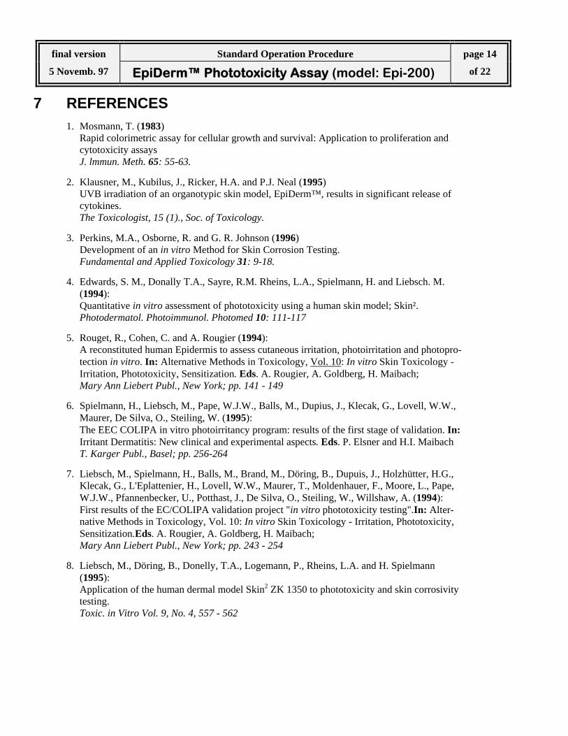
7 REFERENCES 1. Mosmann, T. (1983)
Rapid colorimetric assay for cellular growth and survival: Application to proliferation and cytotoxicity assays J. lmmun. Meth. 65: 55-63.
2. Klausner, M., Kubilus, J., Ricker, H.A. and P.J. Neal (1995) UVB irradiation of an organotypic skin model, EpiDerm™, results in significant release of cytokines. The Toxicologist, 15 (1)., Soc. of Toxicology.
3. Perkins, M.A., Osborne, R. and G. R. Johnson (1996) Development of an in vitro Method for Skin Corrosion Testing. Fundamental and Applied Toxicology 31: 9-18.
4. Edwards, S. M., Donally T.A., Sayre, R.M. Rheins, L.A., Spielmann, H. and Liebsch. M. (1994): Quantitative in vitro assessment of phototoxicity using a human skin model; Skin². Photodermatol. Photoimmunol. Photomed 10: 111-117
5. Rouget, R., Cohen, C. and A. Rougier (1994): A reconstituted human Epidermis to assess cutaneous irritation, photoirritation and photopro-tection in vitro. In: Alternative Methods in Toxicology, Vol. 10: In vitro Skin Toxicology - Irritation, Phototoxicity, Sensitization. Eds. A. Rougier, A. Goldberg, H. Maibach; Mary Ann Liebert Publ., New York; pp. 141 - 149
6. Spielmann, H., Liebsch, M., Pape, W.J.W., Balls, M., Dupius, J., Klecak, G., Lovell, W.W., Maurer, De Silva, O., Steiling, W. (1995): The EEC COLIPA in vitro photoirritancy program: results of the first stage of validation. In: Irritant Dermatitis: New clinical and experimental aspects. Eds. P. Elsner and H.I. Maibach T. Karger Publ., Basel; pp. 256-264
7. Liebsch, M., Spielmann, H., Balls, M., Brand, M., Döring, B., Dupuis, J., Holzhütter, H.G., Klecak, G., L'Eplattenier, H., Lovell, W.W., Maurer, T., Moldenhauer, F., Moore, L., Pape, W.J.W., Pfannenbecker, U., Potthast, J., De Silva, O., Steiling, W., Willshaw, A. (1994): First results of the EC/COLIPA validation project "in vitro phototoxicity testing".In: Alter-native Methods in Toxicology, Vol. 10: In vitro Skin Toxicology - Irritation, Phototoxicity, Sensitization.Eds. A. Rougier, A. Goldberg, H. Maibach; Mary Ann Liebert Publ., New York; pp. 243 - 254
8. Liebsch, M., Döring, B., Donelly, T.A., Logemann, P., Rheins, L.A. and H. Spielmann (1995): Application of the human dermal model Skin2 ZK 1350 to phototoxicity and skin corrosivity testing. Toxic. in Vitro Vol. 9, No. 4, 557 - 562

final version Standard Operation Procedure page 15
5 Novemb. 97 EpiDerm™ Phototoxicity Assay (model: Epi-200) of 22
8 ANNEX A: Procedure Steps First day of testing (Wednesday)
transfer tissues from shipping agarose to assay medium incubate 1 hr. (37°C, 5% CO2)
replace medium
make up 5 concentrations of test chemical
(suspension in oil or water/oil)
make up 5 concentrations of test chemical
(suspension in oil or water/oil) apply each conc.
topically to 2 tissues (5 conc. + 1 vehicle) × 2 = 12 tissues
apply each conc. topically to 2 tissues
(5 conc. + 1 vehicle) × 2 = 12 tissues
incubate over night 18-24 hrs. (37°C, 5% CO2)
Second day of testing
+UVA part of the experiment expose with 6 J/cm2
(= 1.67 mW/cm2 for 60 min) room temperature
+UVA part of the experiment
keep plates covered for 60 min. room temperature
rinse with PBS, replace assay medium rinse with PBS, replace assay medium
incubate over night 18-24 hrs. (37°C, 5% CO2)
Third day of testing (Friday)
transfer tissues in MTT medium transfer tissues in MTT medium
incubate 3 hrs. (37°C, 5% CO2)
rinse with PBS,
add 2 ml extractant (isopropanol) rinse with PBS,
add 2 ml extractant (isopropanol)
shake plates for 2 hrs.
detect OD of formazan extract (cell viability) at 570 nm or 540 nm
9 ANNEX B: Methods Documentation Sheet ASSAY No:.............................. DATE:.................................
XLS file name:......................... Test Chemical....................
final version Standard Operation Procedure page 16
5 Novemb. 97 EpiDerm™ Phototoxicity Assay (model: Epi-200) of 22
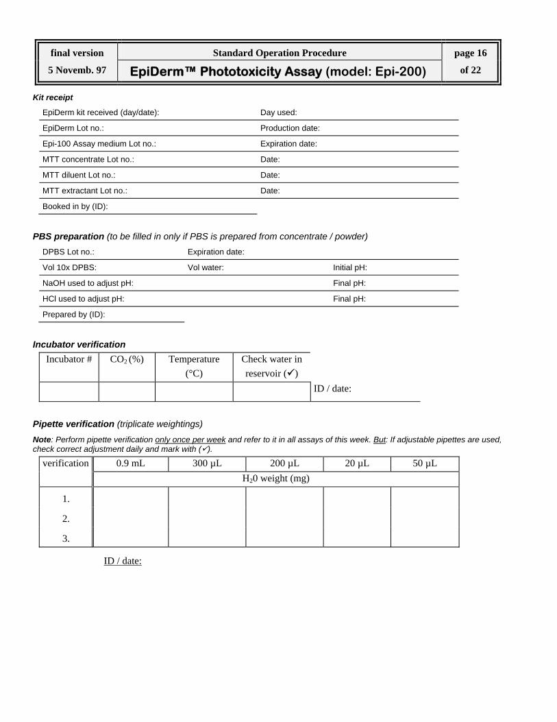
Kit receipt
EpiDerm kit received (day/date): Day used:
EpiDerm Lot no.: Production date:
Epi-100 Assay medium Lot no.: Expiration date:
MTT concentrate Lot no.: Date:
MTT diluent Lot no.: Date:
MTT extractant Lot no.: Date:
Booked in by (ID):
PBS preparation (to be filled in only if PBS is prepared from concentrate / powder) DPBS Lot no.: Expiration date:
Vol 10x DPBS: Vol water: Initial pH:
NaOH used to adjust pH: Final pH:
HCl used to adjust pH: Final pH:
Prepared by (ID):
Incubator verification Incubator # CO2 (%)
Temperature
(°C) Check water in reservoir ( )
ID / date:
Pipette verification (triplicate weightings) Note: Perform pipette verification only once per week and refer to it in all assays of this week. But: If adjustable pipettes are used, check correct adjustment daily and mark with ( ).
verification 0.9 mL 300 µL 200 µL 20 µL 50 µL H20 weight (mg)
1.
2.
3.
ID / date:

final version Standard Operation Procedure page 17
5 Novemb. 97 EpiDerm™ Phototoxicity Assay (model: Epi-200) of 22
Preparation of test chemical and application
concentration (%)
stock preparation:
1. dilution:
2. dilution:
3. dilution:
4. dilution:
5. dilution:
vehicle used (H20 / oil): ...................................
homogenisation technique: ...................................
solution / suspension: ...................................
application voume (µL): ...................................
applied + pad or - pad: ...................................
Time protocols
Procedure Start Stop
1 hr pre-incubation of tissues
over night chemical application (incubator)
3 hrs MTT incubation
Formazan extraction ID / Date:
Check plate photometer filter ( )
reading filter: 570 nm
reading filter: 540 nm ID / Date:
Remarks

final version Standard Operation Procedure page 18
5 Novemb. 97 EpiDerm™ Phototoxicity Assay (model: Epi-200) of 22
10 ANNEX C: EXCEL Spreadsheet chemical: Bergamotteoil solvent: oiltissue-lot no.: 1230 C date of start: 07.05.97exp. no.: 27 Irradiation: 6 J/cm2
application time: 21 hoursmean mean
control 0.920 0.902 0.887 0.903 control 1.117 1.168 1.185 1.1571.058 1.052 1.030 1.047 0.884 0.884 0.896 0.888
0,1 1.045 1.041 1.038 1.041 0,1 1.079 1.076 1.093 1.0830.982 0.951 0.943 0.959 1.060 1.054 1.088 1.067
0.316 1.051 1.055 1.012 1.039 0.316 0.975 0.970 0.979 0.9750.936 0.902 0.907 0.915 0.949 0.930 0.949 0.943
1 0.972 0.961 0.948 0.960 1 0.594 0.595 0.600 0.5960.867 0.861 0.854 0.861 0.622 0.625 0.625 0.624
3.16 0.969 0.969 0.958 0.965 3.16 0.312 0.310 0.313 0.3121.063 1.069 1.051 1.061 0.271 0.268 0.271 0.270
10 0.936 0.917 0.904 0.919 10 0.342 0.325 0.321 0.3290.917 0.933 0.912 0.921 0.281 0.280 0.280 0.280
w/out UVA with UVA
mean Δ tissue % untreated mean Δ tissue % untreated[%] control [%] control
control 0.975 14.74 100 control 1.022 26.28 100 0,1 1.000 8.27 103 0,1 1.075 1.43 105
0.316 0.977 12.72 100 0.316 0.959 3.34 94 1 0.911 10.95 93 1 0.610 4.53 60
3.16 1.013 9.44 104 3.16 0.291 14.33 28 10 0.920 0.18 94 10 0.305 16.07 30
0
20
40
60
80
100
120
140
0,1 0,316 1 3,16 10 [%]
MTT(%untreatedcontrol)
Bergamotte oil21 hours / oilexp. no.: 27

final version Standard Operation Procedure page 19
5 Novemb. 97 EpiDerm™ Phototoxicity Assay (model: Epi-200) of 22
11 ANNEX D: Positive Reference Data
- UVA + UVA - UVA + UVA
0
20
40
60
80
100
120
140
0.001
MTT (% untreated control)
0.002 0.01 0.02 0.1 [%]0.001
Chlorpromazine in water
0
20
40
60
80
100
120
140
0.001
MTT (% untreated control)
0.002 0.01 0.02 0.1 [%]0.001
Chlorpromazine in water

final version Standard Operation Procedure page 20
5 Novemb. 97 EpiDerm™ Phototoxicity Assay (model: Epi-200) of 22
12 ANNEX E: Negative Reference Data - UVA + UVA - UVA + UVA
0
20
40
60
80
100
120
140
0.1
MTT (% untreated control)
0.2 1 2 10 [%]0.1
PABA in oil
0
20
40
60
80
100
120
140
NK
MTT (% untreated control)
0.2 1 2 10 [%]NC
PABA in oil

final version Standard Operation Procedure page 21
5 Novemb. 97 EpiDerm™ Phototoxicity Assay (model: Epi-200) of 22
13 ANNEX F: EPI-200 UVA-Sensitivity
30
40
50
60
70
80
90
100
0 5 10 15 20 25 30
EPI-200 UVA sensitivityviability (%)
Joule/cm2
40
50
60
70
80
90
100
110
120
0 5 10 15 20 25 30
EPI-200 UVA sensitivityviability (%)
Joule/cm2
0
20
40
60
80
100
120
0 5 10 15 20 25 30
EPI-200 UVA sensitivity
Joule/cm2
viability (%)
0
20
40
60
80
100
120
140
0 2 4 6 8 10 12 14 16 18 20 22
UVA Sensitivity of EPIDERM EPI-200(summarized data of 4 independent experiments performed on 3 tissues per UV-dose)
Joule/cm2
Viability (%)
20
40
60
80
100
0 5 10 15 20 25 30
EPI-200 UVA sensitivity
viability (%)
Joule/cm2
The figures show four independent UV sensitivity experiments performed according to 5.3.1. The dose of 6 J/cm2 used in the EpiDerm™ Phototoxicity Test is not cytotoxic in any of the experiments. In addition, the dose of 6 J/cm² is comparable to doses used in animal tests and has prooved to be sufficient to activate photo-toxins.

final version Standard Operation Procedure page 22
5 Novemb. 97 EpiDerm™ Phototoxicity Assay (model: Epi-200) of 22
14 ANNEX G: Irradiance spectrum of the sun simulator Note: The irradinace spectra of the SOL 3 and SOL 500 are nearly identical up to a wavelength of ~ 550 nm. In the longer wavelength range of visible light (> 550 nm - 700 nm) the SOL 500 irradiance decreases, whereas the irradiance of the SOL 3 remains at the same level. In the EU/COLIPA validation study this difference proved to irrelevant. (spectrum kindly provided by Beiersdorf AG)
ADDENDUM (13 November 1997) The spectrum (provided by Beiersdorf AG) shows the FORMAZAN absorbtion. It explains why the reference filter of 630 nm has been omitted. If the filter is not precise (e.g. 620 nm) the dynamics of the reading will be reduced by ~40%!!!
MTT spectrum
0
0,1
0,2
0,3
0,4
0,5
0,6
0,7
0,8
400 500 600 700
Wavelength [nm]
OD
250 300 350 400 450 500 550 600 650 700
Wavelength [nm]
0,001
0,01
0,1
1
10
100Irradiance [mW/cm²]
SOL 500 + H2 Filter
SOL 500 + H1 Filter
SOL 500 + H1 Filter + lid of 96-well plate