EP1−/− mice have enhanced osteoblast differentiation and accelerated fracture repair
11
EP1 / Mice Have Enhanced Osteoblast Differentiation and Accelerated Fracture Repair Minjie Zhang , 1,2 Hsin-chiu Ho , 1 Tzong-jen Sheu , 1 Matthew D Breyer , 3 Lisa M Flick , 1 Jennifer H Jonason , 1 Hani A Awad, 1 Edward M Schwarz , 1 and Regis J O’Keefe 1 1 Center for Musculoskeletal Research, University of Rochester, Rochester, NY, USA 2 Department of Biomedical Genetics, University of Rochester, Rochester, NY, USA 3 Biotechnology Discovery Research, Eli Lilly and Company, Indianapolis, IN, USA ABSTRACT As a downstream product of cyclooxygenase 2 (COX-2), prostaglandin E 2 (PGE 2 ) plays a crucial role in the regulation of bone formation. It has four different receptor subtypes (EP1 through EP4), each of which exerts different effects in bone. EP2 and EP4 induce bone formation through the protein kinase A (PKA) pathway, whereas EP3 inhibits bone formation in vitro. However, the effect of EP1 receptor signaling during bone formation remains unclear. Closed, stabilized femoral fractures were created in mice with EP1 receptor loss of function at 10 weeks of age. Healing was evaluated by radiographic imaging, histology, gene expression studies, micro–computed tomographic (mCT), and biomechanical measures. EP1 / mouse fractures have increased formation of cartilage, increased fracture callus, and more rapid completion of endochondral ossification. The fractures heal faster and with earlier fracture callus mineralization with an altered expression of genes involved in bone repair and remodeling. Fractures in EP1 / mice also had an earlier appearance of tartrate-resistant acid phosphatase (TRAcP)–positive osteoclasts, accelerated bone remodeling, and an earlier return to normal bone morphometry. EP1 / mesenchymal progenitor cells isolated from bone marrow have higher osteoblast differentiation capacity and accelerated bone nodule formation and mineralization in vitro. Loss of the EP1 receptor did not affect EP2 or EP4 signaling, suggesting that EP1 and its downstream signaling targets directly regulate fracture healing. We show that unlike the PGE 2 receptors EP2 and EP4, the EP1 receptor is a negative regulator that acts at multiple stages of the fracture healing process. Inhibition of EP1 signaling is a potential means to enhance fracture healing. ß 2011 American Society for Bone and Mineral Research. KEY WORDS: EP1; FRACTURE HEALING; CHONDROCYTE; CHONDROCYTE MATURATION; MINERALIZATION; OSTEOBLASTS; OSTEOCLASTS; PGE 2 Introduction A pproximately 7.9 million skeletal fractures occur each year in the United States. (1) Nearly 10% to 20% of all fractures have impaired healing, including delayed union or nonunion. Impaired fracture healing, which requires prolonged or repeated treatments, has a marked impact on both the quality of life and the total cost of care. Since the risk of impaired fracture healing is greatest in the elderly and those with other infirmities, the reduced mobility that occurs with fractures further complicates the management of these other conditions, resulting in additional personal and societal costs. (2) For this reason, identifying targets and potential therapies to enhance the rate of fracture healing has great importance. Nonsteroidal anti-inflammatory drugs (NSAIDs) taken to reduce pain following fracture have been linked to decreased bone repair in several clinical studies. (3,4) NSAIDs inhibit the activity of cyclooxygenase (COX) enzymes COX-1 and COX-2. These enzymes play a critical role in the synthesis of prostaglandins (PGs) from arachidonic acid. While COX-1 is constitutively expressed and plays a largely homeostatic role in bone, COX-2 expression is induced by mitogens and inflamma- tory cytokines to upregulate PG synthesis during repair as well as in settings of inflammation and tumorigenesis. (5,6) Several experiments have confirmed that NSAID inhibition of cycloox- ygenase 2 (COX-2) during the inflammatory stage of healing delays the rate of fracture repair. (7–9) Prior studies show significant delay in fracture healing in COX-2 null mice compared with wild-type littermates and COX-1 null mice. (10,11) COX-2 / mice have reduced callus formation, delayed chondrogenesis and impaired endochondral bone formation, and reduced vascularization, primary bone formation, and remodeling. (10) ORIGINAL ARTICLE J JBMR Received in original form August 18, 2009; revised form July 9, 2010; accepted September 22, 2010. Published online October 11, 2010. Address correspondence to: Regis J O’Keefe, MD, Department of Orthopaedics, Box 665, University of Rochester, 601 Elmwood Avenue, Rochester, NY 14642, USA. E-mail: [email protected] Additional Supporting Information may be found in the online version of this article. Journal of Bone and Mineral Research, Vol. 26, No. 4, April 2011, pp 792–802 DOI: 10.1002/jbmr.272 ß 2011 American Society for Bone and Mineral Research 792
-
Upload
minjie-zhang -
Category
Documents
-
view
213 -
download
1
Transcript of EP1−/− mice have enhanced osteoblast differentiation and accelerated fracture repair

ORIGINAL ARTICLE JJBMR
�/� EP1 Mice Have Enhanced Osteoblast Differentiationand Accelerated Fracture Repair
Minjie Zhang ,1,2 Hsin-chiu Ho ,1 Tzong-jen Sheu ,1 Matthew D Breyer ,3 Lisa M Flick ,1
Jennifer H Jonason ,1 Hani A Awad,1 Edward M Schwarz ,1 and Regis J O’Keefe1
1Center for Musculoskeletal Research, University of Rochester, Rochester, NY, USA2Department of Biomedical Genetics, University of Rochester, Rochester, NY, USA3Biotechnology Discovery Research, Eli Lilly and Company, Indianapolis, IN, USA
ABSTRACTAs a downstream product of cyclooxygenase 2 (COX-2), prostaglandin E2 (PGE2) plays a crucial role in the regulation of bone formation. It
has four different receptor subtypes (EP1 through EP4), each of which exerts different effects in bone. EP2 and EP4 induce bone
formation through the protein kinase A (PKA) pathway, whereas EP3 inhibits bone formation in vitro. However, the effect of EP1 receptor
signaling during bone formation remains unclear. Closed, stabilized femoral fractures were created in mice with EP1 receptor loss of
function at 10 weeks of age. Healing was evaluated by radiographic imaging, histology, gene expression studies, micro–computed
tomographic (mCT), and biomechanical measures. EP1�/� mouse fractures have increased formation of cartilage, increased fracture
callus, and more rapid completion of endochondral ossification. The fractures heal faster and with earlier fracture callus mineralization
with an altered expression of genes involved in bone repair and remodeling. Fractures in EP1�/� mice also had an earlier appearance of
tartrate-resistant acid phosphatase (TRAcP)–positive osteoclasts, accelerated bone remodeling, and an earlier return to normal bone
morphometry. EP1�/� mesenchymal progenitor cells isolated from bone marrow have higher osteoblast differentiation capacity and
accelerated bone nodule formation and mineralization in vitro. Loss of the EP1 receptor did not affect EP2 or EP4 signaling, suggesting
that EP1 and its downstream signaling targets directly regulate fracture healing. We show that unlike the PGE2 receptors EP2 and EP4, the
EP1 receptor is a negative regulator that acts at multiple stages of the fracture healing process. Inhibition of EP1 signaling is a potential
means to enhance fracture healing. � 2011 American Society for Bone and Mineral Research.
KEY WORDS: EP1; FRACTURE HEALING; CHONDROCYTE; CHONDROCYTE MATURATION; MINERALIZATION; OSTEOBLASTS; OSTEOCLASTS; PGE2
Introduction
Approximately 7.9 million skeletal fractures occur each year
in the United States.(1) Nearly 10% to 20% of all fractures
have impaired healing, including delayed union or nonunion.
Impaired fracture healing, which requires prolonged or repeated
treatments, has a marked impact on both the quality of life and
the total cost of care. Since the risk of impaired fracture healing is
greatest in the elderly and those with other infirmities, the
reduced mobility that occurs with fractures further complicates
the management of these other conditions, resulting in
additional personal and societal costs.(2) For this reason,
identifying targets and potential therapies to enhance the rate
of fracture healing has great importance.
Nonsteroidal anti-inflammatory drugs (NSAIDs) taken to
reduce pain following fracture have been linked to decreased
Received in original form August 18, 2009; revised form July 9, 2010; accepted Sep
Address correspondence to: Regis J O’Keefe, MD, Department of Orthopaedics, Box 6
E-mail: [email protected]
Additional Supporting Information may be found in the online version of this artic
Journal of Bone and Mineral Research, Vol. 26, No. 4, April 2011, pp 792–802
DOI: 10.1002/jbmr.272
� 2011 American Society for Bone and Mineral Research
792
bone repair in several clinical studies.(3,4) NSAIDs inhibit the
activity of cyclooxygenase (COX) enzymes COX-1 and COX-2.
These enzymes play a critical role in the synthesis of
prostaglandins (PGs) from arachidonic acid. While COX-1 is
constitutively expressed and plays a largely homeostatic role in
bone, COX-2 expression is induced by mitogens and inflamma-
tory cytokines to upregulate PG synthesis during repair as well as
in settings of inflammation and tumorigenesis.(5,6) Several
experiments have confirmed that NSAID inhibition of cycloox-
ygenase 2 (COX-2) during the inflammatory stage of healing
delays the rate of fracture repair.(7–9) Prior studies show
significant delay in fracture healing in COX-2 null mice compared
with wild-type littermates and COX-1 null mice.(10,11) COX-2�/�
mice have reduced callus formation, delayed chondrogenesis
and impaired endochondral bone formation, and reduced
vascularization, primary bone formation, and remodeling.(10)
tember 22, 2010. Published online October 11, 2010.
65, University of Rochester, 601 Elmwood Avenue, Rochester, NY 14642, USA.
le.

However, the role of COX-2/PGE2 in humans has remained
controversial because other reports describe no reduction in the
rate of fracture healing following administration of NSAIDs.(12–14)
As the major downstream product of COX-2, PGE2 plays
a crucial role in regulating bone formation. PGE2 exerts its
effects through binding to four different G protein–coupled
7-transmembrane receptors, and thus the overall effect of PGE2on fracture healing is due to a combination of signaling events
downstream of these receptors.(15) The EP2, EP3, and EP4
receptors each modulate cAMP levels.(16,17) EP2 and EP4
activation stimulates the production of cAMP through Gs. In
contrast, EP3 activation results in a decrease in cAMP levels
through Gi, Gq, or Gs, depending on the EP3 isoform. Less
is known about the EP1 receptor. While the EP1 receptor
is involved in regulating intracellular calcium levels, the G protein
to which it couples remains to be identified.
EP2 and EP4 receptor knockout mice have been shown to
have impaired fracture healing and impaired bone resorp-
tion.(18,19) An EP2-selective agonist induced bone healing in
beagles(20); similarly, an EP4-selective agonist has been used in a
rat model of bone repair with positive effects.(21,22) Recent work
in our laboratory showed that another EP4-selective agonist
accelerated the delayed fracture healing in aged mice and
compensated for the reduced fracture healing observed in COX-
2�/� mice.(23,24) In contrast, treatment of fractures in COX-2�/�
mice with an EP2 agonist enhanced fracture repair only
marginally.(23) Furthermore, while the EP4 agonist significantly
increased cartilage nodule formation in E11.5 murine limb bud
cultures similar to PGE2, the EP2 agonist had no effect on
chondrogenesis. Thus the different EP receptors appear to
mediate unique effects on the cells and tissues involved in
fracture repair. Indeed, the EP3 receptor, which negatively
regulates cAMP levels, is suggested to have negative effects on
bone formation.(25)
Although EP1 receptor expression in bone was reported more
than a decade ago, its role in bone metabolism is less clear. EP1
agonists were shown to stimulate proliferation and inhibit
differentiation in MC3T3-E1 cells.(26) EP1 agonists also stimulate
growth plate chondrocyte proliferation and influence osteoclast
differentiation in murine bone marrow cell cultures.(27,28) EP1
agonist treatment in rat osteoblast cultures stimulates fibro-
nectin expression.(29) While the work to date clearly demon-
strates that EP1 modulates osteoblast gene expression and
function, there are no studies defining the role of EP1 in fracture
repair. While EP1�/� mice are not dramatically different from
wild-type mice in terms of skeletal strength or size,(18) a similar
observation can bemade for COX-2�/�mice, which have marked
impairment of fracture healing.(10,11,30)
In this study, we observed accelerated fracture healing in
EP1�/� mice compared with wild-type mice. Fractures in EP1�/�
mice have more rapid healing, accelerated fracture callus
mineralization and bone remodeling, and earlier and increased
expression of chondrocyte and osteoblast-specific genes. EP1�/�
bone marrow cell cultures undergo more rapid differentiation
and mineralization and have enhanced expression of osteoblast
marker genes. The findings show that PGE2 effects in fracture
repair are complex and likely involve both stimulatory effects,
through EP2 and EP4, and inhibitory effects mediated by EP1.
EP1 NEGATIVELY REGULATES FRACTURE HEALING
Altogether, our findings establish antagonism of the EP1
receptor as a potentially important target to enhance fracture
healing.
Materials and Methods
Experimental animals
All animal studies were done in accordance with and with
approval of the University of Rochester Committee on Animal
Resources. Wild-type (WT) C57BL/6J mice were purchased from
Jackson Laboratories (Bar Harbor, ME, USA). The EP1�/� mice
(C57BL/6J background) were described previously.(31)
Femoral fracture model
Closed femur fractures were created in 10-week-old EP1�/� mice
andWT C57BL/6J mice. Mice were anesthetized with 60mg/kg of
ketamine and 4mg/kg of xylazine intraperitoneally. A 23-gauge
needle (BD Medical Systems, Franklin Lakes, NJ, USA) was
inserted into the length of medullary canal of the femur from the
distal end, and a mid-diaphyseal fracture was created via three-
point bending with an Einhorn device, as described pre-
viously.(32) Healing of the femur fracture was monitored using
radiographs, which were obtained at 0, 7, 14, and 21 days under
anesthesia using a Faxitron Cabinet X-Ray System (Faxitron X-Ray
Corporation, Lincolnshire, IL, USA).
Histology and analysis
The fractured femurs were collected on days 5, 7, 10, 14, 21, 28,
and 35 after fracture. Excess muscle and soft tissue was excised.
Four specimens from each group were fixed in 10% neutral
buffered formalin. The specimens were decalcified for 21 days in
14% EDTA (pH 7.2), embedded in paraffin, and sectioned at a
thickness of 3mm. Levels were cut at depths of 30mm for
histomorphometric analysis (three levels per animal). The
sections were stained using alcian blue hematoxylin/orange G
eosin (ABH/OGE) and cytochemically for tartrate-resistant acid
phosphatase (TRAcP). Total callus area, total cartilage area, and
total woven bone area were quantified using a standardized
eyepiece grid, as described previously.(33) Immunohistochem-
istry also was performed on these sections using a previously
described method.(34) The mouse anti-EP4 antibody (Cayman
Chemical, Ann Arbor, MI, USA) was used at a dilution of 1:200.
Bone marrow cell culture
Bone marrow cells were isolated from 10-week-old EP1�/� and
C57BL/6J mice. Cells were cultured in 2mL of a modified
essential medium (a-MEM) containing 10% fetal bovine serum
(FBS) at 5� 106 cells/well in 6-well plates. After 7 days in culture,
the medium was replaced with medium containing 10mM b-
glycerophosphate and 50mg/mL of ascorbic acid. This medium
was changed every 2 days thereafter. On days 10, 12, 14, 17, and
21 after plating, cells were fixed for alkaline phosphatase and
alizarin red staining or harvested for mRNA isolation using the
RNeasy Mini Kit (Qiagen, Valencia, CA, USA). Cell proliferation
was examined at day 7 using the Cell Proliferation ELISA,
BrdU (colorimetric) immunoassay kit (Roche, Nutley, NJ, USA)
Journal of Bone and Mineral Research 793
according to the manufacturer’s instructions. Cell viability
was examined at day 7 using the CellTiter-Blue cell viability
assay (Promega, San Luis Obispo, CA, USA) according to the
manufacturer’s instructions. Intracellular cAMP activity in the
bone marrow cells was examined using the cAMP-Glo assay
(Promega) according to the manufacturer’s instructions.
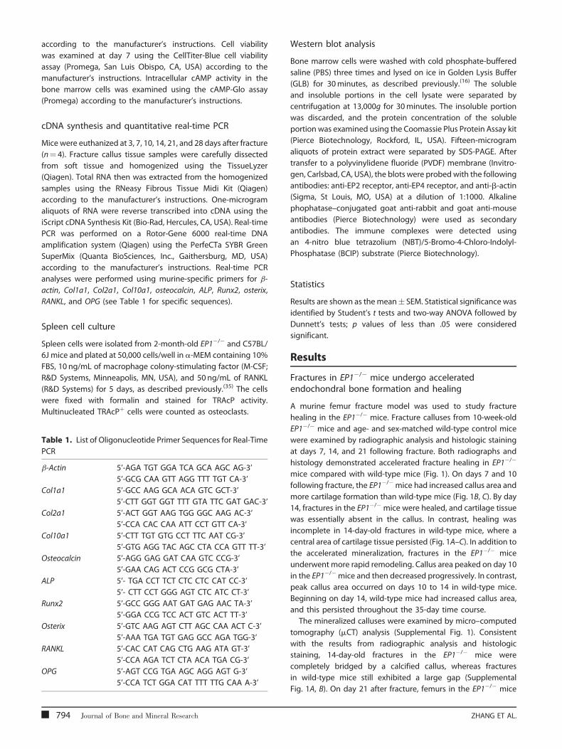
cDNA synthesis and quantitative real-time PCR
Mice were euthanized at 3, 7, 10, 14, 21, and 28 days after fracture
(n¼ 4). Fracture callus tissue samples were carefully dissected
from soft tissue and homogenized using the TissueLyzer
(Qiagen). Total RNA then was extracted from the homogenized
samples using the RNeasy Fibrous Tissue Midi Kit (Qiagen)
according to the manufacturer’s instructions. One-microgram
aliquots of RNA were reverse transcribed into cDNA using the
iScript cDNA Synthesis Kit (Bio-Rad, Hercules, CA, USA). Real-time
PCR was performed on a Rotor-Gene 6000 real-time DNA
amplification system (Qiagen) using the PerfeCTa SYBR Green
SuperMix (Quanta BioSciences, Inc., Gaithersburg, MD, USA)
according to the manufacturer’s instructions. Real-time PCR
analyses were performed using murine-specific primers for b-
actin, Col1a1, Col2a1, Col10a1, osteocalcin, ALP, Runx2, osterix,
RANKL, and OPG (see Table 1 for specific sequences).
Spleen cell culture
Spleen cells were isolated from 2-month-old EP1�/� and C57BL/
6J mice and plated at 50,000 cells/well in a-MEM containing 10%
FBS, 10 ng/mL of macrophage colony-stimulating factor (M-CSF;
R&D Systems, Minneapolis, MN, USA), and 50 ng/mL of RANKL
(R&D Systems) for 5 days, as described previously.(35) The cells
were fixed with formalin and stained for TRAcP activity.
Multinucleated TRAcPþ cells were counted as osteoclasts.
Table 1. List of Oligonucleotide Primer Sequences for Real-Time
PCR
b-Actin 5’-AGA TGT GGA TCA GCA AGC AG-3’
5’-GCG CAA GTT AGG TTT TGT CA-3’
Col1a1 5’-GCC AAG GCA ACA GTC GCT-3’
5’-CTT GGT GGT TTT GTA TTC GAT GAC-3’
Col2a1 5’-ACT GGT AAG TGG GGC AAG AC-3’
5’-CCA CAC CAA ATT CCT GTT CA-3’
Col10a1 5’-CTT TGT GTG CCT TTC AAT CG-3’
5’-GTG AGG TAC AGC CTA CCA GTT TT-3’
Osteocalcin 5’-AGG GAG GAT CAA GTC CCG-3’
5’-GAA CAG ACT CCG GCG CTA-3’
ALP 5’- TGA CCT TCT CTC CTC CAT CC-3’
5’- CTT CCT GGG AGT CTC ATC CT-3’
Runx2 5’-GCC GGG AAT GAT GAG AAC TA-3’
5’-GGA CCG TCC ACT GTC ACT TT-3’
Osterix 5’-GTC AAG AGT CTT AGC CAA ACT C-3’
5’-AAA TGA TGT GAG GCC AGA TGG-3’
RANKL 5’-CAC CAT CAG CTG AAG ATA GT-3’
5’-CCA AGA TCT CTA ACA TGA CG-3’
OPG 5’-AGT CCG TGA AGC AGG AGT G-3’
5’-CCA TCT GGA CAT TTT TTG CAA A-3’
794 Journal of Bone and Mineral Research
Western blot analysis
Bone marrow cells were washed with cold phosphate-buffered
saline (PBS) three times and lysed on ice in Golden Lysis Buffer
(GLB) for 30minutes, as described previously.(16) The soluble
and insoluble portions in the cell lysate were separated by
centrifugation at 13,000g for 30minutes. The insoluble portion
was discarded, and the protein concentration of the soluble
portion was examined using the Coomassie Plus Protein Assay kit
(Pierce Biotechnology, Rockford, IL, USA). Fifteen-microgram
aliquots of protein extract were separated by SDS-PAGE. After
transfer to a polyvinylidene fluoride (PVDF) membrane (Invitro-
gen, Carlsbad, CA, USA), the blots were probedwith the following
antibodies: anti-EP2 receptor, anti-EP4 receptor, and anti-b-actin
(Sigma, St Louis, MO, USA) at a dilution of 1:1000. Alkaline
phophatase–conjugated goat anti-rabbit and goat anti-mouse
antibodies (Pierce Biotechnology) were used as secondary
antibodies. The immune complexes were detected using
an 4-nitro blue tetrazolium (NBT)/5-Bromo-4-Chloro-Indolyl-
Phosphatase (BCIP) substrate (Pierce Biotechnology).
Statistics
Results are shown as themean� SEM. Statistical significance was
identified by Student’s t tests and two-way ANOVA followed by
Dunnett’s tests; p values of less than .05 were considered
significant.
Results
Fractures in EP1�/� mice undergo acceleratedendochondral bone formation and healing
A murine femur fracture model was used to study fracture
healing in the EP1�/� mice. Fracture calluses from 10-week-old
EP1�/� mice and age- and sex-matched wild-type control mice
were examined by radiographic analysis and histologic staining
at days 7, 14, and 21 following fracture. Both radiographs and
histology demonstrated accelerated fracture healing in EP1�/�
mice compared with wild-type mice (Fig. 1). On days 7 and 10
following fracture, the EP1�/�mice had increased callus area and
more cartilage formation than wild-type mice (Fig. 1B, C). By day
14, fractures in the EP1�/� mice were healed, and cartilage tissue
was essentially absent in the callus. In contrast, healing was
incomplete in 14-day-old fractures in wild-type mice, where a
central area of cartilage tissue persisted (Fig. 1A–C). In addition to
the accelerated mineralization, fractures in the EP1�/� mice
underwent more rapid remodeling. Callus area peaked on day 10
in the EP1�/�mice and then decreased progressively. In contrast,
peak callus area occurred on days 10 to 14 in wild-type mice.
Beginning on day 14, wild-type mice had increased callus area,
and this persisted throughout the 35-day time course.
The mineralized calluses were examined by micro–computed
tomography (mCT) analysis (Supplemental Fig. 1). Consistent
with the results from radiographic analysis and histologic
staining, 14-day-old fractures in the EP1�/� mice were
completely bridged by a calcified callus, whereas fractures
in wild-type mice still exhibited a large gap (Supplemental
Fig. 1A, B). On day 21 after fracture, femurs in the EP1�/� mice
ZHANG ET AL.

Fig. 1. EP1�/� fractures exhibit accelerated mineralization. Femur fractures were created in 10-week-old EP1�/� mice and wild-type (WT) controls.
Fractured femurs were harvested at 7, 14, and 21 days after fracture. Representative radiographs of fractured femurs demonstrate the increased
mineralized callus in EP1�/� mice on day 14 compared with the soft callus in wild-type fractures (arrows) (A). Accelerated remodeling on day 21 in EP1�/�
fractures is evident from the contracted callus versus the broad callus that remains in wild-type fractures (arrows). Representative histology (40� original
magnification) of fractured femurs stained with alcian blue hematoxylin/orange G eosin show cartilage (blue) and bone (orange) formation (B).
Histomorphometric measurements (total callus area, cartilage area, woven bone area) were made from 12 sections for each group (n¼ 4 mice/group) (C),
and the results are shown as mean� SEM. Statistical significance was assessed by two-way ANOVA followed by Dunnett’s test; a indicates p< .05
compared with wild-type.
almost returned to normal shape compared with the large callus
seen in wild-type mice. Reconstruction of the mCT data showed
that fracture calluses from the EP1�/� mice had higher bone
mineral density (BMD) than those of wild-type mice after 14 days
of healing (Supplemental Fig. 1B). Torsion testing was performed
to examine the strength of the femurs from the EP1�/� and wild-
type control mice at day 21 after fracture (Supplemental Fig. 2).
Femurs from the EP1�/� mice had higher ultimate torque,
torsional rigidity, ultimate rotation, and torsional energy to
failure.
Quantitative real-time PCR analysis was performed to
determine the expression of genes associated with endochon-
dral bone formation in the fracture callus tissue from wild-type
and EP1�/� mice (Fig. 2A, B). Both collagen, type II (Col2a1) and
collagen, type X (Col10a1) expression peaked earlier during
fracture repair in the EP1�/� mice than in the wild-type mice, so
expression levels of these genes were significantly higher in the
EP1�/� mice 7 days after fracture (Fig. 2A, B). In contrast, peak
expression of Col2a1 and Col10a1 occurred later in wild-type
fractures, so they were increased significantly in the wild-type
mice compared with the EP1�/�mice at between 14 and 21 days
after fracture. The more rapid appearance of these cartilage-
specific genes and their earlier disappearance from the fracture
EP1 NEGATIVELY REGULATES FRACTURE HEALING
callus are consistent with accelerated endochondral bone
formation in the EP1�/� mice.
Osteoblast differentiation is accelerated in fractures inEP1�/� mice
The expression of genes involved in osteoblast differentiation
also was examined in the callus tissue of fractures from EP1�/�
and wild-type mice (Fig. 2C–G). Expression levels of the
osteoblast-specific transcription factors Runx2 and osterix were
accelerated in fracture calluses from the EP1�/� mice (Fig. 2C, D).
Their expression peaked on day 14 in the EP1�/� mice, whereas
in wild-type callus tissue these genes hadmaximal expression on
day 21 (Fig. 2C, D). Expression levels of the osteoblast
differentiation markers alkaline phosphatase (ALP), collagen,
type I (Col1a1), and osteocalcin also were elevated earlier in
fractures from the EP1�/� mice, consistent with accelerated
osteoblast differentiation (Fig. 2E–G). Furthermore, the magni-
tude of both ALP and osteocalcin was higher in fractures in the
EP1�/� mice. Specifically, ALP expression in fractures from the
EP1�/�mice was significantly higher than in wild-type mice from
day 3 to day 14, with peak expression occurring 10 days after
fracture. In contrast, ALP expression in fractures in wild-type mice
Journal of Bone and Mineral Research 795

Fig. 2. The expression of genes involved in chondrogenesis and osteogenesis is altered in EP1�/� mice. Total RNA was extracted from EP1�/� mice and
wild-type controls (n¼ 4) at various time points after fracture. Real-time RT-PCR was performed as described under ‘‘Materials and Methods’’ and
normalized to b-actin expression. The following primer sets were used: Col2a1 (A), Col10a1 (B), Runx2 (C), osterix (D), ALP (E), Col1a1 ( F), and osteocalcin (G).
Statistical comparisons at each time point were performed using two-way ANOVA followed by Dunnett’s test; a indicates p< .05.
had a broad peak of maximal expression at between 10 and 21
days, and the expression levels in fractures from wild-type mice
were increased compared with those from the EP1�/� mice at 21
and 28 days. Expression levels of Col1a1 and osteocalcin also
were elevated earlier in fractures in the EP1�/� mice compared
with wild-type mice. These findings suggest that osteoblasts
undergo a more rapid differentiation in EP1�/� mice.
To determine whether mesenchymal stem cells from EP1�/�
mice have enhanced osteogenic potential, bone marrow
progenitor cells extracted from EP1�/� and wild-type mice were
isolated and placed in culture. After 7 days, osteogenic medium
containing b-glycerol-phosphate (BGP) and ascorbic acid was
added to the cultures, and alkaline phosphatase and alizarin
red staining was performed subsequently over time (Fig. 3A).
Alkaline phosphatase staining showed that more colonies
formed with increased alkaline phosphatase present in the
EP1�/� bone marrow stem cell cultures (Fig. 3B). Similarly,
alizarin red staining was increased in the EP1�/� bone marrow
progenitor cell cultures compared with control cultures,
consistent with accelerated bone nodule formation and
mineralization (Fig. 3C). A BrdU incorporation assay, performed
on day 7, showed that the EP1�/� and wild-type bone marrow
cells have similar rates of proliferation (Fig. 3D). Additionally, a
CellTiter-Blue cell viability assay (Promega, San Luis Obispo, CA,
USA) confirmed that a similar number of viable cells was present
in both cultures on day 7 (Fig. 3D).
Total RNA was extracted from the bone marrow cultures to
examine the expression of genes associated with osteoblast
differentiation. EP1�/� bone marrow stem cell cultures had
796 Journal of Bone and Mineral Research
enhanced early expression of ALP, Col1a1, osteocalcin, Runx2, and
osterix, consistent with accelerated osteoblastogenesis, com-
pared with bone marrow cells from wild-type mice (Fig. 4A–E).
EP1�/� bone marrow stem cell cultures also had increased
expression of RANK ligand (RANKL) and osteoprotegerin (OPG)
(Fig. 4F, G).
Fractures in EP1�/� mice have accelerated remodelingowing to enhanced osteoclast-inducing signals
Since histology and radiographic examination suggested that
the fractures in the EP1�/� mice underwent more rapid
remodeling (Fig. 1), the fracture calluses were stained for the
expression of TRAcP. The number of osteoclasts was increased by
58% (p< .05) in day 14 fracture calluses from EP1�/� mice
compared with wild-type mice, consistent with accelerated bone
remodeling (Fig. 5A). By day 21, fractures in wild-type mice
contained similar numbers of osteoclasts as observed in day 14
fractures in EP1�/� mice. Surprisingly, few osteoclasts were
observed in 21-day-old fractures from EP1�/� mice (Fig. 5A, B).
Total RNA was extracted from the calluses at various times,
and RT-PCR was performed to measure the expression of the
osteoclast-inducing gene RANK ligand (RANKL) and its soluble
receptor antagonist osteoprotegerin (OPG). Fracture calluses from
EP1�/� mice had a higher overall level of expression of both
RANKL and OPG, and both genes were elevated earlier in
fractures in EP1�/� mice compared with wild-type mice (Fig. 5C,
D). The maximum expression levels of RANKL occurred at 14 days
in fracture tissue from EP1�/� mice and at 21 days in wild-type
ZHANG ET AL.

Fig. 3. EP1�/� bone marrow cultures have accelerated osteoblast differ-
entiation. Bonemarrow cells were isolated from 10-week-old EP1�/�mice
and control C57BL/6J mice. (A) Experimental design for in vitro osteoblast
experiments. Cells were cultured in 2mL of a-MEM containing 10% FBS at
5� 106 cells/well in 6-well plates. After 7 days, the medium was replaced
with medium containing b-glycerophosphate and ascorbic acid to
induce osteoblast differentiation. Cells were harvested on days 10, 12,
14, 17, and 21 after plating for alkaline phosphatase (B) and alizarin red
staining (C). Cell proliferation rate was examined by BrdU incorporation
and a CellTiter-Blue cell viability assay on day 7 (D). Statistical compar-
isons were performed using ANOVA. Significance was denoted by the
symbol a (p< .05).
EP1 NEGATIVELY REGULATES FRACTURE HEALING
mice. While RANKL had a sharp peak of expression in facture
tissue from EP1�/� mice, the expression in wild-type mice had a
lower magnitude and was more sustained, consistent with the
observed differences in osteoclast numbers in the fracture
calluses.
To determine whether the accelerated osteoclast formation
observed in EP1�/� fractures is due to a cell-autonomous effect
in osteoclast precursors from the EP1�/� mice, spleen cells were
isolated from EP1�/� and wild-type mice and were placed in cell
culture (Fig. 6). TRAcP staining performed on day 5 showed that
similar numbers of osteoclasts formed in both EP1�/� and wild-
type cultures (Fig. 6). This result suggests that the accelerated
osteoclast formation in EP1�/� fractures is driven primarily by
signals from enhanced osteoblastogenesis.
EP1�/� bone marrow mesenchymal stem cells do nothave increased expression or activation of theEP2 or EP4 receptors
Since prior work has established that activation of the EP2 or
EP4 receptors can accelerate fracture healing, we performed
experiments to confirm that deletion of EP1 does not result in the
increased expression or activity of the EP2 and/or EP4 receptors.
Protein levels of the EP2 and EP4 receptors in EP1�/� and wild-
type bone marrow cultures were examined. While EP1�/� bone
marrow cells had no expression of EP1, the expression of EP2 and
EP4 was unchanged compared with bone marrow cells from
wild-type mice (Fig. 7A). Additional experiments were performed
to examine whether signaling through the EP2 and EP4 receptors
was altered in the absence of EP1. Following administration of
PGE2, cAMP levels were similarly enhanced in bone marrow
cell cultures from wild-type and EP1�/� mice (Fig. 7B). Finally,
immunohistochemistry was used to examine the expression of
the EP4 receptor in fractures from wild-type and EP1�/� mice.
Similar levels of expression were observed in the fractures from
EP1�/� andwild-typemice at 7, 14, and 21 days (Fig. 7C). Thus the
accelerated fracture healing observed in the absence of EP1 was
independent of the EP2 and EP4 receptors.
Discussion
Despite the known importance of COX-2/PGE2 on fracture
healing and bone repair, this study is the first to define the role of
the EP1 receptor in this process. Fractures in EP1�/� mice have
accelerated chondrogenesis, chondrocyte maturation, endo-
chondral bone formation, osteoblast differentiation, and bone
remodeling. Gene expression studies from fracture callus tissues
and bone marrow mesenchymal stem cell cultures demon-
strated an enhanced rate of chondrocyte and osteoblast
differentiation in EP1�/� mice. The effect appears to be directly
due to the EP1 receptor because neither differences in EP2 or EP4
receptor expression nor activity, as measured by intracellular
cAMP levels, were observed between wild-type and EP1�/� bone
marrow cells. The findings show that the EP1 receptor is a
negative regulator in bone repair. Since both the EP2 and EP4
receptors accelerate bone repair, the findings suggest that the
overall effect of COX-2/PGE2 on the process of healing is
determined by the relative activation of these various receptors.
Journal of Bone and Mineral Research 797

Fig. 4. EP1�/� bonemarrow cells have enhanced expression of osteoblast genes in culture. Bonemarrow cells were isolated and cultured as described for
Fig. 3. Total RNA was harvested after 10, 12, 14, 17, and 21 days in culture, and RT-PCR was performed using the primers listed in Table 1. Statistical
comparisons were performed using ANOVA at each time point. The symbol a indicates p< .05.
Nonsteroidal anti-inflammatory drugs (NSAIDs), which are
used widely as pain killers in fractures and other conditions, act
by inhibiting COX-1 and COX-2. These drugs have been reported
to impair the rate of fracture healing in humans.(2,4,17) Prior work
by others and us has clearly established a role for COX-2/PGE2 in
fracture healing in animal models.(7,10,11) PGE2 exerts anabolic
and catabolic effects through its four receptor subtypes EP1
through EP4.(16) Activation of EP2 and EP4 stimulates the
production of cAMP through Gs, whereas activation of EP3
results in a decrease in cAMP levels through Gi, Gq, or Gs
depending on the EP3 isoform.(16,17) In contrast, the EP1 receptor
is involved in regulating intracellular calcium levels. All four
receptor subtypes are expressed in the fracture callus area (data
not shown). Injection of EP2 or EP4 agonist immediately after
fracture accelerates the healing process.(20,21) EP2 and EP4
induce bone formation through the PKA pathway in contrast to
EP3, which has been shown to inhibit bone formation in vitro.(15–
17,19–21,23) These experiments advance our understanding of the
role of COX-2/PGE2 on fracture healing by showing that in
addition to the EP2 and EP4 receptors, the EP1 receptor also
plays a major role in fracture healing.
Fracture healing is a complex process in which bone injury
results in the recruitment of mesenchymal stem cells to the site
of injury, with subsequent proliferation and differentiation into
bone-forming cells.(10) Our findings show that the EP1 receptor
acts as a negative regulator of this process because cell
differentiation and fracture healing are accelerated in mice
deficient in the EP1 receptor. Our in vivo findings in the femur
fracture model established that EP1�/� mice have accelerated
798 Journal of Bone and Mineral Research
chondrogenesis, chondrocyte maturation, and endochondral
bone formation. Furthermore, EP1�/� mice have enhanced
osteoblast differentiation in the fracture callus.
The in vivo findings were confirmed in cultures of bone
marrow stem cells in which osteoblast differentiation and
mineralization of EP1�/� bone marrow cells both were
accelerated compared with cells from wild-type mice. The
mesenchymal stem cell experiments were performed in marrow-
derived cells that are only one component of the stem cell
population that contributes to fracture healing.(36) Since it is
possible that other mesenchymal stem cell populations,
including those from the periosteum, surrounding musculature,
and vascular-derived stem cells could behave differently, the
findings will need confirmation in other cell populations
undergoing osteoblast differentiation. The finding that the
expression levels of the transcription factors Runx2 and osterix
were increased in cells and fracture calluses from EP1�/� mice
suggests an important role for the EP1 receptor in cell fate
determination because both these transcription factors regulate
osteoblastogenesis from mesenchymal precursor cells.(37,38) As
an inhibitor of osteoblast differentiation from mesenchymal
stem cells, EP1 has an important role in the tissue response to
bone injury.
An apparent paradox involving the COX-2/PGE2 signaling
pathway is that while marked effects are observed on fracture
repair, essentially no abnormalities are observed on skeletal
development. While COX-2�/�mice have a profound impairment
in the differentiation of cartilage and bone from mesenchyme,
limb development proceeds normally in COX-2�/� mice.(10,30)
ZHANG ET AL.

Fig. 5. EP1�/� fractures have accelerated bone remodeling. TRAcP staining was performed on sections of fractured femurs, and representative
photomicrographs are shown at 200� original magnification (A). Osteoclast numbers were counted in 12 sections per group (n¼ 4 mice/group),
and mean� SEM is shown (B). RANKL (C) and OPG (D) RNA levels in the fracture callus were examined by RT-PCR. Statistical comparisons were performed
using two-way ANOVA followed by Dunnett’s test; a indicates p< .05.
These observations suggest that while reparative processes
recapitulate many of the events observed during development,
the repair process depends on the activation of unique signaling
pathways. In a similar manner, postnatal growth and develop-
ment also have been shown to depend on unique signaling
pathways. An example is Smad3�/� mice, which have normal
skeletons at birth and grow normally until approximately
3 weeks of age, at which time profound abnormalities of
endochondral bone formation develop.(39) Thus it is not
surprising that the EP1�/� mice develop normally. While recent
work in our laboratory has established that COX-2�/� mice have
subtle differences in bone morphology and bone mass, to date,
no differences in bone metabolism have been identified in the
EP1�/� mice used in this study.(31,40)
There are two different models of EP1 gene deletion. The
EP1�/� mice initially described were produced by homologous
recombination using a targeting vector that replaced a 671-base-
pair sequence involving exon 2.(41) However, since the EP1
receptor gene locus in mouse overlaps with the PKN protein
kinase gene on the antiparallel strand, exon 2 deletion also
resulted in disruption of PKN expression.(42) PKN is activated by
rho GTPase and by fatty acids, including arachidonate, and has a
C-terminal region that is highly homologous to protein
kinase C.(43) Although the role of PKN in bone development
has not been studied, it is possible that PKN regulates bone
formation independent of EP1. In order to disrupt EP1 while
sparing the PKN locus, Guan and colleagues deleted EP1 by
introducing a premature in-frame stop codon into exon 2 by
EP1 NEGATIVELY REGULATES FRACTURE HEALING
nucleotide substitution.(31) Since the latter mice were used in this
study, the findings depend completely on the EP1 receptor.
In addition to enhanced bone formation, our findings also
suggest that fracture remodeling was accelerated in EP1�/�mice
compared with wild-type mice. We performed cell culture
experiments to determine whether deficiency of EP1 was
associated with the potential for accelerated osteoclast forma-
tion through a cell-autonomous process. In cell culture, spleen
cells from EP1�/�and wild-type mice had similar rates of
osteoclastogenesis following treatment with M-CSF and RANKL.
In contrast, the EP1�/� fracture calluses and cultures of EP1�/�
bone marrow stem cells had both accelerated and increased
expression levels of RANKL. Thus the data support a primary role
for enhanced osteoblast differentiation and RANKL expression in
the enhanced osteoclastogenesis and remodeling observed in
the fractures in EP1�/� mice.
Consistent with these histologic and molecular observations,
mCT analysis of fracture callus bone volume and mineral density
(Supplemental Fig. 1) demonstrated an accelerated reduction in
callus volume in EP1�/�mice at 21 days, with a trend of increased
mineral density, also suggesting accelerated remodeling of the
fracture callus.
Biomechanical testing at 28 days following the fracture
confirmed that the fractures in EP1�/� mice healed more
efficiently and with increased torsional strength and rigidity
(Supplemental Fig. 1). The ultimate torque and torsional rigidity
both were increased significantly in the EP1�/� fractures. This
confirms the fact that although EP1�/� fractures have reduced
Journal of Bone and Mineral Research 799

Fig. 6. Increased osteoclastogenesis in EP1�/� mice is not a cell-auton-
omous process. Splenocytes were isolated from 2-month-old EP1�/�
mice and control C57BL/6J mice. Cells were cultured at 50,000 cells/well
in a-MEM containing 10% FBS and 10 ng/mL of M-CSF in 96-well plates.
Then 50 ng/mL of RANKL was added to induce osteoclastogenesis. Five
days after culture, TRAcP staining was performed (A, B). Representative
photographs are shown at 100� original magnification. Similar numbers
of osteoclasts were observed in EP1�/� and wild-type cultures (C). The
data in panel C represent the mean of four different experiments (8
culture wells per group per experiment).
Fig. 7. EP2 and EP4 signaling are not altered in EP1�/� cells. Bone
marrow cells were isolated from 10-week-old EP1�/� mice and wild-type
mice. After culturing for 7 days, cells were treated with PGE2 (3mM) or
vehicle for 24 hours. Total protein was extracted from the cultured cells,
and Western blotting was performed using specific antibodies against
the EP1, EP2, and EP4 receptors (A). A cAMP-Glo assay was performed on
the bone marrow cells from 10-week-old EP1�/� and wild-type mice (B).
Immunohistochemistry was performed using specific antibodies against
the EP4 receptors on the femur fracture tissue samples collected from 10-
week-old EP1�/� and wild-type mice at 7, 14, and 21 days after fracture,
and representative photomicrographs are shown at 200� original mag-
nification (C). Statistical comparisons were performed using Student’s t
test; a indicates p< .05 compared with wild-type, and bindicates p< .05
compared with EP1�/�. b-Actin served as the loading control.
callus area compared with wild-type mice late in the healing
process, the fractures have enhanced biomechanical strength
owing to increased bone remodeling. Since torsional strength in
torsion is inversely related to the cross-sectional area of the
callus,(44) the increased strength despite the reduced callus area
and volume in the EP1�/� fracture callus is indicative of higher-
quality bone.
Compensatory induction of EP2 or EP4 signaling is a possible
explanation for why EP1�/� mice exhibit increased bone repair.
However, no differences were found between wild-type and
EP1�/� bone marrow cells in either EP2 or EP4 receptor level or
intracellular cAMP activity following stimulation with PGE2. This
suggests that the phenotype of accelerated fracture healing in
EP1�/� mice is independent of changes in EP2 or EP4 signaling.
The findings support a primary role for the EP1 receptor as a
negative regulator of bone repair.
All together, these experiments show that the EP1 receptor is a
negative regulator of fracture healing and demonstrate that the
overall effect of COX-2/PGE2 signaling in fracture healing
depends on the complex integration of signals from the various
EP receptors. Our results suggest that inhibition of the EP1
800 Journal of Bone and Mineral Research
receptor may increase fracture healing and support the
importance of further studies to define the expression,
regulation, and effects of this receptor in mesenchymal stem
cells, chondrocytes, and osteoblasts during reparative processes.
Disclosures
All the authors state that they have no conflicts of interest.
Acknowledgments
We wish to thank Ryan Tierney for technical assistance with the
histology. Funding for this study was provided by grants from the
ZHANG ET AL.

National Institutes of Health (R01 AR048681, P50 AR054041, and
R01 DK037097).
References
1. Praemer A, Furner S, Rice DP.Musculoskeletal Conditions in the UnitedStates. Rosemont, IL: American Academy of Orthopaedic Surgeons,
1999.
2. Oni OO, Hui A, Gregg PJ. The healing of closed tibial shaft fractures:
the natural history of union with closed treatment. J Bone Joint SurgBr. 1988;70:787–790.
3. Giannoudis PV, MacDonald DA, Matthews SJ, Smith RM, Furlong AJ,
De Boer P. Nonunion of the femoral diaphysis: the influence of
reaming and nonsteroidal anti-inflammatory drugs. J Bone JointSurg Br. 2000;82:655–658.
4. Glassman SD, Rose SM, Dimar JR, Puno RM, Campbell MJ, Johnson JR.
The effect of postoperative nonsteroidal anti-inflammatory drugadministration on spinal fusion. Spine. 1998;23:834–838.
5. Radi ZA, Khan NK. Effects of cyclooxygenase inhibition on bone,
tendon, and ligament healing. Inflamm Res. 2005;54:358–366.
6. Li L, Pettit AR, Gregory LS, Forwood MR. Regulation of bone biologyby prostaglandin endoperoxide H synthases (PGHS): a rose by any
other name. Cytokine Growth Factor Rev. 2006;17:203–216.
7. Simon AM, O’Connor JP. Dose and time-dependent effects of
cyclooxygenase-2 inhibition on fracture-healing. J Bone Joint SurgAm. 2007;89:500–511.
8. Goodman S, Ma T, TrindadeM, et al. COX-2 selective NSAID decreases
bone ingrowth in vivo. J Orthop Res. 2002;20:1164–1169.
9. Goodman SB, Ma T, Genovese M, Lane Smith R. COX-2 selective
inhibitors and bone. Int J Immunopathol Pharmacol. 2003;16:201–
205.
10. Zhang X, Schwarz EM, Young DA, Puzas JE, Rosier RN, O’Keefe RJ.Cyclooxygenase-2 regulates mesenchymal cell differentiation into
the osteoblast lineage and is critically involved in bone repair. J Clin
Invest. 2002;109:1405–1415.
11. Simon AM, Manigrasso MB, O’Connor JP. Cyclo-oxygenase 2 functionis essential for bone fracture healing. J Bone Miner Res. 2002;17:963–
976.
12. Neal BC, Rodgers A, Gray H, et al. No effect of low-dose aspirin for the
prevention of heterotopic bone formation after total hip replace-ment: a randomized trial of 2,649 patients. Acta Orthop Scand.
2000;71:129–134.
13. Adolphson P, Abbaszadegan H, Jonsson U, Dalen N, SjobergHE, Kalen S. No effects of piroxicam on osteopenia and recovery
after Colles’ fracture. A randomized, double-blind, placebo-con-
trolled, prospective trial. Arch Orthop Trauma Surg. 1993;112:127–
130.
14. Einhorn TA. Cox-2: Where are we in 2003? - The role of cycloox-
ygenase-2 in bone repair. Arthritis Res Ther. 2003;5:5–7.
15. Kobayashi T, Narumiya S. Function of prostanoid receptors: studies
on knockout mice. Prostaglandins Other Lipid Mediat. 2002;68–69:557–573.
16. Li TF, Zuscik MJ, Ionescu AM, et al. PGE2 inhibits chondrocyte
differentiation through PKA and PKC signaling. Exp Cell Res. 2004;300:159–169.
17. Narumiya S, Sugimoto Y, Ushikubi F. Prostanoid receptors: structures,
properties, and functions. Physiol Rev. 1999;79:1193–1226.
18. Akhter MP, Cullen DM, Gong G, Recker RR. Bone biomechanicalproperties in prostaglandin EP1 and EP2 knockout mice. Bone.
2001;29:121–125.
19. Li M, Healy DR, Li Y, et al. Osteopenia and impaired fracture healing in
aged EP4 receptor knockout mice. Bone. 2005;37:46–54.
EP1 NEGATIVELY REGULATES FRACTURE HEALING
20. Paralkar VM, Borovecki F, Ke HZ, et al. An EP2 receptor-selectiveprostaglandin E2 agonist induces bone healing. Proc Natl Acad Sci U
S A. 2003;100:6736–6740.
21. Tanaka M, Sakai A, Uchida S, et al. Prostaglandin E2 receptor (EP4)
selective agonist (ONO-4819. CD) accelerates bone repair of femoralcortex after drill-hole injury. associated with local upregulation of
bone turnover in mature rats. Bone 2004;34:940–948.
22. Ke HZ, Crawford DT, Qi H, et al. A nonprostanoid EP4 receptorselective prostaglandin E2 agonist restores bone mass and strength
in aged, ovariectomized rats. J Bone Miner Res. 2006;21:565–575.
23. Xie C, Liang B, Xue M, et al. Rescue of impaired fracture healing in
COX-2-/- mice via activation of prostaglandin E2 receptor subtype 4.Am J Pathol. 2009;175:772–785.
24. Naik AA, Xie C, Zuscik MJ, et al. Reduced COX-2 Expression in Aged
Mice is Associated with Impaired Fracture Healing. J Bone Miner Res.
2008.
25. Suzawa T, Miyaura C, Inada M, et al. The role of prostaglandin E
receptor subtypes (EP1, EP2, EP3, and EP4) in bone resorption: an
analysis using specific agonists for the respective EPs. Endocrinology.
2000;141:1554–1559.
26. SudaM, Tanaka K, Natsui K, et al. Prostaglandin E receptor subtypes in
mouse osteoblastic cell line. Endocrinology. 1996;137:1698–1705.
27. Brochhausen C, Neuland P, Kirkpatrick CJ, Nusing RM, Klaus G.Cyclooxygenases and prostaglandin E2 receptors in growth plate
chondrocytes in vitro and in situ--prostaglandin E2 dependent pro-
liferation of growth plate chondrocytes. Arthritis Res Ther. 2006;
8:R78.
28. Fujita D, Yamashita N, Iita S, Amano H, Yamada S, Sakamoto K.
Prostaglandin E2 induced the differentiation of osteoclasts in mouse
osteoblast-depleted bone marrow cells. Prostaglandins Leukot
Essent Fatty Acids. 2003;68:351–358.
29. Tang CH, Yang RS, Fu WM. Prostaglandin E2 stimulates fibronectin
expression through EP1 receptor, phospholipase C, protein kinase
Calpha, and c-Src pathway in primary cultured rat osteoblasts. J BiolChem. 2005;280:22907–22916.
30. Dinchuk JE, Car BD, Focht RJ, et al. Renal abnormalities and an altered
inflammatory response in mice lacking cyclooxygenase II. Nature.
1995;378:406–409.
31. Guan Y, Zhang Y, Wu J, et al. Antihypertensive effects of selective
prostaglandin E2 receptor subtype 1 targeting. J Clin Invest. 2007;
117:2496–2505.
32. Bonnarens F, Einhorn TA. Production of a standard closed fracture inlaboratory animal bone. J Orthop Res. 1984;2:97–101.
33. Naik AA, Xie C, Zuscik MJ, et al. Reduced COX-2 expression in aged
mice is associated with impaired fracture healing. J Bone Miner Res.
2009;24:251–264.
34. Mungo DV, Zhang X, O’Keefe RJ, Rosier RN, Puzas JE, Schwarz EM.
COX-1 and COX-2 expression in osteoid osteomas. J Orthop Res.
2002;20:159–162.
35. Wei X, Zhang X, Zuscik MJ, Drissi MH, Schwarz EM, O’Keefe RJ.
Fibroblasts express RANKL and support osteoclastogenesis in a
COX-2-dependent manner after stimulation with titanium particles.
J Bone Miner Res. 2005;20:1136–1148.
36. Schindeler A, Liu R, Little DG. The contribution of different cell
lineages to bone repair: exploring a role for muscle stem cells.
Differentiation. 2009;77:12–18.
37. Matsubara T, Kida K, Yamaguchi A, et al. BMP2 regulates Osterixthrough Msx2 and Runx2 during osteoblast differentiation. J Biol
Chem. 2008;283:29119–29125.
38. Marie PJ. Transcription factors controlling osteoblastogenesis. ArchBiochem Biophys. 2008;473:98–105.
39. Yang X, Chen L, Xu X, Li C, Huang C, Deng CX. TGF-beta/Smad3 signalsrepress chondrocyte hypertrophic differentiation and are required for
maintaining articular cartilage. J Cell Biol. 2001;153:35–46.
Journal of Bone and Mineral Research 801

40. Robertson G, Xie C, Chen D, et al. Alteration of femoral bonemorphology and density in COX-2-/- mice. Bone. 2006;39:767–
772.
41. Stock JL, Shinjo K, Burkhardt J, et al. The prostaglandin E2 EP1
receptor mediates pain perception and regulates blood pressure.J Clin Invest. 2001;107:325–331.
42. Batshake B, Sundelin J. The mouse genes for the EP1 prostanoid
receptor and the PKN protein kinase overlap. Biochem Biophys ResCommun. 1996;227:70–76.
802 Journal of Bone and Mineral Research
43. Mukai H. The structure and function of PKN, a protein kinase havinga catalytic domain homologous to that of PKC. J Biochem. 2003;
133:17–27.
44. Nazarian A, Entezari V, Vartanians V, Muller R, Snyder BD.
An improved method to assess torsional properties of rodent longbones. J Biomech. 2009;42:1720–1725.
45. Reynolds DG, Hock C, Shaikh S, et al. Micro-computed tomography
prediction of biomechanical strength in murine structural bonegrafts. J Biomech. 2007;40:3178–3186.
ZHANG ET AL.