Fatty acid oxidation Ketone bodies Fatty acid synthesis AV. Lipid metabolism 1-2. 2011.

ENZYMES OF FATTY ACID METABOLISM
I. GENERAL INTRODUCTION; CRYSTALLINE CROTONASE*
BY JOSEPH R. STERN,? ALICE DEL CAMPILLO,$ AND ISAIAS RAW5
(From the Department of Pharmacology, New York University College of Medicine, New York, New York)
(Received for publication, July 1, 1955)
The mechanism of the biological oxidation and synthesis of fatty acids has largely been elucidated during the past few years through work in several laboratories (l-7). This work resulted in the complete confirma- tion of Knoop’s B oxidation theory and of the main conclusions derived from the early work of Dakin and Embden. Perhaps one of the most important aspects of the recent work is the demonstration that fatty acid synthesis occurs by a reversal of /3 oxidation.
Fatty acid oxidation is accomplished through the sequence of Reactions 1 through 5. At the end of each sequence (Reaction 5), a 2-carbon frag- ment is split off from the carboxyl end of the fatty acid chain as acetyl CoA.’ This can enter the citric acid cycle for complete oxidation or con- dense with another molecule of acetyl CoA to form acetoacetyl CoA and, through deacylation of the latter in the liver, acetoacetate. The fatty acyl CoA shortened by 2 carbon atoms (the other product of Reaction 5) can undergo a new oxidation and cleavage sequence, etc., through Reac- tions 2 to 5. The reversal of these reactions, from acetyl CoA and hydro-
* Supported by grants from the United States Public Health Service, the American Cancer Society (recommended by the Committee on Growth, National Research Council), and by a contract (N6onr279, T. 0.6) between the Office of Naval Research and New York University College of Medicine.
t Present address, Department of Pharmacology, School of Medicine, Western Reserve University, Cleveland 6, Ohio.
$ Present address, Department of Biochemistry, School of Medicine, University of Puerto Rico, San Juan, Puerto Rico.
§ Present address, Department of Physiological Chemistry, School of Medicine, University of S&o Paulo, Silo Paulo, Brazil.
1 The following abbreviations are employed in Papers I and II: HS-CoA or CoA, reduced coenzyme A; acyl-S-CoA, acyl coenzyme A derivatives; ATP, adenosine triphosphat,e; AMP, adenosine-5’-monophosphate; PP, pyrophosphatc; DPN+ and DPNH, oxidized and reduced diphosphopyridine nucleotide; EDTA, ethylcnedia- minetetraacetate; Tris, tris(hydroxymethyl)aminomethane; Diol, 2-amino-2-methyl- 1,3-propanediol; I,(+) and D(-) designate the dextrorotatory (d) and levorotatory (I) enantiomorphs of p-hydroxybutyric acid. In the case of the higher homologues, whose absolute configurations have not been established, the actual rotation (d or I) is indicated; E, extinction; E, molecular extinction coefficient.
971
by guest on June 24, 2018http://w
ww
.jbc.org/D
ownloaded from
972 ENZYMES OF FATTY ACID METABOLISM. I
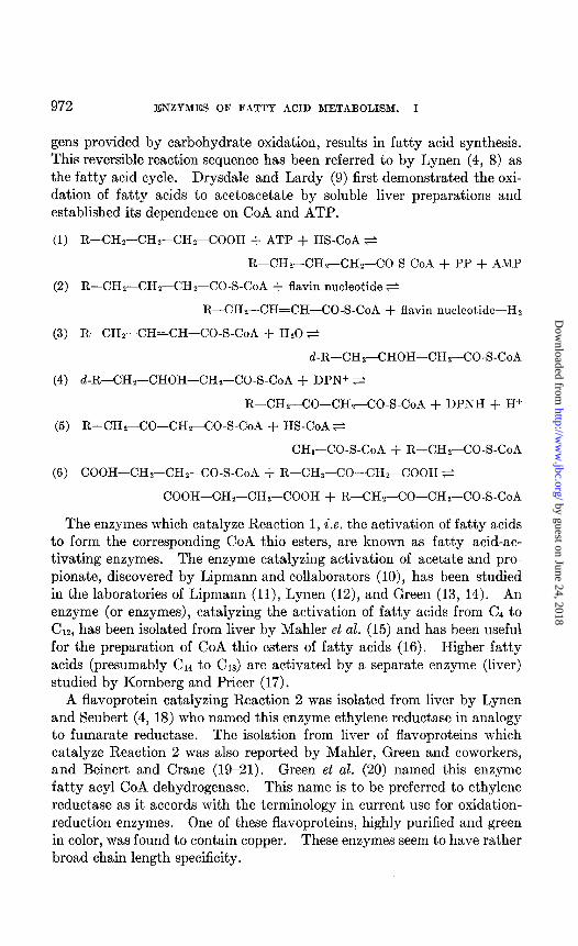
gens provided by carbohydrate oxidation, results in fatty acid synthesis. This reversible reaction sequence has been referred to by Lynen (4, 8) as the fatty acid cycle. Drysdale and Lardy (9) first demonstrated the oxi- dation of fatty acids to acetoacetate by soluble liver preparations and established its dependence on CoA and ATP.
(1) R-CH,CHr-CHrCOOH + ATP + HS-CoA $
R-CHr-CHs-CHr-CO-S-CoA + PP + AMP
(2) R-CHz-CHs-CHZ-CO-S-CoA + flavin nucleotide ti
R-CHY-CH=CH-CO-S-CoA + flavin nucleotide-Hz
(3) R-CHr-CH=CH-CO-S-CoA + Hz0 e
d-R-CHr-CHOH-CHr-CO-S-CoA
(4) d-R-CHr--CHOH-CHn-CO-S-CoA + DPN+ s
R-CHs-CO-CH2-CO-S-CoA + DPNH + H+
(5) R-CHrCO-CHr-CO-S-CoA + HS-CoA s
CHa-CO-S-CoA + R-CHrCO-S-CoA
(6) COOH-CHr-~:I-&-CO-S-CoA + R-C&--CO-CH-COOH e
COOH-CHz-CHz-COOH + R-CHr-CO-CHr-CO-S-CoA
The enzymes which catalyze Reaction 1, i.e. the activation of fatty acids to form the corresponding CoA thio esters, are known as fatty acid-ac- tivating enzymes. The enzyme catalyzing activation of acetate and pro- pionate, discovered by Lipmann and collaborators (lo), has been studied in the laboratories of Lipmann (ll), Lynen (12), and Green (13, 14). An enzyme (or enzymes), catalyzing the activation of fatty acids from Cd to CIZ, has been isolated from liver by Mahler et al. (15) and has been useful for the preparation of CoA thio esters of fatty acids (16). Higher fatty acids (presumably C14 to C18) are activated by a separate enzyme (liver) studied by Kornberg and Pricer (17).
A flavoprotein catalyzing Reaction 2 was isolated from liver by Lynen and Seubert (4, 18) who named this enzyme ethylene reductase in analogy to fumarate reductase. The isolation from liver of flavoproteins which catalyze Reaction 2 was also reported by Mahler, Green and coworkers, and Beinert and Crane (19-21). Green et al. (20) named this enzyme fatty acyl CoA dehydrogenase. This name is to be preferred to ethylene reductase as it accords with the terminology in current use for oxidation- reduction enzymes. One of these flavoproteins, highly purified and green in color, was found to contain copper. These enzymes seem to have rather broad chain length specificity.
by guest on June 24, 2018http://w
ww
.jbc.org/D
ownloaded from

J. R. STERN, A. DEL CAMPILLO, AND 1. RAW 973
The occurrence in heart and liver of an enzyme catalyzing Reaction 3 was first reported by Stern and de1 Campillo (22) who named the enzyme crotonase. This name is to be preferred for simplicity to the name un- saturated fatty acyl CoA hydrase later used by Wakil and Mahler (23) who described the properties of crude preparations of this enzyme from liver. Crotonase was crystallized from ox liver by Stern et al. (24) and is thus the first enzyme of the fatty acid cycle to be obtained in crystalline form. Crystalline crotonase is specific for L(+)-@-hydroxyacyl CoA deriv- atives but has otherwise broad specificity. The work on crotonase was greatly facilitated by Lynen’s study of the N-acetylthioethanolamine ana- logue of crotonyl CoA (4) and the discovery of its characteristic ultraviolet absorption spectrum which, as found by Stern and Raw (cf. (3)), is shared by synthetic crotonyl CoA. This permitted the use of a sensitive optical assay for the enzyme.
The occurrence of an enzyme catalyzing Reaction 4 was first reported by Lynen et al. (8) who isolated this enzyme from liver in a highly purified form. They named the enzyme P-keto reductase because the equilibrium position markedly favors the direction to the left. Use was made here for the first time of analogues of CoA thio esters of fatty acids as substrates. Some of them react with certain enzymes of the fatty acid cycle although at lower rates than the CoA derivatives. The assay for the enzyme was based on the oxidation of DPNH by S-acetoacetyl-N-acetylthioethanola- mine. The presence of this enzyme in heart was noted by Stern et al. (25). Wakil et al. (5, 26) later reported the isolation and the purification of the enzyme from liver and named it P-hydroxyacyl CoA dehydrogenase. This name accords better with current enzyme terminology than fi-keto reductase. We owe to Lehninger and Greville (27) the demonstration that P-keto reductase is specific for the dextrorotatory (L(+)) stereoisomer of ,&hydroxyacyl derivatives of CoA, while /3-hydroxybutyric dehydrogenase, a separate enzyme, specifically catalyzes the reversible oxidation of D(-)-
P-hydroxybutyrate to acetoacetate by DPNf. This work called attention to the stereospecificity of crotonase.
The occurrence of Reaction 5 had been postulated by Lynen et al. (28) to account for the fact that 2 molecules of activated acetate are involved in the synthesis of 1 molecule of acetoacetate (29) and for the observation that ATP and CoA are required for the conversion of acetoacetate and other p-keto acids to citrate by liver enzyme fractions (30). Evidence for this reaction was obtained independently in three laboratories (8, 25, 31, 32). Lynen et al. (8) observed that enzyme preparations from liver were able to catalyze the oxidation of DPNH with acetyl CoA as substrate when supplemented with purified fl-hydroxyacyl CoA dehydrogenase (B-keto re- ductase) and demonstrated for the first time the biological formation of
by guest on June 24, 2018http://w
ww
.jbc.org/D
ownloaded from

974 ENZYMES OF FATTY ACID METABOLISM. I
&hydroxybutyryl CoA. Stern et al. (31) and Green et al. (32) followed the reaction through either the conversion of acetoacetate to acetyl CoA, which was trapped as citrate in the presence of oxalacetate and condensing enzyme (33), or the reverse reaction. Since, in the direction to the right, Reaction 5 is a cleavage of the P-ketoacyl CoA derivative by a thiol (HS- CoA), it can be considered to be a thiolysis. For this reason Lynen et al. (8) have proposed the name /3-ketothiolase, or simply thiolase, for the en- zyme catalyzing this reaction. For its simplicity and accuracy this name is to be preferred to the names acetoacetate-condensing enzyme (25, 31) and /?-ketoacyl CoA cleavage enzyme (32) used by other investigators.
In liver, B-keto acids can be activated through a reaction with ATP and CoA (30). However, in heart muscle, kidney, and possibly in skeletal muscle, activation of some P-keto acids occurs specifically by transfer of CoA from succinyl CoA (Reaction 6). The enzyme concerned with this reaction was discovered independently by Green et al. (32, 34) and Stern et al. (25, 31) and was named CoA transferase by the latter authors. Ace- toacetyl CoA was first isolated and characterized through the transfer of CoA from succinyl CoA to acetoacetate in the presence of partially purified preparations of CoA transferase (25). It was later obtained by Beinert (35) through enzymatic oxidation of B-hydroxybutyryl CoA. Through the formation of acetoacetyl CoA, the enzyme activates acetoacetate, produced in the liver and carried by the circulation to the peripheral tissues, for oxi- dation in these tissues via the citric acid cycle. The latter in turn gener- ates the necessary succinyl CoA through a-ketoglutarate oxidation (6). A similar enzyme had previously been found by Stadtman in extracts of Clos- tridium kluyveri (36). The specificity of this enzyme, named CoA trans- phorase, is quite different, since it catalyzes the reversible transfer of CoA from acetyl CoA to acids such as propionate and butyrate.
The isolation of CoA transferase and thiolase was greatly facilitated by the finding of Lynen et al. (8) that the N-acetylthioethanolamine analogue of acetoacetyl CoA has a characteristic ultraviolet absorption spectrum. As would be expected, this spectrum is shared by acetoacetyl CoA (25). This led to the use of rapid and sensitive optical assay methods which permitted the isolation of CoA transferase and thiolase in highly purified form from pig heart at New York University (3, 6). While the pig heart thiolase was found to be highly specific for acetoacetyl CoA, reacting but very slowly with B-ketovaleryl CoA and not at all with higher P-ketoacyl CoA derivatives, an ox liver thiolase preparation (37) was reported to act on ,&ketoacyl CoA derivatives from c/4 to Cl,. Since the latter enzyme appeared to be relatively crude, and, on the other hand, crude pig heart preparations showed broader specificity (31) than the purified thiolase, the occurrence of several thiolases with different chain length specificity is in- dicated.
by guest on June 24, 2018http://w
ww
.jbc.org/D
ownloaded from

J. R. STERN, A. DEL CAMPILLO, AND I. RAW 975
Papers of this series will be concerned with a detailed account of the enzymes of fatty acid metabolism studied in this laboratory. This work includes studies on crotonase, on the mechanism of breakdown and syn- thesis of fi-keto fatty acids, and on the enzymes CoA transferase and thi- olase.
Crystalline Crotonase
Crotonase catalyzes the hydration-dehydration step (Reaction 3) of the fatty acid cycle. It is also involved in the metabolism of branched chain fatty acids (38) derived from amino acids and, by implication, in the me- tabolism of isoprenoid structures like cholesterol (39, 40) and rubber (41). The isolation and crystallization of crotonase from ox liver (24) are de- scribed in this paper. Data on the distribution of the enzyme are also presented.
Enzyme Assay and Unit-The assay is based on the observation of Seu- bert and Lynen (18) that X-crotonyl-N-acetylthioethanolamine has a char- acteristic absorption spectrum in the ultraviolet with maxima at 224 rnp and 263 rng. The latter band may be attributed to diene conjugation be- tween the 2-ethylenic bond and the carbonyl double bond of the thio ester, which is lacking in the free acid. In the case of crotonyl-S-CoA the band at 263 rnp is largely obscured by the absorption of the adenine moiety and only becomes apparent when this interference is eliminated by taking a difference spectrum between the intact and the hydrolyzed thio ester (Fig. 1). The product of hydration of crotonyl-S-CoA, L(+)-&hydroxybutyryl- S-CoA, exhibits no specific absorption in the region of 263 rnp other than that due to its adenine moiety. Vinylacetyl-S-CoA (3-butenoyl-S-CoA), also, does not possess a specific absorption at 263 rnp, since a conjugate double bond system is lacking. It may be pointed out that, since only minute amounts of enzyme are required, a direct optical assay can also be performed at wave-length 232 to 240 rnp (22), at which adenine interfer- ence is considerably less.
The optical assay is performed with a Beckman model DU spectropho- tometer equipped with a photomultiplier attachment. 2.0 ml. quartz cu- vettes of 0.5 cm. light path are used. To the experimental cuvette are added 0.20 ml. of assay mixture, 0.05 ml. of crotonyl-S-CoA solution (3.2 X 10-s M), enzyme, and water to a final volume of 1.50 ml. The assay mixture consists of 1.0 ml. of 1.0 M Tris buffer, pH 7.5, 1.0 ml. of 0.1 per cent egg albumin, 0.15 ml. of 0.1 M potassium EDTA, pH 7.4, and 0.85 ml. of water. Crotonyl-S-CoA is omitted from the blank cuvette and in its place about 0.15 ml. of adenylic acid (adenosine-5’-phosphate) solution (1 mg. per ml.) is added to compensate for the absorption of the adenine moiety of the CoA thio ester. The optical density of a solution of adenylic acid decreases slowly on standing at 3”. An amount should be added
by guest on June 24, 2018http://w
ww
.jbc.org/D
ownloaded from

976 ENZYMES OF FATTY ACID METABOLISM. I
which gives an initial optical density of about 0.4 for the experimental cuvette.
Crotonase has such a high turnover number that even in crude tissue extracts only 1 to 10 y of protein needs to be used in the assay. For pur- poses of assay the enzyme fraction is diluted with 0.02 M potassium phos- phate buffer, pH 7.4, to a protein concentration of 0.5 mg. per ml. Any necessary further dilution is made with the same buffer containing 1OV M
WAVELENGTH (mp)
FIG. 1
i;J , , ,h o.o, '012 3 4 5 6
MINUTES
FIG. 2
FIG. 1. Ultraviolet absorption spectrum of crotonyl-S-CoA. A difference spec- trum of intact versus hydrolyzed thio ester. Both cuvettes (d = 0.5 cm.) contained -6 X 1O-5 M crotjonyl-SR (of which 80 per cent was the CoA ester) in 0.1 M Tris-HCl buffer, pH 7.5. Crotonyl thio ester in the blank cuvette was hydrolyzed with alkali and then neutralized. The experiment,al cuvette contained the equivalent a.mount of salt. Final volume, 1.50 ml.
FIG. 2. Optical assay of crotonase. The assay conditions are given in the text. Ox liver fraction, specific activity 20, was used as source of crotonase.
potassium EDTA and 0.1 per cent egg albumin. It is essential to insure adequate mixing in making high dilutions of enzyme.
The reaction is started by addition of 0.01 ml. of enzyme solution (con- taining 0.0025 to 2 y of protein, depending on the purity) and the decrease in optical density at 263 rnp is recorded at 0.5 minute intervals. The ini- tial rate of AL& is proportional to enzyme concentration, provided it does not exceed 0.03 per minute (Fig. 2). 1 unit of crotonase is defined as the amount which causes a decrease in optical density of 0.01 per minute at 25” under the above conditions. The specific activity is expressed as units per microgram of protein. Protein is determined by the method of War-
by guest on June 24, 2018http://w
ww
.jbc.org/D
ownloaded from

J. R. STERN, A. DEL CAMPILLO, AND I. RAW 977
burg and Christian (42). The molecular extinction coefficient of crotonyl- S-CoA at 263 rnp is taken to be 6700 as for X-crotonyl-N-acetylthioethanol- amine (18). Therefore 1 unit corresponds to the hydration of 0.0045 pmole of crotonyl-S-CoA.
It should be noted that glutathione and other thiols at relatively low concentrations (1w3 M and above) react rapidly and non-enzymatically with the 2-ethylenic bond of crotonyl-S-CoA, abolishing its ELM; hence, they must be excluded from the components of the assay.
Isolation of Enzyme
Step 1. Preparation of Extract-Ox liver is removed immediately after slaughter, frozen in dry ice, and chopped into small pieces. All subsequent steps are performed at O-3” unless otherwise stated. 150 gm. portions of partially thawed liver are placed in a Waring blendor and 100 ml. of 0.2 M potassium bicarbonate, containing 0.005 M n-cysteine and adjusted to pH 8.2, are added. The suspension is homogenized until it is of uniform consistency (about 5 minutes) ; then another 200 ml. of the solution are added and homogenization is continued for 5 minutes. The suspension is passed through two layers of cheese-cloth and centrifuged in a Servall angle centrifuge at 13,000 r.p.m. for 20 minutes. The supernatant fluid is passed through cheese-cloth again to remove fat particles. Approximately 2400 ml. of an opalescent dark reddish brown extract are obtained from 1 kilo of frozen liver.
Step 2. Acid and Heat Treatment-1200 ml. of liver extract are diluted to 6000 ml. with 0.02 M potassium phosphate buffer, pH 7.4. The pH of the solution is now 7.6. To each liter of solution 43 ml. of 1.0 N acetic acid are added, with efficient stirring at O”, to bring the pH to 5.5. 1 liter portions of the acidified suspension in a 2 liter glass beaker are placed in a bath at 55” and stirred continuously. The temperature of the sus- pension rises to 49-50’ over a period of 10 minutes and is maintained at 50” for 3 minutes; the beaker is then placed in an ice bath. The pH is adjusted to 7.0 with 1.0 M potassium bicarbonate (37 ml.) and the pre- cipitate is removed by centrifugation at 13,000 r.p.m. for 5 minutes.
Step 3. Acetone Precipitation-The clear red supernatant fluid (pH 7.0) is divided into two equal portions. To each portion (2880 ml.) cold ace- tone (2360 ml.) is added slowly, with continuous stirring, to achieve a final concentration of 45 volumes per cent, the temperature of the suspen- sion being lowered gradually to -5”. Practically no precipitation of pro- tein occurs below 20 per cent acetone. The suspension is centrifuged at 2000 r.p.m. for 20 minutes, the temperature being maintained at -5”. The precipitate is dissolved in 150 ml. of 0.02 M potassium phosphate buf- fer, pH 7.4, containing 0.003 M potassium EDTA, and the solution is
by guest on June 24, 2018http://w
ww
.jbc.org/D
ownloaded from

978 ENZYMES OF FATTY .4CID MET.4BOLISM. I
dialyzed overnight against 10 liters of the same buffer. A small precipitate may form on dialysis; it is removed by centrifugation and discarded. Al- though crystallization can be effected at this stage, the crystals are of low activity and admixed with other protein. It is best to crystallize crotonase after salt fractionation.
Step 4. Fractionation with Ammonium Sulfate-The 045 acetone frac- tion (402 ml.) is diluted to 1000 ml. with 0.02 M potassium phosphate buf- fer, pH 7.4, containing 0.003 M potassium EDTA, to bring the protein concentration to about 15 mg. per ml. Powdered ammonium sulfate (280 gm.) is added slowly over a period of 20 minutes, with mechanical stirring, to bring the solution to 40 per cent saturation. After standing for 10 min- utes the suspension is centrifuged at 13,000 r.p.m. for 20 minutes. The precipitate is discarded. The supernatant fluid is brought to 65 per cent saturation (based on the original volume) by addition of 180 gm. of am- monium sulfate in the manner indicated. The mixture is centrifuged and the precipitate is dissolved in about 100 ml. of 0.02 M potassium phosphate buffer, pH 7.4, containing 0.003 M potassium EDTA and 0.001 M neutral- ized glutathione, and dialyzed against 4 liters of the same buffer with glutathione omitted.
Step 5. Crystallization-O.1 volume of cold 95 per cent ethanol is added slowly to the dialyzed 0.40-0.65 ammonium sulfate fraction (152 ml.) at 0”. During this addition the solution becomes opalescent and the onset of crystallization is signaled by the appearance of “schlieren” patterns when the solution is stirred and viewed in transmitted light. The solution is kept at 0”. The crystals settle gradually on standing. After 1 hour, 65 per cent of the enzyme has crystallized. Within 16 to 18 hours essentially all the enzyme is recovered as crystals. At this stage, microscopic exami- nation at 0” reveals the presence of the typical crotonase crystals and many fine, circular, highly refractile bodies (crystals?). Since the crotonase crys- tals redissolve as the drop evaporates, one must work rapidly at 0” to ob- serve them or suspend the drop in 65 per cent ethanol and seal t.he cover- slip. The crystals are harvested by centrifugation at 10,000 r.p.m. for 5 minutes. The clear red supernatant fluid is discarded. The white resi- due is dissolved in an excess of 0.02 M potassium phosphate buffer, pH 7.4, containing 0.003 M potassium EDTA. Any residue is centrifuged and washed with buffer and the wash is combined with t’he main solution. The residue, which contains the highly refractile particles but no crotonasc crystals, as observed microscopically, is discarded.
The enzyme is readily recrystallized from the phosphate-EDTA solution by adding 0.1 to 0.2 volume of 95 per cent ethanol and allowing crystal- lization to proceed for 16 to 18 hours at 0” before harvesting as above. Although not essential, seeding may be used to facilitate crystallization. Since the solubility of crotonase is only about 0.9 per cent at 0’ in 0.02 M
by guest on June 24, 2018http://w
ww
.jbc.org/D
ownloaded from

J. R. STERN, A. DEL CAMPILLO, AND I. RAW 979
potassium phosphate buffer, pII 7.4, containing 0.003 M potassium EDTA, care must be taken not to discard crystalline enzyme on redissolving. With these precautions the recovery of enzyme can be made almost com- plete at each recrystallization. The specific activity reaches a maximal value of 780 after one recrystallization and remains unchanged after three recrystallizations. A summary of the purification procedure is shown in Table I.
The crystalline enzyme is soluble in water as well as in dilute salt solu- tion. It is quite stable in the presence of EDTA and can be kept frozen for many months without appreciable loss of activity. From the specific
TABLE I
Crystallization of Crotonase
Step No. VOlUllX Units
ml. x 10-3
1. Bicarbonate extract* . 6OOOt 2. Acid-heat supernatant fluid.. 5760 3. Acetone ppt., O-0.45. 402 4. (NH4)&Q, 0.40-0.65. . 152 5. Crystals (ethanol). 25.8 6. Supernatant fluid of crystals.. . 150 7. 1st recrystallizat,ion. 10.7 8. 2nd “ 10.2
* From 500 gm. of ox liver. t After diluting 1:5.
140,000 146,000 124,000 53,900 137 ) 000 14,000 96,300 6,020 95,800 223
5,210 5,210 87,400 112 68,900 89
Protein
WT.
Specific activity
units per y protein
0.96 2.3 9.8
16.0 429
1.0 780 773
Yield
per cent
100 89 98 69 68
4 62 49
activity of the crystals, it can be calculated that about 0.12 per cent of the protein in the initial liver extract is crotonase.
Crotonase crystallizes from ethanol as irregular hexagonal plates which have alternate long and short sides (Fig. 3). The crystals can readily be observed under the microscope at room temperature if they are suspended in 65 per cent ethanol in the manner indicated above.
Purity-The twice recrystallized enzyme was homogeneous on ultra- centrifugal analysis and showed a single peak. Its sedimentation con- stant (a,,,) was 7.84 X I(>-‘” per second for a 0.94 per cent solution in 0.02 M potassium phosphate buffer, pH 7.4. Assuming a partial specific volume of 0.75 and a frictional ratio (f:fo) between 1.0 and 1.8, the molec- ular weight would be 120,000 to 288,000. By the method of light scat- tering,2 the molecular weight of crotonase was determined to be 210,000.
2 We arc indebted to Dr. G. Oster, Polytechnic Institute of Brooklyn, for the de- termination, and to Dr. I. B. Wilson, Columbia University, for the ultracentrifuge run.
by guest on June 24, 2018http://w
ww
.jbc.org/D
ownloaded from

980 ENZYMES OF FATTY ACID METABOLISM. I
FIG. 3. Crystalline crotonase (X 1200)
TABLE II
Distribution of Crotonase
The organs were removed immediately after death and placed in ice. Rat tissues were homogenized at 0” in a glass homogenizer, pig kidney and rabbit muscle in a Waring blendor, with 5 volumes of 0.02 M potassium phosphate buffer, pH 7.4, con- taining 1OP M potassium ISDTA. The mixture was allowed to stand for 30 minutes with frequent stirring, then frozen overnight, thawed, and centrifuged at 13,000 r.p.m. (Servall) and 0” for 30 minutes. Clear supernatant fluids used for assay. Bacterial extracts were made from dried or fresh cells by grinding with alumina. Spinach extract was made by grinding the leaf with a juice extractor. The values recorded are specific activities determined by the standard assay.
Tissue ox Pig Rat Rabbit
Liver...... Kidney.. Brain...... Skeletal
muscle. Heart
muscle.
0.96 0.06
0.54 0.72 0.15
0.36 0.01
2.8 --
Pigeon* Microorganisms, etc.
0.10 R. rubrum,t 3.5 Clostridium (strain HF),t 0.80 Clostridium acetobutylicutn,t 0.26
E. coli (strain B), 0
Spinach, 0.002
* Acetone powder extract. t Extracts of these organisms also contain I,(+)-P-hydroxybutyryl-S-CoA dehy-
drogenase and thiolase.
by guest on June 24, 2018http://w
ww
.jbc.org/D
ownloaded from
J. R. STERN, A. DEL CAMPILLO, AND I. RAW 981
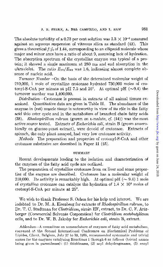
The absolute turbidity of a 0.73 per cent solution was 5.3 X 10v3 measured against an aqueous suspension of vitreous silica as standard (43). This gives a theoretical f:fo of 1.44, corresponding to an ellipsoid molecule whose major and minor axes have a ratio of about 9, assuming lack of hydration. The absorption spectrum of the crystalline enzyme was typical of a pro- tein; it showed a single maximum at 280 rnp and end absorption in the ultraviolet. The ratio E280/E260 was 1.6, indicating almost complete ab- sence of nucleic acid.
Turnover Number-On the basis of the determined molecular weight of 210,000, 1 mole of crystalline crotonase hydrated 730,000 moles of cro- tonyl-S-CoA per minute at pH 7.5 and 25“. At optimal pH (-9.4) the turnover number was 1,400,OOO.
Distribution-Crotonase is present in extracts of all animal tissues ex- amined. Quantitative data are given in Table II. The abundance of the enzyme in (rat) muscle tissue is noteworthy in view of its r61e in the fatty acid thio ester cycle and in the metabolism of branched chain fatty acids (38). Rhodospirillum rubrum (grown on L-malate, cf. (44)) was the most active source tested. Extracts of Escherichia coli, strain B (grown anaero- bically on glucose-yeast extract), were devoid of crotonase. Extracts of spinach, the only plant assayed, had very low crotonase activity.
Methods-The preparation and properties of crotonyl-S-CoA and other crotonase substrates are described in Paper II (45).
SUMMARY
Recent developments leading to the isolation and characterization of the enzymes of the fatty acid cycle are outlined.
The preparation of crystalline crotonase from ox liver and some proper- ties of the enzyme are described. Crotonase has a molecular weight of 210,000. Its activity is remarkably high. At optimal pH (- 9.4) 1 mole of crystalline crotonase can catalyze the hydration of 1.4 X lo6 moles of crotonyl-S-CoA per minute at 25”.
We wish to thank Professor S. Ochoa for his help and interest. We are indebted to Dr. M. A. Eisenberg for extracts of Rhodospirillum rubrum, to Dr. T. C. Stadtman for CZo.stridium, strain HF, extract, to Dr. C. F. Artz- berger (Commercial Solvents Corporation) for Clostridium ucetobutylicum cells, and to Dr. W. B. Jakoby for Escherichia coli, strain B, extract.
Addendum-A committee on nomenclature of enzymes of fatty acid metabolism, convened at the Second International Conference on Biochemical Problems of T,ipidcs, Ghent, Belgium, July 27 to 30, 1955, recommended systematic and trivial names for the enzymes catalyzing Reactions 1 through 6 as follows (trivial names being given in parentheses): (1) thiokinases, (2) acyl dehydrogenases, (3) enoyl
by guest on June 24, 2018http://w
ww
.jbc.org/D
ownloaded from

982 ENZYMES OF FATTY ACID METABOLISM. I
hydrases (crotonase) , (4) fl-hydroxyacyl dehydrogenases, (5) P-ketoacyl thiolases (thiolase), and (6) thiophorases.
BIBLIOGRAPHY
1. Barker, II. A., in McElroy, W. D., and Glass, B., Phosphorus metabolism, Balti- more, 1, 204 (1951).
2. Kennedy, E. P., and Lehninger, A. L., in McElroy, W. D., and Glass, B., Phos- phorus metabolism, Baltimore, 2, 253 (1952).
3. Lynen, F., and Ochoa, S., Biochim. et biophys. acta, 12,299 (1953). 4. Lynen, F., Federation Proc., 12, 683 (1953). 5. Mahler, H. R., Federation Proc., 12, 694 (1953). 6. Ochoa, S., Advances in Enzymol., 16, 183 (1954). 7. Greville, G. D., and Stewart, H. B., Ann. Rep. Progress Chem., 60, 301 (1954). 8. Lynen, F., Wessely, L., Wieland, O., and Rueff, I,., Bnnew. Chem., 64,687 (1952). 9. Drysdale, G. R., and Lardy, H. A., J. Biol. Chem., 202,119 (1953).
10. Chou, T. C., and Lipmann, F., J. Biol. Chem., 196,89 (1952). 11. Jones, M. E., Black, S., Flynn, R. M., and Lipmann, F., Biochim. et biophys. acta,
12, 141 (1953). 12. Lynen, F., Bull. Sot. chirn. biol., 35, 1061 (1953). 13. Beinert, II., Green, D. I~., Hele, P., Hift, H., Von Korff, R. W., and Ramakrish-
nan, C. V., 1. Biol. Chem., 203, 35 (1953). 14. Hele, P., J. Biol. Chem., 206, 671 (1954). 15. Mahler, H. R., Wakil, S. J., and Bock, R. M., J. Biol. Chem., 204, 453 (1953). 16. Beinert, H., Federation Proc., 12, 681 (1953). 17. Kornberg, A., and Pricer, W. E., Jr., J. Biol. Chem., 204, 329 (1953). 18. Seubert, W., and Lynen, F., J. Am. Chem. Sot., 75, 2787 (1953). 19. Mshler, H. R., J. Am. Chem. Sot., 76, 3288 (1953); J. Biol. Chem., 206, 13 (1954). 20. Green, D. E., Mii, S., Mahler, I-1. R., and Bock, 11. RI., J. BioZ. Chem., 206, 1
(1954). 21. Beinert, I-I., and Crane, F. I,., Federation Proc., 13, 181 (1954). 22. Stern, J. R., and de1 Campillo, A., J. Am. Chem. Sot., 76, 2277 (1953). 23. Wakil, S. J., and Mahler, H. R., J. Biol. Chem., 207, 125 (1954). 24. Stern, J. R., Raw, I., and de1 Campillo, A., Federation PTOC., 13, 304 (1954). 25. Stern, J. R., Coon, M. J., and de1 Campillo, A., J. Am. Chem. Xoc., 75,1517 (1953). 26. Wakil, S. J., Green, D. E., Mii, S., and Mahler, H. R., J. Biol. Chem., 207, 631
(1954). 27. Lehninger, A. I,., and Greville, G. D., J. Am. Chem. Sot., 76,1515 (1953) ; Biochim.
et biophys. acta, 12, 188 (1953). 28. Lynen, F., Reichert, E., and Rueff, L., Ann. Chem., 674, 1 (1951). 29. Stadtman, E. R., Doudoroff, M., and Lipmann, F., J. Biol. Chem., 191,377 (1951). 30. Stern, J. R., and Ochoa, S., J. Biol. Chem., 179,491 (1949); 191,161 (1951). 31. Stern, J. R., Coon, M. J., and de1 Campillo, A., Nature, 171,28 (1953). 32. Green, D. E., Goldman, D. S., Mii, S., and Beinert, H., J. Biol. Chem., 202, 137
(1953). 33. Ochoa, S., Stern, J. It., and Schneider, M. C., J. Biol. Chem., 193, 691 (1951).
Stern, J. R., Shapiro, B., Stadtman, E. R., and Ochoa, S., J. Biol. Chem., 193, 703 (1951).
34. Green, D. E., Science, 116, 661 (1952). 35. Beinert, H., J. Biol. Chem., 206, 575 (1953). 36. Stadtman, E. R., J. BioZ. Chem., 203, 501 (1953).
by guest on June 24, 2018http://w
ww
.jbc.org/D
ownloaded from
J. R. STERN, A. DEL CAMPILLO, AND I. RAW 983
37. Goldman, D. S., J. Biol. Chem., 208, 345 (1954). 38. Bachhawat, B. K., Robinson, W. G., and Coon, M. J., J. Am. Chem. Xoc., 76,309s
(1954). 39. Rabinowitz, J. L., and Gurin, S., J. Am. Chem. Sot., 76, 5168 (1954). 40. Bloch, K., Clarke, L. C., and Harary, I., J. Am. Chem. SOL, 76,3859 (1954). 41. Bonner, J., Park, M. W., and Montermoso, J. C., Science, 120, 549 (1954). 42. Warburg, O., and Christian, W., Biochem. Z., 310,384 (1941-42). 43. Oster, G., Anal. Chem., 25, 1165 (1953). 44. Eisenberg, M. A., J. Biol. Chem., 203, 815 (1953). 45. Stern, J. R., and de1 Campillo, A., J. Biol. Chem., 218, 985 (1956).
by guest on June 24, 2018http://w
ww
.jbc.org/D
ownloaded from

RawJoseph R. Stern, Alice del Campillo and Isaias
CROTONASEINTRODUCTION; CRYSTALLINE
METABOLISM: I. GENERAL ENZYMES OF FATTY ACID
1956, 218:971-983.J. Biol. Chem.
http://www.jbc.org/content/218/2/971.citation
Access the most updated version of this article at
Alerts:
When a correction for this article is posted•
When this article is cited•
alerts to choose from all of JBC's e-mailClick here
tml#ref-list-1
http://www.jbc.org/content/218/2/971.citation.full.haccessed free atThis article cites 0 references, 0 of which can be
by guest on June 24, 2018http://w
ww
.jbc.org/D
ownloaded from