Enzymes from marine microorganisms for the preparation of ... · Enzymes from marine microorganisms...
113
DIPARTIMENTO DI BIOTECNOLOGIE MEDICHE E MEDICINA TRASLAZIONALE DOTTORATO IN SCIENZE BIOCHIMICHE CICLO XXX – BIO/10 Enzymes from marine microorganisms for the preparation of biologically active molecules Docente guida: Prof. Patrizia Ferraboschi Direttore del corso di dottorato: Prof. Sandro Sonnino Tesi di Dottorato di: BENEDETTA GUIDI Matricola n. R10868 Anno Accademico 2016/2017
Transcript of Enzymes from marine microorganisms for the preparation of ... · Enzymes from marine microorganisms...

DIPARTIMENTO DI BIOTECNOLOGIE MEDICHE E MEDICINA TRASLAZIONALE
DOTTORATO IN SCIENZE BIOCHIMICHE
CICLO XXX – BIO/10
Enzymes from marine microorganisms for the preparation of
biologically active molecules
Docente guida: Prof. Patrizia Ferraboschi
Direttore del corso di dottorato: Prof. Sandro Sonnino
Tesi di Dottorato di:
BENEDETTA GUIDI
Matricola n. R10868
Anno Accademico 2016/2017


A tutte le famiglie che ho avuto in questi tre anni,

« If the doors of perception were cleansed, everything would appear to man as it is, infinite. »
W. Blake
“In dreams begins responsibility.”
W. B. Yeats
“Quando la tempesta sarà finita, probabilmente non saprai neanche tu come hai fatto ad
attraversarla e a uscirne vivo. Anzi, non sarai neanche sicuro se sia finita per davvero. Ma su un
punto non c'è dubbio...Ed è che tu, uscito da quel vento, non sarai lo stesso che vi è entrato.”
H. Murakami

ABSTRACT
This PhD project focuses on the identification, isolation and characterization of new biocatalysts able
to generate biologically active molecules with significant enantioselectivity. Through screening, we
identified marine strains, from MaCuMBA (Marine Culturable Microorganism for Biotechnological
Applications) and BIODEEP (Biotechnologies from the deep) European project collections, which
show a marked enantioselectivity on intermediates of molecules of biological interest.
Biotransformation substrate range included pramipexole, as main target, but it also embraces other
common building blocks for synthetic industrial preparation.
The stereoselective reduction of structurally different ketones using halotolerant marine yeasts
(Meyerozyma guilliermondii and Rhodotorula mucilaginosa) was studied using cells grown and bio-
converted in seawater. The preparation of valuable chemicals through water-saving (bio)processes
based on the direct exploitation of seawater is a significant step towards sustainable biocatalysis. By
choosing a suitable strain, high yields and stereoselectivity could be achieved in most cases. Notably,
high chemoselectivity and enantioselectivity were observed using R. mucilaginosa in the reduction
of aromatic β-ketonitriles, which allowed the recovery of the optically pure corresponding alcohols;
notably, reduction with whole cells of yeasts generally gives a mixture of undesired products, as
observed with M. guilliermondii.
Keto-reduction potential of thirty-three marine bacterium species was checked and afterwards the
possibility to convert this substrate directly into the optically pure amine was investigated: marine
bacteria were screened to identify transaminase activity. Based on the previous results in terms of
halotolerance and transaminase activity, the marine bacterium strain Virgibacillus pantothenticus
21D was selected for the genome sequencing in order to clone and express an ω-transaminase enzyme.
A recombinant non-marine ketoreductase from Pichia glucozyma (KRED1-Pglu) was used for the
enantioselective reduction of various cyclic ketones including pramipexole ketone intermediate.
Thanks to co-factor recycling system, the purified enzyme showed very promising results.
The soluble expression of a novel ω-transaminase from a newly isolated halotolerant marine
bacterium Virgibacillus pantothenticus was attained. Despite of several standard methodologies
applied, the marine wild-type enzyme was total insoluble in E. coli host and it was satisfactorily
solubilized by one single-point mutation, allowing the characterization of the new omega
transaminase. The enzyme shows an interesting salt and solvent tolerance, in accordance to its origin
and it results particularly active on some interesting building block molecules.

INDEX
ABSTRACT .............................................................................................................................................................. 5
INTRODUCTION ..................................................................................................................................................... 9
BLUE FOR GREEN ............................................................................................................................................ 10
BIOCATALYSIS ................................................................................................................................................ 13
BIOCATALYTIC APPLICATIONS .............................................................................................................. 16
ENZYMES .......................................................................................................................................................... 21
CLASSIFICATION ......................................................................................................................................... 21
KINETIC PARAMETERS .............................................................................................................................. 22
SPECIFICITY AND SELECTIVITY ............................................................................................................. 24
STABILITY ..................................................................................................................................................... 24
ENZYME ENGINEERING ............................................................................................................................. 25
KETO-REDUCTASES........................................................................................................................................ 26
TRANSAMINASES ............................................................................................................................................ 26
TARGET MOLECULES..................................................................................................................................... 27
PRAMIPEXOLE ............................................................................................................................................. 27
REFERENCES .................................................................................................................................................... 29
AIM OF THE WORK ............................................................................................................................................. 33
WHOLE CELL SCREENING ............................................................................................................................ 35
MARINE YEASTS ACTIVITY ...................................................................................................................... 35
MARINE BACTERIA ACTIVITY ................................................................................................................. 35
RECOMBINANT ENZYME SCREENING ....................................................................................................... 35
KETO-REDUCTASE ACTIVITY .................................................................................................................. 35
ESTERASE AND LIPASE ACTIVITY .......................................................................................................... 35
MARINE ω-TRANSAMINASE ACTIVITY .................................................................................................. 36
VIRGIBACILLUS PANTOTHENTICUS ω-TRANSAMINASE ...................................................................... 36
WHOLE CELL SCREENING - MARINE YEASTS ............................................................................................. 36
BACKGROUND ................................................................................................................................................. 37
MARINE YEASTS ......................................................................................................................................... 37
KETO-REDUCTASE ...................................................................................................................................... 38
PROJECT AIM.................................................................................................................................................... 41
RESULTS AND DISCUSSION .......................................................................................................................... 42

KETO-REDUCTASE SCREENING .............................................................................................................. 42
HALOTOLERANCE SCREENING ............................................................................................................... 48
MATERIALS AND METHODS ........................................................................................................................ 52
MATERIALS .................................................................................................................................................. 52
CHARACTERISATION ................................................................................................................................. 53
MICROORGANISMS ..................................................................................................................................... 53
GROWING MEDIUM .................................................................................................................................... 55
HALOTOLERANCE SCREENING ............................................................................................................... 55
BIOTRANSFORMATIONS ........................................................................................................................... 57
PURIFICATION AND CHEMICAL CHARACTERISATION ..................................................................... 58
REFERENCES .................................................................................................................................................... 60
WHOLE CELL SCREENING – MARINE BACTERIA ........................................................................................ 62
BACKGROUND ................................................................................................................................................. 63
MARINE BACTERIA..................................................................................................................................... 63
MARINE BACTERIA BIOCATALYSIS ....................................................................................................... 65
ω-TRANSAMINASE ...................................................................................................................................... 66
PROJECT AIM.................................................................................................................................................... 68
RESULTS AND DISCUSSION .......................................................................................................................... 68
KETO-REDUCTASE ...................................................................................................................................... 68
ω-TRANSAMINASE ...................................................................................................................................... 69
VIRGIBACILLUS PANTOTHENTICUS ...................................................................................................... 69
MATERIALS AND METHODS ........................................................................................................................ 70
MATERIALS .................................................................................................................................................. 70
CHARACTERISATION ................................................................................................................................. 71
MICROORGANISMS ..................................................................................................................................... 71
MEDIA AND GROWING CONDITIONS ..................................................................................................... 72
BIOTRANSFORMATION .............................................................................................................................. 73
PURIFICATION AND CHEMICAL CHARACTERISATION ..................................................................... 73
REFERENCES .................................................................................................................................................... 74
RECOMBINANT ENZYMES ................................................................................................................................ 76
BACKGROUND ................................................................................................................................................. 77
KETO-REDUCTASE ...................................................................................................................................... 77
ESTERASE AND LIPASE ............................................................................................................................. 78
ω-TRANSAMINASE ...................................................................................................................................... 79

PROJECT AIM.................................................................................................................................................... 80
RESULTS ............................................................................................................................................................ 80
KETO-REDUCTASE ...................................................................................................................................... 80
ESTERASE AND LIPASE ............................................................................................................................. 81
ω-TRANSAMINASE ...................................................................................................................................... 82
MATERIALS AND METHODS ........................................................................................................................ 82
MATERIALS .................................................................................................................................................. 82
CHARACTERISATION ................................................................................................................................. 82
KETO-REDUCTASE ...................................................................................................................................... 83
ESTERASE AND LIPASE ............................................................................................................................. 83
ω-TRANSAMINASE ...................................................................................................................................... 84
PURIFICATION AND CHEMICAL CHARACTERISATION ..................................................................... 84
REFERENCES .................................................................................................................................................... 85
VPTA - Virgibacillus pantothenticus ω-transaminase ............................................................................................. 86
BACKGROUND ................................................................................................................................................. 87
ω-TRANSAMINASE ...................................................................................................................................... 87
SOLUBILITY ISSUE ...................................................................................................................................... 88
MUTAGENESIS ............................................................................................................................................. 89
PROJECT AIM.................................................................................................................................................... 89
RESULTS AND DISCUSSIONS ........................................................................................................................ 90
WILD-TYPE VPTA AND SOLUBILISATION STRATEGIES .................................................................... 90
MUTAGENESIS ............................................................................................................................................. 92
b .................................................................................................................. Errore. Il segnalibro non è definito.
c .................................................................................................................. Errore. Il segnalibro non è definito.
d .................................................................................................................. Errore. Il segnalibro non è definito.
a .................................................................................................................. Errore. Il segnalibro non è definito.
VPTA T16F ..................................................................................................................................................... 94
EFFECT OF pH AND TEMPERATURE ....................................................................................................... 95
EFFECT OF CO-SOLVENTS AND SALTS .................................................................................................. 96
AMINO DONORS .......................................................................................................................................... 97
AMINO ACCEPTORS .................................................................................................................................... 98
ENZYME KINETICS ................................................................................................................................... 100
MATERIALS AND METHODS ...................................................................................................................... 100
MARINE MICROORGANISM, GENE IDENTIFICATION AND CLONING .......................................... 100

EXPRESSION OF WILD-TYPE VPTA ....................................................................................................... 101
MUTAGENESIS ON VPTA ......................................................................................................................... 101
EXPRESSION OF VPTA T16F .................................................................................................................... 101
PURIFICATION ........................................................................................................................................... 102
SDS-PAGE ANALYSIS ............................................................................................................................... 102
SPECTROPHOTOMETRIC ENZYMATIC ASSAY ................................................................................... 102
ENZYMATIC REACTION ........................................................................................................................... 103
ANALYTICAL METHODS ......................................................................................................................... 104
REFERENCES .................................................................................................................................................. 104
CONCLUSIONS ................................................................................................................................................... 108
WHOLE CELL SCREENING – MARINE YEASTS ....................................................................................... 109
WHOLE CELL SCREENING – MARINE BACTERIA .................................................................................. 109
RECOMBINANT ENZYME ............................................................................................................................ 109
VPTA ................................................................................................................................................................. 110
LIST OF PUBLICATIONS & CONFERENCE PROCEEDINGS ....................................................................... 111
INTRODUCTION

BLUE FOR GREEN
The marine ecosystem is the largest habitat on Earth, representing 70% of the surface of our planet
and thus it is a critical driver of hydrologic cycle and climate system, important for commerce,
transport, and tourism, a source of economically important resources, as well as new therapeutic
compounds. Seas and oceans include the
greatest extremes of temperature, light and
pressure and habitats that still remain largely
unexplored, understudied and
underexploited in comparison with
terrestrial ecosystems and organisms.
Marine organisms are sources of natural
bioactive substances with potential
therapeutic activity, and they should also be
valued as a source of genetic material to be
explored by the bioprospecting industry1
(fig. 1.1).

Fig. 1.1. A new term for an ancient process: bioprospecting refers to discovery and commercialization of new
products starting from biological resources1.
Only recently, indeed, the sea has been rediscovered as untapped source of novel biocatalysts and its
extraordinary micro-biodiversity can offer new possibilities for conducting highly selective, enzyme-
catalysed reactions in industrial processing, laboratory analyses, and medical therapy1 (fig. 1.2).
Fig. 1.2. Web of Knowledge Scientific hits
containing “marine enzymes” (5528 total
hits).
Biocatalysts produced by marine microorganisms are naturally endowed with an excellent stability
towards high salinity, as a consequence of different structural adjustments adopted to cope with high
ionic strength conditions. Interestingly, adaptation to high salinity corresponds to an increased
tolerance towards organic solvents, a requisite for many enzymes to be used as biocatalysts in organic
chemistry2. From this point of view, marine habitats containing high salt concentrations, such a salt
lake, the Dead Sea and deep hypersaline anoxic basins (DHABs), provide ideal locations to source
novel marine biocatalysts3 (figure 1.3).

Fig. 1.3. Biodiversity
still represents an
incredible resource and
source of new and
naturally high
performing biocatalysts,
thanks to a unique
evolutionary pathway.
The ability to tolerate high NaCl concentrations was investigated, since these extreme environments
are usually characterised by high concentrations of this salt. In this condition, the cell is exposed to
two different stimuli from the environment: osmotic and ionic stress. Typically, hyperosmotic stress
causes immediate water outflow from the cell, reduces turgor pressure and consequently dehydrates
the cytosol4. Ionic stress, on the other hand, results in an intake of ions (e.g. Na+) by the cell that
increases intracellular ionic concentration leading damages to membranes and water discharge from
the cell. One of the strategies adopted by marine microorganisms to counteract changes in turgor
pressure is to lower the absorption of water and compensate with a build of compatible solutes that
do not interfere with cell metabolism and maintaining internal concentration of Na+ at non-toxic
levels5. Those microorganisms that can grow at high NaCl concentrations preferring to other
conditions are defined as halophiles. Differently, the category of halotolerants is representative of
those microorganisms that survive at different concentrations of NaCl and outweigh osmotic and
saline stress but are not, however, in their optimal growth condition. Non-halophiles are all those

microorganisms that do not overcome the stress due to the presence of salt at high concentrations and
therefore are not able to perform their vital functions in this particular condition.
Taking into account what deals with molecular adaptation of proteins that are stable and function at
high salt concentration, understanding how these enzymes maintain their fold stable and avoid
aggregation under harsh conditions is of great interest for biotechnological applications. Comparisons
between the sequences of halophilic/non-halophilic homologous protein pairs indicated that Asp and
Glu are significantly more frequent, while Lys, Ile and Leu are less frequent in halophilic proteins.
Homologous halophilic and non-halophilic proteins have similar overall structure, secondary
structure content, and number of residues involved in the formation of H-bonds. On the other hand,
on the halophilic protein surface, a decrease of nonpolar residues and an increase of charged residues
are observed. Particularly, halophilic adaptation correlates with an increase of Asp and Glu,
compensated by a decrease of basic residues, mainly Lys, on protein surface6. Future studies will give
further insights into the adaptation strategies to high salinity environment and will indicate strategies
for rational protein engineering in order to improve enzyme industrial performance.
BIOCATALYSIS
Biocatalysis, assumed as applications of enzymes or whole cells for chemical synthesis, is an
appealing technology for fine chemical, food and pharmaceutical industry for several reasons.
The employment of biologic systems, such as whole cells or isolated enzymes, offers important
advantages:
High efficiency – high turnover numbers; rates (108 – 1010).
Selectivity/specificity – chemo-, regio- and stereoselectivity.
Mild conditions – mild temperature and pH; atmospheric pressure.
Low energy consumption.
Not bound to their natural role (substrate tolerance).
Highly selective in complex mixtures (no side reactions).
Fewer by-products.
Biodegradable (natural bioproducts).
Can be overproduced.

The excellent regio- and stereoselectivity of enzyme catalysts along with their ability to work under
mild reaction conditions (thus protecting existing functionality within a molecule) enables
transformations without the need for multiple protection and deprotection steps within a synthesis.
Additionally, biocatalysis offers both economic and environmental advantages over chemocatalytic
methods7,8.
Enzymes are produced from
inexpensive renewable resources
and are themselves biodegradable,
fulfilling the central tenants of
Green Chemistry9 and sustainable
development (fig. 1.4).
Fig. 1.4. The Twelve Principles of
Green Chemistry by Anastas and
Warner (1998)9.
Image from Compound of Interest® (http://www.compoundchem.com/2015/09/24/green-
chemistry/) .
Besides the above-mentioned advantages, some drawbacks coming from the application of
biocatalysis in chemistry must be recognized, such as:
Biocatalysts often show lower stability than conventional catalysts.
Development of industrial biocatalytic processes is usually much longer to establish.
Low number of commercially available biocatalysts.
Necessity of microbiological facilities if the biocatalyst is not a commercial enzyme.

Most of these disadvantages might be overcome by modern techniques (screening, molecular biology,
protein engineering, immobilization) able to furnish a much higher number of biocatalysts with
improved performances.
The whole process that sees the industrial application of a new enzyme usually begins with in vivo or
in silico screening. The dominant strategy to discover new biocatalysts has varied from
straightforward experimental assays for certain functions (experimental assays for functions) to
deduction of protein functions from bioinformation data-bases (from sequence to function, from
structure to function). After a biocatalyst characterization, process design has to fit with enzyme
specifications and vice versa (fig.1. 5).
Fig. 1.5. Flowchart of essential elements of
biocatalyst discovery, characterization and
improvement by protein engineering1.
As Bornscheuer et al. described10, we are living the third wave of biocatalysis progress. During the
first wave of biocatalysis (fig. 1.6), which started more than a century ago, scientists recognized that
components of living cells could be applied to useful chemical transformations (in contrast to the
fermentation processes, which had been commonplace for millennia already). The main challenge for
these applications is the limited stability of the biocatalyst, and such shortcomings were primarily
overcome by immobilization of the enzyme, which also facilitated the reuse of the enzyme. During
the second wave of biocatalysis, in the 1980s and 1990s, initial protein engineering technologies,

typically structure based, extended the substrate range of enzymes to allow the synthesis of unusual
synthetic intermediates. This change expanded biocatalysis to the manufacture of pharmaceutical
intermediates and fine chemicals. Apart from stabilization, the challenges now included optimizing
the biocatalyst for the non-natural substrates. The third, and present, wave of biocatalysis started with
the work of Pim Stemmer and Frances Arnold in the mid and late 1990s. They pioneered molecular
biology methods that rapidly and extensively modify biocatalysts via an in vitro version of Darwinian
evolution. The methods are now commonly called directed evolution (see “Enzyme engineering”
paragraph).
Fig. 1.6. Based on protein structures (a) or homology models, rational design (b) approach is able to identify
distinct point mutations, whereas for directed evolution experiments random mutagenesis (c) combined with
screening is the starting point. Combining these methods makes it possible to create smaller, but smarter,
libraries (d). The classical screening of enzymes by enrichment cultures (e) is now replaced by key motif
database searches (f) to guide identification of novel enzymes or those with desired properties. Still in its
infancy is the computational de novo design of enzymes (g)10.
BIOCATALYTIC APPLICATIONS
Despite the clear benefits of biocatalytic processes, the historical number of industrial applications
has been modest, with a dramatic increase in the use of biocatalysis only occurring within the last two
decades (fig. 1.7).

Fig. 1.7. Number of publications and patents discussing
“pharmaceutical biocatalysis” for each 5-year period of the
last 50 years. Metrics from Google Scholar
The use of biocatalyst has acquired importance as a complement to conventional chemical techniques
in various application fields. In particular, biocatalyst selectivity is important for obtaining
biologically active molecules, whose stereochemistry affect their in vivo behaviour. The specific
reactions that can be replaced with biocatalysis have been identified in the synthesis of biologically
active molecules, including chiral amine preparation, stereo and regiospecific hydroxylation of
complex molecules, and other redox reactions10,11.
One of the first enzymatic applications in biologically active molecule synthesis was lipase resolution
for the preparation of Naproxen in 1987 by Gu and co-workers12 (scheme 1.1). (+)-2-(6-Methoxy-2-
naphthyl) propionic acid has been prepared via enzymatic enantiospecific hydrolysis of (±)-
chloroethyl-2-(6-methoxy-2-naphthyl) propionate, catalyzed by the lipase of Candida cylindracea
with E >100 (39% conversion; 98% e.e.).
Scheme 1.1. Biocatalytic resolution of Naproxen intermediate by Candids cylindracea.
Other examples of smart commercial enzyme applications are described for the antitumoral
Capecitabine13 and anti-thrombotic Clopiogrel14 synthesis, both exploiting a serine protease catalysis.
Alcalase CLEA enzyme is able to catalyse alcoholysis allowing the selective deprotection of primary
acetyl ester of the N1-(2’, 3’, 5’-tri-O-acetyl-β-D-ribofuranosyl)-5-fluoro-N4-(n-pentyloxycarbonyl)
cytosine and thus affording the corresponding 5’-hydroxyderivative, an advanced intermediate of
capecitabine synthesis with a good yield (80% after purification). The (S)-2-chlorophenylglycine
moiety is well recognized in the structure of (S)-clopidogrel. The enantiomerically pure chiral

building block synthesis was performed via an enzyme-catalyzed resolution of (RS)-N-Boc-2-
chlorophenylglycine methylester. The high enantiomeric excess of the synthon was obtained by
immobilized subtilisin (Alcalase CLEA). The simplicity of the process makes this pathway suitable
for large-scale preparation.
One of the most successful examples in the practical application of enzymes in the pharmaceutical
industry is the anti-diabetic compound, sitagliptin15,16. Sitagliptin is a drug for type II diabetes that
has been marketed under the trade name Januvia by Merck15. Researchers at Codexis and Merck
engineered R-selective transaminase (R-ATA, ATA-117) from Arthrobacter sp. for the asymmetric
amination of prositagliptin ketone. By applying a substrate walking, modeling, and mutation
approach, they were able to overcome the limitation of the substrate's size for the enzyme. A
combination of the further directed enzyme evolution and process engineering yielded a variant that
converts 200 g/L of prositagliptin ketone into sitagliptin with enantio-purity higher than 99.95% even
in the presence of 1 M i-PrNH2, 50% DMSO and 40 °C 16 (scheme 1.2). Immobilization of engineered
(R) selective-ATA enables the maintenance of the enzyme activity and stability in an organic solvent,
simplifying the workup and allowing a repetitive use of the enzyme17.
Scheme 1.2. Synthesis of sitaglipin from prositagliptin ketone using engineered (R)-selective ATA.
Codexis recently developed a biocatalytic process for producing intermediates for blockbuster drugs
such as atorvastatin, montelukast, duloxetine, phenylephrine, ezetimibe, and crizotinib based on
stereo and regio-specific hydroxylation using keto-reductase (KRED) from Lactobacillus18,19.The
anti-asthmatic drug, montelukast, was developed and marketed under the trade name Singulair by
Merck20. Combined with a directed evolution and process optimization, the engineered KRED
exhibits a high enantio-selectivity (> 99.9%) and stability even in the presence of ∼ 70% organic
solvents at 45 °C (scheme 1.3). The biocatalytic process is currently operated on a > 200 kg scale
substrate. The most intriguing point in KRED engineering is the increase of the enzyme stability even
at a high organic solvent concentration and temperature. Because of the low solubility of the substrate
in water, the high organic solvent concentration and temperature are necessary. Based on the
correlation between the thermostability and solvent tolerance19, researchers at Codexis primarily

screened enzyme mutants with increased thermal stability followed by a screening for solvent tolerant
mutants19.
Scheme 1.3. Regio-specific hydroxylation of key intermediate in synthesis of montelukast using engineered
KRED20.
In regards to whole cell biocatalysis, a high yielding bioprocess for 11-α hydroxylation of canrenone
using Aspergillus ochraceus ATCC 18500 was described21. The optimization of the bioprocess
involved both fermentation (for achieving highly active mycelium of A. ochraceus) and
biotransformation with the aim to obtain 11-α hydroxylation with high selectivity and yield. A
medium based on sucrose as C-source resulted particularly suitable for conversion of canrenone into
the corresponding 11-hydroxy derivative, whereas the use of O2-enriched air and DMSO as a co-
solvent for increasing substrate solubility played a crucial role for obtaining high yields (>95%) of
the desired product in high chemical purity starting from 30 mM (10.2 g/L) of substrate (scheme 1.4).
Scheme 1.4. Conversion of canrenone into the corresponding 11-hydroxy derivative by Aspergillus
ochraceus21.
Another successful whole cell bioconversion led to the achievement of the enantiomerically pure
advanced intermediate of the synthesis of (S)-pramipexole, a anti-Parkinson drug, showing the
Baker’s yeast efficiency (figure 1.5). The use of readily available and inexpensive Saccharomyces
cerevisiae, the easy preparation of the biotransformation substrate, and the simple steps required to
accomplish the synthesis, make the method applicable to a preparative scale.
This synthon, not yet described in the literature, was transformed through very simple steps into the
desired dihydrochloride monohydrate derivative of (S)-pramipexole in 21% overall yield. A complete

inversion of the configuration realized under the Mitsunobu22 conditions allowed the (S)-alcohol
achievement.
The (S)-alcohol obtained is a suitable substrate for dexpramipexole, the (R)-isomer of pramipexole,
which is currently under investigation in the treatment of ALS (amyotrophic lateral sclerosis)23.
Scheme 1.5. Enantioselective reduction of the keto-intermediate in pramipexole synthesis by Saccharomyces
cerevisiae.
BIOCATALYST FORM
Biocatalysis can be performed by both whole cells and isolated enzymes. The type of bioconversion
and enzyme features adress the choice. Whole cells allow the production of compounds also ensuring
cofactor regeneration, with high regio- and stereoselectivity, under mild operational and environment-
friendly conditions. A limit of employing whole cells is sterile initial conditions and prevention of
biological contamination. However, they are quite effective in multi-step reactions, they provide a
protective environment to enzymes (e.g. in non-conventional media) and are significantly cheaper to
produce than free enzymes which require several isolation and purification steps24,25. In one-step
reactions, isolated enzymes should provide significant benefit when compared to whole cells as no
side-reactions should occur and substrates do not have to be transported across membranes. The
enzymes are able to catalyse more efficient reactions and under mild conditions, within a narrow
range of pH and temperature. Furthermore, enzymes are able to maintain their activity under in vitro
conditions and can catalyse reactions in conditions not suitable for cell growth. The use of pure
enzymes in biocatalysis has several advantages such as the specificity for selected reactions, simple
apparatus and procedures and better tolerance to co-solvents used to solubilise low-water soluble
substrates26. However, enzyme isolation and purification can be quite expensive and time consuming,
the addition of co-factors or their recycling may be required and, in general, it is more difficult to
carry out reactions requiring more than one enzyme, contrarily to the use of whole microbial cells
(table 1.1).
BIOCATALYST FORM PROS CONS
ISOLATED ENZYME any
simple apparatus,
simple work-up,
better productivity due
to higher concentration
tolerance
cofactor recycling
necessary

dissolved in water high enzyme activities
side reaction possible,
lipophilic substrates
insoluble,
work-up requires
extraction
suspended in organic solvents
easy to perform,
easy work-up,
lipophilic substrates
soluble,
enzyme recovery easy
low activities
immobilised enzyme recovery easy loss of activity during
immobilisation
WHOLE CELLS
any no cofactor recycling
necessary
expensive equipment,
tedious work-up due to
large volumes,
low productivity due to
lower concentration
tolerance,
low tolerant of organic
solvents,
side reactions likely due
to uncontrolled
metabolism
growing culture higher activities
large biomass,
more by-products,
process control difficult
resting cells work-up easier,
fewer by-products lower activities
immobilised cells cell re-use possible lower activities
Table 1.1. Pros and cons of using isolated enzymes and whole cell systems27.
An important issue related to employing isolated enzyme is stability under relatively harsh conditions,
such as high temperatures, pH and in the presence of solvents28. Limitations of biocatalysts can be
overcome by integrating different techniques, such as genetic engineering, that allows the production
of large quantities of enzymes at relatively low costs, biocatalyst immobilization, and suited reactor
technology. Numerous molecular techniques have been developed to improve the activity or substrate
specificity of an enzyme with a particular industrial application in mind. This includes random
mutagenesis of a target gene followed by screening and rational protein engineering28,29 and directed
evolution30,31.
ENZYMES
CLASSIFICATION
The identity of the biocatalyst must be specified as per name of reaction type: EC number; strain
deposit, GenBank sequence accession number.
In according to International Union of Biochemistry and Molecular Biology, Enzyme commission
(IUBMB EC) classification:
EC 1 oxidoreductases: oxygenation of C-H, C-C, C = C bonds; transfer of electrons.
EC 2 transferases: transfer of functional groups: aldehyde, ketone, acyl, phosphoryl, or methyl.
EC 3 hydrolases: formation/breakdown of esters, amides, lactones, lactams, epoxides, nitriles,
anhydrides, glucosides, etc.
EC 4 lyases: removal or addition on C = C, C = N, C = O bonds.
EC 5 isomerases: racemization and epimerization.
EC 6 ligases: formation- cleavage of C-O, C-S, C-N, C-C bonds requiring ATP cleavage.
KINETIC PARAMETERS
The core kinetic parameters of an enzyme are Km and Vmax values, described by the Henry-Michaelis-
Menten equation32 that correlates the dependence of the reaction velocity (v) on the free substrate
concentration (S). Briggs and Haldane (1925) provided a derivation of this equation introducing the
steady-state approximation33 which assumes that the enzyme-substrate complex (ES) after initial
formation reaches its climax and remains unchanged over the time the reaction is monitored.
Km and Vmax are determined by directly plotting v over S (Michaelis- Menten plot) or more favourably
by one of the linear transformations such as Lineweaver-Burk (1/v vs. 1/S), Hanes (S/v vs. S) or
Eadie-Hofstee (v /S vs. v).
Transformation of the Michaelis-Menten equation32 shows that Km specifies the substrate
concentration where the reaction proceeds at half of Vmax. kcat, often referred to as the turnover
number, can be calculated directly as the quotient of Vmax and the total enzyme concentration.
Turnover numbers are normally reported as molecules product produced per molecules of enzyme
per time (e.g. mol/mol s). Thus, if the same units are used for product and enzyme they eventually
cancel each other and the unit for reporting kcat will be reciprocal time (e.g. s−1). The kcat/Km ratio,
originally referred to as “specificity34” or “performance35” constant and representing a second-order
rate constant (M−1 s−1) is used as a measure for catalytic efficiency of an enzyme. In general, the
higher kcat/Km the better is the enzymatic performance which can be used to compare enzymes or
different substrates of one enzyme.

Fig. 1.7. Michaelis Menten Plot versus Lineweaver-Burk linear kinetic representation32.
In a simple enzyme reaction over time one substrate is converted to yield one single product. This of
course is not totally true since it often requires coenzymes (e.g. NAD(P)H for oxidoreductases) or co-
substrates (e.g. water for hydrolases) or, in the case of lyases (synthases), yields, technically speaking,
two products or converts two products into one substrate. To kinetically describe these kinds of
reactions it is important to define one speed-limiting factor which could be substrate, co- substrate,
etc., at a time and supply the respective others in excess. During the enzymatic assay, substrate
concentration is much higher than enzyme concentration remaining virtually unchanged, and product
accumulation is linear and there is no back reaction into substrate. Substrate concentrations are crucial
and should be in the range of 0.1 to 10 times Km which for unknown enzymes often has to be
determined using trial approaches (fig. 1.8).
Fig. 1.8. Progress curve for an enzyme reaction. The
slope in the initial rate period is the initial rate of
reaction v. The Michaelis-Menten equation describes
how this slope varies with the concentration of
substrate.
In an enzymatic assay, one of the key parameters to check is the initial rate measured as specific
activity (SA). SA gives a measure of enzyme processivity, at a specific (usually saturating) substrate
concentration, and is usually constant for a pure enzyme. It is conventionally expressed in Units (U)
as µmol min−1 mg−1 or in katal kg−1. Enzyme Unit (U) is defined as the amount able to catalyse the
transformation of 1 micromole of the substrate per minute under standard conditions. Turnover

number, on the other hand, is dimensionless referring to the ratio of number of moles of product per
mole of catalyst used over the reaction period. On enzyme assays, depending on what methods are
being used (e.g. spectrophotometry or measurement of fluorescence using a fluorometer), it may be
critical to find the optimum conditions of pH, temperature, buffer concentrations (ionic strength),
metal or cofactor requirements. Importantly, the enzyme concentration used has to be appropriate
since accurate determination of the enzyme’s SA requires the presence of excess amounts of
substrate(s) and possible co-substrate(s).
SPECIFICITY AND SELECTIVITY
One of the characteristics of an enzymatic catalysis is the specificity for substrate(s) and reactions.
Enzymes are generally highly specific for one reaction type, but promiscuous activities can be found
in nature or can be introduced into an enzyme by protein engineering. Specificity and selectivity are
often used as synonyms to describe the ability of an enzyme to distinguish between substrates; more
correctly, substrate specificity refers to a reaction where one (and only one) substrate can react,
whereas substrate selectivity describes a reaction where one substrate is preferentially transformed
over others. Therefore, the term enantioselectivity is preferentially used when the substrates are
enantiomers. Enzymes also exhibit chemoselectivity for similar functional group, and regioselectivity
for analogous functional groups with different chemical neighbourhood.
To better characterize the stereoselective properties of an enzyme and thus to quantitatively assess its
potential for kinetic resolution36 the dimensionless enantiomeric ratio (E)36 was introduced. It is
expressed as the quotient of the second-order rate constants (kcat/Km) R and (kcat/Km) S. This is a
measure for the ‘selectivity’ of an enzymatic resolution37. E values can be experimentally calculated
by measuring the enantiomeric excess (ee) of either the residual substrate or the corresponding
product at the specific degree of conversion (C). To obtain accurate E values, the general rule is to
stop the reaction at about 50% conversion.
STABILITY
In an industrial setting, stability of a biocatalyst is one of the most important characteristics. Even the
most active enzyme will be practically useless if it does not maintain its activity over the envisioned
treatment or production process time. For process development, the fundamental question is whether
the enzyme has to be designed to meet an existing protocol or whether the process can be designed
to adapt to the enzyme’s properties. One can distinguish between several forms of enzyme stability,
such as chemical stability (e.g. the influence of pH, salt or solvent concentration), thermodynamic
stability (e.g. reversible unfolding of the protein structure due to increasing temperature) and kinetic

stability (describing the time the enzyme remains active before undergoing irreversible
denaturation)28,38. Enzyme stability studies, however, are often performed under laboratory
conditions (e.g. in a simple buffer) and thus, although giving some insight, they only have limited
value for assessing the enzyme’s bioprocess suitability. Under process conditions, enzyme behaviour
can be very different with substrate(s), accumulating product(s) and possible solvents modulating its
activity in either way: positive (protection) or negative (destabilization)39.It should be noted that
enzyme stability often depends on the concentration that it was assayed, thus adding another variable.
ENZYME ENGINEERING
Enzymes were optimized by Darwinian evolution over millions of years to catalyse reactions while
ensuring high substrate specificity, as well as exquisite enantioselectivity and stereoselectivity.
However, there are often significant discrepancies between an enzyme’s function in nature and the
specific requirements for ex vivo applications envisioned by scientists and engineers.
The modification of enzymes for adaptation to the environment in a chemical process is often
considered necessary, for which many strategies have been developed40. Frequently the compounds
in the process are poor substrates, the enantioselectivity may be insufficient, or a more heat stable or
co-solvent tolerable enzyme is desired. By using an experimentally determined structure of the
enzyme (determined by X-ray crystallography or NMR) the enzyme engineer can locate specific
amino acid residues suitable for mutation. This is called rational design; arguably, the amount of
knowledge which is available regarding the function of a given enzyme dictates the likelihood of
success with this strategy. When such attempts are unsuccessful, or a structure is unavailable, the
amino acid chain can be altered in a randomised fashion and a library of mutants can be screened for
the desired properties. An improved clone can be further randomised and a new library can be
screened. The iterative process is repeated for the potential discovery of the required enzyme. This
strategy is called directed evolution41–45. Further, to increase the probability of finding improved
variants, semi-rational40 approaches have been devised, such as CASTing46,47 and saturation
mutagenesis. The more recent scientific publications involving enzyme engineering contain examples
of rational and semi-rational approaches with a higher frequency than in the past. This phase in
biocatalytic progress was called “third wave” by Bornscheuer10 as we previously reported. The
method advancements, increased understanding of function and examples of successful enzyme
variants have made researchers less inclined to resorting to directed evolution, which most often
involves a tedious screening procedure. The choice of method is usually not straightforward, and
remains a challenge for the enzyme engineering48.

For what concerns the enzyme activities taken into account, keto-reductase and transaminase
activities were the main biocatalytic strategies investigated.
KETO-REDUCTASES
Ketoreductases (KREDs) are enzymes useful for enantiomeric preparation of chiral alcohols from
ketones, ketoacids and ketoesters19 (scheme 1.6).
Scheme 1.6. Simplified catalysis by KRED.
KREDs can induce an asymmetric attack of the hydride with total or prevalent formation of one of
the two enantiomers. The cofactors (mostly NADH or NADPH) provide the hydride ion giving rise
to the reduction of the substrate. Yeasts are usually employed for synthetic purposes with main
application in stereoselective reduction of ketones affording optically pure secondary alcohols49–52.
In particular, marine yeasts have already been investigated for the production of pharmaceutical and
enzymatic products, such as astaxanthin, siderophore, riboflavin, inulinase and amylases. Yet, the
commercial application of marine yeasts is still limited. The current research, however, indicates the
promising features of the marine yeasts for the potential industrial application and their superiority
over the terrestrial ones in certain field. More direct comparison studies should be carried out to give
further evidence on the advantages of marine yeasts over terrestrial yeasts53, as explained in the
second chapter.
TRANSAMINASES
Transaminases, also called amino transaminase (ATA), are enzymes capable of the enantioselective
transfer of an amino group from the amino donor to the acceptor substrate. The α-ATAs are able to
operate on the amino moieties of the α-carbon of amino acids, while the ω-ATAs mainly accept as
substrates donors with amines distal to the carboxyl moiety, as well as other amine donors and their
respective non-keto acid acceptors (scheme 1.7).

Scheme 1.7. Simplified catalysis by ω-ATA.
Combining ATAs with other enzymatic or chemical routes has been demonstrated to be a smart
method for practical applications, particularly in regard to shortening the reaction routes, avoiding
protecting steps, reducing chemical waste and achieving a high atom-efficiency. This makes these
enzymes useful for synthesis of chiral amines, which are of high importance as building blocks for
production of optically pure amino synthons in pharmaceutical like sitagliptin16, ethambutol54,
imagabalin55, norephedrine and pseudoephedrine56, food and cosmetic additives57, agrochemical and
material industries. The development of protein engineering has tried to answer to this need and it is
achieving remarkable progresses through random and rational mutagenesis approach but some issues
need to be completely clarify yet. The current trend in the research field is directing to the
identification and designing of new ω-transaminases with defined substrate selectivity and capable of
adapting to the uncommon catalytic conditions of industrial processes. The issue will be widely
discussed in the following chapters.
TARGET MOLECULES
The range of molecules evaluated in this PhD project was selected in order to obtain asymmetric
catalysis or kinetic resolution providing chiral building blocks aimed at the synthesis of biologically
active molecules with a solid industrial appeal.
PRAMIPEXOLE
One of the target molecules investigated in this project is pramipexole. A recent example is the
preparation58 of an optically pure new intermediate of (R)- or (S)-pramipexole; this thiazole derivative
is endowed with anti-Parkinson activity59–61 if its stereocenter is in the (S)-configuration, while the
(R)-isomer is under investigation for the treatment of ALS (amyotrophic lateral sclerosis) 23 (fig. 1.9).

Fig. 1.9. Pramipexole enantiomers could have different applications as therapeutics.
Numerous new synthetic pathways were recently developed and patented62–64. The strategy which
this experimental work is based on involves a biocataytic keto-reduction step in order to obtain an
optically pure alcohol, intermediate in this new synthetic method58(scheme 1.8).
Scheme 1.8. This synthetic pathway based on the previous work by Ferraboschi et al.58 combines chemical
and biocatalytic steps.
MIRAPEX® by Boehringer Ingelheim® is the most common drug formulation containing as active
substance (S)-pramipexole dihydrochloride. Currently, also the generic drug is available on the
market. For what concerns anti-Parkinson drug distribution, the global prevalence in 2012 was
approximately 2.18 million cases world-wide; this resulted in $3.56 billion in global market sales of
therapies, including $1.15 billion in the U.S. market. Through advances in technology and drug
development, the Parkinson treatment’s market in 2022 is expected to yield $5.26 billion in global
market sales, with $2.33 billion from the U.S. market65.
Because of the increasing spread of the disease, the global market of anti-Parkinson drugs is
expanding and, consequently, Mirapex®, representing one of the most prescribed therapy, is getting
an important slice of this market66.
REFERENCES
1. Trincone, A. Marine Enzymes for Biocatalysis. (Woodhead Publishing, 2013).
2. Elleuche, S., Schröder, C., Sahm, K. & Antranikian, G. Extremozymes-biocatalysts with unique
properties from extremophilic microorganisms. Curr. Opin. Biotechnol. 29, 116–123 (2014).
3. De Vitis, V. et al. Marine Microorganisms as Source of Stereoselective Esterases and Ketoreductases:
Kinetic Resolution of a Prostaglandin Intermediate. Mar. Biotechnol. 17, 144–152 (2015).
4. Petelenz-Kurdziel, E. et al. Quantification of cell volume changes upon hyperosmotic stress in
Saccharomyces cerevisiae. Integr. Biol. Integr. Biol 3, 1120–1126 (1120).
5. Wood, J. M. Bacterial responses to osmotic challenges. J. Gen. Physiol. 145, 381–388 (2015).
6. Graziano, G. & Merlino, A. Molecular bases of protein halotolerance. Biochim. Biophys. Acta -
Proteins Proteomics 1844, 850–858 (2014).
7. Ran, N., Zhao, L., Chen, Z. & Tao, J. Recent applications of biocatalysis in developing green chemistry
for chemical synthesis at the industrial scale. Green Chem. 10, 361–372 (2008).
8. Tao, J. & Xu, J. H. Biocatalysis in development of green pharmaceutical processes. Curr. Opin. Chem.
Biol. 13, 43–50 (2009).
9. Anastas, P. & Warner, J. Green Chemistry: Theory and Practice. (Oxford University Press, 1998).
10. Bornscheuer, U. T. et al. Engineering the third wave of biocatalysis. Nature 485, 185–194 (2012).
11. Choi, J. M., Han, S. S. & Kim, H. S. Industrial applications of enzyme biocatalysis: Current status and
future aspects. Biotechnol. Adv. 33, 1443–1454 (2015).
12. Gu, Q.-M., Chen, C.-S. & Sih, C. . A facile enzymatic resolution process for the preparation of (+)-S-
2-(6-hethoxy-2-naphthyl)propionic acid (Naproxen). Tetrahedron Lett. 27, (1986).
13. Ciceri, S., Ciuffreda, P., Grisenti, P. & Ferraboschi, P. Synthesis of the antitumoral nucleoside
capecitabine through a chemo-enzymatic approach. Tetrahedron Lett. 56, 5909–5913 (2015).
14. Ferraboschi, P., Mieri, M. D. & Galimberti, F. Chemo-enzymatic approach to the synthesis of the
antithrombotic clopidogrel. Tetrahedron Asymmetry 21, 2136–2141 (2010).
15. Desai, A. A. Sitagliptin manufacture: A compelling tale of green chemistry, process intensification, and
industrial asymmetric catalysis. Angew. Chemie - Int. Ed. 50, 1974–1976 (2011).
16. Savile, C. K. et al. Biocatalytic Asymmetric Synthesis of Chiral Amines from Ketones Applied to
Sitagliptin Manufacture. Science (80-. ). 329, 305–309 (2010).

17. Truppo, M. D., Strotman, H. & Hughes, G. Development of an Immobilized Transaminase Capable of
Operating in Organic Solvent. ChemCatChem 4, 1071–1074 (2012).
18. Huisman, G. W. & Collier, S. J. On the development of new biocatalytic processes for practical
pharmaceutical synthesis. Curr. Opin. Chem. Biol. 17, 284–292 (2013).
19. Huisman, G. W., Liang, J. & Krebber, A. Practical chiral alcohol manufacture using ketoreductases.
Curr. Opin. Chem. Biol. 14, 122–129 (2010).
20. Liang, J. et al. Development of a biocatalytic process as an alternative to the (-)-DIP-Cl-mediated
asymmetric reduction of a key intermediate of montelukast. Org. Process Res. Dev. 14, 193–198
(2010).
21. Contente, M. L. et al. Development of a high-yielding bioprocess for 11-?? hydroxylation of canrenone
under conditions of oxygen-enriched air supply. Steroids 116, 1–4 (2016).
22. Mitsunobu, O. The Use of Diethyl Azodicarboxylate and Triphenylphosphine in Synthesis and
Transformation of Natural Products. Synthesis (Stuttg). 1981, 1–28 (1981).
23. Bozik, M. E., Mather, J. L., Kramer, W. G., Gribkoff, V. K. & Ingersoll, E. W. Safety, Tolerability,
and Pharmacokinetics of KNS-760704 (Dexpramipexole) in Healthy Adult Subjects. J. Clin.
Pharmacol. 51, 1177–1185 (2011).
24. Schmid, A. et al. Industrial biocatalysis today and tomorrow. Nature 409, 258–268 (2001).
25. de Carvalho, C. C. C. R. Whole cell biocatalysts: essential workers from Nature to the industry. Microb.
Biotechnol. 10, 250–263 (2017).
26. Roberts, R. J. et al. On Base Flipping Minireview. Cell 92, 9–12 (1995).
27. Faber, K. Biotransformations in Organic Chemistry: A Textbook. (Springer Berlin Heidelberg, 2011).
28. Polizzi, K. M., Bommarius, A. S., Broering, J. M. & Chaparro-Riggers, J. F. Stability of biocatalysts.
Curr. Opin. Chem. Biol. 11, 220–225 (2007).
29. Morley, K. L. & Kazlauskas, R. J. Improving enzyme properties: When are closer mutations better?
Trends Biotechnol. 23, 231–237 (2005).
30. Eijsink, V. G. H., GÅseidnes, S., Borchert, T. V. & Van Den Burg, B. Directed evolution of enzyme
stability. Biomol. Eng. 22, 21–30 (2005).
31. Collins, C. H., Arnold, F. H. & Leadbetter, J. R. Directed evolution of. Mol. Microbiol. 55, 712–723
(2005).
32. Michaelis, L. & Menten, M. L. Die Kinetik der Invertinwirkung. Biochem Z 49, 333–369 (1913).
33. Briggs, G. E. & Haldane, J. B. S. A Further Note on the Kinetics of Enzyme Action. Biochem. J. 19,
1037–1038 (1925).
34. Fersht, A. R. et al. Hydrogen bonding and biological specificity analysed by protein engineering.
Nature 316, 452–457 (1985).
35. Dean, A. M., Shiau, A. K., And, S. & Koshland, D. E. Determinants of performance in the isocitrate
dehydrogenase of Escherichia coli. Protein Sci. 5, 341–347 (1996).
36. Chen, C. S., Fujimoto, Y., Girdaukas, G. & Sih, C. J. Quantitative Analyses of Biochemical Kinetic
Resolutions of Enantiomers. J. Am. Chem. Soc. 104, 7294–7299 (1982).
37. Faber, K., Hönig, H. & Kleewein, A. in Preparative Biotransformations (Wiley, 1995).
38. Illanes, A. Stability of biocatalysts. Electron. J. Biotechnol. 2, 1–9 (1999).
39. Illanes, A., Altamirano, C. & Zuniga, M. E. Thermal Inactivation of Immobilized Penicillin Acylase in
the Presence of Substrate and Products. Biotechnol. Bioeng. 50, 609–616 (1996).
40. Lutz, S. Beyond directed evolution-semi-rational protein engineering and design. Curr. Opin.
Biotechnol. 21, 734–743 (2010).
41. Stemmer, W. P. DNA shuffling by random fragmentation and reassembly: in vitro recombination for
molecular evolution. Proc. Natl. Acad. Sci. U. S. A. 91, 10747–10751 (1994).
42. Koltermann, A. & Kettling, U. Principles and methods of evolutionary biotechnology. Biophys. Chem.
66, 159–177 (1997).
43. Arnold, F. H. Design by Directed Evolution. Acc. Chem. Res. 31, 125–131 (1998).
44. Turner, N. J. Directed evolution of enzymes for applied biocatalysis. Trends Biotechnol. 21, 474–478
(2003).
45. Arnold, F. H. & Volkov, A. A. Directed evolution of biocatalysts. Curr. Opin. Chem. Biol. 3, 54–59
(1999).
46. Reetz, M. T., Wang, L. W. & Bocola, M. Directed evolution of enantioselective enzymes: Iterative
cycles of CASTing for probing protein-sequence space. Angew. Chemie - Int. Ed. 45, 1236–1241
(2006).
47. Reetz, M. T., Bocola, M., Carballeira, J. D., Zha, D. & Vogel, A. Expanding the range of substrate
acceptance of enzymes: Combinatorial active-site saturation test. Angew. Chemie - Int. Ed. 44, 4192–
4196 (2005).
48. Kazlauskas, R. J. & Bornscheuer, U. T. Finding better protein engineering strategies. Nat. Chem. Biol.
5, 526–9 (2009).
49. Servi, S. Baker’s Yeast as a Reagent in Organic Synthesis. Synthesis (Stuttg). (1990).

50. Kuhlmann, A. U., Bursy, J., Gimpel, S., Hoffmann, T. & Bremer, E. Synthesis of the compatible solute
ectoine in Virgibacillus pantothenticus is triggered by high salinity and low growth temperature. Appl.
Environ. Microbiol. 74, 4560–4563 (2008).
51. Csuk, R. & Glänzer, B. I. Baker’s Yeast Mediated Transformations in Organic Chemistry. Chem. Rev.
91, 49–97 (1991).
52. Santaniello, E., Ferraboschi, P., Grisenti, P. & Manzocchi, A. The Biocatalytic Approach to the
Preparation of Enantiomerically Pure Chiral Building Blocks. Chem. Rev. 92, 1071–1140 (1992).
53. Zaky, A. S., Tucker, G. A., Daw, Z. Y. & Du, C. Marine yeast isolation and industrial application.
FEMS Yeast Res. 14, 813–825 (2014).
54. Malik, M. S., Park, E.-S. & Shin, J.-S. ω-Transaminase-catalyzed kinetic resolution of chiral amines
using l-threonine as an amino acceptor precursor. Green Chem. 14, 2137 (2012).
55. Midelfort, K. S. et al. Redesigning and characterizing the substrate specificity and activity of Vibrio
fluvialis aminotransferase for the synthesis of imagabalin. Protein Eng. Des. Sel. 26, 25–33 (2013).
56. Sehl, T. et al. Efficient 2-step biocatalytic strategies for the synthesis of all nor(pseudo)ephedrine
isomers. Green Chem. 16, 3341–3348 (2014).
57. Contente, M. L., Dall’Oglio, F., Tamborini, L., Molinari, F. & Paradisi, F. Highly efficient oxidation
of amines to aldehydes via flow-based biocatalysis. ChemCatChem (2017).
doi:10.1002/cctc.201701147 IF:4.803
58. Ferraboschi, P. et al. Baker’s yeast catalyzed preparation of a new enantiomerically pure synthon of
(S)-pramipexole and its enantiomer (dexpramipexole). Tetrahedron Asymmetry 25, 1239–1245 (2014).
59. Antonini, A. et al. Role of Pramipexole in the Management of Parkinson ’ s Disease. 24, 829–841
(2010).
60. Perez Lloret, S. & Rascol, O. Pramipexole extended-release (once-daily formulation) for the treatment
of Parkinson’s disease. Expert Opin. Pharmacother. 11, 2221–2230 (2010).
61. Hametner, E.-M., Seppi, K. & Poewe, W. Role and clinical utility of pramipexole extended release in
the treatment of early Parkinson’s disease. Clin. Interv. Aging 7, 83–8 (2012).
62. Patil, P., Pansare, P., Jagtap, A. & Krishnamurth, D. An improved process for the preparation of
pramipexole dihydrochloride monohydrate. (2015). PATENT
63. Castaldi, G., Bologna, A., Allegrini, P., Razzetti, G. & Lucchini, V. Intermediates for the preparation
of pramipexole. (2010). PATENT
64. Zivec, M., Gobec, S., Anzic, B., Zupet, R. & Kolenc, I. Novel process for synthesis of pramipexole and
its pharmaceutically acceptable salts. (2008). PATENT

65. Parkinson’s Disease - Global Drug Forecast and Market Analysis to 2022. (2014).
66. Gohil, K. Steady progress on Parkinson’s disease. PIPELINE PLUS Steady 39, 712–3 (2014).
AIM OF THE WORK

This PhD project focuses on the identification, isolation and characterization of new biocatalysts able
to generate biologically active molecules with significant enantioselectivity. Through screening, we
identified marine strains, from MaCuMBA (Marine Culturable Microorganism for Biotechnological
Applications) and BIODEEP (Biotechnologies form the deep) European project collections, which
show a marked enantioselectivity on intermediates of molecules of biological interest. Once identified
the strains and optimised the biotransformation conditions with whole cells, the most promising
enzyme was cloned and overexpressed in a suitable host. After biochemical characterization of the
biocatalyst, the final challenge was the rational engineering of the protein to improve its biocatalytic
features.

Biotransformation substrate range included pramipexole, as the main target, but it also embraced
other common building blocks for synthetic industrial preparation.
The following chapters are organised according to what reported below.
WHOLE CELL SCREENING
MARINE YEASTS ACTIVITY
Due to cofactor requirement, keto-reductase biotransformations are more efficiently carried out in
whole cell system. The stereoselective reduction of structurally different ketones using halo-tolerant
marine yeasts was studied using cells grown and bio-converted in seawater.
For what concerns pramipexole, the goal was to find a marine yeast able to reduce the ketone
intermediate with an opposite stereochemical outcome in comparison with Saccharomyces
cerevisiae. In order to compare biocatalytic performances, also non-marine yeasts (Rhodotorulae
species) were tested on pramipexole intermediates.
MARINE BACTERIA ACTIVITY
Taking into account pramipexole, firstly keto-reduction potential of thirty-three marine bacterium
species was checked and afterwards the possibility to convert this substrate directly into the optically
pure amine was investigated: marine bacteria were screened to identify transaminase activity.
According to transaminase biocatalytic applications, where cofactor recycling is not needed, the aim
in this case was to express and employ a recombinant enzyme.
RECOMBINANT ENZYME SCREENING
KETO-REDUCTASE ACTIVITY
A recombinant non-marine ketoreductase from Pichia glucozyma (KRED1-Pglu) was used for the
enantioselective reduction of various cyclic ketones including pramipexole ketone intermediate.
Thanks to a co-factor recycling system, the purified enzyme showed very promising results.
ESTERASE AND LIPASE ACTIVITY
Other enzymatic activities were investigated in order to achieve optically pure intermediates for the
preparation of both pramipexole enantiomers. Ten of the most common commercial lipases and one
new recombinant esterase from Bacillus coagulans were tested on pramipexole ester intermediates.

MARINE ω-TRANSAMINASE ACTIVITY
Chromobacterium violaceum and Halomonas elongata ω-transaminases were screened for
biocatalityc conversion of pramipexole intermediates.
VIRGIBACILLUS PANTOTHENTICUS ω-TRANSAMINASE
Trying to improve the pramipexole synthetic pathway by a biocatalytic approach, the promising
marine bacteria Virgibacillus pantothenticus was selected and a new ω-TA was cloned, overexpressed
and characterised.
WHOLE CELL SCREENING - MARINE YEASTS

In this chapter, a screening for keto-reductase activity and halotolerance on a heterogeneous group of
marine yeasts, recently isolated and characterized1,2, will be described.
BACKGROUND
MARINE YEASTS
As it was deeply discussed in the previous chapter, through million years of evolution, marine
microorganisms belonging to the three kingdoms of life Bacteria, Archaea and Eukarya, have
developed unique metabolic and physiological abilities in order to respond to the most extreme
stresses3.
Specifically, for this experimental project, different strains of marine yeasts belonging to
Basidiomycota and Ascomycota phyla have been studied and screened with biocatalytic purposes.
The microorganisms were provided by MaCuMBA European research project (Marine
Microorganisms: Cultivation Methods for Improving their Biotechnological Applications, FP7, Grant
Agreement 311975, Brussels, Belgium), which explored unique marine ecosystems focusing on
isolation and setting up culturing methods for marine microorganisms. From the biocatalytic point of
view this collection offers new opportunities for studying significant enzymatic activities. The yeast
strains were isolated at different depths of the Pacific and Atlantic Ocean seafloor and in particular
from deep-subseafloor sediments and hydrothermal vents. These unique environments are
characterized by a variety of chemical and physical properties including low availability of nutrients,
exposure to high saline concentrations, high hydrostatic pressure, extreme pH and temperature
changes.
The strains originating from the sediments of seafloor were isolated at different depths from the
Canterbury Basin in New Zealand (44° 56 26.62 "S, 172 ° 1 36.30" E)2.
For what concerns hydrothermal springs, the samples were collected in rifts characterized by the
presence of faults, magma, basalt and volcanic rocks. Water infiltration allows the dissolution of
minerals in the springs. The hot winds, "black smokers"(fig. 2.1), can reach a temperature of 270-380
°C4.They are characterized by the lack of dissolved oxygen, high acidity (pH 2 or 3), high
concentration of electron donor molecules (e.g. reduced compounds such as methane and hydrogen
sulphide) and the presence of heavy metals5. Continuous mixing with cold ocean water (2 to 4 °C),
rich in electron acceptor molecules, creates a chemical imbalance that is a source of energy for
microorganisms that control the speed of redox reactions6.

Fig. 2.1. “Black smokers” from hydrothermal vents on the sea floor.
KETO-REDUCTASE
The oxidoreductases have been classified, according to the primary sequence and secondary structure
folding into four main superfamilies:
Medium-chain Dehydrogenases/Reductases (MDRs) participate in numerous oxidation
reactions of alcohols, detoxification of aldehydes and alcohols and they are active in the
metabolism of bile acids. Other components of this family are cinnamyl alcohol
dehydrogenase (CAD), polyhydrodehydrogenase (PHD) and quinone oxidoreductase (QOR).
All MDR enzymes use NADH and NAD(P)H as cofactor and not all but several members of
this family possess a zinc ion for catalytic activity in the active site7.
Flavin mononucleotide dependent reductases, also known as the old yellow enzymes (OYE)
in relation to the colour associated with flavin cofactor, are exploited for stereoselective
biorheduction of double bonds C = C8.
Aldo-Keto Reductases (AKRs) are a superfamily of dependent NADP(H) oxidoreductases
that can perform their action on a wide variety of both endogenous and exogenous substrates.
These enzymes are made up of monomeric proteins of about 320 amino acid residues of length
with an 8-barrel ad (α / β) structure. Their active site contains a conserved catalytic tetrade
composed of Tyr, His, Asp and Lys. They are located in most living organisms and metabolize
steroids, sugars, prostaglandins, polycyclic aromatic hydrocarbons, and a large variety of non-
steroidal aldehydes and ketones9.

Short chain dehydrogenases/reductases (SDRs) are a superfamily that consists of enzymes of
250 - 300 amino acid residues. Their functionality is independent of metal cofactors and its
members are easily distinguishable from the superfamily of alcohol medium and long chain
dehydrogenases. They are in some cases membrane proteins mostly soluble homodimers or
homotetramers with the classic βαβ folding pattern of Rossman able to link both NADH and
NAD(P)H cofactors. SDRs include a large and highly diverging superfamily of enzymes with
over 3,000 known forms (including varieties of species), preserving a gender identity of 15-
30% and covering a broad spectrum of activities on substrates such as alcohols, sugars,
steroids, prostaglandins, aromatic and xenobiotic compounds10,11.
Keto-reductases (KREDs) were originally classified, based on their functionality, in the superfamily
of AKRs. Subsequently, with the discovery of some homologues amino acid residues of SDR
superfamily they were included in this superfamily. KREDs are structurally characterized by the
typical domain involved in binding to the cofactor, called Rossmann-fold, with a GlyXXXGlyXGly
preserved sequence in the N-terminal of the enzyme. Another preserved amino acid which constitutes
the active site of some of these enzymes is the Tyr at position 194 which is part of the TyrXXXLys
common sequence of SDRs.
KREDs are, indeed, enzymes useful for chemo, regio, and stereoselective reductions/oxidations,
being particularly useful for the synthesis of chiral alcohols from ketones, keto-acids and keto-esters.
They are also able to catalyse the reaction of reduction of numerous aldehydes and more generally
catalyse a series of stereoselective reactions on a wide range of substrates:
reduction of carbonyl groups;
oxidation of secondary alcohols;
reduction of 2-substituted-3-ketoesters.
KREDs also usually exhibit high stereoselectivity, in fact they are able to act on molecules with two
prostereogenic centers reducing only one with regioselectivity12.

DH CATALYTIC MECHANISM
The catalytic mechanism of carbonyl-reductase indicated by the generic term of dehydrogenase (DH)
is closely related to two nicotinamide-deriving co-factor: NAD+ and NADP+. DHs transfer electrons
from the substrate to these electron carriers that are reduced and considered oxidizers of the substrate,
in this way the cofactor will require its regeneration to continue the reduction of other substrate
molecules (scheme 2.1).
Scheme 2.1. Mechanism of an aldehyde dehydrogenase, note the use of NAD+ as an electron acceptor13.
The configuration of the alcohol obtained as product generally follows an empirical rule determined
by Prelog, even if the catalytic mechanisms to date are still not entirely clear. In the early 1960s,
Prelog proposed a simple model to predict the stereochemistry of the products based on the steric
requirements of the substrates.
Scheme 2.2. Stereochemistry of the hydride transfer from NAD(P)H to the carbonyl carbon on a ketone substrate.
ADPR = adenosine diphosphoribose, R1 is more sterically hindered and has higher Cahn–Ingold–Prelog priority
than R2 14.
In the asymmetric reduction of a prochiral ketone, there are four possible pathways to deliver the
hydride from NAD(P)H, as shown in scheme 2.2. The pro-(R) or pro-(S)-hydride can attack from the
re face of a prochiral ketone, to produce the (S)-alcohol, according to Prelog's Rule, or it can also
attack from the si face of a ketone to produce the (R)-alcohol. (It should be noted that sometimes the
products have opposite assignments because the small substituent, if alkenyl or alkynyl, has a higher
Cahn–Ingold–Prelog priority than the larger alkyl one). The majority of commercially available

ADHs, like yeast ADH (YADH), horse liver ADH (HLADH), and Thermoanaerobium brockii ADH
(TbADH), fall in the first category (i.e. they deliver the pro-(R)-hydride from the re face of a prochiral
ketone). A few ADHs are known to have anti-Prelog stereopreference; however, very few of them
are commercially available, one of which is Lactobacillus kefir ADH (LkADH)15.
A limit of employing recombinant ADHs is the regeneration of cofactor. These compounds are very
expensive, unstable and consumed in a stoichiometric amount. For the development of an industrial
application of oxidoreductases, this problem has been overcome by the in situ recycling techniques
allowing the use of catalytic amounts of these compounds.
PROJECT AIM
Due to cofactor requirement, keto-reductase biotransformations are more efficiently carried out in
whole cell system. The stereoselective reduction of structurally different ketones using halotolerant
marine yeasts was studied using cells grown and bioconverted in seawater.
For what concerns pramipexole, the goal was to find a marine yeast able to reduce the ketone
intermediate with an opposite stereochemical outcome in comparison with Saccharomyces cerevisiae
(scheme 2.3).
Scheme 2.3. Steroeselective reduction of keto-intermediate of pramipexole by yeasts.

RESULTS AND DISCUSSION
KETO-REDUCTASE SCREENING
Enzymatic activity screening was carried out on a first more homogeneous group of marine yeasts
including genera with already known reductive potentials16–18.
Eight strains of Meyerozyma guilliermondii and twelve strains of Rhodotorula mucilaginosa isolated
previously from different deep-sub-seafloor sediment depths at Canterbury Basin (New Zealand)2
were grown in a liquid YPD (yeast extract, peptone, D-glucose) prepared with purified freshwater or
micro-filtered seawater. After 48h the optical density was checked and three strains appeared to be
particularly halotolerant showing a similar production of biomass in freshwater- and in seawater-
based media: Meyerozyma guilliermondii UBOCC-A-214008, Rhodotorula mucilaginosa UBOCC-
A-214025 and Rhodotorula mucilaginosa UBOCC-A-214036. Therefore, the keto-reductase activity
of these strains was checked towards structurally different substrates.
ARYLMETHYLKETONES
Scheme 2.4. Arylmethylketones employed as substrate.
The biocatalytic potential of these three marine yeasts was first tested on arylmethylketones (1 a–d),
substrates of choice to test stereoselective keto-reductase activity.

Table 2.1. Reduction of arylmethylketones 1 a–d (10 mm) with marine yeasts.
No activity was found towards acetophenone 1 a with strains of Meyerozyma guilliermondii, whereas
the selected strains of Rhodotorula mucilaginosa gave (S)-1-phenylethanol with high yield and
Entry Strain Medium Substrate Yield [%] ee [%] t [h]
1 M. guilliermondii UBOCC-A-214008 freshwater 1 a <5 n.d. 48
2 M. guilliermondii UBOCC-A-214008 seawater 1 a <5 n.d. 48
3 M. guilliermondii UBOCC-A-214008 freshwater 1 b 90 97 24
4 M. guilliermondii UBOCC-A-214008 seawater 1 b 65 88 48
5 M. guilliermondii UBOCC-A-214008 freshwater 1 c <5 n.d. 48
6 M. guilliermondii UBOCC-A-214008 seawater 1 c <5 n.d. 48
7 M. guilliermondii UBOCC-A-214008 freshwater 1 d <5 n.d. 48
8 M. guilliermondii UBOCC-A-214008 seawater 1 d <5 n.d. 48
9 R. mucilaginosa UBOCC-A-214025 freshwater 1 a > 97 > 98 8
10 R. mucilaginosa UBOCC-A-214025 seawater 1 a > 97 > 98 8
11 R. mucilaginosa UBOCC-A-214025 freshwater 1 b <5 n.d. 48
12 R. mucilaginosa UBOCC-A-214025 seawater 1 b <5 n.d. 48
13 R. mucilaginosa UBOCC-A-214025 freshwater 1 c 72 > 98 24
14 R. mucilaginosa UBOCC-A-214025 seawater 1 c 60 > 98 24
15 R. mucilaginosa UBOCC-A-214025 freshwater 1 d 94 > 98 3
16 R. mucilaginosa UBOCC-A-214025 seawater 1 d 87 > 98 3
17 R. mucilaginosa UBOCC-A-214036 freshwater 1 a > 97 > 98 8
18 R. mucilaginosa UBOCC-A-214036 seawater 1 a > 97 > 98 8
19 R. mucilaginosa UBOCC-A-214036 freshwater 1 b <5 n.d. 48
20 R. mucilaginosa UBOCC-A-214036 seawater 1 b <5 n.d. 48
21 R. mucilaginosa UBOCC-A-214036 freshwater 1 c 72 > 98 48
22 R. mucilaginosa UBOCC-A-214036 seawater 1 c 61 > 98 48
23 R. mucilaginosa UBOCC-A-214036 freshwater 1 d 80 >98 3
24 R. mucilaginosa UBOCC-A-214036 seawater 1 d 72 >98 3

enantioselectivity. The biotransformation of 1 a occurred with no significant differences between
freshwater or seawater. The two strains of Rhodotorula mucilaginosa were not active for 1 b but
reduced 1 c and 1 d to a different extent. 1-(Furan-2-yl)ethanone (1 d) was reduced enantioselectively
with high yields and high rates by the two strains of R. mucilaginosa both in freshwater and seawater.
With 1 c, a higher efficiency in the reduction to enantiopure (S)-1-(thiophen-2-yl)ethanol (2 c) could
be observed in freshwater.
From the point of view of stereoselectivity, all the tested strains gave ketone reduction that followed
the so-called Prelog rule with the formation of the alcohols with an S configuration. Overall, bio-
processes performed entirely in seawater gave good results, although a slightly poorer activity was
generally observed than for that in freshwater.
β - KETONITRILES
Scheme 2.5. β-Ketonitriles employed as substrate.
Asymmetric β-hydroxy nitriles are valuable chiral intermediates for the synthesis of a number of
biologically active compounds; hence, we evaluated the reduction of 3-oxo-3-arylpropanenitriles 1 e–
g with marine yeasts in seawater. The biocatalytic reduction of β-ketonitriles has been explored using
both isolated enzymes (which need systems for co-factor regeneration)19–23 and whole cells24; the
latter approach has the advantage of that it can supply co-factors and systems naturally for their
regeneration but it is often hampered by the occurrence of competitive alkylation24, which limits the
preparative scope of these biotransformations.
Entry Strain Substrate Yield [%] ee [%]
1 R. mucilaginosa UBOCC-A-214025 1 e 93 >98
2 R. mucilaginosa UBOCC-A-214025 1 f 85 >98
3 R. mucilaginosa UBOCC-A-214025 1 g 88 >98
4 R. mucilaginosa UBOCC-A-214036 1 e 92 >98

5 R. mucilaginosa UBOCC-A-214036 1 f 93 >98
6 R. mucilaginosa UBOCC-A-214036 1 g 79 >98
Table 2.2. Reduction of β-ketonitriles 1 e–g (10 mm) with R. mucilaginosa grown in YPD-seawater medium.
Two distinct behaviours were observed for the yeasts employed: strains of R. mucilaginosa gave the
reduction products 2 e–g (table 2.2) as the only detectable compounds obtained by biotransformation
with a high enantioselectivity to afford optically pure (S)-2 e–g. The yields and enantioselectivity
obtained using the two strains are remarkable; the high selectivity (both because of the avoidance of
by-products and enantioselectivity) in the reduction of aromatic β-ketonitriles with whole cells of
yeasts was observed previously mostly with strains that belong to the genus Rhodotorula. However,
M. guilliermondii UBOCC-A-214008 gave the 2-ethylketones 3 e–g as major products and traces of
(S)-3-hydroxy-3-arylpropanenitriles 2 e–g. 2-Ethylketones resulted from non-enzymatic aldol
condensation between aromatic β-ketonitriles and acetaldehyde (formed by the yeasts in the presence
of glucose) followed by the reduction of activated C=C by enoate reductases (Scheme 2.5)25. This
different behaviour seems to indicate the presence of pyruvate decarboxylase activity in M.
guillermondii, which is lacking in R. mucilaginosa strains. The biotransformations with cells grown
and used in freshwater gave similar results (data not shown).
Scheme 2.6. Formation of 2-benzoylbutanenitrile (3 e) from 3-oxo-3-phenylpropanenitrile (1 e) in the presence of
glucose26.
STEROIDAL ETHYL SECODIONE
Scheme 2.7. Bioconversion of steroidal ethyl secodione.

To further explore the substrate range accepted by ketoreductases of the selected marine yeasts,
steroidal ethyl secodione derivative 1 h was tested as a substrate (scheme 2.7 and table 2.3). The
stereoselective reduction of the diketone 1 h provides the key chiral precursor for the synthesis of a
number of hormonal contraceptives (i.e., desogestrel, norgestrel, gestodene)23. The biotransformation
of 1 h is complicated by the low solubility of the substrate in water; recently, we reported the use of
different biocatalysts for the stereoselective mono-reduction of 1 h and noticed that the use of EtOH
(both to increase solubility and favour co-factor regeneration) was beneficial for biotransformations
with whole microbial cells27. Thus, the reduction of 1 h with marine yeasts was performed in the
presence of different amounts of EtOH (1–5 %), and the best results were found with an EtOH
concentration of 3.5 %.
Table 2.3. Reduction of ethyl secodione 1 h (10 mM) with marine yeasts grown in YPD-seawater medium.
The biotransformation of 1 h in seawater occurred only with R. mucilaginosa; the reduction of the
carbonyl occurred with high enantioselectivity to form a mixture of the stereoisomers (13S, 17S)-2 h
and (13R, 17S)-3 h with a different stereoselectivity that depended on the strain employed. Reduction
with R. mucilaginosa UBOCC-A-214036 was highly stereoselective and furnished (13S, 17S)-2 h as
the main product.
PRAMIPEXOLE KETO INTERMEDIATE
Scopo: sintesi R per S pramipex
Entry Strain Yield [%] 2 h/3 h
1 M. guilliermondii UBOCC-A-214008 <5 n.d.
2 R. mucilaginosa UBOCC-A-214025 97 63/37
3 R. mucilaginosa UBOCC-A-214036 96 92/8

Scheme 2.8. Reduction of keto intermediate of pramipexole.
Finally, the reduction of the pro-chiral bicyclic ketone 1 i was evaluated (table 2.4). Enantiomerically
pure 2-acetylamino-6-hydroxy-4,5,6,7-tetrahydrobenzothiazole (2 i) is the synthon of the anti-
Parkinson (S)-pramipexole28. An initial screening with growing cells after 24h had led to an
interesting result by R. mucilaginosa UBOCC-A-214025.
Microorganisms UBOCC number Conversion e.e. Configuration
Meyerozyma guilliermondii strain UBOCC-A-214022 100% 7,5% R
Meyerozyma guilliermondii strain UBOCC-A-214008 100% 5% R
Meyerozyma guilliermondii strain UBOCC-A-214007 100% 1,3% R
Rhodotorula mucilaginosa strain UBOCC-A-214045 100% 40% S
Meyerozyma guilliermondii strain UBOCC-A-214013 100% 2% R
Meyerozyma guilliermondii strain UBOCC-A-214014 100% 1% R
Not sequenced UBOCC-A-214004 84% 26% R
Rhodotorula mucilaginosa strain UBOCC-A-214143 100% 45% S
Rhodotorula mucilaginosa strain UBOCC-A-214015 100% 0,15% S
Rhodotorula mucilaginosa strain UBOCC-A-214039 100% 42% S
Rhodotorula mucilaginosa strain UBOCC-A-214049 100% 62% S
Rhodotorula mucilaginosa strain UBOCC-A-214040 98% 50% S
Rhodotorula mucilaginosa strain UBOCC-A-214034 100% 56% S
Rhodotorula mucilaginosa strain UBOCC-A-214025 100% 64% S
Rhodotorula mucilaginosa strain UBOCC-A-214046 100% 48% S
Rhodotorula mucilaginosa strain UBOCC-A-214036 100% 38% S
Rhodotorula mucilaginosa strain UBOCC-A-214051 100% 48% S
Rhodotorula mucilaginosa strain UBOCC-A-214043 100% 50% S
Rhodotorula mucilaginosa strain UBOCC-A-214006 100% 50% S
Rhodotorula mucilaginosa strain UBOCC-A-214026 100% 49% S
Table 2.4. Reduction of ketone 1 i (10 mm) with marine yeasts grown in YPD-seawater medium after 24 h.
All the tested strains gave a high conversion of 1 i into the desired alcohol with the predominant
formation of (S)-2 i; R. mucilaginosa UBOCC-A-214025 gave the highest ee (64 %), whereas

reduction with M. guilliermondii was not enantioselective. The relatively low enantioselectivity
observed in the reduction of 1 i performed under standard conditions led us to investigate the effect
of biotransformation conditions in order to improve enantioselectivity. The best results were observed
using 40 mgdry weight mL−1 of cells, 25 mM substrate in the presence of 7 % iPrOH at 27 °C; under
these conditions the total conversion of the substrate was detected after 5 h with the formation of (S)-
2 i in 72 % ee. At higher substrate concentrations (150 mM) the same enantioselectivity was observed
and a recovered yield of 73 % was obtained, which shows the possibility to apply these
biotransformations on a preparative scale.
HALOTOLERANCE SCREENING
In this second part, a heterogeneous group of marine yeasts was studied from a physiological point
of view in order to identify possible biocatalysts with enhanced performance.
PRIMARY SCREENING
Having the possibility to work on a second heterogeneous group of marine yeasts29, the ability to
growth at different NaCl concentrations was tested on 18 strains with different genera/species, plus
three of previous strains with interesting biocatalytic application (table 2.5). The screened strains
belong to UBO Culture Collection of marine microorganisms isolated from different basins and,
consequently, they are physiologically interesting for the identification of potential candidates to
implement a biocatalytic strategy in sea water. The first screening was developed in YNB (yeast
nitrogen base medium) plates at various NaCl concentrations, buffered at pH 6, with glucose as the
only carbon source. In this way, the growth of microorganisms was mainly influenced by the presence
of NaCl and monitored day by day by observing colonies formation (table 2.5; figure 2.2).
24h 48h 120h
Strain
% NaCl % NaCl8 % NaCl
0 3 6 9 12 0 3 6 9 12 0 3 6 9 12
(Mo 40) Debaryomyces
hansenii ** * * * **** **** **** *** **** **** **** **** ****
(Mo 29) Cryptococcus sp ** * **** **** *** **** **** **** ***
(Mo 35) Rhodotorula
mucilaginosa ** * **** **** **** * **** **** **** ****
(Mo 38) Rhodosporidium
diobovatum ** * **** **** *** **** **** **** *** *
(Mo 39) Candida marinus ** * * **** **** *** *** * **** **** **** **** ****
(Ex 7) Rhodotorula
mucilaginosa ** * **** **** *** * **** **** **** ****
Ex 15 (Pichia
guilliermondii) *** ** * **** **** **** ** **** **** **** **** ****
(Bio 1) Candida
viswanathii *** ** **** **** **** ** **** **** **** **** **

(Bio2) Debaryomyces
hansenii ** * ** * **** **** **** ** * **** **** **** **** ****
(LM 16) Rhodotorula
mucilaginosa ** * **** **** *** * **** **** **** ****
(LM18) Rhodotorula
mucilaginosa ** * **** **** ** * **** **** **** ****
(LM 2) Meyerozyma
guillermondi *** *** ** * **** **** **** ** **** **** **** **** ****
(Mo 36) Leucosporidium
scottii * *** *** * **** **** **** ***
(Mo 34) Hortaea
werneckii * * * *** **** **** *** **
(Mo 31) Candida
atlantica * * **** **** **** ** **** **** **** **** ****
(Mo 30) Phaeotheca
triangularis * ** **
(Mo22) Sporobolomyces
roseus ** ** **** **** ***
(LM 9) Meyerozyma
guillermondi *** ** * **** **** **** ** **** **** **** **** **
Table 2.5. Primary screening, colony growth (*) at different NaCl concentrations at 24.48 and 120 hours. The * is a
qualitative indication of growth based on dilution spots.
Figure 2.2. Plate growing was analysed at different concentrations of NaCl: 0%, 3%, 6%, 9%, 12% and at various
times. The picture shows some examples.
As result of the first screening, 10 microorganisms (table 2.6) were selected for a deeper investigation
under saline stress.
ID Strain
O.D. (optical density) Limiting [NaCl]
Bio 2 Debaryomyces hansenii 47 12% (A)
Mo 31 Candida atlantica 33 9% (B)
Mo 38 Rhodosporidium diobovatum 29 9% (B)
Mo 39 Candida marinus 48 12% (A)
Mo 40 Debaryomyces hansenii 36 12% (A)
Mo 34 Hortaea werneckii 19 9% (B)

Table 2.6. 48 h O.D. and limiting conditions: (A) for 12% NaCl and (B) for 9% NaCl.
SECONDARY SCREENING
The subsequent screening was set up in liquid medium with 4% sea salts that mimic the marine
mineral composition and with a limited concentration of NaCl. The selected strains were inoculated
at initial concentration of 0.1 O.D. and the growth was followed for 96 hours. O.D. values were
recorded every two hours at 600 nm.
Figure 2.3. A standard yeast growth curve. The y axis represents the optical density at 600nm in logarithmic
scale, while on the x axis there is time (h) of readings. The μmax corresponds to the slope of the line in the linear
exponential phase.
Secondary screening shows the presence of halotolerant strains. Based on the previous data, no strain
has a μmax greater in the limit condition than the control. µmax is also higher in the medium with 4%
sea salts than with 9% or 12% NaCl. This phenomenon is shown for all strains with the exception of
Debaryomyces hansenii (UBOCC-A-208002) and Candida atlantica (UBOCC-A-208026). For these
two microorganisms, the growth rate in 12% NaCl was higher than with 4% sea salts. The candidate
strains for this work were chosen based on the results of the growth kinetics: Debaryomyces hansenii
LM 2 Meyerozyma guillermondi 30 12% (A)
LM 9 Meyerozyma guillermondi 31 9% (B)
LM 16 Rhodotorula mucilaginosa 28 9% (B)
LM 18 Rhodotorula mucilaginosa 26 9% (B)
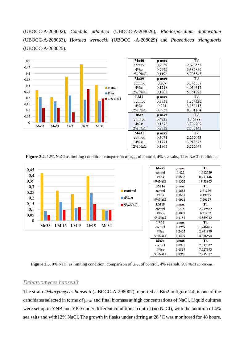
(UBOCC-A-208002), Candida atlantica (UBOCC-A-208026), Rhodosporidium diobovatum
(UBOCC-A-208033), Hortaea werneckii (UBOCC -A-208029) and Phaeotheca triangularis
(UBOCC-A-208025).
Figure 2.4. 12% NaCl as limiting condition: comparison of μmax of control, 4% sea salts, 12% NaCl conditions.
Figure 2.5. 9% NaCl as limiting condition: comparison of μmax of control, 4% sea salt, 9% NaCl conditions.
Debaryomyces hansenii
The strain Debaryomyces hansenii (UBOCC-A-208002), reported as Bio2 in figure 2.4, is one of the
candidates selected in terms of μmax and final biomass at high concentrations of NaCl. Liquid cultures
were set up in YNB and YPD under different conditions: control (no NaCl), with the addition of 4%
sea salts and with12% NaCl. The growth in flasks under stirring at 28 °C was monitored for 48 hours.

Figure 2.6. Growth curves in YNB and YPD in three different conditions: no NaCl (control); with the addition of
4% sea salts (4% ss) and with 12% NaCl. On the x axis there is the time (h) while on y axis the optical density at
600nm expressed in semi-logarithmic scale is reported.
Observing the curves in figure 2.6, there is a manifest difference between the two media in terms of
growth rate (μmax), saline stress response and final biomass. The choice of employing two media, one
minimal and the other one rich in nutrient elements was aimed at understanding the optimal condition
for salt stress response. A greater difference was observed in limit condition, which in the case of
Debaryomyces hansenii (UBOCC-A-208002) is represented by the culture medium (YNB / YPD)
with the addition of 12% NaCl. The strain shows a faster adaptation response in rich medium resulting
in a shorter latency phase in comparison with the same condition in YNB. On the contrary, for the
control condition and with 4% of sea salt, for both YNB and YPD, the lag phase is absent. Moreover,
the difference between μmax of the control and μmax of 4% sea salts is minimal and the growth curves
are almost overlapped. This would suggest that both the control condition and 4% sea salts condition
do not create any adjustments due to stress adaptation for the strain tested.
MATERIALS AND METHODS
MATERIALS All reagents and solvents were obtained from Sigma–Aldrich-Fluka and used without further
purification or drying. TLC was performed with Merck silica gel 60 F254 pre-coated plates. Silica
gel column chromatography was performed on silica gel 60 (40–63 mm particle size). Substrate 1h
was kindly gifted by Dr. Roberto Lenna (Industriale Chimica, Italy); substrate 1i (2-acetylamino-6-
oxo-4,5,6,7- tetrahydrobenzothiazole) was prepared following a procedure described previously27.
For each compound, the alcohol racemic mixture was obtained by NaBH4 reduction (0.25 mm of
substrate). Natural seawater (pH 7.5) was collected from the Camogli beach (Genova, Italy) and
maintained at 4 °C after microfiltration; a water salinity of 35 PSU was reported by ARPA (Agenzia
Regionale Prevenzione e Ambiente) for this area.

CHARACTERISATION
1H and 13C NMR spectra were recorded by using a Nuclear magnetic resonance (NMR) spectra at
300 K on a Bruker-Avance 500 MHz spectrometer operating at 500.13 and 125.76 MHz for 1H and
13C acquisitions, respectively. Chemical shifts (δ) of the 1H NMR and 13C NMR spectra are reported
in ppm using the signal for residual solvent proton resonance as the internal standard (1H NMR:
CDCl3 7.26, DMSO-d6 2.49, CD3OD 3.31 ppm; 13C NMR: CDCl3 77.0 (central line), DMSO-d6 39.50
(central line), CD3OD 49.00 (central line) ppm). HPLC analyses were performed by using a Jasco
Pu-980 equipped with a UV/Vis detector Jasco UV-975. Chiral HPLC columns used: Chiralcel OD
(4.6x250 mm, 5 mm Daicel), Chiralcel OJ-H (4.6x250 mm, 5 mm, Daicel), Chiralcel OD-H (4.6x250
mm, 5 mm, Daicel), Chiralpak IA (4.6x250 mm, 5 mm, Daicel), Lux cellulose-2 column (4.6x250
mm, 5 mm, Phenomenex) and Lux cellulose 3 column (4.6x250 mm, 5 mm, Phenomenex). Optical
rotatory power determinations were performed by a Perkin–Elmer (mod. 241) polarimeter in a 1 dm
cell at 20 °C, setting the wavelength at 589 nm or at 546 nm. Elemental analyses were performed by
using a Carlo Erba Model 1106 Elemental Analyzer for C, H and N, and the obtained results were
within 0.4% of the theoretical values.
MICROORGANISMS
All isolated microorganisms are available in the UBO Culture Collection (http://www.univ-
brest.fr/ubocc).
ID strain Phylum UBOCC number Depth
LM 1 Meyerozyma guillermondi Ascomycota UBOCC-A-214022 37,1 mbsf
LM 2 Meyerozyma guillermondi Ascomycota UBOCC-A-214008 137,13 mbsf
LM 3 Meyerozyma guillermondi Ascomycota UBOCC-A-214007 24,6 mbsf
LM 4 Rhodotorula mucilaginosa Basidiomycota UBOCC-A-214045 137,13 mbsf
LM 5 Meyerozyma guillermondi Ascomycota UBOCC-A-214013 24,6 mbsf
LM 6 Meyerozyma guillermondi Ascomycota UBOCC-A-214014 24,6 mbsf
LM 7 not sequenced UBOCC-A-214004 3,76 mbsf
LM 8 Rhodotorula mucilaginosa Basidiomycota UBOCC-A-214143 24,6 mbsf
LM 9 Meyerozyma guillermondi Ascomycota UBOCC-A-214015 21,1 mbsf
LM 10 Rhodotorula mucilaginosa Basidiomycota UBOCC-A-214039 24,6 mbsf
LM 13 Rhodotorula mucilaginosa Basidiomycota UBOCC-A-214049 21,1 mbsf
LM 14 Rhodotorula mucilaginosa Basidiomycota UBOCC-A-214040 34,1 mbsf
LM 15 Rhodotorula mucilaginosa Basidiomycota UBOCC-A-214034 37,1 mbsf
LM 16 Rhodotorula mucilaginosa Basidiomycota UBOCC-A-214025 3,76 mbsf
LM 17 Rhodotorula mucilaginosa Basidiomycota UBOCC-A-214046 345,5 mbsf
LM 18 Rhodotorula mucilaginosa Basidiomycota UBOCC-A-214036 765 mbsf

LM 19 Rhodotorula mucilaginosa Basidiomycota UBOCC-A-214051 37,1 mbsf
LM 20 Rhodotorula mucilaginosa Basidiomycota UBOCC-A-214043 34,1 mbsf
LM 21 Rhodotorula mucilaginosa Basidiomycota UBOCC-A-214006 3,76 mbsf
LM 22 Rhodotorula mucilaginosa Basidiomycota UBOCC-A-214026 34,1 mbsf
Bio1 Candida viswanathii Ascomycota UBOCC-A-208001 2620 mbsl
Bio2 Debaryomyces hansenii Ascomycota UBOCC-A-208002 2620 mbsl
Ex15 Pichia guilliermondii Ascomycota UBOCC-A-208004 700 mbsl
Ex7 Rhodotorula mucilaginosa Basidiomycota UBOCC-A-208010 2300 mbsl
Mo22 Sporobolomyces roseus Basidiomycota UBOCC-A-208018 2300 mbsl
Mo29 Cryptococcus sp. Basidiomycota UBOCC-A-208024 2300 mbsl
Mo30 Phaeotheca triangularis Ascomycota UBOCC-A-208025 2300 mbsl
Mo31 Candida atlantica Ascomycota UBOCC-A-208026 2300 mbsl
Mo34 Hortaea werneckii Ascomycota UBOCC-A-208029 2300 mbsl
Mo35 Rhodotorula mucilaginosa Basidiomycota UBOCC-A-208030 2300 mbsl
Mo36 Leucosporidium scottii Basidiomycota UBOCC-A-208031 2300 mbsl
Mo38 Rhodosporidium diobovatum Basidiomycota UBOCC-A-208033 2300 mbsl
Mo39 Candida marinus Ascomycota UBOCC-A-208034 2300 mbsl
Mo40 Debaryomyces hansenii Ascomycota UBOCC-A-208035 2300 mbsl
Table 2.7. Marine yeasts employed and and sampling depths: mbsf = meter below seafloor; mbsl = meter below
sea level.
The first group presents 8 strains of Meyerozyma guilliermondii and 12 strains of Rhodotorula
mucilaginosa isolated previously2 from different deep-sub-seafloor sediment depths (mbsf= meter
below the seafloor) at Canterbury Basin (New Zealand 44°56 26.62"S, 172°1 36.30"E).
The second group, on the other hand, derives from a second collection of marine microorganisms of
different origins: South Pacific West (Lau Basin), Mid-Atlantic Ridge (Rainbow), Mid-Atlantic
Ridge (Lost City). These strains were isolated from marine fauna present in "hydrothermal vents"
(table 2.8).
Table 2.8. Collection of marine yeasts associated with fauna in the hydrothermal vents1.

GROWING MEDIUM
YNB (YEAST NITROGEN BASE) MEDIUM
It is a defined mineral medium containing specific concentrations of mineral salts, vitamins and other
elements (traces). As nitrogen source, ammonium sulphate can be added.
YPD MEDIUM
It is a rich medium in term of carbon and
nitrogen sources.
HALOTOLERANCE SCREENING
SCREENING ON PLATE
Plate screening was set up for 18 yeast strains and allowed a first analysis of strain growth and strain
patterns at different NaCl concentrations. For this preliminary analysis YNB plates were prepared at
all different concentrations of NaCl and growth was evaluated up to 120 hours.
From glycerol stocks, the yeasts were re-activated on YPD Solid Broth and grown to 28 °C for 48
hours. They were then pre-inoculated in buffered YNB in Erlenmeyer flasks, allowed to grow at 28
°C, 150 rpm for 48 hours. Growth was evaluated by reading optical density (O.D.) at 600 nm. Each
strain was deposited on plate in four dilutions: 105, 104, 103, 102 cell number (figure 2.2). The protocol
was repeated for all the four conditions of increasing salinity: 0% NaCl, 3% NaCl, 6% NaCl, 9%
NaCl, 12% NaCl.
COMPOSITION CONCENTRATION
YNB (BD-Difco) 1.7 g/L
MES buffer 1 M
Glucose 20 g/L
(NH4)2SO4 5 g/L
COMPOSITION CONCENTRATION
Yeast extract 10 g/L
Tryptone 20 g/L
Glucose 20 g/L
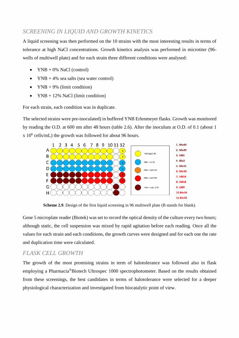
SCREENING IN LIQUID AND GROWTH KINETICS
A liquid screening was then performed on the 10 strains with the most interesting results in terms of
tolerance at high NaCl concentrations. Growth kinetics analysis was performed in microtiter (96-
wells of multiwell plate) and for each strain three different conditions were analysed:
YNB + 0% NaCl (control)
YNB + 4% sea salts (sea water control)
YNB + 9% (limit condition)
YNB + 12% NaCl (limit condition)
For each strain, each condition was in duplicate.
The selected strains were pre-inoculated) in buffered YNB Erlenmeyer flasks. Growth was monitored
by reading the O.D. at 600 nm after 48 hours (table 2.6). After the inoculum at O.D. of 0.1 (about 1
x 106 cells/mL) the growth was followed for about 96 hours.
Scheme 2.9. Design of the first liquid screening in 96 multiwell plate (B stands for blank).
Gene 5 microplate reader (Biotek) was set to record the optical density of the culture every two hours;
although static, the cell suspension was mixed by rapid agitation before each reading. Once all the
values for each strain and each conditions, the growth curves were designed and for each one the rate
and duplication time were calculated.
FLASK CELL GROWTH
The growth of the most promising strains in term of halotolerance was followed also in flask
employing a Pharmacia®Biotech Ultrospec 1000 spectrophotometer. Based on the results obtained
from these screenings, the best candidates in terms of halotolerance were selected for a deeper
physiological characterization and investigated from biocatalytic point of view.

BIOTRANSFORMATIONS
The screening of growth was performed in Erlenmeyer flasks (100 mL) that contained 10 mL of YPD
liquid prepared with freshwater or natural seawater30 and incubated in an orbital shaker at 180 rpm at
28 °C for 48 h. Samples were withdrawn at appropriate intervals and used to monitor the cell growth
by measuring the O.D. at 600 nm by using a spectrophotometer after appropriate dilution. For dry
weight determination, washed culture samples were filtered through a 0.45 μm glass microfiber GF/A
filter (Whatman) and dried for 24 h at 110 °C. The results were an average of five replicates.
Reductions of all substrates were performed with freshly prepared cells, grown in freshwater-YPD or
in YPD-seawater. Cells grown in freshwater-YPD medium (50 mg dry weight/mL) were suspended in
phosphate buffer (pH 7.5, 0.1m), whereas cells grown in seawater-YPD medium (50 mg dry weight/mL)
were suspended in natural microfiltered seawater. The biotransformations of 1a–g were performed in
the presence of 5% glucose by adding the substrates as concentrated solution in DMSO (final
concentration of co-solvent 1%). The biotransformations of ethyl secodione derivative 1h was
performed using freshly cells suspended in seawater in the presence of 3.5% EtOH both to increase
the solubility and to favour cofactor regeneration. The biotransformations of 1i were performed using
fresh cells suspended in seawater in the presence of different co-solvents.
The biotransformations were performed on a 25 mL scale and kept under reciprocal shaking (150
rpm) at the desired temperature. Once the reaction was over, EtOAc (12 mL) was added, and the
resulting mixture was shaken and centrifuged; the aqueous phase was extracted twice more with 12
mL of EtOAc. The organic phases were collected and dried over Na2SO4 and the solvent was
evaporated. The crude residues were purified by flash chromatography.
ARYL METHYLKETONI
Cells were grown in freshwater-YPD or in seawater-YPD medium. Biotransformations were
performed for 24 h with freshly prepared cells (50 mg dry weight mL−1). Cells grown in freshwater-YPD
medium were suspended in phosphate buffer (pH 7.5, 0.1 M), whereas cells grown in seawater-YPD
medium were suspended in seawater. Biotransformations were performed in the presence of 5 %
glucose.
β – KETONITRILES
Biotransformations were performed for 24 h with freshly prepared cells (50 mg dry weight mL−1)
suspended in seawater in the presence of 5 % glucose. Yields and enantioselectivity after 24 h.

ETHYL SECODIONE DERIVATIVE
Biotransformations were performed for 24 h with freshly prepared cells (50 mg dry weight mL−1)
suspended in seawater in the presence of 3.5 % EtOH. Yields and diastereoselectivity after 24 h.
PRAMIPEXOLE KETO INTERMEDIATE
Biotransformations were performed for 24 h with freshly prepared cells (50 mg dry weight mL−1)
suspended in seawater (pH 5.6) in the presence of 5 % glucose. Yields and enantioselectivity were
determined after 24 h. In this case, seawater was used at pH 5.6 because of the low stability of the
substrate at a higher pH.
PURIFICATION AND CHEMICAL CHARACTERISATION
(S)-1-Phenylethanol (2 a)
The reaction progress and ee were determined by using HPLC using a Chiralcel OD column (n-
hexane/iPrOH 95:5, 0.7 mL min−1, 254 nm): tr (R)-2 a 14.2 min, tr (S)-2 a 18.0 min.31 Oil. Rf=0.4
(hexane/EtOAC=8:2); [α] =−40.0 (c=0.5 chloroform) lit32. [α] =−39.2 (c=2.54 chloroform); 1H
NMR (300 MHz, CDCl3): δ=7.20–7.35 (m, 5 H), 4.85 (q, J=6.4 Hz, 1 H), 1.46 ppm (d, J=6.4 Hz,
3 H); 13C NMR (75 MHz, CDCl3): δ=145.9, 128.5, 127.5, 125.4, 70.4, 25.2 ppm; elemental analysis
calcd (%) for C8H10O (122.07): C 78.65, H 8.25; found: C 78.38, H 8.01.
(S)-1-Pyridin-3yl-ethanol (2 b)
The reaction progress and ee were determined by using HPLC using a Phenomenex Lux cellulose 2
column (n-hexane/iPrOH 90:10, 0.7 mL min−1, 254 nm): tr (R)-2 b 20.46 min, tr (S)-2 b 25.30 min.33
Oil. Rf=0.3 (EtOAc); [α] =−38.04 (c=0.4 ethanol) lit34. =−39.0 (c=0.93 ethanol); 1H NMR (300
MHz, CDCl3): δ=8.46–8.33 (m, 2 H), 7.75–7.70 (m, 1 H), 7.24 (dd, J=8.0, 5.0 Hz, 1 H), 4.90 (q,
J=6.5 Hz, 1 H), 4.5 (br s, 1 H, OH), 1.49 ppm (d, J=6.5 Hz, 3 H); 13C NMR (75 MHz, CDCl3):
δ=148.0, 147.0, 141.8, 133.5, 123.5, 67.5 25.1 ppm; elemental analysis calcd (%) for C7H9ON
(123.07): C 68.27, H 7.37, N 11.37; found: C 68.55, H 7.67, N 11.10.
(S)-(1-Thiophen-2-yl)ethanol (2 c)
The reaction progress and ee were determined by using HPLC using a Phenomenex Lux cellulose–3
column (n-hexane/iPrOH 90:10, 0.5 mL min−1, 254 nm): tr (S)-2 c 13.5 min, tr (R)-2 c 15.90 min35.
Oil. Rf=0.3 (hexane/EtOAc=8:2); [α] =−20.80 (c=0.2 chloroform) lit35. [α] =−23.2 (c=0.79
chloroform); 1H NMR (300 MHz, CDCl3): δ=7.25–7.18 (m, 1 H), 6.95–6.92 (m, 2 H), 5.07 (q,
J=6.5 Hz, 1 H), 2.54 (br s, 1 H, OH), 1.56 ppm (d, J=6.5 Hz, 3 H); 13C NMR (75 MHz, CDCl3):
δ=150.0, 126.6, 124.4, 123.2, 66.2, 25.2 ppm; elemental analysis calcd (%) for C6H8OS (128.03): C
56.22, H 6.29; found: C 56.04, H 6.58.

(S)-(1-Furan-2-yl)ethanol (2 d)
The reaction progress and ee were determined by using HPLC using a Phenomenex Lux cellulose–3
column (n-hexane/iPrOH 98:2, 0.5 mL min−1, 220 nm): tr (S)-2 d 19.3 min, tr (R)-2 d 21.3 min.35
Oil. Rf=0.3 (hexane/EtOAc=8:2); [α] =−20.5 (c=0.2 chloroform) lit34. [α] =−13.6 (c=0.55
chloroform); 1H NMR (400 MHz, CDCl3): δ=7.36 (dd, J=2.0, 1.0 Hz, 1 H), 6.32 (dd, J=3.0, 2.0 Hz,
1 H), 6.22 (d, J=3.0 Hz, 1 H), 4.86 (q, J=6.5 Hz, 1 H), 2.35 (br s, 1 H, OH), 1.52 ppm (d, J=6.5 Hz,
3 H); 13C NMR (100 MHz, CDCl3): δ=157.7, 141.8, 110.1, 105.1, 63.6, 21.2 ppm26; elemental
analysis calcd (%) for C6H8O2 (112.05): C 64.27, H 7.19; found: C 63.97, H 7.34.
(S)-3-Hydroxy-3-phenylpropanenitrile (2 e)
The reaction progress and ee were determined by using HPLC using a Chiralcel OJ-H column (n-
hexane/iPrOH 90:10, 1.0 mL min−1, 220 nm), tr (S)-2 e 24.5, tr (R)-2 e 31.1 min.23Oil. Rf=0.4
(hexane/EtOAc=7:3); [α] =−58.5 (c=1.0, EtOH) lit.24 [α] =−57.7 (c=2.6, EtOH)]; 1H NMR (300
MHz,CDCl3): δ=2.74 (d, J=6.1 Hz, 2 H), 5.01 (t, J=6.1 Hz, 1 H), 7.36–7.39 ppm (m, 5 H); 13C
NMR (75 MHz,CDCl3): δ=27.9, 70.0, 117.4, 125.5, 128.8, 128.9, 141.1 ppm; elemental analysis
calcd (%) for C9H9ON (147.07): C 73.45, H 6.16, N 9.52; found: C 73.01, H 6.31, 9.32.
(S)-3-(2-Thienyl)-3-hydroxypropanenitrile (2 f)
The reaction progress and ee were determined by using HPLC using a Chiralcel OJ-H column (n-
hexane/iPrOH=90:10, 1.0 mL min−1, 220 nm), tr (S)-2 f 27.0, tr (R)-2 f 32.3 min.23 Oil. Rf=0.3
(hexane/EtOAc=7:3); [α] =−19.4 (c=1.0, CHCl3) lit.34 [α] =−16.71 (c=1.01, CHCl3);
1H NMR
(300 MHz,CDCl3): δ=2.85 (d, J=6.2 Hz, 2H), 3.04 (s, br, OH), 5.26 (t, J=6.2 Hz, 1 H), 6.98–7.00
(m, 2 H), 7.06–7.07 (d, J=3.5 Hz, 1 H), 7.29–7.32 ppm (m, 1 H); 13C NMR (75 MHz, CDCl3):
δ=28.2, 66.2, 117.1, 124.7, 125.7, 127.1, 144.5 ppm; elemental analysis calcd (%) for C7H7ONS
(153.02): C 54.88, H 4.61, N 9.14; found: C 54.81, H 4.81, 8.92.
(S)-3-(2-Furyl)-3-hydroxypropanenitrile (2 g)
The ee was determined by using HPLC using a Chiralcel OJ-H column (n-hexane/iPrOH=90:10, 1.0
mL min−1, 220 nm), tr (S)-2 g 21.9, tr (R)-2 g 25.6 min.23 Oil. Rf=0.3 (hexane/EtOAc=7:3); [α]
=−40.2 (c=1.0, EtOH) lit.24 [α] =−38.6 (c=1.3, CHCl3); 1H NMR (300 MHz,CDCl3): δ=2.57 (s, br,
OH), 2.88 (d, J=6.2 Hz, 2 H), 5.05 (t, J=6.2 Hz, 1 H), 6.37–6.41 (m, 2 H), 7.41–7.42 ppm (m, 1 H); 13C NMR (75 MHz,CDCl3): δ=24.9, 63.8, 107.5, 110.6, 116.9, 142.9, 152.8 ppm; elemental
analysis calcd (%) for C7H7O2N (137.05): C 61.31, H 5.14, N 10.21; found: C 61.06, H 5.36, 9.94.
1 -Benzoylbutanenitrile (3 e)
Oil. Rf=0.4 (hexane/EtOAc=7:3). 1H NMR (300 MHz, CDCl3): δ=1.16 (t, J=7.7 Hz, 3 H), 2.02–2.15
(m, 2 H), 4.30 (dd, J=6.2, 4.3 Hz, 1), 7.49–7.56 (m, 2 H), 7.65 (d, J=7.6 Hz, 1 H), 7.95 ppm (d,
J=6.7 Hz, 2 H); 13C NMR (75 MHz, CDCl3): δ=11.89, 23.77, 41.38, 128.68, 128.92, 129.31,
130.38, 133.93, 134.63, 170.91, 190.88 ppm; elemental analysis calcd (%) forC11H11ON (173.08):
C 76.28, H 6.40, N 8.09; found: C 75.92, H 6.50, 7.97.

(13S,17S)-E-13-Ethyl-3-methoxy-8,14-secogona-1,3,5(10),9(11)-tetraene-17-
ol-14-one (2 h)
The reaction progress, ee and diastereomeric excess (de) were determined by HPLC using a Lux
Cellulose-2 column (n-hexane/iPrOH 85:15; 0.5 mL min−1 254 nm); tr (13S, 17S)-2 h 18.87 min,
(13S, 17R)-2 h 20.62 min, (13R, 17S)-2 h 23.34 min, (13R, 17S)-2 h, 24.61 min.27White solid.
Rf=0.3 (hexane/EtOAc=1:1). ee=84 %. 1H NMR (300 MHz,C3D6O): δ=0.82 (t, J=7.5 Hz, 3 H), 1.53
(non, J=7.5 Hz, 2 H), 1.78 (qui, J=6.2 Hz, 2 H), 1.95–2.07 (m, 1 H), 2.23–2.31 (m, 3 H;), 2.43–2.46
(m, 2 H; CH2), 2.51 (t, J=6.2 Hz, 2 H), 2.74 (t, J=6.2 Hz, 2 H), 3.77 (s, 3 H), 4.13 (d, J=4.2 Hz, 1 H;
OH), 4.32 (q, J=5.3 Hz, 1 H), 6.0 (t, J=7.5 Hz, 1 H), 6.64 (d, J=2.8 Hz, 1 H), 6.72 (dd, J=2.8, 8.7
Hz, 1 H), 7.50 ppm (d, J=8.7 Hz, 1 H); 13C NMR (300 MHz,C3D6O): δ=8.2, 23.6, 25.3, 26.6, 26.8,
28.4, 30.9, 34.8, 54.9, 57.2, 75.1, 112.8, 113.2, 118.1, 125.3, 129.6, 135.4, 138.7, 159.0, 219.1 ppm;
elemental analysis calcd (%) for C20H26O3 (314.19): C 76.40, H 8.33; found: C 76.11, H 8.39.
(S)-2-Acetylamino-6-hydroxy-4,5,6,7-tetrahydrobenzothiazole (2 i)
The reaction progress and ee were determined by using HPLC using a Chiralpak IA column (n-
hexane/iPrOH 8:2 as eluent, flow rate: 0.7 mL min−1, 254 nm), tr (R)-2 i 10.9 min, tr (S)-2 i 14.8
min27. Yellowish solid. Rf=0.25 (CH2Cl2/MeOH, 95:5). ee=64 %. 1H NMR (300 MHz,CD3OD):
δ=1.91 (dddd, J=14.2, 8.6, 7.7, 5.9 Hz, 1 H), 2.04 (dddd, J=14.2, 6.2, 5.9, 2.9 Hz, 1 H), 2.26 (s,
CH3, 3 H), 2.61–2.71 (m, 2 h), 2.79 (dddd, J=16.5, 5.9, 5.9, 1.8 Hz, 1 H), 2.99 (ddd, J=15.7, 4.7, 1.8
Hz, 1 H), 4.15 ppm (dddd, J=8.6, 6.9, 4.7, 2.9 Hz, 1 H); 13C NMR (300 MHz, CD3OD): δ=21.2,
23.2, 30.5, 30.7, 66.2, 120.9, 143.2, 156.4, 169.1 ppm; elemental analysis calcd (%) for
C9H12O2N2S (212.06): C 50.92, H 5.70, N 13.20; found: C 50.96, H 5.89 N 12.85.
REFERENCES
1. Burgaud, G., Arzur, D., Durand, L., Cambon-Bonavita, M. A. & Barbier, G. Marine culturable yeasts in
deep-sea hydrothermal vents: Species richness and association with fauna. FEMS Microbiol. Ecol. 73,
121–133 (2010).
2. Rédou, V., Navarri, M., Meslet-Cladiére, L., Barbier, G. & Burgaud, G. Species richness and adaptation of
marine fungi from deep-subseafloor sediments. Appl. Environ. Microbiol. 81, 3571–3583 (2015).
3. Trincone, A. Marine enzymes for biocatalysis.
4. Munn, C. B. Marine Microbiology – Ecology and Applications.. (Bios-Garland Scientific, 2003).
5. Tivey, M. K. Generation of Seafloor Hydrothermal Vent Fluids and Associated Mineral Deposits.
Oceanography 20, 50–65 (2007).
6. Jørgensen, B. B. & Boetius, A. Feast and famine — microbial life in the deep-sea bed. Nat. Rev. Microbiol. 5, 770–781 (2007).
7. Nordling, E., Jörnvall, H. & Persson, B. Medium-chain dehydrogenases/reductases (MDR): Family
characterizations including genome comparisons and active site modelling. Eur. J. Biochem. 269, 4267–
4276 (2002).
8. Stuermer, R., Hauer, B., Hall, M. & Faber, K. Asymmetric bioreduction of activated C=C bonds using
enoate reductases from the old yellow enzyme family. Curr. Opin. Chem. Biol. 11, 203–213 (2007).
9. Hoffmann, F. & Maser, E. Carbonyl reductases and pluripotent hydroxysteroid dehydrogenases of the
short-chain dehydrogenase/reductase superfamily. Drug Metab. Rev. 39, 87–144 (2007).
10. Kallberg, Y., Oppermann, U. & Persson, B. Classification of the short-chain dehydrogenase/reductase
superfamily using hidden Markov models. FEBS J. 277, 2375–2386 (2010).
11. Forrest, G. L. & Gonzalez, B. Carbonyl reductase. Chem. Biol. Interact. 129, 21–40 (2000).
12. Zhu, D. & Hua, L. How carbonyl reductases control stereoselectivity: Approaching the goal of rational
design. Pure Appl. Chem. 82, 117–128 (2010).
13. Liu, Z.-J. et al. The first structure of an aldehyde dehydrogenase reveals novel interactions between NAD
and the Rossmann fold. Nat. Struct. Biol. 4, 317–326 (1997).
14. V. Prelog. Specification of the stereospecificity of some oxido-reductases by diamond lattice sections.
Pure Appl. Chem. 9, 119–130 (1964).
15. Musa, M. M. & Phillips, R. S. Recent advances in alcohol dehydrogenase-catalyzed asymmetric
production of hydrophobic alcohols. Catal. Sci. Technol. 1, 1311 (2011).
16. Kurbanoglu, E. B., Zilbeyaz, K., Ozdal, M., Taskin, M. & Kurbanoglu, N. I. Asymmetric reduction of
substituted acetophenones using once immobilized Rhodotorula glutinis cells. Bioresour. Technol. 101,
3825–3829 (2010).
17. Papon, N. et al. Candida guilliermondii: Biotechnological applications, perspectives for biological control,
emerging clinical importance and recent advances in genetics. Curr. Genet. 59, 73–90 (2013).
18. Zhang, B., Zheng, L., Lin, J. & Wei, D. Characterization of an ene-reductase from Meyerozyma
guilliermondii for asymmetric bioreduction of a,b-unsaturated compounds. Biotechnol. Lett. 38, 1527–
1534 (2016).
19. Zhu, D. et al. Asymmetric reduction of β-ketonitriles with a recombinant carbonyl reductase and
enzymatic transformation to optically pure β-hydroxy carboxylic acids. Org. Lett. 9, 2560–2563 (2007).
20. Ankati, H., Zhu, D., Yang, Y., Biehl, E. R. & Hua, L. Asymmetric synthesis of both antipodes of beta-
hydroxy nitriles and beta-hydroxy carboxylic acids via enzymatic reduction or sequential
reduction/hydrolysis. J. Org. Chem. 74, 1658–1662 (2009).
21. Nowill, R. W. et al. Biocatalytic strategy toward asymmetric ??-hydroxy nitriles and ??-amino alcohols.
Tetrahedron Lett. 52, 2440–2442 (2011).
22. Xu, G. C., Yu, H. L., Zhang, Z. J. & Xu, J. H. Stereocomplementary bioreduction of β-ketonitrile without
ethylated byproduct. Org. Lett. 15, 5408–5411 (2013).
23. Contente, M. L. et al. Preparation of enantiomerically enriched aromatic β-hydroxynitriles and halohydrins
by ketone reduction with recombinant ketoreductase KRED1-Pglu. Tetrahedron 72, 3974–3979 (2016).
24. Dehli, J. R. & Gotor, V. Enantio- and chemoselective bioreduction of β-keto nitriles by the fungus
Curvularia lunata. Tetrahedron Asymmetry 11, 3693–3700 (2000).
25. Aguirre-Pranzoni, C., Bisogno, F. R., Orden, A. A. & Kurina-Sanz, M. Lyophilized Rhodotorula yeast as
all-in-one redox biocatalyst: Access to enantiopure building blocks by simple chemoenzymatic one-pot
procedures Dedicated to Professor Carlos E. Tonn for his great contribution to the development of the
chemistry of natura. J. Mol. Catal. B Enzym. 114, 19–24 (2015).
26. Gotor, V., Dehli, J. R. & Rebolledo, F. Biotransformations of benzoylacetonitrile with the fungus. R. Soc.
Chem. 8, 307–309 (2000).
27. Ferraboschi, P. et al. Baker’s yeast catalyzed preparation of a new enantiomerically pure synthon of (S)-
pramipexole and its enantiomer (dexpramipexole). Tetrahedron Asymmetry 25, 1239–1245 (2014).
28. Romano, D., Gandolfi, R., Guglielmetti, S. & Molinari, F. Enzymatic hydrolysis of capsaicins for the
production of vanillylamine using ECB deacylase from Actinoplanes utahensis. Food Chem. 124, 1096–
1098 (2011).
29. Burgaud, G., Arzur, D., Durand, L., Cambon-Bonavita, M. A. & Barbier, G. Marine culturable yeasts in
deep-sea hydrothermal vents: Species richness and association with fauna. FEMS Microbiol. Ecol. 73,
121–133 (2010).
30. Chi, Z., Ma, C., Wang, P. & Li, H. F. Optimization of medium and cultivation conditions for alkaline
protease production by the marine yeast Aureobasidium pullulans. Bioresour. Technol. 98, 534–538

(2007).
31. Contente, M. L. et al. Biotransformation of aromatic ketones and ketoesters with the non-conventional
yeast Pichia glucozyma. Tetrahedron Lett. 55, 7051–7053 (2014).
32. Kantam, M. L. et al. Asymmetric Hydrosilylation of Ketones Catalyzed by magnetically recoverable and
reusable copper ferrite nanoparticles. J. Org. Chem 4608–4611 (2009).
33. Contente, M. L. et al. Stereoselective reduction of aromatic ketones by a new ketoreductase from Pichia
glucozyma. Appl. Microbiol. Biotechnol. 100, 193–201 (2016).
34. Vázquez-Villa, H., Reber, S., Ariger, M. A. & Carreira, E. M. Iridium diamine catalyst for the asymmetric
transfer hydrogenation of ketones. Angew. Chemie - Int. Ed. 50, 8979–8981 (2011).
35. Li, Y. et al. Iron catalyzed asymmetric hydrogenation of ketones. J. Am. Chem. Soc. 136, 4031–4039
(2014).
WHOLE CELL SCREENING – MARINE BACTERIA

In this section the focus will be on biocatalytic potential of varied range of bacterial strains collected
from one of the most peculiar marine habitats. Keto-reductase and transaminase enzymatic activities
were investigated with whole cells in order to identify a possible gene target to express as recombinant
enzyme with enhanced performances.
BACKGROUND
MARINE BACTERIA
In the context of this doctoral thesis, the enzymatic activities of 33 marine strains from four different
deep hypersaline anoxic basins (DHABs) located in the eastern area of the Mediterranean Sea have
been studied. This project was involved in the European research project BIODEEP1
(Biotechnologies from the Deep), which aimed to explore unique marine habitats in order to isolate
and cultivate marine microorganisms, offering new opportunities for the study of new significant
enzymatic activities. The bacteria collected from DHABs have adapted to high salinity, anoxia and
high pressures that may have affected the expression of unique cellular features of interest in many
industrial applications.
The DHABs are located in an area of the Mediterranean called Mediterranean Ridge (figure 3.1); it
is the result of the convergence between the African, European and Aegean platforms. The
hypersaline basins are probably the result of the melting of underground deposits (3200-3600m of
depth) of salt (Messinians evaporites) that were exposed to seawater because of tectonic activities
during Miocene period (from 26 million to 2.5 million years ago). The brine in DHABs is
characterized by anoxia conditions, high pressure (≈35 MPa), saturated salts concentrations2–5, high
concentrations of MgCl2 (≈5 M) and no light that make them some of the most extreme habitats on
earth5,6.
Figure 3.1. Localisation of deep hypersaline anoxic basins in Mediterranean Sea. The L’Atalante, Urania and
Discovery are located in a and Bannock in b.
The brine high density limits the mixing with the overlying oxygen-rich waters, creating a chemocline
of 1-3 m of the thickness. It is clear from previous studies that every DHABs have distinct

geochemical characteristics from the other ones2,4,7. The L’Atalante, Bannock and Urania brine has a
similar ionic composition, but the salinity of Urania is lower in contrast to the concentration of
methane and sulphate which is, however, considerably higher than the other two. The main difference
with the Discovery basin lies in its high concentration of Mg2+ions (about 5M) and a lower
concentration of Na+ions.
Table 3.1. Element composition of deep hypersaline anoxic basins and sea water6.
Physical separation due to different water density and thousands of year of evolutionary pathway
have probably made possible the development of specific and diverse microbial communities in the
four different basins. In the past, these high cation concentrations convinced the researchers that this
condition was incompatible with life8,9, but recently the presence of metabolically active microbial
communities has been demonstrated6. Through the combined study of 16S rRNA and enzymatic tests,
in all four basins, an active reduction of methane, sulfate and an important heterotrophic and
chemoautotrophic activities has been demonstrated. Instead, the waters above them show a different
structure of the microbial community10. Microbial communities prevailing in the Discovery basin
belong to the Bacteria kingdom and same happens for L’Atalante and Bannock basins, while in
Urania basin there is a prevalent prevalence of the Archaea microorganisms.
In all four hypersaline anoxic basins, a microbial population belonging to Bacteria kingdom is highly
diversified compared to the Archaea one; a biodiversity that was not found in the Urania basin. After
a more specific analysis of 16S rRNA gene sequences, it has also emerged that hypersaline anoxic
basins are populated by high percentages of microbial species belonging to γ-, δ-, and ε-
proteobacteria, sphingobacteria and halobacteria. This isolation may have resulted in the evolution of
specific microbial communities in each DHAB. In fact, through biodiversity analysis of the seawater
interface, several taxonomic categories have been identified, including new phylogenetic groups,
organized in colonies and heavily stratified in the “deep-water ecosystem”11–16.

MARINE BACTERIA BIOCATALYSIS
As fairly described by De Vitis et al.17 these peculiar microorganisms isolated from DHABs on the
Mediterranean Ridge have already showed interesting biocatalytic features applied in a kinetic
resolution of the propyl ester of anti-2-oxotricyclo[2.2.1.0]heptan-7-carboxylic acid, a key
intermediate for the prostaglandin D-cloprostenol18 (scheme 3.1).
Scheme 3.1. Kinetic resolution of anti-2-oxotricyclo[2.2.1.0]heptan-7-carboxylic acid by newly isolated marine
bacteria17.
The 33 strains were screened on racemic (R,S)-1 and some of them have showed enantioselective
keto-reductase and esterase activity. Virgibacillus pantotheticus 21D showed high halotolerance,
enantioselective keto-reductase activity in the presence of glucose, and esterase activity was prevalent
in the absence of glucose.
Table 3.2. Activity on racemic propyl ester 1 of whole cells of marine bacteria. a Biotransformation in the
presence of glucose (5%); b Biotransformation in the absence of glucose.

In both cases, enantiomerically pure unreacted substrate could be easily recovered and purified at
molar conversion below 57–58 %.
ω-TRANSAMINASE
As it was reported in the introduction of this PhD thesis, ω-transaminases (ω-TAs), also called
aminotransferases, are enzymes that transfers an amino group from an amino-donor into a carbonyl
moiety of an amino-acceptor, whereby at least one of the two substances is not an α-amino acid or an
a-keto acid. ω-TAs offer a unique opportunity for the asymmetric synthesis or kinetic resolution
(scheme 3.2) of bioactive compounds that possess a chiral amine moiety, starting from prochiral
ketones or low-cost racemic amines19.
Scheme 3.2. Kinetic resolution starting with racemic amines is
limited by 50% maximum yield. Theoretically, a 100% yield is
possible in asymmetric synthesis from prochiral ketones if the
equilibrium can be shifted appropriately19.
This enzymatic catalysis consists of two steps:
deamination of an amino acid or amine (amino-donor) releasing amino-donor product;
amination of a keto acid, ketone or aldehyde (amino-acceptor) producing a new amino acid
or amine.
All aminotransferases (EC 2.6.1.X) reported to date require the same coenzyme, namely pyridoxal-
5’-phosphate (PLP), which serves as a molecular shuttle for ammonia and electrons between the
amino donor and the amino acceptor (scheme 3.3). During the reaction, PLP is reversibly
interconverted to pyridoxamine (PMP). In the catalytic cycle, the amino donor binds first to the
enzyme, PLP is aminated to PMP, and the respective keto product of the amino donor is released.
The transamination is finalized by transferring the amino group from PMP to the acceptor molecule,
thereby closing the catalytic cycle through the liberation of PLP.
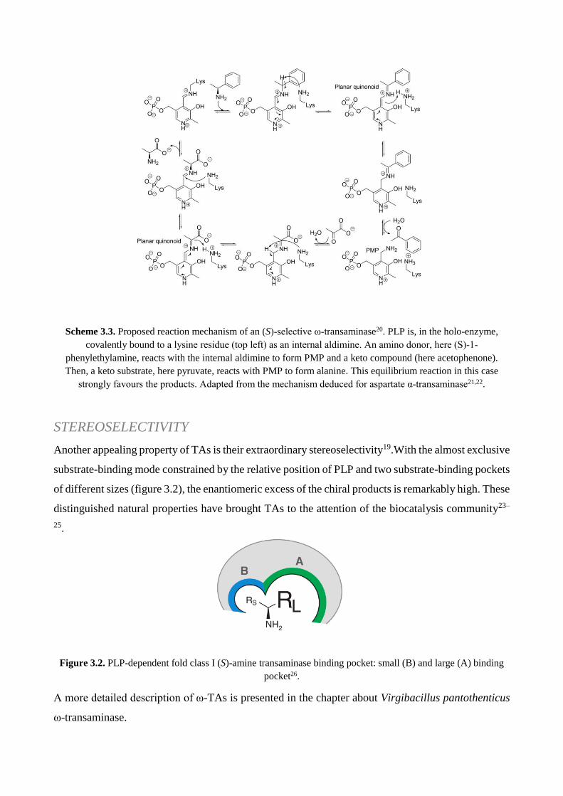
Scheme 3.3. Proposed reaction mechanism of an (S)‐selective ω‐transaminase20. PLP is, in the holo-enzyme,
covalently bound to a lysine residue (top left) as an internal aldimine. An amino donor, here (S)‐1‐
phenylethylamine, reacts with the internal aldimine to form PMP and a keto compound (here acetophenone).
Then, a keto substrate, here pyruvate, reacts with PMP to form alanine. This equilibrium reaction in this case
strongly favours the products. Adapted from the mechanism deduced for aspartate α‐transaminase21,22.
STEREOSELECTIVITY
Another appealing property of TAs is their extraordinary stereoselectivity19.With the almost exclusive
substrate-binding mode constrained by the relative position of PLP and two substrate-binding pockets
of different sizes (figure 3.2), the enantiomeric excess of the chiral products is remarkably high. These
distinguished natural properties have brought TAs to the attention of the biocatalysis community23–
25.
Figure 3.2. PLP-dependent fold class I (S)-amine transaminase binding pocket: small (B) and large (A) binding
pocket26.
A more detailed description of ω-TAs is presented in the chapter about Virgibacillus pantothenticus
ω-transaminase.

PROJECT AIM
Starting from the encouraging results achieved by marine bacteria keto-reductions, the idea was to
exploit this ability on pramipexole keto-intermediate, as done with marine yeasts previously.
On the other hand, the possibility to convert pramipexole ketone intermediate into the optically pure
amine could be realised by a ω-transaminase enzyme (ω-TA) in a one-pot bioreaction instead of a
multi-step synthetic pathway. Therefore, thirty-three marine bacteria species were screened to
identify a transaminase activity. According to transaminase biocatalytic applications, where cofactor
recycling is not needed, the aim in this case was to express and employ a recombinant enzyme.
RESULTS AND DISCUSSION
KETO-REDUCTASE
The most promising strains belonging to BIODEEP collection were selected and screened on keto-
intermediate of pramipexole. The results showed a no particular enantioselectivity, with a general
preference for (R)-enantiomer formation like Saccharomyces cerevisiae (table 3.3).
STRAIN C (%) e.e.(%) SPECIFICITY
11D 100 47 S
15A 100 74 R
3U 100 45 R
3B 46,57 47 R
5B tris 20 46 S
6B 35,97 75 R
7B 41,67 52 R
26A 99 25 S
9A 100 55 R
12D 100 35 R
21D 100 29 S
Table 3.3. Bioconversion data with marine yeasts.
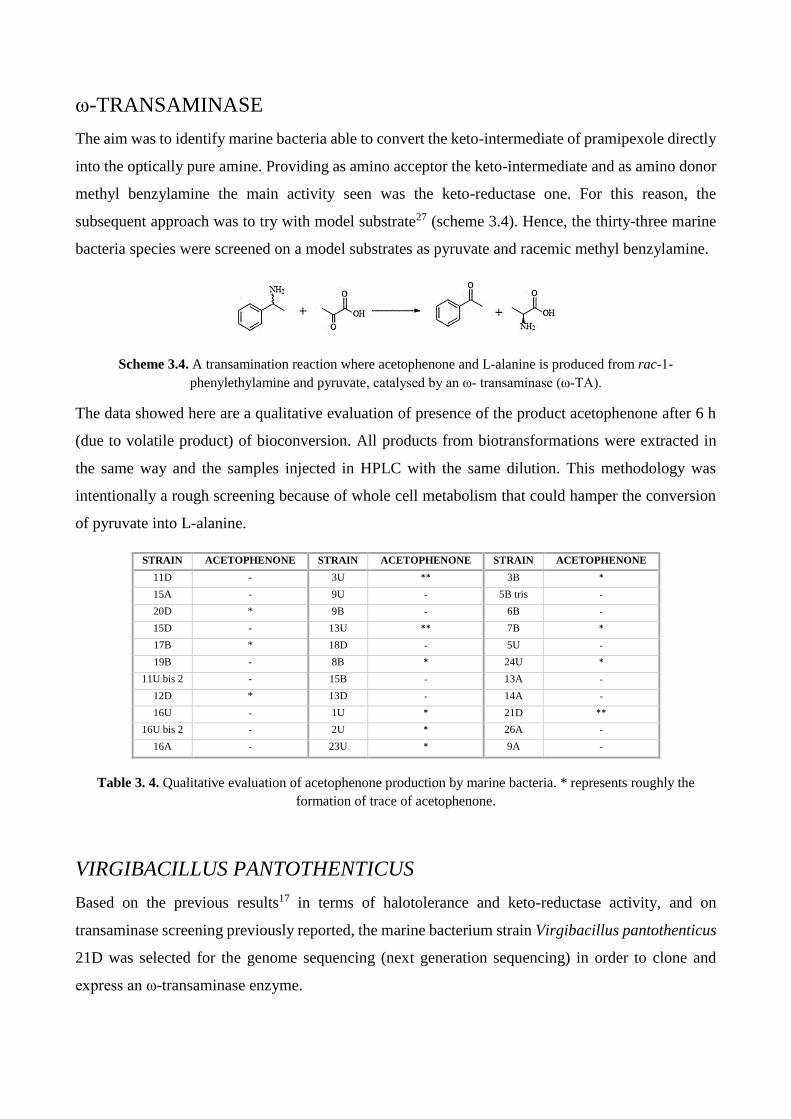
ω-TRANSAMINASE
The aim was to identify marine bacteria able to convert the keto-intermediate of pramipexole directly
into the optically pure amine. Providing as amino acceptor the keto-intermediate and as amino donor
methyl benzylamine the main activity seen was the keto-reductase one. For this reason, the
subsequent approach was to try with model substrate27 (scheme 3.4). Hence, the thirty-three marine
bacteria species were screened on a model substrates as pyruvate and racemic methyl benzylamine.
Scheme 3.4. A transamination reaction where acetophenone and L-alanine is produced from rac‐1-
phenylethylamine and pyruvate, catalysed by an ω‐ transaminase (ω‐TA).
The data showed here are a qualitative evaluation of presence of the product acetophenone after 6 h
(due to volatile product) of bioconversion. All products from biotransformations were extracted in
the same way and the samples injected in HPLC with the same dilution. This methodology was
intentionally a rough screening because of whole cell metabolism that could hamper the conversion
of pyruvate into L-alanine.
STRAIN ACETOPHENONE STRAIN ACETOPHENONE STRAIN ACETOPHENONE
11D - 3U ** 3B *
15A - 9U - 5B tris -
20D * 9B - 6B -
15D - 13U ** 7B *
17B * 18D - 5U -
19B - 8B * 24U *
11U bis 2 - 15B - 13A -
12D * 13D - 14A -
16U - 1U * 21D **
16U bis 2 - 2U * 26A -
16A - 23U * 9A -
Table 3. 4. Qualitative evaluation of acetophenone production by marine bacteria. * represents roughly the
formation of trace of acetophenone.
VIRGIBACILLUS PANTOTHENTICUS
Based on the previous results17 in terms of halotolerance and keto-reductase activity, and on
transaminase screening previously reported, the marine bacterium strain Virgibacillus pantothenticus
21D was selected for the genome sequencing (next generation sequencing) in order to clone and
express an ω-transaminase enzyme.

The strain 21D isolated from seawater-brine interface of the deep hypersaline anoxic basin
Discovery (35° 17′ N, 21° 41′ E), on the Mediterranean Ridge6. The isolate grows in presence of NaCl
and MgCl2 at concentrations up to 9% and 9.52%, respectively, with optimum growth at 6-9% NaCl
and 4.76% MgCl2; pH optimum (8-9) and temperature optimum (30-37°C) have been also
determined.
Genome sequencing data were deposited in The Seed database and genes were annotated by RAST
software28. Automatic annotation of the genome showed that it contains genes that could help the
bacterium to thrive osmotic stresses typically of extreme environments (osmotically activated L-
carnitine/choline ABC transporters, glycine betaine transporters, choline uptake and an ectoine
synthase). The complete genome sequence of Virgibacillus pantothenticus 21D has been deposited
in EMBL database (accession numbers: HG799644) and GenBank database (accession number:
CP018622).
Figure 3.3. The Seed representation of Rast Genome Annotations System of Virgibacillus pantothenticus 21D.
MATERIALS AND METHODS
MATERIALS
All reagents and solvents were obtained from Sigma–Aldrich-Fluka and used without further
purification or drying. TLC was performed with Merck silica gel 60 F254 pre-coated plates. Silica
gel column chromatography was performed on silica gel 60 (40–63 mm particle size). Pramipexole
keto intermediate (2-acetylamino-6-oxo-4,5,6,7- tetrahydrobenzothiazole) was prepared following a
procedure previously described by Ferraboschi et al.29
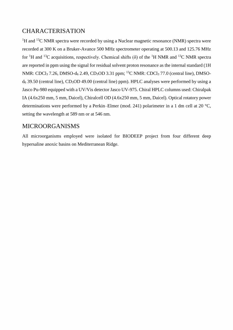
CHARACTERISATION
1H and 13C NMR spectra were recorded by using a Nuclear magnetic resonance (NMR) spectra were
recorded at 300 K on a Bruker-Avance 500 MHz spectrometer operating at 500.13 and 125.76 MHz
for 1H and 13C acquisitions, respectively. Chemical shifts (δ) of the 1H NMR and 13C NMR spectra
are reported in ppm using the signal for residual solvent proton resonance as the internal standard (1H
NMR: CDCl3 7.26, DMSO-d6 2.49, CD3OD 3.31 ppm; 13C NMR: CDCl3 77.0 (central line), DMSO-
d6 39.50 (central line), CD3OD 49.00 (central line) ppm). HPLC analyses were performed by using a
Jasco Pu-980 equipped with a UV/Vis detector Jasco UV-975. Chiral HPLC columns used: Chiralpak
IA (4.6x250 mm, 5 mm, Daicel), Chiralcell OD (4.6x250 mm, 5 mm, Daicel). Optical rotatory power
determinations were performed by a Perkin–Elmer (mod. 241) polarimeter in a 1 dm cell at 20 °C,
setting the wavelength at 589 nm or at 546 nm.
MICROORGANISMS
All microorganisms employed were isolated for BIODEEP project from four different deep
hypersaline anoxic basins on Mediterranean Ridge.
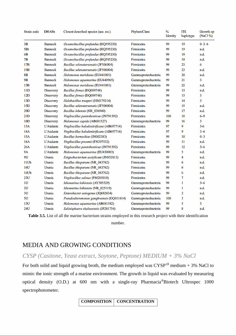
Table 3.5. List of all the marine bacterium strains employed in this research project with their identification
number.
MEDIA AND GROWING CONDITIONS
CYSP (Casitone, Yeast extract, Soytone, Peptone) MEDIUM + 3% NaCl
For both solid and liquid growing broth, the medium employed was CYSP18 medium + 3% NaCl to
mimic the ionic strength of a marine environment. The growth in liquid was evaluated by measuring
optical density (O.D.) at 600 nm with a single-ray Pharmacia®Biotech Ultrospec 1000
spectrophotometer.
COMPOSITION CONCENTRATION

casitone 15 g/L
yeast extract 5 g/L
soitone 3 g/L
peptone 2 g/L
MgSO4*7H2O 15 mg/L
FeCl3*6H2O 116 mg/L
MnCl2*4H2O 20 mg/L
NaCl 30 g/L (3%)
BIOTRANSFORMATION
Cells were grown in CYSP medium + 3% NaCl for 24h in an orbital shaker at 180 rpm at 28 °C for
24 h. Biotransformations were performed suspending freshly prepared cells (50 mg dry weight mL−1) in
10 mL of phosphate buffer (pH 7.5, 0.1 M) in Erlenmeyer flasks (100 mL). Bioconversions were kept
at 28 °C at 180 rpm for 24h.Yields and enantioselectivity were checked during this time.
For dry weight determination, washed culture samples were filtered through a 0.45 μm glass
microfiber GF/A filter (Whatman) and dried for 24 h at 110 °C. The results were an average of five
replicates.
The biotransformation of pramipexole keto intermediate (5 mM) were performed by adding the
substrate as concentrated solution in 2-propanol (final concentration of co-solvent 5%).
The biotransformation of rac-1-phenylethylamine and sodium pyruvate were performed in the
presence of 1% DMSO to increase the solubility and 50 g/L glucose.
For extraction, EtOAc was added in each sample collected at various time for monitoring conversion
and the resulting mixture was shaken and centrifuged; the organic phases were collected, dried under
N2 and analysed by HPLC.
PURIFICATION AND CHEMICAL CHARACTERISATION
2-Acetylamino-6-hydroxy-4,5,6,7-tetrahydrobenzothiazole The reaction progress and ee were determined by HPLC using a Chiralpak IA column (n-
hexane/iPrOH 8:2 as eluent, flow rate: 0.7 mL min−1, 254 nm), tr (R)-2 i 10.9 min, tr (S)-2 i 14.8 min.29
Yellowish solid. Rf=0.25 (CH2Cl2/MeOH, 95:5). 1H NMR (300 MHz,CD3OD): δ=1.91 (dddd,
J=14.2, 8.6, 7.7, 5.9 Hz, 1 H), 2.04 (dddd, J=14.2, 6.2, 5.9, 2.9 Hz, 1 H), 2.26 (s, CH3, 3 H), 2.61–
2.71 (m, 2 h), 2.79 (dddd, J=16.5, 5.9, 5.9, 1.8 Hz, 1 H), 2.99 (ddd, J=15.7, 4.7, 1.8 Hz, 1 H), 4.15

ppm (dddd, J=8.6, 6.9, 4.7, 2.9 Hz, 1 H); 13C NMR (300 MHz, CD3OD): δ=21.2, 23.2, 30.5, 30.7,
66.2, 120.9, 143.2, 156.4, 169.1 ppm; elemental analysis calcd (%) for C9H12O2N2S (212.06): C
50.92, H 5.70, N 13.20; found: C 50.96, H 5.89 N 12.85.
Acetophenone
The reaction progress and e.e. were determined by using HPLC using a Chiralcell OD column (n-
hexane/iPrOH 95:5 as eluent, flow rate: 0.7 mL min−1, 254 nm), tr 7.6 min.
REFERENCES
1. BIODEEP. Available at: http://cordis.europa.eu/project/rcn/57219_it.html.
2. Camerlenghi, A. Anoxic basins of the eastern Mediterranean: geological framework. Mar.
Chem. 31, 1–19 (1990).
3. Jongsma, D. et al. Discovery of an anoxic basin within the Strabo Trench, eastern
Mediterranean. Lett. to Nat. 305, (1983).
4. Langendijk, P. S., Hanssen, J. T. & Van der Hoeven, J. S. Sulfate-reducing bacteria in
association with human periodontitis. J. Clin. Periodontol. 27, 943–950 (2000).
5. Wallmann, K., Suess, E., Westbrook, G. H., Winckler, G. & Cita, M. B. Salty brines on the
Mediterranean sea floor. Nat. - Sci. Corresp. 387, (1997).
6. Daffonchio, D. et al. The Enigma of Prokaryotic Life in Deep Hypersaline Anoxic Basins.
Science 4, 121–123 (2005).
7. Henneke, E. & Lange, G. J. D. The distribution of DOC and POC in the water column and
brines of the Tyro and Bannock Basins. Mar. Chem. 31, (1990).
8. Horowitz, N. H., Cameron, R. E. & Hubbard, J. S. Microbiology of the Dry Valleys of
Antarctica. Science (80-. ). 176, 242–245 (1972).
9. Siegel, B. Z., Mcmurty, G., Siegel, S. M., Chen, J. & Larock, P. Life in the calcium chloride
environment of Don Juan Pond, Antarctica. Nature 280, 828–829 (1979).
10. Sørensen, K. B., Canfield, D. E., Teske, A. P. & Oren, A. Community Composition of a
Hypersaline Endoevaporitic Microbial Mat Community Composition of a Hypersaline
Endoevaporitic Microbial Mat. Appl. Environ. Microbiol. 71, 7352–7365 (2005).
11. Daffonchio, D. et al. Stratified prokaryote network in the oxic–anoxic transition of a deep-
sea halocline. Nature 440, 203–207 (2006).
12. Yakimov, M. M., Giuliano, L., Cappello, S., Denaro, R. & Golyshin, P. N. Microbial
community of a hydrothermal mud vent underneath the deep-sea anoxic brine lake Urania
(Eastern Mediterranean). Orig. Life Evol. Biosph. 37, 177–188 (2007).
13. Borin, S. et al. Sulfur cycling and methanogenesis primarily drive microbial colonization of
the highly sulfidic Urania deep hypersaline basin. Proc. Natl. Acad. Sci. U. S. A. 106, 9151–
9156 (2009).
14. Danovaro, R. et al. Deep-sea biodiversity in the Mediterranean Sea: The known, the
unknown, and the unknowable. PLoS One 5, (2010).
15. Sass, A. M., Sass, H., Coolen, M. J. L., Cypionka, H. & Overmann, J. Microbial

Communities in the Chemocline of a Hypersaline Deep-Sea Basin (Urania Basin,
Mediterranean Sea). Appl. Environ. Microbiol. 67, 5392–5402 (2001).
16. Welch, D. B. M. & Huse, S. M. Microbial Diversity in the Deep Sea and the Underexplored
‘Rare Biosphere’. Handb. Mol. Microb. Ecol. II Metagenomics Differ. Habitats
17. De Vitis, V. et al. Marine Microorganisms as Source of Stereoselective Esterases and
Ketoreductases: Kinetic Resolution of a Prostaglandin Intermediate. Mar. Biotechnol. 17,
144–152 (2015).
18. Romano, A., Romano, D., Molinari, F., Gandolfi, R. & Costantino, F. A new
chemoenzymatic synthesis of d-cloprostenol. Tetrahedron Asymmetry 16, 3279–3282 (2005).
19. Koszelewski, D., Tauber, K., Faber, K. & Kroutil, W. ω-Transaminases for the synthesis of
non-racemic α-chiral primary amines. Trends Biotechnol. 28, 324–332 (2010).
20. Cassimjee, K. E. ω-Transaminase in Biocatalysis Methods , Reactions and Engineering.
(KTH Royal Institute of Technology, Stockholm, 2012).
21. Hammes, G. G. & Haslam, J. L. A kinetic investigation of the interaction of erythro-beta-
hydroxyaspartic acid with aspartate aminotransferase. Biochemistry 8, 1591–8. (1969).
22. Silverman, R. B. The Organic Chemistry of Enzyme-Catalyzed Reactions. (Elsevier Science,
2000).
23. Ghislieri, D. & Turner, N. J. Biocatalytic approaches to the synthesis of enantiomerically
pure chiral amines. Top. Catal. 57, 284–300 (2014).
24. Kohls, H., Steffen-Munsberg, F. & Höhne, M. Recent achievements in developing the
biocatalytic toolbox for chiral amine synthesis. Curr. Opin. Chem. Biol. 19, 180–192 (2014).
25. Fuchs, M., Farnberger, J. E. & Kroutil, W. The Industrial Age of Biocatalytic
Transamination. European J. Org. Chem. 2015, 6965–6982 (2015).
26. Höhne, M., Schätzle, S., Jochens, H., Robins, K. & Bornscheuer, U. T. Rational assignment
of key motifs for function guides in silico enzyme identification. Nat. Chem. Biol. 6, 807–
813 (2010).
27. Guo, F. & Berglund, P. Transaminase Biocatalysis: Optimization and Application. Green
Chem. 19, 333–360 (2016).
28. Aziz, R. K. et al. The RAST Server: Rapid Annotations using Subsystems Technology. BMC
Genomics 9, 75 (2008).
29. Ferraboschi, P. et al. Baker’s yeast catalyzed preparation of a new enantiomerically pure
synthon of (S)-pramipexole and its enantiomer (dexpramipexole). Tetrahedron Asymmetry
25, 1239–1245 (2014).

RECOMBINANT ENZYMES
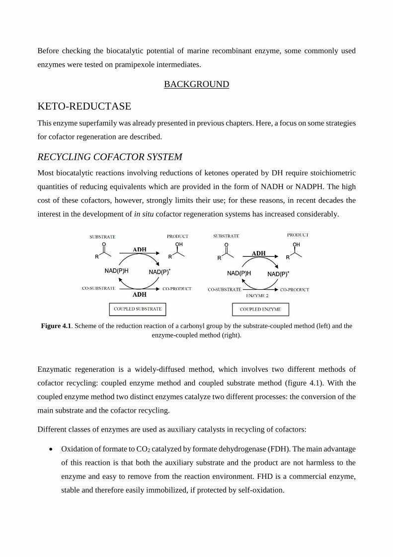
Before checking the biocatalytic potential of marine recombinant enzyme, some commonly used
enzymes were tested on pramipexole intermediates.
BACKGROUND
KETO-REDUCTASE
This enzyme superfamily was already presented in previous chapters. Here, a focus on some strategies
for cofactor regeneration are described.
RECYCLING COFACTOR SYSTEM
Most biocatalytic reactions involving reductions of ketones operated by DH require stoichiometric
quantities of reducing equivalents which are provided in the form of NADH or NADPH. The high
cost of these cofactors, however, strongly limits their use; for these reasons, in recent decades the
interest in the development of in situ cofactor regeneration systems has increased considerably.
Figure 4.1. Scheme of the reduction reaction of a carbonyl group by the substrate-coupled method (left) and the
enzyme-coupled method (right).
Enzymatic regeneration is a widely-diffused method, which involves two different methods of
cofactor recycling: coupled enzyme method and coupled substrate method (figure 4.1). With the
coupled enzyme method two distinct enzymes catalyze two different processes: the conversion of the
main substrate and the cofactor recycling.
Different classes of enzymes are used as auxiliary catalysts in recycling of cofactors:
Oxidation of formate to CO2 catalyzed by formate dehydrogenase (FDH). The main advantage
of this reaction is that both the auxiliary substrate and the product are not harmless to the
enzyme and easy to remove from the reaction environment. FHD is a commercial enzyme,
stable and therefore easily immobilized, if protected by self-oxidation.

Glucose dehydrogenase (GDH) or glucose-6-phosphate dehydrogenase (G6PDH) are
enzymes that oxidise glucose, allowing effective regeneration of both NADH and NADPH.
The balance of this reaction is totally directed towards the formation of gluconolactone due
to its spontaneous hydrolysis to gluconic acid. Active system, stable but very expensive.
Oxidation of alcohols with aldehyde dehydrogenase (ADH). The low cost of ADH and the
volatility of ethanol and acetaldehyde make this method attractive for large-scale use. Due to
low redox potential, only reactive carbonyl derivatives such as aldehydes or cyclic ketones
can be reduced. With other substrates, the balance must be forced by using an excess of
ethanol or by removing the aldehyde. This latter result can be obtained by oxidizing the
aldehyde to acetate with an aldehyde dehydrogenase, resulting in regeneration of a second
low cofactor equivalent.
Direct reduction of NAD+ with hydrogenase; hydrogenases are bidirectional enzymes that
catalyze the production and oxidation of molecular hydrogen. The regeneration of the
NADPH directed by using this enzyme is probably the most elegant solution, as there is no
by-product formation.
An alternative method that does not require the implication of a second cofactor enzyme recirculation
is referred to as a "substrate-coupled method" and is based on the addition of an auxiliary substrate
reaction that provides the reducing power necessary for the primary reaction of reduction. The
auxiliary substrate most widely added to the biotransformations is glucose as well as 2-propanol
which in the process of producing chiral alcohols is oxidized to acetone as a coproduct of the
reaction1,2. In this sense, the possibility of carrying biocatalysis reactions with enzymes that need
cofactors is a huge advantage if you are working with whole cells. Indeed, these processes with
respect to processes with isolated enzymes do not require cofactor recirculation systems and offer the
ability to selectively synthesise the target molecules using large, abundant and low cost raw materials.
However, these systems, though advantageous, require expensive and time consuming equipment. In
some cases, the development of uncontrolled metabolic processes during cell growth, with the
production of toxic products for cells, sometimes makes it difficult to separate products from the rest
of cell culture3.
ESTERASE AND LIPASE
Esterases and lipases are generally employed in stereospecific hydrolysis, transesterification, ester
synthesis and other organic reactions.
The hydrolases (EC 3 -...) form a group of enzymes capable of catalysing the hydrolysis of a covalent
bond between a carbon atom and a heteroatom by water intervention. Their natural function is purely

"digestive". Precisely because of the need to hydrolyse a wide variety of nutrients, this family includes
several enzymatic groups often associated with a wide substrate specificity4.
Carboxyesterases (EC 3.1.1-) belong to this group of enzymes, and catalyse hydrolysis and the
formation of ester bonds; they are classified in two enzymatic classes: lipases (EC 3.1.1.3) and
properly esterases (EC 3.1.1.1), which differ in some catalytic properties5:
Lipases preferably catalyse hydrolysis of non-soluble water esters such as long chain fatty
acid triglycerides, while esterases hydrolyse low molecular weight and water soluble esters.
Esterases follow Michaelis-Menten kinetics6, while lipases are active on the organic water-
substrate interface and therefore they need a minimum substrate concentration for enzymatic
activity: this phenomenon is known as "interface activation" and, thanks to structural studies,
has been correlated with the presence of a hydrophobic domain that renders the active site
inaccessible to the substrate; only in the presence of a minimal concentration of a hydrophobic
molecule (the substrate itself or an organic solvent) the "lid" moves to show a conformational
change able to access the substrate at the active lipase site7,8.
Finally, while lipases generally show high enantioselectivity, especially to secondary
alcohols, and organic solvent stability, esterases have greater variability in these two
characteristics and are generally poorly active in organic solvents.
ω-TRANSAMINASE
The topic of ω-ATA was already introduced in previous chapters. A focus on two of these enzymes
widely studied and applied in biocatalytic conversion is presented here.
The ω -ATA from Chromobacterium violaceum DSM30191 which was described for the first time
by Kaulmann et at.9 shows 38% sequence identity to the V. fluvialis JS17 enzyme and resembles the
latter in its preference for aromatic amine substrates. In fact, studies on the CV2025 ω-ATA from C.
violaceum showed a very broad substrate specificity especially with respect to amine acceptors. The
production of chiral arylic aminodiols from prochiral ketodiols looks promising and at present we are
exploring possibilities to optimise ketodiol conversion by the native enzyme.
On the other hand, Halomonas elongata enzyme (HEWT) was recently characterised by Cerioli et
al.10 with interesting results. The ω-ATA from this moderate halophile bacterium tolerated 20% (v/v)
co-solvents over 22 h, the best solvents were MeOH (47%) and DMSO (27%). Acceptance of
isopropylamine as amino donor is an advantage in asymmetric synthesis and a cost-effective benefit
for industrial applications. With its high enantioselectivity, large substrate spectra and stability in
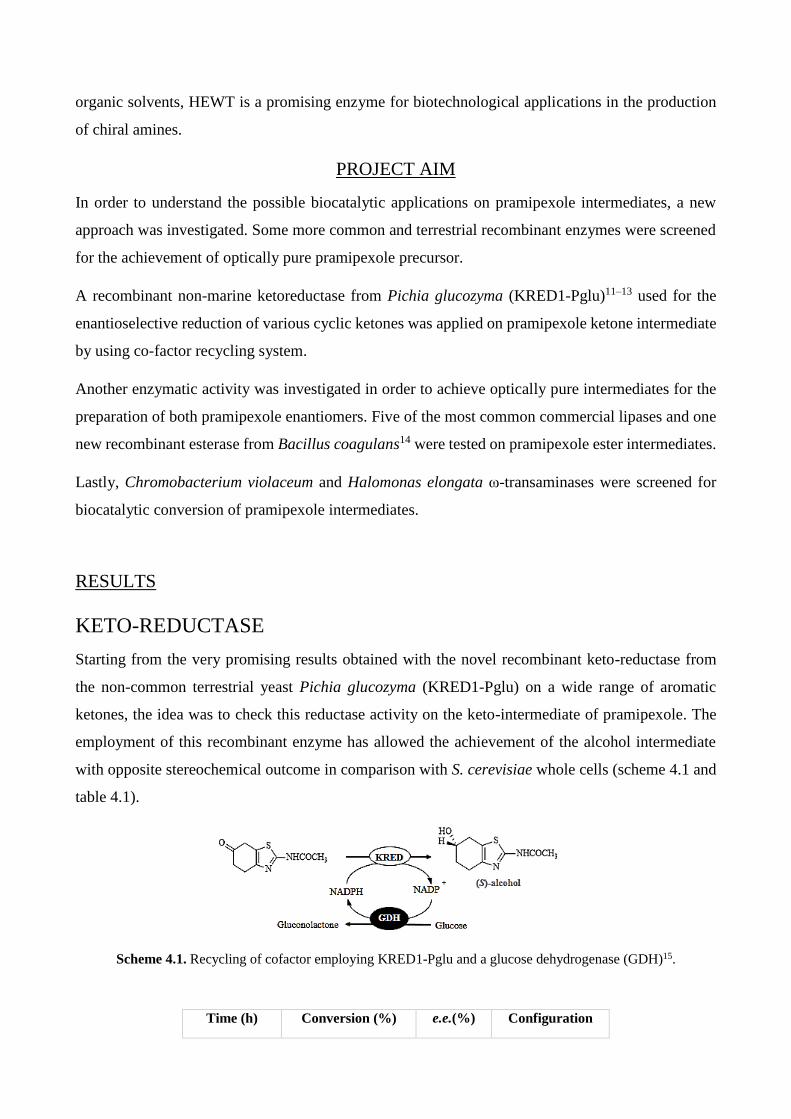
organic solvents, HEWT is a promising enzyme for biotechnological applications in the production
of chiral amines.
PROJECT AIM
In order to understand the possible biocatalytic applications on pramipexole intermediates, a new
approach was investigated. Some more common and terrestrial recombinant enzymes were screened
for the achievement of optically pure pramipexole precursor.
A recombinant non-marine ketoreductase from Pichia glucozyma (KRED1-Pglu)11–13 used for the
enantioselective reduction of various cyclic ketones was applied on pramipexole ketone intermediate
by using co-factor recycling system.
Another enzymatic activity was investigated in order to achieve optically pure intermediates for the
preparation of both pramipexole enantiomers. Five of the most common commercial lipases and one
new recombinant esterase from Bacillus coagulans14 were tested on pramipexole ester intermediates.
Lastly, Chromobacterium violaceum and Halomonas elongata ω-transaminases were screened for
biocatalytic conversion of pramipexole intermediates.
RESULTS
KETO-REDUCTASE
Starting from the very promising results obtained with the novel recombinant keto-reductase from
the non-common terrestrial yeast Pichia glucozyma (KRED1-Pglu) on a wide range of aromatic
ketones, the idea was to check this reductase activity on the keto-intermediate of pramipexole. The
employment of this recombinant enzyme has allowed the achievement of the alcohol intermediate
with opposite stereochemical outcome in comparison with S. cerevisiae whole cells (scheme 4.1 and
table 4.1).
Scheme 4.1. Recycling of cofactor employing KRED1-Pglu and a glucose dehydrogenase (GDH)15.
Time (h) Conversion (%) e.e.(%) Configuration

4 75 86 S
Table 4.1. KRED1-Pglu-catalyzed bioconversion of pramipexole keto intermediate by using a cofactor recycling
system.
ESTERASE AND LIPASE
Based on former studies not already published on a new recombinant esterase from Bacillus
coagulans (BCE), a benzoyl ester was synthesized in order to verify the enantioselective hydrolysis
by BCE. Moreover, a commercial esterase active on benzoyl esters was assayed: pig liver esterase
PLE (scheme 4.2 and table 4.2).
Scheme 4.2. Bioconversion of the benzoyl ester intermediate into alcohol intermediate and benzoic acid.
ESTERASE
24h Conversion (%) e.e.(%) Configuration
BCE 12 100 R
PLE - - -
Table 4.2. BCE- and PLE-catalyzed conversion.
For what concerns lipase activity, 5 commercial lipases as Pseudomonas fluorescens lipase (PFL),
Candida antarctica lipase A and B (CAL A, CAL B), lipase from porcine pancreas (PPL) and from
Candida cylindracea (CCL) were screened toward the acetyl ester intermediate of pramipexole
(scheme 4.3 and table 4.3). The use of commercial lipases showed a high activity on this substrate
but low molar conversion percentages, although the high e.e. with R-isomer preference like BCE.
Scheme 4.3. Bioconversion of acetyl ester intermediate into alcohol intermediate and acetic acid.
LIPASE 20h Conversion (%) e.e.(%) Configuration
CAL A-CLEA* 5 80 R
CAL B 49 71 R
PFL 5 100 R
PPL 7 100 R
CCL 9 20 R

Table 4.3. Biotransformation molar conversion and enantiomeric excesses. *CLEA stands for cross-linked
enzyme aggregates (CLEA Technologies).
ω-TRANSAMINASE
The application of H. elongata (HEWT) and C. violaceum (CV2025) ω-ATAs was exploited in
bioconversion of pramipexole keto intermediate with (S)-1-phenylethylamine ((S)-1-PEA) and L-
alanine as amine-donor (scheme 4.4). With both recombinant enzymes, no conversion of substrate
has been seen.
Scheme 4.4. Transaminase catalysis on pramipexole intermediate.
MATERIALS AND METHODS
MATERIALS
All reagents and solvents were obtained from Sigma–Aldrich-Fluka and used without further
purification or drying. TLC was performed with Merck silica gel 60 F254 pre-coated plates. Silica
gel column chromatography was performed on silica gel 60 (40–63 mm particle size). Pramipexole
keto intermediate (2-acetylamino-6-oxo-4,5,6,7- tetrahydrobenzothiazole) was prepared following a
procedure previously described16.
Commercial enzymes were employed following the patent WO 2006012277 A217 and the work by
Ferraboschi et al18–20. Lipase from porcine pancreas (PPL, 23.9 U/mg), Candida cylindracea lipase
(CCL, 3.86 U/mg), Pseudomonas fluorescens (PFL, 40.2 U/mg), Esterase from porcine liver (PLE,
≥15 U/mg) and Candida antarctica lipase B (CAL B, 5 U/mg) were purchased by Sigma-Aldrich.
Candida antarctica lipase A CLEA (CAL A CLEA, 1.56 U/mg) was obtained from CLEA
Technologies.
CHARACTERISATION
Nuclear magnetic resonance (NMR) spectra were recorded at 300 K on a Bruker-Avance 500 MHz
spectrometer operating at 500.13 and 125.76 MHz for 1H and 13C acquisitions, respectively. Chemical
shifts (δ) of the 1H NMR and 13C NMR spectra are reported in ppm using the signal for residual
solvent proton resonance as the internal standard (1H NMR: CDCl3 7.26, DMSO-d6 2.49, CD3OD
3.31 ppm; 13C NMR: CDCl3 77.0 (central line), DMSO-d6 39.50 (central line), CD3OD 49.00 (central
line) ppm). HPLC analyses were performed by using a Jasco Pu-980 equipped with a UV/Vis detector

Jasco UV-975. Chiral HPLC columns used: Chiralpak IA (4.6V250 mm, 5 mm, Daicel), Optical
rotatory power determinations were performed by a Perkin–Elmer (mod. 241) polarimeter in a 1 dm
cell at 20 °C, setting the wavelength at 589 nm or at 546 nm.
KETO-REDUCTASE
KRED-Pglu was kindly provided by Professor Francesco Molinari’s research group (University of
Milan, DEFENS department, Milano, Italy). Recombinant GDH from Bacillus megaterium was
kindly provided by Prof Daniela Monti (Istituto di Chimica del Riconoscimento Molecolare, C.N.R.,
Milano, Italy).
BIOTRANSFORMATIONS
Molar conversion and enantioselectivity were determined using an enzyme-coupled system with
glucose-glucose dehydrogenase (GDH) from Bacillus megaterium15 for cofactor recycling.
Reductions were carried out in 5-mL screw-capped test tubes with a reaction volume of 1 mL with
KRED1-Pglu (20 mU/ mL), GDH (1 U/mL), NADP+ (0.1 mM), substrate (1 g/L), glucose (4×mmol
of substrate) suspended in 50 mMTris-HCl buffer pH 8.0. The biotransformation was kept under
stirring at 30 °C for 24 h.
Although the pH suitable for pramipexole ketone intermediate bioconversion is around pH 5-6,
KRED1-Pglu is able to catalyse alcohol production with high rate avoiding substrate degradation.
For extraction, EtOAc was added in each sample collected at various time for monitoring conversion
and the resulting mixture was shaken and centrifuged; the organic phases were collected, dried under
N2 and analysed by HPLC.
ESTERASE AND LIPASE
BCE was kindly provided by Prof. Francesco Molinari’s research group (University of Milan,
DEFENS department, Milano, Italy).
ESTER SYNTHESIS
For what concerns the acetyl ester, the procedure described by Ferraboschi et al.16 was followed. For
the preparation of pramipexole benzoyl ester intermediate, 6-hydroxy derivative was treated with
benzoyl chloride in pyridine.
BIOTRANSFORMATIONS
Benzoyl ester hydrolysis was carried out in 2 mL Eppendorf tubes with a reaction volume of 1 mL
with BCE (20 mU/ mL), substrate (0.1 g/L) suspended in 50mMTris-HCl buffer pH 8.0, NaCl 100
mM. The biotransformation was kept under stirring at 30 °C for 24h.
DMSO as co-solvent was not employed in order to avoiding keto substrate degradation.
Acetyl ester hydrolysis, instead, was performed in 10-mL screw-capped test tubes with a reaction
volume of 3 mL with the proper amount of commercial lipases17,19,18,20, H2O (5×mmol of substrate),
substrate (20 mM) suspended in acetonitrile (ACN). The biotransformation was kept under stirring at
30 °C for 24h.
For extraction, EtOAc was added in each sample collected at various time for monitoring conversion
and the resulting mixture was shaken and centrifuged; the organic phases were collected, dried under
N2 and analysed by HPLC.
ω-TRANSAMINASE
During this experimental work, we had the opportunity to test aminotransferase potential of
Chromobacterium violaceum and Halomonas elongate ω-ATAs on keto intermediate of pramipexole
synthesis.
Both enzymes were kindly provided by Prof. Francesca Paradisi’s research group.
BIOTRANSFORMATIONS
The enzymatic reactions were carried out at 37 °C in 100 mM potassium phosphate buffer pH 8.0,
0.1 mM PLP using a purified enzyme solution (0.25 U/mL). The reaction mixture contained 10 mM
(S)-1-PEA or 10 mM L-alanine and 10 mM pramipexole keto intermediate as amine-acceptor in a
reaction volume of 200 μL.
For HPLC analysis, samples were collected at different time, diluted 1:50 with HCl 0.2% solution for
quenching the reaction and injected.
PURIFICATION AND CHEMICAL CHARACTERISATION
2-Acetylamino-6-hydroxy-4,5,6,7-tetrahydrobenzothiazole
The reaction progress and ee were determined by using HPLC using a Chiralpak IA column (n-
hexane/iPrOH 8:2 as eluent, flow rate: 0.7 mL min−1, 254 nm), tr (R)-2 i 10.9 min, tr (S)-2 i 14.8 min.16

Yellowish solid. Rf=0.25 (CH2Cl2/MeOH, 95:5). ee=64 %. 1H NMR (300 MHz,CD3OD): δ=1.91
(dddd, J=14.2, 8.6, 7.7, 5.9 Hz, 1 H), 2.04 (dddd, J=14.2, 6.2, 5.9, 2.9 Hz, 1 H), 2.26 (s, CH3, 3 H),
2.61–2.71 (m, 2 h), 2.79 (dddd, J=16.5, 5.9, 5.9, 1.8 Hz, 1 H), 2.99 (ddd, J=15.7, 4.7, 1.8 Hz, 1 H),
4.15 ppm (dddd, J=8.6, 6.9, 4.7, 2.9 Hz, 1 H); 13C NMR (300 MHz, CD3OD): δ=21.2, 23.2, 30.5,
30.7, 66.2, 120.9, 143.2, 156.4, 169.1 ppm; elemental analysis calcd (%) for C9H12O2N2S (212.06):
C 50.92, H 5.70, N 13.20; found: C 50.96, H 5.89 N 12.85.
2-Acetylamino-6-hydroxy-4,5,6,7-tetrahydrobenzothiazole, 6-benzoate
HPLC analysis: Chiralpak IA, hexane/2-propanol 8:2 as eluent, flow rate 0.7 mL/min. Rt (R)-isomer
14.32 min; (S)-isomer 16.25 min. 1H NMR (CDCl3): δ 8.02 (2H, d, J = 8.0 Hz, o-Ph H), 7.58 (1H, t,
J = 8.0 Hz, p-Ph H), 7.46 (2H, dd, J = 8.0, 8.0 Hz, m-Ph H), 5.58 (1H, dddd, J = 7.6, 5.5, 4.8, 2.5 Hz,
6-H), 3.20 (1H, ddd, J = 16.5, 4.8, 1.2 Hz, 7a-H), 3.02 (1H, dd, J = 16.5, 5.5 Hz, 7b-H), 2.93 (1H,
ddd, J = 16.8, 8.4, 5.9 Hz, 4a-H), 2.86 (1H, dddd, J = 16.8, 6.2, 5.8, 1.4 Hz, 4b-H), 2.30 (3H, s, CH3),
2.30 (1H, dddd, J = 13.5, 7.6, 5.9, 5.8 Hz, 5a-H), 2.00 (1H, dddd, J = 13.5, 8.4, 6.2, 2.5 Hz, 5b-H).
REFERENCES
1. Hollmann, F., Arendsa, I. W. C. E. & Holtmannb, D. Enzymatic reductions for the chemist. Green Chem.
13, 2285–2314 (2011).
2. Somogyi, L. P. The flavour and fragrance industry: Serving a global market. Chem Ind. 4, 170–173 (1996).
3. Graßmann, J. Terpenoids as Plant Antioxidants. Vitam. Horm. 72, 505–535 (2005).
4. Bornscheuer, U.T., Kazlauskas, R.J. Hydrolases in Organic Synthesis. (Weinheim: Wiley-VCH, 1999).
5. Bornscheuer, U. T. Microbial carboxyl esterases: classification, properties and application in biocatalysis.
FEMS Microbiol Rev. 26, 73–81. (2002).
6. Michaelis, L. & Menten, M. L. Die Kinetik der Invertinwirkung. Biochem Z 49, 333–369 (1913).
7. Holmquist, M. Alpha/Beta-hydrolase fold enzymes: structures, functions and mechanisms. Curr Protein
Pept Sci 1, 209–35 (2000).
8. Schrag, J. D., Li, Y., Wu, S. & Cygler, M. Ser-His-Glu triad forms the catalytic site of the lipase from
Geotrichum candidum. Nature 351, 761–764 (1991).
9. Kaulmann, U., Smithies, K., Smith, M. E. B., Hailes, H. C. & Ward, J. M. Substrate spectrum of ω-
transaminase from Chromobacterium violaceum DSM30191 and its potential for biocatalysis. Enzyme
Microb. Technol. 41, 628–637 (2007).
10. Cerioli, L., Planchestainer, M., Cassidy, J., Tessaro, D. & Paradisi, F. Characterization of a novel amine
transaminase from Halomonas elongata. J. Mol. Catal. B Enzym. 120, 141–150 (2015).
11. Contente, M. L. et al. Enzymatic reduction of acetophenone derivatives with a benzil reductase from Pichia
glucozyma (KRED1-Pglu): electronic and steric effects on activity and enantioselectivity. Org. Biomol.
Chem. 14, 3404–3408 (2016).
12. Contente, M. L. et al. Preparation of enantiomerically enriched aromatic β-hydroxynitriles and halohydrins
by ketone reduction with recombinant ketoreductase KRED1-Pglu. Tetrahedron 72, 3974–3979 (2016).
13. Contente, M. L. et al. Stereoselective reduction of aromatic ketones by a new ketoreductase from Pichia
glucozyma. Appl. Microbiol. Biotechnol. 100, 193–201 (2016).

14. Romano, D. et al. Enhanced enantioselectivity of Bacillus coagulans in the hydrolysis of 1,2-O-
isopropylidene glycerol esters by thermal knock-out of undesired enzymes. Tetrahedron Asymmetry 16,
841–845 (2005).
15. Bechtold, M. et al. Biotechnological Development of a Practical Synthesis of Ethyl (S)-2-Ethoxy-3-(p-
methoxyphenyl)propanoate (EEHP): Over 100-Fold Productivity Increase from Yeast Whole Cells to
Recombinant Isolated Enzymes. Org. Process Res. Dev. 16, 269–276 (2012).
16. Ferraboschi, P. et al. Baker’s yeast catalyzed preparation of a new enantiomerically pure synthon of (S)-
pramipexole and its enantiomer (dexpramipexole). Tetrahedron Asymmetry 25, 1239–1245 (2014).
17. Valivety, R. H., Michels, P. C., Pantaleone, D. P. & Khmelnitsky, Y. L. Biocatalytic process for preparing
enantiomerically enriched pramipexole. (2005).
18. Ciceri, S., Ciuffreda, P., Grisenti, P. & Ferraboschi, P. Synthesis of the antitumoral nucleoside capecitabine
through a chemo-enzymatic approach. Tetrahedron Lett. 56, 5909–5913 (2015).
19. Ferraboschi, P., Mieri, M. D. & Ragonesi, L. Lipase-catalyzed preparation of corticosteroid 17a-esters
endowed with antiandrogenic activity. Tetrahedron Lett. 49, 4610–4612 (2008).
20. Ferraboschi, P., Mieri, M. D. & Galimberti, F. Chemo-enzymatic approach to the synthesis of the
antithrombotic clopidogrel. Tetrahedron Asymmetry 21, 2136–2141 (2010).
VPTA - Virgibacillus pantothenticus ω-transaminase

BACKGROUND
ω-TRANSAMINASE
Although the transaminase potential is under a magnifying glass since decades, their applicability in
biotechnological fields for the preparation of chiral amino compounds is not entirely exploited yet.
Combining ATAs with other enzymatic or chemical routes has been demonstrated to be a smart
method for practical applications, particularly in regard to shortening the reaction routes, avoiding
protecting steps, reducing chemical waste and achieving a high atom-efficiency1–3.
This makes these enzymes useful for synthesis of chiral amines, which are of high importance as
building blocks for production of optically pure amino synthons in pharmaceutical like sitagliptin4,
ethambutol5, imagabalin6, norephedrine and pseudoephedrine7, food, agrochemical and cosmetic
additives8.
Synthesis by ATAs suffers from the unfavourable reaction equilibrium, especially in the asymmetric
synthesis of amines from an amino acid as amino donor9. To maximize the productivity of ATAs,
shifting the equilibrium to the side of product is extremely important10,11. Equilibrium displacement
has been done by removal of the products or large excess of amino donor12,13.
Nevertheless, maintaining the quaternary and tertiary structures under non-physiological conditions
(high concentrations of substrate and product, organic solvent, mechanical force, etc.) for a long
period of time (10–100 h working time and an even longer storage time) is a huge task for all
biocatalysts. The current trend in this research field is directing to the identification and designing of

new ω-transaminases with defined substrate specificity and capable of adapting to the uncommon
catalytic conditions of industrial processes.
SOLUBILITY ISSUE
Protein overexpression systems, such as the E. coli T7 system, have been widely used for protein
purification. However, due to improper folding of the overexpressed proteins, they often aggregate
to form inclusion bodies, greatly reducing the protein yield. Different methods have been used to
solve this problem14, such as optimization of bioprocess parameters or using dedicated screening tools
for finding conditions for soluble expression15, expression at reduced temperature16, solubility-
enhancing fusion tags17,18, co-expression of chaperones19, consideration of codon usage20 and
refolding of proteins after dissolving inclusion bodies in denaturing reagents21–23 (fig. 5.1).
Fig. 5.1. All these methods were taken into account and the results illustrated below.
Solubility on overexpression of some proteins has been found to be altered by certain mutations24.
Individual mutations differ in the extent to which they influence the solubility on overexpression. For
example, the W131A and V165K mutants of human HIV type 1 integrase are only marginally more
soluble, whereas the F185K is significantly more soluble relative to the wild-type under the same
conditions of overexpression25.
Systematic replacement of hydrophobic residues has also been attempted, but only some of these
mutations resulted in improved solubility25–27. Mutations involving substitution of less polar residues
with more polar ones, identified by multiple sequence alignment of the insoluble protein with related
soluble proteins27 or predicted solvent accessibilities28 have also been attempted, but only some
mutations have manifested in improved solubility. Many of these types of mutations are identified by
random mutagenesis procedures29,30. Asano et al.31, have created highly active and in vivo soluble
mutants of MeHNL1 (hydroxynitrile lyase from Manihot esculenta, cassava plant) in E. coli by
directed evolution, where several mutants of this enzyme were generated including His103 and also
Lys-Pro mutants.

There are other examples of correlation between one single point mutation and folding changing.
Particulary, regarding human transaminases, some specific mutations such as P11L (major allele) and
G47R (minor allele) on alanine:glyoxylate aminotransferase (AGT) was proved to have a role in
misfolding and pathogenic aggregation leading to Primary Hyperoxaluria Type I. These mutations
seem to be involved in dimerization and stabilization of enzyme complex32,33.
MUTAGENESIS
In recombinant applications, high levels of soluble expressed protein are usually required and often
this entails an alteration of the physiological requirements for the enzyme natural folding leading to
the formation of inclusions bodies. Therefore, thinking about a solubility improvement of enzyme
sequences could be more than appreciable but not universally possible through a rational strategy.
Moreover, utilization of proteins outside of their usual biological context, for example in industrial
applications, often requires improvement of biophysical properties such as stability34,35. In the
Introduction the mutagenesis strategies aimed at solving this issue were already presented.
Another approach based on multiple sequence alignments (MSAs) consists in statistical amino acid
frequencies analysis36,37. Data-driven mutagenesis design is based on the simple assumption that the
frequency of a given residue in an MSA of homologous proteins correlates with that amino acid's
contribution to protein stability38–40. A de novo designed protein possessing the most frequent residue
at each position should accordingly show maximum stability. Given the difficulty of predicting how
individual residues contribute to overall stability41, this approach to protein stabilization is often
preferable to classical rational design or is adopted as a complement of it, particularly as it does not
depend on the availability of structural information.
PROJECT AIM
The present work descibes the attaining of soluble expression of a novel ω-transaminase from a newly
isolated halotolerant marine bacteria Virgibacillus pantothenticus(VPTA). This marine bacterium is
part of the micro-biodiversity of Deep Hypersaline Anoxic Basins on the Mediterranean Ridge, a new
and uncommon source of biocatalysts. Despite of several standard methodologies applied, the marine
wild-type enzyme VPTA was total insoluble in E. coli host and it was satisfactorily solubilized by
one single-point mutation, allowing the characterization of the new ω-transaminase. Combining
statistical amino acid frequencies analysis with a rational approach based on 3-D model, a target
residue likely involved in structural stabilization and dimerization was selected. This finding has
significant implications for ω-transaminase structure-stability-solubility understanding and
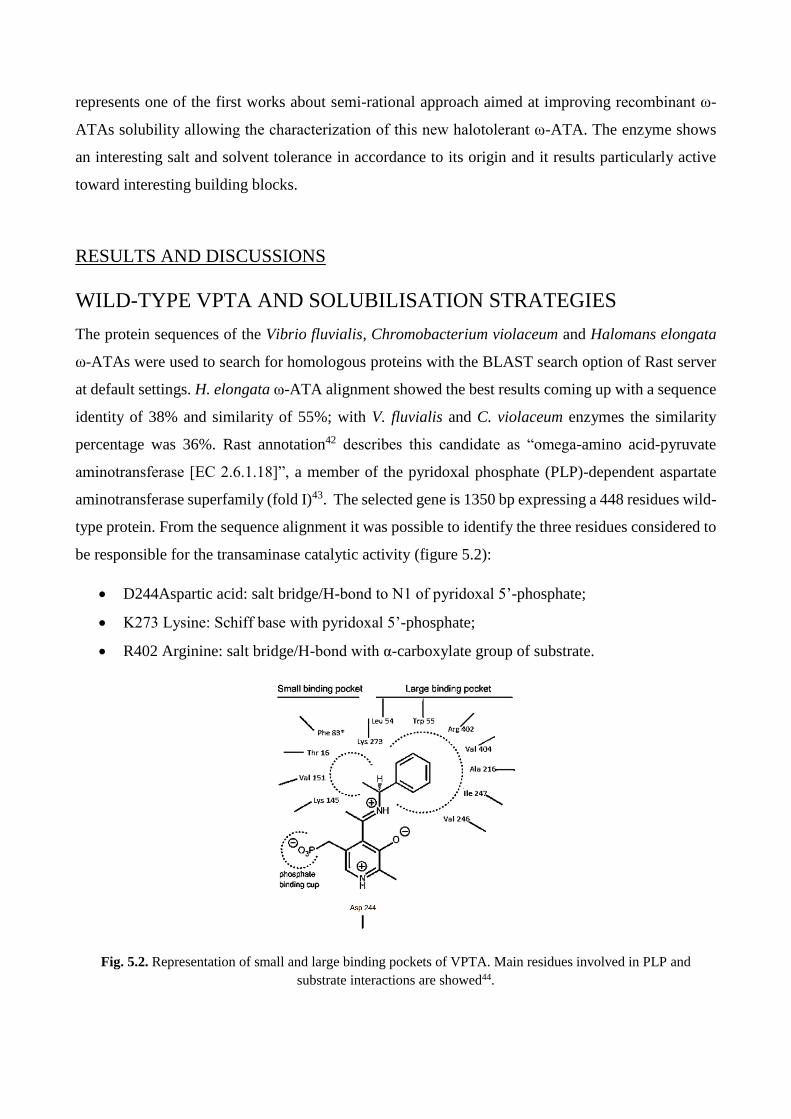
represents one of the first works about semi-rational approach aimed at improving recombinant ω-
ATAs solubility allowing the characterization of this new halotolerant ω-ATA. The enzyme shows
an interesting salt and solvent tolerance in accordance to its origin and it results particularly active
toward interesting building blocks.
RESULTS AND DISCUSSIONS
WILD-TYPE VPTA AND SOLUBILISATION STRATEGIES
The protein sequences of the Vibrio fluvialis, Chromobacterium violaceum and Halomans elongata
ω-ATAs were used to search for homologous proteins with the BLAST search option of Rast server
at default settings. H. elongata ω-ATA alignment showed the best results coming up with a sequence
identity of 38% and similarity of 55%; with V. fluvialis and C. violaceum enzymes the similarity
percentage was 36%. Rast annotation42 describes this candidate as “omega-amino acid-pyruvate
aminotransferase [EC 2.6.1.18]”, a member of the pyridoxal phosphate (PLP)-dependent aspartate
aminotransferase superfamily (fold I)43. The selected gene is 1350 bp expressing a 448 residues wild-
type protein. From the sequence alignment it was possible to identify the three residues considered to
be responsible for the transaminase catalytic activity (figure 5.2):
D244Aspartic acid: salt bridge/H-bond to N1 of pyridoxal 5’-phosphate;
K273 Lysine: Schiff base with pyridoxal 5’-phosphate;
R402 Arginine: salt bridge/H-bond with α-carboxylate group of substrate.
Fig. 5.2. Representation of small and large binding pockets of VPTA. Main residues involved in PLP and
substrate interactions are showed44.
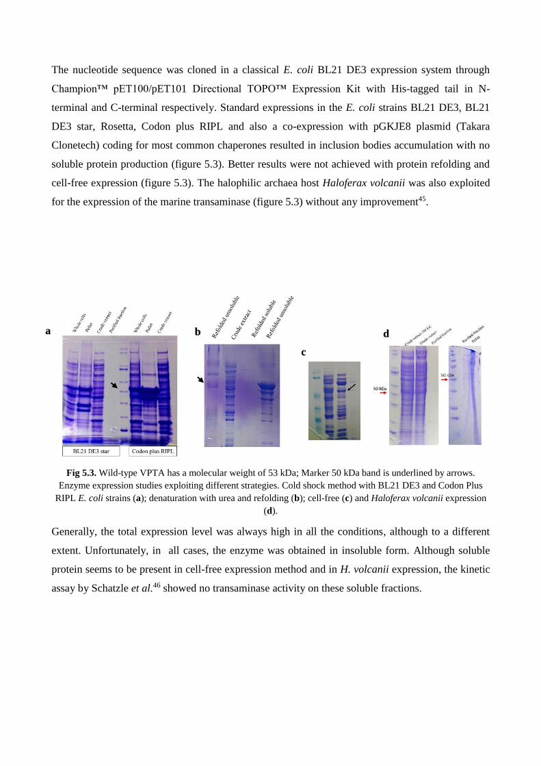
The nucleotide sequence was cloned in a classical E. coli BL21 DE3 expression system through
Champion™ pET100/pET101 Directional TOPO™ Expression Kit with His-tagged tail in N-
terminal and C-terminal respectively. Standard expressions in the E. coli strains BL21 DE3, BL21
DE3 star, Rosetta, Codon plus RIPL and also a co-expression with pGKJE8 plasmid (Takara
Clonetech) coding for most common chaperones resulted in inclusion bodies accumulation with no
soluble protein production (figure 5.3). Better results were not achieved with protein refolding and
cell-free expression (figure 5.3). The halophilic archaea host Haloferax volcanii was also exploited
for the expression of the marine transaminase (figure 5.3) without any improvement45.
Fig 5.3. Wild-type VPTA has a molecular weight of 53 kDa; Marker 50 kDa band is underlined by arrows.
Enzyme expression studies exploiting different strategies. Cold shock method with BL21 DE3 and Codon Plus
RIPL E. coli strains (a); denaturation with urea and refolding (b); cell-free (c) and Haloferax volcanii expression
(d).
Generally, the total expression level was always high in all the conditions, although to a different
extent. Unfortunately, in all cases, the enzyme was obtained in insoluble form. Although soluble
protein seems to be present in cell-free expression method and in H. volcanii expression, the kinetic
assay by Schatzle et al.46 showed no transaminase activity on these soluble fractions.
a b
c
d

MUTAGENESIS
Starting from ConSurf47 analysis in which 500 homologous sequences were compared getting a
statistical frequency (table 5.1) and a level of conservation (figure 5.4) for each position, some
residues less conserved than the catalytic one were taken into account.
Table 5.1. ConSurf results
in terms of statistical amino acid frequency for each position. In the table, only the first 20 positions are shown as
an example.
POSITION AA with the HIGHEST
FREQUENCY % FREQUENCY
1 M 100
2 N 38
3 S 37
4 L 88
5 Q 29
6 E 35
7 L 59
8 D 88
9 A/R 23
10 A 38
11 H 88
12 H 41
13 L 52
14 H 88
15 P 97
16 F 45
17 T 49
18 D 31
19 L 14
20 K 34

Fig. 5.4. Conservation scale by ConSurf on VPTA amino acidic sequence. The position 16 shows a quite high
conservation.
After target residues localization in tertiary structure model, threonine in position 16 was selected.
Position 16 has a good level of conservation but the residue with the highest frequency in that position
is phenylalanine (T16F).
The protein model was built using the SWISS-MODEL Homology Modeling tool48, superimposing
the VTPA protein sequence on the resolved structure of the homologous amino transaminase from
Vibrio fluvialis (PDB: 4E3Q). The result elaboration and the structural evaluation was achieved using
the molecular visualization system PyMOL (open source license). Sequence analysis and alignment
was performed with CunSurf, a software for multiple blast provided by BioSoft.
b

Fig. 5.5. VPTA model with threonine in yellow. This residue is localised on a small loop at N-terminal in a
distinct motif above the catalytic K in light blue (a); the homodimerisation (b) involves hydrophobic amino acids
(red) from both monomers in what is called hydrophobic pocket (d). In binding pockets of V. fluvialis ATA many
hydrophobic residues are present (c) likely with a role in dimer formation.
The mutation choice reflects the current positions in regard to this residue involved in the active site
with no particular predisposition to central catalytic role6,40,44,49, despite of proximity to substrate
binding pocket and moderately high level of conservation of the positions (ConSurf analysis figure
5.4). The amino acids involved with high probability in the structural stabilization of the functional
homodimer are less conserved than the catalytic ones at least, but they are preserved by evolution
anyway. This could shed light on the structural role of this residue in the ω-ATAs homodimerization
and stabilization.
VPTA T16F
The expression of the N-terminal his-tagged transaminase was performed and compared in different
conditions and best results were achieved with ZYM-5052 auto-induction medium at 150 rpm, at 30
°C for 24 h. Purification was performed by immobilized metal affinity chromatography (IMAC). The
enzyme was obtained in the pure form, expressed at higher concentration in auto-induction medium
with a specific activity 2.4 U/mg and a volumetric yield 4 mg/L (figure 5.6).
a
c
d

Fig. 5.6. VPTA T16F SDS-PAGE purifications fractions from auto-induction expression. The purified fractions
present the molecular weight attended (53 kDa).
EFFECT OF pH AND TEMPERATURE
VPTA preparation was suspended in a universal buffer solution at the desired pH and stored at 4 °C
for 24 h. After incubation, the residual activity was determined with the spectrophotometric enzyme
assay in standard condition46 (figure 5.7). Only at pH 8 the enzyme is stable and active.
Fig. 5.7. Stability test at different pH: data are reported as relative percentages in comparison with the control in
standard conditions. Every reaction was performed in three replicates and the results are reported as the average of
the data obtained.
50 kDa

Thermal stability was tested after incubation of the enzyme at variable temperatures. After 20 h the
enzyme mantains 80% of initial activity up to 45 °C.
The activity was determined with the spectrophotometric enzyme assay performed at the indicated
temperature. The highest activity was reached between 25 °C and 45 °C.
Fig. 5.8. The X axis shows the set of temperature selected while on Y axis the residual activity in percentage is
reported. Left panel: stability test of VPTA in the range 25-60 °C. Right panel: activity test of VPTA in the range
25-60 °C. Each reaction was performed in triplicate. Results are reported as the average of the data obtained.
As described for the first time by Ikai et at.50, this high mesophilic profile could be confirmed by the
VPTA high aliphatic index of 94.44 (ProtParam tool, accessible from the ExPASy website
www.expasy.ch).
EFFECT OF CO-SOLVENTS AND SALTS
The use of co-solvents is very useful if the bioconversion deals with a very low soluble substrate, for
this reason the effect of different co-solvents on VPTA activity and stability was investigated. The
stability analysis was carried out incubating the enzyme with 10% and 20% of a selection of common
water-miscible organic solvent for 45 h (figure 5.9). In all cases, a decrease in enzyme activity was
observed, even if VPTA shows a higher stability than HEWT under the same condition. With regard
to the activity in presence of co-solvents, the best results were obtained with MeOH 10%. Hence, this
enzyme is suitable for enantioselective amination of ketones and aldehydes where the presence of a
co-solvent is necessary.

Fig. 5.9. The X axis shows the set of solvents selected while on Y axis the residual activity in percentage is
reported. Left panel: stability test of VPTAwith 10 and 20% solvents after 45 h. Right panel: activity test of
VPTAwith 10 and 20% solvents. Each reaction was performed in triplicate. Results are reported as the average of
the data obtained.
The effect of different salt (NaCl and KCl) concentrations on enzyme stability and activity was also
investigated. Under activity test conditions in the presence of salts, enzyme activity is very low, when
compared to the enzyme at pH 8.0 in 50 mM phosphate buffer (figure 5.10). On the other hand,
incubation of the enzyme in the presence of salts for seven days resulted in a satisfactory residual
activity only when 1 M KCl was used.
Fig. 5.10. The X axis shows the set of salts selected while on Y axis the residual activity in percentage is reported.
On the left stability test: incubation was carried out at the indicated salt concentration for 45 h at 4 ◦C. On the
right activity test in presece of salts: the residual activity of the enzymatic solution was determined by the standard
spectrophotometric assay carried out in presence of the salt concentration indicated and expressed in function of
the activity under standard conditions. Every reaction was performed in three replicates and the results are
reported as the average of the data obtained.
AMINO DONORS
A number of amino donors was investigated in order to understand the catalytical potential of VPTA
and consequently its possible applications (table 5.2). VPTA does not display an activity for classical
amino donors (i.e., L-alanine isomolar with amino acceptor higher than that of HEWT, CV2025 and

V. fluvialis ω-ATA. However, VPTA shows to be highly (S)-selective showing no detectable activity
on (R)-(-)-1-phenylethylamine. The final conversion was determined after 24 h of reaction time.
AMINO DONOR STRUCTURE FINAL CONVERSION (%)
rac-1-phenylethylamine
43
S-(-)-1-phenylethylamine
71
R-(-)-1-phenylethylamine
/
Isopropylamine*
/
benzylamine
47
β-alanine*
/
L-alanine*
8
D-alanine*
/
cinnamyl-amine
74
3,4-(methylenedioxy)benzylamine
70
vanillylamine
90
putrescine* /
cadaverine* /
4-phenylbenzylamine
16
Table 5.2. VPTA specificity on different amino donors are reported. The enzymatic reactions with VPTA were
carried out as reported in Materials and Methods. Every reaction was performed in three replicates and the results
are reported as the average of the data obtained. Concentration of amino donors was kept constant at 10 mM, as
amino acceptor 10 mM pyruvate was normally employed, except for the substrate with *, where pyruvate was
substituted with10 mM benzaldehyde. The substrate was added in methanol solution to guarantee the correct
concentration of the substrate in reaction mixture (final concentration 10% MeOH). Final conversions with VPTA
were determined by HPLC analysis as reported in materials and methods.
AMINO ACCEPTORS
VPTA shows no interesting activity on standard amino acceptors in comparison with HEWT,
CV2025 and V. fluvialis ω-ATA. Some promising conversions resulted for substrates such as
phenylacetaldehyde, cinnamalaldehyde, biphenyl-4-carboxaldehyde and 3-phenylpropionaldehyde
(table 5.3). These bulky aldehydes are well accepted by the enzyme. The lack of activity with D-
fructose and L-ribulose could be caused by a steric clash hampering the accomodation of the substrate
in the catalytic pocket. The final conversion was determined after 24 h of reaction time.
AMINE ACCEPTOR STRUCTURE FINAL CONVERSION (%)

pyruvic acid*
71
β-hydroxypyruvic acid*
/
glyoxylic acid*
/
benzaldehyde
49
phenylacetaldehyde
90
2-phenylpropionaldehyde
53
vanillin
/
cinnamalaldehyde
93
cyclohexanone*
/
1,3-dihydroxyacetone*
/
L-erythrulose*
/
L-ribulose*
/
D-fructose*
/
4-nitrobenzaldehyde
50
3-nitrobenzaldehyde
86
2-nitrobenzaldehyde
68
biphenyl-4-carboxaldehyde
81
3-phenylpropionaldehyde
91
Table 5.3. VPTA specificity with different amino acceptors are reported. The enzymatic reactions with VPTA
were carried out as reported in materials and methods. Every reaction was performed in three replicates and
results reported as the average value. Concentration of amino acceptors was kept constant at 10 mM, as amino
donor 1 M L-alanine was normally employed, except for the substrate with *, where L-alanine was substituted
with 50 mM (S)-PEA. The substrate was added in 10% methanol solution to guarantee the correct concentration of
the substrate in reaction mixture. Final conversion with VPTA were determined by HPLC analysis as reported in
materials and methods.

ENZYME KINETICS
The maximum velocity (Vmax) was 0.19 µM/s and the Michaelis–Menten constants (Km) were 1.9
mM and 10.7 mM for (S)-(–)-1-phenylethylamine and pyruvate, respectively. The turnover number
was also determined as kcat 0.099 s−1 (figure 5.13).
Fig. 5.13. Comparison between HEWT and VPTA kinetic parameters.
MATERIALS AND METHODS
Unless stated otherwise, all chemicals and media components were purchased from Sigma-Aldrich
(Steinheim, Germany), New England Biolabs (Ipswich, MA, USA) or Merck (Darmstadt,
Germany), Fisher BioReagents (Loughborough, United Kingdom). Primers and sequencing were
furnished by Eurofins MWG Operon (Ebersberg, Germany). Plasmid pG-KJE8 harboring
Chaperonins was from Takara Clontech (Saint-Germain-en-Laye, France). Champion pET100
Directional TOPO Expression Kit and QuikChange Lightning Site-Directed Mutagenesis Kit were
provided respectively by Invitrogen (Loughborough, United Kingdom) and Agilent Technologies
(Santa Clara, CA, USA).
MARINE MICROORGANISM, GENE IDENTIFICATION AND
CLONING
Virgibacillus pantothenticus marine bacterial strain (EMBL database accession numbers HG799644)
belongs to the European project Biodeep collection isolated from water-brine interface of the deep
hypersaline anoxic basin Discovery (35° 17′ N, 21° 41′ E), on the Mediterranean Ridge 51. Due to the
high halotolerant features52, Virgibacillus pantothenticus genome was selected for sequencing, the

data were deposited in The Seed database and genes were annotated by RAST software42 (Rapid
Annotation using Subsytems Technology).
Virgibacillus pantothenticus was grown in CYSP medium (casitone 15 g/L, yeast extract 5 g/L,
soitone 3 g/L, peptone 2 g/L, MgSO4*7H2O 15 mg/L, FeCl3*6H2O 116 mg/L, MnCl2*4H2O 20 mg/L,
NaCl 30 g/L) at 30° C 53. After 24 h the cells in stationary phase were harvested and genomic DNA
was extracted with GenElute Bacterial Genomic DNA Kit (Sigma-Aldrich).
VPTA gene was identified by homology blasting Chromobacterium violaceum, Vibrio fluvialis and
Halomonas elongata transaminase amino acidic sequences on V. pantothenticus genome at default
settings of RAST software. All the three enzyme sequences match with the gene annotated as omega-
amino acid-pyruvate aminotransferase. The gene was cloned in pET100, pET101 plasmid employing
Champion pET Directional TOPO Expression Kit.
EXPRESSION OF WILD-TYPE VPTA
Expression of the recombinant VPTA protein was performed using BL21 DE3, BL21 DE3 star,
BL21(DE3), Rosetta, Codon Plus RIPL E. coli strains and pET100, pET101 expression vectors (His
tag respectively in N-terminal and C-terminal). A following transformation with pG-KJE8 plasmid
allowed the co-expression of VPTA with the chaperonins dnaK, dnaJ, grpE, groES, groEL following
the protocol provided by Takata Clontech. Expression cultures were prepared growing a single colony
of E. coli cells carrying the recombinant plasmid for 24h at 18, 25, 30, 37 °C, on a rotatory shaker at
200 rpm, in flasks containing LB/TB/ZYM-5052 auto-induction medium54 with 100 µg/mL
ampicillin. For LB and TB medium the IPTG added was 0.5 mM at 0.6 OD of cell growth, with 24
hours of expression time, unless stated otherwise. Experiments were carried out in 1 L baffled
Erlenmeyer flasks containing 300 mL of liquid medium.
MUTAGENESIS ON VPTA
The VPTA gene harbored in a pET100 plasmid was mutated employing the QuikChange Lightning
Site-Directed Mutagenesis Kit provided by Agilent Technologies. The oligonucleotide primer was
designed using the QuickChange Primer Design tool (Agilent Technologies). The T16F mutant was
achieved using the following primer: 5′-atgttgttgctcttgtatcgatgagaatggatggatgaaatgcttcttatccaac-3′
(mutated codon is underlined).
EXPRESSION OF VPTA T16F
Expression of the recombinant VPTA protein was performed using BL21 DE3 E. coli strain with
pET100 as expression vectors. Preliminary trials showed that this host yielded the higher VPTA

expression as compared to Codon plus RIPL cells. The best expression results were achieved in the
following conditions: expression cultures were prepared growing a single colony carrying the
recombinant plasmid in 1L flasks containing 300 mL of auto-induction broth with 100 µg/mL
ampicillin at 30 ° C, with 180 rpm of stirring, for 24h. Cells were harvested by centrifugation.
PURIFICATION
Pellets of 300 mL cultures were suspended in about 12 mL (2 mL per g pellet) of washing buffer (50
mM Tris-HCl pH8, 100 mM NaCl, 0.1 mM PLP, 30 mM imidazole) and lysed by sonication as
described before55. The lysate was clarified by centrifugation at 13,000 × g for 1 h at 4°C, filtered
through a 0.45 μm filter (Millipore, Bedford, MA, USA). Using an ÄKTA Start System (GE
Healthcare, Little Chalfont, UK), crude extract was loaded at a flow rate of 1 ml/min into a 1mL
HisTrap HP column pre-charged with NiSO4 (0.1 M). The column was washed at a flow rate of
1mL/min with at least 10 column volumes of washing buffer and the enzyme was eluted with eluition
buffer (50 mM Tris-HCl pH8, 100 mM NaCl, 0.1 mM PLP, 300 mM imidazole), after an intermediate
step with the 15% of elution buffer to remove non-specifically bound proteins. The fractions
containing the enzyme were desalted via overnight dialysis against 50 mM phosphate buffer pH 8,
containing 0.1 mM PLP. The purified enzyme was quantified by Epoch Take3 and stored at 4◦C. The
spectrophotometric enzymatic assays described by Deszsn et al.40 was employed for testing the
enzymatic activity. The acronym VPTA was adopted to recognize Virgibacillus pantothenticus
omega-transaminase. Fractions of 0.5 ml were collected, analyzed by SDS-PAGE using a 12%
polyacrylamide gel. For separation into soluble and insoluble fractions, samples were centrifuged at
13,000 × g for 1 h at 4°C, and the pellet was suspended in the same volume of 50 mM Tris-HCl buffer
pH8.
SDS-PAGE ANALYSIS
The concentration of the purified protein was determined spectrophotometrically by UV absorption
at 280nm. The extinction coefficient 40.130 M−1 cm−1, at 280nm, measured in water, was estimated
by ExPASy ProtParam tool, accessible from the ExPASy website (www.expasy.ch). SDS-PAGE was
carried out employing a 12% polyacrylamide gels, stained with Coomassie Brilliant Blue R250 and
a broad range protein marker was used for determination of relative molecular weight.
SPECTROPHOTOMETRIC ENZYMATIC ASSAY
A kinetic assay derived from Schatzle et al.46 was used as standard enzymatic assay. The reactions
were carried out at 25 °C employing a reaction mixture containing 1 mL phosphate buffer (50 mM,
pH 8), 2.5 mM (S)-(–)-1-phenylethylamine, 2.5 mM pyruvate, 0.25% DMSO, 0.1 mM PLP and a

proper amount of enzyme. These parameters were modified in order to investigate the behavior of the
enzyme in different reaction conditions. The activity was estimated following the production of
acetophenone during the first three minutes of reaction at 245 nm using a Bioteck Epoch Microplate
Spectrophotometer. In the study of the effect of the pH on enzyme activity and stability, instead of
the phosphate buffer, a universal buffer was employed. This system contains 25 mM citric acid, 25
mM KH2PO4, 25 mM Tris, 12.5 mM Na2B4O7, and 25 mM KCl56.
VPTA stability at different temperatures was checked with a standard activity assay after enzyme
incubation at 25, 37, 45, 50, 55, 60 °C for 20h. For what concerns activity studies, the
spectrophotometric enzymatic activity test was carried on at these temperatures.
The effect of co-solvents on VPTA stability was studied storing the enzyme in the presence of either
10 or 20% (v/v) of co-solvent at 4 °C for 45 h. Before and after incubation the residual activity was
determined by the standard spectrophotometric enzymatic assay. The effect of co-solvents on VPTA
activity was checked in presence of the corresponding amount of co-solvents in the reaction mixture.
As done with solvents, stability was tested in presence of different concentrations of NaCl and KCl,
the residual activity after 7 days was measured by the standard spectrophotometric assay. For activity
analysis, the kinetic assay was carried on in the presence of salts. Residual activity was measured in
both cases by the standard spectrophotometric assay.
ENZYMATIC REACTION
The enzymatic reactions with different amino-donor and acceptors were carried out at 37 °C in 100
mM phosphate buffer pH 8. In amino-donor screening, the reaction mixture contained 10 mM
enantiopure amino donor (20 mM if racemic), 10 mM pyruvate or benzaldehyde as amino acceptor
(see table 5.2) and 0.1 mg/mL of VPTA in a reaction volume of 200 μL (10% MeOH as co-solvent).
Differently, amino-acceptors screening was carried out with 10 mM enantiopure amino-acceptor (20
mM if racemic), 1 M L-alanine or 50 mM (S)-PEA as amino-donor (see table 5.3) and 0.1 mg/mL of
VPTA in a reaction volume of 200 μL (10% MeOH as co-solvent). As a control, the reactions were
set up as previously described but without the addition of VPTA. One enzymatic unit is defined as
the amount of enzyme that converts 1 μmol of (S)-(–)-1-phenylethylamine in 1 min in standard
conditions. For storage conditions, VPTA was stable at 4 °C for ten days without evident loss of
activity. Data were obtained by averaging the measurements on three independent samples.

ANALYTICAL METHODS
SPECTROPHOTOMETRIC ANALYSIS
The kinetic catalytic constants Vmax and Km were measured at pH 8.0 and 25 °C using Bioteck Epoch
Microplate Spectrophotometer as described above in the presence of 0.25% DMSO, 0.1 mM PLP and
an appropriate amount of enzyme (0.1 mg/mL). Reactions were carried out at 2.5 mM pyruvate and
various concentrations (0.5–5 mM) of (S)-(–)-1-phenylethylamine, at 2.5 mM (S)-(–)-1-
phenylethylamine and various concentrations (2.5–12.5 mM) of pyruvate (figure 5.13).
Concentrations higher than 5 mM of (S)-(–)-1-phenylethylamine gave significant interference with
the UV detection signal and for this reason it was kept below saturation conditions. The initial-
velocity data were fitted to the Michaelis–Menten equation using SigmaPlot software (Version 11.0).
HPLC ANALYSIS
The final conversion of the different amino acceptor were determined employing a Thermo Scientific
HPLC instrument equipped with Accucore C18, LC column, Particle size 2.6 microm, diameter 4.6
mm, length 150 mm. The substrates were detected at 210, 245, 280 nm using the following mobile
phase A: formic acid (0.1% in water), B: ACN; the gradien eluition method is 15% B (10 min),
increasing to 80% B (over 8 min), decreasing 15% B (over 2 min) at 25 °C with a flow rate of 1
mL/min. The depletion of aromatic amines, aldehydes and the formation of acetophenone were
evaluated by calibration curve. The samples were injected after a dilution of 1:50 with HCl 0.2% in
the quenching step. The retention times in minutes are: acetophenone (8 min), benzaldehyde (6.3
min), benzylamine (4.1), cinnamaldehyde (16 min), 1-phenylethylamine (5 min), phenylacetaldehyde
(14 min), 2-phenylpropionaldehyde (14.5 min), vanillin (1.8 min), 4-nitrobenzaldehyde (14 min), 3-
nitrobenzaldehyde (14 min), 2-nitrobenzaldehyde (15 min), biphenyl-4-carboxaldehyde (18.7 min),
3-phenylpropionaldehyde (16 min), cinnamyl-amine (9 min), 3,4-(methylenedioxy)benzylamine
(1.54 min), vanillylamine (5 min), 4-phenylbenzylamine (18.7 min). All samples were quenched with
HCl 0.2% and then centrifuged before the HPLC analysis.
REFERENCES
1. Simon, R. C., Richter, N., Busto, E. & Kroutil, W. Recent developments of cascade reactions involving ω-
transaminases. ACS Catal. 4, 129–143 (2014).
2. Sattler, J. H. et al. Introducing an in situ capping strategy in systems biocatalysis to access 6-aminohexanoic
acid. Angew. Chemie - Int. Ed. 53, 14153–14157 (2014).
3. Villegas-Torres, M. F. et al. Multi-step biocatalytic strategies for chiral amino alcohol synthesis. Enzyme
Microb. Technol. 81, 23–30 (2015).
4. Savile, C. K. et al. Biocatalytic Asymmetric Synthesis of Chiral Amines from Ketones Applied to Sitagliptin
Manufacture. Science (80-. ). 329, 305–309 (2010).

5. Malik, M. S., Park, E.-S. & Shin, J.-S. ω-Transaminase-catalyzed kinetic resolution of chiral amines using
l-threonine as an amino acceptor precursor. Green Chem. 14, 2137 (2012).
6. Midelfort, K. S. et al. Redesigning and characterizing the substrate specificity and activity of Vibrio fluvialis
aminotransferase for the synthesis of imagabalin. Protein Eng. Des. Sel. 26, 25–33 (2013).
7. Sehl, T. et al. Efficient 2-step biocatalytic strategies for the synthesis of all nor(pseudo)ephedrine isomers.
Green Chem. 16, 3341–3348 (2014).
8. Contente, M. L., Dall’Oglio, F., Tamborini, L., Molinari, F. & Paradisi, F. Highly efficient oxidation of
amines to aldehydes via flow-based biocatalysis. ChemCatChem (2017). doi:10.1002/cctc.201701147
IF:4.803
9. Mathew, S., Nadarajan, S. P., Chung, T., Park, H. H. & Yun, H. Biochemical characterization of
thermostable ω-transaminase from Sphaerobacter thermophilus and its application for producing aromatic β-
and γ-amino acids. Enzyme Microb. Technol. 87–88, 52–60 (2016).
10. Tufvesson, P. et al. Process considerations for the asymmetric synthesis of chiral amines using
transaminases. Biotechnol. Bioeng. 108, 1479–1493 (2011).
11. Abu, R. & Woodley, J. M. Application of Enzyme Coupling Reactions to Shift Thermodynamically Limited
Biocatalytic Reactions. ChemCatChem 7, 3094–3105 (2015).
12. Koszelewski, D., Tauber, K., Faber, K. & Kroutil, W. ω-Transaminases for the synthesis of non-racemic α-
chiral primary amines. Trends Biotechnol. 28, 324–332 (2010).
13. Green, A. P., Turner, N. J. & O’Reilly, E. Chiral amine synthesis using ω-transaminases: An amine donor
that displaces equilibria and enables high-throughput screening. Angew. Chemie - Int. Ed. 53, 10714–10717
(2014).
14. Sørensen, H. P. & Mortensen, K. K. Soluble expression of recombinant proteins in the cytoplasm of
Escherichia coli. Microb. Cell Fact. 4, 1 (2005).
15. Schultz, T., Martinez, L. & de Marco, A. The evaluation of the factors that cause aggregation during
recombinant expression in E. coli is simplified by the employment of an aggregation-sensitive reporter.
Microb. Cell Fact. 5, 28 (2006).
16. Schein, C. H. & Noteborn, M. H. M. Formation of Soluble Recombinant Proteins in Escherichia Coli is
Favored by Lower Growth Temperature. Nat. Biotechnol. 6, 291–294 (1988).
17. Waugh, D. S. Making the most of affinity tags. Trends Biotechnol. 23, 316–320 (2005).
18. Esposito, D. & Chatterjee, D. K. Enhancement of soluble protein expression through the use of fusion tags.
Curr. Opin. Biotechnol. 17, 353–358 (2006).
19. Kolaj, O., Spada, S., Robin, S. & Wall, J. G. Use of folding modulators to improve heterologous protein
production in Escherichia coli. Microb. Cell Fact. 8, 9 (2009).
20. Rosano, G. L. & Ceccarelli, E. A. Rare codon content affects the solubility of recombinant proteins in a
codon bias-adjusted Escherichia coli strain. Microb. Cell Fact. 8, 41 (2009).
21. Guise, A. D., West, S. M. & Chaudhuri, J. B. Protein folding in vivo and renaturation of recombinant
proteins from inclusion bodies. Mol Biotechnol. 6, 53–64 (1996).
22. Mukhopadhyay, A. Inclusion bodies and purification of proteins in biologically active forms. Adv Biochem
Eng Biotechnol. 56, 61–109 (1997).
23. Kapust, R. B. & Waugh, D. S. Escherichia coli maltose-binding protein is uncommonly effective at
promoting the solubility of polypeptides to which it is fused. Protein Sci. 8, 1668–1674 (1999).
24. Idicula-thomas, S. & Balaji, P. V. Understanding the relationship between the primary structure of proteins
and its propensity to be soluble on overexpression in Escherichia coli. Protein Sci 14, 582–592 (2005).
25. Jenkins, T. M. et al. Catalytic domain of human immunodeficiency virus type 1 integrase: identification of
a soluble mutant by systematic replacement of hydrophobic residues. Proc Natl Acad Sci U S A 92, 6057–
6061 (1995).
26. Murby, M. et al. Hydrophobicity Engineering to Increase Solubility and Stability of a Recombinant Protein
from Respiratory Syncytial Virus. Eur. J. Biochem. 230, 38–44 (1995).
27. Malissard, M. & Berger, E. G. Improving solubility of catalytic domain of human beta-1,4-
galactosyltransferase 1 through rationally designed amino acid replacements. Eur. J. Biochem. 268, 4352–
8 (2001).
28. Dale, G. E., Broger, C., Langen, H., D’Arcy, A. & Stüber, D. Improving protein solubility through rationally
designed amino acid replacements: solubilization of the trimethoprim-resistant type S1 dihydrofolate
reductase. Protein Eng. (1994).
29. Lin, Z., Thorsen, T. & Arnold, F. H. Functional expression of horseradish peroxidase in E. coli by directed
evolution. Biotechnol. Prog. 15, 467–71 (1999).
30. Perry, M. Z. Improving protien solubility via directed evolution. (Queen’s University Kingston, Ontario,
Canada, 2009).
31. Dadashipour, M., Fukuta, Y. & Asano, Y. Comparative expression of wild-type and highly soluble mutant
His103Leu of hydroxynitrile lyase from Manihot esculenta in prokaryotic and eukaryotic expression
systems. Protein Expr. Purif. 77, 92–97 (2011).
32. Lumb, M. J. & Danpure, C. J. Functional synergism between the most common polymorphism in human
alanine:glyoxylate aminotransferase and four of the most common disease-causing mutations. J. Biol. Chem.
275, 36415–36422 (2000).
33. Montioli, R. et al. Misfolding caused by the pathogenic mutation G47R on the minor allele of
alanine:glyoxylate aminotransferase and chaperoning activity of pyridoxine. Biochim. Biophys. Acta -
Proteins Proteomics 1854, 1280–1289 (2015).
34. Schmid, A. et al. Industrial biocatalysis today and tomorrow. Nature 409, 258–268 (2001).
35. Fasan, R., Chen, M. M., Crook, N. C. & Arnold, F. H. Engineered alkane-hydroxylating cytochrome
P450BM3 exhibiting nativelike catalytic properties. Angew. Chemie - Int. Ed. 46, 8414–8418 (2007).
36. Polizzi, K. M., Bommarius, A. S., Broering, J. M. & Chaparro-Riggers, J. F. Stability of biocatalysts. Curr.
Opin. Chem. Biol. 11, 220–225 (2007).
37. Lehmann, M. & Wyss, M. Engineering proteins for thermostability: The use of sequence alignments versus
rational design and directed evolution. Curr. Opin. Biotechnol. 12, 371–375 (2001).
38. Steipe, B., Schiller, B., Plückthun, A. & Steinbacher, S. Sequence Statistics Reliably Predict Stabilizing
Mutations in a Protein Domain. J. Mol. Biol. 240, 188–192 (1994).
39. Jäckel, C., Bloom, J. D., Kast, P., Arnold, F. H. & Hilvert, D. Consensus protein design without phylogenetic
bias. J. Mol. Biol. 399, 541–546 (2010).
40. Deszcz, D. et al. Single active-site mutants are sufficient to enhance serine:pyruvate ??-transaminase activity
in an ω-transaminase. FEBS J. 282, 2512–2526 (2015).
41. Wintrode, P. L. & Arnold, F. H. Temperature adaptation of enzymes: lessons from laboratory evolution.
Adv Protein Chem. 55, 161–225. (2000).

42. Aziz, R. K. et al. The RAST Server: Rapid Annotations using Subsystems Technology. BMC Genomics 9,
75 (2008).
43. Rudat, J., Brucher, B. R. & Syldatk, C. Transaminases for the synthesis of enantiopure beta-amino acids.
AMB Express 2, 11 (2012).
44. Genz, M. et al. Engineering the Amine Transaminase from Vibrio fluvialis towards Branched-Chain
Substrates. ChemCatChem 8, 3199–3202 (2016).
45. Strillinger, E., Gr??tzinger, S. W., Allers, T., Eppinger, J. & Weuster-Botz, D. Production of halophilic
proteins using Haloferax volcanii H1895 in a stirred-tank bioreactor. Appl. Microbiol. Biotechnol. 100,
1183–1195 (2016).
46. Schätzle, S., Höhne, M., Redestad, E., Robins, K. & Bornscheuer, U. T. Rapid and sensitive kinetic assay
for characterization of ω-transaminases. Anal. Chem. 81, 8244–8248 (2009).
47. ConSurf. Available at: http://consurf.tau.ac.il/2016/.
48. Swiss-Model Homology modeling tool. Available at: https://swissmodel.expasy.org/.
49. Abrahamsson, E. Master thesis in Biochemistry Changing or improving the enantioselectivity of omega-
transaminase ... Changing or improving the enantioselectivity of ω -transaminase towards ( R ) -amines ,
utilizing a semi-rational design approach Erik Abrahamsson Master. (2014).
50. Ikai, A. Thermostability and aliphatic index of globular proteins. J. Biochem. 1898, 1895–1898 (1980).
51. Daffonchio, D. et al. The Enigma of Prokaryotic Life in Deep Hypersaline Anoxic Basins. 4, 121–123
(2005).
52. De Vitis, V. et al. Marine Microorganisms as Source of Stereoselective Esterases and Ketoreductases:
Kinetic Resolution of a Prostaglandin Intermediate. Mar. Biotechnol. 17, 144–152 (2015).
53. Romano, D. et al. Enhanced enantioselectivity of Bacillus coagulans in the hydrolysis of 1,2-O-
isopropylidene glycerol esters by thermal knock-out of undesired enzymes. Tetrahedron Asymmetry 16,
841–845 (2005).
54. Studier, F. W. Protein production by auto-induction in high-density shaking cultures. Protein Expr. Purif.
41, 207–234 (2005).
55. Cerioli, L., Planchestainer, M., Cassidy, J., Tessaro, D. & Paradisi, F. Characterization of a novel amine
transaminase from Halomonas elongata. J. Mol. Catal. B Enzym. 120, 141–150 (2015).
56. Davies, M. T. A universal buffer solution for use in ultra-violet spectrophotometry. Analyst 84, (1959).

CONCLUSIONS

WHOLE CELL SCREENING – MARINE YEASTS
The stereoselective reduction of structurally different ketones using halotolerant marine yeasts (Meyerozyma
guilliermondii and Rhodotorula mucilaginosa) was studied using cells grown and bio-converted in seawater. The
preparation of valuable chemicals through water-saving (bio)processes based on the direct exploitation of seawater
is a significant step towards sustainable biocatalysis. By choosing a suitable strain, high yields and stereoselectivity
could be achieved in most cases. Notably, high chemoselectivity and enantioselectivity were observed using R.
mucilaginosa in the reduction of aromatic β-ketonitriles, which allowed the recovery of the optically pure
corresponding alcohols; notably, reduction with whole cells of yeasts generally give a mixture of undesired
products, as observed with M. guilliermondii. Although the examples reported here are still limited to the laboratory
scale, these results show that freshwater can be replaced by seawater in a whole bio-catalysis process using marine
yeasts. It can be foreseen that halophilic and/or halotolerant marine yeasts (and more generally halophilic and/or
halotolerant microorganisms) could be a plentiful source of enzymes to accomplish bio-catalysis in seawater.
WHOLE CELL SCREENING – MARINE BACTERIA
Starting from the new synthetic pathway developed by Ferraboschi et al., the possibility of employing
an omega-transaminase enzyme to convert the ketone directly into optically pure amine intermediate
was taken into account.
Therefore, thirty-three marine bacteria species were screened to identify a transaminase activity on
model substrates like pyruvate and racemic methyl benzylamine to produce acetophenone. The
acetophenone detected in some biotransformations confirms the presence of transaminase activity.
The best performance was achieved with Virgibacillus pantothenticus that was selected for genome
sequencing.
RECOMBINANT ENZYME
About ketoreductase activity, recombinant enzymes were also employed. A newly isolated
ketoreductase from the uncommon yeast Pichia glucozyma was able to produce the pramipexole S-
alcohol intermediate with 86% e.e. in four hours.
Other enzymatic activities were investigated in order to achieve optical pure intermediate for the
preparation of both pramipexole enantiomers. Some of the most common commercial lipases and a
new recombinant esterase from Bacillus coagulans were tested on pramipexole ester intermediates
without achieving optically pure compounds.

VPTA
In recombinant applications, high levels of soluble expressed protein are usually required and often
this entails an alteration of the physiological requirements for the enzyme natural folding leading to
the formation of inclusions bodies. Therefore, thinking about a solubility improvement of enzyme
sequences could be more than appreciable but not universally possible through a rational strategy.
The present work illustrates the attaining of soluble expression of a novel omega transaminase from
a newly isolated halo-tolerant marine bacteria Virgibacillus pantothenticus. This marine bacterium is
part of the micro-biodiversity of Deep Hypersaline Anoxic Basins on the Mediterranean Ridge, a new
and uncommon source of biocatalysts.
Despite of several standard methodologies applied, the marine wild-type enzyme was total insoluble
in E. coli host and it was satisfactorily solubilized by one single-point mutation, allowing the
characterization of the new omega transaminase. The enzyme shows an interesting salt and solvent
tolerance, in accordance to its origin and it results particularly active on some interesting building
blocks molecules.
The constant demand of chiral amines by the Food, Pharmaceutical and Fine Chemical industries has
made ω-transaminases (ω-ATAs) some of the most interesting biocatalysts of the last decades. The
research current trend is directing to the identification and designing of new ω-ATAs with defined
substrate specificity and capable of high stress-tolerance, in order to complement or replace the
traditional synthetic methods. To date, ω-ATAs biocatalytic potential is not entirely exploited yet,
despite of remarkable progresses achieved either by random and rational approaches.
This latter strategy was followed to select a target residue likely involved in structural stabilization.
Comparison of primary sequences and three-dimensional models has shed light on a possible
structural involvement of this amino acid in the ω-ATAs homo-dimerization and stabilization.
This finding has significant implications for omega transaminase structure-stability-solubility
understanding and represents one of the first works about a rational approach aimed at improving
recombinant ω-ATAs solubility. Further studies will be addressed to VPTA crystallization and
tertiary structure investigation in order to understand the real effect of T16F mutation on the protein
soluble folding.

LIST OF PUBLICATIONS & CONFERENCE PROCEEDINGS

CONFERENCES
Riunione dei Biochimici dell'Area Milanese, Palazzo Feltrinelli in Gargnano(BS), 12th-14th
April 2015 (poster presentation);
Biotrans Conference 2015, Vienna (Austria), 20th-30th July 2015 (poster presentation);
Workshop "INDUSTRIAL ENZYMES", Pavia (PV), 22nd-23rd September 2015;
Riunione dei Biochimici dell'Area Milanese, Palazzo Feltrinelli in Gargnano(BS), 20th-22nd
March 2016 (poster presentation);
Frontiere nelle Biotecnologie Enzimatiche, The Protein Factory, Università degli Studi
dell'Insubria –Varese, 18th May 2016;
28° Riunione nazionale "A. Castellani" dei Dottorandi di Ricerca in Discipline Biochimiche,
Brallo di Pregola, 6th-10th June 2016 (oral and poster presentaions);
Gordon Research Conference Biocatalysis, University of New England (USA), 10th-15th July
2016 (poster presentation);
XIVth Annual UK Workshop on Archaea, University of Bristol, 5th-6th January 2017
Riunione dei Biochimici dell'Area Milanese, Palazzo Feltrinelli in Gargnano(BS), 25th-27th
June 2017 (oral presentaion);
Biotrans 2017, Budapest (Hungary), 9th-13th July 2017 (poster presentation).
COURSES
Corso di formazione in modalità e-learning sul tema della Prevenzione e Sicurezza sul Lavoro
organizzato da UNIMI;
Corso sulla "Comunicazione della Ricerca Scientifica", 5-6 maggio, 16-17giugno 2015,
Milano;
Corso sulla Sicurezza "Prevenzione dei rischi biologico e chimico nel laboratorio biologico e
nel laboratorio di analisi", 20 marzo 2015, Segrate.
PUBLICATIONS

Serra, I., Guidi, B., Burgaud, G., Contente, M.L., Ferraboschi, P., Pinto, A., Compagno, C.,
Molinari, F., Romano, D. Seawater-Based Biocatalytic Strategy: Stereoselective Reductions
of Ketones with Marine Yeasts (2016) ChemCatChem, 8 (20), pp. 3254-3260.
Contente, M.L., Guidi, B., Serra, I., De Vitis, V., Romano, D., Pinto, A., Lenna, R., de Souza
Oliveira, R.P., Molinari, F. Development of a high-yielding bioprocess for 11-α hydroxylation
of canrenone under conditions of oxygen-enriched air supply (2016) Steroids, 116, pp. 1-4.
Contente, M.L., Serra, I., Palazzolo, L., Parravicini, C., Gianazza, E., Eberini, I., Pinto, A.,
Guidi, B., Molinari, F., Romano, D. Enzymatic reduction of acetophenone derivatives with a
benzil reductase from Pichia glucozyma (KRED1-Pglu): electronic and steric effects on
activity and enantioselectivity (2016) Organic and Biomolecular Chemistry, 14 (13), pp.
3404-3408.
De Vitis, V., Guidi, B., Contente, M.L., Granato, T., Conti, P., Molinari, F., Crotti, E.,
Mapelli, F., Borin, S., Daffonchio, D., Romano, D. Marine Microorganisms as Source of
Stereoselective Esterases and Ketoreductases: Kinetic Resolution of a Prostaglandin
Intermediate (2015) Marine Biotechnology, 17 (2), pp. 144-152.
![Cytochrome P450 Enzymes and Microbial Drug Preparation...arteannuic acid after CYP450 enzymes catalysis [1]. Enzyme modification . The application value of CYP450 enzymes in microbial](https://static.fdocuments.in/doc/165x107/5eccdb00dac3cb715e57d5ad/cytochrome-p450-enzymes-and-microbial-drug-preparation-arteannuic-acid-after.jpg)