Enzymatic modifications of exopolysaccharides enhance bacterial persistence · bacterial...
21
REVIEW published: 15 May 2015 doi: 10.3389/fmicb.2015.00471 Edited by: Jochen Schmid, Technische Universität München, Germany Reviewed by: Wensheng Lan, Shenzhen Entry–Exit Inspection and Quarantine Bureau, China Hans-Curt Flemming, University of Duisburg-Essen, Germany *Correspondence: P. Lynne Howell, Program in Molecular Structure and Function, Research Institute, The Hospital for Sick Children, 686 Bay Street, Toronto, ON M5G OA4, Canada [email protected] † These authors have contributed equally to this work. Specialty section: This article was submitted to Microbiotechnology, Ecotoxicology and Bioremediation, a section of the journal Frontiers in Microbiology Received: 11 March 2015 Accepted: 29 April 2015 Published: 15 May 2015 Citation: Whitfield GB, Marmont LS and Howell PL (2015) Enzymatic modifications of exopolysaccharides enhance bacterial persistence. Front. Microbiol. 6:471. doi: 10.3389/fmicb.2015.00471 Enzymatic modifications of exopolysaccharides enhance bacterial persistence Gregory B. Whitfield 1,2† , Lindsey S. Marmont 1,2† and P. Lynne Howell 1,2 * 1 Program in Molecular Structure and Function, Research Institute, The Hospital for Sick Children, Toronto, ON, Canada, 2 Department of Biochemistry, Faculty of Medicine, University of Toronto, Toronto, ON, Canada Biofilms are surface-attached communities of bacterial cells embedded in a self- produced matrix that are found ubiquitously in nature. The biofilm matrix is composed of various extracellular polymeric substances, which confer advantages to the encapsulated bacteria by protecting them from eradication. The matrix composition varies between species and is dependent on the environmental niche that the bacteria inhabit. Exopolysaccharides (EPS) play a variety of important roles in biofilm formation in numerous bacterial species. The ability of bacteria to thrive in a broad range of environmental settings is reflected in part by the structural diversity of the EPS produced both within individual bacterial strains as well as by different species. This variability is achieved through polymerization of distinct sugar moieties into homo- or hetero-polymers, as well as post-polymerization modification of the polysaccharide. Specific enzymes that are unique to the production of each polymer can transfer or remove non-carbohydrate moieties, or in other cases, epimerize the sugar units. These modifications alter the physicochemical properties of the polymer, which in turn can affect bacterial pathogenicity, virulence, and environmental adaptability. Herein, we review the diversity of modifications that the EPS alginate, the Pel polysaccharide, Vibrio polysaccharide, cepacian, glycosaminoglycans, and poly-N-acetyl-glucosamine undergo during biosynthesis. These are EPS produced by human pathogenic bacteria for which studies have begun to unravel the effect modifications have on their physicochemical and biological properties. The biological advantages these polymer modifications confer to the bacteria that produce them will be discussed. The expanding list of identified modifications will allow future efforts to focus on linking these modifications to specific biosynthetic genes and biofilm phenotypes. Keywords: Biofilm, exopolysaccharide, PNAG, PIA, alginate, PEL, VPS, cepacian Abbreviations: Bcc, Burkholderia cepacia complex; CF, cystic fibrosis; CPS, capsular polysaccharide; EPS, exopolysaccharide; G-blocks, guluronate blocks; GAGs, glycosaminoglycans; GBS, Group B Streptococcus; GlcA, glucuronic acid; GulA, guluronate; GulNAcA, N-acetyl guluronate; HS, heparan sulfate; IdoA, iduronic acid; LPS, lipopolysaccharide; M-blocks, mannuronate blocks; ManA, mannuronate; MBOAT, membrane-bound O-acetyltransferase; MG-blocks, mixed mannuronate and guluronate blocks; PEL, pel polysaccharide; PIA, polysaccharide intercellular adhesin; PNAG, poly-N- acetylglucosamine; polyM, polymannuronate; ROS, reactive oxygen species; VPS, Vibrio polysaccharide. Frontiers in Microbiology | www.frontiersin.org 1 May 2015 | Volume 6 | Article 471
Transcript of Enzymatic modifications of exopolysaccharides enhance bacterial persistence · bacterial...

REVIEWpublished: 15 May 2015
doi: 10.3389/fmicb.2015.00471
Edited by:Jochen Schmid,
Technische Universität München,Germany
Reviewed by:Wensheng Lan,
Shenzhen Entry–Exit Inspectionand Quarantine Bureau, China
Hans-Curt Flemming,University of Duisburg-Essen,
Germany
*Correspondence:P. Lynne Howell,
Program in Molecular Structureand Function, Research Institute,
The Hospital for Sick Children,686 Bay Street, Toronto,ON M5G OA4, Canada
†These authors have contributedequally to this work.
Specialty section:This article was submitted to
Microbiotechnology, Ecotoxicologyand Bioremediation,
a section of the journalFrontiers in Microbiology
Received: 11 March 2015Accepted: 29 April 2015Published: 15 May 2015
Citation:Whitfield GB, Marmont LS
and Howell PL (2015) Enzymaticmodifications of exopolysaccharides
enhance bacterial persistence.Front. Microbiol. 6:471.
doi: 10.3389/fmicb.2015.00471
Enzymatic modifications ofexopolysaccharides enhancebacterial persistenceGregory B. Whitfield1,2†, Lindsey S. Marmont1,2† and P. Lynne Howell1,2*
1 Program in Molecular Structure and Function, Research Institute, The Hospital for Sick Children, Toronto, ON, Canada,2 Department of Biochemistry, Faculty of Medicine, University of Toronto, Toronto, ON, Canada
Biofilms are surface-attached communities of bacterial cells embedded in a self-produced matrix that are found ubiquitously in nature. The biofilm matrix is composedof various extracellular polymeric substances, which confer advantages to theencapsulated bacteria by protecting them from eradication. The matrix compositionvaries between species and is dependent on the environmental niche that thebacteria inhabit. Exopolysaccharides (EPS) play a variety of important roles in biofilmformation in numerous bacterial species. The ability of bacteria to thrive in a broadrange of environmental settings is reflected in part by the structural diversity ofthe EPS produced both within individual bacterial strains as well as by differentspecies. This variability is achieved through polymerization of distinct sugar moietiesinto homo- or hetero-polymers, as well as post-polymerization modification of thepolysaccharide. Specific enzymes that are unique to the production of each polymercan transfer or remove non-carbohydrate moieties, or in other cases, epimerizethe sugar units. These modifications alter the physicochemical properties of thepolymer, which in turn can affect bacterial pathogenicity, virulence, and environmentaladaptability. Herein, we review the diversity of modifications that the EPS alginate,the Pel polysaccharide, Vibrio polysaccharide, cepacian, glycosaminoglycans, andpoly-N-acetyl-glucosamine undergo during biosynthesis. These are EPS producedby human pathogenic bacteria for which studies have begun to unravel the effectmodifications have on their physicochemical and biological properties. The biologicaladvantages these polymer modifications confer to the bacteria that produce them willbe discussed. The expanding list of identified modifications will allow future effortsto focus on linking these modifications to specific biosynthetic genes and biofilmphenotypes.
Keywords: Biofilm, exopolysaccharide, PNAG, PIA, alginate, PEL, VPS, cepacian
Abbreviations: Bcc, Burkholderia cepacia complex; CF, cystic fibrosis; CPS, capsular polysaccharide; EPS, exopolysaccharide;G-blocks, guluronate blocks; GAGs, glycosaminoglycans; GBS, Group B Streptococcus; GlcA, glucuronic acid; GulA,guluronate; GulNAcA, N-acetyl guluronate; HS, heparan sulfate; IdoA, iduronic acid; LPS, lipopolysaccharide;M-blocks, mannuronate blocks; ManA, mannuronate; MBOAT, membrane-bound O-acetyltransferase; MG-blocks, mixedmannuronate and guluronate blocks; PEL, pel polysaccharide; PIA, polysaccharide intercellular adhesin; PNAG, poly-N-acetylglucosamine; polyM, polymannuronate; ROS, reactive oxygen species; VPS,Vibrio polysaccharide.
Frontiers in Microbiology | www.frontiersin.org 1 May 2015 | Volume 6 | Article 471

Whitfield et al. Exopolysaccharide modifications
Introduction
Bacteria faced with fluctuating or stressful environmentalconditions can undergo a number of physiological changes. Onecommon tactic that bacteria use to adapt to their surroundingsis to grow as a multicellular community or biofilm. Biofilmformation begins with attachment of the bacteria to a surfaceor, in the case of some infectious biofilms, embedding of thebacteria in host-derived tissue or mucous. This is followed bybacterial aggregation, colony development, and the secretionof self-produced polymeric substances, which form a matrixthat encapsulates and protects the bacteria (Ohman, 1986;Allesen-Holm et al., 2006). This matrix is composed of nucleicacids, proteins, lipids, and extracellular polysaccharides (EPS;Flemming andWingender, 2010), with the types and ratio of eachcomponent varying between bacterial species and environmentalconditions. Once the biofilm has matured into a robust structureit becomes exceedingly difficult to eradicate, and is typicallycapable of enduring mechanical, biological, and chemical meansof elimination. Bacteria form biofilms in nearly all environmentsstudied to date (Bjarnsholt et al., 2013b), and are implicatedin the contamination of surfaces as diverse as the InternationalSpace Station (Kim et al., 2013), ship hulls (Schultz et al., 2011),and oil storage and transfer infrastructure (Lenhart et al., 2014).Biofilms are also of major concern in medical settings, where
they are responsible for the chronic infection of burn wounds,eye and skin lacerations, and pneumonia in CF patients (Lyczaket al., 2000). The contamination of medical devices such ascatheters, prosthetic joints, and ventilators (Veerachamy et al.,2014) has also been well documented. In these environments EPSoften contribute to the formation, growth, and preservation ofbiofilm architecture and also serve to protect the bacteria againstantibiotics, desiccation, and the host’s immune defenses.
Biosynthesis of EPS begins in the cytoplasm with thegeneration of activated precursor sugars. These precursorsare often taken from common cellular sugar pools and aremodified for specific use in EPS biosynthesis pathways, priorto polymerization (Figure 1). In Gram-negative bacteria, thepolymer is transported across the inner membrane to theperiplasm during synthesis; whereas in Gram-positive bacteriathe polymer is transported directly to the extracellular space.Modifications to the polymer can occur in the cytoplasm (Atkinet al., 2014) or the periplasm (Colvin et al., 2013; Baker et al.,2014; Little et al., 2014b; Wolfram et al., 2014) prior to exportacross the outer membrane, and in the extracellular space in bothGram-positive and Gram-negative bacteria (Rozeboom et al.,2008; Little et al., 2014a).
The chemical structure of EPS from different bacterial species,or even within the same organism, can vary greatly. BacterialEPS are usually composed of hexose sugars, but pentose sugars
FIGURE 1 | Generalized EPS biosynthetic platforms. Not to scale. InGram-negative bacteria (A) modifications to the polymer can occur in thecytoplasm, periplasm and in the extracellular space. In Gram-positive bacteria(B) modifications can occur in the cytoplasm and extracellular space. Polymerbiosynthetic systems are represented here as follows: activated sugars (bluehexagons with green inverted triangles) are assembled by a polymerase (blue),
transported across the inner membrane (teal; polymerization and transport maybe coupled and performed by a single protein), and exported (purple) across theouter membrane in Gram-negative bacteria, or exported across the cytoplasmicmembrane in Gram-positive bacteria. Modifications can be performed in any ofthese cellular compartments (red). Also shown, LPS (tan circles), teichoic acids(light purple circles), and EPS (blue hexagons).
Frontiers in Microbiology | www.frontiersin.org 2 May 2015 | Volume 6 | Article 471

Whitfield et al. Exopolysaccharide modifications
have also been identified. Rhizobium huakuii EPS contains ribose(Hisamatsu et al., 1997), while some marine bacteria produceEPS with xylose and ribose moieties (Kwon et al., 2002). EPScan be homo- or hetero-polymers, and have branching sidechains (Byrd et al., 2009; Cescutti et al., 2010) or be simplelinear sugar polymers (Linker and Jones, 1966; Maira-Litrànet al., 2002). They can be as short as dimers and trimers, orthousands of saccharide repeat units long (González et al., 1998),depending on the mechanisms of chain length regulation, andcan even be woven together to form fibers (Benziman et al.,1980).
Exopolysaccharides can be modified by the action oftransferases and hydrolases which add or remove functionalgroups such as acetyls, pyruvyls (Marzocca et al., 1991),glyceryls (Kuo et al., 1986), succinyls (Reuber and Walker,1993), lactyls (Maalej et al., 2014), or a combination of these,leading to variations in polymer surface electrostatics andsolubility. Additionally, epimerization can drastically alter thestructural conformation of polysaccharides, affecting polymerinteractions within the biofilm (Steigedal et al., 2008). Someof these modifications have been studied with respect totheir importance in bacterial virulence, pathogenesis, biofilmformation, or symbiosis (Figure 2; Ridout et al., 1997), as wellas their commercial utility in the food and cosmetic industries.
Despite this wealth of knowledge, there remain a number ofunresolved questions regarding the biological implications of EPSmodifications. In this review, we explore the modifications thatbiofilm-forming EPS produced by human pathogenic bacteriaundergo and discuss the proteins involved in modification, aswell as the role modifications play in bacterial persistence in theenvironment and host.
FIGURE 2 | Exopolysaccharide modifications offer protection tobacteria. Modifications to EPS contribute to evasion of host immunemechanisms such as complement deposition (C3b), and specific antibodyproduction (lavender). Modifications have also been shown to protect againstROS produced by immune cells, antimicrobial peptides, and EPS degradationenzymes produced by competing microorganisms.
Alginate
Alginate synthesis has been characterized in several speciesof brown algae, as well as in the genera Azotobacter andPseudomonas (Gorin and Spencer, 1966; Evans and Linker,1973; Govan et al., 1981; Gacesa, 1988). Bacterial alginate isa high molecular weight, linear polysaccharide composed ofβ-1,4-linked D-ManA and variable amounts of its C5 epimer L-GulA (Smidsrød and Draget, 1996). In Pseudomonas aeruginosaand Azotobacter vinelandii, alginate is initially synthesized aspolyM in the cytoplasm and is shuttled across the innermembrane to the periplasm, where it is randomly acetylatedat the O2 and/or O3 hydroxyl positions (Table 1; Figure 3;Franklin and Ohman, 1993, 1996, 2002). ManA residues thatare not acetylated serve as substrates for epimerization at theC5 position by the enzyme AlgG in the periplasm, leading tothe formation of mixed ManA and GulA segments (MG-blocks)as well as non-epimerized sections (M-blocks; Gacesa, 1988;Jain et al., 2003).
Pseudomonas aeruginosa chronic lung infections in CFpatients are the leading cause of morbidity andmortality. In theseinfections the production of alginate is often linked to poorerpatient prognosis (Lyczak et al., 2000). In the lung, the clinicalisolate P. aeruginosa FRD1 displays an alginate-overproducing,or mucoid phenotype due to mutations in negative regulatoryelements, providing P. aeruginosa with the capability of adheringto respiratory tract epithelial cells and mucin (Marcus and Baker,1985; Doig et al., 1987; Ramphal et al., 1987). In addition,alginate production has been linked to the hindrance of host cell-mediated phagocytosis and neutralization of ROS (Learn et al.,1987; Mai et al., 1993; Pier et al., 2001). Mucoid conversionis still not well understood, but in vitro experimentation hasshown that nutrient and aeration levels (Buckmire, 1984; Krieget al., 1986; Speert et al., 1990) and external stressors (Terryet al., 1991; Damron et al., 2011; Limoli et al., 2014) can inducemucoidy.
Azotobacter vinelandii is a soil-borne bacterium that isoften used as a model organism for nitrogen fixation studies(Bulen et al., 1964). Although A. vinelandii is not a humanpathogen, understanding the process of alginate biosynthesisin this organism has provided valuable insight into thebiological significance of alginate production by P. aeruginosa.In A. vinelandii, alginate plays a unique and essential role where,under conditions of nutrient and environmental stress such asnitrogen starvation, A. vinelandii converts from a vegetative cellto a dormant cyst (Socolofsky andWyss, 1961). Cyst developmentproceeds through deposition of a protective extracellular materialcomposed primarily of alginate. The cyst layers are richin proteins, lipids and carbohydrates, with the exine (outerlayer) and intine (inner layer) containing carbohydrate materialconsisting of approximately 40 and 72% polyuronic acids,respectively (Lin and Sadoff, 1969). Cyst formation, much likethe biofilm matrix, protects A. vinelandii from desiccation, andonly when environmental conditions become more favorable willA. vinelandii convert back to the vegetative state by degradingthe alginate barrier. In contrast to P. aeruginosa, A. vinelandiicontains seven additional extracellular epimerases (AlgE1-7),
Frontiers in Microbiology | www.frontiersin.org 3 May 2015 | Volume 6 | Article 471

Whitfield et al. Exopolysaccharide modifications
TAB
LE
1|E
xop
oly
sacc
har
ide
mo
dify
ing
enzy
mes
of
pat
ho
gen
icb
acte
ria.
Pro
tein
nam
eG
enB
ank
Acc
essi
on
nu
mb
er
Org
anis
m(s
)st
ud
ied
aF
un
ctio
nC
ellu
lar
loca
lizat
ion
PD
Bco
deb
Ad
dit
ion
alco
mm
ents
Ref
eren
ce
Alg
GN
P_2
5223
5.1
AA
P46
694.
1Y
P_0
0279
8298
.1
P.ae
rugi
nosa
P.flu
ores
cens
A.v
inel
andi
i
Man
nuro
nan
C5-
epim
eras
e;in
trodu
ces
MG
-blo
cks
into
poly
Mal
gina
te
Per
ipla
sm4N
K6
Req
uire
dfo
ral
gina
tepr
oduc
tion
and
epim
eriz
atio
nin
P.ae
rugi
nosa
and
P.flu
ores
cens
;ca
nnot
epim
eriz
eac
etyl
ated
algi
nate
Chi
tnis
and
Ohm
an(1
990)
,Fr
ankl
inet
al.(
1994
),R
ehm
etal
.(19
96),
Mor
eaet
al.(
2001
),G
imm
esta
det
al.(
2003
),Ja
inet
al.(
2003
),W
olfra
met
al.(
2014
)
Alg
E1
Alg
E2
Alg
E3
Alg
E4
Alg
E5
Alg
E6
Alg
E7
YP
_002
8021
78Y
P_0
0280
2177
YP
_002
8021
76Y
P_0
0280
2179
YP
_002
8004
96Y
P_0
0280
2180
YP
_002
8021
82
A.v
inel
andi
iM
annu
rona
nC
5-ep
imer
ases
;alg
inat
ely
ase
(Alg
E2,
Alg
E7)
;va
ryin
gM
G-
and
G-b
lock
form
ing
activ
ities
Ext
race
llula
rA
lgE
4:2P
YG
(act
ive
do
mai
n),
2AG
M(r
egu
lato
ryd
om
ain
)A
lgE
6:2M
L2
and
2ML
3(r
egu
lato
ryd
om
ain
s)
Ca2
+ -de
pend
ent;
mod
ular
arch
itect
ure
com
pose
dof
one
orm
ore
epim
eras
eac
tive
dom
ains
and
activ
ityen
hanc
ing
regu
lato
rydo
mai
ns
Ert
esvå
get
al.(
1994
,199
5,19
98),
Ert
esvå
gan
dVa
lla(1
999)
,Sva
nem
etal
.(19
99,
2001
),A
achm
ann
etal
.(2
006)
,R
ozeb
oom
etal
.(20
08),
Buc
hing
eret
al.(
2014
)
Alg
JN
P_2
5223
9.1
P.ae
rugi
nosa
Exh
ibits
invi
tro
acet
yles
tera
seac
tivity
Per
ipla
sm-
IMte
ther
ed4O
8VR
equi
red
for
algi
nate
acet
ylat
ion
Fran
klin
and
Ohm
an(1
996,
2002
),B
aker
etal
.(20
14)
Alg
FN
P_2
5224
0.1
YP
_002
7982
93.1
P.ae
rugi
nosa
A.v
inel
andi
iA
cety
latio
n;sp
ecifi
cro
leun
know
nP
erip
lasm
ND
Req
uire
dfo
ral
gina
teac
etyl
atio
nFr
ankl
inan
dO
hman
(199
3),
Shi
naba
rger
etal
.(19
93),
Vazq
uez
etal
.(19
99),
Fran
klin
and
Ohm
an(2
002)
Alg
IN
P_2
5223
8.1
P.ae
rugi
nosa
Ace
tyla
tion;
pred
icte
dM
BO
ATIn
ner
mem
bran
eN
DR
equi
red
for
algi
nate
acet
ylat
ion
Fran
klin
and
Ohm
an(1
996,
2002
),Fr
ankl
inet
al.(
2004
)
Alg
XN
P_2
5223
6.1
P.ae
rugi
nosa
A.v
inel
andi
iA
cety
latio
n;ex
hibi
tsin
vitr
oac
etyl
este
rase
activ
ity;
term
inal
algi
nate
acet
ylas
e
Per
ipla
sm4K
NC
Req
uire
dfo
ral
gina
tepr
oduc
tion
and
acet
ylat
ion
Mon
day
and
Sch
iller
(199
6),
Rob
les-
Pric
eet
al.(
2004
),R
iley
etal
.(20
13),
Bak
eret
al.(
2014
)
Pel
AN
P_2
5175
4.1
P.ae
rugi
nosa
Dea
cety
latio
n;in
vitr
ode
acet
ylas
eac
tivity
Per
ipla
sm2V
YO
(30%
;511
-794
)M
odel
edre
gion
ofde
acet
ylas
e,al
soha
sN
-ter
min
alhy
drol
ase
dom
ain
Col
vin
etal
.(20
13)
Bce
OB
ceS
Bce
U
YP
_001
1169
03Y
P_0
0111
6907
YP
_001
1169
10
B.c
epac
iaco
mpl
exA
cety
ltran
sfer
ase
Inne
rm
embr
ane
(pre
dict
ed)
ND
Del
etio
nof
bceS
–re
duce
dac
etyl
atio
nFe
rrei
raet
al.(
2010
)
Vps
CV
psG
NP
_230
566.
1N
P_2
3057
0.1
V.ch
oler
aeA
cety
ltran
sfer
ase
(pro
pose
d)N
D1T
3D(9
2%;2
-172
)1T
3D(9
7%;1
-140
)M
odel
edpr
otei
nis
anac
etyl
tran
sfer
ase
Fong
etal
.(20
10)
Pga
BN
P_4
1554
2.1
E.co
liD
eace
tyla
se;C
-ter
min
alca
rboh
ydra
tebi
ndin
gm
odul
efa
cilit
ates
PN
AG
expo
rt
Out
erm
embr
ane
lipop
rote
in4F
9J/4
F9D
Low
cata
lytic
effic
ienc
yal
low
sfo
rpa
rtia
lPN
AG
deac
etyl
atio
nW
ang
etal
.(20
04),
Itoh
etal
.(2
008)
,Li
ttle
etal
.(20
12,2
014b
)
Hm
sFN
P_4
1554
2.1
Y.pe
stis
Dea
cety
lase
Out
erm
embr
ane
4F9D
(88%
;43-
646)
Mod
eled
onP
gaB
Form
anet
al.(
2006
)
IcaB
AA
C06
118.
1A
AD
5205
7.1
S.e
pide
rmid
isS
.aur
eus
Dea
cety
lase
Ext
race
llula
r4W
CJ
Low
cata
lytic
effic
ienc
yal
low
sfo
rpa
rtia
lPIA
deac
etyl
atio
nP
okro
vska
yaet
al.(
2013
),Li
ttle
etal
.(20
14a )
IcaC
AA
C06
119.
1A
AD
5205
8.1
S.e
pide
rmid
isS
.aur
eus
Suc
ciny
ltran
sfer
ase
(pro
pose
d)M
embr
ane
embe
dded
ND
Not
expe
rimen
tally
dete
rmin
edA
tkin
etal
.(20
14)
aG
enus
abbr
evia
tions
asfo
llow
s:P.
aeru
gino
sa,
Pse
udom
onas
aeru
gino
sa;
P.flu
ores
cens
,P
seud
omon
asflu
ores
cens
;A
.vin
elan
dii,
Azo
toba
cter
vine
land
ii;B
.cep
acia
com
plex
,Bcc
;V.c
hole
rae,
Vib
rioch
oler
ae;
E.co
li,Es
cher
ichi
aco
li;Y.
pest
is,Y
ersi
nia
pest
is;
S.e
pide
rmid
is,S
taph
yloc
occu
sep
ider
mid
is;
S.a
ureu
s,S
taph
yloc
occu
sau
reus
.bA
cces
sion
code
for
stru
ctur
eor
stru
ctur
ally
rela
ted
prot
ein,
bold
lette
rsin
dica
teth
atth
est
ruct
ure
has
been
expe
rimen
tally
dete
rmin
ed.
Whe
reap
plic
able
,pr
otei
nco
vera
ge(%
)an
dre
sidu
esm
odel
edar
elis
ted
inbr
acke
ts.
ND
:not
dete
rmin
ed,
Phy
re2
(Kel
ley
and
Ste
rnbe
rg,
2009
)was
unab
leto
mod
elth
epr
otei
n.
Frontiers in Microbiology | www.frontiersin.org 4 May 2015 | Volume 6 | Article 471
Whitfield et al. Exopolysaccharide modifications
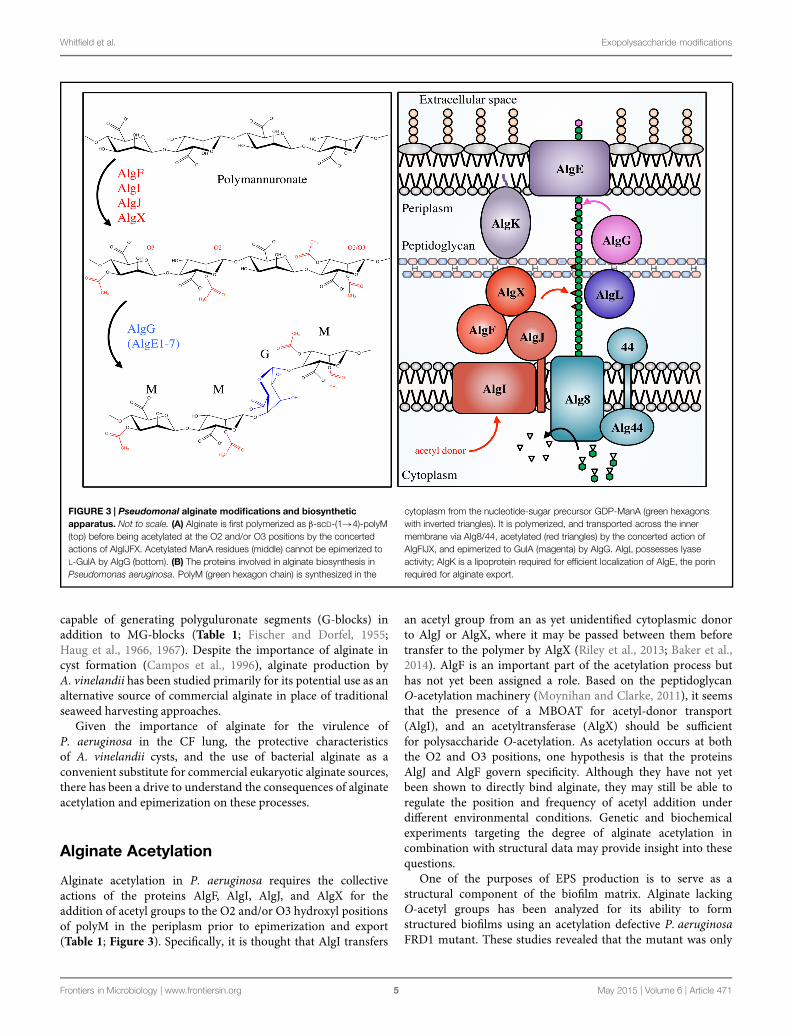
FIGURE 3 | Pseudomonal alginate modifications and biosyntheticapparatus. Not to scale. (A) Alginate is first polymerized as β-scD-(1→4)-polyM(top) before being acetylated at the O2 and/or O3 positions by the concertedactions of AlgIJFX. Acetylated ManA residues (middle) cannot be epimerized toL-GulA by AlgG (bottom). (B) The proteins involved in alginate biosynthesis inPseudomonas aeruginosa. PolyM (green hexagon chain) is synthesized in the
cytoplasm from the nucleotide-sugar precursor GDP-ManA (green hexagonswith inverted triangles). It is polymerized, and transported across the innermembrane via Alg8/44, acetylated (red triangles) by the concerted action ofAlgFIJX, and epimerized to GulA (magenta) by AlgG. AlgL possesses lyaseactivity; AlgK is a lipoprotein required for efficient localization of AlgE, the porinrequired for alginate export.
capable of generating polyguluronate segments (G-blocks) inaddition to MG-blocks (Table 1; Fischer and Dorfel, 1955;Haug et al., 1966, 1967). Despite the importance of alginate incyst formation (Campos et al., 1996), alginate production byA. vinelandii has been studied primarily for its potential use as analternative source of commercial alginate in place of traditionalseaweed harvesting approaches.
Given the importance of alginate for the virulence ofP. aeruginosa in the CF lung, the protective characteristicsof A. vinelandii cysts, and the use of bacterial alginate as aconvenient substitute for commercial eukaryotic alginate sources,there has been a drive to understand the consequences of alginateacetylation and epimerization on these processes.
Alginate Acetylation
Alginate acetylation in P. aeruginosa requires the collectiveactions of the proteins AlgF, AlgI, AlgJ, and AlgX for theaddition of acetyl groups to the O2 and/or O3 hydroxyl positionsof polyM in the periplasm prior to epimerization and export(Table 1; Figure 3). Specifically, it is thought that AlgI transfers
an acetyl group from an as yet unidentified cytoplasmic donorto AlgJ or AlgX, where it may be passed between them beforetransfer to the polymer by AlgX (Riley et al., 2013; Baker et al.,2014). AlgF is an important part of the acetylation process buthas not yet been assigned a role. Based on the peptidoglycanO-acetylation machinery (Moynihan and Clarke, 2011), it seemsthat the presence of a MBOAT for acetyl-donor transport(AlgI), and an acetyltransferase (AlgX) should be sufficientfor polysaccharide O-acetylation. As acetylation occurs at boththe O2 and O3 positions, one hypothesis is that the proteinsAlgJ and AlgF govern specificity. Although they have not yetbeen shown to directly bind alginate, they may still be able toregulate the position and frequency of acetyl addition underdifferent environmental conditions. Genetic and biochemicalexperiments targeting the degree of alginate acetylation incombination with structural data may provide insight into thesequestions.
One of the purposes of EPS production is to serve as astructural component of the biofilm matrix. Alginate lackingO-acetyl groups has been analyzed for its ability to formstructured biofilms using an acetylation defective P. aeruginosaFRD1 mutant. These studies revealed that the mutant was only
Frontiers in Microbiology | www.frontiersin.org 5 May 2015 | Volume 6 | Article 471

Whitfield et al. Exopolysaccharide modifications
TABLE 2 | Biological implications of EPS modifications.
Modification Proteins involved Organism studied Implication of modification Reference
Alginate
Acetylation AlgF, AlgI, AlgJ, AlgX Pseudomonasaeruginosa
Required for surface attachment and formation ofstructured microcolonies
Nivens et al. (2001), Tielenet al. (2005)
Increased polymer viscosity Tielen et al. (2005)
Decreased neutrophil locomotion and lymphocytetransformation
Mai et al. (1993)
Reduced activation of complement and opsonic killingby phagocytes
Pier et al. (2001)
Scavenging of ROS Learn et al. (1987)
Reduced susceptibility to enzymatic degradation Farrell and Tipton (2012)
Increased gel thickness Skjåk-Braek et al. (1989)
Epimerization AlgG, AlgE1-7 (Azotobactervinelandii)
P. aeruginosa Improved gel forming ability (cohesion) Grant et al. (1973), Donatiet al. (2005)
Upregulation of virulence factors through Ca2+sequestration
Horsman et al. (2012)
A. vinelandii Maintain biofilm structure during changingenvironmental conditions
Ertesvåg et al. (1998)
Preserve N2-fixing capability Sabra et al. (2000)
Required for formation of functional cyst coat Steigedal et al. (2008)
PEL
Deacetylation PelA P. aeruginosa Required for biofilm formation (in PSL deficient strains) Colvin et al. (2013)
Cepacian
Acetylation BceOSU Bcc Reduced susceptibility to enzymatic degradation Cescutti et al. (2006)
Scavenging of ROS Cuzzi et al. (2012)
Vibrio polysaccharide (VPS)
Acetylation VpsG Vibrio cholerae Required for robust biofilm formation and wild-typephenotypes
Fong et al. (2010)
Poly-N-acetyl-glucosamine (PNAG)
Deacetylation PgaB (Escherichia coli),HmsF (Yersinia pestis) IcaB(Staphylococcus epidermidisand S. aureus)
S. epidermidis Required for biofilm formation and surface attachment Vuong et al. (2004a)
Resistance to human cationic antimicrobial peptides
Resistance to neutrophil phagocytosis
Persistence in mouse model of infection
S. aureus Required for biofilm formation and surface attachment Cerca et al. (2007)
Resistance to phagocytosis
Persistence in mouse model of infection
E. coli Required for export of polymer and biofilm formation Itoh et al. (2008)
Y. pestis Required for biofilm formation Forman et al. (2006)
Succinylation IcaC S. aureus Modulation improves in vitro fitness Brooks and Jefferson (2014)
able to produce small, unstructured microcolonies that sparselypopulated the examined surface, suggesting an attachment defect(Table 2; Nivens et al., 2001). In contrast, FRD1 formedextensive biofilm structures that exhibited significant structuralheterogeneity. In a separate study, an aggregation defect wasrevealed when the capacity for an acetylation-deficient FRD1mutant to adhere to a steel surface was tested (Tielen et al.,2005). Additionally, the viscosity of extracellular material fromthe acetylation-defective mutant was significantly reduced incomparison to FRD1, suggesting that the loss of O-acetyl groupsled to weakening of inter- and intra-polymer interactions withinthe biofilm matrix. This is supported by rheological studiesof FRD1 biofilms, which suggested that inter-chain alginateinteractions occur primarily through physical entanglements
(Wloka et al., 2005). These entanglements supported an elasticbiofilm architecture, which differed from O-acetylation-defectiveFRD1 mutants which produced weaker biofilms with reducedresistance to tensile forces. Based on these results, it was suggestedthat O-acetyl groups in alginate act as molecular hooks thatimprove the resistance of the entangled alginate structuralnetwork against applied forces (Wloka et al., 2005). While theimportance of alginate acetyl groups for cell aggregation andmicrocolony formation in vitro is well established, the influenceof O-acetyl groups on biofilm formation phenotypes in clinicallyrelevant P. aeruginosa infections or related in vivomodel systemsof infection remain uncharacterized.
P. aeruginosa biofilm formation in the CF lung has been shownto provide significant protection from a variety of host immune
Frontiers in Microbiology | www.frontiersin.org 6 May 2015 | Volume 6 | Article 471
Whitfield et al. Exopolysaccharide modifications
factors. For example, decreased locomotion of neutrophils, aswell as reduced lymphocyte transformation, have been observedwhen these cell types are incubated with alginate (Simpson et al.,1988). However, chemical removal of acetyl groups from alginateled to a complete loss of these inhibitory effects on neutrophiland lymphocyte function, suggesting that alginate O-acetylationis essential for their suppression (Mai et al., 1993). The activationof complement is also affected by the presence of acetyl groups(Pier et al., 2001). This is not surprising given that interactionsbetween alginate and the complement component C3b likelyoccurs through unsubstituted hydroxyl groups (Hostetter et al.,1982), suggesting that the addition of acetyl groups to alginate inP. aeruginosamay have evolved as a mechanism for complementevasion. Activation of the alternative pathway of complement canlead to phagocytic killing, which is also impaired by the presenceof O-acetyl groups. Opsonic killing of the FRD1 O-acetylationdeficient mutant by phagocytes was readily observed, whilewild-type FRD1 was resistant to these attacks (Pier et al.,2001). Alginate is also known to scavenge ROS produced byphagocytic cells during infection. Hypochlorite is a common ROSproduced by phagocytes, and the presence of alginate in mucoidP. aeruginosa provides a significant protective advantage againsthypochlorite over non-mucoid cells in vitro (Learn et al., 1987).This protective effect was, in part, attributed to the O-acetylgroups, as chemically deacetylated alginate exhibited impairedhypochlorite scavenging. Furthermore, addition of hypochloriteto native alginate led to a decrease in viscosity, similar to thatseen for the chemically deacetylated alginate, suggesting thathypochlorite may be specifically reacting with O-acetyl groupsfrom native alginate (Learn et al., 1987).
When bacteria are contending for control of the sameenvironment, they can release extracellular enzymes to degradecritical structural components of cohabiting organisms togive them a competitive advantage (Korotkov et al., 2012).This is observed in the CF lung, where instances of multi-species biofilms are common (Elias and Banin, 2012). Duringcolonization of the CF lung, alginate acetyl groups may serveas a protective mechanism to prevent unwanted degradationof alginate within the biofilm by bacteria that could secretean AlgL-like lyase as an offensive tactic. The P. aeruginosaalginate lyase AlgL preferentially degrades deacetylated alginateor polyM over mature, acetylated alginate (Farrell and Tipton,2012). Furthermore, O-acetyl groups prevent the epimerizationof ManA to GulA by the epimerases AlgE1-7 in A. vinelandii,whichmay allow for control over the degree of epimerization and,in turn, regulation of the cyst coat composition.
Alginate acetylation content ranges from 4 to 57%, dependingon the percentage of ManA present (Skjåk-Braek et al., 1986).The degree of O-acetylation is often observed to vary notonly between different alginate-producing organisms, but alsobetween different strains of the same organism and even withinthe same strain under differing growth conditions (Marty et al.,1992; Peña et al., 2006). For example, modulation of carbonsource during growth for a single alginate-producing P. syringaestrain led to significant differences in acetyl content, rangingfrom 9 to 34% of total uronic acids bearing an acetyl group(Day, 1988). In another study, the alginate produced by several
different strains of P. aeruginosa grown on nutritionally distinctmedia was examined. This study revealed that between differentstrains O-acetyl content of alginate varied between 2 to 56%(Marty et al., 1992). Furthermore, in both studies alginate acetylcontent changed over the course of a single growth experimentby as much as 40%, possibly owing to the availability ofacetyl-CoA, the proposed acetyl donor (Lee and Day, 1998). Inaddition to acetyl-CoA availability, differences in acetyl contentcould conceivably be a means to optimize attachment, nutrientuptake, or nutrient diffusion within the biofilm in the face ofdifferent media compositions and nutrient sources. This notion issupported by findings which suggest that alginate O-acetylationcan enhance the swelling ability of calcium alginate gels (Skjåk-Braek et al., 1989). Deacetylated alginate exhibited poor swellingability in comparison to chemically acetylated variants, withincreasing degrees of acetylation leading to greater swellingvolume. Conversely, increased O-acetylation led to a decrease inthe affinity of alginate gels for calcium ions (Skjåk-Braek et al.,1989). Thus, alginate acetyl content has specific consequenceswith respect to calcium ion binding and the thickness of alginategels, which may influence nutrient diffusion in the biofilm. Thesefindings could potentially be extrapolated to other componentsof the growth media, and suggests a mechanism by whichalginate-producing bacteria could regulate the uptake of essentialnutrients.
Alginate Epimerization
Pseudomonas aeruginosa has a single alginate C5-epimerasein the periplasm, AlgG. In A. vinelandii, there is an AlgGortholog that performs the same function, and seven additionalextracellular epimerases, AlgE1 through AlgE7 (Table 1).Alginate can form strong gels through interactions withGulA residues, mediated by Ca2+ ions. This feature wasthought to be limited to alginates containing G-blocks, andwould therefore exclude the MG-block alginates produced byP. aeruginosa (Grant et al., 1973). However, it is now thoughtthat alginates containing exclusively MG-blocks can also formgels in the presence of Ca2+ (Donati et al., 2005), suggestingthat epimerization by AlgG in P. aeruginosa may serve as amechanism to improve the cohesion of alginate during biofilmformation. It was found that addition of CaCl2 to growth medialed to the production of biofilms that were 10- to 20-fold thickerthan that produced in the absence of Ca2+ (Sarkisova et al.,2005). Ca2+-alginate interactions also regulate virulence factorexpression, as chelation of Ca2+ by alginate induces expressionof the Type 3 secretion system (Horsman et al., 2012). Therefore,it appears that there are mechanisms in place in P. aeruginosa forvirulence factors to be upregulated by the expression of another,thus allowing for concerted actions that improve fitness (Table 2).Despite advances in understanding the interplay between Ca2+and alginate in P. aeruginosa, and the extensive studies performedon acetyl-deficient alginate, there are no reports on the effects ofepimerization on biofilm formation, pathogenicity, or virulence.
In contrast, the role of epimerization in A. vinelandii cystformation has been well characterized. One hypothesis regarding
Frontiers in Microbiology | www.frontiersin.org 7 May 2015 | Volume 6 | Article 471

Whitfield et al. Exopolysaccharide modifications
the ability of A. vinelandii to express multiple epimeraseswith unique activities is that these enzymes allow the alginatestructures to be tailored to different layers of the cyst underdiverse environmental conditions. For example, the epimeraseAlgE1 has two catalytic domains that introduce primarilyMG-blocks and G-blocks, respectively (Ertesvåg et al., 1998).Decreasing the availability of Ca2+ in the presence of AlgE1in vitro led to greater incorporation of G-blocks into polyMalginate. This may provide a means in vivo to maintain thestrength of Ca2+-mediated inter-alginate bonds in the face ofdecreased environmental Ca2+ availability (Ertesvåg et al., 1998).Regulation of alginate structure is also observed during vegetativegrowth of A. vinelandii, where nitrogen fixation is mediated bythe expression of highly oxygen-sensitive nitrogenases. In thisstate, alginate is utilized as a barrier to prevent the diffusion ofoxygen into the cell. In the presence of increasing environmentalO2 concentrations, A. vinelandii was able to produce alginatewith greater G-content, which led to the formation of a thicker,denser alginate layer around the cell and thus limited oxygenpenetration (Sabra et al., 2000). The expression of differentmannuronan C5-epimerases is also regulated over the courseof the A. vinelandii life cycle, including during vegetativegrowth, cyst development, and cyst germination (Høidal et al.,2000). Although the exact biological function for the expressionof specific epimerases at unique points in the life cycle ofA. vinelandii has not been determined, preferential expression ofAlgE7 during cyst germination could be linked to the apparentlyase activity of this enzyme, which may be utilized to degradethe cyst coat (Høidal et al., 2000).
Unlike AlgG in P. aeruginosa, the importance of the AlgE1-7 epimerases in the formation of the cyst coat and toleranceto desiccation has been explored. Inactivation of the AlgE1-7epimerases, either through chromosomal deletion inA. vinelandii(Steigedal et al., 2008) or by inactivating the Type 1 secretionsystem responsible for their export (Gimmestad et al., 2006)led to the production of low G-content alginate, suggestingthat the periplasmic A. vinelandii AlgG is active but not veryefficient. In both cases, these mutants were unable to forma cyst coat and could not survive desiccation. In contrast,deletion of individual algE genes, with the exception of algE3,did not have an appreciable effect on the G-content of alginate.Deletion of algE3 showed a significant reduction in G-content(Steigedal et al., 2008). However, since each of the individual algEdeletion mutants was able to form a functional cyst and survivedesiccation, it appears that no single epimerase is absolutelyessential for cyst formation or germination. This suggests thatthe presence of multiple extracellular epimerases may increaseredundancy of epimerase activity to ensure formation of afunctional cyst coat (Steigedal et al., 2008). It remains tobe determined whether cyst formation under unique stressfulconditions may require specific epimerases, and little work hasbeen done to date to examine the role of mannuronan C5-epimerases during the vegetative stage of A. vinelandii growth.
While a great deal is understood about the regulation ofalginate biosynthesis and its modification at the genetic andprotein level (Hay et al., 2014), the implications of alginateacetylation and epimerization in terms of biofilm formation,
pathogenicity and virulence, environmental adaptability andsurvivability remain largely uncharacterized.
The Pel Polysaccharide
In addition to alginate, P. aeruginosa is capable of synthesizingtwo other polymers that have been implicated in biofilmformation, the Pel and Psl polysaccharides (PEL and PSL;Franklin et al., 2011). Unlike alginate, PEL and PSL are primarilyassociated with the establishment of non-mucoid biofilms. PSL isa neutral, branched polysaccharide with a five-sugar repeat unitcomposed of D-mannose, D-glucose, and L-rhamnose and is notthought to undergo any modifications after polymerization (Byrdet al., 2009). The exact structure of PEL is currently unknown, butit is predicted to be glucose rich (Friedman and Kolter, 2004; Maet al., 2012). Colvin and colleagues have demonstrated that thePEL biosynthesis protein PelA has deacetylase activity in vitroand when residues predicted to be required for deacetylationwere mutated, this activity was lost. Introduction of these PelAdeacetylation mutations in P. aeruginosa PA14, which uses PELas the primary EPS, led to a biofilm deficient phenotype and alack of material recognizable by PEL-reactive antisera (Table 1;Colvin et al., 2013). Given the localization of PelA to theperiplasm (Colvin et al., 2013), deacetylation of PEL followingpolymerization may be necessary for biofilm formation, andsuggests that an acetylated sugar is likely a feature of the PEL.Our current understanding of PEL biosynthesis is limited, andit remains to be determined whether PelA acts directly on PEL,the degree of PEL deacetylation by PelA, and what effect thismodification has on virulence.
Cepacian
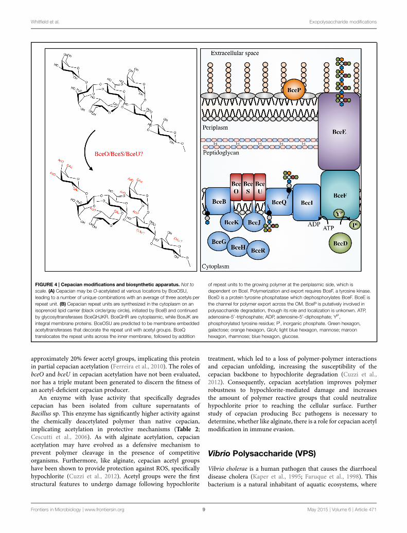
The Bcc is a group of at least 17 different bacterial species,including beneficial environmental isolates, as well asrhizosphere parasites, and plant and animal pathogens(Mahenthiralingam et al., 2002; Vanlaere et al., 2009). TheBcc have become increasingly important as opportunisticpathogens of immunocompromised individuals and thosewith CF (Mahenthiralingam et al., 2005). In CF patients, Bccinfections occasionally develop into a form of necrotisingpneumonia, known as cepacia syndrome, which often leads topatient death (Govan and Deretic, 1996). The majority of bothclinical and environmental Bcc isolates produce the EPS cepacian(Ferreira et al., 2010); a known virulence factor that contributessignificantly to bacterial pathogenicity. Cepacian is composed ofglucose, GlcA, mannose, rhamnose, and galactose in a 1:1:1:1:3ratio and is decorated with acetyl groups (Figure 4; Cérantolaet al., 1999; Cescutti et al., 2000; Linker et al., 2001). The acetylgroups can be found at 12 different locations on the polymerrepeating unit with an average of three acetyl groups presentper repeat unit (Cescutti et al., 2011). The genes responsible forcepacian acetylation were discovered by Ferreira and colleagues,and include the putative acetyltransferases bceO, bceS, and bceU(Ferreira et al., 2010). Mutations in bceS produced cepacian with
Frontiers in Microbiology | www.frontiersin.org 8 May 2015 | Volume 6 | Article 471
Whitfield et al. Exopolysaccharide modifications
FIGURE 4 | Cepacian modifications and biosynthetic apparatus. Not toscale. (A) Cepacian may be O-acetylated at various locations by BceOSU,leading to a number of unique combinations with an average of three acetyls perrepeat unit. (B) Cepacian repeat units are synthesized in the cytoplasm on anisoprenoid lipid carrier (black circle/gray circle), initiated by BceB and continuedby glycosyltransferases BceGHJKR. BceGHR are cytoplasmic, while BceJK areintegral membrane proteins. BceOSU are predicted to be membrane embeddedacetyltransferases that decorate the repeat unit with acetyl groups. BceQtranslocates the repeat units across the inner membrane, followed by addition
of repeat units to the growing polymer at the periplasmic side, which isdependent on BceI. Polymerization and export requires BceF, a tyrosine kinase.BceD is a protein tyrosine phosphatase which dephosphorylates BceF. BceE isthe channel for polymer export across the OM. BceP is putatively involved inpolysaccharide degradation, though its role and localization is unkonwn. ATP,adenosine-5′ -triphosphate; ADP, adenosine-5′-diphosphate; YP,phosphorylated tyrosine residue; Pi, inorganic phosphate. Green hexagon,galactose; orange hexagon, GlcA; light blue hexagon, mannose; maroonhexagon, rhamnose; blue hexagon, glucose.
approximately 20% fewer acetyl groups, implicating this proteinin partial cepacian acetylation (Ferreira et al., 2010). The roles ofbceO and bceU in cepacian acetylation have not been evaluated,nor has a triple mutant been generated to discern the fitness ofan acetyl-deficient cepacian producer.
An enzyme with lyase activity that specifically degradescepacian has been isolated from culture supernatants ofBacillus sp. This enzyme has significantly higher activity againstthe chemically deacetylated polymer than native cepacian,implicating acetylation in protective mechanisms (Table 2;Cescutti et al., 2006). As with alginate acetylation, cepacianacetylation may have evolved as a defensive mechanism toprevent polymer cleavage in the presence of competitiveorganisms. Furthermore, like alginate, cepacian acetyl groupshave been shown to provide protection against ROS, specificallyhypochlorite (Cuzzi et al., 2012). Acetyl groups were the firststructural features to undergo damage following hypochlorite
treatment, which led to a loss of polymer-polymer interactionsand cepacian unfolding, increasing the susceptibility of thecepacian backbone to hypochlorite degradation (Cuzzi et al.,2012). Consequently, cepacian acetylation improves polymerrobustness to hypochlorite-mediated damage and increasesthe amount of polymer reactive groups that could neutralizehypochlorite prior to reaching the cellular surface. Furtherstudy of cepacian producing Bcc pathogens is necessary todetermine, whether like alginate, there is a role for cepacian acetylmodification in immune evasion.
Vibrio Polysaccharide (VPS)
Vibrio cholerae is a human pathogen that causes the diarrhoealdisease cholera (Kaper et al., 1995; Faruque et al., 1998). Thisbacterium is a natural inhabitant of aquatic ecosystems, where
Frontiers in Microbiology | www.frontiersin.org 9 May 2015 | Volume 6 | Article 471
Whitfield et al. Exopolysaccharide modifications
it forms biofilms on a variety of surfaces, including plankton,plants, crustaceans, insects, and sediment (Huq et al., 1983, 1995;Halpern et al., 2004; Broza et al., 2005). In areas where cholerais endemic, V. cholerae has been shown to form suspendedbiofilm-like aggregates in surface waters, however, when particles>20 μm in diameter are removed from water sources, theincidence of cholera can be reduced (Huq et al., 1996; Colwellet al., 2003). Furthermore, it has been shown that the averageinfectivity of the aggregate form of V. cholerae is significantlyhigher than that of planktonic cells (Faruque et al., 2006),and biofilm formation within aquatic ecosystems significantlyimproves V. cholerae fitness and persistence (Matz et al., 2005).A major component of the biofilm produced by V. cholerae isan EPS called VPS. This polymer is thought to be producedduring infection and contributes to bacterial colonization andsurvival (Yildiz and Schoolnik, 1999; Fong et al., 2010). Thechemical structure of VPS revealed a backbone containingthe unusual constituent GulNAcAGly: the amide formed from2-acetamido-2-deoxy-L-guluronic acid and glycine (Figure 6;Yildiz et al., 2014). Of the genes involved in VPS biosynthesis,originally identified using a transposon mutagenesis screen(Yildiz and Schoolnik, 1999), two putative acetyltransferases,vpsG and vpsC, were identified (Fong et al., 2010). Deletion ofvpsG results in reduced biofilm formation and altered biofilm-related phenotypes, as well as weak reactivity with VPS antisera,suggesting that it may modify the polymer, perhaps throughacetylation (Table 1). In contrast, deletions of vpsC do notaffect biofilm formation or VPS production, suggesting thatvpsC is inactive, not expressed, or is performing some otherfunction in VPS biosynthesis besides polymermodification (Fonget al., 2010). The chemical composition of VPS produced byvpsG and vpsC mutants was not studied for alterations inacetyl content. The presence of GulNAcA in VPS may bethe result of epimerization by the predicted GDP-mannosedehydrogenase VpsB, a conversion similar to the ManA toGulA epimerization catalyzed by AlgG in the biosynthesis ofalginate by P. aeruginosa (Wolfram et al., 2014; Yildiz et al.,2014). The unusual glycine modification in VPS requires furtherexploration, as the enzyme responsible for its addition is presentlyunknown. Given the important role of VPS in V. choleraepathogenesis and environmental persistence, and the recentdetermination of its precise chemical structure, we anticipate thatthe proteins involved in VPS modification will soon be identifiedand characterized.
Glycosaminoglycans (GAGs)
GAGs are a group of polymers that are typically composed ofa disaccharide repeat unit containing an amino sugar and ahexuronic acid (Laurent and Fraser, 1992; Esko and Lindahl,2001; DeAngelis, 2002; Silbert and Sugumaran, 2002). GAGswere initially thought to exist only in the animal kingdom, wherethey serve essential biological functions, however, there hasbeen an emergence of GAG-like polymers amongst prokaryotes(Vann et al., 1981; Rodriguez et al., 1988; DeAngelis et al.,2002). Prokaryotic GAGs are typically less complex than their
eukaryotic counterparts due to an absence of modifications suchas sulfation (Raedts et al., 2011). HS, for example, is an essentialGAG in animals and is composed of repeating disaccharidesof GlcA and GlcNAc (Kjellén and Lindahl, 1991). HS can bemodified post-polymerization by a glucuronyl C5-epimerase,which converts GlcA to IdoA, as well as by the addition of sulfategroups to GlcNAc or IdoA moieties. Mouse embryos lackingthe GlcA C5-epimerase display a lethal phenotype characterizedby skeletal malformations and lung defects (Li et al., 2003),highlighting the importance of HS epimerization in murinedevelopment. Interestingly, the K5 antigen of E. coli O10:K5:H4has an identical structure to heparosan, the unsulfated, non-epimerized backbone structure of HS (Vann et al., 1981). K5heparosan is a form of molecular camouflage, as it impartslow immunogenicity to the bacterium in humans and henceincreased pathogenicity (Vann et al., 1981).
Although sulfation has not yet been observed amongstprokaryotic GAGs, IdoA residues have been found to beconstituents of bacterial GAGs (Figure 6). The identificationof the bacteria glucuronyl C5-epimerase has proven elusive(Raedts et al., 2011), however, an enzyme (RED65_08024) fromthe marine bacterium Bermanella marisrubi that shares 37%sequence similarity with the human glucuronyl C5-epimerasehas been characterized and shown in vitro to convert GlcAto IdoA in de-sulfated mouse HS (Raedts et al., 2013). Thisglucuronyl C5-epimerase represents the first prokaryotic proteincapable of generating IdoA residues, and is the only identifiedepimerase that can function on bacterial polysaccharides post-polymerization, besides AlgG and AlgE1-7. Unfortunately, theEPS produced by B. marisrubi has not been characterized, soits target remains unknown. Nevertheless, the ability of bacteriato more closely replicate the structures of essential humanpolysaccharides by expression of homologous modificationenzymes likely serves as a mechanism to mask their presencefrom the host immune system (Cress et al., 2014).
Poly-β-1,6-N-Acetyl-glucosamine(PNAG)
Poly-β-1,6-N-acetyl-glucosamine is a poly-GlcNAc polymerthat is produced by a wide range of Gram-positive andGram-negative bacterial pathogens, including Staphylococcusepidermidis, Staphylococcus aureus, Escherichia coli, Yersiniapestis, Bordetella sp., Acinetobacter baumanii, Actinobacilluspleuropneumoniae, Burkholderia cepacia complex (Bcc), andAggregatibacter actinomycetemcomitans (Cramton et al., 1999;Vuong et al., 2004b; Wang et al., 2004; Izano et al., 2007,2008; Parise et al., 2007; Bobrov et al., 2008; Choi et al., 2009).These organisms are responsible for a wide spectrum of diseases,including but not limited to, hospital acquired infections, toxicshock syndrome, plague, and whooping cough. Depending onthe source or organism in question, PNAG may also be referredto as PGA (polyglucosamine, in Gram-negative bacteria), PIA(in Gram-positive bacteria), poly-NAG, hms+ (in Y. pestis),or BPS (Bordetella polysaccharide, in Bordetella sp.). Given thedifferences in PNAG modifications between Gram-positive and
Frontiers in Microbiology | www.frontiersin.org 10 May 2015 | Volume 6 | Article 471

Whitfield et al. Exopolysaccharide modifications
Gram-negative bacteria, as described below, we will use PGA andPIA to refer to PNAG polymer produced by Gram-negative andGram-positive organisms, respectively.
Initially S. epidermidis was thought to produce severaldifferent polymers, but the discovery of the icaADBC operon(Heilmann et al., 1996a,b; Gerke et al., 1998) revealed thatonly a single polymer, PIA, was produced (Tojo et al., 1988;Christensen et al., 1990; Heilmann et al., 1996a; Mack et al.,1996; McKenney et al., 1998). PIA is a partially deacetylatedβ-1,6-GlcNAc polymer. In S. epidermidis and S. aureus 15–20% of the N-acetyls are removed by the extracellular, cell-surface associated polysaccharide deacetylase IcaB (Table 1;Figure 5; Vuong et al., 2004a; Cerca et al., 2007). In additionto deacetylation, approximately 6 and 10% of GlcNAc residues
in S epidermidis and S. aureus, respectively, are O-succinylated(Joyce et al., 2003; Sadovskaya et al., 2005). This modification isthought to be performed by themembrane localized protein, IcaC(Atkin et al., 2014). Interestingly, a mechanism of phase variation,where bacteria modulate virulence phenotypes at the genomelevel in a rapid on/off fashion, was noted in S. aureus whereinslipped-strand mispairing led to inactivation of icaC (Brooks andJefferson, 2014). This phenotype confers a fitness advantage thatwas not seen when the ica operon was deleted, which may be aresponse to modulate PIA O-succinylation and thus decrease theoverall anionic charge of the polymer.
The production of PGA has been extensively characterizedin E. coli, where the pgaABCD operon encodes the proteinsnecessary for its biosynthesis (Wang et al., 2004). In E. coli,
FIGURE 5 | The PNAG and PIA modifications and biosyntheticapparatus. Not to scale. (A) PNAG polymers are partially deacetylated byPgaB in Escherichia coli, or IcaB in Staphylococcal species. It has beenproposed that IcaC O-succinylates the polymer in certain Staphylococcalspecies, however, the location of the succinyl groups and the order ofdeacetylation/succinylation has not yet been determined. The proteins involvedin PNAG biosynthesis in E. coli (B), and PIA synthesis in Staphylococcal species
(C). PNAG (blue hexagon chain) is synthesized in the cytoplasm from thenucleotide-sugar precursor UDP-GlcNAc (blue hexagons with green invertedtriangles). The polymer is transported across the inner membrane via PgaCD,deacetylated (light blue hexagons) by PgaB in the periplasm, and then exportedthrough the PgaA porin. PIA is transported across the cytoplasmic membraneby IcaAD, then partially deacetylated by IcaB in the extracellular space. PIA hasbeen proposed to be O-succinylated by IcaC (magenta star).
Frontiers in Microbiology | www.frontiersin.org 11 May 2015 | Volume 6 | Article 471

Whitfield et al. Exopolysaccharide modifications
FIGURE 6 | Exopolysaccharide modifications of VPS and Heparosan.(A) VPS is produced by Vibrio cholerae O1 El Tor and contains an O-acetylgroup, likely added by VpsG. In addition the polymer has a glycine modificationand an N-acetyl group; the enzymes responsible for these modifications havenot been determined. VPS contains a GulA residue which is epimerized by an
unknown C5-epimerase. (B) Heparosan, a GAG composed of a disacchariderepeating unit of GlcA and GlcNAc, produced by select bacteria such as theurinary tract pathogen E. coli O10:K5:H4. A C5-epimerase introduces IdoAresidues. The proteins involved in, and mechanism of, biosynthesis of the aboveEPS have not been fully resolved.
approximately 3–5% of N-acetyls are removed by the lipoproteinPgaB (Wang et al., 2004; Itoh et al., 2008; Little et al., 2012). TheN-terminal domain of PgaB is homologous to IcaB in Gram-positive bacteria but the protein is located on the inner leaflet ofthe outer membrane. There is no IcaC ortholog in the pgaABCDoperon, which is consistent with an observed lack of O-succinylgroups in PGA.
Partial deacetylation of PNAG by both IcaB and PgaB isimportant for a variety of biofilm-associated phenotypes inS. epidermidis, S. aureus, and E. coli. Deletion of icaB inS. epidermidis led to the production of fully acetylated PIA,suggesting that IcaB is not necessary for polymer production(Vuong et al., 2004a). However, the fully acetylated polymerwas not retained at the cellular surface and was shed into theculture media, which led to deficiencies in biofilm formationand surface attachment (Table 2). The lack of deacetylationled to a loss of cationic charge in the polymer, which maybe essential for interactions with the anionic cell surfaceof S. epidermidis (Vuong et al., 2004a). Furthermore, icaB-deficient mutants of S. epidermidis were more susceptible tohuman cationic antimicrobial peptides and phagocytosis byneutrophils, and were unable to persist in a mouse modelof device-related infections (Vuong et al., 2004a). Deletion oficaB in S. aureus produced similar phenotypes (Cerca et al.,2007). Intriguingly, the production of wall teichoic acids,the predominant anionic component of the Gram-positivebacterial envelope, was dispensable for adherence of PIA tothe cell surface of S. aureus (Vergara-Irigaray et al., 2008),suggesting that other less prevalent anionic species mediate thisinteraction.
In contrast to IcaB, inactivation of PgaB in E. coli preventedpolymer export, suggesting that partial deacetylation is necessaryfor export through the predicted outer membrane porin PgaA(Itoh et al., 2008). This is in line with findings that suggestconformational changes in the C-terminal domain of PgaB, uponbinding of deacetylated PNAG, assist in targeting PNAG forexport (Little et al., 2014b). Deacetylation has also been studiedin Y. pestis, where a PNAG-like polymer is thought to mediatebiofilm formation. Biofilm formation in Y. pestis is crucial for
its zoonotic transmission (Jarrett et al., 2004). In the flea, theproventriculus, a feeding tube covered in spines that connects themidgut to the esophagus, provides a platform for the adhesionof Y. pestis aggregates. Subsequent colonization impedes bloodpassage and leads to transposition of Y. pestis from flea tomammal when a flea attempts excessive feeding due to a partialor completely blocked proventriculus (Jarrett et al., 2004). ThehmsHFRS operon in Y. pestis is orthologous to the pgaABCDoperon, where HmsF is the outer membrane localized deacetylasewith structural similarity to PgaB (Forman et al., 2006). Mutationor deletion of hmsF led to a deficiency in biofilm formation.This suggests HmsF in Y. pestis may be analogous to PgaB inE. coli in terms of de-N-acetylation activity and importance forpolysaccharide export and biofilm formation.
While PNAG production has been studied primarily inS. aureus, S. epidermidis, and E. coli, there are a multitude ofadditional pathogenic bacteria, fungi, and protozoans that mayproduce this polymer (Cywes-Bentley et al., 2013). PNAG couldrepresent the first example of an EPS that is broadly utilized bypathogenic organisms as a mechanism to improve fitness in theenvironment or during infection.
Insights from Modification of OtherMicrobial Polysaccharides
The implications of EPS modifications in pathogenic bacteriahave been studied to some extent, particularly in alginate andPNAG producing bacteria (Figure 2; Table 2). However, thebreadth of our knowledge in this field remains limited. Despitethis, comparable modifications found on LPS and CPSs havebeen studied extensively in an effort to identify vaccine targets,and can be used for comparison purposes to generate newhypotheses regarding EPS modifications (Cody et al., 2003; Pintoand Berti, 2014). In particular, the study of polysaccharide acetylmodifications has clarified their role in mediating a variety ofsurvival mechanisms.
Many of the protective benefits of EPS acetyl modificationsdescribed above have been noted for other bacterial pathogens.
Frontiers in Microbiology | www.frontiersin.org 12 May 2015 | Volume 6 | Article 471
Whitfield et al. Exopolysaccharide modifications
For instance, in Haemophilus influenzae, an opportunisticpathogen of the upper respiratory tract, acetylation of LPSby the acetyltransferase OafA leads to increased resistance tocomplement-mediated killing by human serum (Fox et al.,2005). Similarly, in S. aureus, the acetyltransferase Cap5H, whichis responsible for the O-acetylation of type 5 CPS, confersprotection against opsonophagocytic killing and improvespropagation in a murine model of infection (Bhasin et al.,1998). Type 5 CPS producers also exhibit increased survivalrates in murine models of bacteremia and renal abscessformation and resistance to killing in whole mouse bloodand opsonophagocytic assays, in comparison to producers ofthe structurally similar type 8 CPS which have reduced levelsof N-acetylation (Watts et al., 2005). Beyond the prokaryoticdomain, acetyl modifications are also incorporated into theCPS of the pathogenic fungus Cryptococcus neoformans toevade complement activation (Fujihara et al., 1997), decreasethe efficiency of capsule clearance by the host (Kozel et al.,2003), and inhibit neutrophil migration (Ellerbroek et al., 2004)during cryptococcosis. An excellent example of the benefits ofpolysaccharide acetyl modifications comes from a survey ofclinical isolates of Streptococcus pneumoniae and E. coliK1, whichfound that the bacteria expressing acetyl-decorated polymerswere more virulent and invasive than those that expressedpolymers lacking the modification (Frasa et al., 1993; Melinet al., 2010). Therefore, the immunomodulatory characteristicsof acetyl modifications are utilized by a wide range of pathogenicorganisms and likely represent a general mechanism for survivaland proliferation within the host.
The above notion is firmly supported by studies of serotypevariation within the context of CPS biosynthesis. In Streptococcuspneumoniae, a causative agent of meningitis, bacteremia, andpneumonia, there are more than 90 different capsule serotypeswith unique carbohydrate structures and biosynthetic loci.This has evolved, in part, as a mechanism to overcomeserotype-specific host mechanisms of adaptive immunity that canefficiently clear infections. In some serotypes, such as 9V/9A,11A/11E, and 15B/15C, the CPS structures differ only in thedegree of O-acetylation (Jansson et al., 1987; Rutherford et al.,1991; Zartler et al., 2009). Mechanisms within S. pneumoniaehave been revealed that allow for serotype switching duringinfection as a means to actively evade the host immune response.In the case of serotypes 9V and 11A, inactivating mutations in theacetyltransferase-encoding gene wceJ led to expression of non-acetylated 9A and 11E capsule (Calix andNahm, 2010; Calix et al.,2011). Moreover, certain wceJ mutations only partially inhibitacetyltransferase activity, which have led to intermediate 9V/9Aand 11A/11E phenotypes (Calix et al., 2011, 2014). In the caseof 15B/15C serotype switching, the process is reversible due toslipped-strand mispairing of the acetyltransferase-encoding genewciZ (van Selm et al., 2003). Regardless of the mechanism, thismid-infection serotype variation provides significant protectiveadvantages to S. pneumoniae in terms of antibody evasion. Forexample, antibodies generated against O-acetylated serotype 15Bwere unreactive with non-acetylated 15C polymer (Rajam et al.,2007), and serotype 9V specific antibodies exhibited reducedspecificity for 9A polymer (Calix et al., 2012). Furthermore,
10–20% of individuals receiving a S. pneumoniae vaccine targetedagainst the 9V polysaccharide did not generate antibodiestargeting serotype 9A (McNeely et al., 1998). In addition, theability of acetyl groups to mask protective epitopes of bacterialpolysaccharides has been noted for the Vi antigen of Salmonellatyphi (Szu et al., 1991), Salmonella typhimurium O-antigen (Kimand Slauch, 1999) and Neisseria meningitidis serogroup A, C,and Y CPS (Michon et al., 2000; Berry et al., 2002; Fusco et al.,2007). Therefore, through modulation of acetyl groups on thepolymer, a wide variety of pathogenic bacteria are able to evadehost-mediated mechanisms of adaptive immunity.
The above examples illustrate scenarios in which acetylmodification is an all-or-nothing response to adaptive immunity,however, in the case of GBS, acetyl levels on its sialic acid CPScan be fine-tuned by the actions of the acetyltransferase NeuDand the acetylesterase NeuA (Lewis et al., 2006, 2007). Differentdegrees ofO-acetylation in GBS CPS have been linked to differentstages of invasion and infection. For instance, it is thoughtthat during the asymptomatic stages of initial colonization andpersistence in the human gastrointestinal and vaginal tracts, GBSproduces an extensively acetylated form of CPS to protect againstdegradation by sialidases introduced by competing microbesin these environments (Weiman et al., 2009). However, highlyacetylated CPS renders GBS more susceptible to killing byneutrophils and reduces virulence during stages of opportunisticinfections (Weiman et al., 2010). Therefore, during infectionit is thought that GBS produces a sparsely acetylated formof CPS that improves resistance to neutrophil-mediated killingthrough reduced neutrophil activation and production of pro-inflammatory cytokines, and enhances survival in the murinebladder (Kline et al., 2011). Interestingly, this variant of CPSis also able to promote the persistence of uropathogenic E. coliin co-culture urinary tract infection models (Kline et al.,2012). Therefore, in certain pathogens, specific degrees ofpolysaccharide acetylation allow for adaptation during differentstages of colonization and infection.
The above examples of acetyl modulation in LPS andCPS not only reinforce the importance of EPS acetylationfor pathogenicity and persistence, but also provide additionalperspectives in considering the variability of this modificationobserved in alginate and cepacian. For instance, cepacian hason average three acetyl groups per repeat unit, each located onone of 12 potential positions (Cescutti et al., 2011). Therefore,each cepacian repeat unit can have one or more acetyl groupsat any of 12 positions, generating an overwhelming number ofunique acetyl decoration patterns. Given the importance of acetylgroups in forming or masking antibody epitopes, this level ofdiversity would make the generation of protective antibodies orthe development of an effective vaccine extraordinarily difficult.Furthermore, production of such a heterogeneous polymer likelyrequires an arsenal of regulatory factors and/or acetyltransferases,very few of which have been discovered in the context of cepacianbiosynthesis (Ferreira et al., 2011). Similar to S. pneumoniaeand N. meningitidis CPS production, members of the Bcc maymodulate the presentation of cepacian acetyl groups through anas yet unknown mechanism as a means to evade host adaptiveimmune mechanisms.
Frontiers in Microbiology | www.frontiersin.org 13 May 2015 | Volume 6 | Article 471

Whitfield et al. Exopolysaccharide modifications
The degree of acetylation and epimerization of alginate haslong been known to differ depending on the organism andstrain, as well as the composition of the growth medium (Day,1988; Marty et al., 1992; Peña et al., 2006). This reflects, inpart, a need to adapt to the specific conditions imposed bydifferent nutritional media, and may mirror other features of theenvironment from which the organism was isolated. In line withthis concept, additional promoters within the alginate operonhave been identified upstream of algG and algIJF in P. aeruginosa,suggesting that there may be modes of regulating the levelsof these modifying enzymes independently of the rest of thealginate biosynthesis machinery (Paletta and Ohman, 2012). Theupregulation ofO-acetylation machinery would not only increasealginate acetyl content, but would also decrease the availabilityof substrate for AlgG and thus decrease epimerization levels.Conversely, upregulation of algG would increase the numberof G-residues that cannot act as substrates for O-acetylation.Therefore, there is the potential for a complicated regulatoryinterplay between these processes, much like the reciprocalO-acetylation/de-O-acetylation of GBS CPS that allows for fine-tuning of acetyl levels at different stages of infection.
The degree of PNAG de-N-acetylation is not known to varyconsiderably, and the exact processes involved in VPS acetylationand epimerization, PIA O-succinylation, PEL deacetylationand GAG epimerization are poorly understood. However, theability to perform these types of modifications in a randomfashion may increase the difficulty in generating antibodiesthat recognize specific epitopes on these EPS, either duringhost adaptive immune responses or in vaccine development(Gening et al., 2010). As such, EPS modifications are capable ofimparting beneficial characteristics upon polymers that improvepersistence, survival, or evasion of the immune response in theircognate bacteria regardless of their frequency, mechanism ofaddition to the polymer, or chemical properties.
Reflection and Future Perspectives
Identifying and characterizing biofilm EPS is difficult andthere are a number of hurdles that need to be overcome.One of the initial challenges involves culturing biofilm-formingbacteria. Identification of an appropriate medium and growthconditions is required to study EPS production of certainmicroorganisms in the laboratory (Stewart, 2012). Of thosethat can be cultured, it is imperative to use similar growthconditions when making experimental comparisons in theliterature, as variations can affect the presence or degree ofdifferent polysaccharide modifications. With alginate, varyinglevels of acetylation and epimerization are observed dependingon the culture conditions, as well as varying biofilm phenotypesof identical P. aeruginosa strains (Pier et al., 2001; Tielen et al.,2005). Additionally, conflicting studies on the levels of pyruvyland O-acetyl modifications to xanthan gum were attributed todifferent media conditions (Bradshaw et al., 1983). Differentmedia or culturing equipment may also affect experiments suchas surface attachment assays. For example, different types ofplastics were found to affect PIA-mediated surface attachment
in microtitre plates (Maira-Litràn et al., 2004). This suggeststhat during preliminary analyses, multiple types of media anddifferent materials including plastics and glass should be tested toensure the validity of observed biofilm phenotypes. Variations inthe abundance or type of modifications on a given polymer underdifferent experimental conditions can be difficult to quantify;however, this variation likely reflects the ability of differentbacteria to adapt to unique situations. Many EPS-producingbacteria naturally exist in diverse environments and are alsoable to infect various hosts and survive in specific tissues.Additionally, during the course of infection, the environmentwithin the host will change as immune mechanisms attempt toeradicate the bacteria and the surrounding tissue suffers damage.Variations in the degree of modifications under different growthconditions or stressors may therefore provide valuable insightinto bacterial adaptation.
The majority of biofilm studies focusing on EPS modificationshave been performed using in vitro model systems with mono-species cultures. However, the majority of biofilms from chronicinfections differ significantly from those studied in the laboratory(Bjarnsholt et al., 2013a). One significant issue lies in the use ofabiotic surfaces, such as cover slides or the plastic surfaces of96-well plates for the growth and study of biofilms. While somesystems can closely approximate the conditions encountered invivo, such as flow cell models for catheter associated infections,biofilms from infections like CF pneumonia or those encounteredin epidermal wounds are thought to involve attachment to hostcells or host-derived molecules (Bjarnsholt et al., 2009). In theseinstances it is challenging to extrapolate results obtained invitro to chronic biofilm infections, as modifications that appearimportant for attachment to abiotic surfaces may be disposablefor attachment to host tissues, or vice versa (Leuck et al., 2014).Furthermore, many biofilms encountered in the clinic are notcomprised of a single species of bacteria, but rather containmixtures that can include pathogenic or non-pathogenic bacteriaof host or environmental origin, as well as fungi (Wargo andHogan, 2006; Elias and Banin, 2012). These types of microbialinteractions have been studied extensively in the CF lung,where P. aeruginosa has been shown to interact with the Bccopportunistic pathogens B. cepacia and B. cenocepacia (Chattorajet al., 2010; Schwab et al., 2014), as well as the pathogenicfungus Aspergillus fumigatus (Chotirmall and McElvaney, 2014).In the case of mixed P. aeruginosa and B. cenocepacia biofilms,the production of alginate by P. aeruginosa has been shownto promote B. cenocepacia persistence in a mouse model ofCF (Chattoraj et al., 2010). This highlights how the productionof EPS in mixed species biofilms can have implications thatextend beyond the source organism, and suggests that dataobtained regarding the presence or absence of EPS modificationsin monospecies biofilms in vitromay have additional significancein multispecies biofilms.
Following bacterial culturing, difficulties in polysaccharideisolation due to the compositional complexity of the biofilmmatrix or polymer insolubility may necessitate extensiveoptimization of purification protocols. Typically, separationof the polymer from the cell surface may require the useof procedures that lyse the associated bacteria, introducing
Frontiers in Microbiology | www.frontiersin.org 14 May 2015 | Volume 6 | Article 471

Whitfield et al. Exopolysaccharide modifications
additional contaminants (Bales et al., 2013). Therefore, it ispreferable if possible to isolate EPS from culture supernatants;however, such polymers may exhibit different properties fromtheir cell-associated counterparts leading to discrepant analysis(Maira-Litràn et al., 2002). In either case, once the polymer hasbeen obtained from the cell surface or supernatant, contaminantssuch as DNA, RNA and protein must be removed. This can beachieved through enzymatic digestion or chemical precipitationof contaminants, or precipitation of EPS. Contaminatingcarbohydrates can be removed by chromatographic techniquessuch as size exclusion or ion exchange (Bales et al., 2013).In some cases, purification of EPS leads to insufficient yieldsfor subsequent compositional or structural analysis, often as aresult of polymer insolubility. As a result, harsher conditionsmay need to be employed to solubilize the polymer, includingthe use of strong acids or bases. During initial structuralstudies of PIA, strong alkaline purification conditions led to theincorrect identification of N-succinyl groups on the polymer(McKenney et al., 1999; Maira-Litràn et al., 2002), which wasactually a degradation product of glucosamine monosaccharides(Sadovskaya et al., 2005). Additionally, phosphates have beenreported in the monosaccharide composition of polymers, whichare occasionally remnants from purification buffers, or teichoicacids in the case of Gram-positive bacteria (Sadovskaya et al.,2005). Harsher conditions can also partially or completelyremove functional groups, such as acetyls, or even degradethe polymer, leading to incorrect calculations of molecularweight. The EPS described in this review are the select fewwhose structures have been determined, or for which we have
biochemical and genetic data supporting the importance ofthe polysaccharide modifications. With improved culturing,purification and structural determination procedures, additionalEPS will be discovered and their modifications characterized.We will then be able to formulate trends between differenttypes of modifications and their effect on biofilm formation,pathogenicity, and virulence. Furthermore, the details regardingthe genes and proteins involved in the addition of polysaccharidemodifications remain largely unavailable. Characterization ofthese genes and proteins will likely provide details on how thelevels and types of modifications are regulated under differentconditions. Such findings will have a significant impact onour understanding of bacterial pathogenicity, and may revealnovel drug targets aimed at inhibiting the biosynthesis of theseimportant virulence factors.
Acknowledgments
The authors thank Drs. Dustin Little, Perrin Baker, and MarkNitz for their critical reading and contributions to this review.This work was supported by the Canadian Institutes of HealthResearch Operating Grant 13337 to PLH. GBW is supportedby a graduate scholarship from the Natural Sciences andEngineering Research Council of Canada (NSERC). LSM issupported by graduate scholarships from NSERC, the OntarioGraduate Scholarship Program and The Hospital for SickChildren Foundation Student Scholarship Program. PLH is therecipient of a Canada Research Chair.
References
Aachmann, F. L., Svanem, B. I. G., Güntert, P., Petersen, S. B., Valla, S., andWimmer, R. (2006). NMR structure of the R-module: a parallel beta-roll subunitfrom an Azotobacter vinelandii mannuronan C-5 epimerase. J. Biol. Chem. 281,7350–7356. doi: 10.1074/jbc.M510069200
Allesen-Holm, M., Barken, K. B., Yang, L., Klausen, M., Webb, J. S., Kjelleberg, S.,et al. (2006). A characterization of DNA release in Pseudomonas aeruginosacultures and biofilms. Mol. Microbiol. 59, 1114–1128. doi: 10.1111/j.1365-2958.2005.05008.x
Atkin, K. E., Macdonald, S. J., Brentnall, A. S., Potts, J. R., and Thomas, G. H.(2014). A different path: revealing the function of staphylococcal proteins inbiofilm formation. FEBS Lett. 588, 1869–1872. doi: 10.1016/j.febslet.2014.04.002
Baker, P., Ricer, T., Moynihan, P. J., Kitova, E. N., Walvoort, M. T. C., Little, D. J.,et al. (2014). P. aeruginosa SGNH hydrolase-like proteins AlgJ and AlgX havesimilar topology but separate and distinct roles in alginate acetylation. PLoSPathog. 10:e1004334. doi: 10.1371/journal.ppat.1004334
Bales, P. M., Renke, E. M., May, S. L., Shen, Y., and Nelson, D. C. (2013).Purification and characterization of biofilm-associated EPS exopolysaccharidesfrom ESKAPE organisms and other pathogens. PLoS ONE 8:e67950. doi:10.1371/journal.pone.0067950
Benziman, M., Haigler, C. H., Brown, R. M., White, A. R., and Cooper, K. M.(1980). Cellulose biogenesis: polymerization and crystallization are coupledprocesses in Acetobacter xylinum. Proc. Natl. Acad. Sci. U.S.A. 77, 6678–6682.doi: 10.1073/pnas.77.11.6678
Berry, D. S., Lynn, F., Lee, C.-H., Frasch, C. E., and Bash, M. C. (2002). Effect ofO acetylation of Neisseria meningitidis serogroup A capsular polysaccharide ondevelopment of functional immune responses. Infect. Immun. 70, 3707–3713.doi: 10.1128/IAI.70.7.3707-3713.2002
Bhasin, N., Albus, A., Michon, F., Livolsi, P. J., Park, J. S., and Lee, J. C. (1998).Identification of a gene essential for O-acetylation of the Staphylococcus aureus
type 5 capsular polysaccharide. Mol. Microbiol. 27, 9–21. doi: 10.1046/j.1365-2958.1998.00646.x
Bjarnsholt, T., Alhede, M., Alhede, M., Eickhardt-Sørensen, S. R., Moser, C.,Kühl, M., et al. (2013a). The in vivo biofilm. Trends Microbiol. 21, 466–474.doi: 10.1016/j.tim.2013.06.002
Bjarnsholt, T., Ciofu, O., Molin, S., Givskov, M., and Høiby, N. (2013b). Applyinginsights from biofilm biology to drug development – can a new approach bedeveloped?Nat. Rev. Drug Discov. 12, 791–808. doi: 10.1038/nrd4000
Bjarnsholt, T., Jensen, P. Ø., Fiandaca, M. J., Pedersen, J., Hansen, C. R., Andersen,C. B., et al. (2009). Pseudomonas aeruginosa biofilms in the respiratory tract ofcystic fibrosis patients. Pediatr. Pulmonol. 44, 547–558. doi: 10.1002/ppul.21011
Bobrov, A. G., Kirillina, O., Forman, S., Mack, D., and Perry, R. D. (2008). Insightsinto Yersinia pestis biofilm development: topology and co-interaction of Hmsinner membrane proteins involved in exopolysaccharide production. Environ.Microbiol. 10, 1419–1432. doi: 10.1111/j.1462-2920.2007.01554.x
Bradshaw, I. J., Nisbet, B. A., Kerr, M. H., and Sutherland, I. W. (1983). Modifiedxanthan—its preparation and viscosity. Carbohydr. Polym. 3, 23–38. doi:10.1016/0144-8617(83)90010-3
Brooks, J. L., and Jefferson, K. K. (2014). Phase variation of poly-N-acetylglucosamine expression in Staphylococcus aureus. PLoS Pathog.10:e1004292. doi: 10.1371/journal.ppat.1004292
Broza, M., Gancz, H., Halpern, M., and Kashi, Y. (2005). Adult non-biting midges:possible windborne carriers of Vibrio cholerae non-O1 non-O139. Environ.Microbiol. 7, 576–585. doi: 10.1111/j.1462-2920.2005.00745.x
Buchinger, E., Knudsen, D. H., Behrens, M. A., Pedersen, J. S., Aarstad, O. A.,Tøndervik, A., et al. (2014). Structural and functional characterization of theR-modules in alginate C-5 epimerases AlgE4 and AlgE6 from Azotobactervinelandii. J. Biol. Chem. 289, 31382–31396. doi: 10.1074/jbc.M114.567008
Buckmire, F. L. (1984). Influence of nutrient media on the characteristics of theexopolysaccharide produced by three mucoid Pseudomonas aeruginosa strains.Microbios 41, 49–63.
Frontiers in Microbiology | www.frontiersin.org 15 May 2015 | Volume 6 | Article 471
Whitfield et al. Exopolysaccharide modifications
Bulen, W. A., Burns, R. C., and LeComte, J. R. (1964). Nitrogen fixation: cell-free system with extracts of Azotobacter. Biochem. Biophys. Res. Commun. 17,265–271. doi: 10.1016/0006-291X(64)90395-X
Byrd, M. S., Sadovskaya, I., Vinogradov, E., Lu, H., Sprinkle, A. B., Richardson,S. H., et al. (2009). Genetic and biochemical analyses of the Pseudomonasaeruginosa Psl exopolysaccharide reveal overlapping roles for polysaccharidesynthesis enzymes in Psl and LPS production.Mol. Microbiol. 73, 622–638. doi:10.1111/j.1365-2958.2009.06795.x
Calix, J. J., Brady, A. M., Du, V. Y., Saad, J. S., and Nahm, M. H. (2014). Spectrumof pneumococcal serotype 11A variants results from incomplete loss of capsuleO-acetylation. J. Clin. Microbiol. 52, 758–765. doi: 10.1128/JCM.02695-13
Calix, J. J., and Nahm, M. H. (2010). A new pneumococcal serotype, 11E, hasa variably inactivated wcjE gene. J. Infect. Dis. 202, 29–38. doi: 10.1086/653123
Calix, J. J., Oliver, M. B., Sherwood, L. K., Beall, B. W., Hollingshead, S. K.,and Nahm, M. H. (2011). Streptococcus pneumoniae serotype 9A isolatescontain diverse mutations to wcjE that result in variable expression ofserotype 9V-specific epitope. J. Infect. Dis. 204, 1585–1595. doi: 10.1093/infdis/jir593
Calix, J. J., Saad, J. S., Brady, A. M., and Nahm, M. H. (2012). Structuralcharacterization of Streptococcus pneumoniae serotype 9A capsulepolysaccharide reveals role of glycosyl 6-O-acetyltransferase wcjE in serotype9V capsule biosynthesis and immunogenicity. J. Biol. Chem. 287, 13996–14003.doi: 10.1074/jbc.M112.346924
Campos, M., Martínez-Salazar, J. M., Lloret, L., Moreno, S., Núñez, C.,Espín, G., et al. (1996). Characterization of the gene coding for GDP-mannosedehydrogenase (algD) fromAzotobacter vinelandii. J. Bacteriol. 178, 1793–1799.
Cérantola, S., Lemassu-Jacquier, A., and Montrozier, H. (1999). Structuralelucidation of a novel exopolysaccharide produced by a mucoid clinical isolateof Burkholderia cepacia. Characterization of a trisubstituted glucuronic acidresidue in a heptasaccharide repeating unit. Eur. J. Biochem. 260, 373–383. doi:10.1046/j.1432-1327.1999.00171.x
Cerca, N., Jefferson, K. K., Maira-Litràn, T., Pier, D. B., Kelly-Quintos, C.,Goldmann, D. A., et al. (2007). Molecular basis for preferential protectiveefficacy of antibodies directed to the poorly acetylated form of staphylococcalpoly-N-acetyl-beta-(1-6)-glucosamine. Infect. Immun. 75, 3406–3413. doi:10.1128/IAI.00078-07
Cescutti, P., Bosco, M., Picotti, F., Impallomeni, G., Leitão, J. H., Richau, J. A., et al.(2000). Structural study of the exopolysaccharide produced by a clinical isolateof Burkholderia cepacia. Biochem. Biophys. Res. Commun. 273, 1088–1094. doi:10.1006/bbrc.2000.3059
Cescutti, P., Foschiatti, M., Furlanis, L., Lagatolla, C., and Rizzo, R. (2010).Isolation and characterisation of the biological repeating unit of cepacian, theexopolysaccharide produced by bacteria of the Burkholderia cepacia complex.Carbohydr. Res. 345, 1455–1460. doi: 10.1016/j.carres.2010.03.029
Cescutti, P., Impallomeni, G., Garozzo, D., and Rizzo, R. (2011). O-Acetyl locationon cepacian, the principal exopolysaccharide of Burkholderia cepacia complexbacteria. Carbohydr. Res. 346, 2905–2912. doi: 10.1016/j.carres.2011.10.011
Cescutti, P., Scussolin, S., Herasimenka, Y., Impallomeni, G., Bicego, M., andRizzo, R. (2006). First report of a lyase for cepacian, the polysaccharideproduced by Burkholderia cepacia complex bacteria. Biochem. Biophys. Res.Commun. 339, 821–826. doi: 10.1016/j.bbrc.2005.11.104
Chattoraj, S. S., Murthy, R., Ganesan, S., Goldberg, J. B., Zhao, Y., Hershenson,M. B., et al. (2010). Pseudomonas aeruginosa alginate promotes Burkholderiacenocepacia persistence in cystic fibrosis transmembrane conductance regulatorknockout mice. Infect. Immun. 78, 984–993. doi: 10.1128/IAI.01192-09
Chitnis, C. E., and Ohman, D. E. (1990). Cloning of Pseudomonas aeruginosa algG,which controls alginate structure. J. Bacteriol. 172, 2894–2900.
Choi, A. H. K., Slamti, L., Avci, F. Y., Pier, G. B., and Maira-Litràn, T. (2009).The pgaABCD locus of Acinetobacter baumannii encodes the production ofpoly-beta-1-6-N-acetylglucosamine, which is critical for biofilm formation.J. Bacteriol. 191, 5953–5963. doi: 10.1128/JB.00647-09
Chotirmall, S. H., and McElvaney, N. G. (2014). Fungi in the cystic fibrosislung: bystanders or pathogens? Int. J. Biochem. Cell Biol. 52, 161–173. doi:10.1016/j.biocel.2014.03.001
Christensen, G. D., Barker, L. P., Mawhinney, T. P., Baddour, L. M., and Simpson,W. A. (1990). Identification of an antigenic marker of slime production forStaphylococcus epidermidis. Infect. Immun. 58, 2906–2911.
Cody, A. J., Field, D., Feil, E. J., Stringer, S., Deadman, M. E., Tsolaki, A. G., et al.(2003). High rates of recombination in otitis media isolates of non-typeableHaemophilus influenzae. Infect. Genet. Evol. 3, 57–66. doi: 10.1016/S1567-1348(02)00152-1
Colvin, K. M., Alnabelseya, N., Baker, P., Whitney, J. C., Howell, P. L., andParsek, M. R. (2013). PelA deacetylase activity is required for Pel polysaccharidesynthesis in Pseudomonas aeruginosa. J. Bacteriol. 195, 2329–2339. doi:10.1128/JB.02150-12
Colwell, R. R., Huq, A., Islam, M. S., Aziz, K. M. A., Yunus, M., Khan, N. H., et al.(2003). Reduction of cholera in Bangladeshi villages by simple filtration. Proc.Natl. Acad. Sci. U.S.A. 100, 1051–1055. doi: 10.1073/pnas.0237386100
Cramton, S. E., Gerke, C., Schnell, N. F., Nichols, W. W., and Götz, F. (1999).The intercellular adhesion (ica) locus is present in Staphylococcus aureus andis required for biofilm formation. Infect. Immun. 67, 5427–5433.
Cress, B. F., Englaender, J. A., He, W., Kasper, D., Linhardt, R. J., and Koffas,M. A. G. (2014). Masquerading microbial pathogens: capsular polysaccharidesmimic host-tissue molecules. FEMS Microbiol. Rev. 38, 660–697. doi:10.1111/1574-6976.12056
Cuzzi, B., Cescutti, P., Furlanis, L., Lagatolla, C., Sturiale, L., Garozzo, D., et al.(2012). Investigation of bacterial resistance to the immune system response:cepacian depolymerisation by reactive oxygen species. Innate Immun. 18,661–671. doi: 10.1177/1753425911435954
Cywes-Bentley, C., Skurnik, D., Zaidi, T., Roux, D., Deoliveira, R. B., Garrett, W. S.,et al. (2013). Antibody to a conserved antigenic target is protective againstdiverse prokaryotic and eukaryotic pathogens. Proc. Natl. Acad. Sci. U.S.A. 110,E2209–E2218. doi: 10.1073/pnas.1303573110
Damron, F. H., Davis, M. R., Withers, T. R., Ernst, R. K., Goldberg, J. B., Yu, G.,et al. (2011). Vanadate and triclosan synergistically induce alginate productionby Pseudomonas aeruginosa strain PAO1. Mol. Microbiol. 81, 554–570. doi:10.1111/j.1365-2958.2011.07715.x
Day, D. F. (1988). Bacterial alginate control of enzyme modification. Ann. N. Y.Acad. Sci. 542, 107–110. doi: 10.1111/j.1749-6632.1988.tb25813.x
DeAngelis, P. L. (2002). Evolution of glycosaminoglycans and theirglycosyltransferases: implications for the extracellular matrices of animalsand the capsules of pathogenic bacteria. Anat. Rec. 268, 317–326. doi:10.1002/ar.10163
DeAngelis, P. L., Gunay, N. S., Toida, T., Mao, W.-J., and Linhardt, R. J. (2002).Identification of the capsular polysaccharides of Type D and F Pasteurellamultocida as unmodified heparin and chondroitin, respectively.Carbohydr. Res.337, 1547–1552. doi: 10.1016/S0008-6215(02)00219-7
Doig, P., Smith, N. R., Todd, T., and Irvin, R. T. (1987). Characterization of thebinding of Pseudomonas aeruginosa alginate to human epithelial cells. Infect.Immun. 55, 1517–1522.
Donati, I., Holtan, S., Mørch, Y. A., Borgogna, M., Dentini, M., and Skjak-Braek, G.(2005). New hypothesis on the role of alternating sequences in calcium-alginategels. Biomacromolecules 6, 1031–1040. doi: 10.1021/bm049306e
Elias, S., and Banin, E. (2012). Multi-species biofilms: living with friendlyneighbors. FEMS Microbiol. Rev. 36, 990–1004. doi: 10.1111/j.1574-6976.2012.00325.x
Ellerbroek, P. M., Lefeber, D. J., van Veghel, R., Scharringa, J., Brouwer, E.,Gerwig, G. J., et al. (2004). O-acetylation of cryptococcal capsularglucuronoxylomannan is essential for interference with neutrophilmigration. J. Immunol. 173, 7513–7520. doi: 10.4049/jimmunol.173.12.7513
Ertesvåg, H., Doseth, B., Larsen, B., Skjåk-Braek, G., and Valla, S. (1994). Cloningand expression of an Azotobacter vinelandii mannuronan C-5-epimerase gene.J. Bacteriol. 176, 2846–2853.
Ertesvåg, H., Høidal, H. K., Hals, I. K., Rian, A., Doseth, B., and Valla, S.(1995). A family of modular type mannuronan C-5-epimerase genes controlsalginate structure in Azotobacter vinelandii. Mol. Microbiol. 16, 719–731. doi:10.1111/j.1365-2958.1995.tb02433.x
Ertesvåg, H., Høidal, H. K., Skjåk-Braek, G., and Valla, S. (1998). The Azotobactervinelandii mannuronan C-5-epimerase AlgE1 consists of two separatecatalytic domains. J. Biol. Chem. 273, 30927–30932. doi: 10.1074/jbc.273.47.30927
Ertesvåg, H., and Valla, S. (1999). The A modules of the Azotobacter vinelandiimannuronan-C-5-epimerase AlgE1 are sufficient for both epimerization andbinding of Ca2+. J. Bacteriol. 181, 3033–3038.
Frontiers in Microbiology | www.frontiersin.org 16 May 2015 | Volume 6 | Article 471
Whitfield et al. Exopolysaccharide modifications
Esko, J. D., and Lindahl, U. (2001). Molecular diversity of heparan sulfate. J. Clin.Invest. 108, 169–173. doi: 10.1172/JCI13530
Evans, L. R., and Linker, A. (1973). Production and characterization of the slimepolysaccharide of Pseudomonas aeruginosa. J. Bacteriol. 116, 915–924.
Farrell, E. K., and Tipton, P. A. (2012). Functional characterization of AlgL, analginate lyase from Pseudomonas aeruginosa. Biochemistry 51, 10259–10266.doi: 10.1021/bi301425r
Faruque, S. M., Albert, M. J., and Mekalanos, J. J. (1998). Epidemiology, genetics,and ecology of toxigenic Vibrio cholerae. Microbiol. Mol. Biol. Rev. 62, 1301–1314.
Faruque, S. M., Biswas, K., Udden, S. M. N., Ahmad, Q. S., Sack, D. A., Nair,G. B., et al. (2006). Transmissibility of cholera: in vivo-formed biofilms and theirrelationship to infectivity and persistence in the environment. Proc. Natl. Acad.Sci. U.S.A. 103, 6350–6355. doi: 10.1073/pnas.0601277103
Ferreira, A. S., Leitão, J. H., Silva, I. N., Pinheiro, P. F., Sousa, S. A., Ramos, C. G.,et al. (2010). Distribution of cepacian biosynthesis genes among environmentaland clinical Burkholderia strains and role of cepacian exopolysaccharide inresistance to stress conditions. Appl. Environ. Microbiol. 76, 441–450. doi:10.1128/AEM.01828-09
Ferreira, A. S., Silva, I. N., Oliveira, V. H., Cunha, R., and Moreira, L. M.(2011). Insights into the role of extracellular polysaccharides in Burkholderiaadaptation to different environments. Front. Cell Infect. Microbiol. 1:16. doi:10.3389/fcimb.2011.00016
Fischer, F. G., and Dorfel, H. (1955). Die polyuronsäuren der braunalgen(Kohlenhydrate der Algen I). Biol. Chem. 302, 186–203. doi:10.1515/bchm2.1955.302.1-2.186
Flemming, H.-C., and Wingender, J. (2010). The biofilm matrix. Nat. Rev.Microbiol. 8, 623–633. doi: 10.1038/nrmicro2415
Fong, J. C. N., Syed, K. A., Klose, K. E., and Yildiz, F. H. (2010).Role of Vibrio polysaccharide (vps) genes in VPS production, biofilmformation and Vibrio cholerae pathogenesis.Microbiology 156, 2757–2769. doi:10.1099/mic.0.040196-0
Forman, S., Bobrov, A. G., Kirillina, O., Craig, S. K., Abney, J., Fetherston, J. D.,et al. (2006). Identification of critical amino acid residues in the plague biofilmHms proteins.Microbiology 152, 3399–3410. doi: 10.1099/mic.0.29224-0
Fox, K. L., Yildirim, H. H., Deadman, M. E., Schweda, E. K. H., Moxon, E. R., andHood, D. W. (2005). Novel lipopolysaccharide biosynthetic genes containingtetranucleotide repeats in Haemophilus influenzae, identification of a genefor adding O-acetyl groups. Mol. Microbiol. 58, 207–216. doi: 10.1111/j.1365-2958.2005.04814.x
Franklin, M. J., Chitnis, C. E., Gacesa, P., Sonesson, A., White, D. C., andOhman, D. E. (1994). Pseudomonas aeruginosa AlgG is a polymer level alginateC5-mannuronan epimerase. J. Bacteriol. 176, 1821–1830.
Franklin, M. J., Douthit, S. A., and McClure, M. A. (2004). Evidence that thealgI/algJ gene cassette, required for O acetylation of Pseudomonas aeruginosaalginate, evolved by lateral gene transfer. J. Bacteriol. 186, 4759–4773. doi:10.1128/JB.186.14.4759-4773.2004
Franklin, M. J., Nivens, D. E., Weadge, J. T., and Howell, P. L. (2011).Biosynthesis of the Pseudomonas aeruginosa Extracellular Polysaccharides,Alginate, Pel, and Psl. Front. Microbiol. 2:167. doi: 10.3389/fmicb.2011.00167
Franklin, M. J., and Ohman, D. E. (1993). Identification of algF in the alginatebiosynthetic gene cluster of Pseudomonas aeruginosa which is required foralginate acetylation. J. Bacteriol. 175, 5057–5065.
Franklin, M. J., and Ohman, D. E. (1996). Identification of algI and algJ in thePseudomonas aeruginosa alginate biosynthetic gene cluster which are requiredfor alginate O acetylation. J. Bacteriol. 178, 2186–2195.
Franklin, M. J., and Ohman, D. E. (2002). Mutant analysis and cellularlocalization of the AlgI, AlgJ, and AlgF proteins required for O acetylationof alginate in Pseudomonas aeruginosa. J. Bacteriol. 184, 3000–3007. doi:10.1128/JB.184.11.3000-3007.2002
Frasa, H., Procee, J., Torensma, R., Verbruggen, A., Algra, A., Rozenberg-Arska, M., et al. (1993). Escherichia coli in bacteremia: O-acetylated K1 strainsappear to be more virulent than non-O-acetylated K1 strains. J. Clin. Microbiol.31, 3174–3178.
Friedman, L., and Kolter, R. (2004). Genes involved in matrix formation inPseudomonas aeruginosa PA14 biofilms. Mol. Microbiol. 51, 675–690. doi:10.1046/j.1365-2958.2003.03877.x
Fujihara, H., Kagaya, K., and Fukazawa, Y. (1997). Anti-chemotactic activityof capsular polysaccharide of Cryptococcus neoformans in vitro. Microbiol.Immunol. 41, 657–664. doi: 10.1111/j.1348-0421.1997.tb01908.x
Fusco, P. C., Farley, E. K., Huang, C.-H., Moore, S., and Michon, F. (2007).Protective meningococcal capsular polysaccharide epitopes and the role of Oacetylation. Clin. Vaccine Immunol. 14, 577–584. doi: 10.1128/CVI.00009-07
Gacesa, P. (1988). Alginates. Carbohydr. Polym. 8, 161–182. doi: 10.1016/0144-8617(88)90001-X
Gening, M. L., Maira-Litràn, T., Kropec, A., Skurnik, D., Grout, M., Tsvetkov, Y. E.,et al. (2010). Synthetic {beta}-(1->6)-linked N-acetylated and nonacetylatedoligoglucosamines used to produce conjugate vaccines for bacterial pathogens.Infect. Immun. 78, 764–772. doi: 10.1128/IAI.01093-09
Gerke, C., Kraft, A., Süssmuth, R., Schweitzer, O., and Götz, F. (1998).Characterization of the N-acetylglucosaminyltransferase activity involved inthe biosynthesis of the Staphylococcus epidermidis polysaccharide intercellularadhesin. J. Biol. Chem. 273, 18586–18593. doi: 10.1074/jbc.273.29.18586
Gimmestad, M., Sletta, H., Ertesvåg, H., Bakkevig, K., Jain, S., Suh, S. J., et al.(2003). The Pseudomonas fluorescens AlgG protein, but not its mannuronan C-5-epimerase activity, is needed for alginate polymer formation. J. Bacteriol. 185,3515–3523. doi: 10.1128/JB.185.12.3515-3523.2003
Gimmestad, M., Steigedal, M., Ertesvåg, H., Moreno, S., Christensen, B. E.,Espìn, G., et al. (2006). Identification and characterization of an Azotobactervinelandii type I secretion system responsible for export of the AlgE-type mannuronan C-5-epimerases. J. Bacteriol. 188, 5551–5560. doi:10.1128/JB.00236-06
González, J. E., Semino, C. E., Wang, L. X., Castellano-Torres, L. E., and Walker,G. C. (1998). Biosynthetic control of molecular weight in the polymerizationof the octasaccharide subunits of succinoglycan, a symbiotically importantexopolysaccharide of Rhizobium meliloti. Proc. Natl. Acad. Sci. U.S.A. 95,13477–13482. doi: 10.1073/pnas.95.23.13477
Gorin, P., and Spencer, J. (1966). Exocellular alginic acid from Azotobactervinelandii. Can. J. Chem. 44, 993–998. doi: 10.1139/v66-147
Govan, J. R., and Deretic, V. (1996). Microbial pathogenesis in cystic fibrosis:mucoid Pseudomonas aeruginosa and Burkholderia cepacia.Microbiol. Rev. 60,539–574.
Govan, J. R., Fyfe, J. A., and Jarman, T. R. (1981). Isolation of alginate-producingmutants of Pseudomonas fluorescens, Pseudomonas putida and Pseudomonasmendocina. J. Gen. Microbiol. 125, 217–220. doi: 10.1099/00221287-125-1-217
Grant, G. T., Morris, E. R., Rees, D. A., Smith, P., and Thom, D. (1973). Biologicalinteractions between polysaccharides and divalent cations: the egg-box model.FEBS Lett. 32, 195–198. doi: 10.1016/0014-5793(73)80770-7
Halpern, M., Broza, Y. B., Mittler, S., Arakawa, E., and Broza, M. (2004).Chironomid egg masses as a natural reservoir of Vibrio cholerae non-O1 and non-O139 in freshwater habitats. Microb. Ecol. 47, 341–349. doi:10.1007/s00248-003-2007-6
Haug, A., Larsen, B., and Smidsrød, O. (1966). A study of the constitution ofalginic acid by partial acid hydrolysis. Acta Chem. Scand. 20, 183–190. doi:10.3891/acta.chem.scand.20-0183
Haug, A., Larsen, B., and Smidsrød, O. (1967). Alkaline degradation of alginate.Acta Chem. Scand. 21, 2859–2870. doi: 10.3891/acta.chem.scand.21-2859
Hay, I. D., Wang, Y., Moradali, M., Rehman, Z. U., and Rehm, B. H. A. (2014).Genetics and regulation of bacterial alginate production. Environ. Microbiol. 16,2997–3011. doi: 10.1111/1462-2920.12389
Heilmann, C., Gerke, C., Perdreau-Remington, F., and Götz, F. (1996a).Characterization of Tn917 insertion mutants of Staphylococcus epidermidisaffected in biofilm formation. Infect. Immun. 64, 277–282.
Heilmann, C., Schweitzer, O., Gerke, C., Vanittanakom, N., Mack, D., and Götz, F.(1996b). Molecular basis of intercellular adhesion in the biofilm-formingStaphylococcus epidermidis.Mol. Microbiol. 20, 1083–1091. doi: 10.1111/j.1365-2958.1996.tb02548.x
Hisamatsu, M., Nomura, S., Shutsrirung, A., Obata, H., Teranishi, K., Yamada, T.,et al. (1997). Structural characterization of a new acidic exopolysaccharideand cyclic (1→2) β-glucan produced by Rhizobium huakuii forming noduleson Astragalus sinicus. J. Ferment. Bioeng. 83, 315–320. doi: 10.1016/S0922-338X(97)80135-3
Høidal, H. K., Glaerum Svanem, B. I., Gimmestad, M., and Valla, S. (2000).Mannuronan C-5 epimerases and cellular differentiation of Azotobactervinelandii. Environ. Microbiol. 2, 27–38. doi: 10.1046/j.1462-2920.2000.00074.x
Frontiers in Microbiology | www.frontiersin.org 17 May 2015 | Volume 6 | Article 471

Whitfield et al. Exopolysaccharide modifications
Horsman, S. R., Moore, R. A., and Lewenza, S. (2012). Calcium chelationby alginate activates the type III secretion system in mucoid Pseudomonasaeruginosa biofilms. PLoS ONE 7:e46826. doi: 10.1371/journal.pone.0046826
Hostetter, M. K., Thomas, M. L., Rosen, F. S., and Tack, B. F. (1982). Binding ofC3b proceeds by a transesterification reaction at the thiolester site. Nature 298,72–75. doi: 10.1038/298072b0
Huq, A., Colwell, R. R., Chowdhury, M. A., Xu, B., Moniruzzaman, S. M., Islam,M. S., et al. (1995). Coexistence of Vibrio cholerae O1 and O139 Bengal inplankton in Bangladesh. Lancet 345, 1249. doi: 10.1016/S0140-6736(95)92038-2
Huq, A., Small, E. B.,West, P. A., Huq, M. I., Rahman, R., and Colwell, R. R. (1983).Ecological relationships between Vibrio cholerae and planktonic crustaceancopepods. Appl. Environ. Microbiol. 45, 275–283.
Huq, A., Xu, B., Chowdhury, M. A., Islam, M. S., Montilla, R., and Colwell,R. R. (1996). A simple filtration method to remove plankton-associatedVibrio cholerae in raw water supplies in developing countries. Appl. Environ.Microbiol. 62, 2508–2512.
Itoh, Y., Rice, J. D., Goller, C., Pannuri, A., Taylor, J., Meisner, J., et al. (2008). Rolesof pgaABCD genes in synthesis, modification, and export of the Escherichiacoli biofilm adhesin poly-beta-1,6-N-acetyl-D-glucosamine. J. Bacteriol. 190,3670–3680. doi: 10.1128/JB.01920-07
Izano, E. A., Sadovskaya, I., Vinogradov, E., Mulks, M. H., Velliyagounder, K.,Ragunath, C., et al. (2007). Poly-N-acetylglucosamine mediates biofilmformation and antibiotic resistance in Actinobacillus pleuropneumoniae.Microb. Pathog. 43, 1–9. doi: 10.1016/j.micpath.2007.02.004
Izano, E. A., Sadovskaya, I., Wang, H., Vinogradov, E., Ragunath, C.,Ramasubbu, N., et al. (2008). Poly-N-acetylglucosamine mediates biofilmformation and detergent resistance in Aggregatibacter actinomycetemcomitans.Microb. Pathog. 44, 52–60. doi: 10.1016/j.micpath.2007.08.004
Jain, S., Franklin, M. J., Ertesvåg, H., Valla, S., and Ohman, D. E. (2003). Thedual roles of AlgG in C-5-epimerization and secretion of alginate polymers inPseudomonas aeruginosa. Mol. Microbiol. 47, 1123–1133. doi: 10.1046/j.1365-2958.2003.03361.x
Jansson, P. E., Lindberg, B., Lindquist, U., and Ljungberg, J. (1987). Structuralstudies of the capsular polysaccharide from Streptococcus pneumoniae types 15Band 15C. Carbohydr. Res. 162, 111–116. doi: 10.1016/0008-6215(87)80205-7
Jarrett, C. O., Deak, E., Isherwood, K. E., Oyston, P. C., Fischer, E. R., Whitney,A. R., et al. (2004). Transmission of Yersinia pestis from an infectious biofilm inthe flea vector. J. Infect. Dis. 190, 783–792. doi: 10.1086/422695
Joyce, J. G., Abeygunawardana, C., Xu, Q., Cook, J. C., Hepler, R., Przysiecki,C. T., et al. (2003). Isolation, structural characterization, and immunologicalevaluation of a high-molecular-weight exopolysaccharide from Staphylococcusaureus. Carbohydr. Res. 338, 903–922. doi: 10.1016/S0008-6215(03)00045-4
Kaper, J. B., Morris, J. G., and Levine, M. M. (1995). Cholera. Clin. Microbiol. Rev.8, 48–86.
Kelley, L. A., and Sternberg, M. J. E. (2009). Protein structure prediction onthe Web: a case study using the Phyre server. Nat. Protoc. 4, 363–371. doi:10.1038/nprot.2009.2
Kim, M. L., and Slauch, J. M. (1999). Effect of acetylation (O-factor 5) on thepolyclonal antibody response to Salmonella typhimurium O-antigen. FEMSImmunol. Med. Microbiol. 26, 83–92. doi: 10.1111/j.1574-695X.1999.tb01375.x
Kim, W., Tengra, F. K., Young, Z., Shong, J., Marchand, N., Chan, H. K., et al.(2013). Spaceflight promotes biofilm formation by Pseudomonas aeruginosa.PLoS ONE 8:e62437. doi: 10.1371/journal.pone.0062437
Kjellén, L., and Lindahl, U. (1991). Proteoglycans: structures and interactions.Annu. Rev. Biochem. 60, 443–475. doi: 10.1146/annurev.bi.60.070191.002303
Kline, K. A., Schwartz, D. J., Gilbert, N. M., Hultgren, S. J., and Lewis, A. L. (2012).Immune modulation by group B Streptococcus influences host susceptibility tourinary tract infection by uropathogenic Escherichia coli. Infect. Immun. 80,4186–4194. doi: 10.1128/IAI.00684-12
Kline, K. A., Schwartz, D. J., Lewis, W. G., Hultgren, S. J., and Lewis, A. L.(2011). Immune activation and suppression by group B streptococcus in amurine model of urinary tract infection. Infect. Immun. 79, 3588–3595. doi:10.1128/IAI.00122-11
Korotkov, K. V., Sandkvist, M., and Hol, W. G. J. (2012). The type II secretionsystem: biogenesis, molecular architecture and mechanism. Nat. Rev. Microbiol.10, 336–351. doi: 10.1038/nrmicro2762
Kozel, T. R., Levitz, S. M., Dromer, F., Gates, M. A., Thorkildson, P., andJanbon, G. (2003). Antigenic and biological characteristics of mutant strains of
Cryptococcus neoformans lacking capsular O acetylation or xylosyl side chains.Infect. Immun. 71, 2868–2875. doi: 10.1128/IAI.71.5.2868-2875.2003
Krieg, D. P., Bass, J. A., and Mattingly, S. J. (1986). Aeration selects for mucoidphenotype of Pseudomonas aeruginosa. J. Clin. Microbiol. 24, 986–990.
Kuo, M.-S., Mort, A. J., and Dell, A. (1986). Identification and location ofl-glycerate, an unusual acyl substituent in gellan gum. Carbohydr. Res. 156,173–187. doi: 10.1016/S0008-6215(00)90109-5
Kwon, K. K., Lee, H. S., Jung, S., Yim, J., Lee, J., and Lee, H. K. (2002). Isolation andidentification of biofilm-forming marine bacteria on glass surfaces in dae-hodike, Korea. J. Microbiol. 40, 260–266.
Laurent, T. C., and Fraser, J. R. (1992). Hyaluronan. FASEB J. 6, 2397–2404.Learn, D. B., Brestel, E. P., and Seetharama, S. (1987). Hypochlorite scavenging by
Pseudomonas aeruginosa alginate. Infect. Immun. 55, 1813–1818.Lee, J. W., and Day, F. (1998). The separation of alginate biosynthesis and
acetylation in Pseudomonas syringae. Can. J. Microbiol. 44, 394–398. doi:10.1139/w98-008
Lenhart, T. R., Duncan, K. E., Beech, I. B., Sunner, J. A., Smith,W., Bonifay, V., et al.(2014). Identification and characterization of microbial biofilm communitiesassociated with corroded oil pipeline surfaces. Biofouling 30, 823–835. doi:10.1080/08927014.2014.931379
Leuck, A.-M., Johnson, J. R., and Dunny, G. M. (2014). A widely used invitro biofilm assay has questionable clinical significance for enterococcalendocarditis. PLoS ONE 9:e107282. doi: 10.1371/journal.pone.0107282
Lewis, A. L., Cao, H., Patel, S. K., Diaz, S., Ryan, W., Carlin, A. F., et al. (2007).NeuA sialic acid O-acetylesterase activity modulates O-acetylation of capsularpolysaccharide in group B Streptococcus. J. Biol. Chem. 282, 27562–27571. doi:10.1074/jbc.M700340200
Lewis, A. L., Hensler, M. E., Varki, A., and Nizet, V. (2006). The group Bstreptococcal sialic acid O-acetyltransferase is encoded by neuD, a conservedcomponent of bacterial sialic acid biosynthetic gene clusters. J. Biol. Chem. 281,11186–11192. doi: 10.1074/jbc.M513772200
Li, J.-P., Gong, F., Hagner-McWhirter, A., Forsberg, E., Abrink, M., Kisilevsky, R.,et al. (2003). Targeted disruption of a murine glucuronyl C5-epimerase generesults in heparan sulfate lacking L-iduronic acid and in neonatal lethality.J. Biol. Chem. 278, 28363–28366. doi: 10.1074/jbc.C300219200
Limoli, D. H., Rockel, A. B., Host, K. M., Jha, A., Kopp, B. T., Hollis, T.,et al. (2014). Cationic antimicrobial peptides promote microbial mutagenesisand pathoadaptation in chronic infections. PLoS Pathog. 10:e1004083. doi:10.1371/journal.ppat.1004083
Lin, L. P., and Sadoff, H. L. (1969). Chemical composition ofAzotobacter vinelandiicysts. J. Bacteriol. 100, 480–486.
Linker, A., Evans, L. R., and Impallomeni, G. (2001). The structure of apolysaccharide from infectious strains of Burkholderia cepacia. Carbohydr. Res.335, 45–54. doi: 10.1016/S0008-6215(01)00205-1
Linker, A., and Jones, R. S. (1966). A new polysaccharide resembling alginic acidisolated from pseudomonads. J. Biol. Chem. 241, 3845–3851.
Little, D. J., Bamford, N. C., Podrovskaya, V., Robinson, H., Nitz, M., and Howell,P. L. (2014a). Structural basis for the de-N-acetylation of poly-β-1,6-N-acetyl-D-glucosamine in Gram-positive bacteria. J. Biol. Chem. 289, 35907–35917. doi:10.1074/jbc.M114.611400
Little, D. J., Li, G., Ing, C., DiFrancesco, B. R., Bamford, N. C., Robinson, H.,et al. (2014b). Modification and periplasmic translocation of the biofilmexopolysaccharide poly-β-1,6-N-acetyl-d-glucosamine. Proc. Natl. Acad. Sci.U.S.A. 111, 11013–11018. doi: 10.1073/pnas.1406388111
Little, D. J., Poloczek, J., Whitney, J. C., Robinson, H., Nitz, M., and Howell,P. L. (2012). The structure- and metal-dependent activity of Escherichiacoli PgaB provides insight into the partial de-N-acetylation of poly-β-1,6-N-acetyl-D-glucosamine. J. Biol. Chem. 287, 31126–31137. doi: 10.1074/jbc.M112.390005
Lyczak, J. B., Cannon, C. L., and Pier, G. B. (2000). Establishment of Pseudomonasaeruginosa infection: lessons from a versatile opportunist. Microbes Infect. 2,1051–1060. doi: 10.1016/S1286-4579(00)01259-4
Ma, L., Wang, J., Wang, S., Anderson, E. M., Lam, J. S., Parsek, M. R.,et al. (2012). Synthesis of multiple Pseudomonas aeruginosa biofilm matrixexopolysaccharides is post-transcriptionally regulated. Environ. Microbiol. 14,1995–2005. doi: 10.1111/j.1462-2920.2012.02753.x
Maalej, H., Boisset, C., Hmidet, N., Buon, L., Heyraud, A., and Nasri, M. (2014).Purification and structural data of a highly substituted exopolysaccharide
Frontiers in Microbiology | www.frontiersin.org 18 May 2015 | Volume 6 | Article 471

Whitfield et al. Exopolysaccharide modifications
from Pseudomonas stutzeri AS22. Carbohydr. Polym. 112, 404–411. doi:10.1016/j.carbpol.2014.06.003
Mack, D., Fischer, W., Krokotsch, A., Leopold, K., Hartmann, R., Egge, H.,et al. (1996). The intercellular adhesin involved in biofilm accumulationof Staphylococcus epidermidis is a linear beta-1,6-linked glucosaminoglycan:purification and structural analysis. J. Bacteriol. 178, 175–183.
Mahenthiralingam, E., Baldwin, A., and Vandamme, P. (2002). Burkholderiacepacia complex infection in patients with cystic fibrosis. J. Med. Microbiol. 51,533–538.
Mahenthiralingam, E., Urban, T. A., and Goldberg, J. B. (2005). The multifarious,multireplicon Burkholderia cepacia complex. Nat. Rev. Microbiol. 3, 144–156.doi: 10.1038/nrmicro1085
Mai, G. T., Seow, W. K., Pier, G. B., McCormack, J. G., and Thong, Y. H.(1993). Suppression of lymphocyte and neutrophil functions by Pseudomonasaeruginosa mucoid exopolysaccharide (alginate): reversal by physicochemical,alginase, and specific monoclonal antibody treatments. Infect. Immun. 61,559–564.
Maira-Litràn, T., Kropec, A., Abeygunawardana, C., Joyce, J., Mark, G., Goldmann,D. A., et al. (2002). Immunochemical properties of the staphylococcal poly-N-acetylglucosamine surface polysaccharide. Infect. Immun. 70, 4433–4440.
Maira-Litràn, T., Kropec, A., Goldmann, D., and Pier, G. B. (2004).Biologic properties and vaccine potential of the staphylococcal poly-N-acetyl glucosamine surface polysaccharide. Vaccine 22, 872–879. doi:10.1016/j.vaccine.2003.11.033.
Marcus, H., and Baker, N. R. (1985). Quantitation of adherence of mucoid andnonmucoid Pseudomonas aeruginosa to hamster tracheal epithelium. Infect.Immun. 47, 723–729.
Marty, N., Dournes, J. L., Chabanon, G., and Montrozier, H. (1992). Influence ofnutrient media on the chemical composition of the exopolysaccharide frommucoid and non-mucoid Pseudomonas aeruginosa. FEMS Microbiol. Lett. 77,35–44. doi: 10.1111/j.1574-6968.1992.tb05486.x
Marzocca, M. P., Harding, N. E., Petroni, E. A., Cleary, J. M., and Ielpi, L. (1991).Location and cloning of the ketal pyruvate transferase gene of Xanthomonascampestris. J. Bacteriol. 173, 7519–7524.
Matz, C., McDougald, D., Moreno, A. M., Yung, P. Y., Yildiz, F. H., andKjelleberg, S. (2005). Biofilm formation and phenotypic variation enhancepredation-driven persistence ofVibrio cholerae.Proc. Natl. Acad. Sci. U.S.A. 102,16819–16824. doi: 10.1073/pnas.0505350102
McKenney, D., Hübner, J., Muller, E., Wang, Y., Goldmann, D. A., and Pier, G. B.(1998). The ica locus of Staphylococcus epidermidis encodes production of thecapsular polysaccharide/adhesin. Infect. Immun. 66, 4711–4720.
McKenney, D., Pouliot, K. L., Wang, Y., Murthy, V., Ulrich, M., Döring, G.,et al. (1999). Broadly protective vaccine for Staphylococcus aureusbased on an in vivo-expressed antigen. Science 284, 1523–1527. doi:10.1126/science.284.5419.1523
McNeely, T. B., Staub, J. M., Rusk, C. M., Blum, M. J., and Donnelly, J. J. (1998).Antibody responses to capsular polysaccharide backbone and O-acetate sidegroups of Streptococcus pneumoniae type 9V in humans and rhesus macaques.Infect. Immun. 66, 3705–3710.
Melin, M., Trzciñski, K., Antonio, M., Meri, S., Adegbola, R., Kaijalainen, T., et al.(2010). Serotype-related variation in susceptibility to complement depositionand opsonophagocytosis among clinical isolates of Streptococcus pneumoniae.Infect. Immun. 78, 5252–5261. doi: 10.1128/IAI.00739-10
Michon, F., Huang, C. H., Farley, E. K., Hronowski, L., Di, J., and Fusco, P. C.(2000). Structure activity studies on group C meningococcal polysaccharide-protein conjugate vaccines: effect of O-acetylation on the nature of theprotective epitope.Dev. Biol. (Basel) 103, 151–160.
Monday, S. R., and Schiller, N. L. (1996). Alginate synthesis in Pseudomonasaeruginosa: the role of AlgL (alginate lyase) and AlgX. J. Bacteriol. 178, 625–632.
Morea, A., Mathee, K., Franklin, M. J., Giacomini, A., O’Regan, M., and Ohman,D. E. (2001). Characterization of algG encoding C5-epimerase in the alginatebiosynthetic gene cluster of Pseudomonas fluorescens. Gene 278, 107–114. doi:10.1016/S0378-1119(01)00685-0
Moynihan, P. J., and Clarke, A. J. (2011). O-Acetylated peptidoglycan: controllingthe activity of bacterial autolysins and lytic enzymes of innate immune systems.Int. J. Biochem. Cell Biol. 43, 1655–1659. doi: 10.1016/j.biocel.2011.08.007
Nivens, D. E., Ohman, D. E., Williams, J., and Franklin, M. J. (2001).Role of alginate and its O acetylation in formation of Pseudomonas
aeruginosa microcolonies and biofilms. J. Bacteriol. 183, 1047–1057. doi:10.1128/JB.183.3.1047-1057.2001
Ohman, D. E. (1986). Molecular genetics of exopolysaccharide productionby mucoid Pseudomonas aeruginosa. Eur. J. Clin. Microbiol. 5, 6–10. doi:10.1007/BF02013452
Paletta, J. L., and Ohman, D. E. (2012). Evidence for two promoters internal to thealginate biosynthesis operon in Pseudomonas aeruginosa. Curr. Microbiol. 65,770–775. doi: 10.1007/s00284-012-0228-y
Parise, G., Mishra, M., Itoh, Y., Romeo, T., and Deora, R. (2007). Role of aputative polysaccharide locus in Bordetella biofilm development. J. Bacteriol.189, 750–760. doi: 10.1128/JB.00953-06
Peña, C., Hernández, L., and Galindo, E. (2006). Manipulation of the acetylationdegree ofAzotobacter vinelandii alginate by supplementing the culture mediumwith 3-(N-morpholino)-propane-sulfonic acid. Lett. Appl. Microbiol. 43, 200–204. doi: 10.1111/j.1472-765X.2006.01925.x
Pier, G. B., Coleman, F., Grout, M., Franklin, M., and Ohman, D. E.(2001). Role of alginate O acetylation in resistance of mucoid Pseudomonasaeruginosa to opsonic phagocytosis. Infect. Immun. 69, 1895–1901. doi:10.1128/IAI.69.3.1895-1901.2001
Pinto, V., and Berti, F. (2014). Exploring the Group B Streptococcus capsularpolysaccharides: the structural diversity provides the basis for developmentof NMR-based identity assays. J. Pharm. Biomed. Anal. 98, 9–15. doi:10.1016/j.jpba.2014.05.004
Pokrovskaya, V., Poloczek, J., Little, D. J., Griffiths, H., Howell, P. L., and Nitz, M.(2013). Functional Characterization of Staphylococcus epidermidis IcaB, a De-N-acetylase important for biofilm formation. Biochemistry 52, 5463–5471. doi:10.1021/bi400836g
Raedts, J., Kengen, S. W. M., and van der Oost, J. (2011). Occurrence of L-iduronicacid and putative D-glucuronyl C5-epimerases in prokaryotes. Glycoconj. J. 28,57–66. doi: 10.1007/s10719-011-9324-7
Raedts, J., Lundgren, M., Kengen, S. W. M., Li, J.-P., and van der Oost, J. (2013).A novel bacterial enzyme with D-glucuronyl C5-epimerase activity. J. Biol.Chem. 288, 24332–24339. doi: 10.1074/jbc.M113.476440
Rajam, G., Carlone, G.M., and Romero-Steiner, S. (2007). Functional antibodies tothe O-acetylated pneumococcal serotype 15B capsular polysaccharide have lowcross-reactivities with serotype 15C.Clin. Vaccine Immunol. 14, 1223–1227. doi:10.1128/CVI.00184-07
Ramphal, R., Guay, C., and Pier, G. B. (1987). Pseudomonas aeruginosa adhesinsfor tracheobronchial mucin. Infect. Immun. 55, 600–603.
Rehm, B. H., Ertesvåg, H., and Valla, S. (1996). A new Azotobacter vinelandiimannuronan C-5-epimerase gene (algG) is part of an alg gene cluster physicallyorganized in a manner similar to that in Pseudomonas aeruginosa. J. Bacteriol.178, 5884–5889.
Reuber, T. L., and Walker, G. C. (1993). Biosynthesis of succinoglycan, asymbiotically important exopolysaccharide of Rhizobium meliloti. Cell 74, 269–280. doi: 10.1016/0092-8674(93)90418-P
Ridout, M. J., Brownsey, G. J., York, G. M., Walker, G. C., and Morris, V. J.(1997). Effect of o-acyl substituents on the functional behaviour of Rhizobiummeliloti succinoglycan. Int. J. Biol. Macromol. 20, 1–7. doi: 10.1016/S0141-8130(96)01140-3
Riley, L. M., Weadge, J. T., Baker, P., Robinson, H., Codée, J. D. C., Tipton,P. A., et al. (2013). Structural and functional characterization of Pseudomonasaeruginosa AlgX: role of AlgX in alginate acetylation. J. Biol. Chem. 288,22299–22314. doi: 10.1074/jbc.M113.484931
Robles-Price, A., Wong, T. Y., Sletta, H., Valla, S., and Schiller, N. L. (2004).AlgX is a periplasmic protein required for alginate biosynthesis in Pseudomonasaeruginosa. J. Bacteriol. 186, 7369–7377. doi: 10.1128/JB.186.21.7369-7377.2004
Rodriguez, M. L., Jann, B., and Jann, K. (1988). Structure and serologicalcharacteristics of the capsular K4 antigen of Escherichia coli O5:K4:H4,a fructose-containing polysaccharide with a chondroitin backbone. Eur. J.Biochem. 177, 117–124. doi: 10.1111/j.1432-1033.1988.tb14351.x
Rozeboom, H. J., Bjerkan, T. M., Kalk, K. H., Ertesvåg, H., Holtan, S.,Aachmann, F. L., et al. (2008). Structural and mutational characterization of thecatalytic A-module of the mannuronan C-5-epimerase AlgE4 from Azotobactervinelandii. J. Biol. Chem. 283, 23819–23828. doi: 10.1074/jbc.M804119200
Rutherford, T. J., Jones, C., Davies, D. B., and Elliott, A. C. (1991). Location andquantitation of the sites of O-acetylation on the capsular polysaccharide fromStreptococcus pneumoniae type 9V by 1H-n.m.r. spectroscopy: comparison
Frontiers in Microbiology | www.frontiersin.org 19 May 2015 | Volume 6 | Article 471
Whitfield et al. Exopolysaccharide modifications
with type 9A. Carbohydr. Res. 218, 175–184. doi: 10.1016/0008-6215(91)84096-W
Sabra, W., Zeng, A. P., Lünsdorf, H., and Deckwer, W. D. (2000). Effect ofoxygen on formation and structure of Azotobacter vinelandii alginate and itsrole in protecting nitrogenase. Appl. Environ. Microbiol. 66, 4037–4044. doi:10.1128/AEM.66.9.4037-4044.2000
Sadovskaya, I., Vinogradov, E., Flahaut, S., Kogan, G., and Jabbouri, S. (2005).Extracellular carbohydrate-containing polymers of a model biofilm-producingstrain, Staphylococcus epidermidis RP62A. Infect. Immun. 73, 3007–3017. doi:10.1128/IAI.73.5.3007-3017.2005
Sarkisova, S., Patrauchan, M. A., Berglund, D., Nivens, D. E., and Franklin, M. J.(2005). Calcium-induced virulence factors associated with the extracellularmatrix of mucoid Pseudomonas aeruginosa biofilms. J. Bacteriol. 187, 4327–4337. doi: 10.1128/JB.187.13.4327-4337.2005
Schultz, M. P., Bendick, J. A., Holm, E. R., and Hertel, W. M. (2011). Economicimpact of biofouling on a naval surface ship. Biofouling 27, 87–98. doi:10.1080/08927014.2010.542809
Schwab, U., Abdullah, L. H., Perlmutt, O. S., Albert, D., Davis, C. W., Arnold,R. R., et al. (2014). Localization of Burkholderia cepacia complex bacteriain cystic fibrosis lungs and interactions with Pseudomonas aeruginosain hypoxic mucus. Infect. Immun. 82, 4729–4745. doi: 10.1128/IAI.01876-14
Shinabarger, D., May, T. B., Boyd, A., Ghosh, M., and Chakrabarty, A. M.(1993). Nucleotide sequence and expression of the Pseudomonas aeruginosaalgF gene controlling acetylation of alginate.Mol. Microbiol. 9, 1027–1035. doi:10.1111/j.1365-2958.1993.tb01232.x
Silbert, J. E., and Sugumaran, G. (2002). Biosynthesis of chondroitin/dermatansulfate. IUBMB Life 54, 177–186. doi: 10.1080/15216540214923
Simpson, J. A., Smith, S. E., and Dean, R. T. (1988). Alginate inhibition of theuptake of Pseudomonas aeruginosa by macrophages. J. Gen. Microbiol. 134,29–36. doi: 10.1099/00221287-134-1-29
Skjåk-Braek, G., Grasdalen, H., and Larsen, B. (1986). Monomer sequence andacetylation pattern in some bacterial alginates. Carbohydr. Res. 154, 239–250.doi: 10.1016/S0008-6215(00)90036-3
Skjåk-Braek, G., Zanetti, F., and Paoletti, S. (1989). Effect of acetylation on somesolution and gelling properties of alginates. Carbohydr. Res. 185, 131–138. doi:10.1016/0008-6215(89)84028-5
Smidsrød, O., and Draget, K. I. (1996). Chemistry and physical properties ofalginates. Carbohydr. Eur. 14, 6–13.
Socolofsky, M. D., and Wyss, O. (1961). Cysts of Azotobacter. J. Bacteriol. 81,946–954.
Speert, D. P., Farmer, S. W., Campbell, M. E., Musser, J. M., Selander, R. K.,and Kuo, S. (1990). Conversion of Pseudomonas aeruginosa to the phenotypecharacteristic of strains from patients with cystic fibrosis. J. Clin. Microbiol. 28,188–194.
Steigedal, M., Sletta, H., Moreno, S., Maerk, M., Christensen, B. E., Bjerkan, T.,et al. (2008). The Azotobacter vinelandii AlgE mannuronan C-5-epimerasefamily is essential for the in vivo control of alginate monomer compositionand for functional cyst formation. Environ. Microbiol. 10, 1760–1770. doi:10.1111/j.1462-2920.2008.01597.x
Stewart, E. J. (2012). Growing unculturable bacteria. J. Bacteriol. 194, 4151–4160.doi: 10.1128/JB.00345–312
Svanem, B. I., Skjåk-Braek, G., Ertesvåg, H., and Valla, S. (1999). Cloning andexpression of three new Aazotobacter vinelandii genes closely related toa previously described gene family encoding mannuronan C-5-epimerases.J. Bacteriol. 181, 68–77.
Svanem, B. I., Strand, W. I., Ertesvåg, H., Skjåk-Braek, G., Hartmann, M.,Barbeyron, T., et al. (2001). The catalytic activities of the bifunctionalAzotobacter vinelandii mannuronan C-5-epimerase and alginate lyase AlgE7probably originate from the same active site in the enzyme. J. Biol. Chem. 276,31542–31550. doi: 10.1074/jbc.M102562200
Szu, S. C., Li, X. R., Stone, A. L., and Robbins, J. B. (1991). Relation betweenstructure and immunologic properties of the Vi capsular polysaccharide. Infect.Immun. 59, 4555–4561.
Terry, J.M., Piña, S. E., andMattingly, S. J. (1991). Environmental conditions whichinfluence mucoid conversion Pseudomonas aeruginosa PAO1. Infect. Immun.59, 471–477.
Tielen, P., Strathmann, M., Jaeger, K. -E., Flemming, H. -C., and Wingender, J.(2005). Alginate acetylation influences initial surface colonization bymucoid Pseudomonas aeruginosa. Microbiol. Res. 160, 165–176. doi:10.1016/j.micres.2004.11.003
Tojo, M., Yamashita, N., Goldmann, D. A., and Pier, G. B. (1988). Isolationand characterization of a capsular polysaccharide adhesin from Staphylococcusepidermidis. J. Infect. Dis. 157, 713–722. doi: 10.1093/infdis/157.4.713
Vanlaere, E., Baldwin, A., Gevers, D., Henry, D., De Brandt, E., Lipuma, J. J.,et al. (2009). Taxon K, a complex within the Burkholderia cepacia complex,comprises at least two novel species, Burkholderia contaminans sp. nov. andBurkholderia lata sp. nov. Int. J. Syst. Evol. Microbiol. 59, 102–111. doi:10.1099/ijs.0.001123-0
Vann, W. F., Schmidt, M. A., Jann, B., and Jann, K. (1981). The structure ofthe capsular polysaccharide (K5 antigen) of urinary-tract-infective Escherichiacoli 010:K5:H4. A polymer similar to desulfo-heparin. Eur. J. Biochem. 116,359–364. doi: 10.1111/j.1432-1033.1981.tb05343.x
van Selm, S., van Cann, L. M., Kolkman, M. A. B., van der Zeijst, B. A. M., andvan Putten, J. P. M. (2003). Genetic basis for the structural difference betweenStreptococcus pneumoniae serotype 15B and 15C capsular polysaccharides.Infect. Immun. 71, 6192–6198. doi: 10.1128/IAI.71.11.6192-6198.2003
Vazquez, A., Moreno, S., Guzmán, J., Alvarado, A., and Espín, G. (1999).Transcriptional organization of the Azotobacter vinelandii algGXLVIFA genes:characterization of algF mutants. Gene 232, 217–222. doi: 10.1016/S0378-1119(99)00119-5
Veerachamy, S., Yarlagadda, T., Manivasagam, G., and Yarlagadda, P. K. (2014).Bacterial adherence and biofilm formation on medical implants: a review. Proc.Inst. Mech. Eng. H 228, 1083–1099. doi: 10.1177/0954411914556137
Vergara-Irigaray, M., Maira-Litràn, T., Merino, N., Pier, G. B., Penadés, J. R., andLasa, I. (2008). Wall teichoic acids are dispensable for anchoring the PNAGexopolysaccharide to the Staphylococcus aureus cell surface. Microbiology(Reading, Engl.) 154, 865–877. doi: 10.1099/mic.0.2007/013292-0
Vuong, C., Kocianova, S., Voyich, J. M., Yao, Y., Fischer, E. R., DeLeo, F. R., et al.(2004a). A crucial role for exopolysaccharide modification in bacterial biofilmformation, immune evasion, and virulence. J. Biol. Chem. 279, 54881–54886.doi: 10.1074/jbc.M411374200
Vuong, C., Voyich, J. M., Fischer, E. R., Braughton, K. R., Whitney, A. R.,DeLeo, F. R., et al. (2004b). Polysaccharide intercellular adhesin (PIA)protects Staphylococcus epidermidis against major components of the humaninnate immune system. Cell. Microbiol. 6, 269–275. doi: 10.1046/j.1462-5822.2004.00367.x
Wang, X., Preston, J. F., and Romeo, T. (2004). The pgaABCD locus of Escherichiacoli promotes the synthesis of a polysaccharide adhesin required for biofilmformation. J. Bacteriol. 186, 2724–2734. doi: 10.1128/JB.186.9.2724-2734.2004
Wargo, M. J., and Hogan, D. A. (2006). Fungal–bacterial interactions: amixed bag of mingling microbes. Curr. Opin. Microbiol. 9, 359–364. doi:10.1016/j.mib.2006.06.001
Watts, A., Ke, D., Wang, Q., Pillay, A., Nicholson-Weller, A., and Lee, J. C.(2005). Staphylococcus aureus strains that express serotype 5 or serotype 8capsular polysaccharides differ in virulence. Infect. Immun. 73, 3502–3511. doi:10.1128/IAI.73.6.3502-3511.2005
Weiman, S., Dahesh, S., Carlin, A. F., Varki, A., Nizet, V., and Lewis, A. L. (2009).Genetic and biochemical modulation of sialic acid O-acetylation on group BStreptococcus: phenotypic and functional impact. Glycobiology 19, 1204–1213.doi: 10.1093/glycob/cwp111
Weiman, S., Uchiyama, S., Lin, F.-Y. C., Chaffin, D., Varki, A., Nizet, V.,et al. (2010). O-Acetylation of sialic acid on Group B Streptococcusinhibits neutrophil suppression and virulence. Biochem. J. 428, 163–168. doi:10.1042/BJ20100232
Wloka, M., Rehage, H., Flemming, H. -C., and Wingender, J. (2005). Structureand rheological behaviour of the extracellular polymeric substance networkof mucoid Pseudomonas aeruginosa biofilms. Biofilms 2, 275–283. doi:10.1017/S1479050506002031
Wolfram, F., Kitova, E. N., Robinson, H., Walvoort, M., Codée, J. D. C.,Klassen, J. S., et al. (2014). Catalytic mechanism and mode of action of theperiplasmic alginate epimerase AlgG. J. Biol. Chem. 289, 6006–6019. doi:10.1074/jbc.M113.533158
Yildiz, F., Fong, J., Sadovskaya, I., Grard, T., and Vinogradov, E. (2014).Structural characterization of the extracellular polysaccharide from Vibrio
Frontiers in Microbiology | www.frontiersin.org 20 May 2015 | Volume 6 | Article 471
Whitfield et al. Exopolysaccharide modifications
cholerae O1 El-Tor. PLoS ONE 9:e86751. doi: 10.1371/journal.pone.0086751
Yildiz, F. H., and Schoolnik, G. K. (1999). Vibrio choleraeO1 El Tor: identificationof a gene cluster required for the rugose colony type, exopolysaccharideproduction, chlorine resistance, and biofilm formation. Proc. Natl. Acad. Sci.U.S.A. 96, 4028–4033. doi: 10.1073/pnas.96.7.4028
Zartler, E. R., Porambo, R. J., Anderson, C. L., Chen, L. H., Yu, J.,and Nahm, M. H. (2009). Structure of the capsular polysaccharide ofpneumococcal serotype 11A reveals a novel acetylglycerol that is the structuralbasis for 11A subtypes. J. Biol. Chem. 284, 7318–7329. doi: 10.1074/jbc.M807952200
Conflict of Interest Statement: The authors declare that the research wasconducted in the absence of any commercial or financial relationships that couldbe construed as a potential conflict of interest.
Copyright © 2015 Whitfield, Marmont and Howell. This is an open-access articledistributed under the terms of the Creative Commons Attribution License (CC BY).The use, distribution or reproduction in other forums is permitted, providedthe original author(s) or licensor are credited and that the original publicationin this journal is cited, in accordance with accepted academic practice. Nouse, distribution or reproduction is permitted which does not comply with theseterms.
Frontiers in Microbiology | www.frontiersin.org 21 May 2015 | Volume 6 | Article 471


![Monodisperse Emulsion Drop Microenvironments for Bacterial ... · bial world: of persistence in bacterial subpopulations, [ 25 ] of the acceleration and emergence of bacterial antibiotic](https://static.fdocuments.in/doc/165x107/5f10c9d97e708231d44ad240/monodisperse-emulsion-drop-microenvironments-for-bacterial-bial-world-of-persistence.jpg)

![A dot hybridization assay for the diagnosis of bacterial ... · Bacterial keratitis (BK) is the most common microbial keratitis in temperate countries [ 1,2] and is a leading cause](https://static.fdocuments.in/doc/165x107/5cea8a6c88c9934e128d5f12/a-dot-hybridization-assay-for-the-diagnosis-of-bacterial-bacterial-keratitis.jpg)