Mussel Seed Collection Strategies for Maine Mussel Raft Cult

Management of Biological Invasions (2021) Volume 12, Issue 3: 578–598
Yip et al. (2021), Management of Biological Invasions 12(3): 578–598, https://doi.org/10.3391/mbi.2021.12.3.05 578
CORRECTED PROOF Research Article
Environmental DNA detection of the invasive mussel Mytella strigata as a surveillance tool
Zhi Ting Yip1,*, Chin Sing Lim2, Ywee Chieh Tay3, Yong How Jonathan Tan4, Stephen Beng5, Karenne Tun4, Serena Lay-Ming Teo2 and Danwei Huang1,2,6 1Department of Biological Sciences, National University of Singapore, Singapore 117558, Singapore 2Tropical Marine Science Institute, National University of Singapore, Singapore 119227, Singapore 3Temasek Life Sciences Laboratory, Singapore 117604, Singapore 4National Biodiversity Centre, National Parks Board, Singapore 259569, Singapore 5Marine Conservation Group, Nature Society (Singapore), Singapore 389466, Singapore 6Centre for Nature-based Climate Solutions, National University of Singapore, Singapore 117558, Singapore Author e-mails: [email protected] (ZTY), [email protected] (CSL), [email protected] (YCT), [email protected] (YHJT), [email protected] (SB), [email protected] (KT), [email protected] (SLMT), [email protected] (DH) *Corresponding author
Abstract The American charru mussel Mytella strigata (Hanley, 1843) is an invasive species of great concern along the shores of North America and Asia. As with most invasive mussels, it is very difficult to eradicate once established. Surveillance therefore plays a vital role in controlling its spread. Molecular tools like environmental DNA (eDNA) have proved to be useful in recent years to assist in the early detection and management of invasive species, with considerable advantages over conventional methods like substrate monitoring and sampling, which can be relatively laborious and time-intensive. This technique can be particularly useful in the initial stages of invasion when the population density is often too low to be detected by visual surveys alone. In the present study, we developed a species-specific quantitative polymerase chain reaction (qPCR) approach targeting a cytochrome c oxidase subunit I (COI) DNA fragment aimed at detecting the presence of M. strigata from water samples. We also investigated the relationship between mussel cover and eDNA concentration. Our approach was tested on coastal seawater samples from 14 sites in Singapore, supported by conventional visual quadrat surveys. The results showed clear, positive M. strigata eDNA detection for all sites where this species was observed visually during field surveys. However, there was a weakly negative correlation between percent mussel cover and eDNA concentration, indicating that mussel abundance could not be estimated reliably using seawater eDNA alone. Nevertheless, this study underscores the effectiveness of eDNA in informing the presence and distribution of M. strigata along extensive coastlines comprising different habitats. This approach contributes to a robust toolkit for routine surveillance at sites where invasion may be impending to control the spread of the invasive mussel.
Key words: biomonitoring, eDNA, introduced species, Mollusca, quantitative PCR, Southeast Asia Introduction
The charru mussel, Mytella strigata (Hanley, 1843), is native to Central and South America (Fofonoff et al. 2018) but has spread beyond its native
Citation: Yip ZT, Lim CS, Tay YC, Tan YHJ, Beng S, Tun K, Teo SLM, Huang D (2021) Environmental DNA detection of the invasive mussel Mytella strigata as a surveillance tool. Management of Biological Invasions 12(3): 578–598, https://doi.org/10.3391/mbi.2021.12.3.05
Received: 27 July 2020 Accepted: 7 February 2021 Published: 19 April 2021
Handling editor: Joana Dias
Copyright: © Yip et al. This is an open access article distributed under terms of the Creative Commons Attribution License (Attribution 4.0 International - CC BY 4.0).
OPEN ACCESS.

Environmental DNA detection of the invasive mussel Mytella strigata
Yip et al. (2021), Management of Biological Invasions 12(3): 578–598, https://doi.org/10.3391/mbi.2021.12.3.05 579
range to many parts of the world such as India, Philippines and southeastern USA (Spinuzzi et al. 2013; Vallejo et al. 2017; Biju Kumar et al. 2019; Sanpanich and Wells 2019). It has been referred to by its synonym M. charruana in recent literature (Huber 2010; Coan and Valentich-Scott 2012; Calazans et al. 2017) due to the confusion over the origin of Hanley’s strigata, until Lim et al. (2018) resolved its taxonomy. Recently, this invasive mussel has been reported to establish and spread quickly along the coasts of Southeast Asia, in the Philippines (Rice et al. 2016; Mediodia et al. 2017; Vallejo et al. 2017), Singapore (Lim et al. 2018) and Thailand (Sanpanich and Wells 2019). It has been postulated that the introduction of M. strigata onto the shores of Southeast Asia was due to shipping, with the most probable source population from the Caribbean coast of South America (Lim et al. 2018).
The successful colonisation of charru mussels globally has been attributed to its life history and hardiness (Yuan et al. 2010; Rice et al. 2016), which may allow it to withstand long-distance travel in ballast waters (Briski et al. 2012; Lim et al. 2018, 2020a; Jayanchandran et al. 2019; Sanpanich and Wells 2019), similar to other invasive bivalves like Dreissena polymorpha and Limnoperna fortunei which have established beyond its native range through ballast transfer (Johnson and Padilla 1996; Ricciardi 1998). Mytella strigata larvae are capable of settlement within 14 days post-fertilisation, and with sufficient feed, most juveniles reach 1 mm within 30 days (Tay et al. 2018). They are also tolerant of a wide range of salinities (marine and brackish waters) and temperatures (Yuan et al. 2010; Brodsky 2011), and can form dense mats on undisturbed and unexploited habitats (Santos et al. 2010). Mytella strigata is considered invasive outside its native range due to the numerous negative impacts reported at introduced areas (Grosholz 2002). For example, it competes with native oyster populations in Florida, USA, for food resources due to its significantly higher rates of filtration, affecting the dynamics of the established food web in the ecosystem (Galimany et al. 2017). In Asia, M. strigata poses a threat to coastal communities depending on local resources as it can displace key aquaculture taxa such as Perna viridis and P. perna (Vallejo et al. 2017; Biju Kumar et al. 2019; Sanpanich and Wells 2019). As a biofouling species, M. strigata also covers infrastructure used in aquaculture like nets and ropes by attaching themselves and forming an impenetrable barrier for water and oxygen flow to the farmed organisms (Lim et al. 2018).
Singapore, a maritime port with high vessel traffic, is highly vulnerable to bioinvasions via ballast water or hull fouling and can act as a stepping stone to bioinvasions worldwide (Seebens et al. 2013). Mytella strigata can be extremely abundant along the Johor Straits, but it has not established along much of the southern coast and islands except previously at Jurong River and on subtidal fouling panels at the Republic of Singapore Yacht Club (Figure 1) (Lim et al. 2018). Mytella strigata was first observed along a stretch of mudflats on the northern coastlines at Mandai during routine

Environmental DNA detection of the invasive mussel Mytella strigata
Yip et al. (2021), Management of Biological Invasions 12(3): 578–598, https://doi.org/10.3391/mbi.2021.12.3.05 580
Figure 1. Map of survey sites for both eDNA sampling and transect surveys. Red circles represent positive detection of Mytella strigata by both eDNA-qPCR analysis and field surveys, blue circles represent no detection by both methods, and black circles indicate M. strigata presence recorded previously (Lim et al. 2018). The inset map shows the location of Singapore within Southeast Asia, with red boxes indicating M. strigata invasions in the region. Photos show substrate types with dense mats of M. strigata.
volunteer surveys in Singapore in 2015, and the impact was documented following field surveys and observations at a study site at Changi along the Johor Straits (Lim et al. 2018). Intervention through early detection in the invasion process, followed by rapid response and eradication, was indicated as a much needed action to prevent further establishment of M. strigata populations around Singapore (Hulme 2006; Lodge et al. 2006). In practice, conventional routine monitoring methods such as visual surveys or settlement arrays require extensive and laborious field work, as well as expertise on a wide variety of taxa (Hayes et al. 2005; Darling and Mahon 2011; Smart et al. 2015; Lim et al. 2018).
Environmental DNA (eDNA) can be described as DNA shed by organisms in various forms (e.g. cells, mucous) into their surrounding environment, such as sediment and water (Ficetola et al. 2008; Taberlet et al. 2012; Jerde and Mahon 2015; Thomsen and Willerslev 2015). Methods involving DNA
Environmental DNA detection of the invasive mussel Mytella strigata
Yip et al. (2021), Management of Biological Invasions 12(3): 578–598, https://doi.org/10.3391/mbi.2021.12.3.05 581
detection from environmental samples such as seawater are paving the way for rapid, sensitive and efficient monitoring of invasive species (Dejean et al. 2011; Fukumoto et al. 2015; Muñoz-Colmenero et al. 2018). Environmental DNA monitoring has been applied in a wide array of bioinvasion studies successfully, from assessing community diversity with the use of universal primers and metabarcoding of ballast water (Egan et al. 2015; Rey et al. 2019), to targeted detection of species using specific primers and quantitative PCR (qPCR) (Kim et al. 2018, 2020; Knudsen et al. 2019). The latter allows for a “one-step” real-time monitoring of amplification and relative quantification of the target DNA. High detection rates of primarily benthic marine organisms like bryozoans and hydroids in surface seawater have been achieved in recent studies utilising eDNA sampling (Kim et al. 2018, 2020). In the context of surveillance of non-indigenous species, specific primers have been used to detect invasive crayfish (Cai et al. 2017) and clams (Ardura et al. 2015), while invasive molluscs in the Great Lakes have been monitored using universal primers (Klymus et al. 2017). Although metabarcoding can be more informative than qPCR as it is conducted with universal primers on eDNA or pools of settlement array scrapes for community analysis (von Ammon et al. 2018), qPCR remains advantageous for single-species detection, as the process is sensitive, simpler, cheaper and faster (Harper et al. 2018; Bylemans et al. 2019). Indeed, metabarcoding analyses are more bioinformatically challenging and computationally intensive (Coissac et al. 2012), while qPCR-based approaches are more convenient and yield results faster.
The abundance of target species has been shown to be associated with eDNA concentrations in some recent studies (Lacoursière‐Roussel et al. 2016; Doi et al. 2017). However, this correlation remains equivocal across various studies (Currier et al. 2018; Knudsen et al. 2019). Environmental DNA quantification can be challenging and less predictable in an open and dynamic habitat, owing to a myriad of factors like hydrodynamics that influence eDNA residence time and transport (Harrison et al. 2019). In situ experiments in marine environments found weak correlation of biomass with DNA copy number (Knudsen et al. 2019), while in vitro experiments showed strong correlation between biomass and cell copy number using qPCR (Takahara et al. 2012). However, it is possible to accurately predict the species’ abundance from eDNA concentrations if the system is well-characterised. For example, Tillotson et al. (2018) estimated that 75% of the salmon eDNA variance observed in a freshwater stream were accounted for by biotic (live/dead fish ratio) and abiotic (sampling locality along the stream, hydrodynamics and water temperature) variables.
The aim of our study is to develop an effective surveillance strategy to: 1) detect M. strigata from water samples, 2) estimate the likelihood of its presence at various sites in Singapore, especially at sites with high risk of population establishment, and 3) estimate mussel cover from eDNA

Environmental DNA detection of the invasive mussel Mytella strigata
Yip et al. (2021), Management of Biological Invasions 12(3): 578–598, https://doi.org/10.3391/mbi.2021.12.3.05 582
quantification. If both the presence and abundance of the target species are predictable with eDNA, then this labour- and cost-efficient approach can potentially replace visual surveys, especially for sites where water sampling is more convenient, including the turbid and intertidal environment preferred by M. strigata (Nishida and Leonel 1995; Beasley et al. 2005). The development of an optimised eDNA protocol will help shape surveillance strategies for rapid identification and early detection of M. strigata, improve management of the invasive mussel, and consequently decrease the risk of spread beyond its established boundaries.
Materials and methods
Conventional field surveys and eDNA sampling
Field surveys were conducted from August 2018 to June 2019 at 14 intertidal sites along the Singapore coast (Figure 1). Each survey was performed on foot for approximately 2–2.5 h during low tides based on tidal predictions (Hydrographic Department, Maritime and Port Authority of Singapore 2018). The survey area was maximised by considering the overall size, profile and habitat type of each site, available survey hours, ease of manoeuvrability on substrate type and available personnel. A tape measure (3–22 per site depending on site expanse) was placed perpendicular to the shoreline, and an average of eight 0.09 m2 quadrats (30 cm width by 30 cm length) were laid at sampling points approximately 10–20 m apart. Each quadrat was imaged, and the GPS coordinates, substratum type and percent cover of M. strigata were recorded (Supplementary material Table S1). Percent cover was estimated from each quadrat photo using the Coral Point Count with Excel extensions (CPCe) software (Kohler and Gill 2006).
In preparation for water collection, we sterilised all equipment items by autoclaving and exposing them to ultraviolet light for 30 min in the biosafety cabinet hood. The water sampler was sterilised using anti-bacterial soap and double-rinsed in autoclaved reverse osmosis (RO) water before use and between sites. The cooler box used for transport of collection bottles and all work bench surfaces were decontaminated using 10% bleach solution and 70% ethanol wipes. Sterile gloves were worn when handling samples in the field and in the laboratory.
Seawater samples, each 2L, were collected from 14 sites along both northern and southern shores of Singapore (Table S2), on independent occasions from the visual surveys (Table S1) for some sites (Figure 1). Samples were obtained using a 5L Van Dorn water sampler at various depths depending on bottom depth and transferred to Nalgene HDPE plastic bottles. For intertidal sites, surface samples were collected directly into the plastic bottles (Table S2). Field negative controls (1L Milli-Q water) were included for each sampling set. Water samples were stored on ice in the cooler box (4 °C) and filtered at the laboratory < 2 h from time of collection. Each

Environmental DNA detection of the invasive mussel Mytella strigata
Yip et al. (2021), Management of Biological Invasions 12(3): 578–598, https://doi.org/10.3391/mbi.2021.12.3.05 583
sample was mixed thoroughly by shaking before being filtered through 0.2 μm pore size nylon membrane. A change of filter (maximum of four) was made when the flow rate slowed down. Filters were immediately placed in 50 ml falcon tubes and stored at −80 °C until DNA extraction.
Primer design and validation
Mytella strigata (n = 12) and two other common bivalves, Perna viridis (n = 3) and Anadara gubernaculum (n = 1), were collected from the intertidal environment of Pasir Ris Park (GPS: 1°3846′N; 103°9464′E, Figure 1) in November 2018. The whole animal tissue was removed from the shell with forceps and preserved in 100% molecular grade ethanol at −80 ℃ until extraction.
Tissue samples of 0.5 cm × 0.5 cm were dried and placed in a solution consisting of 900 µL of cetyltrimethylammonium bromide (CTAB) and 20 µl of proteinase K (Doyle and Doyle 1987), and incubated at 55 °C overnight. Total genomic DNA was isolated from each digested tissue sample by phase separation using phenol:chloroform:isoamyl alcohol (25:24:1). The extracted DNA was stored at −20 ℃. Primers were designed to target the female mitochondrial DNA as it is present in abundance in both male and female somatic cells (Breton et al. 2007) due to the doubly uniparental mode of mitochondrial inheritance in Mytilidae (Hoeh et al. 1996, 2002). Cytochrome c oxidase subunit I (COI) sequences published by Lim et al (2018) for M. strigata and P. viridis, as well as from other publicly available sequences of Mytilidae species found on local shores (Arcuatula arcuatula, Arcuatula senhousia, Modiolus elongatus, Modiolus modulaides, Modiolus rumphii, Septifer excisus, Trichomya hirsuta and Xenostrobus atratus; Tan and Woo 2010), were downloaded from Genbank and aligned in Geneious v9.1.6 (Kearse et al. 2012). Potential primer regions targeting a short fragment of COI (~ 200 bp) were screened by maximising interspecific nucleotide differences at the primer annealing region between M. strigata and non-target mytilids, while minimising polymorphic sites within M. strigata. Primers designed were tested in silico by searching against the NCBI GenBank database using BLASTn to ensure that they matched only M. strigata COI sequences. In silico PCR with a maximum of two mismatched bases allowed in the primer annealing region was performed on locally recorded mytilids using the M. strigata-specific primers.
To validate primer specificity in vitro, we used the extracted genomic DNA of M. strigata, P. viridis and A. gubernaculum to test the primers for possible cross-species amplification. A M. strigata-positive eDNA sample from Sembawang (GPS: 1°4706′N; 103°8200′E; Figure 1, see details below) was also included. PCR was conducted under the following cycling parameters: 95 °C for 1 min followed by 35 cycles of denaturing at 95 °C for 30 s, annealing at 50 °C for 30 s, extension at 72 °C for 30 s, and a final extension at 72 °C for 2 min. The amplification reaction was performed in

Environmental DNA detection of the invasive mussel Mytella strigata
Yip et al. (2021), Management of Biological Invasions 12(3): 578–598, https://doi.org/10.3391/mbi.2021.12.3.05 584
a total volume of 25 μl consisting of 12.5 μl of GoTaq (Promega, Massachusetts, USA), 9.5 μl of water, 1 μl of each forward and reverse primers (5 μM) and 1 μl of template DNA. PCR products were visualised with 2% agarose gel electrophoresis. The identity of amplicons was verified using Sanger sequencing. Sequences were searched against GenBank with BLASTn to check if they matched M. strigata COI only.
To evaluate primer sensitivity, we performed PCR on serial dilutions of extracted M. strigata DNA from a known concentration (125 ng/μl): 1 (no dilution), 1:5, 1:10, 1:25, 1:50, 1:100, 1:500, 1:1000, 1:5000, 1:1×10⁴, 1:5×10⁴, 1:1×10⁵, 1:5×10⁵, 1:1×10⁶, 1:5×10⁶, 1:1×10⁷, 1:5×10⁷, 1:1×10⁸. Primer sensitivity is defined as the detection limit where the PCR amplicon can be visibly observed through gel visualisation. DNA concentration was quantified using a Qubit 3.0 Fluorometer (Life Technologies, USA). PCR and gel visualisation was carried out as described above.
eDNA amplification and analysis
DNA was isolated from sample filtrates in a separate sterile environment (biosafety cabinet) where tissue samples were not handled. Using sterile forceps and scissors, filter membranes were cut and pooled in a 50 ml falcon tube with 3600 μl CTAB and 120 μl proteinase K per filter, and vortexed at maximum speed for 2 min before incubation for 3 h at 55 °C at 120 rpm (Innova 42 Incubator Shaker). The digested extracts and cut filter membranes were distributed equally into four 1.5 ml tubes with phenol:chloroform:isoamyl alcohol (25:24:1), mixed well and centrifuged at 12000 rcf for 10 min. The supernatant was transferred to a fresh set of phenol:chloroform:isoamyl alcohol (Renshaw et al. 2015). A second wash was performed before transferring the supernatant into isopropanol for DNA precipitation at −30 °C for 15 h, followed by centrifugation at 12000 rcf to obtain a pellet. The eDNA pellet obtained was eluted in varying volumes of molecular grade water (Table S2) and pooled into a single tube before storing in −20 °C. Phenol:chloroform:isoamyl alcohol (25:24:1) was the chosen extraction method as it returned higher eDNA yield and detection probability compared to commercial kits and other precipitation methods (Piggott 2016). The optimised protocol was adapted from Deiner and Altermatt (2014).
Relative M. strigata eDNA concentrations were determined by qPCR using the species-specific COI primer designed above. Each qPCR reaction consisted of 10 μl including 5 μl of SsoAdvanced Universal SYBR Green Supermix (Bio-Rad Laboratories, California, USA), 0.4 μl of each forward and reverse primers (5 μM), 2.2 μl of molecular grade water, 1 μl of Bovine Serum Albumin (BSA) and 1 μl of template DNA. Triplicate qPCR reactions were performed in a CFX96 Touch Real-Time PCR Detection System (Bio-Rad, USA) under the following parameters: 95 °C for 3 min, followed by 40 cycles of denaturation at 95 °C for 10 s and extension at 50 °C for 30 s. A melting curve analysis was conducted from 65 °C to 95 °C.

Environmental DNA detection of the invasive mussel Mytella strigata
Yip et al. (2021), Management of Biological Invasions 12(3): 578–598, https://doi.org/10.3391/mbi.2021.12.3.05 585
Concentration standards consisted of 10-fold serial dilutions of extracted tissue samples from 1 ng/μl to 10-7 ng/μl and were run alongside each qPCR run. Field and PCR negative controls were included in every qPCR run to further control for contamination. All seawater samples were tested for inhibition (which could lead to false negatives or delayed amplification) by spiking eDNA field subsamples from all 14 sites with an additional 1 μl of M. strigata DNA extract per qPCR reaction and comparing the cycle threshold (Ct) values of the spiked eDNA subsamples with qPCR standards of the same concentration. Dilution of environmental samples was conducted (10- and 30-fold dilutions) and samples reanalysed. A Ct shift of more than 3 cycles beyond the qPCR standard was regarded as inhibitory (Hartman et al. 2005).
The amount of M. strigata DNA per litre of seawater filtered was determined using a formula obtained from the y-intercept and slope. PCR efficiency was calculated by substituting the slope value into the formula: PCR efficiency = (10(-1/slope)) – 1 (Zhang et al. 2016), with the x-axis log10 transformed. The melt peaks generated by the melting curve analysis were checked for species-specific amplification. All statistical analyses were performed using R version 3.6.2 (R Core Team 2019). We fitted a logistic regression model to test the relationship between the observed presence of M. strigata at each site and eDNA concentration of COI. The concentration threshold for positive detection of M. strigata was based on the logistic regression. For sites where M. strigata was visually determined to be present, we fitted a linear model to test the relationship between average percent cover of M. strigata and eDNA concentration. Specifically, percent quadrat cover was averaged for each site and compared with eDNA concentration calculated from each qPCR triplicate.
Results
Conventional field surveys
Living M. strigata were visually recorded at five sites and absent at the remaining nine sites (Figure 1). Populations of M. strigata were found in increasing average percent cover at Mandai mudflats (4.44% ± SD 13.7%), Lim Chu Kang mangroves (5.44% ± SD 20.1%), Kranji Reservoir Park mudflat (11.4% ± SD 20.0%), Sungei Buloh Wetland Reserve (22.2% ± SD 34.3%) and Sembawang Park (23.5% ± SD 34.1%). Dead Mytella shells were found along the rocky shores of Punggol Point, presumably washed ashore by the receding tides.
Mytella strigata populations were found at the fringes of the mangroves at Lim Chu Kang and Kranji Reservoir Park but have yet to settle on the main mudflat areas. Similarly, at Sembawang Park, dense numbers of live mussels were found on hard structures and along the mouth of the canalized monsoon drain, but not on the mudflats.
Environmental DNA detection of the invasive mussel Mytella strigata
Yip et al. (2021), Management of Biological Invasions 12(3): 578–598, https://doi.org/10.3391/mbi.2021.12.3.05 586
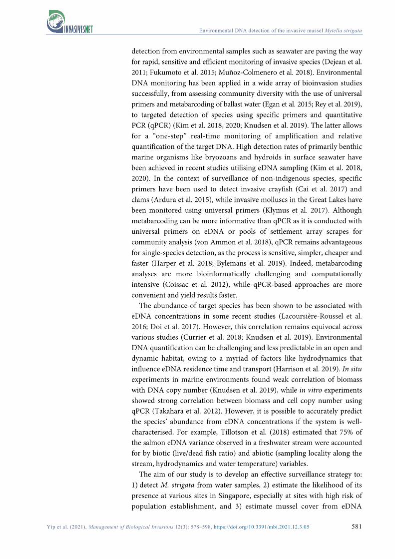
Figure 2. Agarose gels showing PCR products obtained with M. strigata-specific primers for: a) PCR amplification of target species M. strigata and other non-target bivalves; b) PCR products from Sembawang water sample; c) Determination of primer sensitivity; starting DNA extract concentration of 125 ng/μl was diluted for each PCR reaction: 1) no dilution, 2) 1:5, 3) 1:10, 4) 1:25, 5) 1:50, 6) 1:100, 7) 1:500, 8) 1:1000, 9) 1:5000, 10) 1:1×10⁴, 11) 1:5×10⁴, 12) 1:1×10⁵, 13) 1:5×10⁵, 14) 1:1×10⁶, 15) 1:5×10⁶, 16) 1:1×10⁷, 17) 1:5×10⁷, 18) 1:1×10⁸.
Primer design and validation
The specific primer pair designed in this study comprised CO1mytellaFf (forward; 5’-GGG TTA ATA GGA AGA AGG TTG AGA-3’) and CO1mytellaFr (reverse; 5’-ACA ACC ACC GAT ACA TAA AGG-3’), which targeted a short barcode region of mitochondrial COI with a length of 196 bp, The primers matched with all available COI M. strigata sequences in GenBank with 100% identity and 100% coverage.
No cross-amplification occurred during the PCR specificity test in silico and in vitro for the other bivalves tested, including P. viridis and A. gubernaculum (Figure 2a). Sanger sequences were deposited in the NCBI database (MW579566–MW579581). PCR amplification of M. strigata DNA from the positive Sembawang seawater sample was also successful, with the species identity verified by Sanger sequencing (Figure 2b). The sensitivity of the primers, evaluated by PCR-visualisation on a 2% agarose gel, showed that 0.00125 ng/μl (1:1×10⁵ from 125 ng/μl) was the lowest DNA concentration to visibly amplify (Figure 2c, lane 12). However, it should be noted that the 1:5×10⁴ standard did not appear to amplify (Figure 2c, lane 11).
eDNA amplification and analysis
A total of 14 sites were sampled for eDNA (see Table S2 for relative eDNA concentrations). The PCR efficiency was calculated to be 82% from the

Environmental DNA detection of the invasive mussel Mytella strigata
Yip et al. (2021), Management of Biological Invasions 12(3): 578–598, https://doi.org/10.3391/mbi.2021.12.3.05 587
Figure 3. a) Logistic regression of presence/absence of M. strigata against eDNA concentration in ng/L; red and blue circles represent presence and absence of shells observed in the field respectively; the dashed vertical line indicates the eDNA concentration at which Ct = 35. b) Plot of average percent cover of M. strigata against eDNA concentration in ng/L.
standard curve (Figure S1). Among the 14 sites surveyed, all five sites where M. strigata populations were visually observed showed qPCR amplification from 25 to 30 Ct. For sites where M. strigata was not visually observed, amplification was late (Ct > 35) or not successful. Negative controls failed to amplify (Table S3) and all eDNA samples showed no PCR inhibition (Table S4). The melting curve analysis confirmed the specificity of the eDNA amplification as melt peaks for eDNA samples and tissue extracts were identical at 78 °C (Figure S2).
The eDNA detections were positively associated with the visually confirmed presence of M. strigata (p = 0.00114), with the lowest M. strigata eDNA concentration where it was visually observed being 0.000335 ng/L (Figure 3a, Table S5). There was however a significant negative linear relationship between percent cover of mussels and eDNA concentration (p = 0.0368), explaining 24% of the variation (Figure 3b, Table S6).

Environmental DNA detection of the invasive mussel Mytella strigata
Yip et al. (2021), Management of Biological Invasions 12(3): 578–598, https://doi.org/10.3391/mbi.2021.12.3.05 588
Discussion
In the present study, we developed an eDNA approach for detecting the invasive M. strigata from water samples in Singapore that can be applied worldwide. This method is less field intensive than conventional visual survey techniques and is more cost effective by around threefold (Tables S7, S8). We observed high detection rate of M. strigata eDNA where the mussels were present, thus demonstrating high accuracy for presence prediction (Figure 3a). While this study found a perfect correlation between sites with visible shells and eDNA detection, other mussel studies have recovered eDNA at sites where the target species has not been detected visually (e.g. Currier et al. 2018). For example, Sepulveda et al. (2019) detected invasive dreissenid mussel eDNA even though no adults and veligers were observed using non-molecular tools during the non-spawning season, thus extending the seasonal window for dreissenid mussel surveillance. The higher sensitivity of detection using eDNA as compared with non-molecular methods have been recorded in many aquatic mussel invasion studies (Darling and Mahon 2011; Hosler 2017; Currier et al. 2018; Sepulveda et al. 2019). This has been justified in part because mussel larvae and gametes that are present in the water column can be captured by eDNA surveys even before populations are established (Trebitz et al. 2019). This provides an avenue for early detection and eradication where invasive mussel populations have yet to be established (Gingera et al. 2017) or are established but remain in low abundance to be detected by visual surveys or planktonic tows (Trebitz et al. 2019). Regular water sampling at sites where M. strigata has been detected sporadically by conventional survey is crucial in improving the accuracy of the eDNA concentration threshold for positive detection. In the present study we consider eDNA negative detections and late amplifications (Ct > 35), backed by negative detection from visual surveys to correspond to a lack of mussel establishment at nine sites. However, a temporal study should be conducted at these sites to document the changes in eDNA concentration over time and that could account for spawning times and larval presence to explain the late Ct values. Such a more comprehensive study would also be useful to guide future surveys and include further areas that could have been missed during the short window of low tide for visual surveys in this study.
Primer design for targeted detection of species is a critical step in eDNA studies, with several related considerations including amplicon length, primer specificity and sensitivity. Targeting short DNA fragments (100–200 bp) that are easily amplifiable by PCR is beneficial for recovering eDNA as environmental samples can contain a fair amount of degraded DNA (Barnes et al. 2014; Strickler et al. 2015). Validating the specificity and sensitivity of the primers is also crucial for determining the effectiveness of the eDNA method in detecting invasive species with low false error rates. Although we did not design qPCR probes which may increase the specificity

Environmental DNA detection of the invasive mussel Mytella strigata
Yip et al. (2021), Management of Biological Invasions 12(3): 578–598, https://doi.org/10.3391/mbi.2021.12.3.05 589
of detection (Kutyavin et al. 2000; Yao et al. 2006), our tests for cross-species amplification on tissue and eDNA samples showed that the primers amplified only M. strigata and not the other species present in the environments sampled (Figure 2a, b). Sensitivity tests also revealed that the primer pair could detect low starting DNA material using conventional PCR (Figure 2c) and even lower concentrations with qPCR (Table S3). Although both conventional PCR and qPCR can be used to infer species presence and absence, the latter offers higher sensitivity and semi-quantitative data on the abundance of target DNA (Wilcox et al. 2013). While for some labs the PCR test can lower the cost of surveillance by relying on more commonly available PCR machines and gel visualisation for a presence-absence outcome, reproducibility of consistent positive detection is also a challenge for conventional PCR at low concentrations.
Amplification efficiency is an essential criterion assessed in qPCR experiments. An optimal efficiency of 100% indicates that the PCR copy number doubles with every cycle (Svec et al. 2015) and an efficiency of 90–110% is recommended (Klymus et al. 2020). Although our newly designed primers have an efficiency of 82%, this is comparable to other published eDNA primer sets (Klymus et al. 2020), and we found the triplicates performed remarkably consistently across all qPCR reactions (R2 > 0.99) (Figure S1). It is also important to consider the possibility of false negatives in eDNA analyses which can arise from PCR inhibition in environmental samples due to compounds like humic acid restricting DNA amplification, or from low starting eDNA concentrations. The latter may be common among organisms with low population sizes (Ficetola et al. 2008) or low shedding rates (Mauvisseau et al. 2019). It is therefore generally essential to validate primer sensitivity and test for inhibition in eDNA studies (Raymaekers et al. 2009; Klymus et al. 2020).
While our detection of M. strigata was effective, the data also showed a very weak negative correlation between eDNA concentration and percent mussel cover. That only 24% of the variation could be attributed to the relationship between percent cover of mussels and eDNA concentration suggests many other confounding environmental factors, including hydrodynamics in an open system, bacterial and fungal activity, UV radiation, salinity and pH, could be affecting the retention and degradation rates of eDNA (Stoeckle et al. 2017; Collins et al. 2018; Harrison et al. 2019). Similarly, Shogren et al. (2019) found weak but positive relationships between eDNA concentration, zebra mussel abundance, and biophysical parameters. Mussel densities were only moderately predicted by eDNA concentrations, but the latter was most strongly influenced by nutrient concentrations and water velocity in the river (Shogren et al. 2019). Another plausible reason for the weak correlation is the average difference of two months between the visual survey and water sampling times. There could have been changes to mussel population sizes leading to variation in

Environmental DNA detection of the invasive mussel Mytella strigata
Yip et al. (2021), Management of Biological Invasions 12(3): 578–598, https://doi.org/10.3391/mbi.2021.12.3.05 590
the amount of shed DNA in the water samples. Furthermore, the life history and reproduction of the species should be considered. Gametes or veligers released into the water column from a reproducing population could potentially account for the discrepancy between our eDNA quantification and mussel coverage. Water samples should thus, in the future, be examined for gametes and veligers. Finally, the variability in mussel cover was clearly larger for sites with high average percent cover, which could also have contributed to the discrepancy.
The relationship between biomass and eDNA copy number has been explored with great success largely in aquaria and in situ closed systems such as lotic environments (Takahara et al. 2012; Thomsen et al. 2012; Biggs et al. 2015; Buxton et al. 2017). It is, however, met with more challenges in lentic environments and open systems (i.e. coastal and marine environments) as the correlation is frequently moderate at best with high levels of unexplained variation due to the above confounding environmental factors (Currier et al. 2018; Knudsen et al. 2019). Nevertheless, studies have shown relatively strong correlation between cod biomass as well as catch per unit effort (CPUE) with eDNA concentration in oceanic waters (Salter et al. 2019), and precisely quantified the infestation level of invasive dreissenids with nuclear markers, as opposed to mitochondrial markers since the amount of mtDNA may vary across cells (Peñarrubia et al. 2016). Marshall and Stepien (2019) have also demonstrated that sampling eDNA near the benthos where adult colonies are found may provide a more accurate estimation of mussel biomass. Recent studies on the prospect of strengthening this relationship have led to the use of allometrically scaled mass (ASM) in models of eDNA concentration and abundance that better fit the data (Yates et al. 2020), particularly where there is substantial intraspecific size variation (Yates et al. 2020).
Our analysis clearly produced true positive detection when the M. strigata eDNA concentration in a water sample exceeds 0.000335 ng/L. Studies on other invasive bivalves have found similar sensitivity levels of their primers at 10-4 ng of target DNA (e.g. Pie et al. 2017). This ability to distinguish presence and absence of the invasive mussel based on an eDNA concentration threshold raises the capacity to monitor the distribution and spread of M. strigata and other invasive mussels (Amberg et al. 2019). Indeed, seawater can be sampled from areas under high invasion risk but where populations have yet to be established, including the southern coast and islands of Singapore like Jurong River and West Coast where M. strigata populations were found during a previous study conducted by Lim et al. (2018). These are areas with a high density of port terminals but adjacent to natural habitats like Labrador Nature Reserve and Cyrene Reefs that have rich marine biodiversity and distinct intertidal assemblages from the northern shores (Lim et al. 2020b). Routine surveillance to prevent the establishment of viable populations at these sites is crucial as they may act

Environmental DNA detection of the invasive mussel Mytella strigata
Yip et al. (2021), Management of Biological Invasions 12(3): 578–598, https://doi.org/10.3391/mbi.2021.12.3.05 591
as a steppingstone for the colonisation of M. strigata in the southern islands of Singapore.
The observations at Lim Chu Kang, Kranji and Sembawang Park suggest that M. strigata colonies initially dominate man-made hard substrata and extend to the mudflat after they have attained high densities (see Table S1 for percent cover of M. strigata). Studies on M. strigata substrate preference have revealed similar colonising behaviour, with mussels found more commonly on man-made structures than natural substrates like mangrove roots or oyster shells, possibly due to the higher habitat suitability for longer-term survival (Gilg et al. 2010). Therefore, future surveys at sites where it has not been visually observed should prioritise artificial structures over natural substrates. Enhanced monitoring efforts should also couple water eDNA testing with examination of settlement plates and planktonic tows (e.g. Koziol et al. 2019) in order to obtain a more comprehensive picture of colonisation and establishment potential. More generally, this approach would greatly reduce the manpower and effort needed for early detection of invasive species, allow surveillance in areas where field surveys cannot be easily conducted (e.g. restricted military sites, unsafe environments, or logistically-challenging areas that are extremely muddy and/or turbid), and enable a rapid response toward its control when needed. We also note that the M. strigata invasion in Singapore is occurring along the Johor Strait coastline in close proximity to Malaysia, posing an enhanced risk of M. strigata being transported across the Strait. Extensive shipping activities between Singapore and Indonesia could also result in the mussel spreading to the latter’s vast coastlines (Lim et al. 2017). Therefore, surveillance at sites near Singapore’s borders would help in detecting and mitigating its spread into neighbouring countries.
Studies have shown that an ecosystem can be more prone to invasion if the area has an invasion history, prompting an “invasional meltdown” (Simberloff and Von Holle 1999). This phenomenon has been observed in marine ecosystems, where the introduction of a non-indigenous organism induced the colonisation of more non-indigenous species (Grosholz 2005; Gavira-O’Neill et al. 2018). Such incidents are potentially driven by facilitative interactions like mutualism and commensalism between introduced species, or the change in dynamics of prey-predator relationships caused by an invader that favours the survivorship of another invader, thus compounding the effects of the initial invasion (Ricciardi 2001). Future monitoring efforts to detect early invasions should thus target areas with higher risk due to past invasions.
To help achieve higher sensitivity and more reliable eDNA detection of invasive species, new technologies such as the digital droplet PCR (ddPCR) could be adopted, albeit at a higher cost (Nathan et al. 2014; Simmons et al. 2016). ddPCR is especially useful for monitoring and managing elusive invasive species, as the amount of DNA material from the target species
Environmental DNA detection of the invasive mussel Mytella strigata
Yip et al. (2021), Management of Biological Invasions 12(3): 578–598, https://doi.org/10.3391/mbi.2021.12.3.05 592
captured in environmental samples is typically extremely low (Doi et al. 2015a; Simmons et al. 2016). The quantification accuracy of ddPCR is higher than qPCR at low eDNA concentrations (~ 10 DNA copies), with lower variation in measurements (Doi et al. 2015b). It also provides stronger correlation between species abundance and eDNA concentration (Doi et al. 2015b). Nevertheless, even as new technologies are emerging that would be suitable for eDNA surveillance of invasive species, there needs to be continuing development of more streamlined and effective protocols for qPCR-based approaches which remain relatively cost-effective.
In conclusion, this study has validated the efficacy of routine qPCR testing of eDNA water samples for monitoring the colonisation and spread of the invasive species, M. strigata. While percent mussel cover was not reliably predicted by water eDNA concentrations here, the data indicate that our detection procedure, along with the primers designed, are highly sensitive and suitable for eDNA-based detection of M. strigata. Currently, Singapore uses a multi-pronged approach that encompasses precautionary measures taken at each step of the invasion pathway to deal with marine bioinvasions effectively. Strict regulations are enforced on ballast water management along with antifouling systems to prevent the introduction of NIMS through shipping. The food and pet trades are also monitored regularly for NIMS (Wells et al. 2019), and the public are educated on the harmful effects of the release of NIMS into the wild. There is, however, no regular seawater monitoring programme established for early detection of invasive species. Molecular approaches like eDNA may therefore be useful for monitoring their potential incursion into Singapore’s southern islands where the richest biodiversity lies (Lim et al. 2020b). Neither visual surveys nor eDNA detection are absolutely accurate although the former is a more time- and labour-intensive monitoring method. Environmental DNA techniques with an optimised sampling strategy to reduce the chance of false negatives can potentially replace conventional surveys up until the point where eDNA concentration approaches the threshold for detection (Xia et al. 2018). In general, the two methods should be used concurrently for higher detection accuracy. For M. strigata which has already established along the northern Singapore coast, surveillance efforts must continue in order to curb its spread to other coastlines in Southeast Asia.
Acknowledgements We would like to thank volunteers of the Nature Society (Singapore) for helping with the field visual surveys, as well as members of the Reef Ecology Laboratory, National University of Singapore, for assisting in the water sampling surveys. We are grateful to the Singapore Armed Forces Yacht Club, Sembawang, for access to survey the site, and the National Parks Board and Singapore Land Authority for research permits (NP/RP16-049-1b and NP/RP19-038a). We would also like to thank the anonymous reviewers and the editorial staff at Management of Biological Invasions for their invaluable feedback and insight.

Environmental DNA detection of the invasive mussel Mytella strigata
Yip et al. (2021), Management of Biological Invasions 12(3): 578–598, https://doi.org/10.3391/mbi.2021.12.3.05 593
Funding declaration This research is supported by the National Research Foundation, Prime Minister’s Office, Singapore under its Marine Science R&D Programme (MSRDP-P03 and MSRDP-P39), and the National Parks Board (R-154-000-B34-490).
References Amberg JJ, Merkes CM, Stott W, Rees CB, Erickson RA (2019) Environmental DNA as a tool
to help inform zebra mussel, Dreissena polymorpha, management in inland lakes. Management of Biological Invasions 10: 96, https://doi.org/10.3391/mbi.2019.10.1.06
Ardura A, Zaiko A, Martinez JL, Samulioviene A, Semenova A, Garcia-Vazquez E (2015) eDNA and specific primers for early detection of invasive species - A case study on the bivalve Rangia cuneata, currently spreading in Europe. Marine Environmental Research 112: 48–55, https://doi.org/10.1016/j.marenvres.2015.09.013
Barnes MA, Turner CR, Jerde CL, Renshaw MA, Chadderton WL, Lodge DM (2014) Environmental conditions influence eDNA persistence in aquatic systems. Environmental Science & Technology 48: 1819–1827, https://doi.org/10.1021/es404734p
Beasley CR, Fernandes CM, Gomes CP, Brito BA, Santos SML, Tagliaro CH (2005) Molluscan diversity and abundance among coastal habitats of northern Brazil. Ecotropica 11: 9–20
Biggs J, Ewald N, Valentini A, Gaboriaud C, Dejean T, Griffiths RA, Foster J, Wilkinson JW, Arnell A, Brotherton P, Williams P, Dunn F (2015) Using eDNA to develop a national citizen science-based monitoring programme for the great crested newt (Triturus cristatus). Biological Conservation 183: 19–28, https://doi.org/10.1016/j.biocon.2014.11.029
Biju Kumar A, Ravinesh R, Oliver PG, Tan SK, Sadasivan K (2019) Rapid bioinvasion of alien mussel Mytella strigata (Hanley, 1843) (Bivalvia: Mytilidae) along Kerala Coast, India: Will this impact the livelihood of fishers in Ashtamudi Lake? Journal of Aquatic Biology & Fisheries 7: 31–45
Breton S, Beaupre HD, Stewart DT, Hoeh WR, Blier PU (2007) The unusual system of doubly uniparental inheritance of mtDNA: isn’t one enough? Trends in Genetics 23: 465–474, https://doi.org/10.1016/j.tig.2007.05.011
Briski E, Ghabooli S, Bailey SA, MacIsaac HJ (2012) Invasion risk posed by macroinvertebrates transported in ships’ ballast tanks. Biological Invasions 14: 1843–1850, https://doi.org/10. 1007/s10530-012-0194-0
Brodsky S (2011) Cold temperature effects on byssal thread production by the native mussel Geukensia demissa versus the non-native mussel Mytella charruana. The Pegasus Review: UCF Undergraduate Research Journal (URJ) 5(1): 1
Buxton AS, Groombridge JJ, Zakaria NB, Griffiths RA (2017) Seasonal variation in environmental DNA in relation to population size and environmental factors. Scientific Reports 7: 46294, https://doi.org/10.1038/srep46294
Bylemans J, Gleeson DM, Duncan RP, Hardy CM, Furlan EM (2019) A performance evaluation of targeted eDNA and eDNA metabarcoding analyses for freshwater fishes. Environmental DNA 1: 402–414, https://doi.org/10.1002/edn3.41
Cai W, Ma Z, Yang C, Wang L, Wang W, Zhao G, Geng Y, Yu DW (2017) Using eDNA to detect the distribution and density of invasive crayfish in the Honghe-Hani rice terrace World Heritage site. PLoS ONE 12: e0177724, https://doi.org/10.1371/journal.pone.0177724
Calazans CSH, Walters LJ, Fernandes FC, Ferreira CEL, Hoffman EA (2017) Genetic structure provides insights into the geographical origins and temporal change in the invasive charru mussel (Sururu) in the southeastern United States. PLoS ONE 12: e0180619, https://doi.org/10.1371/journal.pone.0180619
Coan EV, Valentich-Scott P (2012) Bivalve Seashells of Tropical West America. Marine Bivalve Mollusks from Baja California to northern Peru. 2 volumes. Santa Barbara Museum of Natural History, Santa Barbara, California, U.S.A.
Coissac E, Riaz T, Puillandre N (2012) Bioinformatic challenges for DNA metabarcoding of plants and animals. Molecular Ecology 21: 1834–1847, https://doi.org/10.1111/j.1365-294X. 2012.05550.x
Collins RA, Wangensteen OS, O'Gorman EJ, Mariani S, Sims DW, Genner MJ (2018) Persistence of environmental DNA in marine systems. Communications Biology 1: 185, https://doi.org/10.1038/s42003-018-0192-6
Currier CA, Morris TJ, Wilson CC, Freeland JR (2018) Validation of environmental DNA (eDNA) as a detection tool for at‐risk freshwater pearly mussel species (Bivalvia: Unionidae). Aquatic Conservation: Marine and Freshwater Ecosystems 28: 545–558, https://doi.org/10. 1002/aqc.2869
Darling JA, Mahon AR (2011) From molecules to management: adopting DNA-based methods for monitoring biological invasions in aquatic environments. Environmental Research 111: 978–988, https://doi.org/10.1016/j.envres.2011.02.001
Deiner K, Altermatt F (2014) Transport distance of invertebrate environmental DNA in a natural river. PLoS ONE 9: e88786, https://doi.org/10.1371/journal.pone.0088786

Environmental DNA detection of the invasive mussel Mytella strigata
Yip et al. (2021), Management of Biological Invasions 12(3): 578–598, https://doi.org/10.3391/mbi.2021.12.3.05 594
Dejean T, Valentini A, Duparc A, Pellier-Cuit S, Pompanon F, Taberlet P, Miaud C (2011) Persistence of environmental DNA in freshwater ecosystems. PLoS ONE 6: e23398, https://doi.org/10.1371/journal.pone.0023398
Doi H, Takahara T, Minamoto T, Matsuhashi S, Uchii K, Yamanaka H (2015a) Droplet digital polymerase chain reaction (PCR) outperforms real-time PCR in the detection of environmental DNA from an invasive fish species. Environmental Science & Technology 49: 5601–5608, https://doi.org/10.1021/acs.est.5b00253
Doi H, Uchii K, Takahara T, Matsuhashi S, Yamanaka H, Minamoto T (2015b) Use of Droplet Digital PCR for Estimation of Fish Abundance and Biomass in Environmental DNA Surveys. PLoS ONE 10: e0122763, https://doi.org/10.1371/journal.pone.0122763
Doi H, Inui R, Akamatsu Y, Kanno K, Yamanaka H, Takahara T, Minamoto T (2017) Environmental DNA analysis for estimating the abundance and biomass of stream fish. Freshwater Biology 62: 30–39, https://doi.org/10.1111/fwb.12846
Doyle J, Doyle J (1987) A rapid procedure for DNA purification from small quantities of fresh leaf tissue. Phytochemical Bulletin 19: 11–15
Egan SP, Grey E, Olds B, Feder JL, Ruggiero ST, Tanner CE, Lodge DM (2015) Rapid molecular detection of invasive species in ballast and harbor water by integrating environmental DNA and light transmission spectroscopy. Environmental Science & Technology 49: 4113–4121, https://doi.org/10.1021/es5058659
Ficetola GF, Miaud C, Pompanon F, Taberlet P (2008) Species detection using environmental DNA from water samples. Biology Letters 4: 423–425, https://doi.org/10.1098/rsbl.2008.0118
Fofonoff PW, Ruiz GM, Steves B, Simkanin C, Carlton JT (2018) National Exotic Marine and Estuarine Species Information System. http://invasions.si.edu/nemesis/ (accessed 26 April 2020)
Fukumoto S, Ushimaru A, Minamoto T (2015) A basin‐scale application of environmental DNA assessment for rare endemic species and closely related exotic species in rivers: A case study of giant salamanders in Japan. Journal of Applied Ecology 52: 358–365, https://doi.org/10.1111/1365-2664.12392
Galimany E, Freeman CJ, Lunt J, Domingos A, Sacks P, Walters L (2017) Feeding competition between the native oyster Crassostrea virginica and the invasive mussel Mytella charruana. Marine Ecology Progress Series 564: 57–66, https://doi.org/10.3354/meps11976
Gavira-O’Neill K, Guerra-García JM, Moreira J, Ros M (2018) Mobile epifauna of the invasive bryozoan Tricellaria inopinata: is there a potential invasional meltdown? Marine Biodiversity 48: 1169–1178, https://doi.org/10.1007/s12526-016-0563-5
Gilg MR, Hoffman EA, Schneider KR, Ryabinov J, El-Khoury C, Walters LJ (2010) Recruitment preferences of non-native mussels: interaction between marine invasions and land-use changes. Journal of Molluscan Studies 76: 333–339, https://doi.org/10.1093/mollus/ eyq017
Gingera TD, Bajno R, Docker MF, Reist JD (2017) Environmental DNA as a detection tool for zebra mussels Dreissena polymorpha (Pallas, 1771) at the forefront of an invasion event in Lake Winnipeg, Manitoba, Canada. Management of Biological Invasions 8: 287, https://doi.org/10.3391/mbi.2017.8.3.03
Grosholz E (2002) Ecological and evolutionary consequences of coastal invasions. Trends in Ecology & Evolution 17: 22–27, https://doi.org/10.1016/S0169-5347(01)02358-8
Grosholz ED (2005) Recent biological invasion may hasten invasional meltdown by accelerating historical introductions. Proceedings of the National Academy of Sciences 102: 1088–1091, https://doi.org/10.1073/pnas.0308547102
Harper LR, Lawson HL, Hahn C, Boonham N, Rees HC, Gough KC, Lewis E, Adams IP, Brotherton P, Phillips S, Hänfling B (2018) Needle in a haystack? A comparison of eDNA metabarcoding and targeted qPCR for detection of the great crested newt (Triturus cristatus). Ecology and Evolution 8: 6330–6341, https://doi.org/10.1002/ece3.4013
Harrison JB, Sunday JM, Rogers SM (2019) Predicting the fate of eDNA in the environment and implications for studying biodiversity. Proceedings of the Royal Society B 286: 20191409, https://doi.org/10.1098/rspb.2019.1409
Hartman LJ, Coyne SR, Norwood DA (2005) Development of a novel internal positive control for Taqman® based assays. Molecular and Cellular Probes 19: 51–59, https://doi.org/10. 1016/j.mcp.2004.07.006
Hayes KR, Cannon R, Neil K, Inglis G (2005) Sensitivity and cost considerations for the detection and eradication of marine pests in ports. Marine Pollution Bulletin 50: 823–834, https://doi.org/10.1016/j.marpolbul.2005.02.032
Hoeh WR, Stewart DT, Sutherland BW, Zouros E (1996) Multiple origins of gender-associated mitochondrial DNA lineages in bivalves (Mollusca: Bivalvia). Evolution 50: 2276–2286, https://doi.org/10.1111/j.1558-5646.1996.tb03616.x
Hoeh WR, Stewart DT, Guttman SI (2002) High fidelity of mitochondrial genome transmission under the doubly uniparental mode of inheritance in freshwater mussels (Bivalvia: Unionoidea). Evolution 56: 2252–2261, https://doi.org/10.1111/j.0014-3820.2002.tb00149.x
Hosler DM (2017) Where is the body? Dreissenid mussels, raw water testing, and the real value of environmental DNA. Management of Biological Invasions 8: 335–341, https://doi.org/10. 3391/mbi.2017.8.3.07

Environmental DNA detection of the invasive mussel Mytella strigata
Yip et al. (2021), Management of Biological Invasions 12(3): 578–598, https://doi.org/10.3391/mbi.2021.12.3.05 595
Huber M (2010) Compendium of Bivalves. A full-color guide to 3,300 of the world’s marine bivalves. A status on Bivalvia after 250 years of research. Conchbooks, Hackenheim, Germany, 904 pp
Hulme PE (2006) Beyond control: wider implications for the management of biological invasions. Journal of Applied Ecology 43: 835–847, https://doi.org/10.1111/j.1365-2664.2006.01227.x
Jayachandran PR, Aneesh BP, Oliver PG, Philomina J, Jima M, Harikrishnan K, Nandan SB (2019) First record of the alien invasive biofouling mussel Mytella strigata (Hanley, 1843) (Mollusca: Mytilidae) from Indian waters. BioInvasions Record 8: 828–837, https://doi.org/ 10.3391/bir.2019.8.4.11
Jerde CL, Mahon AR (2015) Improving confidence in environmental DNA species detection. Molecular Ecology Resources 15: 461–463, https://doi.org/10.1111/1755-0998.12377
Johnson LE, Padilla DK (1996) Geographic spread of exotic species: ecological lessons and opportunities from the invasion of the zebra mussel Dreissena polymorpha. Biological Conservation 78: 23–33, https://doi.org/10.1016/0006-3207(96)00015-8
Kearse M, Moir R, Wilson A, Stones-Havas S, Cheung M, Sturrock S, Buxton S, Cooper A, Markowitz S, Duran C, Thierer T, Ashton B, Meintjes P, Drummond A (2012) Geneious Basic: an integrated and extendable desktop software platform for the organization and analysis of sequence data. Bioinformatics 28: 1647–1649, https://doi.org/10.1093/bioinformatics/bts199
Kim P, Kim D, Yoon TJ, Shin S (2018) Early detection of marine invasive species, Bugula neritina (Bryozoa: Cheilostomatida), using species-specific primers and environmental DNA analysis in Korea. Marine Environmental Research 139: 1–10, https://doi.org/10.1016/j. marenvres.2018.04.015
Kim P, Yoon TJ, Shin S (2020) Environmental DNA and specific primers for detecting the invasive species Ectopleura crocea (Hydrozoa: Anthoathecata) in seawater samples. Sustainability 12: 2360, https://doi.org/10.3390/su12062360
Klymus KE, Marshall NT, Stepien CA (2017) Environmental DNA (eDNA) metabarcoding assays to detect invasive invertebrate species in the Great Lakes. PLoS ONE 12: e0177643, https://doi.org/10.1371/journal.pone.0177643
Klymus KE, Merkes CM, Allison MJ, Goldberg CS, Helbing CC, Hunter ME, Jackson CA, Lance RF, Mangan AM, Monroe EM, Piaggio AJ, Stokdyk JP, Wilson CC, Richter CA (2020) Reporting the limits of detection and quantification for environmental DNA assays. Environmental DNA 2: 271–282, https://doi.org/10.1002/edn3.29
Knudsen SW, Ebert RB, Hesselsøe M, Kuntke F, Hassingboe J, Mortensen PB, Thomsen PF, Sigsgaard EE, Hansen BK, Nielsen EE, Møller PR (2019) Species-specific detection and quantification of environmental DNA from marine fishes in the Baltic Sea. Journal of Experimental Marine Biology and Ecology 510: 31–45, https://doi.org/10.1016/j.jembe.2018.09.004
Kohler KE, Gill SM (2006) Coral Point Count with Excel extensions (CPCe): A Visual Basic program for the determination of coral and substrate coverage using random point count methodology. Computers and Geosciences 32: 1259–1269, https://doi.org/10.1016/j.cageo. 2005.11.009
Koziol A, Stat M, Simpson T, Jarman S, DiBattista JD, Harvey ES, Marnane M, McDonald J, Bunce M (2019) Environmental DNA metabarcoding studies are critically affected by substrate selection. Molecular Ecology Resources 19: 366–376, https://doi.org/10.1111/1755-0998.12971
Kutyavin IV, Afonina IA, Mills A, Gorn VV, Lukhtanov EA, Belousov ES, Singer MJ, Walburger DK, Lokhov SG, Gall AA, Dempcy R, Reed MW, Meyer RB, Hedgpeth J (2000) 3′-minor groove binder-DNA probes increase sequence specificity at PCR extension temperatures. Nucleic Acids Research 28: 655–661, https://doi.org/10.1093/nar/28.2.655
Lacoursière‐Roussel A, Côté G, Leclerc V, Bernatchez L (2016) Quantifying relative fish abundance with eDNA: a promising tool for fisheries management. Journal of Applied Ecology 53: 1148–1157, https://doi.org/10.1111/1365-2664.12598
Lim CS, Leong YL, Tan KS (2017) Managing the risk of non-indigenous marine species transfer in Singapore using a study of vessel movement. Marine Pollution Bulletin 115: 332–344, https://doi.org/10.1016/j.marpolbul.2016.12.009
Lim JY, Tay TS, Lim CS, Lee SSC, Teo SM, Tan KS (2018) Mytella strigata (Bivalvia: Mytilidae): an alien mussel recently introduced to Singapore and spreading rapidly. Molluscan Research 38: 170–186, https://doi.org/10.1080/13235818.2018.1423858
Lim CS, Tay TS, Tan KS, Teo SLM (2020a) Removal of larvae of two marine invasive bivalves, Mytilopsis sallei (Récluz, 1849) and Mytella strigata (Hanley, 1843), by water treatment processes. Marine Pollution Bulletin 155: 111154, https://doi.org/10.1016/j. marpolbul.2020.111154
Lim LJW, Loh JBY, Lim AJS, Tan BYX, Ip YCA, Neo ML, Tan R, Huang D (2020b) Diversity and distribution of intertidal marine species in Singapore. Raffles Bulletin of Zoology 68: 1–8
Lodge DM, Williams S, MacIsaac HJ, Hayes KR, Leung B, Reichard S, Mack RN, Moyle PB, Smith M, Andow DA, Carlton JT, McMichael A (2006) Biological invasions: recommendations for US policy and management. Ecological Applications 16: 2035–2054, https://doi.org/10.1890/1051-0761(2006)016[2035:BIRFUP]2.0.CO;2

Environmental DNA detection of the invasive mussel Mytella strigata
Yip et al. (2021), Management of Biological Invasions 12(3): 578–598, https://doi.org/10.3391/mbi.2021.12.3.05 596
Marshall NT, Stepien CA (2019) Invasion genetics from eDNA and thousands of larvae: A targeted metabarcoding assay that distinguishes species and population variation of zebra and quagga mussels. Ecology and Evolution 9: 3515–3538, https://doi.org/10.1002/ece3.4985
Mauvisseau Q, Davy-Bowker J, Bulling M, Brys R, Neyrinck S, Troth C, Sweet M (2019) Combining ddPCR and environmental DNA to improve detection capabilities of a critically endangered freshwater invertebrate. Scientific Reports 9: 1–9, https://doi.org/10.1038/s41598-019-50571-9
Mediodia DP, De Leon SMS, Anasco NC, Baylon CC (2017) Shell morphology and anatomy of the Philippine Charru mussel Mytella charruana (D’Orbigny 1842). Asian Fisheries Science 30: 185–194, https://doi.org/10.33997/j.afs.2017.30.3.004
Muñoz-Colmenero M, Ardura A, Clusa L, Miralles L, Gower F, Zaiko A, Garcia-Vazquez E (2018) New specific molecular marker detects Ficopomatus enigmaticus from water eDNA before positive results of conventional sampling. Journal for Nature Conservation 43: 173–178, https://doi.org/10.1016/j.jnc.2017.12.004
Nathan LM, Simmons M, Wegleitner BJ, Jerde CL, Mahon AR (2014) Quantifying environmental DNA signals for aquatic invasive species across multiple detection platforms. Environmental Science & Technology 48: 12800–12806, https://doi.org/10.1021/ es5034052
Nishida AK, Leonel RMV (1995) Occurrence, population dynamics and habitat characterization of Mytella guyanensis (Lamarck, 1819) (Mollusca, Bivalvia) in the Paraíba do Norte river estuary. Revista Brasileira de Oceanografia 43: 41–49, https://doi.org/10.1590/S0373-55241995 000100004
Peñarrubia L, Alcaraz C, bij de Vaate A, Sanz N, Pla C, Vidal O, Viñas J (2016) Validated methodology for quantifying infestation levels of dreissenid mussels in environmental DNA (eDNA) samples. Scientific Reports 6: 39067, https://doi.org/10.1038/srep39067
Pie MR, Ströher PR, Agostinis AO, Belmonte-Lopes R, Tadra-Sfeir MZ, Ostrensky A (2017) Development of a real-time PCR assay for the detection of the golden mussel (Limnoperna fortunei, Mytilidae) in environmental samples. Anais da Academia Brasileira de Ciências 89: 1041–1045, https://doi.org/10.1590/0001-3765201720160723
Piggott MP (2016) Evaluating the effects of laboratory protocols on eDNA detection probability for an endangered freshwater fish. Ecology and Evolution 6: 2739–2750, https://doi.org/10. 1002/ece3.2083
R Core Team (2019) R: A language and environment for statistical computing. R Foundation for Statistical Computing, Vienna, Austria. https://www.R-project.org/
Raymaekers M, Smets R, Maes B, Cartuyvels R (2009) Checklist for optimization and validation of real‐time PCR assays. Journal of Clinical Laboratory Analysis 23: 145–151, https://doi.org/10.1002/jcla.20307
Renshaw MA, Olds BP, Jerde CL, McVeigh MM, Lodge DM (2015) The room temperature preservation of filtered environmental DNA samples and assimilation into a phenol-chloroform-isoamyl alcohol DNA extraction. Molecular Ecology Resources 15: 168–176, https://doi.org/10.1111/1755-0998.12281
Rey A, Carney KJ, Quinones LE, Pagenkopp Lohan KM, Ruiz GM, Basurko OC, Rodríguez-Ezpeleta N (2019) Environmental DNA metabarcoding: a promising tool for ballast water monitoring. Environmental Science & Technology 53: 11849–11859, https://doi.org/10.1021/ acs.est.9b01855
Ricciardi A (1998) Global range expansion of the Asian mussel Limnoperna fortunei (Mytilidae): another fouling threat to freshwater systems. Biofouling 13: 97–106, https://doi.org/10.1080/ 08927019809378374
Ricciardi A (2001) Facilitative interactions among aquatic invaders: is an “invasional meltdown” occurring in the Great Lakes? Canadian Journal of Fisheries and Aquatic Sciences 58: 2513–2525, https://doi.org/10.1139/f01-178
Rice MA, Rawson PD, Salinas AD, Rosario WR (2016) Identification and salinity tolerance of the Western hemisphere mussel Mytella charruana (D’Orbigny, 1842) in the Philippines. Journal of Shellfish Research 35: 865–873, https://doi.org/10.2983/035.035.0415
Salter I, Joensen M, Kristiansen R, Steingrund P, Vestergaard P (2019) Environmental DNA concentrations are correlated with regional biomass of Atlantic cod in oceanic waters. Communications Biology 2: 1–9, https://doi.org/10.1038/s42003-019-0696-8
Sanpanich K, Wells FE (2019) Mytella strigata (Hanley, 1843) emerging as an invasive marine threat in Southeast Asia. BioInvasions Records 8: 343–356, https://doi.org/10.3391/bir.2019.8.2.16
Santos H, Beasley C, Tagliaro C (2010) Changes in population characteristics of Mytella falcata (D’Orbigny, 1846) beds, an exploited tropical estuarine mussel. Boletim do Instituto de Pesca São Paulo 36: 85–97
Seebens H, Gastner MT, Blasius B, Courchamp F (2013) The risk of marine bioinvasion caused by global shipping. Ecology Letters 16: 782–790, https://doi.org/10.1111/ele.12111
Sepulveda AJ, Amberg JJ, Hanson E (2019) Using environmental DNA to extend the window of early detection for dreissenid mussels. Management of Biological Invasions 10: 342–358, https://doi.org/10.3391/mbi.2019.10.2.09

Environmental DNA detection of the invasive mussel Mytella strigata
Yip et al. (2021), Management of Biological Invasions 12(3): 578–598, https://doi.org/10.3391/mbi.2021.12.3.05 597
Shogren AJ, Tank JL, Egan SP, Bolster D, Riis T (2019) Riverine distribution of mussel environmental DNA reflects a balance among density, transport, and removal processes. Freshwater Biology 64: 1467–1479, https://doi.org/10.1111/fwb.13319
Simberloff D, Von Holle B (1999) Positive interactions of nonindigenous species: invasional meltdown? Biological Invasions 1: 21–32, https://doi.org/10.1023/A:1010086329619
Simmons M, Tucker A, Chadderton WL, Jerde CL, Mahon AR (2016) Active and passive environmental DNA surveillance of aquatic invasive species. Canadian Journal of Fisheries and Aquatic Sciences 73: 76–83, https://doi.org/10.1139/cjfas-2015-0262
Smart AS, Tingley R, Weeks AR, Van Rooyen AR, McCarthy MA (2015) Environmental DNA sampling is more sensitive than a traditional survey technique for detecting an aquatic invader. Ecological Applications 25: 1944–1952, https://doi.org/10.1890/14-1751.1
Spinuzzi S, Schneider KR, Walters LJ, Yuan WS, Hoffman EA (2013) Tracking the distribution of non-native marine invertebrates (Mytella charruana, Perna viridis and Megabalanus coccopoma) along the south-eastern USA. Marine Biodiversity Records 6: 1–13, https://doi.org/10.1017/S1755267213000316
Stoeckle BC, Beggel S, Cerwenka AF, Motivans E, Kuehn R, Geist J (2017) A systematic approach to evaluate the influence of environmental conditions on eDNA detection success in aquatic ecosystems. PLoS ONE 12: e0189119, https://doi.org/10.1371/journal.pone.0189119
Strickler KM, Fremier AK, Goldberg CS (2015) Quantifying effects of UV-B, temperature, and pH on eDNA degradation in aquatic microcosms. Biological Conservation 183: 85–92, https://doi.org/10.1016/j.biocon.2014.11.038
Svec D, Tichopad A, Novosadova V, Pfaffl MW, Kubista M (2015) How good is a PCR efficiency estimate: Recommendations for precise and robust qPCR efficiency assessments. Biomolecular Detection and Quantification 3: 9–16, https://doi.org/10.1016/j.bdq.2015.01.005
Taberlet P, Coissac E, Hajibabaei M, Rieseberg LH (2012) Environmental DNA. Molecular Ecology 21: 1789–1793, https://doi.org/10.1111/j.1365-294X.2012.05542.x
Takahara T, Minamoto T, Yamanaka H, Doi H, Kawabata Z (2012) Estimation of fish biomass using environmental DNA. PLoS ONE 7: e35868, https://doi.org/10.1371/journal.pone.0035868
Tan SK, Woo HPM (2010) A preliminary checklist of the molluscs of Singapore. Raffles Museum of Biodiversity Research, Singapore, 78 pp
Tay TS, Gan BQ, Lee SCS, Lim CS, Tan KS, Teo SLM (2018) Larval development of the invasive charru mussel, Mytella strigata (Bivalvia: Mytilidae). Invertebrate Reproduction & Development 62(4): 248–256
Thomsen PF, Willerslev E (2015) Environmental DNA-An emerging tool in conservation for monitoring past and present biodiversity. Biological Conservation 183: 4–18, https://doi.org/ 10.1016/j.biocon.2014.11.019
Thomsen PF, Iversen LL, Wiuf C, Rasmussen M, Gilbert MTP, Orlando L, Willerslev E (2012) Monitoring endangered freshwater biodiversity using environmental DNA. Molecular Ecology 21: 2565–2573, https://doi.org/10.1111/j.1365-294X.2011.05418.x
Tillotson MD, Kelly RP, Duda JJ, Hoy M, Kralj J, Quinn TP (2018) Concentrations of environmental DNA (eDNA) reflect spawning salmon abundance at fine spatial and temporal scales. Biological Conservation 220: 1–11, https://doi.org/10.1016/j.biocon.2018.01.030
Trebitz AS, Hatzenbuhler CL, Hoffman JC, Meredith CS, Peterson GS, Pilgrim EM, Barge JT, Cotter AM, Wick MJ (2019) Dreissena veligers in western Lake Superior-Inference from new low-density detection. Journal of Great Lakes Research 45: 691–699, https://doi.org/10. 1016/j.jglr.2019.03.013
Vallejo JrB, Conejar-Espedido J, Manubag L, Artiaga KCC, Damatac II AM, Imperial ICV, Itong TAB, Fontanilla IK, Cao EP (2017) First record of the Charru mussel Mytella charruana d’Orbignyi, 1846 (Bivalvia: Mytilidae) from Manila Bay, Luzon, Philippines. BioInvasions Record 6: 49–55, https://doi.org/10.3391/bir.2017.6.1.08
von Ammon U, Wood SA, Laroche O, Zaiko A, Tait L, Lavery S, Inglis GJ, Pochon X (2018) Combining morpho-taxonomy and metabarcoding enhances the detection of non-indigenous marine pests in biofouling communities. Scientific Reports 8: 1–11, https://doi.org/10.1038/ s41598-018-34541-1
Wells FE, Tan KS, Todd PA, Jaafar Z, Yeo, DC (2019) A low number of introduced marine species in the tropics: a case study from Singapore. Management of Biological Invasions 10: 23–45, https://doi.org/10.3391/mbi.2019.10.1.03
Wilcox TM, McKelvey KS, Young MK, Jane SF, Lowe WH, Whiteley AR, Schwartz MK (2013) Robust detection of rare species using environmental DNA: the importance of primer specificity. PLoS ONE 8: e59520, https://doi.org/10.1371/journal.pone.0059520
Xia Z, Zhan A, Gao Y, Zhang L, Haffner GD, MacIsaac HJ (2018) Early detection of a highly invasive bivalve based on environmental DNA (eDNA). Biological Invasions 20: 437–447, https://doi.org/10.1007/s10530-017-1545-7
Yao Y, Nellåker C, Karlsson H (2006) Evaluation of minor groove binding probe and Taqman probe PCR assays: Influence of mismatches and template complexity on quantification. Molecular and Cellular Probes 20: 311–316, https://doi.org/10.1016/j.mcp.2006.03.003

Environmental DNA detection of the invasive mussel Mytella strigata
Yip et al. (2021), Management of Biological Invasions 12(3): 578–598, https://doi.org/10.3391/mbi.2021.12.3.05 598
Yates MC, Glaser D, Post J, Cristescu ME, Fraser DJ, Derry AM (2020) The relationship between eDNA particle concentration and organism abundance in nature is strengthened by allometric scaling. Molecular Ecology 1–15, https://doi.org/10.1111/mec.15543
Yuan W, Walters LJ, Schneider KR, Hoffman EA (2010) Exploring the survival threshold: a study of salinity tolerance of the nonnative mussel Mytella charruana. Journal of Shellfish Research 29: 415–422, https://doi.org/10.2983/035.029.0218
Zhang AN, Mao Y, Zhang T (2016) Development of quantitative real-time pcr assays for different clades of “Candidatus Accumulibacter”. Scientific Reports 6: 23993, https://doi.org/ 10.1038/srep23993
Supplementary material
The following supplementary material is available for this article: Figure S1. Amplification curve, melt curve and melt peak analyses of the qPCR reactions for water eDNA samples. Figure S2. qPCR standard curve quantifying the relative concentration of M. strigata DNA. Table S1. Locality, day, substratum type, habitat, GPS coordinates and percent cover of each quadrat for the field survey. Table S2. Relative M. strigata eDNA concentration for each qPCR triplicate per site. Table S3. Quantification of cycle thresholds (Ct) for the qPCR analysis of water eDNA samples. Table S4. Comparison of average cycle thresholds (Ct) between spiked eDNA reactions and standards. Table S5. Logistic regression analysis of the model presence ~ M. strigata eDNA concentration. Table S6. Linear regression analysis of the model percent cover ~ M. strigata eDNA concentration. Table S7. Estimated researcher-hours for conventional visual and eDNA-qPCR survey methods. Table S8. Costs (SGD$) for conventional visual and eDNA-qPCR survey methods. This material is available as part of online article from: http://www.reabic.net/journals/mbi/2021/Supplements/MBI_2021_Yip_etal_SupplementaryTables.xlsx http://www.reabic.net/journals/mbi/2021/Supplements/MBI_2021_Yip_etal_SupplementaryFigures.pdf