Environmental distribution of Cryptococcus...
32
Environmental distribution of Cryptococcus neoformans and Cryptococcus gattii around the Mediterranean basin Massimo Cogliati 1 , Roberta D'Amicis 1 , Alberto Zani 1 , Maria Teresa Montagna 3 , Giuseppina Caggiano 3 , Osvalda De Giglio 3 , Stella Balbino 3 , Antonella De Donno 4 , Francesca Serio 4 , Serdar Susever 5 , Cagri Ergin 6 , Aristea Velegraki 7 , Mohamed S. Ellabib 8 , Simona Nardoni 9 , Cristina Macci 10 , Salvatore Oliveri 11 , Laura Trovato 11 , Ludovico Dipineto 12 , Volker Rickerts 13 , Ilka McCormick-Smith 13 , Sevim Akcaglar 14 , Okan Tore 14 , Emilija Mlinaric-Missoni 15 , Sebastien Bertout 16 , Michele Mallié 16 , Maria da Luz Martins 17 , Ana C.F. Vencà 17 , Maria L. Vieira 17 , Ana C. Sampaio 18 , Cheila Pereira 18 , Giuseppe Griseo 19 , Orazio Romeo 19 , Stéphane Ranque 20 , Mohammed H. Y. Al-Yasiri 20 , Meltem Kaya 21 , Nilgun Cerikcioglu 21 , Anna Marchese 22 , Luigi Vezzulli 23 , Macit Ilkit 24 , Marie Desnos-Ollivier 25 , Vincenzo Pasquale 26 , Maya Korem 27 , Itzhack Polacheck 27 , Antonio Scopa 28 , Wieland Meyer 29 , Kennio Ferreira-Paim 29 , Ferry Hagen 30 , Bart Theelen 31 , Teun Boekhout 31 , Shawn R. Lockhart 32 , Kathrin Tintelnot 13 , Anna Maria Tortorano 1 , Françoise Dromer 25 , Ashok Varma 33 , Kyung J. Kwon-Chung 33 , Joäo Inácio 34 , Beatriz Alonso 34 , Maria F. Colom 2 1. Dip. Scienze Biomediche per la Salute, Università degli Studi di Milano, Milano, Italy 2. Universidad Miguel Hernández, Alicante, Spain 3. Università degli Studi di Bari “Aldo Moro”, Bari, Italy 4. Università del Salento, Lecce, Italy 5. Cyprus Near East University, Nicosia, Cyprus 6. Pamukkale University, Denizli, Turkey 7. Medical School National and Kapodistrian University of Athens, Athens, Greece 8. Medical College University of Tripoli, Tripoli, Libya 9. Università di Pisa, Pisa, Italy 10. Istituto per lo Studio degli Ecosistemi (ISE), National Research Council (CNR), Pisa, Italy 11. Università degli Studi di Catania, Catania, Italy 12. University of Napoli Federico II, Napoli, Italy 13. Robert-Koch Institute, Berlin, Germany 14. Uludag University, School of Medicine, Bursa, Turkey 15. Croatian National Institute of Public Health, Zagreb, Croatia 16. Unité Mixte Internationale "Recherches Translationnelles sur l'infection à VIH et les Maladies Infectieuses", Université de Montpellier, Montpellier, France 17. Instituto de Higiene e Medicina Tropical, Lisbon, Portugal 18. Universidade de Trás-os-Montes e Alto Douro, CITAB, Quinta dos Prados, Vila Real, Portugal 19. Department of Chemical, Biological, Pharmaceutical and Environmental Sciences, University of Messina, Italy 20. Aix-Marseille University, Marseille, France 21. Marmara University,School of Medicine, Istanbul, Turkey 22. Sezione di Microbiologia del DISC, Università di Genova-IRCCS San Martino IST Genova, Genova, Italy 23. Dipartimento di Scienze della Terra, dell'Ambiente e della Vita (DISTAV), Università di Genova, Genova, Italy 24. University of Çukurova Sarıçam, Adana, Turkey 25. Institut Pasteur, CNRS, Unité de Mycologie Moléculaire, Paris, France 26. Università degli Studi di Napoli Parthenope, Napoli, Italy 27. Hadassah-Hebrew University Medical Center, Jerusalem, Israel 28. Università degli Studi della Basilicata, Potenza, Italy FEMS Yeast Research Advance Access published May 5, 2016
Transcript of Environmental distribution of Cryptococcus...
Environmental distribution of Cryptococcus neoformans and Cryptococcus gattii around the
Mediterranean basin
Massimo Cogliati1, Roberta D'Amicis
1, Alberto Zani
1, Maria Teresa Montagna
3, Giuseppina
Caggiano3, Osvalda De Giglio
3, Stella Balbino
3, Antonella De Donno
4, Francesca Serio
4, Serdar
Susever5, Cagri Ergin
6, Aristea Velegraki
7, Mohamed S. Ellabib
8, Simona Nardoni
9, Cristina
Macci10
, Salvatore Oliveri11
, Laura Trovato11
, Ludovico Dipineto12
, Volker Rickerts13
, Ilka
McCormick-Smith13
, Sevim Akcaglar14
, Okan Tore14
, Emilija Mlinaric-Missoni15
, Sebastien
Bertout16
, Michele Mallié16
, Maria da Luz Martins17
, Ana C.F. Vencà17
, Maria L. Vieira17
, Ana C.
Sampaio18
, Cheila Pereira18
, Giuseppe Griseo19
, Orazio Romeo19
, Stéphane Ranque20
, Mohammed
H. Y. Al-Yasiri20
, Meltem Kaya21
, Nilgun Cerikcioglu21
, Anna Marchese22
, Luigi Vezzulli23
, Macit
Ilkit24
, Marie Desnos-Ollivier25
, Vincenzo Pasquale26
, Maya Korem27
, Itzhack Polacheck27
, Antonio
Scopa28
, Wieland Meyer29
, Kennio Ferreira-Paim29
, Ferry Hagen30
, Bart Theelen31
, Teun
Boekhout31
, Shawn R. Lockhart32
, Kathrin Tintelnot13
, Anna Maria Tortorano1, Françoise
Dromer25
, Ashok Varma33
, Kyung J. Kwon-Chung33
, Joäo Inácio34
, Beatriz Alonso34
, Maria F.
Colom2
1. Dip. Scienze Biomediche per la Salute, Università degli Studi di Milano, Milano, Italy
2. Universidad Miguel Hernández, Alicante, Spain
3. Università degli Studi di Bari “Aldo Moro”, Bari, Italy
4. Università del Salento, Lecce, Italy
5. Cyprus Near East University, Nicosia, Cyprus
6. Pamukkale University, Denizli, Turkey
7. Medical School National and Kapodistrian University of Athens, Athens, Greece
8. Medical College University of Tripoli, Tripoli, Libya
9. Università di Pisa, Pisa, Italy
10. Istituto per lo Studio degli Ecosistemi (ISE), National Research Council (CNR), Pisa, Italy
11. Università degli Studi di Catania, Catania, Italy
12. University of Napoli Federico II, Napoli, Italy
13. Robert-Koch Institute, Berlin, Germany
14. Uludag University, School of Medicine, Bursa, Turkey
15. Croatian National Institute of Public Health, Zagreb, Croatia
16. Unité Mixte Internationale "Recherches Translationnelles sur l'infection à VIH et les Maladies
Infectieuses", Université de Montpellier, Montpellier, France
17. Instituto de Higiene e Medicina Tropical, Lisbon, Portugal
18. Universidade de Trás-os-Montes e Alto Douro, CITAB, Quinta dos Prados, Vila Real, Portugal
19. Department of Chemical, Biological, Pharmaceutical and Environmental Sciences, University
of Messina, Italy
20. Aix-Marseille University, Marseille, France
21. Marmara University,School of Medicine, Istanbul, Turkey
22. Sezione di Microbiologia del DISC, Università di Genova-IRCCS San Martino IST Genova,
Genova, Italy
23. Dipartimento di Scienze della Terra, dell'Ambiente e della Vita (DISTAV), Università di
Genova, Genova, Italy
24. University of Çukurova Sarıçam, Adana, Turkey
25. Institut Pasteur, CNRS, Unité de Mycologie Moléculaire, Paris, France
26. Università degli Studi di Napoli Parthenope, Napoli, Italy
27. Hadassah-Hebrew University Medical Center, Jerusalem, Israel
28. Università degli Studi della Basilicata, Potenza, Italy
FEMS Yeast Research Advance Access published May 5, 2016
29. Molecular Mycology Research Laboratory, CIDM, MBI, Sydney Medical School-Westmead
Hospital, University of Sydney/Westmead Millennium Institute, Westmead, NSW, Australia
30. Canisius-Wilhelmina Hospital, Nijmegen, The Netherlands
31. CBS-KNAW Fungal Biodiversity Centre, Utrecht, The Netherlands
32. Centers for Disease Control and Prevention , Atlanta, USA
33. National Institute of Allergy and Infectious Diseases, Bethesda, USA
34. School of Pharmacy and Biomolecular Sciences, University of Brighton, Brighton, UK
Corresponding author
Massimo Cogliati
Lab. Micologia Medica
Dip. Scienze Biomediche per la Salute
Università degli Studi di Milano
Via Pascal 36, 20133 Milano, Italy
Phone: +39 0250315144
Fax: +39 0250315146
E-mail: [email protected]
Running title:
Cryptococcus in the Mediterranean environment
Abstract
In order to elucidate the distribution of Cryptococcus neoformans and C. gattii in the Mediterranean
basin, an extensive environmental survey was carried out during 2012-15. A total of 302 sites
located in 12 countries were sampled, 6436 samples from 3765 trees were collected, and 5% of
trees were found to be colonized by cryptococcal yeasts. Cryptococcus neoformans was isolated
from 177 trees and C. gattii from 13. Cryptococcus neoformans colonized 27% of Ceratonia, 10%
of Olea, Platanus and Prunus trees and a lower percentage of other tree genera. The 13 C. gattii
isolates were collected from five Eucalyptus, four Ceratonia, two Pinus, and two Olea trees. C.
neoformans was distributed all around the Mediterranean basin, whereas C. gattii was isolated in
Greece, Southern Italy, and Spain, in agreement with previous findings from both clinical and
environmental sources. Among C. neoformans isolates VNI was the prevalent molecular type but
VNII, VNIV and VNIII hybrid strains were also isolated. With the exception of a single VGIV
isolate, all C. gattii isolates were VGI. The results confirmed the presence of both Cryptococcus
species in the Mediterranean environment, and showed that both carob and olive trees represent an
important niche for these yeasts.
Keywords: Cryptococcus; C. neoformans; C. gattii; Environment; Europe; Epidemiology;
molecular typing
Introduction
Cryptococcosis is a life-threatening fungal infection caused by the basidiomycetous yeasts in the
Cryptococcus neoformans and C. gattii species complex. The infection is likely acquired from the
environment by inhalation of spores or dehydrated yeast cells that are able to penetrate the
pulmonary alveoli and then disseminate through the bloodstream causing soft tissue infections,
pneumonia and most often meningoencephalitis (Kwon-Chung et al. 2014).
Cryptococcosis caused by C. neoformans is a major cause of mortality in AIDS patients. An
estimated one million cases of cryptococcal meningitis occur annually among people with HIV
infection worldwide, resulting in nearly 625,000 deaths (Park et al. 2009). Since the introduction of
antiretroviral therapy the cases of cryptococcosis and the number of deaths in people with advanced
HIV infection have decreased substantially in developed countries. While cryptococcosis cases in
HIV-infected patients have been decreasing, an increase in the number of cases has been reported in
non-HIV patients due to the rising number of susceptible patients such as patients with
hematological malignancies, organ transplant recipients, and patients affected by autoimmune
diseases, but also in patients without any other risk factor except that they were exposed to the
pathogen (Bratton et al. 2012; Henao-Martinez and Beckham 2015).
In Europe, the epidemiology of cryptococcosis is difficult to establish for two reasons: there are
only a few outdated reports on epidemiology of cryptococcosis from a limited number of countries
and the lack of coordination to collect epidemiological data among scientists from EU countries.
The epidemiological data thus far available on cryptococcosis are restricted to Croatia, France,
Germany, Italy, Serbia, Spain, Portugal, the Netherlands and the United Kingdom (Baró et al. 1999;
FIMUA Network 2002; Dromer et al. 2004; Mlinaric-Missoni et al. 2011; Hagen et al. 2012a; Patel
et al. 2013; Arsic Arsenijevic et al. 2014; Sanchini et al. 2014; Bitar et al. 2014; Maduro et al.
2015). Data from the rest of the EU are either scarce or completely lacking, especially from Central
and Eastern European countries where a higher incidence of cryptococcosis is expected due to a
heavier burden of HIV infection compared to Western Europe (de Colombani et al. 2004).
A unique attempt for a prospective European survey was performed during a survey from 1997 to
1999 in which 655 cases from 17 countries were reported and 311 cryptococcal isolates were
collected for molecular typing (Viviani et al. 2006). Although the survey represented a milestone in
the elucidation of the European epidemiology of cryptococcosis, the results underestimated the
burden of the disease since many countries did not participate in the study. At the national level, a
recent study carried out in France reported 1850 cases of cryptococcosis from 2001 to 2010 and an
incidence of 0.3 per 100,000 population/year with a fatality rate of 15% (Bitar et al. 2014), while
129 cases were recorded in a study carried out in Germany from 2004 to 2010 (Sanchini et al.
2014).
Due to its geographical location, Europe is also subjected to extensive immigration of people from
both Asia and Africa where cryptococcosis represents the third highest cause of death among HIV-
infected patients (Assogba et al. 2015; Park et al. 2009). This inevitably favors the spread of new
genotypes in Europe through the introduction of the pathogen via vehicles, clothing, and goods
potentially contaminated as also been shown in the Vancouver outbreak caused by C. gattii (Kidd et
al. 2007). Furthermore, the high flow of people to and from Europe for business and tourism allows
the emergence of cryptococcosis cases acquired in endemic areas (Dromer et al. 1992; Hagen et al.
2012b). The recent cryptococcosis outbreaks occurring on Vancouver Island (Canada) and the
Pacific Northwest of North America showed how this fungal threat could spread rapidly in the
environment once it has found a favorable niche (Bartlett et al. 2012; Byrnes and Marr 2011; Hagen
et al. 2012b, 2013). The Centers for Disease Control and Prevention in the USA worked with local
public health authorities to implement a plan to monitor the epidemiology of C. gattii in the states
of Washington and Oregon where the reporting of this fungal disease is now mandatory. The
coordination of such actions is a lengthy process in Europe and needs to be improved for early
documentation of outbreaks as have been reported due to C. gattii.
Few studies to assess the occurrence of the C. neoformans/C. gattii species complex in the
environment have been performed in Europe. Cryptococcus neoformans was mainly reported to be
associated with bird excreta (Colom et al. 1997; Garcia-Hermoso et al. 1997; Pernice et al. 1998;
Montagna et al. 2003, Boekhout et al. 2001; Lagrou et al. 2005; Cafarchia et al. 2006) and only
few isolates were recovered from arboreal sources (Criseo et al. 1995; Criseo and Gallo 1997: Lo
Passo et al. 1997; Campisi et al. 2003; Bauwens et al. 2004, Chowdhary et al. 2012, Colom et al.
2012). Cryptococcus gattii was recovered for the first time from the European environment in
Southern Italy (Montagna et al. 1997, Romeo et al. 2011), and then also in the Netherlands
(Chowdhary et al. 2012) and Spain (Colom et al. 2012). However, these surveys were limited to a
restricted territory and carried out at different periods, as such the results are geographically and
temporally fragmented.
The present study represents the first collaborative effort aimed to understand the environmental
distribution of C. neoformans and C. gattii on trees around the Mediterranean basin and in
continental Europe. In addition, isolates and associated metadata collected during the survey
represent an important source for the comparison and correlation of European and global clinical
data.
Materials and methods
Network and study design
The ISHAM Working Group for Genotyping of Cryptococcus neoformans and C. gattii
(http://www.isham.org/WorkingGroups/Genotyping_neoformans_gattii) established a network to
survey the distribution of C. neoformans and C. gattii in the environment focusing on sampling
around the Mediterranean basin. Thirty-two centers from nine European countries and three non-
European countries (Israel, Libya, and Turkey) participated in the study. Each participating center
was required to collect samples from trees and soil especially in urban area where the finding of the
pathogens could represent a menace for humans, and to record a defined set of metadata, including
sampling site and date, type of sample, tree species, daily mean temperature, number of collected
samples, and number of positive samples. Sample collection and cultivation were performed in each
participating center according to predefined methods described below. Both the metadata and the
isolates were sent to the coordinating center of the study at the Medical Mycology Laboratory,
Università degli Studi di Milano (Italy). All isolates were coded and stocked, and then processed for
molecular analyses. Three additional non-European centers, one from Australia and two from the
USA, joined the study and participated in the molecular strain typing effort.
Environmental samples
Samples were collected in the geographical areas where the different participating centers were
located mainly from the widest public gardens in urban areas but also from some rural areas. In
each site a minimum of 10 and a maximum of 100 samples were collected depending of the
extension of the sampled area and the density of the trees using a non-random sampling
methodology.
The sources of samples were hollows and fissures of trees, flowers, leaves, bark, fruits, decaying
wood, soil underneath trees, and bird excreta on or near the tree. All the sampled trees were
identified as far as possible to the species level.
Hollows and fissures on the tree trunk
Samples were collected by rubbing the inner of the hollows or fissures of the trees with a sterile
cotton-tipped swab moistened in a solution of sterile distilled water supplemented with
chloramphenicol (10 mg/L). The swab was placed into a tube with 3 ml of the solution and the tube
was shaken for 5 min without removing the swab. The swab was removed and the suspension was
left to sediment at least for 10 min. One hundred µl of the supernatant and 100 µl of the diluted
supernatant (1:10 in sterile distilled water) were inoculated onto two different Niger seed agar
plates (Kwon Chung and Bennett 1992). The plates were incubated at 37°C for at least 10 days. All
brown colored colonies grown on the plates were isolated for further species identification.
Flowers and leaves
About 10-20 g of the sample were collected and sealed in zip-lock bags. A portion of the sample (5
g) was transferred in a sterile mortar and fragmented with a pestle. The fragments were suspended
in 50 ml sterile distilled water and vortexed for about 2 min at maximum speed. Sediment was
allowed to settle for 15-20 min. Two ml of the supernatant was mixed with 8 ml of sterile distilled
water containing chloramphenicol (10 mg/L). One hundred µl of the supernatant and 100 µl of the
diluted supernatant were inoculated in two different Niger seed agar plates. The plates were
incubated at 37°C for at least 10 days. All brown colonies grown on the plates were collected for
identification.
Bark and decaying wood
Samples were obtained by scraping the surface of the wood with a scalpel. The obtained shavings
were sealed in a zip-lock bag. Following vigorous grinding with a mortar, 1 g of the sample was
suspended in 50 ml of sterile distilled water containing chloramphenicol at 10 mg/L, shaken for 2
min and allowed to settle for 30 min. One hundred µl of the supernatant and 100 µl of the diluted
supernatant (1:10 in sterile distilled water) were inoculated onto two different Niger seed agar
plates. The plates were incubated at 37°C for at least 10 days. All brown colonies grown on the
plates were collected for identification.
Soil
Approximately 10-20 g of soil was collected and sealed in a zip-lock bag. Part of the soil (5 g) was
suspended in 50 ml sterile distilled water and mixed by vortexing for about 2 min at maximum
speed. Sediment was allowed to settle for at least 15 min. Two ml of supernatant was mixed with 8
ml of sterile distilled water containing chloramphenicol (10 mg/L). One hundred µl of the
supernatant and 100 µl of the diluted supernatant were inoculated on two different Niger seed agar
plates. The plates were incubated at 37°C for at least 10 days. All brown colonies grown on the
plates were collected for identification.
Isolation, species identification, coding and storage
Brown colonies were streaked for isolation on a fresh Niger seed agar plate in order to collect pure
single colonies. Isolates were then examined by microscopy for assessing the yeast morphology and
capsule presence, tested for urease activity, and the ability to grow at 37°C and assimilate myo-
inositol as a carbon source.
The cryptococcal species was identified by inoculating a fresh colony onto canavanine-glycine-
bromothymol blue (CGB) agar differential medium (Kwon-Chung and Bennett 1992).
A code identifying the country, the place of origin, the type of sample and the tree code number was
assigned to each isolate, which were then suspended and stored in a vial containing 3 ml of sterile
distilled water at room temperature.
Molecular analyses
Genomic DNA was extracted as previously reported (Viviani et al. 1997). Molecular type and
mating type of all isolates was determined by four multiplex PCRs specific for both C. neoformans
and C. gattii as described elsewhere (Cogliati et al. 2000; Esposto et al. 2004; Feng et al. 2013;
Cogliati et al. 2015). Molecular types were assigned according to the standard nomenclature of the
ISHAM working group for genotyping of C. neoformans and C. gattii (Meyer et al. 2009). Strains
H99 (VNI-A), JEC20 (VNIV-aD), JEC21 (VNIV-D), IUM 96-2828 (VNII-aA), WM 626
(VNII-A), WM779 (VGIV-C), NIH312 (VGIII-B), NIH191 (VGIII-aC), WM201 (VGI-B),
IUM 00-5363 (VGII-aB) were used as reference strains.
Mating assay

A mating assay was performed to test the fertility of selected environmental isolates from the
survey. The isolates were streaked onto a 90-mm plate containing 20 ml Murashige-Skoog agar
medium (0.44% Murashige-Skoog basal medium; Sigma-Aldrich, St. Louis, MO, U.S.A; and 4%
agar in distilled water) and then mixed with a tester strain of the opposite mating type. Co-cultures
were incubated at 25°C in the dark for at least 3-4 weeks and checked periodically for the formation
of hyphae and basidiospores. Basidiospores were collected by cutting a square of the agar on which
hyphae were produced (avoiding to touch the yeast colony edge), and transferring it to a tube
containing 2 ml sterile distilled water. After gently stirring the tube, the supernatant was transferred
to a new tube and checked microscopically for the presence of basidiospores and the absence of
yeast cells and hyphae. A 100 µl volume of the spore suspensions were plated on Sabouraud
dextrose agar and incubated at 37°C for 48 h. Ten single colonies grown on the plate were collected
and processed for molecular typing. A nearly 1:1 ratio of MATa and MAT spores confirmed the
successful mating between the two tested strains. The strain pairs JEC20 (VNIV-aD) and JEC21
(VNIV-D), and H99 (VNI-A) and IUM 96-2828 (VNII-aA) were used as tester strains.
Taxonomy
Although a new taxonomic classification of Cryptococcus neoformans/C. gattii species complex
has recently been proposed (Hagen et al. 2015), it is still under discussion. Therefore, in the present
study we continue to adopt the classical taxonomy which classifies the agents of cryptococcosis into
two species, C. neoformans and C. gattii, and C. neoformans into two varieties, C. neoformans var.
grubii and C. neoformans var. neoformans.
Statistics
Difference between the percentages of colonized trees observed in different tree populations,
months of isolation, and mean daily temperature ranges were statistically analyzed by chi-square
test using the online statistical calculator at www.vassarstats.net. The chi-square test was also
applied to compare the results obtained in different sampling areas in order to assess whether the
results obtained in a specific area could have influenced the overall results.
Results
Sampling distribution
The survey was performed during 2012-2015. A total of 302 sites in 12 countries were sampled,
primarily in Italy (n=152), followed by Spain (n=47), France (n=27), Turkey (n=19), Croatia
(n=18), Portugal (n=11 ), Germany (n=10), Greece (n=7), Cyprus (n=6), Libya (n=3), and Israel
plus the Netherlands with one site each (Fig. 1).
Trees
Samples were collected from 3765 trees representing more than 100 different genera (Tab. S1).
Most of the trees were Eucalyptus (37%), Olea (olive tree, 14%), Pinus (13%), Quercus (oak, 5%),
Ceratonia (carob tree, 3%), Prunus (2%), and Platanus (plane tree, 2%). The highest percentage of
sampled trees was in Italy (43%), followed by Turkey (12%), Cyprus (10%), Libya (9%), Spain
(6.5%), and Greece (6%). A total of 188 trees (5%) were colonized by C. neoformans or C. gattii.
Colonized trees were found in Cyprus, France, Greece, Italy, Libya, Portugal, Spain, and Turkey,
with the highest percentage of positive trees recorded in Spain and Greece (16.7% and 16.8%
respectively). The percentage of colonized trees relative to each tree genus was the following:
Ceratonia (n=130, 27.7%), Platanus (n=79, 10.1%), Prunus (n=96, 9.4%), Olea (n=536, 9.3%),
Pinus (n=515, 4.7%), Eucalyptus (n=1392, 3.7%), and Quercus (n=179, 1.1%). Sporadic positive
samples were also found on Aesculus hippocastanum, Carpinus betulus, Juglans nigra, Juniperus
spp., Gleditsia triacanthos, and Pyrus communis.
Samples
A total of 6436 samples were collected from trunk hollows (62%), bark (11.5%), leaves (8%),
flowers (1.3%), soil under trees (16%), and other samples (1.2%) that included: fruits, decaying
wood, debris near trees, or bird excreta on trees. The majority of the samples, 44.4% and 9.6%,
were collected in Italy and Greece, respectively. A total of 220 samples (3.4%) were positive.
Colony forming units per sample was variable and ranged from 1 to 100. Cryptococcus neoformans
or C. gattii were recovered from cultures of trunk hollow swabs, bark scrapings, soil, and decaying
wood, whereas no isolates were recovered from leaves and flowers. Trunk hollows had a percentage
positivity of 4.1% (n=3986), bark of 3% (n=738), and soil of 2.4% (n=1039). In addition, nine
samples from debris of decaying wood were positive.
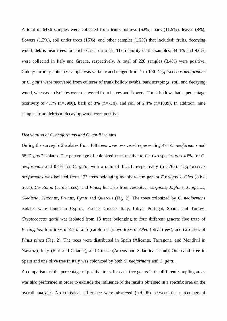
Distribution of C. neoformans and C. gattii isolates
During the survey 512 isolates from 188 trees were recovered representing 474 C. neoformans and
38 C. gattii isolates. The percentage of colonized trees relative to the two species was 4.6% for C.
neoformans and 0.4% for C. gattii with a ratio of 13.5:1, respectively (n=3765). Cryptococcus
neoformans was isolated from 177 trees belonging mainly to the genera Eucalyptus, Olea (olive
trees), Ceratonia (carob trees), and Pinus, but also from Aesculus, Carpinus, Juglans, Juniperus,
Gleditsia, Platanus, Prunus, Pyrus and Quercus (Fig. 2). The trees colonized by C. neoformans
isolates were found in Cyprus, France, Greece, Italy, Libya, Portugal, Spain, and Turkey.
Cryptococcus gattii was isolated from 13 trees belonging to four different genera: five trees of
Eucalyptus, four trees of Ceratonia (carob trees), two trees of Olea (olive trees), and two trees of
Pinus pinea (Fig. 2). The trees were distributed in Spain (Alicante, Tarragona, and Mendivil in
Navarra), Italy (Bari and Catania), and Greece (Athens and Salamina Island). One carob tree in
Spain and one olive tree in Italy was colonized by both C. neoformans and C. gattii.
A comparison of the percentage of positive trees for each tree genus in the different sampling areas
was also performed in order to exclude the influence of the results obtained in a specific area on the
overall analysis. No statistical difference were observed (p>0.05) between the percentage of
positive trees in the different sampling areas confirming that the differences observed are likely
associated with the tree species considered.
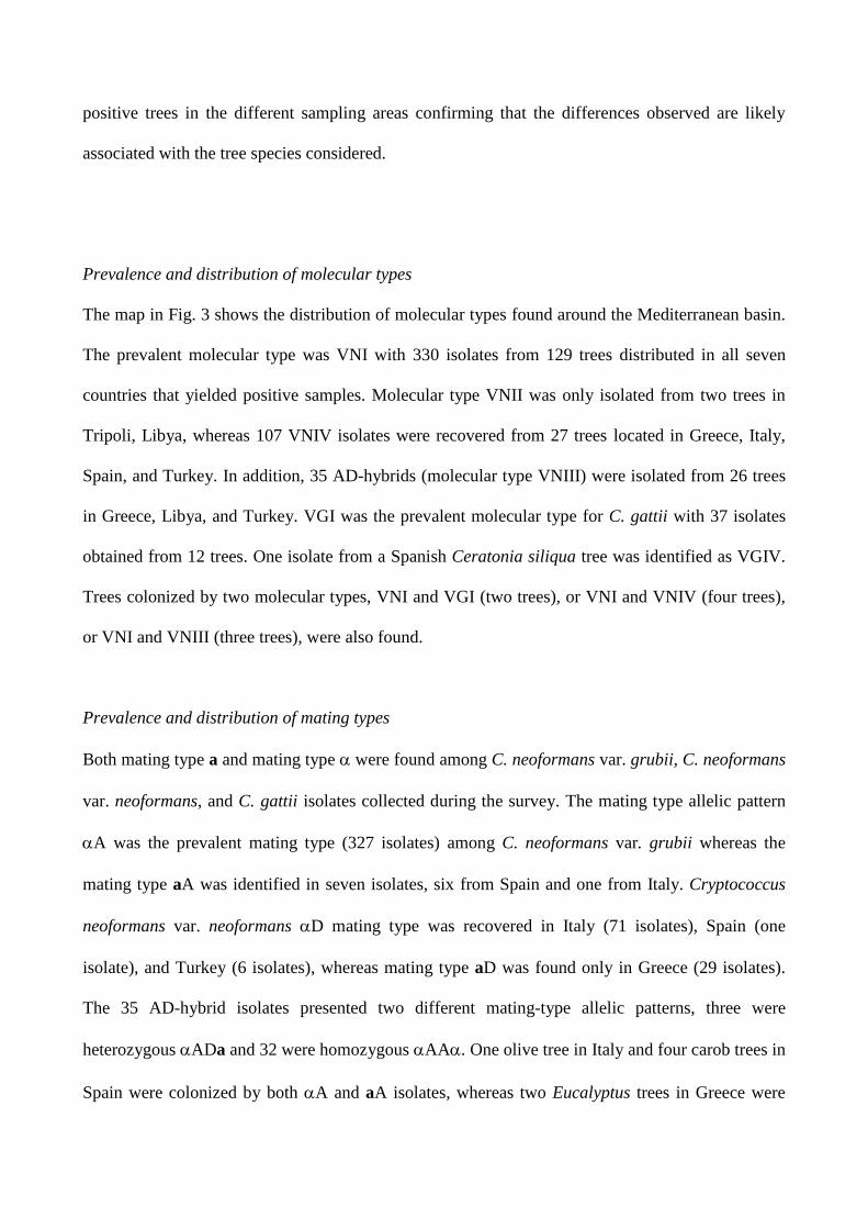
Prevalence and distribution of molecular types
The map in Fig. 3 shows the distribution of molecular types found around the Mediterranean basin.
The prevalent molecular type was VNI with 330 isolates from 129 trees distributed in all seven
countries that yielded positive samples. Molecular type VNII was only isolated from two trees in
Tripoli, Libya, whereas 107 VNIV isolates were recovered from 27 trees located in Greece, Italy,
Spain, and Turkey. In addition, 35 AD-hybrids (molecular type VNIII) were isolated from 26 trees
in Greece, Libya, and Turkey. VGI was the prevalent molecular type for C. gattii with 37 isolates
obtained from 12 trees. One isolate from a Spanish Ceratonia siliqua tree was identified as VGIV.
Trees colonized by two molecular types, VNI and VGI (two trees), or VNI and VNIV (four trees),
or VNI and VNIII (three trees), were also found.
Prevalence and distribution of mating types
Both mating type a and mating type were found among C. neoformans var. grubii, C. neoformans
var. neoformans, and C. gattii isolates collected during the survey. The mating type allelic pattern
A was the prevalent mating type (327 isolates) among C. neoformans var. grubii whereas the
mating type aA was identified in seven isolates, six from Spain and one from Italy. Cryptococcus
neoformans var. neoformans D mating type was recovered in Italy (71 isolates), Spain (one
isolate), and Turkey (6 isolates), whereas mating type aD was found only in Greece (29 isolates).
The 35 AD-hybrid isolates presented two different mating-type allelic patterns, three were
heterozygous ADa and 32 were homozygous AA. One olive tree in Italy and four carob trees in
Spain were colonized by both A and aA isolates, whereas two Eucalyptus trees in Greece were
colonized by both A and aD isolates. Co-existence of A and D as well as A and AD-hybrids
was observed in Italy (two trees) and Turkey (three trees), respectively.
Regarding C. gattii, 10 trees were colonized by mating type B strains, two trees in Italy by mating
type aB, and one in Spain by mating type C. One of the two Italian aB isolates shared the same
olive tree with C. neoformans var. grubii aA and A strains. Similarly, in Spain one C. gattii B
strain co-existed with one C. neoformans var. grubii A strain in a carob tree.
Results of mating assays
Seven mating assays were performed to test the fertility of the isolates that originated from the trees
where two opposite mating type isolates shared the same niche. Five assays were intra-variety
mating assays between aA and A isolates whereas the other two were inter-variety assays between
A and aD isolates. All intra-variety assays produced filaments and basidiospores. In addition,
molecular analysis showed that the progeny included both aA and A mating types in a Mendelian
ratio (Fig. 6). In contrast, none of the inter-varietal matings was fertile after four weeks of
observation.
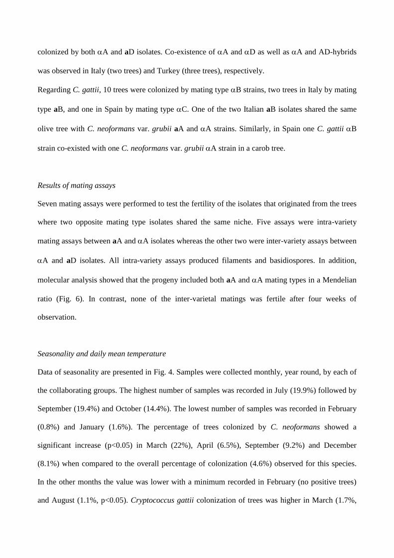
Seasonality and daily mean temperature
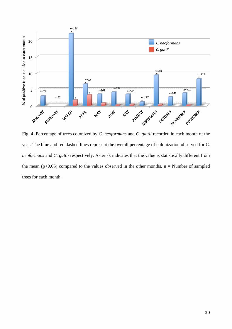
Data of seasonality are presented in Fig. 4. Samples were collected monthly, year round, by each of
the collaborating groups. The highest number of samples was recorded in July (19.9%) followed by
September (19.4%) and October (14.4%). The lowest number of samples was recorded in February
(0.8%) and January (1.6%). The percentage of trees colonized by C. neoformans showed a
significant increase (p<0.05) in March (22%), April (6.5%), September (9.2%) and December
(8.1%) when compared to the overall percentage of colonization (4.6%) observed for this species.
In the other months the value was lower with a minimum recorded in February (no positive trees)
and August (1.1%, p<0.05). Cryptococcus gattii colonization of trees was higher in March (1.7%,
p<0.05), April (3.3%, p<0.05), and May (0.8%, p>0.05) than that observed in overall tree
population (0.4%). No C. gattii positive trees were found in January, February, August, October,
and December.
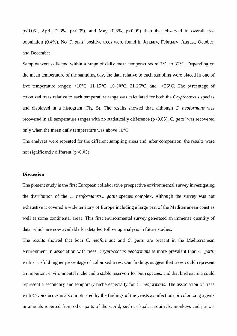
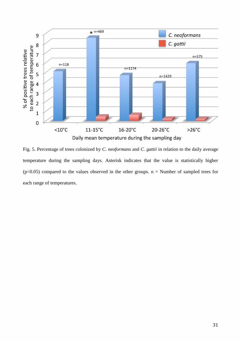
Samples were collected within a range of daily mean temperatures of 7°C to 32°C. Depending on
the mean temperature of the sampling day, the data relative to each sampling were placed in one of
five temperature ranges: <10°C, 11-15°C, 16-20°C, 21-26°C, and >26°C. The percentage of
colonized trees relative to each temperature range was calculated for both the Cryptococcus species
and displayed in a histogram (Fig. 5). The results showed that, although C. neoformans was
recovered in all temperature ranges with no statistically difference (p>0.05), C. gattii was recovered
only when the mean daily temperature was above 10°C.
The analyses were repeated for the different sampling areas and, after comparison, the results were
not significantly different (p>0.05).
Discussion
The present study is the first European collaborative prospective environmental survey investigating
the distribution of the C. neoformans/C. gattii species complex. Although the survey was not
exhaustive it covered a wide territory of Europe including a large part of the Mediterranean coast as
well as some continental areas. This first environmental survey generated an immense quantity of
data, which are now available for detailed follow up analysis in future studies.
The results showed that both C. neoformans and C. gattii are present in the Mediterranean
environment in association with trees. Cryptococcus neoformans is more prevalent than C. gattii
with a 13-fold higher percentage of colonized trees. Our findings suggest that trees could represent
an important environmental niche and a stable reservoir for both species, and that bird excreta could
represent a secondary and temporary niche especially for C. neoformans. The association of trees
with Cryptococcus is also implicated by the findings of the yeasts as infectious or colonizing agents
in animals reported from other parts of the world, such as koalas, squirrels, monkeys and parrots
whose lives revolve around trees. Goats have the habit of eating tree barks and they also develop
cryptococcosis (Roussilhon et al. 1987; López-Martinez and Castañón-Olivares 1995; Torres-
Rodriguez et al. 1999; Krockenberger et al. 2003; Stilwell and Pissarra 2014; Iatta et al. 2015;
Maestrale et al. 2015).
This study reveals a difference in the association of the two Cryptococcus species with specific
trees. In particular, Ceratonia (carob tree) and Olea (olive tree), two tree genera typical for the
Mediterranean region, together with Eucalyptus trees, produced the highest number of positive
samples. However, when the values were normalized as a percentage of the positive tree genus and
stratified for each of the Cryptococcus species and varieties, the results showed that Ceratonia is an
important niche for both C. gattii and C. neoformans var. grubii, but not for C. neoformans var.
neoformans and AD-hybrids, since the latter were not recovered from these trees. In addition, C.
gattii was recovered only from trees typical for the Mediterranean climate (Ceratonia, Olea,
Eucalyptus and Pinus pinea), whereas C. neoformans var. grubii colonized 12 different tree genera
confirming the ability of this pathogen to adapt to different environments and, hence, contributing
to its global distribution. The importance of Ceratonia siliqua as a niche for C. gattii, shown in the
present survey, is in agreement with the data reported by previous environmental studies carried out
in Spain (Colom et al. 2012; Linares et al. 2015). In contrast, C. neoformans var. neoformans and
AD-hybrids showed a preference to colonize trees typical of the sub-continental climate, such as
Platanus, Prunus and Quercus. This could reflect the ability of C. neoformans var. neoformans to
tolerate lower temperature better than C. neoformans var. grubii and C. gattii as previously shown
by other authors (Martinez et al. 2001). However, the survey has the limit that the samples were not
collected with a rigorous randomized method and therefore the conclusions here reported must be
considered as an attempt to describe the observed data and a starting point for future more extensive
studies.
Although the two C. neoformans varieties and C. gattii have differences in tree preference, the
climatic zones do not have sharp boundaries and they overlap along the entire Mediterranean basin.
Therefore, in the Mediterranean environment, the different C. neoformans and C. gattii populations
are continuously in contact with each other. This is confirmed by the finding that isolates belonging
to different species or varieties shared a niche of the same tree, as well as by the presence of hybrids
in the same area.
Hybridization between C. neoformans var. grubii and C. neoformans var. neoformans is well
documented in Europe by the identification of numerous AD-hybrids in clinical isolates which have
a prevalence of about 30% (Viviani et al. 2006). In contrast, only a few hybrids have been isolated
from pigeon droppings (Baró et al. 1999; Ferreira et al. 2014) due to the paucity of environmental
studies carried out in Europe. The current study reports for the first time the association of AD-
hybrids with trees and their presence in the same area where the putative parental C. neoformans
var. grubii and C. neoformans var. neoformans isolates may co-exist. This finding suggests that
hybridization between the two C. neoformans varieties is occurring in the European environment
and this may play an important role as a mechanism of evolution of this species (Cogliati et al.
2009; Li et al. 2012; Desnos-Ollivier et al. 2015). Although the inter-varietal mating assays carried
out in the present study did not succeed, this does not mean that the isolates studied are not
compatible but probably means that the in vitro assay conditions here adopted were not optimal for
hybridization. The high variability of conditions and substrates encountered in the environment may
better favor this process.
The presence of C. gattii in Greece, Southern Italy and Spain confirms the previous results obtained
in these geographical areas where the pathogen was isolated from both clinical and environmental
sources (Montagna et al. 1997; Torres-Rodriguez et al. 1999; Velegraki et al. 2001; Colom et al.
2005; Viviani et al. 2006; Solla et al. 2008; Ropstad et al. 2011; Colom et al. 2012; Hagen et al.
2012b; Romeo et al. 2012; Iatta et al. 2012). The survey also confirmed that VGI is the prevalent C.
gattii molecular type, which is in agreement with a previous analysis carried out by Hagen et al.
(2012b) that identified an endemic VGI cluster in Mediterranean Europe. However, one isolate was
identified as VGIV-C (Linares 2015) suggesting that other molecular types are also able to
colonize the Mediterranean basin. Further molecular analysis of the isolates collected during this
survey by MLST will elucidate the relationships and the clusters present in the European
environment.
Cryptococcus neoformans VNI was the prevalent molecular type distributed all around the
Mediterranean basin from Portugal to Libya confirming the ubiquitous presence of this pathogen in
the region (Cogliati 2013). In this study, a high prevalence of the VNIV molecular type was found
in Greece, where it represented the most common molecular type, and Northern Turkey, where it
was the only molecular type present, whereas it occurred less frequently in Italy and Spain. On the
basis of these results it could be speculated that this molecular type is spreading from sub-
continental areas of the South-Eastern Mediterranean towards the Western part of Europe. Further
sampling and an accurate niche modeling analysis is in progress to corroborate the above
hypothesis.
Our results showed that mating type a and are present in the Mediterranean environment for both
C. neoformans varieties and C. gattii. Interestingly, we found that the occurrence of two strains with
different mating types in the same tree is not rare, as already observed for C. gattii in Australia
(Halliday et al. 1999), therefore, trees are possible niches to complete the sexual cycle. This
hypothesis is supported by the observation that most of the isolates with different mating types
sharing the same niche were able to produce filaments and recombinant basidiospores in the mating
assays. In addition, other authors showed that C. neoformans and C. gattii are able to proliferate and
mate in vitro in a Cryptococcus-Arabidopsis system (Xue et al. 2007).
These findings suggest that the current view of a clonal population structure observed for both C.
neoformans and C. gattii could be due to the genotyping results being mainly obtained from clinical
isolates and the relatively low number of available environmental isolates investigated (Cogliati
2013). The analysis of a larger number of environmental isolates might show a more relevant
involvement of sexual reproduction in the evolution and propagation of the C. neoformans/C. gattii
species complex. Our data are corroborated by previous studies reporting recombination among C.
neoformans var. grubii (VNI and VNB) and C. gattii (VGI and VGII) populations isolated from the
environment (Litvintseva et al. 2003; Saul et al. 2008; Carriconde et al. 2011).
A different trend can be observed with respect to seasonality depending on the Cryptococcus
species considered. Sampling during different seasons did not greatly influence the recovery of C.
neoformans although a peak of positive trees was observed during spring. Similarly, C. gattii
recovery was more likely during spring and early summer, but was absent during the colder
seasons. Both species were isolated less in August, which is the hottest and driest month in the
Mediterranean area, suggesting the difficulty to cultivate these yeasts during such climatic
conditions. A recent study carried out in Colombia reported a similar observation with a low
probability to recover Cryptococcus from trees during the seasons with reduced rainfall (Noguera et al.
2015). When daily mean temperatures were considered, the results confirmed that C. neoformans
can be recovered during a wide range of temperatures, whereas C. gattii seems to be absent or
difficult to cultivate at temperatures below 10 °C.
In conclusion, the present survey established a wide laboratory network that, for the first time,
collected extensive information concerning the environmental distribution and ecology of the C.
neoformans/C. gattii species complex in Europe and the Mediterranean area. The results represent
the basis for future studies on environmental niches of Cryptococcus in Europe and an important
step towards the comparison of clinical and environmental isolates.
Acknowledgements
The work by K.J. Kwon-Chung and A. Varma was supported by funds from the intramural program
of the National Institute of Allergy and Infectious Diseases, NIH. The work by A. Sampaio and C.
Pereira was in part supported by CITAB under the project UID/AGR/04033/2013. W. Meyer was
supported by funding from the NH&MRC, Australia, grant # APP1031943. K. Ferreira-Paim was
supported by a CAPES Science without Borders visiting fellow (Nº 9313133) from Brazil.
Disclosures
The findings and conclusions of this article are those of the authors and do not necessarily represent
the views of the Centers for Disease Control and Prevention
References
Assogba K, Belo M, Wateba MI, et al. Neuromeningeal cryptococcosis in sub-Saharan Africa:
Killer disease with sparse data. J Neurosci Rural Pract 2015;6:221-4.
Arsic Arsenijevic V, Pekmezovic MG, Meis JF, et al. Molecular epidemiology and antifungal
susceptibility of Serbian Cryptococcus neoformans isolates. Mycoses 2014;57:380-7.
Baró T, Torres-Rodríguez JM, Morera Y, et al. Serotyping of Cryptococcus neoformans isolates
from clinical and environmental sources in Spain. J Clin Microbiol 1999;37:1170-2.
Bartlett KH, Cheng PY, Duncan C, et al. A decade of experience: Cryptococcus gattii in British
Columbia. Mycopathologia 2012;173:311-9.
Bauwens L, Vercammen F, Wuytack C, et al. Isolation of Cryptococcus neoformans in Antwerp
Zoo's nocturnal house. Mycoses 2004;47:292-6.
Bitar D, Lortholary O, Le Strat Y, et al. Population-based analysis of invasive fungal infections,
France, 2001-2010. Emerg Infect Dis 2014;20:1149-55.
Boekhout T, Theelen B, Diaz M, et al. Hybrid genotypes in the pathogenic yeast Cryptococcus
neoformans. Microbiology 2001;147:891-907.
Bratton EW, El Husseini N, Chastain CA, et al. Comparison and temporal trends of three groups
with cryptococcosis: HIV-infected, solid organ transplant, and HIV-negative/non-transplant.
PLoS One 2012;7:e43582.
Byrnes EJ 3rd, and Marr KA. The Outbreak of Cryptococcus gattii in Western North America:
Epidemiology and Clinical Issues. Curr Infect Dis Rep 2011;13:256-61.
Cafarchia C, Romito D, Iatta R, et al. Role of birds of prey as carriers and spreaders of
Cryptococcus neoformans and other zoonotic yeasts. Med Mycol 2006;44:485-92.
Campisi E, Mancianti F, Pini G, et al. Investigation in Central Italy of the possible association
between Cryptococcus neoformans var. gattii and Eucalyptus camaldulensis. Eur J Epidemiol
2003;18:357-62.
Carriconde F, Gilgado F, Arthur I, et al. Clonality and α-a recombination in the Australian
Cryptococcus gattii VGII population - an emerging outbreak in Australia. PLoS One
2011;6:e16936.
Chowdhary A, Randhawa HS, Boekhout T, et al. Temperate climate niche for Cryptococcus gattii
in Northern Europe. Emerg Infect Dis 2012;18:172-4.
Cogliati, M., Allaria, M., Tortorano, A.M., et al. Genotyping Cryptococcus neoformans var.
neoformans with specific primers designed from PCR-fingerprinting bands sequenced using a
modified PCR-based strategy. Med Mycol 2000;38:97-103.
Cogliati M, Lin X, and Viviani MA. Hybridization and its importance in the Cryptococcus species
complex. In Cryptococcus: from Pathogen to Model Yeast. Heitman J, Kozel TR, Kwon-Chung
KJ, Perfect JR, Casadevall A (eds). Washington, USA: ASM press. 2009;359-70.
Cogliati M. Global Molecular Epidemiology of Cryptococcus neoformans and Cryptococcus gattii:
An Atlas of the Molecular Types. Scientifica 2013:675213.
Cogliati M, D'Amicis R, and Tortorano AM. Cryptococcus gattii sero-mating type allelic pattern
determined by multiplex PCR. Clin Microbiol Infect 2015;21:190.e1-4.
Colom Valiente MF, Alberdi M, Meseguer I, et al. Isolation of Cryptococcus neoformans from
environmental samples in Alicante. Rev Iberoam Micol 1997;14:63-4.
Colom MF, Frasés S, Ferrer C, et al. First case of human cryptococcosis due to Cryptococcus
neoformans var. gattii in Spain. J Clin Microbiol 2005;43:3548-50.
Colom MF, Hagen F, Gonzalez A, et al. Ceratonia siliqua (carob) trees as natural habitat and
source of infection by Cryptococcus gattii in the Mediterranean environment. Med Mycol
2012;50:67-73.
Criseo G, Bolignano MS, De Leo F, et al. Evidence of canary droppings as an important reservoir
of Cryptococcus neoformans. Zentralbl Bakteriol 1995;282:244-54.
Criseo G, and Gallo M. Serotyping of Cryptococcus neoformans isolates from environmental and
clinical sources in extreme southern Italy (Calabria and Sicily, central Mediterranean area).
Mycoses 1997;40:95-100.
de Colombani P, Banatvala N, Zaleskis R, et al. European framework to decrease the burden of
TB/HIV. Eur Respir J 2004;24:493-501.
Desnos-Ollivier M, Patel S, Raoux-Barbot D, et al. Cryptococcosis serotypes impact outcome and
provide evidence of Cryptococcus neoformans speciation. MBio 2015;6:e00311.
Dromer F, Ronin O, and Dupont B. Isolation of Cryptococcus neoformans var. gattii from an Asian
patient in France: evidence for dormant infection in healthy subjects. J Med Vet Mycol
1992;30:395-7.
Dromer F, Mathoulin-Pélissier S, Fontanet A, et al. Epidemiology of HIV-associated
cryptococcosis in France (1985-2001): comparison of the pre- and post-HAART eras. AIDS.
2004;18:555-62.
Esposto MC, Cogliati M, Tortorano AM, et al. Determination of Cryptococcus neoformans var.
neoformans mating type by multiplex PCR. Clin Microbiol Infect 2004;10:1092-4.
Feng X, Fu X, Ling B, et al. Rapid differentiation of cryptic species within Cryptococcus gattii by a
duplex PCR assay. J Clin Microbiol 2013;51:3110-2.
Ferreira AS, Sampaio A, Maduro AP, et al. Genotypic diversity of environmental Cryptococcus
neoformans isolates from Northern Portugal. Mycoses 2014;57:98-104.
FIMUA Cryptococcosis Network. European Confederation of Medical Mycology (ECMM)
prospective survey of cryptococcosis: report from Italy. Med Mycol 2002;40:507-17.
Garcia-Hermoso D, Mathoulin-Pélissier S, Couprie B, et al. DNA typing suggests pigeon droppings
as a source of pathogenic Cryptococcus neoformans serotype D. J Clin Microbiol
1997;35:2683-5.
Hagen F, Illnait-Zaragozí MT, Meis JF, et al. Extensive genetic diversity within the Dutch clinical
Cryptococcus neoformans population. J Clin Microbiol 2012a;50:1918-26.
Hagen F, Colom MF, Swinne D, et al. Autochthonous and dormant Cryptococcus gattii infections
in Europe. Emerg Infect Dis 2012b;18:1618-24.
Hagen F, Ceresini PC, Polacheck I, et al. Ancient dispersal of the human fungal pathogen
Cryptococcus gattii from the Amazon rainforest. PLoS One 2013;8:e71148.
Hagen F, Khayhan K, Theelen B, et al. Recognition of seven species in the Cryptococcus
neoformans/C. gattii species complex. Fungal Genet Biol 2015;78:16-48.
Halliday CL, Bui T, Krockenberger M, et al. Presence of alpha and a mating types in environmental
and clinical collections of Cryptococcus neoformans var. gattii strains from Australia. J Clin
Microbiol 1999;37:2920-26.
Henao-Martínez AF, and Beckham JD. Cryptococcosis in solid organ transplant recipients. Curr
Opin Infect Dis 2015;28:300-7.
Iatta R, Hagen F, Fico C, et al. Cryptococcus gattii infection in an immunocompetent patient from
Southern Italy. Mycopathologia 2012;174:87-92.
Iatta R, Immediato D, Puttilli et al. Cryptococcus neoformans in the respiratory tract of squirrels,
Callosciurus finlaysonii (Rodentia, Sciuridae). Med Mycol 2015;53:666-73.
Li W, Averette AF, Desnos-Ollivier M, Ni M, et al. Genetic diversity and genomic plasticity of
Cryptococcus neoformans AD Hybrid Strains. G3 (Bethesda) 2012;2:83-97.
Kidd SE, Bach PJ, Hingston AO, et al. Cryptococcus gattii dispersal mechanisms, British
Columbia, Canada. Emerg Infect Dis 2007;13:51-7.
Krockenberger MB, Canfield PJ, and Malik R. Cryptococcus neoformans var. gattii in the koala
(Phascolarctos cinereus): a review of 43 cases of cryptococcosis. Med Mycol 2003;41: 225-34.
Kwon-Chung KJ, and Bennett JE (eds). Medical Mycology. Philadelphia, USA. Lea & Fabiger.
1992.
Kwon-Chung KJ, Fraser JA, Doering TL, et al. Cryptococcus neoformans and Cryptococcus gattii,
the etiologic agents of cryptococcosis. 2014. Cold Spring Harb Perspect Med 2014:a019760;
doi:10.1101/cshperspect.a019760.
Lagrou K, Van Eldere J, Keuleers S, et al. Zoonotic transmission of Cryptococcus neoformans from
a magpie to an immunocompetent patient. J Intern Med 2005;257:385-8.
Lin X. Cryptococcus neoformans: morphogenesis, infection, and evolution. Infect Genet Evol
2009;9:401-16.
Linares C, Colom MF, Torreblanca M, et al. Environmental sampling of Ceratonia siliqua (carob)
trees in Spain reveals the presence of the rare Cryptococcus gattii genotype AFLP7/VGIV. Rev
Iberoam Micol 2015;32:269-72.
Litvintseva, A.P., Marra, R.E., Nielsen, K., et al. Evidence of sexual recombination among
Cryptococcus neoformans serotype A isolates in sub-Saharan Africa. Eukaryot Cell
2003;2:1162-8.
Lo Passo, C., Pernice, I., Gallo, M., et al. Genetic relatedness and diversity of Cryptococcus
neoformans strains in the Maltese Islands. J Clin Microbiol 1997;35:751-5.
López-Martínez R, and Castañón-Olivares LR. Isolation of Cryptococcus neoformans var.
neoformans from bird droppings, fruits and vegetables in Mexico City. Mycopathologia
1995;129:25-28.
Maduro AP, Gonçalves L, Inácio J, et al. HIV/AIDS-associated cryptococcosis in Portugal
spanning the pre- to post-HAART era: a retrospective assessment at the genotypic level based
on URA5-RFLP. Curr Microbiol 2015;71:449-57.
Maestrale C, Masia M, Pintus D, et al. Genetic and pathological characteristics of Cryptococcus
gattii and Cryptococcus neoformans var. neoformans from meningoencephalitis in
autochthonous goats and mouflons, Sardinia, Italy. Vet Microbiol 2015;177:409-13.
Martinez LR, Garcia-Rivera J, and Casadevall A. Cryptococcus neoformans var. neoformans
(serotype D) strains are more susceptible to heat than C. neoformans var. grubii (serotype A)
strains. J Clin Microbiol 2001;39:3365–67.
Meyer W, Aanensen DM, Boekhout T, et al. Consensus multi-locus sequence typing scheme for
Cryptococcus neoformans and Cryptococcus gattii. Med Mycol 2009;47:561-70.
Mlinaric-Missoni E, Hagen F, Chew WH, et al. In vitro antifungal susceptibilities and molecular
typing of sequentially isolated clinical Cryptococcus neoformans strains from Croatia. J Med
Microbiol 2011;60:1487-95.
Montagna MT, Viviani MA, Pulito A, et al. Cryptococcus neoformans var. gattii in Italy - Note 2 -
Environment investigation related to an autochtonous clinical case in Apulia. J Mycol Méd
1997;7:93-96.
Montagna MT, Santacroce MP, Caggiano G, et al. Cavernicolous habitats harbouring Cryptococcus
neoformans: results of a speleological survey in Apulia, Italy, 1999-2000. Med Mycol
2003;41:451-5.
Noguera MC, Escandón P, and Castañeda E. Cryptococcosis in Atlántico, Colombia: an
approximation of the prevalence of this mycosis and the distribution of the etiological agent in
the environment. Rev Soc Bras Med Trop 2015;48:580-6.
Park BJ, Wannemuehler KA, Marston BJ, et al. Estimation of the current global burden of
cryptococcal meningitis among persons living with HIV/AIDS. AIDS 2009;23:525-30.
Patel S, Shin GY, Wijewardana I, et al. The prevalence of cryptococcal antigenemia in newly
diagnosed HIV patients in a Southwest London cohort. J Infect 2013;66:75-9.
Pernice I, Lo Passo C, Criseo G, et al. Molecular subtyping of clinical and environmental strains of
Cryptococcus neoformans variety neoformans serotype A isolated from southern Italy. Mycoses
1998;41:117-24.
Romeo O, Scordino F, and Criseo G. Environmental isolation of Cryptococcus gattii serotype B,
VGI/MATα strains in southern Italy. Mycopathologia 2011;171:423-30.
Romeo O, Scordino F, Chillemi V, et al. Cryptococcus neoformans/Cryptococcus gattii species
complex in southern Italy: an overview on the environmental diffusion of serotypes, genotypes
and mating-types. Mycopathologia 2012;174:283-91.
Ropstad EO, Leiva M, Peña T, et al. Cryptococcus gattii chorioretinitis in a ferret. Vet Ophthalmol
2011;14:262-6.
Roussilhon C, Postal JM, and Ravisse P. Spontaneous cryptococcosis of a squirrel monkey (Saimiri
sciureus) in French Guyana. J Med Primatol 1987;16:39-47.
Sanchini A, Smith IM, Sedlacek L, et al. Molecular typing of clinical Cryptococcus neoformans
isolates collected in Germany from 2004 to 2010. Med Microbiol Immunol 2014;203:333-40.
Saul N, Krockenberger M, and Carter D. Evidence of recombination in mixed-mating-type and
alpha-only populations of Cryptococcus gattii sourced from single eucalyptus tree hollows.
Eukaryot Cell 2008;7: 727-734.
Solla I, Morano LE, Vasallo F, et al. Cryptococcus gattii meningitis: observation in a Spanish
patient. Enferm Infecc Microbiol Clin 2008;26:395-6.
Stilwell G, and Pissarra H. Cryptococcal meningitis in a goat - a case report. BMC Vet Res
2014;10:84.
Torres-Rodríguez JM, Baró T, Morera Y, et al. Molecular characterization of Cryptococcus
neoformans var. gattii causing epidemic outbreaks of cryptococcosis in goats. Rev Iberoam
Micol 1999;16:164-5.
Velegraki A, Kiosses VG, Pitsouni H, et al. First report of Cryptococcus neoformans var. gattii
serotype B from Greece. Med Mycol 2001;39:419-22.
Viviani MA, Wen H, Roverselli A, et al. Identification by polymerase chain reaction fingerprinting
of Cryptococcus neoformans serotype AD. J Med Vet Mycol 1997;35:355-60.
Viviani MA, Cogliati M, Esposto MC, et al. Molecular analysis of 311 Cryptococcus neoformans
isolates from a 30-month ECMM survey of cryptococcosis in Europe. FEMS Yeast Res
2006;6:614-9.
Xue C, Tada Y, Dong X, et al. The human fungal pathogen Cryptococcus can complete its sexual
cycle during a pathogenic association with plants. Cell Host Microbe 2007;1:263-73.
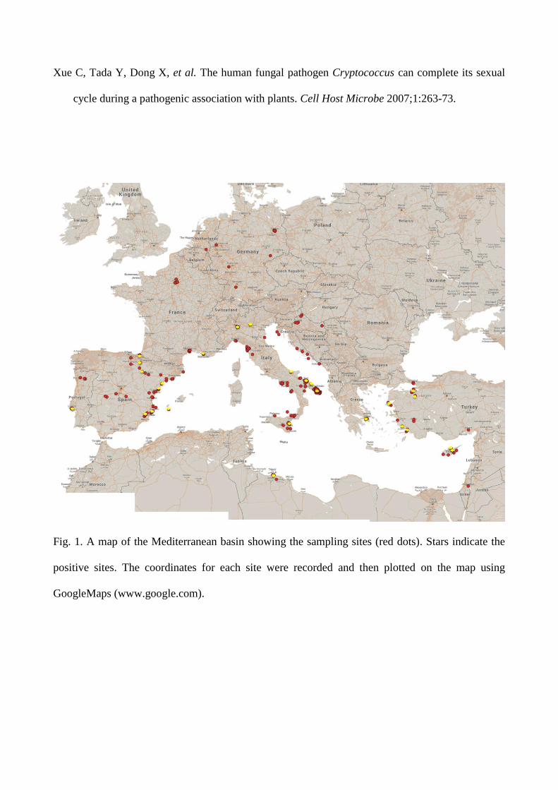
Fig. 1. A map of the Mediterranean basin showing the sampling sites (red dots). Stars indicate the
positive sites. The coordinates for each site were recorded and then plotted on the map using
GoogleMaps (www.google.com).
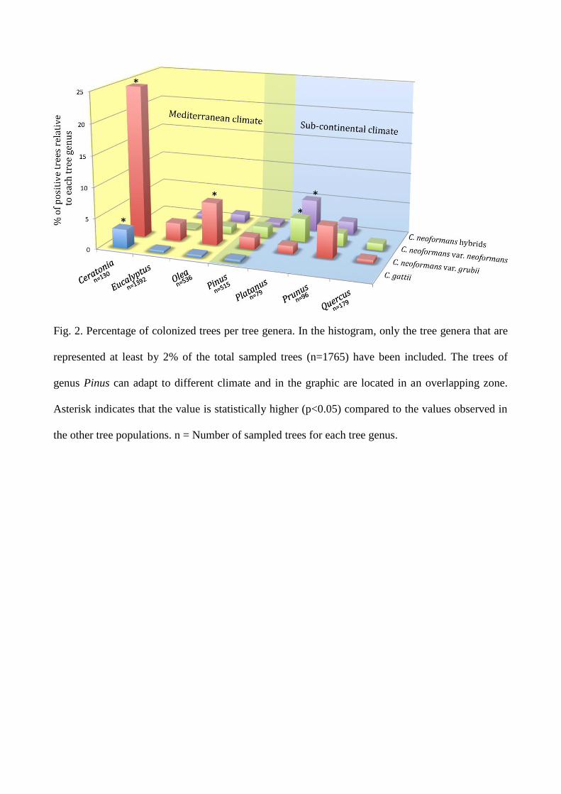
Fig. 2. Percentage of colonized trees per tree genera. In the histogram, only the tree genera that are
represented at least by 2% of the total sampled trees (n=1765) have been included. The trees of
genus Pinus can adapt to different climate and in the graphic are located in an overlapping zone.
Asterisk indicates that the value is statistically higher (p<0.05) compared to the values observed in
the other tree populations. n = Number of sampled trees for each tree genus.
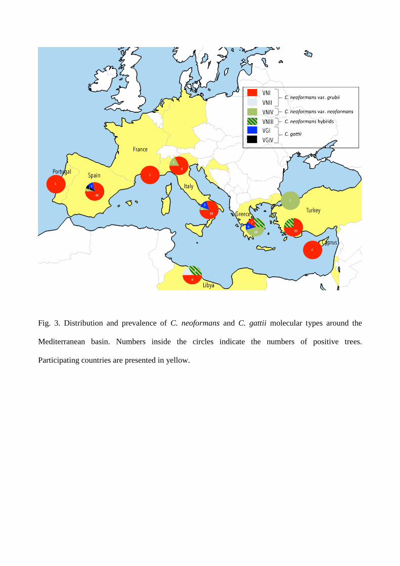
Fig. 3. Distribution and prevalence of C. neoformans and C. gattii molecular types around the
Mediterranean basin. Numbers inside the circles indicate the numbers of positive trees.
Participating countries are presented in yellow.
30
Fig. 4. Percentage of trees colonized by C. neoformans and C. gattii recorded in each month of the
year. The blue and red dashed lines represent the overall percentage of colonization observed for C.
neoformans and C. gattii respectively. Asterisk indicates that the value is statistically different from
the mean (p<0.05) compared to the values observed in the other months. n = Number of sampled
trees for each month.
31
Fig. 5. Percentage of trees colonized by C. neoformans and C. gattii in relation to the daily average
temperature during the sampling days. Asterisk indicates that the value is statistically higher
(p<0.05) compared to the values observed in the other groups. n = Number of sampled trees for
each range of temperatures.
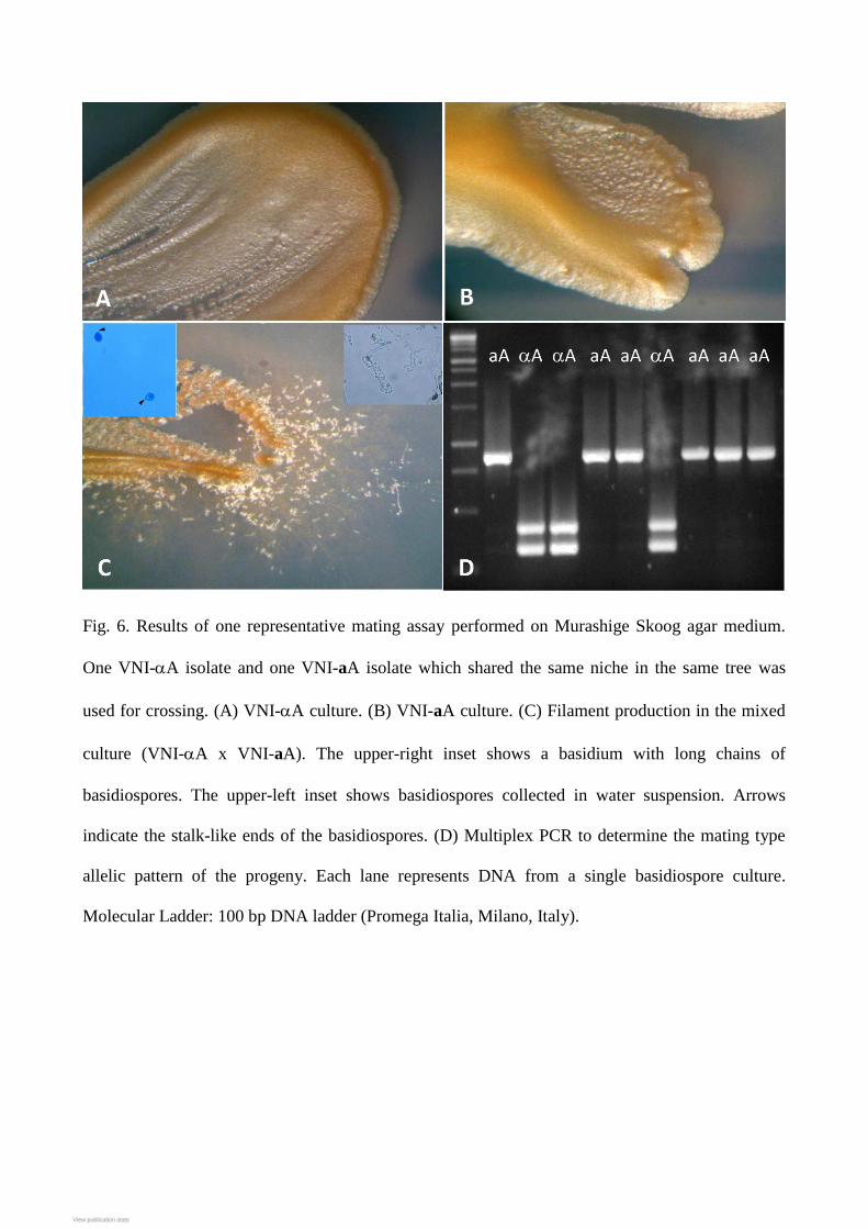
Fig. 6. Results of one representative mating assay performed on Murashige Skoog agar medium.
One VNI-A isolate and one VNI-aA isolate which shared the same niche in the same tree was
used for crossing. (A) VNI-A culture. (B) VNI-aA culture. (C) Filament production in the mixed
culture (VNI-A x VNI-aA). The upper-right inset shows a basidium with long chains of
basidiospores. The upper-left inset shows basidiospores collected in water suspension. Arrows
indicate the stalk-like ends of the basidiospores. (D) Multiplex PCR to determine the mating type
allelic pattern of the progeny. Each lane represents DNA from a single basidiospore culture.
Molecular Ladder: 100 bp DNA ladder (Promega Italia, Milano, Italy).
View publication statsView publication stats