Environmental Chemistry Journal of
26
Journal of Environmental Chemistry and Ecotoxicology Volume 8 Number 3 March 2016 ISSN 2141-226X
Transcript of Environmental Chemistry Journal of

Journal of
Environmental Chemistry
and Ecotoxicology
Volume 8 Number 3 March 2016
ISSN 2141-226X

ABOUT JECE The Journal of Environmental Chemistry and Ecotoxicology (JECE) is published bi-monthly (one volume per year) by Academic Journals.
Journal of Environmental Chemistry and Ecotoxicology (JECE) is an open access journal that provides rapid publication (bi-monthly) of articles in all areas of the subject such as ocean acidification, pesticides use and regulation, arsenic induced genotoxicity by curcumin, heavy metals and its environmental effect etc. The Journal welcomes the submission of manuscripts that meet the general criteria of significance and scientific excellence. Papers will be published shortly after acceptance. All articles published in JECE are peer- reviewed.
Contact Us
Editorial Office: [email protected]
Help Desk: [email protected]
Website: http://www.academicjournals.org/journal/JECE
Submit manuscript online http://ms.academicjournals.me/

Editors Prof. Peter Massanyi Prof. Toyin Ayodele Arowolo
Slovak University of Agriculture, Faculty of Department of Environmental Management &
Biotechnology and Food Sciences, Department of Toxicology,
Animal Physiology, College of Environmental Resources
Management,
Tr. A. Hlinku 2, SK-949 76 Nitra, Slovak Republic, University of Agriculture,
Slovak Republic. P.M.B. 2240, Abeokuta 110001, Ogun State,
Prof. Mostafa El-Sheekh Nigeria.
Faculty of Science, Tanta University,
Tanta 31527, Dr. Xue Song Wang
Egypt. Department of Chemical Engineering,
Huaihai Institute of Technology,
CangWu Road 59#, Lianyungang, Jiangsu, PR. China Prof. Minghua Zhou Nankai University, Dr. Mohamed Nageeb Rashed
No. 94, Road Weijin, Aswan Faculty of Science, South Valley University,
Nankai District, Aswan,
Tianjin 300071, Egypt.
China.
Prof. Muhammad Abdul Rauf Prof. Hamayun Khan
United Arab Emirates University , Department of Chemistry
United Arab Emirates. Islamia College University
Peshawar-25120,
Pakistan. Prof. Shao Hongbo Qingdao University of Science Technology, Zhengzhou Road 53, Qingdao266042, China , China.
Prof. Ghasem D. Najafpour
Oshirvani University of Technology
Babol, Iran
Iran.

Editorial Board Dr. Mohammad Al-Hwaiti Dr. Ghousia Begum
Al-Hussein Bin Talal University Indian Institute of Chemical Technology,
Environmental Engineering Department Hyderabad 500 007, A.P; India
O. Box (20) Ma’an-Jordan Prof. Gang Yang
Jordan. Key Laboratory of Forest Plant Ecology, Ministry of Education,
Prof. Ajai Kumar Srivastav Northeast Forestry University
DDU Gorakhpur University 26 Hexing Road, Harbin
Department of Zoology, DDU Gorakhpur University, China.
Gorakhpur, INDIA. Dr. Anindita Bhattacharyya
Indian Statistical Institute
Nathaniel C. Añasco 203, B. T. Road, Kolkata 700108, West Bengal,
University of the Philippines Visayas India.
Miagao, Iloilo 5023 Philippines Philippines. Dr. Onome Davies
Rivers State University of Science & Technology, Port
Prof. El-Refaie Kenawy Harcourt, Rivers State
King Saud University, Dept. of Fisheries & Aquatic Environment, P.M.B. 5080,
Faculty of Science, Port Harcourt,
Department of Chemistry, Nigeria.
Petrochemicals Research Chair, B.O.Box 2455 Riyadh 11451 ,Saudi Arabia Dr. B. Stephen Inbaraj
Saudi Arabia. Fu Jen University Department of Nutrition & food science,
Dr. K. Senthil Kumar Taipei 242, Taiwan
TÜV SÜD South Asia Pvt. Ltd., Environmental Chemistry & Analytical Chemistry
No: A-151, 2nd C Main, 2nd Stage, Peenya Industrial Taiwan.
Estate, Bangalore 560058, Karnataka State, INDIA Dr. Hala A. Awney
India. Institute of Graduate Studies and Research, Alexandria University,
Dr. Omotayo Sarafadeen Amuda Egypt.
Ladoke Akintola University of Technology Ilorin Road, Ogbomoso, Dr. Suhel Parvez
Nigeria. Leibniz Institute for Neurobiology Brenneckestr. 6, Magdeburg 39118,
Dr. Jitendra Pandey Germany.
Banaras Hindu university Environmental Science Division, Department of Botany, Dr. Mayalagu Rajkumar
Banaras Hindu university, Varanasi - 221005, Institute of Oceanology, Chinese Academy of Sciences
India. 7 Nanhai Road, Qingdao, 266071 China.
Dr. Soumya Chatterjey Defence Research Laboratory, Tezpur (DRDO) Dr. Eldon Raj Rene
Post Bag No. 2, Tezpur 784001, Assam, University of La Coruna
India. Department of Chemical Engineering, Spain
Prof. Bechan Sharma Vyacheslav Khavrus
University of Allahabad L. V. Pisarzhevskii Institute of physical chemistry of
Department of Biochemistry, NAS of the Ukraine (permanent position)
Allahabad-211002, Ukraine.
India.

Table of Content: Volume 8 Number 3 March, 2016
Journal of Environmental Chemistry and Ecotoxicology
Table of Content: Volume 8 Number 1 January, 2016
ARTICLE
Distribution of trace metals in surface water and sediments of
Imo River Estuary (Nigeria): Health risk assessment, seasonal
and physicochemical variability 1 Essien D. Udosen, Nnanake-Abasi O. Offiong, Samuel Edem and John B. Edet
ARTICLES
Quantification of pharmaceutical residues in wastewater impacted surface waters and sewage sludge from Lagos, Nigeria 14 Oluwatosin Olarinmoye, Adekunle Bakare, Obih Ugwumba and Arne Hein Sensitivity, ecotoxicity and histopathological effects on neotropical fish exposed to glyphosate alone and associated to surfactant 25 Claudinei da Cruz, Silvia Patrícia Carraschi, Natália Sayuri Shiogiri, Adilson Ferreira da Silva, Robinson Antonio Pitelli and Marcia Rita Fernandes Machado
Journal of Environmental Chemistry and Ecotoxicology

Vol. 8(3), pp. 14-24, March, 2016
DOI:10.5897/JECE2015.0364
Article Number: 083D7EF57805
ISSN 2141-226X
Copyright ©2016
Author(s) retain the copyright of this article
http://www.academicjournals.org/JECE
Journal of Environmental Chemistry and Ecotoxicology
Full Length Research Paper
Quantification of pharmaceutical residues in wastewater impacted surface waters and sewage
sludge from Lagos, Nigeria
Oluwatosin Olarinmoye1*, Adekunle Bakare2, Obih Ugwumba2 and Arne Hein3
1Lagos State University, Lagos, Nigeria
2University of Ibadan, Oyo State, Nigeria.
3German Environment Protection Agency (Umweltbundesamt), WorlitzerPlatz 1, 06844, Dessau-Roßlau, Germany,
Germany.
Received 3 September, 2015, Accepted 12 October, 2015
Information on the occurrence of pharmaceuticals in the environment of Nigeria is limited, only a single publication previously on pharmaceutical occurrence in the environment of Nigeria, which measured general estrogen levels in Enugu, South-East Nigeria. In order to establish a first overview, surface water samples from six locations as well as ten sewage sludge samples from waste water treatment plants were analysed for a range of different pharmaceuticals, including antibiotics, estrogens, and lipid-lowering drugs. The results of this monitoring campaign were evaluated in comparison to published measured environmental concentrations in Africa and worldwide. In surface water samples, 12 of 37 pharmaceutical substances were detected at concentrations ranging from Limits of Detection (LOD) up to 8.84 µg/l. Four of these pharmaceuticals were found at concentrations exceeding ecotoxicological predicted no-effect concentrations (PNEC). In industrial, domestic, and hospital sewage sludge, nine different pharmaceutical substances were detected with the NSAID diclofenac present in all samples at concentrations of up to 1100 µg/kg dry weight, exceeding the highest measured concentration of 560 µg/ kg reported in sludge samples worldwide. This study proves the presence of several pharmaceuticals at relevant concentrations in the environmental matrices studied. Further, more comprehensive monitoring campaigns, especially in locations with high population density and low dilution of treated or untreated waste water in receiving streams are recommended. Key words: Nigeria, pharmaceuticals, liquid chromatography-tandem mass spectrometry (LC-MS/MS), water, sludge.
INTRODUCTION The global presence of pharmaceuticals in the environment has been validated as a real and ongoing situation of concern by several studies. This has in part
led to the current initiative to have pharmaceuticals included in the list of environmentally and ecologically active chemical entities, under the global SUNEP (UBA,
*Corresponding author. E-mail: [email protected]
Author(s) agree that this article remain permanently open access under the terms of the Creative Commons Attribution
License 4.0 International License
2014). Pharmaceuticals enter subterranean and surface water environments after human/veterinary use in trace amounts regardless of attenuating bio-physical processes, which include degradation, dilution, and sorption (Scheytt et al., 2001; WHO, 2012), primarily through excretion and sewage release as completely non-metabolised parent drugs, partly metabolized bio-active metabolites, and re-activatable pro-drugs (Bound and Voulvoulis, 2005; Jjemba, 2008). Pharmaceutically Active Compounds [PhACs] have also been demonstrated to be incompletely removed from waste waters by current sewage treatment regimes, with som entirely unchanged by such processes (Nakada et al., 2007; Miege et al., 2009).
Since human waste streams have been implicated as the most significant sources of environmental pharmaceutical loading (Bound and Voulvoulis, 2005) the degree of human waste impaction on surface water bodies conceivably should determine the concentrations of detectable PhAC residues for individual locations. In developing country urban situations as studied here, waste water treatment is inadequate and sanitation poor. PhACs could potentially enter surface waters without any preliminary treatment, and the effects consequent on this fact, especially on non-target species such as fish and other aquatic fauna, could potentially be even more profound than conjectured at present.
Sewage Treatment Facility (STF) sludge could be a significant catchment matrix and source for environmental, especially water loading of pharmaceutical residues (Carballa et al., 2004; Ternes et al., 2004), especially where such post-treatment sludge is used as manure for crop production. Lagos, the commercial capital of Nigeria, with an estimated population of about 12,614000 million people (UN, 2014), is one of the world’s fastest growing metropolises, already gaining megacity status. High population density clusters in largely unplanned urban and peri-urban locations, poverty, and the overstretched public infrastructure and works, are largely descriptive of living conditions in Lagos.
The generally poor sanitary conditions in most of the city and suburbs, aggravated by the tropical climate characterized by seasonal heavy rains, and inadequate primary sewage treatment, make wastewater impaction on surface waters a stark and ongoing reality. This study seeks to identify and quantify the levels of pharmaceutical residues in some surface waters and sewage sludge from locations within Lagos municipality, and a nearby state, and to compare detected concentrations with existing information from other studies for perspective.
MATERIALS AND METHODS
Sampling locations
Selection criteria for the locations surveyed included population
Olarinmoye et al. 15 (numbers, density, commercial activities, etc.), proximity to hospitals, pharmaceutical manufacturing concerns, and waste disposal sites. The chosen locations were Amuwo-Odofin, Ajido via Badagry, Isolo, Liverpool/Apapa, Ojo, and from the river Owo, a tributary of River Ogun which receives treated and untreated effluents from the Agbara Industrial Estate in Lagos and municipalities in Ogun States, south west Nigeria which it flows down through (Figure 1).
Sewage sludge
Sewage sludge was sampled in four domestic wastewater treatment plants, five industrial effluent treatment plants and one hospital wastewater treatment plant from January to April 2012 at different locations in Lagos, Oyo and Ogun State, all in South West Nigeria (Sindiku et al., 2013). Table 1 gives details of the sampled plants.
Water collection, processing, and analysis
Water samples were collected (between August and September 2013) in duplicate, in the morning from each of the six locations into 500 mL amber coloured PET capped bottles, ensuring that contamination of the collection equipment, and samples before, during and after collection was minimal. The “clean hands, dirty hands” protocol (USGS, 2013) for ensuring the latter was employed in all cases. All locations were flowing water sites. Water was collected using the dip sampling method, which involved de-capping the bottles and dipping them below the surface till full. A little of the contents were routinely poured away to ensure enough room for expansion during cold storage. Field measurements including temperature, DO, conductivity and pH were done immediately after collection using a handheld Henna instruments®
model HI-98129 meter (Table 2). Following collection, water bottles were placed in Styrofoam coolers with ice packs and transferred to the laboratory where kept at -20°C until analysed.
Analyses of water samples were carried out by IWW Water Centre in Muelheim, Germany according to published guidelines or validated in-house methods.
Polar pharmaceutical substances
Sample aliquots were spiked with an internal standard (4-chlorobenzoic acid), and adjusted to pH 2 by HCl addition. SPE was done using a pre-conditioned SPE column (200 mg ENV+ adsorber material). The analytes were eluted in 9 mL of ethyl acetate, concentrated to a volume of 200 µL under a gentle stream of nitrogen, and derivatized with diazomethane following DIN EN ISO 15913 guidelines prior to GC-MS analysis (column: Phenomenex ZB 50 or J&W DB 5).
Non-polar pharmaceutical substances
Sample aliquots spiked with an internal standard (Fluazifo-butyl) and adjusted to pH 7 by NaOH addition were extracted using pre-conditioned SPE columns (200 mg ENV+) as before. Desired analytes were eluted in 6 mL of acetone and concentrated to a volume of 200 µL prior to GC-MS analysis.
All GC-MS analyses was done using an Agilent GC-MS system (6890 GC and 5973 MSD single quadrupole mass analyzer) equipped with an automatic sampler (MPS 2) and Cooled Injection System (CIS 4, Gerstel, Mülheim, Germany).
Antibiotics
Typical sample aliquots were adjusted to pH 3 by HCl addition and

16 J. Environ. Chem. Ecotoxicol.
Figure 1. Showing water sampling locations.
Table 1. Characteristics of the wastewater treatment plants studied in Nigeria (Sindiku et al., 2013).
Treatment plants Location Type of wastewater treated Treatment type Capacity [m³/h]
Brewery Lagos Industrial Activated sludge system 126
WEMABOD Lagos Industrial Aerated lagoon system n.a.
Dairy Ibadan, Oyo state Industrial Activated sludge system n.a.
AGBARA Ota, Ogun state Industrial Aerated lagoon system n.a.
Food Ota, Ogun state Industrial Activated sludge system 100
Ikeja Lagos Domestic Oxidation pond system 60
Abesan Lagos Domestic Activated sludge system 80-100
Iponri Lagos Domestic Lagoon oxidation system 150
Oke Afa Lagos Domestic Lagoon oxidation system 120
UCH Ibadan, Oyo state Hospital Activated sludge system n.a.
n.a. not available.
extracted on preconditioned SPE columns (500 mg ENV+). The analytes were eluted in 10 mL of methanol, concentrated to
dryness, and transferred into 1 mL of acetonitrile/water (v/v 60:40) solvent prior to analysis by LC-MS/MS (column: Nucleosil C18 HD

Olarinmoye et al. 17
Table 2. Physicochemical data measurements on collection dates by location.
Parameter Location
River Owo Ojo Liverpool Ajido Amuwo Isolo
Conductivity 134 3999 3999 488 3999 492
pH 7.06 7.83 6.6 7.65 7.60 7.45
Temperature (°C) 26.4 31.4 34.7 29 28.2 27.7
solvent prior to analysis by LC MS/MS (column: Nucleosil C18 HD 250 × 3 mm, 5 µm). Tetracyclines The analysis of the tetracyclines was conducted without pre-concentration using SPE by LC-MS/MS (column: Discovery HS C18150 x 2.1 mm, 3 µm). Hormones SPE was done on sample aliquots adjusted to pH 2 by HCl addition using preconditioned SPE columns (1.0 g RP-C18-Polar-Plus-Material). The analytes were eluted four times in 3 mL of methanol, concentrated to a volume of 100 µL, and diluted to a final volume of 500 µL by addition of water/methanol solvent prior to analysis with LC-MS/MS (column: Synergie Polar 4 µm, 250 mm × 2 mm ID). Waters Acquity ultra performance liquid chromatography (UPLC-TQD/PDA and UPLC-TQS) coupled with an electro-spray ionization tandem mass spectrometric system (Milford, USA) was used to carry out LC-MS/MS analyses.
Data acquisition, processing, and evaluation were carried out using Agilent ChemStation software combined with Gerstel Maestro software package. The limit of detection (LOD) was defined by a signal to noise ratio of 3:1 for all applied methods.
Sewage sludge collection, processing, and analysis
Sludge samples were collected from wastewater treatment plants and the respective sewage stabilization ponds in amber glass flasks. Samples were transported to the laboratory on ice for further treatment and processing. Sludge samples had a water content of approximately 95%.
The samples were air-dried, ground, and homogenized by sieving through a stainless steel 2-mm sieve, and consequently stored in a freezer (Sindiku et al., 2013). Analysis of the freeze-dried sewage sludge samples were carried out by the Institute of Energy and Environmental Technology (IUTA), Duisburg, Germany. Samples were extracted by accelerated solvent extraction (ASE 200, Dionex, Idstein, Germany) during a single extraction cycle of 15 min at temperature and pressure settings of 100°C and 100 bar respectively, using methanol as solvent. After evaporation, resolving and filtering, pharmaceuticals were analysed using an Agilent 1100 HPLC (Agilent Technologies, Böblingen, Germany), which was coupled to an API 3000 mass spectrometer (AB Sciex, Darmstadt, Germany).
The separation of the analytes was performed with a Synergi 4u Polar –RP 80A column (Phenomenex). Confirmation measurements were carried out on a SymbiosisTM Pico LC system (Spark Holland, Emmen, Netherlands) connected to a QTRAPTM 6500 mass spectrometer (AB SCIEX, Darmstadt, Germany). Analyst 1.6 software was used for data acquisition and processing. Limits of
detection were defined as a signal to noise ratio of 3:1 for the applied method. Samples were quantified with the method of standard addition according to DIN 32633. Hormone samples were analysed by GC-MS.
Therefore, extracted samples were filtered, dried until dryness, derivatized, and analysed using a Thermo Trace GC and a Thermo ISQ mass analyser (Thermo Fisher Scientific GmbH, Dreieich, Germany). Data acquisition, processing, and evaluation were carried out using Excalibur software.
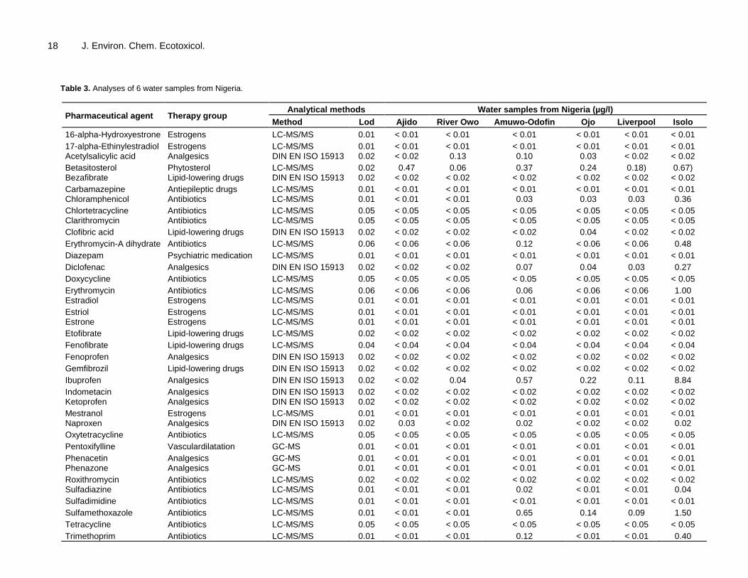
RESULTS Water Preliminary results from analysis of water samples for the presence or absence of a wide variety of pharmaceuticals and their degradates yielded information about the presence of twelve pharmaceutical entities out of Thirty seven checked for, which comprised of six antibiotics, four non-steroidal anti-inflammatory analgesics (NSAIDs), one antilipidemic and, phytoestrogen each (Table 3).
The detected agents were found at concentrations exceeding the detection limit of used analytical methods. The highest individual pharmaceutical concentrations measured were detected in the sample taken from the Isolo sampling location for ibuprofen (8.84 µg/l), sulfamethoxazole (1.5 µg/l) and erythromycin (1.0 µg/l). Total locational concentrations for determined pharmaceuticals were highest for the Isolo location at 13.55 µg/l followed by Amuwo Odofin at which total concentrations were 2.13 µg/l. All compounds detected were found in the water sample from Amuwo Odofin, followed by Isolo where all but acetylsalicylic acid were detected (Table 2).
Betasitosterol was detected in all locations including Isolo where detected levels were highest (0.67 µg/l). Ajido recorded the fewest pharmaceutically active compounds with just two entities detected, betasitosterol and naproxen. Detection frequency of detected residues were in the order betasitosterol (100%) > ibuprofen (45.5%) > chloramphenicol, diclofenac and sulfamethoxazole (33. 37%) > naproxen (27.27%) > erythromycin–A--dihydrate, erythromycin, sulfadiazine and trimetoprim (18%) >clofibrate (9%).
Table 4 displays maximum concentrations measured in the six water samples from Nigeria in comparison to measured concentrations in surface water or groundwater in other African countries, and in comparison
18 J. Environ. Chem. Ecotoxicol.
Table 3. Analyses of 6 water samples from Nigeria.
Pharmaceutical agent Therapy group Analytical methods Water samples from Nigeria (µg/l)
Method Lod Ajido River Owo Amuwo-Odofin Ojo Liverpool Isolo
16-alpha-Hydroxyestrone Estrogens LC-MS/MS 0.01 < 0.01 < 0.01 < 0.01 < 0.01 < 0.01 < 0.01
17-alpha-Ethinylestradiol Estrogens LC-MS/MS 0.01 < 0.01 < 0.01 < 0.01 < 0.01 < 0.01 < 0.01 Acetylsalicylic acid Analgesics DIN EN ISO 15913 0.02 < 0.02 0.13 0.10 0.03 < 0.02 < 0.02
Betasitosterol Phytosterol LC-MS/MS 0.02 0.47 0.06 0.37 0.24 0.18) 0.67) Bezafibrate Lipid-lowering drugs DIN EN ISO 15913 0.02 < 0.02 < 0.02 < 0.02 < 0.02 < 0.02 < 0.02
Carbamazepine Antiepileptic drugs LC-MS/MS 0.01 < 0.01 < 0.01 < 0.01 < 0.01 < 0.01 < 0.01 Chloramphenicol Antibiotics LC-MS/MS 0.01 < 0.01 < 0.01 0.03 0.03 0.03 0.36
Chlortetracycline Antibiotics LC-MS/MS 0.05 < 0.05 < 0.05 < 0.05 < 0.05 < 0.05 < 0.05 Clarithromycin Antibiotics LC-MS/MS 0.05 < 0.05 < 0.05 < 0.05 < 0.05 < 0.05 < 0.05
Clofibric acid Lipid-lowering drugs DIN EN ISO 15913 0.02 < 0.02 < 0.02 < 0.02 0.04 < 0.02 < 0.02
Erythromycin-A dihydrate Antibiotics LC-MS/MS 0.06 < 0.06 < 0.06 0.12 < 0.06 < 0.06 0.48
Diazepam Psychiatric medication LC-MS/MS 0.01 < 0.01 < 0.01 < 0.01 < 0.01 < 0.01 < 0.01
Diclofenac Analgesics DIN EN ISO 15913 0.02 < 0.02 < 0.02 0.07 0.04 0.03 0.27
Doxycycline Antibiotics LC-MS/MS 0.05 < 0.05 < 0.05 < 0.05 < 0.05 < 0.05 < 0.05
Erythromycin Antibiotics LC-MS/MS 0.06 < 0.06 < 0.06 0.06 < 0.06 < 0.06 1.00 Estradiol Estrogens LC-MS/MS 0.01 < 0.01 < 0.01 < 0.01 < 0.01 < 0.01 < 0.01
Estriol Estrogens LC-MS/MS 0.01 < 0.01 < 0.01 < 0.01 < 0.01 < 0.01 < 0.01 Estrone Estrogens LC-MS/MS 0.01 < 0.01 < 0.01 < 0.01 < 0.01 < 0.01 < 0.01
Etofibrate Lipid-lowering drugs LC-MS/MS 0.02 < 0.02 < 0.02 < 0.02 < 0.02 < 0.02 < 0.02
Fenofibrate Lipid-lowering drugs LC-MS/MS 0.04 < 0.04 < 0.04 < 0.04 < 0.04 < 0.04 < 0.04
Fenoprofen Analgesics DIN EN ISO 15913 0.02 < 0.02 < 0.02 < 0.02 < 0.02 < 0.02 < 0.02
Gemfibrozil Lipid-lowering drugs DIN EN ISO 15913 0.02 < 0.02 < 0.02 < 0.02 < 0.02 < 0.02 < 0.02
Ibuprofen Analgesics DIN EN ISO 15913 0.02 < 0.02 0.04 0.57 0.22 0.11 8.84
Indometacin Analgesics DIN EN ISO 15913 0.02 < 0.02 < 0.02 < 0.02 < 0.02 < 0.02 < 0.02 Ketoprofen Analgesics DIN EN ISO 15913 0.02 < 0.02 < 0.02 < 0.02 < 0.02 < 0.02 < 0.02
Mestranol Estrogens LC-MS/MS 0.01 < 0.01 < 0.01 < 0.01 < 0.01 < 0.01 < 0.01 Naproxen Analgesics DIN EN ISO 15913 0.02 0.03 < 0.02 0.02 < 0.02 < 0.02 0.02
Oxytetracycline Antibiotics LC-MS/MS 0.05 < 0.05 < 0.05 < 0.05 < 0.05 < 0.05 < 0.05
Pentoxifylline Vasculardilatation GC-MS 0.01 < 0.01 < 0.01 < 0.01 < 0.01 < 0.01 < 0.01
Phenacetin Analgesics GC-MS 0.01 < 0.01 < 0.01 < 0.01 < 0.01 < 0.01 < 0.01 Phenazone Analgesics GC-MS 0.01 < 0.01 < 0.01 < 0.01 < 0.01 < 0.01 < 0.01
Roxithromycin Antibiotics LC-MS/MS 0.02 < 0.02 < 0.02 < 0.02 < 0.02 < 0.02 < 0.02 Sulfadiazine Antibiotics LC-MS/MS 0.01 < 0.01 < 0.01 0.02 < 0.01 < 0.01 0.04
Sulfadimidine Antibiotics LC-MS/MS 0.01 < 0.01 < 0.01 < 0.01 < 0.01 < 0.01 < 0.01
Sulfamethoxazole Antibiotics LC-MS/MS 0.01 < 0.01 < 0.01 0.65 0.14 0.09 1.50
Tetracycline Antibiotics LC-MS/MS 0.05 < 0.05 < 0.05 < 0.05 < 0.05 < 0.05 < 0.05
Trimethoprim Antibiotics LC-MS/MS 0.01 < 0.01 < 0.01 0.12 < 0.01 < 0.01 0.40

Olarinmoye et al. 19
Table 4. The analyses of ten sewage sludge samples from Nigeria (*positive, but below LOQ; n/e = not evaluable due to high matrix interferences).
Pharmaceutical substance
Unit
Brewery WEMABOD Dairy Agabara Food Ikeja Abesan Iponri Oke AFA UCH
Lagos Lagos Ibadan Ota Ota Lagos Lagos Lagos Lagos Ibadan
I I I I I D D D D H
17-alpha-Ethinylestradiol µg/kg <10 <30 <40 <10 n/e <10 <10 <10 <15 <10
17-beta-Estradiol µg/kg <10 <10 <20 <10 n/e <10 <10 <10 <10 <10
Azithromycin µg/kg <10 <10 <10 <10 <10 <10 <10 <10 <10 <10
Bezafibrate µg/kg <10 <10 <10 <10 <10 11 <10 <10 <10 <10
Bisoprolol µg/kg <10 <10 <10 <10 <10 <10 <10 <10 <10 <80
Carbamazepine µg/kg <10 <10 * 49 * 23 24 39 <30 * <10 * <10 * 71
Clarithromycin µg/kg <10 <10 <10 <10 <10 <10 <10 <10 <10 43
Diclofenac µg/kg 30 38 395 1100 169 635 119 <10 * 71 111
Erythromycin µg/kg <10 <10 <10 <10 <10 39 147 <10 * 38 49
Fenofibric acid µg/kg n/e n/e n/e n/e n/e n/e n/e n/e n/e n/e
Ibuprofen µg/kg 360 <100 <100 210 <100 <100 <100 <100 <100 <60
Ketoprofen µg/kg <10 <10 <10 <10 <10 <10 <10 <10 <10 <10
Metoprolol µg/kg <10 <10 * <10 <10 <10 <10 <10 <10 <10 <30
(Naproxen) µg/kg n/e n/e n/e n/e n/e n/e n/e n/e n/e n/e
Propranolol µg/kg <10 <10 33 <10 <10 25 <10 <10 <10 <10
Roxithromycin µg/kg <10 <10 <10 <10 <10 <10 <10 <10 <10 <10
Sotalol µg/kg <10 <10 <10 <10 <10 <10 <10 <10 <10 <10
Sulfamethoxazole µg/kg <10 <10 <10 11 <10 <10 <10 <10 <10 <10
Trimethoprim µg/kg <10 <10 <10 31 <10 19 <10 <10 <10 38
Source: Sindiku et al. (2013). Key: I, industrial; D, domestic; H: Hospital: *1, positive, but below LOQ; *2, Positive using an optimised measurement method; *3, positive, corrected result. to ecotoxicological effect concentrations (PNEC predicted no effect concentrations). Sewage sludge Nine of nineteen pharmaceutical substances were detected in at least one of the studied ten sludge samples (Table 4). Diclofenac was detected in each of the ten sludge samples in a concentration range of < 10 to 1140 µg/kg dry weight, followed by Carbamazepine with positive detection in nine
samples (< 10 to 70 µg/kg) and Erythromycin in five samples (< 10 to 147 µg/kg). Bezafibrate, Clarithromycin, Ibuprofen, Metoprolol, Propranolol, Sulfamethoxazole, and Trimethoprim were positively detected in one or two sludge samples only (concentration range < 10 to 360 µg/kg). Two substances (Fenofibric acid and Naproxen) could not been evaluated due to high matrix interferences. Tables 5 and 6 contain details of maximum concentrations measured in the ten sewage sludge samples from Nigeria in comparison to maximum concentrations in sewage
sludge samples measured in Africa and worldwide respectively. DISCUSSION There are several routes of entry of human and veterinary pharmaceuticals into environmental matrices including water. Earlier work has firmly established the presence of pharmaceutically active compounds, their derivatives and other forms of the active moieties in all environmental
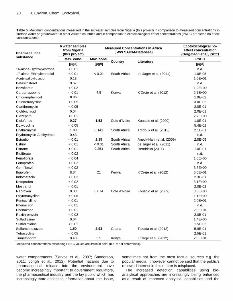
20 J. Environ. Chem. Ecotoxicol. Table 5. Maximum concentrations measured in the six water samples from Nigeria (this project) in comparison to measured concentrations in surface water or groundwater in other African countries and in comparison to ecotoxicological effect concentrations (PNEC predicted no effect concentrations).
Pharmaceutical substance
6 water samples from Nigeria (this project)
Measured Concentrations in Africa (IWW SAICM-Database)
Ecotoxicological no-effect concentration
(Bergmann et al., 2011)
Max. conc. Max. conc. Country Literature
PNEC
[µg/l] [µg/l] [µg/l]
16-alpha-Hydroxyestrone < 0.01 n.d.
17-alpha-Ethinylestradiol < 0.01 < 0.01 South Africa de Jager et al. (2011) 1.0E-05
Acetylsalicylic acid 0.13 1.0E+01
Betasitosterol 0.67 n.d.
Bezafibrate < 0.02 1.2E+00
Carbamazepine < 0.01 4,5 Kenya K'Oreje et al. (2012) 2.5E+00
Chloramphenicol 0.36 1.9E-02
Chlortetracycline < 0.05 3.0E-02
Clarithromycin < 0.05 2.0E-01
Clofibric acid 0.04 2.0E-01
Diazepam < 0.01 2.7E+00
Diclofenac 0.27 1.52 Cote d’Ivoire Kouadio et al. (2009) 1.0E-01
Doxycycline < 0.05 5.4E-02
Erythromycin 1.00 0.141 South Africa Tredoux et al. (2012) 2.1E-01
Erythromycin-A dihydrate 0.48 n.d.
Estradiol < 0.01 2.15 South Africa Aneck-Hahn et al. (2009) 2.0E-05
Estriol < 0.01 < 0.01 South Africa de Jager et al. (2011) n.d.
Estrone < 0.01 0.251 South Africa Hendricks (2011) 1.0E-01
Etofibrate < 0.02 n.d.
Fenofibrate < 0.04 1.6E+00
Fenoprofen < 0.02 n.d.
Gemfibrozil < 0.02 3.8E+00
Ibuprofen 8.84 21 Kenya K'Oreje et al. (2012) 6.0E+01
Indometacin < 0.02 2.3E-01
Ketoprofen < 0.02 3.1E+00
Mestranol < 0.01 2.0E-02
Naproxen 0.03 0.074 Cote d’Ivoire Kouadio et al. (2009) 3.3E+00
Oxytetracycline < 0.05 1.1E+00
Pentoxifylline < 0.01 2.0E+01
Phenacetin < 0.01 n.d.
Phenazone < 0.01 2.0E+01
Roxithromycin < 0.02 2.0E-01
Sulfadiazine 0.04 1.4E+00
Sulfadimidine < 0.01 1.5E-02
Sulfamethoxazole 1.50 2.93 Ghana Takada et al. (2012) 5.9E-01
Tetracycline < 0.05 2.5E-01
Trimethoprim 0.40 5.5 Kenya K'Oreje et al. (2012) 2.0E+01
Measured concentrations exceeding PNEC values are listed in bold. (n.d. = not determined).
water compartments (Servos et al., 2007; Sanderson, 2011; Jongh et al., 2012). Potential hazards due to pharmaceutical release into the environment have become increasingly important to government regulators, the pharmaceutical industry and the lay public which has increasingly more access to information about the issue,
sometimes not from the most factual sources e.g. the popular media. It however cannot be said that the public’s renewed interest in this matter is misplaced.
The increased detection capabilities using bio-analytical approaches are increasingly being enhanced as a result of improved analytical capabilities and the
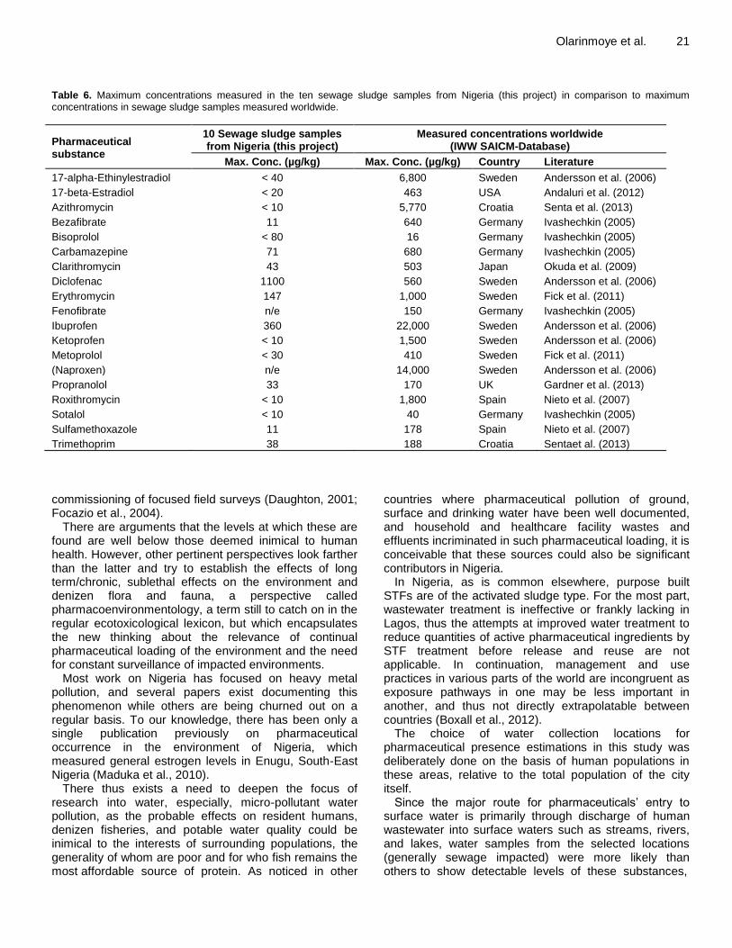
Olarinmoye et al. 21 Table 6. Maximum concentrations measured in the ten sewage sludge samples from Nigeria (this project) in comparison to maximum concentrations in sewage sludge samples measured worldwide.
Pharmaceutical substance
10 Sewage sludge samples from Nigeria (this project)
Measured concentrations worldwide (IWW SAICM-Database)
Max. Conc. (µg/kg) Max. Conc. (µg/kg) Country Literature
17-alpha-Ethinylestradiol < 40 6,800 Sweden Andersson et al. (2006)
17-beta-Estradiol < 20 463 USA Andaluri et al. (2012)
Azithromycin < 10 5,770 Croatia Senta et al. (2013)
Bezafibrate 11 640 Germany Ivashechkin (2005)
Bisoprolol < 80 16 Germany Ivashechkin (2005)
Carbamazepine 71 680 Germany Ivashechkin (2005)
Clarithromycin 43 503 Japan Okuda et al. (2009)
Diclofenac 1100 560 Sweden Andersson et al. (2006)
Erythromycin 147 1,000 Sweden Fick et al. (2011)
Fenofibrate n/e 150 Germany Ivashechkin (2005)
Ibuprofen 360 22,000 Sweden Andersson et al. (2006)
Ketoprofen < 10 1,500 Sweden Andersson et al. (2006)
Metoprolol < 30 410 Sweden Fick et al. (2011)
(Naproxen) n/e 14,000 Sweden Andersson et al. (2006)
Propranolol 33 170 UK Gardner et al. (2013)
Roxithromycin < 10 1,800 Spain Nieto et al. (2007)
Sotalol < 10 40 Germany Ivashechkin (2005)
Sulfamethoxazole 11 178 Spain Nieto et al. (2007)
Trimethoprim 38 188 Croatia Sentaet al. (2013)
commissioning of focused field surveys (Daughton, 2001; Focazio et al., 2004).
There are arguments that the levels at which these are found are well below those deemed inimical to human health. However, other pertinent perspectives look farther than the latter and try to establish the effects of long term/chronic, sublethal effects on the environment and denizen flora and fauna, a perspective called pharmacoenvironmentology, a term still to catch on in the regular ecotoxicological lexicon, but which encapsulates the new thinking about the relevance of continual pharmaceutical loading of the environment and the need for constant surveillance of impacted environments.
Most work on Nigeria has focused on heavy metal pollution, and several papers exist documenting this phenomenon while others are being churned out on a regular basis. To our knowledge, there has been only a single publication previously on pharmaceutical occurrence in the environment of Nigeria, which measured general estrogen levels in Enugu, South-East Nigeria (Maduka et al., 2010).
There thus exists a need to deepen the focus of research into water, especially, micro-pollutant water pollution, as the probable effects on resident humans, denizen fisheries, and potable water quality could be inimical to the interests of surrounding populations, the generality of whom are poor and for who fish remains the most affordable source of protein. As noticed in other
countries where pharmaceutical pollution of ground, surface and drinking water have been well documented, and household and healthcare facility wastes and effluents incriminated in such pharmaceutical loading, it is conceivable that these sources could also be significant contributors in Nigeria.
In Nigeria, as is common elsewhere, purpose built STFs are of the activated sludge type. For the most part, wastewater treatment is ineffective or frankly lacking in Lagos, thus the attempts at improved water treatment to reduce quantities of active pharmaceutical ingredients by STF treatment before release and reuse are not applicable. In continuation, management and use practices in various parts of the world are incongruent as exposure pathways in one may be less important in another, and thus not directly extrapolatable between countries (Boxall et al., 2012).
The choice of water collection locations for pharmaceutical presence estimations in this study was deliberately done on the basis of human populations in these areas, relative to the total population of the city itself. Since the major route for pharmaceuticals’ entry to
surface water is primarily through discharge of human wastewater into surface waters such as streams, rivers, and lakes, water samples from the selected locations (generally sewage impacted) were more likely than others to show detectable levels of these substances,

22 J. Environ. Chem. Ecotoxicol. and measured concentrations conceivably should follow a trend along impaction severity and population. The exception in this deliberate choosing of sample locations was Ajido, a location with a relatively smaller population as compared to other locations. It was thus chosen as the reference location.
In the present study twelve pharmaceutical entities were determined and discovered at environmentally significant concentrations in water samples. Higher concentrations have been previously recorded for ten of eleven pharmaceuticals in other African countries than in the present study.
However, erythromycin has been found in higher concentrations in the Nigerian water samples than in any other African surface water investigation. Notably, four substances (Acetylsalicylic acid, Beta-Sitosterol, Clofibric acid, and Sulfadiazine) detected in the present study have not previously been measured/not been reported in studies from other African countries. Four of the pharmaceuticals measured in Nigerian surface waters were found at concentrations exceeding ecotoxicological effect concentrations (Chloramphenicol, Diclofenac, Erythromycin, and Sulfamethoxazole). It therefore has to be expected that these pharmaceutical substances have adverse effects on ecosystem health in Nigerian surface waters at these locations though as yet undocumented. The frequency of detection of antibiotic and NSAID residues in samples from studied locations is an indication of their widespread use, as observed in other locations in countries worldwide. The especially high concentrations of erythromycin, sulfamethoxazole and Ibuprofen in the sample ISOLO could be attributable to the proximity of the water collection location to a midsized government (referral) hospital, and a large municipal landfill in which a significant quantity of solid wastes from the hospital ends up.
It is conceivable that the common denominator explaining the general presence of beta-sitosterol in study locations would be faecal contamination with coprosterols. No endogenous or synthetic animal estrogens were detected in this study. Considering the dense population clusters in most of the studied locations and the virtually uncontrolled entrees of wastewaters and domestic sewage effluents into the surface waters same, the opposite would have been expected.
This situation could be explained in part considering that untreated wastes which impact the studied waters are often heavily laden with sewage microorganisms. In sewage treatment procedures, heterotrophic bacteria are routinely utilised for the controlled biodegradation of pharmaceuticals in sewage, and though the removal of estrogens is not total following such treatment, the levels are significantly reduced. Benka-Coker and Ojior (1995) reported high levels of heterotrophic bacteria in a sewage impacted river in Nigeria, corroborating results from a similar study by Olayemi (1994) which also established higher heterotrophic bacterial counts during the rainy
season, probably due to increased sewage overflows into surrounding water bodies. Incidentally, our water collection was carried out during the rains. There are at the present time no functional STFs in all the studied locations, thus it is possible that the high levels of heterotrophs could be responsible for the increased mobilization and breakdown of endogenous human estrogens found in domestic sewage, and the dilution effect of higher water levels and flow during the rainy season could account for the null returns from analysis of sampled waters for these compounds.
At present, there are no verifiable figures for clofibrate prescription quantities in Nigeria. It is thus difficult to link concentrations detected and frequencies of detection to usage figures and statistics, information which could help explain the limited occurrence of clofibric acid in the current study. Considering the well documented persistence of clofibric acid in the environment, and specifically its resistance to photodegradation (most important here where concerted sewage treatment is poor), it would have been expected that clofibric acid would be detectable at more locations than found, due to continuing use and the aforementioned inherent environmental persistence. In the absence of more comprehensive usage data, the low levels of detection and concentrations of clofibric acid could attributable to low prescription rates in the studied locales. It is also conceivable that later studies which would include more locations, some highbrow (as obesity and consequent hypercholesteremia in Nigeria, though increasing across socioeconomic divides, is still more of a condition linked to affluence), would yield more representative results.
Pharmaceuticals have been demonstrated to exist in sewage sludge at concentrations much higher than in STF influents and effluents. Generally, study data on pharmaceutical occurrences in sewage sludge are few compared to surface water and STF data. None exist for Nigeria, and indeed Africa except those reported here. The NSAIDs, diclofenac and ibuprofen were found the highest concentrations in sludge with diclofenac occurring in all samples. Carbamezapine, an anticonvulsant and mood stabilising agent, was also generally discovered in all locations in significant quantities.
The well documented biorecalcitrance and environmental persistence coupled with poor removal rates by treatment may account for the near ubiquitous presence and high concentrations of diclofenac and carbamezapine recorded in the current study.
In addition the especially high diclofenac concentrations in the locations Agbara and Ikeja, could be due to the presence of many pharmaceutical production facilities in both locations which are both industrial/residential layouts. In Nigeria the use of STF sludge as fertiliser is not widespread, but studies into the presence of pharmaceuticals in soil are needed to give a more balanced-overall picture of pharmaceutical micropollution of the environment and associated

ecological sequelae.
In summary, this study has confirmed the presence of pharmaceutically active compounds in waste water impacted surface water bodies and sewage sludge in Lagos, Nigeria. The findings from this investigation has also established that the concentrations of detected PHACs, and the locational frequency of detection are comparable to reports from other countries, especially from developing countries from where unfortunately, such reports are few, limiting further comparisons.
Based on the results, it was concluded that several pharmaceuticals occur at relevant concentrations in the environment of Nigeria. It is recommended that more comprehensive water monitoring campaigns be conducted, especially in locations with high population density and low dilution of treated or untreated wastewater in receiving streams. Furthermore, investigations of groundwater, tap water/drinking water, manure, soil, and sediments as additional matrices of concern needs to be done in the nearest future to help generate wholistic pictures of the spatial environmental presences and concentrations of PhACs in them. In doing this, representative sampling strategies, improved sample conservation and pre-treatment should be used. ACKNOWLEGEMENTS This work was funded by the German Federal Environmental Agency (UBA), Project No. 31667. The authors acknowledge sampling of sludge samples by Professor Osibanjo, and Omotayo Sindiku (both of the University of Ibadan). Thanks to Dr. Roland Weber (POPs Environmental Consulting). Authors also especially thank Umweltbundesamt (Dr. Arne Hein, Dr. Anette Küster, and Ina Ebert) for financing, support, and valuable discussion.
Conflict of Interests
The authors have not declared any conflict of interest. REFERENCES Andaluri G, Suri RS, Kumar K (2012). Occurrence of estrogen
hormones in biosolids, animal manure and mushroom compost. Environ. Monit. Assess. 184(2):1197-1205.
Andersson J, Woldegiorgis A, Remberger M, Kaj L, Ekheden Y, Dusan B, Svenson A, Brorström-Lundén E, Dye C, Schlabach M (2006). Results from the Swedish National Screening Programme 2005. Stockholm, Sweden.
Aneck-Hahn NH, Bornman MS, de Jager C (2009). Oestrogenic activity in drinking waters from a rural area in the Waterberg District, Limpopo Province, South Africa. Water SA 35(3):245-251.
Benka-Coker MO, Ojior, OO (1995). Effect of slaughterhouse wastes on the water quality of Ikpoba River, Nigeria. Bioresour Technol. 52(1):5-12.
Bergmann A, Fohrmann R, Weber FA (2011). Zusammenstellung von Monitoringdaten zu Umweltkonzentrationen von Arzneimitteln (in German). UBA-Texte 66/2011,Umweltbundesamt,Dessau-Roßlau.
Olarinmoye et al. 23 http://www.umweltbundesamt.de/sites/default/files/medien/461/publikationen/4188.pdf
Bound JP, Voulvoulis N (2005). Household disposal of pharmaceuticals as a pathway for aquatic contamination in the United Kingdom. Environ. Health Perspect. 113(12):1705-1711.
Boxall A, Rudd M, Brooks B, Caldwell D, Choi K, Hickmann S, Innes E, Ostapyk K, Staveley J, Verslycke T, Ankley G, Beazley K, Belanger S, Berninger J, Carriquiriborde P, Coors A, DeLeo P, Dyer S, Ericson J, Gagné F, Giesy J, Gouin T, Hallstrom L, Karlsson M, Larsson D, Lazorchak J, Mastrocco F, McLaughlin A, McMaster M, Meyerhoff R, Moore R, Parrott J, Snape J, Murray-Smith R, Servos M, Sibley P, Straub J, Szabo N, Topp E, Tetreault G, Vance LT (2012). Pharmaceuticals and personal care products in the environment: What are the big questions? Environ. Health Perspect. pp. 120-129.
Carballa , mila , ema , lompart , arc a- ares C, odr gue I, Gómez M, Ternes T (2004). Behavior of pharmaceuticals, cosmetics and hormones in a sewage treatment plant. Water Res. 38(12):2918-2926.
Daughton C (2001). Pharmaceuticals in the environment: Overarching issues and overview. In: Daughton CG, Jones-Lepp TL (eds.). Pharmaceuticals and Personal Care Products in the Environment: Scientific and Regulatory Issues. Symposium Series 791; American Chemical Society. Washington DC USA.
de Jager C, Swemmer A, Aneck-Hahn NH, van Zijl C, van Wyk S, Bornman MS, Barnhoorn I, Jonker M, van Vuren JHJ, Burger AEC (2011). Endocrine Disrupting Chemical (EDC) Activity and Health Effects of Identified Veterinary Growth Stimulants in Surface and Groundwater; WRC Report No. 1686/1/11; Water Research Commission South Africa.
Federal Environment Agency (UBA) (2014). Pharmaceuticals in the environment, global occurrence, effects, and options for action. Report of an International Workshop held April 8th and 9th 2014, International Environment House II, Geneva, Switzerland. Available online from: http://www.pharmaceuticals-in-theenvironment.org/files/en/bereich_2/application/pdf/workshop_documentation_global-relevance-of-pharmaceuticals-in-the-environment.pdf
Fick J, Lindberg RH, Kaj L, Brorström-Lundén E (2011). Results from the Swedish National Screening Programme 2010. Stockholm, Sweden, P 56.
Focazio MJ, Kolpin DW, Furlong ET (2004). Occurrence of human pharmaceuticals in water resources of the United States: A review. In: Kummerer, K. (ed.) Pharmaceuticals in the Environment: Sources, Fate, Effects and Risks. 2nd ed. Berlin: Springer.
Gardner M, Jones V, Comber S, Scrimshaw MD, Coello‐Garcia T, Cartmell E, Lester J, Ellor B (2013). Performance of UK wastewater treatment works with respect to trace contaminants. Sci. Total Environ. pp. 359-369, 456-457.
Hendricks R (2011). Assessment of the biological quality of raw and treated effluents from three sewage treatment plants in the Western Cape, South Africa. Doctoral dissertation. Retrieved from University of the Western Cape electronic thesis and dissertations repository. Available online from: http://etd.uwc.ac.za/index.php?module=etd&action=viewtitle&id=gen8Srv25Nme4_6966_1331032010
Ivashechkin P (2005). Literaturaus ertung zum Vorkommenge fährlicher Stoffeim Abwasser und in Gewässern. Berichtzum Vorhabenim Auftrag des Ministeriums für Umwelt und Naturschutz, Landwirtschaft und Verbraucherschutz Nordrhein-Westfalen.
Jjemba P (2008). Pharmaecology: The occurrence and fate of pharmaceuticals and personal care products. Hoboken NJ: John Wiley and Sons.
Jongh CD, Kooij P, Voogt PD, Laak TT (2012). Screening and human health risk assessment of pharmaceuticals and their transformation products in Dutch surface waters and drinking water. Sci. Total Environ. pp. 70-77, 427-428.
K'Oreje KO, Demeestere K, De Wispelaere P, Vergeynst L, Dewulf J, Van Langenhove H (2012). From multi-residue screening to target analysis of pharmaceuticals in water: Development of a new approach based on magnetic sector mass spectrometry and application in the Nairobi River Basin, Kenya. Sci. Total Environ. 437:153-164.

24 J. Environ. Chem. Ecotoxicol. Kouadio LD, Traore SK, Bekro YA, Véronique M, Dembele A, Mamadou
K, Mazellier P, Legube B, Houenou P (2009). Contamination des Eaux de Surface par les Produits Pharmaceutiquesen Zones Urbaines de Côte D’ivoire: Cas du District D’abidjan. Eur. J. Scient. Res. 27(1):140-151.
Miege C, Choubert JM, Ribeiro L, Eusebe M, Coquery M (2009). Fate of pharmaceuticals and personal care products in wastewater treatment plants - Conception of a database and first results. Environ. Pollut. 157:1721-1726.
Nakada N, Shinohara H, Murata A, Kiri K, Managakia S, Sato N, Takada H (2007). Removal of selected pharmaceuticals and personal care products [PPCPS] and endocrine-disrupting chemicals [edcs] during sand filtration and ozonation at a municipal sewage treatment plant. Water Res. 41:4373-4382.
Nieto A, Borrull F, Marcé RM, Pocurull E (2007). Selective extraction of sulfonamides, macrolides and other pharmaceuticals from sewage sludge by pressurized liquid extraction. J. Chromatogr. A. 1174(1-2):125-131.
Okuda T, Yamashita N, Tanaka H, Matsukawa H, Tanabe K (2009). Development of extraction method of pharmaceuticals and their occurrences found in Japanese wastewater treatment plants. Environ. Int. 35(5):815-820.
Olayemi AB (1994). Bacteriological water assessment of an urban river in Nigeria. Int. J. Environ. Health Res. 4(3):b156-164.
Sanderson H (2011). Presence and risk assessment of pharmaceuticals in surface water and drinking water. Water Sci. Technol. 63:2143-2148.
Scheytt T, Mersmann P, Heberer T, Reddersen K (2001). Natural attenuation of pharmaceuticals. In Proceedings of the 2nd International Conference on Pharmaceuticals and Endocrine Disrupting Chemicals in Water, October pp. 9-11.
Senta I, Terzic S, Ahel M (2013). Occurrence and fate of dissolved and particulate antimicrobials in municipal wastewater treatment. Water Res. 47(2):705-714.
Servos M, Smith M, McInnis R, Burnison B, Lee B, Seto P, Backus S (2007). The presence of selected pharmaceuticals and the antimicrobial triclosan in drinking water in Ontario, Canada. Water Qual. Res. J. Can. 42:130-137.
Sindiku O, Orata F, Weber R, Osibanjo O (2013). Per- and polyfluoroalkyl substances in selected sewage sludge in Nigeria. Chemosphere 92:329-335.
Takada H, Shimizu A, Koike T, Takeshita A, Nakada N, Suzuki S (2012). Ubiquitous distribution of sulfamethoxazole in tropical Asian and African waters. SETAC 6th World Congress/SETAC Europe 22nd Annual Meeting, Berlin, Berlin.
Ternes TA, Herrmann N, Bonerz M, Knacker T, Siegrist H, Joss A (2004). A rapid method to measure the solid-water distribution coefficient (Kd) for pharmaceuticals and musk fragrances in sewage sludge. Water Res. 38(9):4075-4084.
Tredoux G, Genthe B, Steyn M, Germanis J (2012). Managed aquifer
recharge for potable reuse in Atlantis, South Africa. In: Water reclamation technologies for safe managed aquifer recharge. Eds: Christian Kazner TW, TMelin T, Dillon pp. 121-140.
United Nations Department of Economic and Social Affairs, Population Division (2014). World Urbanization Prospects: The 2014 Revision, Highlights.
USGS (2013). Organic W astewater Compounds in W ater and Sediment in and near Restored Wetlands, Great Marsh, Indiana Dunes National Lakeshore, 2009 - 11. pdf. Available at: http://pubs.usgs.gov/sir/2013/5186/pdf/sir2013-5186.pdf
WHO (2012). Pharmaceuticals in drinking-water. W orld Health Organization, Geneva 35pp.

Vol. 8(3), pp. 25-33, March, 2016
DOI:10.5897/JECE2015.0362
Article Number: 29BD0C957807
ISSN 2141-226X
Copyright ©2016
Author(s) retain the copyright of this article
http://www.academicjournals.org/JECE
Journal of Environmental Chemistry and Ecotoxicology
Full Length Research Paper
Sensitivity, ecotoxicity and histopathological effects on neotropical fish exposed to glyphosate alone and
associated to surfactant
Claudinei da Cruz1*, Silvia Patrícia Carraschi2, Natália Sayuri Shiogiri2, Adilson Ferreira da Silva2, Robinson Antonio Pitelli2 and Marcia Rita Fernandes Machado3
1University Center of Barretos Educational Fundation. Av. Prof. Roberto Frade Monte no 389, 14.783-226, Barretos
(SP), Brazil. 2Weed Science Environmental Research Studies of the College of Agricultural and Veterinary Science at Unesp, Via de
Acesso Prof. Dr. Paulo Donato Castellane, Zona Rural, s/ no, Jaboticabal, SP, Brazil.
3Department of Morphology and Animal Physiology of the College of Agricultural and Veterinary Sciences at Unesp from
Jaboticabal, SP, Brazil.
Received 13 August, 2015; Accepted 22 December, 2015
The aim of this research was to evaluate neotropical fish sensitivity (Piaractus mesopotamicus, Phallocerus caudimaculatus, Hyphessobrycon eques, and Brachydanio rerio) to a reference substance (potassium chloride); to estimate the lethal concentration (LC50; 96 h) for glyphosate, formulated as Rodeo
® alone and in association with 0.5 and 1.0% Aterbane
® BR surfactant and to evaluate the
histopathology of the gills, liver, and kidney from the fish after acute exposure. P. caudimaculatus and H. eques are good bioindicators like B. rerio because they have similar sensitivity. The LC50;96 h for glyphosate alone and in association with 0.5% Aterbane
® BR was similar (>975.0 mg L
-1) for all the fish.
Aterbane® BR alone was the most toxic substance to P. caudimaculatus (5.81 mg L
-1 LC50;96 h) and
glyphosate associated to 1.0% Aterbane® BR was more toxic to H. eques (411.91 mg L
-1 LC50;96 h). The
glyphosate alone and in association with Aterbane® BR was classified as practically non-toxic, whereas
Aterbane® BR alone was considered moderately toxic for the tested organisms. The histopathological
effects caused by glyphosate exposure on gills, liver, and kidneys are reversible, except for the liver necrosis on P. caudimaculatus. H. eques, P. caudimaculatus, and P. mesopotamicus present great potential to be used as standard organisms for herbicides monitoring and the use of glyphosate without surfactant addition is enough to cause histological alterations on H. eques and P. caudimaculatus, which makes them possible to be applied on environmental monitoring studies as biomarkers. Key words: Bioindicator, pesticide, histology, biomarker, gill, herbicide.
INTRODUCTION Glyphosate (GFT) is the most used herbicide for weed control on several crops, due to its wide action spectrum,
*Corresponding author. E-mail: [email protected].
Author(s) agree that this article remain permanently open access under the terms of the Creative Commons Attribution
License 4.0 International License

26 J. Environ. Chem. Ecotoxicol.
Table 1. Concentrations of the substances used on the toxicity assays.
Substances Fish
P. mesopotamicus P. caudimaculatus H. eques B. rerio
KCL (g L-1
) 1.0, 1.5, 2.0, 2.5 0.5, 1.0, 1.5, 2.0, 2.5 1.0, 1.5, 2.0, 2.5 1.0, 1.5, 2.0, 2.5, 3.0
Glyfosate (mg L-1
) 900.0, 925.0, 950.0, 975.0 900.0, 925.0, 950.0, 975.0 900.0, 925.0, 950.0, 975.0 900.0, 925.0, 950.0, 975.0
Surfactant (mg L-1
) 8.0, 9.0, 10.0, 11.0, 12.0 3.0, 4.0, 5.0, 6.0, 7.0, 8.0 4.0, 6.0, 8.0, 10.0, 12.0, 14.0 7.0, 8.0, 9.0, 10.0, 11.0
*GF+0.5% (mg L-1
) 900.0, 925.0, 950.0, 975.0 900.0, 925.0, 950.0, 975.0 900.0, 925.0, 950.0, 975.0 900.0, 925.0, 950.0, 975.0
*GF+1.0% (mg L-1
) 475.0, 500.0, 525.0, 550.0, 575.0 900.0, 925.0, 950.0, 975.0 400.0, 425.0, 450.0, 475.0 900.0, 925.0, 950.0, 975.0
*GFT+0.5% glyphosate + 0.5% surfactant; GFT+1.0% glyphosate + 1.0% surfactant
lower selectivity, post-emergence application, systemic action and does not represent environmental risks to the water bodies (Shiogiri et al., 2010), and has a large potential to control aquatic weeds. The macrophyte present countless benefits to the environment, however they grow in an accelerated rate under eutrophic conditions, causing negative impacts to the use of the water body, like fishing, recreation, and power generation.
The herbicide application on the aquatic environment needs a higher scientific base, due to the lack of knowledge about the molecules for non-target organisms, morph-physiological effects, and control effectiveness (Botelho et al., 2009) and residues on water. Another issue is the toxicity of the other formulation components, since the inert components, such as the surfactants, may be more toxic than the active ingredient (IA) for non-target organisms (Carraschi et al., 2011). For Amarante Jr et al. (2002), Tsui and Chui (2003), and Navarro and Martinez (2014), the surfactants from the glyphosate formulations are more toxic for fish than the molecule itself.
At this context, the acute and chronic toxicity assays may be performed to characterize the effects and to evaluate dangerousness and environmental risks of the formulations (Schmitt-
Jansen et al., 2008). Thus, the organism selection is based on a series of criteria, such as sensibility, short reproduction cycle, operation easiness, and optimization costs (Cruz et al., 2008).
The neotropical species may be used as bioindicators for xenobiotics presence at the aquatic environment. In Brazil, the study about the application of this method for environmental monitoring is scarce (Glusczak et al., 2006; Shiogiri et al., 2012).
Thus, the aim of this study was to evaluate the sensitivity to a reference substance (potassium chloride) and the acute toxicity (LC50;96 h) of the glyphosate and the Aterbane
® BR surfactant
(alkylphenolpolyglycol ether) alone and in association with glyphosate (GFT + 0.5 and 1.0% surfactant) for the neotropical fish Piaractus mesopotamicus, Hyphessobrycon eques and Phallocerus caudimaculatus and the comparison with the standard species Brachydanio rerio and to evaluate possible histological effects caused by the glyphosate on gills, liver and kidney after acute exposure.
MATERIALS AND METHODS The potassium chloride with 99.9% purity was used as reference substance, the Rodeo
® herbicide was the
glyphosate source (480.0 g L-1
) and Aterbane® BR was
used as the surfactant alkylphenolpolyglycol ether source (466.0 g L
-1).
The 50% lethal concentration values were estimated by the Trimmed Spearman-Karber (Hamilton et al., 1977).
Sensitivity and acute toxicity tests
To perform the tests, the fish (P. mesopotamicus, H. eques, B. rerio and P. caudimaculatus) were acclimatized during seven days, under bioassay room conditions (25.0 ± 2°C temperature and 12 h photoperiod).
The fish were exposed to potassium chloride, glyphosate, surfactant and GFT + surfactant associations (Table 1), with three repetitions per treatment, three fish per repetition, at maximum density of 1.0 g L
-1, with 96 h
duration (ABNT, 2011). The substances concentrations on the definitive assays
were previously adjusted by preliminary tests, to find the concentrations interval which caused zero and 100% mortality.
Histological analysis after glyphosate exposure
After 96 h exposure, three representative animals from each treatment and each species (n=3) were euthanized through anesthesia (1.0 g L
-1 benzocaine). Liver, kidney,
and gills were collected. The samples were immersed in buffered formaldehyde solution (0.1 m; pH 7.3) for 24 h. After the fixation, the pieces were dehydrated in alcohol, diaphanized on xylene and included in Histosec
® (Merck).
Cruz et al. 27
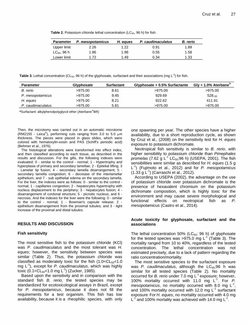
Table 2. Potassium chloride lethal concentration (LC50, 96 h) for fish.
Parameter P. mesopotamicus H. eques P. caudimaculatus B. rerio
Upper limit 2.26 1.22 0.91 1.89
LC50, 96 h 1.86 1.98 0.55 1.58
Lower limit 1.72 1.49 0.34 1.33
Table 3. Lethal concentration (CL50, 96 h) of the glyphosate, surfactant and their associations (mg L
-1) for fish.
Parameter Glyphosate Surfactant Glyphosate + 0.5% Surfactante Gly + 1.0% Aterbane®
B. rerio >975.00 8.61 >975.00 >975.00
P. mesopotamicus >975.00 9.45 929.69 528.50
H. eques >975.00 8.21 922.62 411.91
P. caudimaculatus >975.00 5.81 >975.00 >975.00
*Surfactant: alkylphenolpolyglycol ether (Aterbane®BR)
Then, the microtomy was carried out in an automatic microtome
(RM2155 - Leica®), performing cuts ranging from 3.0 to 5.0 m
thickness. The pieces were placed in glass slides, which were colored with hematoxylin-eosin and PAS (Schiff's periodic acid) (Behmer et al., 1976).
The histological alterations were transformed into effect index, and then classified according to each tissue, as described in the results and discussion. For the gills, the following indexes were evaluated: 0 - similar to the control - normal; 1 - Hypertrophy and hyperplasia of primary and secondary lamellae; 2 - Epitelial lifting; 3 - Lamelar tip fusion; 4 - secondary lamella disarrangement; 5 - secondary lamella congestion; 6 - decrease of the interlamellar epithelium; and 7 - sub epithelial edema on the secondary lamella. For the liver, the indexes were as follows: 0 - similar to the control - normal; 1 - capillaries congestion; 2 - hepatocytes hypertrophy with nucleus displacement to the periphery; 3 - hepatocytes fusion; 4 - disarrangement of cordonal structure; 5 - picnotic nucleus; and 6 - necrosis. And the indexes for the liver were the following: 0 - similar to the control - normal; 1 - Bowman's capsule release; 2 - epithelium disarrangement from the proximal tubules; and 3 - light increase of the proximal and distal tubules. RESULTS AND DISCUSSION Fish sensitivity The most sensitive fish to the potassium chloride (KCl) was P. caudimaculatus and the most tolerant was H. eques; however, the sensitivity between species was similar (Table 2). Thus, the potassium chloride was classified as moderately toxic for the fish (1.0<CL50<1.0 mg L
-1), except for P. caudimaculatus, which was highly
toxic (0.1<CL50<1.0 mg L-1
) (Zucker, 1985). Based upon the sensitivity and in comparison with the
standard fish B. rerio, the tested species may be standardized for ecotoxicological assays in Brazil, except for P. mesopotamicus, because it does not fill the requirements for a test organism. This fish has low availability, because it is a rheophilic species, with only
one spawning per year. The other species have a higher availability, due to a short reproduction cycle, as shown by Cruz et al., (2008) on the sensitivity test for H. eques exposure to potassium dichromate.
Neotropical fish sensitivity is similar to B. rerio, with higher sensibility to potassium chloride than Pimephales promelas (7.62 g L
-1 LC50;96 h) (USEPA, 2001). The fish
sensibilities were similar as described for H. eques (1.5 g L
-1) (Fujimoto et al., 2012) and for P. mesopotamicus
(1.33 g L-1
) (Carraschi et al., 2012). According to USEPA (2002), the advantage on the use
of potassium chloride over potassium dichromate is the presence of hexavalent chromium on the potassium dichromate composition, which is highly toxic for the environment and may cause severe morphological and functional effects on neotropical fish as P. mesopotamicus (Castro et al., 2014). Acute toxicity for glyphosate, surfactant and the associations The lethal concentration 50% (LC50; 96 h) of glyphosate for the tested species was >975.0 mg L
-1 (Table 3). The
mortality ranged from 10 to 40%, regardless of the tested concentration. The lethal concentration was not estimated precisely, due to a lack of pattern regarding the ratio concentration/mortality.
The most sensitive species to the surfactant exposure was P. caudimaculatus, although the LC50;96 h was similar for all tested species (Table 2). No mortality occurred for B. rerio under 7.0 mg L
-1 exposure; however,
100% mortality occurred with 11.0 mg L-1
. For P. mesopotamicus, no mortality occurred with 8.0 mg L
-1,
and 100% mortality occurred with 12.0 mg L-1
surfactant exposure. For H. eques, no mortality occurred with 4.0 mg L
-1 and 100% mortality was achieved with 14.0 mg L
-1.

28 J. Environ. Chem. Ecotoxicol.
For P. caudimaculatus no mortality occurred at 3.0 mg L
-1 and 100% mortality was obtained with 8.0 mg L
-1
surfactant exposure. The LC50;96 h for the glyphosate + 0.5% surfactant
association was > 975.0 mg L-1
for B. rerio and P. caudimaculatus. For P. mesopotamicus and H. eques the LC50;96 h was > 900.0 mg L
-1 (Table 3). For P.
mesopotamicus, 22% mortality occurred with 875.0 mg L-
1 and 100% mortality was achieved with 975 mg L
-1. For
H. eques, no mortality occurred at 875.0 mg L-1
exposure and 100% mortality occurred at 975 mg L
-1. B. rerio and
P. caudimaculatus displayed no mortality during association exposure.
The LC50;96 h observed for the glyphosate + 1.0% surfactant association were as follows: P. caudimaculatus and B. rerio > 975.0 mg L
-1; H. eques > 400.0 mg L
-1; P.
mesopotamicus > 500 mg L-1
(Table 3). With 400.0 mg L-1
exposure, 22.2% mortality occurred for H. eques; 100% mortality occurred with 475 mg L
-1 exposure. No P.
mesopotamicus mortality occurred with 475.0 mg L-1
exposure, whereas 100% mortality occurred with 575.0 mg L
-1. No mortality occurred for B. rerio and P.
caudimaculatus during the association exposure. The glyphosate (Rodeo
®) is practically non toxic (LC50
> 100.0 mg L-1
) for all studied species, different from found for Prochilodus lineatus exposed to Roundup
®
(LC50;96 h = 13.69 mg L-1
) (Langiano and Martinez, 2008). Other authors also found different results for Roundup Ready
® and P. mesopotamicus (LC50;96 h =
3.74 mg L-1
) (Shiogiri et al., 2012); other glyphosate formulations and Rhinella arenarum (LC50;96 h = 6.8 to 72.8 mg L
-1) (Brodeur et al., 2014); and Roundup
Original® and Pseudoplatystoma species (LC50;96 h =
15.0 mg L-1
) (Sinhorin et al., 2014). The glyphosate toxicity observed in this study was
similar to the values found for Salmo gairdneri and Oncorhynchus tshawytscha (LC50;96 h = 1070.0 to 1440.0 mg L
-1) (Mitchell et al., 1987); Hybognathus
amarus and P. promelas (LC50;96 h > 1000.0 mg L-1
) (Beyers, 1995) and P. promelas and Polonichthys macrolepidoyus (LC50;96 h = 1154.0 to 1132.0 mg L
-1)
(Riley and Finlayson, 2004). The commercial formulations type (inert compounds and surfactant) has an important role for the glyphosate toxicity characterization. The lower toxicity of the formulation studied in this paper (Rodeo
®) may be due to the
absence of surfactants. Thus, the knowledge about the formulations toxicity is fundamental at the moment of decision making, concerning the monitoring of possible glyphosate environmental effects.
The fish evaluated for this study were more tolerant to the alkylphenolpolyglycol ether than S. gairdneri, P. promelas, Ictalurus punctatus and Lepomis macrochirus exposed to the MON0818 surfactant (amine ethoxylate) (LC50 = 1.4 to 3.0 mg L
-1) (Wan et al., 1989). Hypomesus
transpacificus, P. promelas and P. macrolepidoy were also less tolerant when exposed to the surfactants
alkylphenolpolyethoxylate and alcohol polyalkoxylate acid (LC50 = 0.7 to 3.9 mg L
-1) (Riley and Finlayson, 2004).
And the surfactant Ammoeng 130® was also classified as
more toxic for P. promelas (LC50;96 h = 0.51 to 1.80 mg L
-1) (Versteeg et al., 2006). However, the toxicity
observed for Onchorynchus mykiss exposed to alkylphenolpolyethoxylate was similar to the toxicity observed in this study, with 5.18 to 6.57 mg L
-1 LC50
(Curran et al., 2004). The surfactant presence in any concentration has
increased the formulation toxicity in about 50% for H. eques and P. mesopotamicus, indicating that the surfactant addition interfere on the glyphosate toxicity, regarding some non-target organisms. According to Giesy et al. (2000), the polyethoxylate amine surfactant (POEA) may be responsible for making the Roundup
®
formulation more toxic for aquatic organisms. Riley and Finlayson (2004) observed that the LC50;96 h for H. transpacificus was 5.5 and 3.9 mg L
-1 for P. promelas,
after the exposure to Rodeo® formulation with the
addition of R-11® surfactant, while the LC50 of the
commercial formulation without the surfactant was 270.0 and 1132.0 mg L
-1 for the fish, respectively, showing the
connection between the surfactant presence and glyphosate toxicity.
Based on the classification by Zucker (1985) and Giesy et al. (2000), the glyphosate and the associations with 0.5 and 1.0% surfactant (recommended doses for herbicide application) can be considered practically non-toxic and the alkyl phenolpolyglycol ether is classified as moderately nontoxic (1>LC50<10 mg L
-1).
Glyphosate histopathological effects for fish Gills The pacu (P. mesopotamicus) gills consist of four branchial arches, supported by two rows of primary lamellae, which are covered with stratified epithelium. The secondary lamellae consist of epithelial, lining, pillar, chloride, and mucous cells (Figure 1A), as described by Severi et al., (2000). The observed histological changes are described below according to each concentration (Figure 1), on Table 4.
The epithelium increase and the blood congestion at the secondary lamellae also occurred on O. mykiss exposed to methiocarb insecticide (Altonok et al., 2006) and on Oreochromis niloticus exposed to diquat herbicide (Henares et al., 2008). The epithelium increase works as a barrier to decrease the xenobiotics absorption (Mallatt, 1985), similar as described for Cyprinus carpio exposed to GFT (Neskovic et al., 1996).
The presence of sub epithelial edema and the disarrangement of the secondary lamellae was reported for C. carpio (Neskovic et al., 1996) and for O. niloticus exposed to GFT (Jiraungkoorskul et al., 2002). The blood

Cruz et al. 29
Table 4. Histological alterations on fish gills after glyphosate exposure.
Concentration (mg L-1
) P. mesopotamicus P. caudimaculatus H. eques
0.0 0 0 0
900.0 0 0 0
925.0 1 1 and 7 3, 4 and 7
950.0 1 1 and 7 5 and 6
975.0 2 - 5 and 6
Figure 1. Gills photomicrography. A. Primary and secondary lamellae (PL and SL) from P. mesopotamicus with no exposure (control). B. Hypertrophy and hiperplasia of the primary lamellae (arrow) and congestion in the secondary lamellae (c) (925.0 mg L
-1) on H. eques. C. Decrease of the interlamellar epithelium height (arrow)
(975.0 mg L-1
) on H. eques. D. Sub epithelial edema (star) and hypertrophy and hyperplasia of primary and secondary lamellae (arrow) (925 mg L
-1) on P. caudimaculatus. Bar =12.5 µm. Color HE.
congestion caused by the blood flow increase leads to the sub epithelial edema and the lamellae disarrangement. According to Shiogiri et al. (2012), P. mesopotamicus exposed under acute conditions to Roundup Ready
® also presented pillar cells hyperplasia
and lamellar epithelium hypertrophy. Although, such alterations are reversible (Henares et al., 2008; Shiogiri et al., 2012) and after some days of exposure, the tissue can remake its common histomorphology, different from
paraquat herbicide, which caused cells disarrangement, uncommon regeneration of the epithelial cells and deformation of the branchial cartilage cells from Colossoma macropomum, exposed to 10.0 mg L
-1
(Salazar-Lugo et al., 2011). H. eques was the most sensitive bioindicator to GFT,
with several histological changes at the gills. Despite the changes not being specific to the GFT toxicity, this kind of biomarkers indicates the presence of xenobiotics on the

30 J. Environ. Chem. Ecotoxicol.
Table 5. Histological alterations on the fish liver after glyphosate exposure.
Concentration (mg.L-1
) P. mesopotamicus P. caudimaculatus H. eques
0.0 0 0 0
900 0 and 1 2 and 4 2, 3, and 5
925 2 4 and 6 2 and 2
950 2, 3 and 4 4 and 6 2 and 3
975 2, 3 and 4 4 and 6 2 and 3
environment. This fact is important to environmental monitoring and identification of contaminants on the aquatic environment.
P. caudimaculatus displayed branchial structure similar as observed for P. mesopotamicus and H. eques on the control and under 900.0 mg L
-1 exposure. The sub
epithelial edema on the secondary lamellae and the increase of the interlamellar epithelium was similar to a report with O. niloticus exposed to 36.0 mg L
-1 glyphosate
as the Roundup® formulation (Jiraungkoorskul et al.,
2002). Liver The evaluated fish liver (Control) showed cordonal organization of the hepatocytes in direct contact with the capillary sinusoids. The hepatocytes showed a hexagonal shape, with central nucleus, basophil, decondensed chromatin, a visible nucleolus and rosy cytoplasm, indicating high acidophilia (Figure 2A).
The main histological changes occurred after acute exposition were as follows: capillary sinusoids congestion (hyperemia), hypertrophy and fusion of the hepatocytes and cordonal structure disarrangement of the hepatocytes, with 900.0 and 925.0 mg L
-1 exposure
(Table 5), followed by tissue necrosis with 925.0 to 975.0 mg L
-1 exposure for P. caudimaculatus (Table 5) (Figure
2C, D, E, F). The cellular nucleus displacement to the periphery and
the hepatocytes hypertrophy was also observed for O. niloticus exposed to GFT (Jiraungkoorskul et al., 2002). The displacement indicates the increase of the organelles and of the enzymes which are responsible for the xenobiotics metabolism, whereas the blood congestion may be a signal of the pressure decrease of the hepatic circulation, similar to O. mykiss exposed to 5.0 mg L
-1
GFT (Topal et al., 2015). The cordonal disarrangement also occurred with Spaurus aurata exposed to imazapyr, terbutrin and triasulfuron (Arufe et al., 2004) and with Trichogaster trichopterus exposed to paraquat (Banaee et al., 2013). According to Shiogiri et al. (2012), the P. mesopotamicus liver showed hepatocytes vacuolization and cells hypertrophy when exposed to 3.0 or 4.0 mg L
-1
glyphosate as Roundup Ready®. The vacuolization may
work as storage of compounds which are difficult to be
metabolized. The liver, used as GFT exposure biomarker, was the
tissue which exhibited the highest histological index, reaching tissue necrosis during P. caudimaculatus exposure to Rodeo
®. This alteration was also described
by Ayoola (2008) on C. gariepinus, same for the fibrosis which occurred on O. mykiss exposed in a chronic manner to 5.0 or 10.0 mg L
-1 GFT (Topal et al., 2015).
The necrosis probably occurred due to an excessive work performed by the hepatocytes, in attempt to eliminate the herbicide during the detoxification process. Kidneys The fish kidneys are formed by a renal corpuscle containing the glomerulus constituted by capillaries and the Bowman's capsule. Around the renal corpuscle are also found the hematopoietic tissue with basophilic cells, proximal and distal tubules, as observed on the control for all studied species on this research (Figure 3A).
The alterations were the shrinkage of the Bowman's capsule and disarrangement of the proximal tubules epithelium in all assessed concentration for P. caudimaculatus and H. eques, whereas did not occurred alterations of the tissue structure on P. mesopotamicus in any tested concentration (Table 6 and Figure 3B).
The glomerulus capsule release, the disarrangement of the proximal tubules epithelium and the proximal and distal tubule light increase was also observed on O. mykiss exposed to linuron herbicide (Oulmi et al., 1995) and on O. niloticus exposed to Roundup
®
(Jiraungkoorskul et al., 2002). The Bowman's capsule space increase, or the capsule release, and the disarrangement of the tubules cells was also observed on C. gariepinus exposed to GFT, which leads to the kidneys physiological functions loss (Ayoola, 2008). Conclusions The fish species H. eques, P. caudimaculatus and P. mesopotamicus present great potential to be used as standard organisms for ecotoxicological assays for herbicides monitoring, for they present an excellent answer to the tested reference substance. The addition of

Cruz et al. 31
Figure 2. A. Cordonal organization of the hepatocytes (line) from H. eques (control). B. Picnotic nucleus (Arrow) (900.0 mg L
-1) on H. eques. C. Hepatocyte hypertrophy (star) (925.0 mg L-1) on H. eques. D.
Necrosis spots (asterisk) (975.0 mg L-1
) on P. caudimaculatus. E. Cell fusion (arrow) (950.0 mg L-1
) on P. mesopotamicus. F. Disarrangement of the hepatocytes cordonal structure (900 mg L
-1) on P.
caudimaculatus. Bar = 12.5 µm. Color HE.
Table 6. Histological alterations on fish kidneys after glyphosate exposure.
Concentration (mg.L-1
) P. mesopotamicus P. caudimaculatus H. eques
0.0 0 0 0
900 0 1, 2 and 3 1, 2 and 3
925 0 1, 2 and 3 1, 2 and 3
950 0 1, 2 and 3 1, 2 and 3
975 0 1, 2 and 3 1, 3 and 3

32 J. Environ. Chem. Ecotoxicol.
Figure 3. Kidneys photomicrography. A. Proximal segment (ps), distal segment (ds) (control), hematopoietic tissue (HT) on P. mesopotamicus. B. Shrinkage of the Bowman's capsule (cs) and disarrangement of the epithelium proximal segment (dps) on 975 mg L
-1
on H. eques, Bar: 12.5 µm. Color HE.
surfactants to glyphosate formulations may change the herbicide acute toxicity pattern.
The use of glyphosate without surfactant addition is enough to cause histological alterations on H. eques and P. caudimaculatus, featuring specially the kidneys and liver as the xenobiotics presence biomarkers, which makes them possible to be applied on environmental monitoring studies.
Conflict of interests
The authors have not declared any conflict of interests.
REFERENCES
ABNT (2011). Brazilian Association of Techicnal Standards.NBR 12713. Aquatic Ecotoxicology. Fish Sao Paulo, Brazil. 19pp.
Arufe MI, Arellano J, Moreno MJ, Sarasquete C (2004). Toxicity of a commercial herbicide containing terbutryn and triasulfuron to seabream (Sparusaurata L.) larvae: A comparison with the microtextest. Ecotoxicol. Environ. Saf. 59(2):209-216.
Ayoola SO (2008). Histopathological effects of Glyphosate on Juvenile African Catfish (Clariasgariepinus). Am. Euras. J. Agric. Environ. Sci. 4(3):362-367.
Altonok I, Capkin E, Karahan S, Boran M (2006). Effects of water quality in fish size on toxicity of methiocarb: A carbamate pesticide, to rainbow trout. Environ. Toxicol. Pharmacol. 22(1):20-26.
Amarante Jr OP, Santos TCR, Brito NM, Ribeiro ML (2002). Glyphosate: Properties, toxicities, uses and legislation. New Chem. 25(4):589-593.

Banaee M, Davoodi MH, Zoheiri F (2013). Histopathological changes
induced by paraquat on some tissues of Gourami fish (Trichogaster trichopterus). Open Vet. J. 3(1):36-42.
Behmer AO,Tolosa EMC, Feritas-Neto AG (1976). Manual of techniques for normal and pathological histology.1ª Edition, Edart, Editora da USP, Sao Paulo. P 239.
Beyers DW (1995). Acute toxicity of Rodeo® herbicide to Rio Grande
silvery minnow as estimated by surrogate species: Plains minnow and fathead minnow. Arch. Environ. Contam. Toxicol. 29:24-26.
Botelho RG, Santos JB, Oliveira TA, Braga RR, Byrro ECM, (2009). Acute toxicity of herbicides to Tilapia (Oreochromis niloticus). Weed Plant 27(3):621-626.
Brodeur JC, Poliserpi MB, D’Andrea MF, Sánchez M (2014). Synergy between glyphosate- and cypermethrin-based pesticides during acute exposures in tadpoles of the common South American Toad Rhinella arenarum. Chemosphere 112:70-76.
Castro MP, Moraes FR, Fujimoto RY, Cruz C, Andrade Belo MA, Moraes JRE (2014). Acute Toxicity by Water Containing Hexavalent or Trivalent Chromium in Native Brazilian Fish, Piaractus mesopotamicus: Anatomopathological Alterations and Mortality. Bull Environ. Contam.Toxicol. 92:213-219.
Carraschi SP, Cubo P, Schiavetti BL, Shiogiri NS, Cruz C, Pitelli RA (2011). Efeitos tóxicos de surfactantes fitossanitários para o peixe mato grosso (Hyphessobryconeques). Acta Sci. Biol. Sci. 33(2):191-196.
Carraschi S, Luna L, Neto A, Gírio A, Cruz C, Pitelli R, (2012). Ecotoxicity of agricultural surfactants for guaru Phalloceros caudimaculatus. Ecotoxicol. Environ. Contam. 7(1):27-32.
Cruz C, Cubo P, Gomes GR, Venturini FP, Guilherme PE,Pitelli RA (2008). Sensibilidade de peixes neotropicais ao dicromato de potássio. J. Bra. Soc. Ecotoxicol. 3(1):53-55.
Curran CA, Grassley JM, Grue CE (2004). Toxicity of R-11® surfactant
to juvenile rainbow trout: Does size matter? Bull. Environ. Contam. Toxicol. 72:401-404.
Giesy JP, Dobson S, Solomon KR (2000). Ecotoxicological risk assessment for Roundup
®herbicide. Rev. Environ. Contam. Toxicol.
167:35-120. Fujimoto RY, Gabbay MI, Anjos ECS, Carraschi SP, Cruz C (2012).
Toxicidade e risco ambiental da oxitetraciclina e efeito em leucócitos de mato grosso (Hyphessobryconeques). Ecotoxicol. Environ. Contam. 7(2):11-15.
Glusczak L, Miron DS, Crestani M, Fonseca MB, Pedron FA, Duarte MF, VieiraVLP (2006). Effect of glyphosate herbicide on acetylcholinesterase activity and embolic and hematological parameters in piava (Leporinusobtusidens). Ecotoxicol. Environ. Saf. 65(2):237-241.
Hamilton MA, Russo RC, Thurston V (1977). Trimmed Spearman-Karber method for estimating medial lethal concentrations in toxicoty bioassays. Environ. Sci. Technol. 7:714-719.
Henares MNP, Cruz C, Gomes GR, Pitelli RA, Machado MRF (2008). Acute toxicity and histopathological effects of herbicide diquat in gill and liver of tilapianile (Oreochromisniloticus). Acta Sci. Biol. Sci. 30:77-81.
Jiraungkoorskul W, Upatham ES, Kruatrachue M, Sahaphong S, Vichasri-Grams S, Pokethitiyook P (2002). Histopathological effects of roundup, a glyphosate herbicide, on nile tilapia (Oreochromisniloticus). Sci. Asia 28:121-127.
Langiano VC, Martinez CBR (2008).Toxicity and effects of a glyphosate-based herbicide on the Neotropical fish Prochiloduslineatus. Comp. Biochem. Physiol. C. 147:222-231.
Mallatt J (1985). Fish gill structural changes induced by toxicants and other irritants: A statistical review. Can. J. Fish Aquat. Sci. 42(4):630-648.
Mitchell DG, Chapman PM, Long TJ (1987). Acute toxicity of Roundup®
and Rodeo® herbicides to rainbow trout, Chinook and coho salmon.
Bull. Environ. Contam. Toxicol. 39:1028-1035. Navarro CD, Martinez CB (2014). Effects of the surfactant
polyoxyethylene amine (POEA) on genotoxic, biochemical and physiological parameters of the freshwater teleost Prochilodus lineatus. Comp. Biochem. Physiol. C Toxicol. Pharmacol. 165:83-90.
Neskovic NK, Poleksic V, Elezovic I, Karan V, Budimir M (1996). Biochemical and histopathological effects of glyphosate on carp,
Cruz et al. 33
Cyprinus carpio L. Bull. Environ. Contam. Toxicol. 56:295-302. Oulmi Y, Negele RD, Braunbeck T (1995). Cytopathology of liver and
kidney in rainbow trout (Oncorhynchus mykiss) after long-term exposure to sublethal concentrations of linuron. Dis. Aquat. Org. 21:35-52.
Riley F, Finlayson S (2004). Acute toxicities of herbicides used to control water hyacinth and Brazilian Elodea on larval delta smelt and sacramentosplittail. Office of Spill Prevention and Response 9pp.
Salazar-Lugo R, Mata C, Oliveros A, Rojas LM, Lemus M, Villarroel ER (2011). Histopathological changes in gill, liver and kidney of neotropical fish Colossoma macropomum exposed to paraquat at different temperatures. Environ. Toxicol. Pharm. 31:490-495.
Severi W, Rantin FT, Fernandes MN (2000). Structural and morphological features of Piaractus mesopotamicus (Holmberg, 1887) gills. Rev. Braz. Biol. 60(3):493-501.
Schmitt-Jansen M, Veit U, Dudel G, Altenburger R. (2008). An Ecological perspective in aquatic ecotoxicology: Approaches and challenges. Basic Appl. Ecol. 9:337-345.
Shiogiri NS, Carraschi SP, Cubo P, Schiavetti BL, Cruz C, Pitelli RA (2010). Ecotoxicity of glyphosate aterbane
®br surfactant on guaru
(Phallocerus caudimaculatus). Acta Sci. Biol. Sci. 32(3):285-289. Shiogiri NS, Paulino MG, Carraschi SP, Baraldi FG, Cruz C, Fernandes
MN (2012). Acute exposure of a glyphosate-based herbicide affects the gills and liver of the Neotropical fish, Piaractus mesopotamicus. Environ. Toxicol. Pharmacol. 34:388-396.
Sinhorin VDG, Sinhorin AP, Santos Teixeira JM, Miléski KML, Hansen PC, Moreira, PSA, Kawashita NH, Baviera AM, Loro VL (2014). Effects of the acute exposition to glyphosate-based herbicide on oxidative stress parameters and antioxidant responses in a hybrid Amazon fish surubim (Pseudoplatystoma sp). Ecotoxicol Environ. Saf. 106:181-187.
Topal A, Atamanalp M, Uçar A, Oruç E, Kocaman EM, Sulukan E, Akdemir F, Beydemir S, Kilinç N, Erdogan O, Ceyhum SB (2015). Effects of glyphosate on juveline rainbow trout (Oncorhynchus mykiss): Transcriptional and enzymatic alayses of antioxidant defence system, histopathological liver damage and swimming performance. Ecotoxicol. Environ. Saf. 111:206-214.
Tsui M, Chu L (2003). Aquatic toxicity of glyphosate-based formulations: Comparison between different organisms and the effects of environmental factors. Chemosphere 52:1189-1197.
USEPA - United States Environmental Protection Agency. (2001). Final report: Interlaboratory study of EPA short-term chronic and acute whole effluent toxicity test methods. v. 1. Office of Water, Washington, D.C. 20460EPA/821/B-01/004.
USEPA - United States Environmental Protection Agency. (2002). Methods for measuring the acute toxicity of effluents and receiving waters to freshwater and marine organisms.5ª ed. October. 275pp.
Versteeg DJ, Rawlings J, Bozso E, Shi J (2006). The acute and chronic of hexadecyl and heptadecyl sulfate to aquitic organisms. Arch. Environ. Contam. Toxicol. 51:43-53.
Wan MT, Watts RG, Moul DJ (1989). Effects of different dilution water types on the acute toxicity to juvenile pacific salmonids and and rainbow trout of glyphosate and formulated products. Bull. Environ. Contam. Toxicol. 4:378-385.
Zucker E (1985). Hazard evaluation division.Standard evaluation procedure: Acute toxicity test for freshwater fish.USEPA publication 540/9-85-006, Washington. 17pp.

Journal of
Environmental Chemistry
and Ecotoxicology
Related Journals Published by Academic Journals
African Journal of Pure and Applied Chemistry
International Journal of Physical Sciences
Journal of Geology and Mining Research
African Journal of Mathematics and Computer Science
Research
Journal of Internet and Information Systems
Journal of Oceanography and Marine Science
Journal of Petroleum Technology and Alternative Fuels