Pathological and bacteriological studies on Clostridium perfringens ...
ENVIRONMENT AGENCY
The Microbiology of Drinking Water (2010) – Part 6 – Methods for theisolation and enumeration of sulphite-reducing clostridia
and Clostridium perfringens by membrane filtration
Methods for the Examination of Waters and Associated Materials

2

The Microbiology of Drinking Water (2010) - Part 6 - Methods for the isolation and enumeration of sulphite-reducing clostridia and Clostridium perfringens by membrane filtration Methods for the Examination of Waters and Associated Materials This booklet contains two methods for the isolation and enumeration of sulphite-reducing clostridia and Clostridium perfringens by membrane filtration, and replaces “The Microbiology of Drinking Water (2009) - Part 6 - Methods for the isolation and enumeration of sulphite-reducing clostridia and Clostridium perfringens by membrane filtration”. The major difference between the two documents appears in section B8.6 and this booklet contains the correct formulation for the acid phosphatase reagent. Whilst specific commercial products may be referred to in this document, this does not constitute an endorsement of these products but serves only as illustrative examples of the type of products available. Equivalent products may be available and it should be understood that the performance of the method might differ when other materials are used and all should be confirmed by validation of the method.
3

Within this series there are separate booklets, each dealing with different topics concerning the microbiology of drinking water. Other booklets include The Microbiology of Drinking Water (2002) Part 1 - Water quality and public health Part 2 - Practices and procedures for sampling (currently under revision) Part 3 - Practices and procedures for laboratories (currently under revision) Part 10 - Methods for the isolation and enumeration of Yersinia, Vibrio and Campylobacter by selective enrichment. The Microbiology of Drinking Water (2004) Part 11 - Taste, odour and related aesthetic problems Part 12 - Methods for micro-organisms associated with taste, odour and related aesthetic problems. The Microbiology of Drinking Water (2006) Part 9 - The isolation and enumeration of Salmonella and Shigella by selective enrichment, membrane filtration and multiple tube-most probable number techniques The Microbiology of Drinking Water (2007) Part 7 - Methods for the enumeration of heterotrophic bacteria (currently under revision) Part 13 - The isolation and enumeration of aerobic spore-forming bacteria by membrane filtration The Microbiology of Drinking Water (2009) Part 4 - Methods for the isolation and enumeration of coliform bacteria and Escherichia coli (including E. coli O157:H7) Part 14 - Methods for the isolation, identification and enumeration of Cryptosporidium oocysts and Giardia cysts The Microbiology of Drinking Water (2010) Part 5 - The isolation and enumeration of enterococci by membrane filtration Part 6 - Methods for the isolation and enumeration of sulphite-reducing clostridia and Clostridium perfringens by membrane filtration Part 8 - The isolation and enumeration of Aeromonas and Pseudomonas aeruginosa by membrane filtration
4

Contents
About this series 6 Warning to users 6
Methods for the isolation and enumeration of sulphite-reducing clostridia and Clostridium perfringens by membrane filtration A Enumeration of sulphite-reducing clostridia by membrane filtration 7 A1 Introduction 7 A2 Scope 7 A3 Definitions 7 A4 Principle 7 A5 Limitations 7 A6 Health and safety 7 A7 Apparatus 8 A8 Media and reagents 8 A9 Analytical procedure 9 A10 Calculations 10 A11 Expression of results 11 A12 Quality assurance 11 A13 References 11
B Enumeration of Clostridium perfringens by membrane filtration 12 B1 Introduction 12 B2 Scope 12 B3 Definitions 12 B4 Principle 12 B5 Limitations 12 B6 Health and safety 12 B7 Apparatus 13 B8 Media and reagents 13 B9 Analytical procedure 16 B10 Calculations 21 B11 Expression of results 21 B12 Quality assurance 21 B13 References 21 Appendix 1 Verification of the acid phosphatase test for the confirmation of Clostridium perfringens isolated from various waters 23 Address for correspondence 30 Members assisting with these methods 30
5

About this series Introduction This booklet is part of a series intended to provide authoritative guidance on recommended methods of sampling and analysis for determining the quality of drinking water, ground water, river water and sea water, waste water and effluents as well as sewage sludges, sediments and biota. Performance of methods Ideally, all methods should be fully evaluated with results from performance tests. These methods should be capable of establishing, within specified or pre-determined and acceptable limits of deviation and detection, whether or not any sample contains concentrations of parameters above those of interest. In the procedures described in each method any reference to the tolerances to be adopted with respect to, for example the amount or volume of reagents to be used is left to the discretion of the laboratory. These tolerances should be as low as possible in order to satisfy stringent performance criteria. Tolerances of between 1 - 5 % have been shown to be satisfactory for most purposes. Lower tolerances should result in improved precision. In the methods described, for example for wavelengths, storage conditions, concentrations of the same or similar reagents, etc, differences may be noted. This information is provided by individual laboratories operating under their own management systems and is dependent on specific conditions pertaining to each laboratory. It is assumed this information is supported by sufficient data to justify its inclusion. Users intending to use or vary the quoted wavelengths, storage conditions, concentrations, etc, should ensure they are appropriate to their own laboratory and verify their application to demonstrate
appropriate performance of the method. In addition, good laboratory practice and analytical quality control are essential if satisfactory results are to be achieved. Standing Committee of Analysts The preparation of booklets within the series “Methods for the Examination of Waters and Associated Materials” and their continuing revision is the responsibility of the Standing Committee of Analysts. This committee was established in 1972 by the Department of the Environment and is now managed by the Environment Agency. Methods are produced by panels of experts in the appropriate field, often in co-operation with working groups and the main committee. The names of those members principally associated with these methods are listed at the back of this booklet. A report describing all SCA activities for the period 1 July to 30 June is produced annually and is available from the Agency’s web-page (www.environment-agency.gov.uk/nls). Users should ensure they are aware of the most recent version of the draft they seek. If users wish to receive copies or advance notice of forthcoming publications, or obtain details of the index of methods then contact the Secretary on the Agency’s internet web-page or by post, see address listed at the back of this booklet. Great efforts are made to avoid errors appearing in the published text. If, however, any are found, please notify the Secretary. Dr D Westwood Secretary February 2010
___________________________________________________________________________ Warning to users The analytical procedures described in this booklet should only be carried out under the proper supervision of competent, trained analysts in properly equipped laboratories. All possible safety precautions should be followed and appropriate regulatory requirements complied with. This should include compliance with the Health and Safety at Work etc Act 1974 and all regulations made under the Act, and the Control of Substances Hazardous to Health Regulations 2002 (SI 2002/2677). Where particular or exceptional hazards exist in carrying out the procedures described in this booklet, then specific attention is noted.
Numerous publications are available giving practical details on first aid and laboratory safety. These should be consulted and be readily accessible to all analysts. Amongst such publications are; “Safe Practices in Chemical Laboratories” and “Hazards in the Chemical Laboratory”, 1992, produced by the Royal Society of Chemistry; “Guidelines for Microbiological Safety”, 1986, Portland Press, Colchester, produced by Member Societies of the Microbiological Consultative Committee; and “Safety Precautions, Notes for Guidance” produced by the Public Health Laboratory Service. Another useful publication is “Good Laboratory Practice” produced by the Department of Health.
6

7
A Enumeration of sulphite-reducing clostridia by membrane filtration
A1 Introduction
Tests for sulphite-reducing clostridia play only a subsidiary role in water examination. Theorganisms form spores which are environmentally resistant and their presence may indicatesoil contamination, although some species may grow in deposits, and be associated withcorrosion of distribution pipes. Clostridium perfringens is a sulphite-reducing species and isassociated with faecal contamination. The significance of sulphite-reducing clostridia andClostridium perfringens in water treatment and supply are described elsewhere(1) in thisseries.
A2 Scope
The method is suitable for the examination of drinking waters, including samples from allstages of treatment and distribution, and those source waters of moderate turbidity.
Users wishing to employ this method should verify its performance under their ownlaboratory conditions(2).
A3 Definitions
Sulphite-reducing clostridia are Gram-positive anaerobic spore-forming rod-shapedbacteria, which in the context of this method reduce sulphite to sulphide at 37 °C within24 hours.
A4 Principle
A volume of sample is filtered and the membrane filter placed on the surface of an agarmedium containing sulphite, iron(III) and D-cycloserine (which inhibits other bacteria andreduces the size of colonies that develop). The agar medium is then incubated underanaerobic conditions at 37 °C. Sulphite-reducing clostridia usually produce black coloniesas a result of the reduction of sulphite to sulphide, which then reacts with the iron(III) salt.If only a spore count is required then the sample is heat-treated at 60 °C prior to filtrationin order to kill vegetative bacteria.
A5 Limitations
The method is suitable for most types of aqueous samples except those with high turbiditieswhich tend to block the membrane filter. This will limit the volume of sample that can befiltered. Accumulated deposit on the membrane filter may mask or inhibit the growth ofindicator organisms. The maximum number of colonies that should be counted from a singlemembrane filter is approximately 100. Some clostridia may produce spreading colonieswhich may reduce the potential maximum count.
A6 Health and safety
Media, reagents and bacteria used in this method are covered by the Control of SubstancesHazardous to Health Regulations(3) and appropriate risk assessments should be madebefore adopting this method. Standard laboratory microbiology safety procedures should befollowed and guidance is given elsewhere(2) in this series.

8
A7 Apparatus
Standard laboratory equipment should be used which conforms to the performance criteriaoutlined elsewhere(2) in this series. Principally, appropriate membrane filtration apparatusand incubators (fan assisted, static temperature) are required. Other items include:
A7.1 Sterile sample bottles of appropriate volume, made of suitable material, containingsufficient sodium thiosulphate pentahydrate to give a final concentration in the sample of notless than 18 mg/l (for example, 0.1 ml of a 1.8 % m/v solution of Na2S2O3.5H2O per100 ml of sample, or equivalent).
A7.2 Incubator capable of maintaining a temperature of 37.0 ± 1.0 °C.
A7.3 Anaerobic jars, or similar equipment, and anaerobic gas-generating system (forgenerating atmospheres of approximately 9 - 13 % carbon dioxide).
A7.4 Filtration apparatus, sterile filter funnels, and source of vacuum.
A7.5 Sterile gridded membrane filters, for example, white, 47 mm diameter cellulose-based, 0.45 µm nominal pore size.
A7.6 Smooth-tipped forceps.
A8 Media and reagents
Commercial formulations of these media and reagents may be available, but may possessminor variations to their formulation. The performance of all media and reagents should beverified prior to their use in the method(2). Variations in the preparation and storage of mediashould also be verified. Water should be distilled, deionised or of similar quality. Unlessotherwise stated chemical constituents should be added as anhydrous salts. If the pH ofthe medium is not within the stated range, then, before heating, it should be adjustedaccordingly. Where media are stored in a refrigerator they should be allowed to reach roomtemperature before use.
A8.1 Tryptose sulphite cycloserine agar without egg yolk(4, 5)
Yeast extract 5 gTryptose 15 gSoya peptone 5 gSodium metabisulphite 1 gIron(III) ammonium citrate 1 gAgar 14 gWater 1 litre
Suspend the ingredients in the water and dissolve by heating and stirring the mixture.Sterilise the solution by autoclaving at 121 °C for 15 minutes. Allow the medium to cool to46 ± 2 °C. Add 4 ml of a filter-sterilised solution of D-cycloserine in water at a concentrationof 100 mg/ml. Mix the solution thoroughly, and dispense into Petri dishes. The final pH ofthe medium should be 7.6 ± 0.2. The Petri dishes should be the vented type to ensureanaerobic conditions for the medium during storage and incubation.
Performance of the medium deteriorates during storage due to exposure to oxygen.Prepared media may be stored in a refrigerator under anaerobic conditions at a temperature
9
of 5 ± 3 °C for up to one week. However, some anaerobic generating systems may not worksatisfactorily at this temperature. When fresh medium is used, the colony characteristics thatare observed tend to be more defined. The medium, once removed from the refrigerator,should be discarded if not used.
A8.2 Other media
Standard and commercial formulations of other media and reagents used in this methodinclude quarter-strength Ringer’s solution and maximum recovery diluent.
A9 Analytical procedure
A9.1 Sample preparation
The volumes, and dilutions, of samples should be chosen so that the number of colonies tobe counted on the membrane filter lies, if possible, between 20 and 80. With some waters, itmay be advantageous to filter a selection of different volumes of sample so that the numberof colonies on one of the membrane filters is likely to fall within this range. For treatedwaters, filter 100 ml of the sample. For polluted waters, either filter smaller volumes ordilutions of the sample made with quarter-strength Ringer’s solution or maximum recoverydiluent.
If it is the intention to count only the spores of sulphite-reducing clostridia then the volume ofsample should be heated to 60 ± 2 °C (for example, in a water bath) and the whole volumemaintained at this temperature for 15 ± 1 minutes. The temperature may be monitored byplacing an appropriate thermometer in a similar bottle containing a volume of water similarto the volume of sample being treated.
A9.2 Sample processing
Place the sterile filtration apparatus in position and connect to a source of vacuum, with thestopcock turned off. Remove the funnel and, holding the edge of the membrane filter withsterile smooth-tipped forceps, place a sterile membrane filter, grid-side upwards, onto theporous disc of the filter base. Replace the sterile funnel securely on the filter base. Pour orpipette the required volume of sample into the funnel. When the volume of sample to befiltered is less than 10 ml, add 10 - 20 ml of sterile diluent (for example, quarter-strengthRinger’s solution or maximum recovery diluent) to the funnel before addition of the sample.This aids the dispersion of the bacteria over the entire surface of the membrane filter duringthe filtration. Open the stopcock and apply a vacuum not exceeding 65 kPa (500 mm ofmercury) and filter the sample slowly through the membrane filter. Close the stopcock assoon as the sample has been filtered.
Remove the funnel and transfer the membrane filter carefully to a Petri dish of well-driedtryptose sulphite cycloserine agar. Ensure that no air bubbles are trapped between themembrane filter and the medium. ‘Rolling’ the membrane filter onto the medium minimisesthe likelihood of air bubbles becoming trapped.
As the spores of sulphite-reducing clostridia are very resilient, funnels that have been usedonce should be sterilised by autoclaving before being used again. Placing funnels in a waterbath at this stage may not be sufficient to kill spores. If different volumes of the samesample are to be examined, the funnel may be re-used without sterilising the funnelprovided that the smallest volume, or highest dilution of the sample, is filtered first. Fordifferent samples, take a fresh pre-sterilised funnel and repeat the filtration process. During

10
the filtration of a series of samples, the filter base need not be sterilised unless it becomes,or is suspected of being, contaminated or a membrane filter becomes damaged. Whenfunnels are not in use they should be covered with a sterile lid or a sterile Petri dish lid.
The time between the end of the filtration step and the beginning of the incubation stageshould be as short as possible and no longer than 2 hours.
Incubate the Petri dishes at 37 °C in an anaerobic jar or similar system containing anindicator of anaerobiosis and an atmosphere containing 9 - 13 % carbon dioxide. Examinethe dishes after 21 ± 3 hours incubation.
A9.3 Reading of results
After incubation, count all black or grey colonies (see Figure A1).
Figure A1 Typical colonies of sulphite-reducing clostridia on tryptose sulphitecycloserine agar
A9.4 Confirmation tests
The specificity of tryptose sulphite cycloserine agar is such that confirmation of isolates isnot usually required.
A10 Calculations
A10.1 Confirmed sulphite-reducing clostridia
The number of confirmed sulphite-reducing clostridia colonies is generally quoted as thenumber of colonies per 100 ml. Calculate the confirmed count as follows:
Confirmed count/100 ml = Number of colonies x 100 x DF Volume of sample filtered (ml)
where DF is the appropriate dilution factor.

11
A11 Expression of results
Counts for sulphite-reducing clostridia are expressed in colony forming units per volume ofsample. For drinking water, the volume is typically 100 ml.
A12 Quality assurance
New batches of media and reagents should be tested with appropriate reference strains oftarget bacteria (for example Clostridium perfringens) and non-target bacteria (for exampleBacillus species). Petri dishes should be incubated for 21 ± 3 hours at 37 °C. Further detailsof media and analytical quality control are given elsewhere(2) in this series.
A13 References
1. Standing Committee of Analysts, The Microbiology of Drinking Water (2002) - Part 1 -Water quality and public health, Methods for the Examination of Waters and AssociatedMaterials, in this series, Environment Agency.
2. Standing Committee of Analysts, The Microbiology of Drinking Water (2002) - Part 3 -Practices and procedures for laboratories, Methods for the Examination of Waters andAssociated Materials, in this series, Environment Agency. (currently undergoing revision.)
3. The Control of Substances Hazardous to Health Regulations 2002, StatutoryInstrument 2002 No. 2677, The Stationery Office.
4. Enumeration of food-borne Clostridium perfringens in egg yolk free tryptose-sulphite-cycloserine agar, Applied Microbiology, A H W Hauschild and R Hillsheimer, 1974, 27,pp521-526.
5. Membrane filtration enumeration of faecal clostridia and Clostridium perfringens inwater, Water Research, D P Sartory, 1986, 20, pp1255-1260.
12
B Enumeration of Clostridium perfringens by membrane filtration
B1 Introduction
Tests for Clostridium perfringens play only a subsidiary role in water examination. Theorganisms form spores which are resistant to environmental stress and can persist in theenvironment for some time. Clostridium perfringens is associated with faecal contamination.If found at a time when other faecal indicator organisms are no longer detectable, theorganism may indicate remote or intermittent pollution. The significance of Clostridiumperfringens in water treatment and supply are described elsewhere(1) in this series.
B2 Scope
The method is suitable for the examination of drinking waters, including samples from allstages of treatment and distribution, and those source waters of moderate turbidity.
Users wishing to employ this method should verify its performance under their ownlaboratory conditions(2).
B3 Definitions
Clostridium perfringens is a Gram-positive anaerobic spore-forming rod-shaped bacterium,which in the context of this method reduces sulphite to sulphide at 44 °C within 24 hours.Clostridium perfringens reduces nitrate, is non-motile, ferments lactose and liquefies gelatin.Clostridium perfringens also produces the enzyme acid phosphatase, which is a diagnosticcharacteristic for this species amongst the clostridia.
B4 Principle
A volume of sample is filtered and the membrane filter placed on the surface of an agarmedium containing sulphite, iron(III) and D-cycloserine (which inhibits other bacteria andreduces the size of colonies that develop). The agar medium is incubated under anaerobicconditions at 44 °C. Clostridium perfringens usually produces black colonies as a result ofthe reduction of sulphite to sulphide, which then reacts with the iron(III) salt. If only a spore count is required, then the sample is heat-treated at 60 °C prior to filtration in order to killvegetative bacteria.
B5 Limitations
The method is suitable for most types of aqueous samples except those with high turbiditieswhich tend to block the membrane filter. This will limit the volume of sample that can befiltered. Accumulated deposit on the membrane filter may mask or inhibit the growth ofindicator organisms. The maximum number of colonies that should be counted from a singlemembrane is approximately 100.
B6 Health and safety
Media, reagents and bacteria used in this method are covered by the Control of SubstancesHazardous to Health Regulations(3) and appropriate risk assessments should be madebefore adopting this method. Standard laboratory microbiology safety procedures should befollowed and guidance is given elsewhere(2) in this series.
13
B7 Apparatus
Standard laboratory equipment should be used which conforms to the performance criteriaoutlined elsewhere(2) in this series. Principally, appropriate membrane filtration apparatusand incubators (fan assisted, static temperature) are required. Others items include:
B7.1 Sterile sample bottles of appropriate volume, made of suitable material, containingsufficient sodium thiosulphate pentahydrate to give a final concentration in the sample of notless than 18 mg/l (for example, 0.1 ml of a 1.8 % m/v solution of Na2S2O3.5H2O per100 ml of sample, or equivalent).
B7.2 Incubators capable of maintaining temperatures of 37.0 ± 1.0 °C and 44.0 ± 0.5 °C.
B7.3 Anaerobic jars, or similar equipment, and anaerobic gas-generating system (forgenerating atmospheres of approximately 9 - 13 % carbon dioxide).
B7.4 Filtration apparatus, sterile filter funnels, and source of vacuum.
B7.5 Sterile gridded membrane filters, for example, white, 47 mm diameter, cellulose-based, 0.45 µm nominal pore size.
B7.6 Smooth-tipped forceps.
B8 Media and reagents
Commercial formulations of these media and reagents may be available, but may possessminor variations to their formulation. The performance of all media and reagents should beverified prior to their use in the method(2). Variations in the preparation and storage of mediashould also be verified. Water should be distilled, deionised or of similar quality. Unlessotherwise stated chemical constituents should be added as anhydrous salts. If the pH ofthe medium is not within the stated range, then, before heating, it should be adjustedaccordingly. Where media are stored in a refrigerator they should be allowed to reach roomtemperature before use.
B8.1 Tryptose sulphite cycloserine agar without egg yolk(4, 5)
Yeast extract 5 gTryptose 15 gSoya peptone 5 gSodium metabisulphite 1 gIron(III) ammonium citrate 1 gAgar 14 gWater 1 litre
Suspend the ingredients in the water and dissolve by heating and stirring the mixture.Sterilise the solution by autoclaving at 121 °C for 15 minutes. Allow the medium to cool to46 ± 2 °C. Add 4 ml of a filter-sterilised solution of D-cycloserine in water at a concentrationof 100 mg/ml. Mix the solution thoroughly, and dispense into Petri dishes. The final pH ofthe medium should be 7.6 ± 0.2. The Petri dishes should be the vented type to ensureanaerobic conditions for the medium during storage and incubation.
Performance of the medium deteriorates during storage due to exposure to oxygen.Prepared media may be stored in a refrigerator under anaerobic conditions at a temperature
14
of 5 ± 3 °C for up to one week. However, some anaerobic generating systemsmay not work satisfactorily at this temperature. When fresh medium is used, the colonycharacteristics that are observed tend to be more defined. Medium, once removed from therefrigerator, should be discarded if not used.
B8.2 Buffered nitrate-motility medium(6)
Beef extract 3 gPeptone 5 gPotassium nitrate 5 gD-Galactose 5 gGlycerol 5 gDisodium hydrogen phosphate 2.5 gAgar 3 gWater 1 litre
Dissolve the solid ingredients in 950 ml of water by heating to boiling point whilst stirring themixture continuously. Dissolve the glycerol in 50 ml of water in a separate container andadd this solution to the base medium and mix thoroughly. Dispense the resulting solution,typically in 10 ml aliquots, in appropriately sized tubes. Cap the tubes. Sterilise the mediumby autoclaving at 121 °C for 15 minutes. The final pH of the medium should be 7.3 ± 0.2.Prepared tubes may be stored at a temperature of 5 ± 3 °C for up to one month if protectedagainst dehydration.
Before use, stored media should be heated for 10 - 15 minutes in a boiling water bath, toensure that the contents have melted and to eliminate any absorbed oxygen. The tubesshould then be allowed to cool and the media to solidify ready for use.
B8.3 Nitrate reduction test reagents(7)
Reagent A
Sulphanilic acid 0.8 gAcetic acid 30 mlWater 100 ml
Warm gently to aid dissolution.
Reagent B
N, N-dimethyl-1-naphthylamine 0.6 mlAcetic acid 30 mlWater 100 ml
Dissolve the amine in the acetic acid solution. To aid dissolution, warm gently (for example,by placing in a water bath at 40 - 60 °C).
The reagents may be stored at a temperature of 5 ± 3 °C for up to several months.
For the combined reagent, mix equal volumes of reagents A and B immediately prior to use.Prepare in small volumes sufficient for the tests to be performed. The combined reagentmay be stored at a temperature of 5 ± 3 °C, protected from direct light, and should be usedwithin 24 hours.
15
B8.4 Lactose-gelatin medium(4)
Tryptose 15 gYeast extract 10 gDisodium hydrogen phosphate 5 gGelatin 120 gLactose 10 gPhenol red (0.4 % m/v solution) 12.5 mlWater 1 litre
Dissolve the ingredients, except the gelatin, lactose and phenol red, in the water. Add thegelatin gradually whilst stirring the mixture continuously and warming gently to aiddissolution. Adjust the pH to 7.5 ± 0.2. Add the lactose and phenol red and mix thoroughlyto dissolve. Dispense the resulting solution, typically in 10 ml aliquots, in appropriately sizedtubes. Cap the tubes. Sterilise the medium at 121 °C for 15 minutes. The final pH should be7.5 ± 0.2. Prepared media may be stored at a temperature of 5 ± 3 °C for up to one month ifprotected against dehydration.
Before use stored media should be heated for 10 - 15 minutes in a boiling water bath, toensure that the contents have melted and to eliminate any absorbed oxygen. The tubesshould then be allowed to cool and the media to solidify ready for use.
B8.5 Columbia agar base
Special peptone 23.0 gStarch 1.0 gSodium chloride 5.0 gAgar 10.0 gWater 1 litre
Suspend the ingredients in the water and dissolve by heating and stirring the mixture.Sterilise the solution by autoclaving at 121 °C for 15 minutes. Cool and dispense into Petridishes. The Petri dishes should be the vented type to ensure anaerobic conditions for themedium during incubation. The final pH of the medium should be 7.3 ± 0.2. Sterile mediamay be stored at a temperature of 5 ± 3 °C for up to one month, if protected againstdehydration.
B8.6 Acid phosphatase reagent(8)
Acetate buffer
Glacial acetic acid 0.3 mlSodium acetate 0.4 gWater to 100 ml
Thoroughly mix the ingredients. The final pH value should be 4.6 ± 0.2.
16
Complete reagent
1-naphthyl phosphate monosodium salt 0.4 go-dianisidine tetrazotized zinc chloride complex 0.8 g(Fast Blue B)Acetate buffer 20 ml
Add the ingredients to the acetate buffer and shake well to dissolve. Store the reagent at5 ± 3 °C for one hour. Filter the solution to remove any precipitate. The reagent may bestored at 5 ± 3 °C for up to two weeks.
B8.7 Other media
Standard and commercial formulations of other media and reagents used in this methodinclude zinc powder, quarter-strength Ringer’s solution and maximum recovery diluent.
B9 Analytical procedure
B9.1 Sample preparation
The volumes, and dilutions, of samples should be chosen so that the number of colonies tobe counted on the membrane filter lies, if possible, between 20 and 80. With some waters, itmay be advantageous to filter a selection of different volumes of sample so that the numberof colonies on one of the membrane filters is likely to fall within this range. For treatedwaters, filter 100 ml of the sample. For polluted waters, either filter smaller volumes ordilutions of the sample made with quarter-strength Ringer’s solution or maximum recoverydiluent.
If it is the intention to count only the spores of Clostridium perfringens then the volume ofsample should be heated to 60 ± 2 °C (for example in a water bath) and the whole volumemaintained at this temperature for 15 ± 1 minutes. The temperature may be monitored byplacing an appropriate thermometer in a similar bottle containing a volume of water similarto the volume of sample being treated.
B9.2 Sample processing
Place the sterile filtration apparatus in position and connect to a source of vacuum, with thestopcock turned off. Remove the funnel and, holding the edge of the membrane filter withsterile smooth-tipped forceps, place a sterile membrane filter, grid-side upwards, on theporous disc of the filter base. Replace the sterile funnel securely on the filter base. Pour orpipette the required volume of sample into the funnel. When the volume of sample to befiltered is less than 10 ml, add 10 - 20 ml of sterile diluent (for example, quarter-strengthRinger’s solution or maximum recovery diluent) to the funnel before addition of the sample.This aids dispersion of the bacteria over the entire surface of the membrane filter duringfiltration. Open the stopcock and apply a vacuum not exceeding 65 kPa (500 mm ofmercury) and filter the sample slowly through the membrane filter. Close the stopcock assoon as the sample has been filtered.
Remove the funnel and transfer the membrane filter carefully to a Petri dish of well-driedtryptose sulphite cycloserine agar. Ensure that no air bubbles are trapped between themembrane filter and the medium. ‘Rolling’ the membrane filter onto the medium minimisesthe likelihood of air bubbles becoming trapped.

17
As the spores of Clostridium perfringens are very resilient, funnels that have been usedonce should be sterilised by autoclaving before being used again. Placing funnels in a waterbath at this stage may not be sufficient to kill spores. If different volumes of the samesample are to be examined, the funnel may be re-used without sterilising the funnelprovided that the smallest volume, or highest dilution of sample, is filtered first. For differentsamples, take a fresh pre-sterilised funnel and repeat the filtration process. During thefiltration of a series of samples, the filter base need not be sterilised unless it becomes, or issuspected of being, contaminated or a membrane filter becomes damaged. When funnelsare not in use they should be covered with a sterile lid or a sterile Petri dish lid.
The time between the end of the filtration step and the beginning of the incubation stageshould be as short as possible and no longer than 2 hours.
Incubate the Petri dishes at 44 °C in an anaerobic jar or similar system containing anindicator of anaerobiosis and an atmosphere containing 9 - 13 % carbon dioxide. Examinethe dishes after 21 ± 3 hours incubation.
B9.3 Reading of results
Under anaerobic conditions at 44 °C colonies of clostridia are typically black or grey incolour (see Figure B1). However, on occasion colourless colonies may be encountered. Allcolonies growing on tryptose sulphite cycloserine agar at 44 °C should, therefore, becounted as presumptive Clostridium perfringens.
Figure B1 Typical colonies of Clostridium perfringens on tryptose sulphitecycloserine agar
B9.4 Confirmation tests
Depending on the intended purpose of the analysis and the required accuracy, sub-culture asuitable number of colonies. If the aim is to estimate the number of organisms present, thenfor the greatest accuracy, all colonies should be sub-cultured if fewer than ten are presentor, at least ten colonies should be sub-cultured if more than ten are present.

18
Clostridium perfringens can be confirmed by testing for reduction of nitrate, motility,fermentation of lactose and liquefaction of gelatin (i.e. the NMLG tests). Alternatively,Clostridium perfringens can be confirmed by testing for the production of acid phosphatase.
B9.4.1 Confirmation by the NMLG tests
For each isolate, inoculate a tube of buffered nitrate-motility medium by stabbing themedium with a straight wire or inoculator to just above the bottom of the tube and incubateanaerobically at 37 °C for 21 ± 3 hours. Growth of non-motile clostridia will be restricted to along the length of the stab (see Figure B2). Growth of motile clostridia will be seenas cloudy growth throughout the medium (see Figure B2).
Figure B2 Motility test reactions for non-motile Clostridium perfringens and motileClostridium bifermentans in buffered nitrate-motility medium
To test for nitrate reduction, add a few drops, approximately 0.2 - 0.5 ml, of the combinednitrate reduction test reagent to each tube. A red colour forming within 15 minutes indicatesnitrate reduction to nitrite and the test is regarded as being positive (See Figure B3).
Figure B3 Nitrate reduction test reactions for Clostridium perfringens (positive) andClostridium bifermentans (negative) in buffered nitrate-motility medium

19
If a red colour does not develop within 15 minutes, add a small amount of zinc powder andleave to stand for 10 minutes. If, after this time, there is still no red colour, this indicates thatnitrate has been reduced to nitrite, which has been further reduced to nitrogen. The test isregarded as being positive. However, if a red colour subsequently develops after theaddition of zinc powder, this indicates that nitrate has not been reduced and the test isregarded as being negative.
In addition, inoculate a tube of lactose-gelatin medium by stabbing the medium with astraight wire or inoculator and incubate anaerobically at 37 °C for 44 ± 4 hours. After incubation, the medium will be liquid, irrespective of whether gelatin liquefaction has occurred or not. Inorder to establish whether gelatin liquefaction has occurred, the tube should be placed in arefrigerator for at least one hour. Gelatin liquefaction will have occurred in tubes where themedium remains liquid after refrigeration. If necessary, the tubes may be examined afterincubating at 37 °C for 21 ± 3 hours and refrigerated (for example, for about one hour) and ifgelatin liquefaction occurs, i.e. the test is regarded as positive, the result is recorded. Ifnegative, i.e. the medium remains solid after refrigeration, the tubes should be returned tothe incubator. Incubation should be continued until the total incubation period of44 ± 4 hours has been achieved. The tubes are then re-examined.
A set of control tubes inoculated with appropriate positive and negative strains should beincubated and tested in parallel.
Clostridium perfringens is confirmed by the following reactions:
(i) Non-motile - growth along the line of the stab and not spread through bufferednitrate-motility medium.
(ii) Nitrate reduction - red colour after addition of combined nitrate reduction test reagentto buffered nitrate-motility medium, or remaining colourless after addition of zincpowder.
(iii) Lactose fermentation - orange/yellow colouration of lactose-gelatin medium.
(iv) Gelatin liquefaction - contents of the lactose-gelatin medium tube become liquefied.
Further identification may be carried out by means of appropriate biochemical and othertests. Suitable commercial identification kits may be used following appropriate performanceverification at the laboratory.
B9.4.2 Confirmation by the acid phosphatase test
Clostridium perfringens can be confirmed by demonstration of production of acidphosphatase. Data on the verification of the performance of the acid phosphataseconfirmation procedure are given in appendix 1.
Sub-culture presumptive colonies onto Columbia agar base and incubate anaerobically at37 °C for 21 ± 3 hours. Place two or three drops of acid phosphatase reagent onto thegrowth. Development of a purplish or dark brown colour within three minutes is consideredpositive (see Figure B4).

Figure B4 Positive (Clostridium perfringens) and negative (Clostridium bifermentans) acid phosphatase reactions by dropping acid phosphatase reagent on colonies on Columbia agar base
Left side of Petri dishes show Clostridium perfringens (growth stained dark brown) and right side of Petri dishes show Clostridium bifermentans (growth not dark brown) Alternatively, soak a filter paper with the acid phosphatase reagent, transfer some of the colonies from the Columbia agar base and smear them onto the pre-soaked filter paper. The development of a purplish colour within three minutes is considered positive (see Figure B5). Figure B5 Positive (Clostridium perfringens) and negative (Clostridium bifermentans) acid phosphatase reactions by streaking colonies from Columbia agar base onto filter paper soaked in acid phosphatase reagent
Clostridium perfringens Clostridium perfringens (purple colour) left side of Petri dish) Clostridium bifermentans Clostridium bifermentans (light brown colour) (right side of Petri dish) At the same time, test positive (for example Clostridium perfringens) and negative (for example Clostridium bifermentans) controls. Isolates producing acid phosphatase are confirmed as Clostridium perfringens. Further identification may be carried out by means of appropriate biochemical and other tests. Suitable commercial identification kits may be used following appropriate performance verification in the laboratory.
20
21
B10 Calculations
B10.1 Presumptive Clostridium perfringens
The number of presumptive Clostridium perfringens colonies is generally quoted as thenumber of colonies per 100 ml. Calculate the presumptive count as follows:
Presumptive count/100 ml = Number of colonies x 100 x DF Volume of sample filtered (ml)
where DF is the appropriate dilution factor.
B10.2 Confirmed Clostridium perfringens
The number of confirmed Clostridium perfringens colonies is calculated by multiplying thenumber of presumptive Clostridium perfringens by the proportion of the isolates that areeither non-motile, reduce nitrate, ferment lactose and liquefy gelatin, or produce acidphosphatase.
B11 Expression of results
The number of presumptive and confirmed Clostridium perfringens is expressed in colonyforming units per volume of sample. For drinking waters, the volume is typically 100 ml.
B12 Quality assurance
New batches of isolation medium should be tested with appropriate reference strains oftarget bacteria (for example Clostridium perfringens) and non-target bacteria (for exampleBacillus species). Petri dishes should be incubated for 21 ± 3 hours at 37 °C or 44 °C asappropriate. New batches of confirmatory media and reagents should be tested withappropriate reference strains of bacteria chosen to verify positive and negative reactions ineach case. Further details of media and analytical quality control are given elsewhere(2) inthis series.
B13 References
1. Standing Committee of Analysts, The Microbiology of Drinking Water (2002) - Part 1 -Water quality and public health, Methods for the Examination of Waters and AssociatedMaterials, in this series, Environment Agency.
2. Standing Committee of Analysts, The Microbiology of Drinking Water (2002) - Part 3 -Practices and procedures for laboratories, Methods for the Examination of Waters andAssociated Materials, in this series, Environment Agency. (currently undergoing revision.)
3. The Control of Substances Hazardous to Health Regulations 2002, StatutoryInstrument 2002 No. 2677, The Stationery Office.
4. Enumeration of food-borne Clostridium perfringens in egg yolk free tryptose-sulphite-cycloserine agar, Applied Microbiology, A H W Hauschild and R Hillsheimer, 1974, 27,pp521-526.

22
5. Membrane filtration enumeration of faecal clostridia and Clostridium perfringens inwater, Water Research, D P Sartory, 1986, 20, pp1255-1260.
6. Media for confirming Clostridium perfringens from food and feces, Journal of FoodProtection, S M Harmon and D A Kautter, 1978, 41, pp626-630.
7. Medical Microbiology, Volume Two: The Practice of Medical Microbiology, TwelfthEdition, Edited by R Cruikshank, J P Duguid, B P Marmion and R H A Swain, Edinburgh,Churchill Livingstone,1975.
8. A study of rapid and simplified confirmatory tests for Clostridium perfringens, Journalof Applied Bacteriology, G C Mead, L Paez de Leon and B W Adams, 1981, 51, pp355-361.
23
Appendix 1 Verification of the acid phosphatase test for the confirmation ofClostridium perfringens isolated from various waters
1 Introduction
In an earlier document(1) in this series procedures are described for confirming presumptiveClostridium perfringens from membrane filters incubated on tryptone sulphite cycloserine(TSC) agar involving sub-culture to buffered nitrate-motility medium and lactose-gelatinmedium (i.e. the NMLG tests) to test for nitrate reduction, motility, lactose fermentation andgelatin liquefaction. Isolates that reduce nitrate, are non-motile, ferment lactose and liquefygelatin are considered to be confirmed as Clostridium perfringens.
An alternative method for confirming Clostridium perfringens based upon the demonstrationof the production of acid phosphatase has been reported(2) where the acid phosphatase testwas reportedly more specific for Clostridium perfringens than the NMLG tests. However,some strains of other species of Clostridium were also found to be acid phosphatase-positive. Of 114 environmental isolates of Clostridium perfringens, 108 (i.e. 94.7 %) wereacid phosphatase-positive compared to 104 (i.e. 91.2 %) that produced typical reactions inthe NMLG tests(2). Failure to reduce nitrate was the most common atypical result from theNMLG tests, which complements reports(3, 4) that about 10 % of strains of Clostridiumperfringens are nitrate-negative.
A multi-laboratory study was therefore organised under the auspices of the StandingCommittee of Analysts to assess the acid phosphate test for the confirmation of Clostridiumperfringens and to demonstrate the equivalency of the method to the NMLG testsprocedure(1).
2 Materials and methods
Samples from a range of environmental water types were analysed according to previouslypublished procedures(1) using membrane filtration and enumeration on TSC agar. Followingincubation, membranes filters exhibiting between 10 - 30 colonies were selected forconfirmation, and colonies were counted and presumptive counts recorded.
Colonies of presumptive Clostridium perfringens were sub-cultured onto Columbia agarbase and incubated anaerobically at 37.0 ± 1.0 °C for 21 ± 3 hours. For each isolate, a tubeof buffered nitrate-motility medium was inoculated by stabbing with a straight wire orinoculator to just above the bottom of the tube and incubated anaerobically at 37 °C for21 ± 3 hours. Testing for nitrate reduction was achieved by adding a few drops,approximately 0.2 - 0.5 ml, of the combined nitrate test reagent to each tube. A red colourforming within 15 minutes indicates nitrate reduction to nitrite and the test was regarded aspositive. If a red colour did not develop within this time, a small amount of zinc powder wasadded and the tube left to stand for 10 minutes. If, after this time there was still no redcolour, this indicates that the nitrate has been reduced to nitrite, which had been furtherreduced to nitrogen, and the test was, therefore, deemed positive. However, if a red coloursubsequently developed after the addition of the zinc powder, this indicates that nitrate hadnot been reduced and the test was regarded as being negative. Motility was assessed asgrowth along the line of the stab spreading through buffered nitrate-motility medium.
In addition, a tube of lactose-gelatin medium was inoculated by stabbing with a straight wireor inoculator and incubated anaerobically at 37 °C for a minimum of 21 ± 3 hours and amaximum of 44 ± 4 hours. After incubation the tubes were placed in a refrigerator

24
for at least one hour. A positive gelatin liquefaction reaction was recorded for tubes wherethe medium remained liquid after refrigeration. Tubes examined after 21 ± 3 hoursincubation that were negative, i.e. did not exhibit gelatin liquefaction were returned forfurther incubation until the total incubation period of 44 ± 4 hours had been achieved. Thetubes were then re-examined.
Lactose fermentation was indicated by an orange/yellow colouration of the lactose-gelatinmedium.
The remaining growth on the Columbia agar base plate was used for the acid phosphatasetest. Two or three drops of acid phosphatase reagent(5) were placed onto growth of eachculture. The development of a purplish or brown colour within three minutes was consideredas positive. Alternatively, a filter paper was soaked with the acid phosphatase reagent andsome growth was smeared onto the filter paper. Development of a purplish colour withinthree minutes was considered as positive. At the same time positive (Clostridiumperfringens) and negative (Clostridium bifermentans) controls were tested.
The participating laboratories were also requested to test a selection of strains givingunusual results by sub-culturing to cooked meat medium (BioMerieux), staining using Gramstain (clostridia being Gram-positive) and identified, for example using API 20A miniaturised identification system (BioMerieux).
The water types examined ranged from surface freshwaters (for example, river, stream,canal and reservoir waters) groundwaters, raw sewage and sewage effluents and salinewaters (marine and bathing beach).
3 Results and discussion
Fourteen laboratories participated, of which data from 13 laboratories were suitable foranalysis (Table 1). Data were generated for 274 samples of surface freshwaters and similarwaters (including one drinking water sample and one unclassified sample) 6 samples ofgroundwaters, 12 samples of raw sewage, 29 samples of sewage effluent and 4 samples ofsaline waters, i.e. a total of 325 samples.
Table 1 Numbers and types of waters and isolates of presumptive Clostridiumperfringens analysed by 13 participating laboratories
Laboratory Fresh waters
Groundwaters
Rawsewage
Sewageeffluent
Salinewaters
Numberof
samples
Numberof
isolates1 8 12 6 4 30 3762 30 30 2973 30 30 4264 28 2 30 4915 26 2 28 1206 26 26 6457 32 32 5148 31 31 3859 13 2 15 20410 10 2 12 18711 30 30 30012 8 4 12 12013 2 17 19 81
Total 274 6 12 29 4 325 4146

25
From these samples, 4146 isolates of presumptive Clostridium perfringens were tested bythe NMLG confirmation tests and the acid phosphatase test, the data for which aresummarised in a 2x2 matrix shown in Table 2. The data for each laboratory with respect toall possible combinations of the NMLG profiles and acid phosphatase reactions aresummarised in Table 3.
Of the 4146 isolates, 3499 (i.e. 84.4 %) were acid phosphatase-positive, of which 3270(i.e. 78.9 %) were confirmed as Clostridium perfringens according to the NMLG tests (thetest profile being + − + + respectively) (see Table 2). Of the 647 (15.6 %) acidphosphatase-negative isolates, 462 (11.1 %) gave NMLG profiles other than that forClostridium perfringens. Of the remaining isolates, 229 (5.5 %) were acid phosphatase-positive but did not confirm as Clostridium perfringens by the NMLG tests and 185 (4.5 %)were acid phosphatase-negative but confirmed as Clostridium perfringens by the NMLGtests. Thus, there is 90.0 % agreement between the NMLG tests and the acidphosphatase test for confirming Clostridium perfringens.
Table 2 Summary of comparative results from NMLG tests and acidphosphatase test for presumptive Clostridium perfringens isolated fromvarious waters
NMLG profile+ − + + other
+ 3270 (78.9 %) 229 (5.5 %) 3499 (84.4 %)Acidphosphatase
reaction − 185 (4.5 %) 462 (11.1 %) 647 (15.6 %)
3455 (83.4%) 691 (16.6 %) 4146
Three laboratories (laboratories 8, 9 and 10) provided data on the identification ofpresumptive Clostridium perfringens isolates, principally targeting isolates that gavediscrepant results between some of the NMLG tests and the acid phosphatase test. Theseare summarised in Table 4.
For 67 isolates that were confirmed as Clostridium perfringens by the NMLG tests (testprofile being + − + + respectively), 33 (i.e. 49. 3 %) were identified as Clostridiumperfringens, of which 13 (19.4 %) were acid phosphatase-negative and 34 (50.7 %) wereidentified as species other than Clostridium perfringens. These were, principally,Clostridium beijerinckii / Clostridium butyricum, i.e. 26 (38.8 %) of the 67 isolates.
Additionally, 29 acid phosphatase-positive isolates, with non-perfringens NMLG profiles,were identified, of which 6 (20.7 %) were identified as Clostridium perfringens. Three ofthese isolates were nitrate-negative, one was gelatin-negative and one was negative forboth tests. Thus, of the 39 isolates (i.e. 33 + 6) identified as Clostridium perfringens, 10.3% were nitrate-negative. This figure agrees with figures reported elsewhere(2, 3, 4). Of the29 non-perfringens acid phosphatase-positive isolates, 14 (48.3 %) were species ofClostridium beijerinckii / Clostridium butyricum and Clostridium bifermentans, i.e. beingreported as 7 and 7 respectively.

26
Table 3 NMLG reactions of acid phosphatase-positive and acid phosphatase-negative presumptive Clostridium perfringensisolates from various waters
Acid phosphatase-positive Acid phosphatase-negative
Nitrate + + + + + + + + − − − − − − − − + + + + + + + + − − − − − − − −
Motility − − − − + + + + − − − − + + + + − − − − + + + + − − − − + + + +
Lactose + + − − + + − − + + − − + + − − + + − − + + − − + + − − + + − −
Gelatin + − + − + − + − + − + − + − + − + − + − + − + − + − + − + − + −
Lab n1 376 256 8 3 1 10 2 1 1 51 11 1 1 3 11 9 1 3 2 12 297 260 2 1 17 4 1 1 1 1 7 1 13 426 296 8 5 18 2 9 22 4 6 5 21 6 2 20 24 491 379 10 7 1 13 1 23 1 13 11 1 28 2 15 120 109 2 3 5 16 645 625 2 3 11 4
7 514 487 1 1 1 11 1 1 1 1 1 2 4 2
8 385 233 8 1 1 2 54 7 52 21 1 2 2 19 204 123 3 3 3 4 1 1 5 5 5 9 5 15 6 3 10 1
10 187 132 1 1 1 4 1 1 5 8 4 4 2 6 6 1 2 4 1 2 1
11 300 216 12 1 3 1 1 2 2 2 1 1 21 10 2 1 3 1 2 16 1 1
12 120 106 4 8 1 113 81 48 9 2 9 2 1 1 1 7 1
Total 4146 3270 35 1 16 28 2 0 0 85 13 2 11 28 4 1 3 185 47 0 9 37 18 2 11 132 69 3 24 86 16 3 6
+ = Nitrate reduction-positive, motility-positive, lactose fermentation-positive and gelatin liquefaction-positive.− = Nitrate reduction-negative, non-motile, lactose fermentation-negative and gelatin liquefaction-negative.
27
Table 4 Identification of isolates of presumptive Clostridium perfringens withrespect to their NMLG profiles and acid phosphatase reactions
NMLG profile Clostridium identification Acidphosphatase
positive
Acidphosphatase
negative
+ − + +Cl. perfringensCl. baratii / Cl. paraputrificumCl. beijerinckii / Cl. butyricumCl. bifermentans
20
2
131247
+ − + −
Cl. perfringensCl. baratiiCl. baratii / Cl. paraputrificumCl. beijerinckii / Cl. butyricumCl. bifermentansCl. histolyticumCl. ramosum
1
1
213311
+ − − −Cl. clostridioformeCl. ramosumCl. tertium
111
+ + + +Cl. perfringensCl. beijerinckii / Cl. butyricumCl. innocuum
12 1
1+ + + − Cl. beijerinckii / Cl. butyricum 1
− − + +
Cl. perfringensCl. beijerinckii / Cl. butyricumCl. bifermentansCl. bifermentans / Cl. cadaverisCl. paraputrificumCl. ramosum
336111
1101
− − + −
Cl. beijerinckii / Cl. butyricumCl. bifermentansCl. innocuumCl. paraputrificumCl. histolyticum
1
1
3111
− + + + Cl. bifermentans 1 2
− + + −Cl. perfringensCl. beijerinckii / Cl. butyricumCl. septicum
1
11
− + − − Cl. clostridioforme 151 80
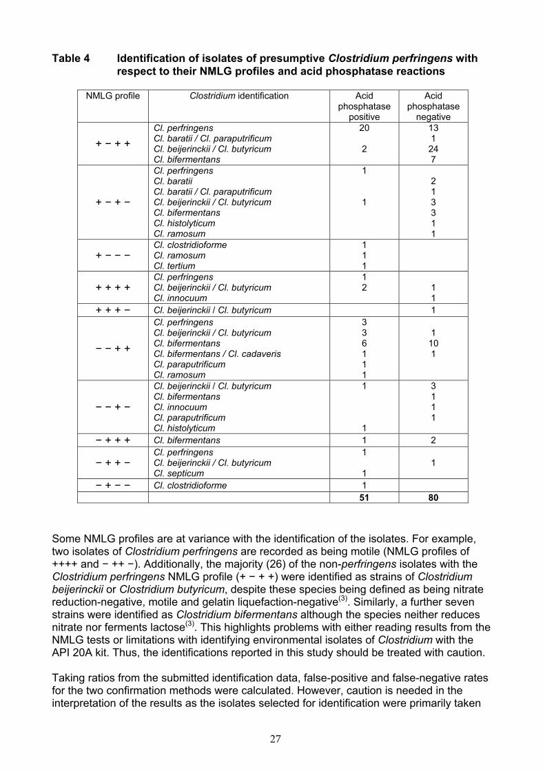
Some NMLG profiles are at variance with the identification of the isolates. For example,two isolates of Clostridium perfringens are recorded as being motile (NMLG profiles of ++++ and − ++ −). Additionally, the majority (26) of the non-perfringens isolates with theClostridium perfringens NMLG profile (+ − + +) were identified as strains of Clostridiumbeijerinckii or Clostridium butyricum, despite these species being defined as being nitratereduction-negative, motile and gelatin liquefaction-negative(3). Similarly, a further sevenstrains were identified as Clostridium bifermentans although the species neither reducesnitrate nor ferments lactose(3). This highlights problems with either reading results from theNMLG tests or limitations with identifying environmental isolates of Clostridium with theAPI 20A kit. Thus, the identifications reported in this study should be treated with caution.
Taking ratios from the submitted identification data, false-positive and false-negative ratesfor the two confirmation methods were calculated. However, caution is needed in theinterpretation of the results as the isolates selected for identification were primarily taken
28
from those that produced discrepant confirmation results, particularly with respect to theacid phosphatase test. This will skew any assessment of the data, especially as thetargeted isolates represent strains from only about 10 % of all the isolates tested and thedata are derived from only three of the thirteen participating laboratories. Taking this intoaccount and applying the identification data to that shown in Table 3, the false-positive andfalse-negative rates of the NMLG and acid phosphatase test methods for the confirmationof Clostridium perfringens are:-
False-positive rate for NMLG tests = 3.9 % False-positive rate for acid phosphatase test = 4.9 %False-negative rate for NMLG tests = 1.3 %False-negative rate for acid phosphatase test = 1.6 %
The false-negative rate for the NMLG tests is lower than expected, particularly as 10.3 %of the strains identified as Clostridium perfringens did not reduce nitrate.
4 Conclusions
The results of this study indicate that the acid phosphatase test for the confirmation ofClostridium perfringens from water is at least as reliable as the current method(1) basedupon the demonstration of reduction of nitrate, lack of motility, fermentation of lactose andliquefaction of gelatin. The two procedures show similar false-positive and false-negativerates, at a level expected from application to a large number of a wide range ofenvironmental isolates. There is an agreement rate of 90.0 %. The false-positive rates forboth procedures are less than 5 % and appear to be primarily due to species ofClostridium beijerinckii or Clostridium butyricum and Clostridium bifermentans, althoughthese identifications need to be treated with caution. The acid phosphatase test isconsiderably simpler to perform and is potentially more specific(2).
5 References
1. Standing Committee of Analysts, The Microbiology of Drinking Water (2004) - Part 6 -Methods for the isolation and enumeration of Sulphite-Reducing Clostridia and Clostridiumperfringens by membrane filtration, Methods for the Examination of Waters and AssociatedMaterials, in this series, Environment Agency.
2. Evaluation of acid phosphatase as a confirmation test for Clostridium perfringensisolated from water, Letters in Applied Microbiology, D P Sartory, R Waldock, C E Daviesand A M Field, 2006, 42, pp418-424.
3. Genus Clostridium Prazmowski 1880. In Bergey’s Manual of SystematicBacteriology Volume 2 (Edited by P H A Sneath, N S Mair, M E Sharpe and J G Holt),E P Cato, W L George and S M Finegold, pp. 1141-1200, Baltimore, Williams and Wilkins,1986.
4. Cowan and Steel’s Manual for the Identification of Medical Bacteria, 3rd Edition.Edited by G I Barrow and R K A Feltham, Cambridge, Cambridge University Press, 1993.
5. A study of rapid and simplified confirmatory tests for Clostridium perfringens.Journal of Applied Bacteriology, G C Mead, L Paez de Leon and B W Adams, 1981, 51,pp355-361.
29
6 Acknowledgements
The Standing Committee of Analysts is indebted to the managers and analysts of thefollowing laboratories that participated in this study: AES Laboratories (Newcastle-upon-Tyne)CREH Analytical (Leeds)Northern Ireland Water Services (Londonderry)Scottish Water (Dundee)Scottish Water (Edinburgh)Scottish Water (Turriff)Severn Trent Laboratories (Bridgend)Severn Trent Laboratories (Coventry)Severn Trent Water (Nottingham)Severn Trent Water (Shrewsbury)South West Water (Exeter)Southern Water (Winchester)United Utilities (Warrington)Wessex Water (Bath).

Address for correspondence However well procedures may be tested, there is always the possibility of discovering hitherto unknown problems. Analysts with such information are requested to contact the Secretary of the Standing Committee of Analysts at the address given below. In addition, if users wish to receive advance notice of forthcoming publications, please contact the Secretary. Secretary Standing Committee of Analysts Environment Agency (National Laboratory Service) 56 Town Green Street Rothley Leicestershire LE7 7NW www.environment-agency.gov.uk/nls Environment Agency Standing Committee of Analysts Members assisting with these methods Without the good will and support given by these individuals and their respective organisations SCA would not be able to continue and produce the highly valued and respected blue book methods. P Boyd Health Protection Agency S Cole Wessex Water R Down Southern Water D Gaskell United Utilities H Hawkins Veolia Water D Mortimer States Analyst's Laboratory, Guernsey D Sartory SWM Consulting R Stott Northumbrian Water Scientific Services J Watkins CREH (Analytical) Ltd K Woolnough Eurofins Grateful acknowledgement is made to D Gaskell and J Watkins for providing colour photographs.
30

CONTACTS:ENVIRONMENT AGENCY HEAD OFFICE
Rio House, Waterside Drive, Aztec West, Almondsbury, Bristol BS32 4UD
www.environment-agency.gov.ukwww.environment-agency.wales.gov.uk
ENVIRONMENT AGENCY REGIONAL OFFICESANGLIANKingfisher HouseGoldhay WayOrton GoldhayPeterborough PE2 5ZR
MIDLANDSSapphire East550 Streetsbrook RoadSolihull B91 1QT
NORTH EASTRivers House21 Park Square SouthLeeds LS1 2QG
NORTH WESTPO Box 12Richard Fairclough HouseKnutsford RoadWarrington WA4 1HG
SOUTHERNGuildbourne HouseChatsworth RoadWorthingWest Sussex BN11 1LD
SOUTH WESTManley HouseKestrel WayExeter EX2 7LQ
THAMESKings Meadow HouseKings Meadow RoadReading RG1 8DQ
WALESCambria House29 Newport RoadCardiff CF24 0TP
NORTH EAST
Leeds
Warrington
Solihull
MIDLANDSANGLIAN
Peterborough
SOUTHERNSOUTH WEST
Exeter
Cardiff
BristolTHAMES London
Worthing
Reading
WALES
NORTH WEST
E N V I R O N M E N T A G E N C YG E N E R A L E N Q U I R Y L I N E
08708 506 506
E N V I R O N M E N T A G E N C YE M E R G E N C Y H O T L I N E
0800 80 70 60
E N V I R O N M E N T A G E N C YF L O O D L I N E
0845 988 1188