Entrapment Neuropathies in the Upper and Lower Limbs: Anatomy
13
Hindawi Publishing Corporation Radiology Research and Practice Volume 2012, Article ID 230679, 12 pages doi:10.1155/2012/230679 Review Article Entrapment Neuropathies in the Upper and Lower Limbs: Anatomy and MRI Features Qian Dong, Jon A. Jacobson, David A. Jamadar, Girish Gandikota, Catherine Brandon, Yoav Morag, David P. Fessell, and Sung-Moon Kim Division of Musculoskeletal Radiology, Department of Radiology, University of Michigan Health System, 1500 East Medical Center Drive, TC 2910R, Ann Arbor, MI 48109-5326, USA Correspondence should be addressed to Qian Dong, [email protected] Received 20 June 2012; Revised 30 August 2012; Accepted 25 September 2012 Academic Editor: Avneesh Chhabra Copyright © 2012 Qian Dong et al. This is an open access article distributed under the Creative Commons Attribution License, which permits unrestricted use, distribution, and reproduction in any medium, provided the original work is properly cited. Peripheral nerve entrapment occurs at specific anatomic locations. Familiarity with the anatomy and the magnetic resonance imaging (MRI) features of nerve entrapment syndromes is important for accurate diagnosis and early treatment of entrapment neuropathies. The purpose of this paper is to illustrate the normal anatomy of peripheral nerves in the upper and lower limbs and to review the MRI features of common disorders affecting the peripheral nerves, both compressive/entrapment and noncompressive, involving the suprascapular nerve, the axillary nerve, the radial nerve, the ulnar nerve, and the median verve in the upper limb and the sciatic nerve, the common peroneal nerve, the tibial nerve, and the interdigital nerves in the lower limb. 1. Introduction Peripheral neuropathies are relatively common clinical disor- ders, which may be classified, according to cause, into com- pressive or entrapment and noncompressive neuropathies [1]. Although nerves may be injured anywhere along their course, peripheral nerve compression or entrapment occurs more at specific locations, such as sites where a nerve courses through fibroosseous or fibromuscular tunnels or penetrates muscles [2, 3]. Typically, the diagnosis has been based mainly on the combination of clinical history, physical examination, and electrodiagnostic studies. However, such clinical evaluations may provide insufficient information in making an accurate diagnosis, and imaging is being used often to confirm diagnoses. Magnetic resonance imaging (MRI) and high-resolution ultrasonography (US), as noninvasive techniques, provide valuable spatial information in making important diagnostic distinctions that cannot be readily accomplished by using other existing methods [2, 4, 5]. While both allow direct anatomic visualization of a nerve, identification of the cause, and location of primary abnormalities, MRI has the ability to demonstrate intrinsic signal abnormalities within the nerve itself and is considered superior in delineating the associated indirect signs related to muscle denervation [2, 4]. The purpose of this paper is to illustrate the anatomical course of peripheral nerves and review a broad spectrum of common peripheral neuropathies, both compressive/ entrapment and noncompressive, involving the suprascapu- lar, axillary, radial, ulnar, and median nerves in the upper limb, and the sciatic, common peroneal, tibial, and the interdigital nerves in the lower limb. 2. MRI Features The signal intensity of a normal nerve on MRI is of intermediate to low on T1-weighted sequences becoming slightly higher on T2-weighted and other fluid-sensitive sequences [3, 4]. Enlargement with apparent increase in T2 signal is considered an abnormal MRI appearance [3]. In addition, a hyperintense signal of the denervated muscle is usually identified when entrapment is acute, and fatty infiltration and muscle atrophy are the signs of chronic neuropathy in longstanding cases [2–4]. Peripheral nerve sheath tumors (PNSTs) appear as a well-defined mass continuous with a peripheral nerve. Such
Transcript of Entrapment Neuropathies in the Upper and Lower Limbs: Anatomy

Hindawi Publishing CorporationRadiology Research and PracticeVolume 2012, Article ID 230679, 12 pagesdoi:10.1155/2012/230679
Review Article
Entrapment Neuropathies in the Upper and Lower Limbs:Anatomy and MRI Features
Qian Dong, Jon A. Jacobson, David A. Jamadar, Girish Gandikota, Catherine Brandon,Yoav Morag, David P. Fessell, and Sung-Moon Kim
Division of Musculoskeletal Radiology, Department of Radiology, University of Michigan Health System,1500 East Medical Center Drive, TC 2910R, Ann Arbor, MI 48109-5326, USA
Correspondence should be addressed to Qian Dong, [email protected]
Received 20 June 2012; Revised 30 August 2012; Accepted 25 September 2012
Academic Editor: Avneesh Chhabra
Copyright © 2012 Qian Dong et al. This is an open access article distributed under the Creative Commons Attribution License,which permits unrestricted use, distribution, and reproduction in any medium, provided the original work is properly cited.
Peripheral nerve entrapment occurs at specific anatomic locations. Familiarity with the anatomy and the magnetic resonanceimaging (MRI) features of nerve entrapment syndromes is important for accurate diagnosis and early treatment of entrapmentneuropathies. The purpose of this paper is to illustrate the normal anatomy of peripheral nerves in the upper and lower limbs and toreview the MRI features of common disorders affecting the peripheral nerves, both compressive/entrapment and noncompressive,involving the suprascapular nerve, the axillary nerve, the radial nerve, the ulnar nerve, and the median verve in the upper limb andthe sciatic nerve, the common peroneal nerve, the tibial nerve, and the interdigital nerves in the lower limb.
1. Introduction
Peripheral neuropathies are relatively common clinical disor-ders, which may be classified, according to cause, into com-pressive or entrapment and noncompressive neuropathies[1]. Although nerves may be injured anywhere along theircourse, peripheral nerve compression or entrapment occursmore at specific locations, such as sites where a nervecourses through fibroosseous or fibromuscular tunnels orpenetrates muscles [2, 3]. Typically, the diagnosis has beenbased mainly on the combination of clinical history, physicalexamination, and electrodiagnostic studies. However, suchclinical evaluations may provide insufficient information inmaking an accurate diagnosis, and imaging is being usedoften to confirm diagnoses.
Magnetic resonance imaging (MRI) and high-resolutionultrasonography (US), as noninvasive techniques, providevaluable spatial information in making important diagnosticdistinctions that cannot be readily accomplished by usingother existing methods [2, 4, 5]. While both allow directanatomic visualization of a nerve, identification of the cause,and location of primary abnormalities, MRI has the ability todemonstrate intrinsic signal abnormalities within the nerve
itself and is considered superior in delineating the associatedindirect signs related to muscle denervation [2, 4].
The purpose of this paper is to illustrate the anatomicalcourse of peripheral nerves and review a broad spectrumof common peripheral neuropathies, both compressive/entrapment and noncompressive, involving the suprascapu-lar, axillary, radial, ulnar, and median nerves in the upperlimb, and the sciatic, common peroneal, tibial, and theinterdigital nerves in the lower limb.
2. MRI Features
The signal intensity of a normal nerve on MRI is ofintermediate to low on T1-weighted sequences becomingslightly higher on T2-weighted and other fluid-sensitivesequences [3, 4]. Enlargement with apparent increase in T2signal is considered an abnormal MRI appearance [3]. Inaddition, a hyperintense signal of the denervated muscleis usually identified when entrapment is acute, and fattyinfiltration and muscle atrophy are the signs of chronicneuropathy in longstanding cases [2–4].
Peripheral nerve sheath tumors (PNSTs) appear as awell-defined mass continuous with a peripheral nerve. Such
2 Radiology Research and Practice
IS
SS
SSN
Figure 1: The drawing shows anatomy of the suprascapular nerve from the posterior view. Note the nerve courses through the suprascapularnotch (open arrow) and spinoglenoid notch (curved arrow). SSN: suprascapular nerve, SS: supraspinatus muscle, IS: infraspinatus muscle.
IS
SS
(a)
IS
SS
(b)
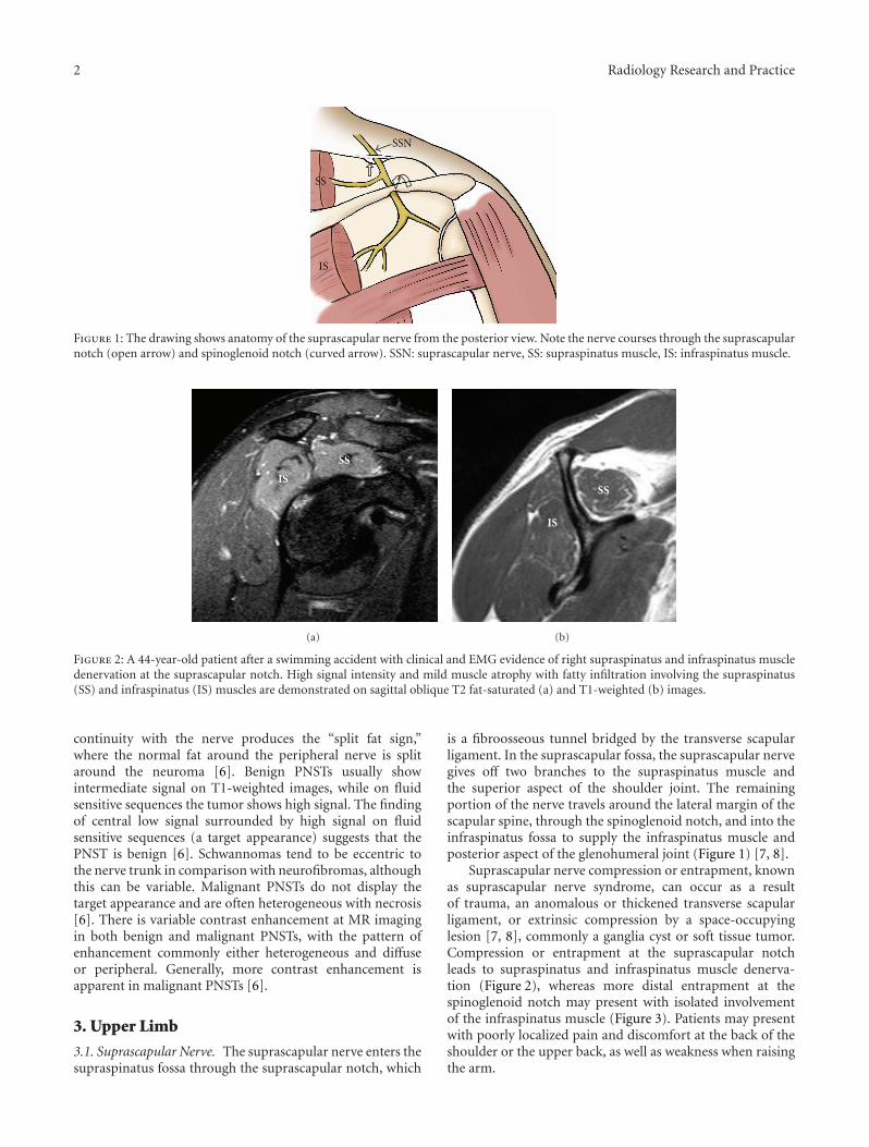
Figure 2: A 44-year-old patient after a swimming accident with clinical and EMG evidence of right supraspinatus and infraspinatus muscledenervation at the suprascapular notch. High signal intensity and mild muscle atrophy with fatty infiltration involving the supraspinatus(SS) and infraspinatus (IS) muscles are demonstrated on sagittal oblique T2 fat-saturated (a) and T1-weighted (b) images.
continuity with the nerve produces the “split fat sign,”where the normal fat around the peripheral nerve is splitaround the neuroma [6]. Benign PNSTs usually showintermediate signal on T1-weighted images, while on fluidsensitive sequences the tumor shows high signal. The findingof central low signal surrounded by high signal on fluidsensitive sequences (a target appearance) suggests that thePNST is benign [6]. Schwannomas tend to be eccentric tothe nerve trunk in comparison with neurofibromas, althoughthis can be variable. Malignant PNSTs do not display thetarget appearance and are often heterogeneous with necrosis[6]. There is variable contrast enhancement at MR imagingin both benign and malignant PNSTs, with the pattern ofenhancement commonly either heterogeneous and diffuseor peripheral. Generally, more contrast enhancement isapparent in malignant PNSTs [6].
3. Upper Limb
3.1. Suprascapular Nerve. The suprascapular nerve enters thesupraspinatus fossa through the suprascapular notch, which
is a fibroosseous tunnel bridged by the transverse scapularligament. In the suprascapular fossa, the suprascapular nervegives off two branches to the supraspinatus muscle andthe superior aspect of the shoulder joint. The remainingportion of the nerve travels around the lateral margin of thescapular spine, through the spinoglenoid notch, and into theinfraspinatus fossa to supply the infraspinatus muscle andposterior aspect of the glenohumeral joint (Figure 1) [7, 8].
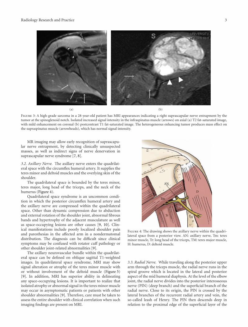
Suprascapular nerve compression or entrapment, knownas suprascapular nerve syndrome, can occur as a resultof trauma, an anomalous or thickened transverse scapularligament, or extrinsic compression by a space-occupyinglesion [7, 8], commonly a ganglia cyst or soft tissue tumor.Compression or entrapment at the suprascapular notchleads to supraspinatus and infraspinatus muscle denerva-tion (Figure 2), whereas more distal entrapment at thespinoglenoid notch may present with isolated involvementof the infraspinatus muscle (Figure 3). Patients may presentwith poorly localized pain and discomfort at the back of theshoulder or the upper back, as well as weakness when raisingthe arm.
Radiology Research and Practice 3
(a) (b)
Figure 3: A high-grade sarcoma in a 28-year-old patient has MRI appearances indicating a right suprascapular nerve entrapment by thetumor at the spinoglenoid notch. Isolated increased signal intensity in the infraspinatus muscle (arrows) on axial (a) T2 fat-saturated image,with mild enhancement on coronal (b) postcontrast T1 fat-saturated image. The heterogeneous enhancing tumor produces mass effect onthe supraspinatus muscle (arrowheads), which has normal signal intensity.
MR imaging may allow early recognition of suprascapu-lar nerve entrapment, by detecting clinically unsuspectedmasses, as well as indirect signs of nerve denervation insuprascapular nerve syndrome [7, 8].
3.2. Axillary Nerve. The axillary nerve enters the quadrilat-eral space with the circumflex humeral artery. It supplies theteres minor and deltoid muscles and the overlying skin of theshoulder.
The quadrilateral space is bounded by the teres minor,teres major, long head of the triceps, and the neck of thehumerus (Figure 4).
Quadrilateral space syndrome is an uncommon condi-tion in which the posterior circumflex humeral artery andthe axillary nerve are compressed within the quadrilateralspace. Other than dynamic compression due to abductionand external rotation of the shoulder joint, abnormal fibrousbands and hypertrophy of the adjacent musculature as wellas space-occupying lesions are other causes [9, 10]. Clin-ical manifestations include poorly localized shoulder painand paresthesias in the affected arm in a nondermatomaldistribution. The diagnosis can be difficult since clinicalsymptoms may be confused with rotator cuff pathology orother shoulder joint-related abnormalities [9].
The axillary neurovascular bundle within the quadrilat-eral space can be defined on oblique sagittal T1-weightedimages. In quadrilateral space syndrome, MRI may showsignal alteration or atrophy of the teres minor muscle withor without involvement of the deltoid muscle (Figure 5)[9]. In addition, MRI has superior ability in delineatingany space-occupying lesions. It is important to realize thatisolated atrophy or abnormal signal in the teres minor musclemay occur in asymptomatic patients or patients with othershoulder abnormalities [9]. Therefore, care must be taken toassess the entire shoulder with clinical correlation when suchimaging findings are present on MRI.
Tm
TrH
D
TM
AN
Figure 4: The drawing shows the axillary nerve within the quadri-lateral space from a posterior view. AN: axillary nerve, Tm: teresminor muscle, Tr: long head of the triceps, TM: teres major muscle,H: humerus, D: deltoid muscle.
3.3. Radial Nerve. While traveling along the posterior upperarm through the triceps muscle, the radial nerve runs in thespiral groove which is located in the lateral and posterioraspect of the mid humeral diaphysis. At the level of the elbowjoint, the radial nerve divides into the posterior interosseousnerve (PIN) (deep branch) and the superficial branch of theradial nerve. Close to its origin, the PIN is crossed by thelateral branches of the recurrent radial artery and vein, theso-called leash of Henry. The PIN then descends deep inrelation to the proximal edge of the superficial layer of the

4 Radiology Research and Practice
(a) (b) (c)
Figure 5: A 46-year-old patient with right shoulder pain and clinical and EMG evidence of quadrilateral space syndrome. Oblique coronal(a), oblique sagittal (b) T2 fat-saturated, and oblique sagittal T1-weighted (c) images demonstrate severe fatty atrophy of the teres minormuscle (arrows).
RN
ECRB
SPPIN
SRN
RRA
Figure 6: The drawing provides an anterior view of the course ofthe radial nerve at the elbow. Posterior interosseous nerve (PIN)entrapment may occur due to prominent radial recurrent artery(RRA), medial edge of the extensor carpi radialis brevis (ECRB),and proximal edge of the supinator muscle (SP) (arcade of Frohse).RN: radial nerve, SRN: superficial radial nerve.
supinator muscle, which is known as the arcade of Frohse(Figure 6). The superficial branch of the radial nerve coursesalongside the radial artery and then distally courses lateralover the first extensor wrist compartment [9].
The radial nerve is predisposed to injury and entrapmentat several locations along its course, which include the radialnerve in the spiral groove of the humerus (spiral groovesyndrome) above the elbow joint, where the PIN travelsthrough the radial tunnel, and the superficial branch ofthe radial nerve where it crosses over the first dorsal wristcompartment (Wartenberg’s syndrome).
Compression or entrapment of the PIN in the radial tun-nel may yield two different clinical presentations: posteriorinterosseous nerve syndrome and radial tunnel syndrome.The radial tunnel is a musculoaponeurotic furrow or space
extending from the lateral epicondyle of the humerus tothe distal edge of the supinator muscle [11]. In patientswith posterior interosseous nerve syndrome, the clinicalpresentation includes motor deficits of the extensor musclegroup without significant sensory loss. The most commonsite of nerve compression of PIN within the radial tunnelis posterior interosseous nerve syndrome at the arcade ofFrohse. Other structures can potentially cause compressionor entrapment including the medial edges of the extensorcarpi radialis brevis, fibrous bands at the radial head, and theleash of Henry [12] (Figure 6). Patients with radial tunnelsyndrome, on the other hand, typically present with painover the proximal lateral forearm [12, 13], which can becaused by acute trauma, masses, and compression fromadjacent structures. MR imaging features range from directvisualization of nerve thickening with increased T2 signaland muscle signal alterations (Figure 7) to the detection ofcompressive lesions or abnormal structures, which may causecompression or entrapment as mentioned above.
Entrapment of the superficial branch of the radial nerveat the level of distal wrist is called Wartenberg’s syndrome[5]. The superficial location of the superficial branch of theradial nerve predisposes it to injury and may result fromfixation of a distal radius fracture, penetrating injury, andiatrogenic injury related to vein cannulation or adjacenttendon sheath injection [5].
3.4. Ulnar Nerve. The ulnar nerve enters the cubital tunnel,a fibroosseous canal, at the level of the medial epicondyleof the elbow. The roof of the tunnel is formed by afascial band between the olecranon process and the medialepicondyle known as the cubital tunnel retinaculum [14].The nerve then passes beneath the arcuate ligament whichis an aponeurosis between the humeral and ulnar heads ofthe flexor carpi ulnaris muscle (Figure 8). The cubital tunnelretinaculum and arcuate ligament typically blend with eachother. After passing through the anterior compartment ofthe forearm, the ulnar nerve, artery, and vein enter the wristthrough Guyon’s canal (Figure 10), which is a fibroosseoustunnel between the pisiform and the hook of the hamate,

Radiology Research and Practice 5
(a) (b) (c)
Figure 7: An 18-year-old patient with clinical and EMG evidence of PIN entrapment. Axial T2 fat-saturated (a) and T1-weighted (b) imagesat the level of right distal humerus show thickening and high T2 signal of the radial nerve (arrows). Supinator muscle edema (arrowheads)is present (c), axial T2 fat-saturated image.
FCU
AL
Ulnar n.
Figure 8: The drawing demonstrates the course of the ulnar nervefrom posterior view at the elbow. Note the nerve travels deep tothe flexor carpi ulnaris muscle (FCU) beneath the arcuate ligament(AL).
with the roof formed proximally by the palmar carpalligament and distally by the short palmar muscle, and thefloor formed by the flexor retinaculum. Within the canal,the ulnar nerve divides into the superficial sensory and deepmotor branches [14].
Compressive or entrapped ulnar nerve neuropathiesinclude cubital tunnel syndrome and Guyon’s canal syn-drome.
Cubital tunnel syndrome is the second most commonperipheral neuropathy of the upper extremity. It may becaused by abnormal fascial bands, subluxation, or dis-location of the ulnar nerve over the medial epicondyle,trauma, or direct compression by soft tissue masses. Clinicalsymptoms include a sensory abnormality of the ulnar handand weakness of the flexor carpi muscle group of the 4thand 5th fingers. MRI findings of cubital tunnel syndromeare enlargement and hyperintense signal just proximal tothe cubital tunnel, and a caliber change with flatteningdistally (Figure 9). Mild enlargement and edema of the ulnar
(a)
(b)
Figure 9: A 17-year-old patient suffered right cubital tunnelsyndrome. MR arthrogram of the elbow was performed to evaluatefor loose body. Axial T2-weighted with fat-saturated (a, b) imagesreveal swollen and edematous ulnar nerve (arrow) at the level ofproximal cubital tunnel, and normal size distally (arrowhead).

6 Radiology Research and Practice
P
S
(a)
T H
(b)
Figure 10: Drawings of the carpal tunnel and Guyon’s canal at the levels of pisiform (a) and hamate (b). The median nerve (open arrows)passes through the carpal tunnel under the flexor retinaculum lying superficial to the flexor digitorum superficialis tendons. Guyon’s canalcontains the ulnar nerve (arrows) and ulnar artery with the floor formed by the flexor retinaculum. The ulnar nerve divides into thesuperficial sensory and deep motor branches at the level of the hamate. P: pisiform, S: scaphoid, T: trapezium, H: hamate.
(a) (b) (c)
Figure 11: A 73-year-old patient with clinical evidence of right ulnar nerve compression at wrist. Axial T1-weighted (a), T2-weighted withfat saturation (b), and axial SENSE MIPs (c) identify the ulnar nerve (arrows) in a crowded Guyon’s canal compressed by a tortuous ulnarartery (arrowheads).
nerve at the medial epicondyle can be seen in asymptomaticindividuals on MRI [15]. Careful assessment of the flexormuscle group for signal abnormalities and correlation withthe patient’s symptoms are important.
The causes of Guyon’s canal syndrome include space-occupying lesions, trauma, anomalous muscles, and ulnarartery aneurysms. Entrapment at Guyon’s canal can beassociated with either motor or sensory findings, dependingon whether the nerve compression involves the ulnar nerveprior to its bifurcation to the superficial (sensory) anddeep (motor) branches or if the compression is limitedto one of the branches [5]. MR imaging is useful indepicting the ulnar nerve in Guyon’s canal demonstratingthe etiology of entrapment with additional information ofmuscle denervation (Figure 11), if present.
3.5. Median Nerve. The median nerve enters the forearmbetween the humeral and ulnar heads of the pronator teresmuscle, where the anterior interosseous branch is given off(Figure 12). The nerve then continues distally in the forearmsandwiched between flexor digitorum profundus and flexordigitorum superficialis muscles. It then passes through thecarpal tunnel under the flexor retinaculum, lying superficialto the flexor digitorum superficialis tendons. The carpaltunnel is formed by the carpal bones (floor), the transversecarpal ligament (roof), the scaphoid and trapezium (radial
Figure 12: The drawing of the median nerve shows that it coursesalong the anterior elbow, through the two heads of the pronatorteres muscle (stars), and into the forearm beneath the edge of thefibrous arch of the flexor digitorum sublimis (open arrow).
side), and the pisiform and hook of the hamate (ulnar side)(Figure 9). The median nerve subdivides into digital andmuscular branches as it exits the carpal tunnel.
Radiology Research and Practice 7
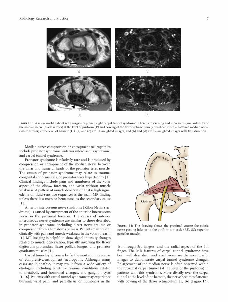
P
S
(a)
P
S
(b)
T
H
(c)
H
T
(d)
Figure 13: A 48-year-old patient with surgically proven right carpal tunnel syndrome. There is thickening and increased signal intensity ofthe median nerve (black arrows) at the level of pisiform (P) and bowing of the flexor retinaculum (arrowhead) with a flattened median nerve(white arrows) at the level of hamate (H). (a) and (c) are T1-weighted images, and (b) and (d) are T2-weighted images with fat saturation.
Median nerve compression or entrapment neuropathiesinclude pronator syndrome, anterior interosseous syndrome,and carpal tunnel syndrome.
Pronator syndrome is relatively rare and is produced bycompression or entrapment of the median nerve betweenthe ulnar and humeral heads of the pronator teres muscle.The causes of pronator syndrome may relate to trauma,congenital abnormalities, or pronator teres hypertrophy [1].Clinical findings include pain and numbness of the volaraspect of the elbow, forearm, and wrist without muscleweakness. A pattern of muscle denervation that is high signaledema on fluid-sensitive sequences is the main MR findingunless there is a mass or hematoma as the secondary cause[1].
Anterior interosseous nerve syndrome (Kilon-Nevin syn-drome) is caused by entrapment of the anterior interosseousnerve in the proximal forearm. The causes of anteriorinterosseous nerve syndrome are similar to those describedin pronator syndrome, including direct nerve trauma orcompression from a hematoma or mass. Patients may presentclinically with pain and muscle weakness in the volar forearm[1]. MR imaging is helpful to show signal intensity changesrelated to muscle denervation, typically involving the flexordigitorum profundus, flexor pollicis longus, and pronatorquadratus muscles [1].
Carpal tunnel syndrome is by far the most common causeof compressive/entrapment neuropathy. Although manycases are idiopathic, it may result from a wide variety ofetiologies, including repetitive trauma, conditions relatedto metabolic and hormonal changes, and ganglion cysts[1, 16]. Patients with carpal tunnel syndrome may experienceburning wrist pain, and paresthesia or numbness in the
PF
SGSciatic n.
Figure 14: The drawing shows the proximal course the sciaticnerve passing inferior to the piriformis muscle (PS). SG: superiorgemellus muscle.
1st through 3rd fingers, and the radial aspect of the 4thfinger. The MR features of carpal tunnel syndrome havebeen well described, and axial views are the most usefulimages to demonstrate carpal tunnel syndrome changes.Enlargement of the median nerve is often observed withinthe proximal carpal tunnel (at the level of the pisiform) inpatients with this syndrome. More distally over the carpaltunnel at the level of the hamate, the nerve becomes flattenedwith bowing of the flexor retinaculum [1, 16] (Figure 13),

8 Radiology Research and Practice
(a)
(b) (c)
Figure 15: Extensive tear of the left hamstring muscle origin with sciatic nerve scarring in a 54-year-old patient after a water skiing injury.Axial T1-weighted images (a) and (b) identify a tear of left hamstring at the level of origin (black arrow in (a)). There is thickeningand abnormal signal of the adjacent sciatic nerve (white arrowheads) extending distally (b), consistent with secondary entrapment fromscarring. This finding is also demonstrated on a sagittal fat-saturated T2-weighted image (c). On the right side, the right sciatic nerve (blackarrowheads) has normal caliber and signal intensity, and an intact hamstring muscle origin is present (star).
(a) (b)
Figure 16: Surgically proven neurofibroma of the left sciatic nerve in a 33-year-old patient. Axial T2-weighted fat-saturated (a) and sagittalT1-weighted postcontrast (b) images show a lobulated enhancing mass (arrowheads) with isointense T1 (not shown) and high T2 signaloriginating from the left sciatic nerve. Proximally, the left S1 nerve root is thickened (white arrow). Note the target appearance of theneurofibroma.
and a hyperintense signal of the nerve on T2-weighted orSTIR imaging is often observed. Although the sensitivity andspecificity of the MR findings for carpal tunnel syndromeare low (sensitivity, 23%–96%; specificity, 39%–87%), MRimaging is useful in detecting a space-occupying lesion,inflammatory arthritis, or a congenital anomaly as the causeof carpal tunnel syndrome [1].
4. Lower Limb
4.1. Sciatic Nerve. The sciatic nerve originates from theupper division of the sacral plexus and typically leaves thepelvis through the greater sciatic foramen at the inferiorborder of the piriformis muscle (Figure 14). The sciatic nervethen divides into the tibial and common peroneal nerves justabove the knee.
Sciatic nerve entrapment may occur in the hip regionand less commonly in the thigh, and clinical presentations
are based upon the level of injury [3]. Sciatic neuropathymay result from conditions such as fibrous or muscu-lar entrapment, vascular compression, scarring related totrauma (Figure 15) or radiation, tumors (Figure 16), andhypertrophic neuropathy [3, 17, 18]. Piriformis syndromeis a controversial diagnosis, often thought to be relatedto sciatic nerve compression or irritation related to thepiriformis muscle. MRI can show variations in anatomy,muscle hypertrophy, as well as abnormal signal of the sciaticnerve [19]. MR imaging is not only a sensitive techniquein identifying and characterizing the causative abnormalitiesbut also can provide useful information for surgical planning[20].
4.2. Common Peroneal Nerve. The common peroneal nervearises from the sciatic nerve at the level of popliteal fossa.It travels distally and laterally posterior to the short head

Radiology Research and Practice 9
TN
SN
CPN
PL
Figure 17: Sagittal oblique projection of the knee illustrates the common peroneal nerve (CPN) arising from the sciatic nerve (SN) at thelevel of popliteal fossa. It travels around the fibular head deep to the origin of the peroneus longus muscle (PL). TN: tibial nerve.
TA
(a)
ED
TA
(b)
Figure 18: Common peroneal nerve entrapment secondary to a surgically proven intraneural ganglion cyst in a 44-year-old patient with a 6-month history of right foot drop. Axial T2-weighted fat-saturated images (a, b) reveal a multilobulated high T2 signal structure (arrowheads)compressing the adjacent common peroneal nerve (arrow). Associated patchy high signal in tibialis anterior (TA) and extensor digitorumlongus (ED) muscles.
of the biceps femoris muscle, and lateral and superficial tothe lateral head of the gastrocnemius muscle. It then passesaround the fibular head laterally entering the anterolateralaspect of the leg deep to the peroneus longus muscle(Figure 17), where the nerve splits into deep and superficialperoneal branches.
Nerve impingement of the common peroneal nerve mayoccur around the level of fibular head due to its superficiallocation, or as it travels deep to the origin of the peroneuslongus muscle [17]. The etiologies of common peronealneuropathy may include idiopathic mononeuritis, intrinsicand extrinsic space-occupying lesions including an intra-neural ganglion cyst (Figure 18) [21], or traumatic injuryof the nerve, especially related to proximal fibular fractures[22]. Clinically, patients may experience pain at the site ofentrapment with foot drop and a slapping gait [17, 23].
MR imaging is superior in depicting the location andcause of peroneal nerve compression and assessing the stageof the neuropathy, indicated by early muscle denervation orlater changes such as atrophy.
4.3. Tibial Nerve. After dividing from the sciatic nerve, thetibial nerve descends into the posterior compartment of thelower leg deep to the soleus, plantaris, and gastrocnemiusmuscles. It then crosses the ankle behind the medial malle-olus, where it divides into its terminal branches, the medialcalcaneal nerve, and medial and lateral plantar nerves. Thename of the “posterior tibial nerve” is used as the tibialnerve reaches the ankle region [17]. The tarsal tunnel refersto a fibroosseous tunnel in the medial aspect of the anklewith the flexor retinaculum as the roof [17, 23]. The tunnelcontains the flexor digitorum longus and flexor hallucis

10 Radiology Research and Practice
MPN
FR
TN
MCN
LPN
Figure 19: The drawing of the medial aspect of the ankle showingthe course of the tibial nerve (TN) and its branches, the medialcalcaneal nerve (MCN), and medial and lateral plantar nerves(MPN and LPN), passing through the tarsal tunnel. FR: flexorretinaculum.
longus tendons, and the posterior tibial artery and veins, andthe posterior tibial nerve and its branches (Figure 19).
Tarsal tunnel syndrome is a well-known compres-sion/entrapment neuropathy of the posterior tibial nerve.Common etiologies include posttraumatic fibrosis due tofracture, tenosynovitis, ganglion cysts (Figure 20), space-occupying lesions, and dilated or tortuous veins. Mostpatients with tarsal tunnel syndrome have burning pain andparesthesia along the plantar foot and toes. MR imaging isuseful for localizing pathologies within the tarsal tunnel anddepicting the lesion extent and relationship to the nerve andbranches [24].
Compression of the proximal tibial nerve, the so-calledsoleal sling syndrome, is uncommon. It occurs when theproximal tibial nerve travels beneath the tendinous slingat the origin of the soleus muscle [25, 26]. The clinicalpresentation includes numbness, paresthesias in the sole ofthe foot, and posterior calf pain. MR imaging is useful indetecting increased T2 signal intensity of the nerve, as wellas signal alteration in denervated gastrocnemius and soleusmuscles [25].
4.4. Interdigital Nerve. The medial and lateral plantar nerves,which are terminal branches of the tibial nerve, divideinto interdigital nerves at the level of metatarsal bases. Theinterdigital nerves pass deep to the transverse intermetatarsalligament into a relatively small space between the metatarsalheads.
While repetitive mechanical stress with subsequent per-ineural fibrosis is the most commonly accepted cause ofMorton neuroma, other possibilities include ischemia andcompression of the nerve by an inflamed and enlargedintermetatarsal bursa [27]. Morton neuroma most frequently
(a)
(b)
Figure 20: Tarsal tunnel syndrome caused by a ganglion cystin a 32-year-old patient. Axial T1-weighted (a) and T2-weighted(b) fat-saturated images show a multilobulated cystic structure(arrowheads) within the right tarsal tunnel. Note the adjacent tibialnerve (arrows).
occurs in the second and third intermetatarsal spaces, oftenassociated with an intermetatarsal bursa (Figure 21). TheMRI appearance of the Morton neuroma is characteristic,typically manifested as an enhancing tear-drop-shaped softtissue mass with intermediate signal on both T1- and T2-weighted images between the metatarsal heads (Figure 22).MR imaging provides very helpful information in localiza-tion and accurate size assessment of Morton neuromas.
5. Conclusion
Peripheral neuropathies may be underdiagnosed in patientswith complicated clinical presentations. MR imaging pro-vides valuable information in making a precise diagnosis

Radiology Research and Practice 11
Figure 21: The drawing of the forefoot shows a Morton neuroma(star) at the site of the entrapment of the interdigital nerve betweenthe third and fourth metatarsal heads.
(a)
(b)
(c)
Figure 22: Morton neuroma in a 38-year-old patient. Coronal T2-weighted with fat-saturation (a), T1-weighted (b), and T1-weightedfat-saturated postcontrast (c) images identify an enhancing tear-drop-shaped soft tissue mass (white arrows) with intermediatesignal on both T1- and T2-weighted images in the third inter-metatarsal space. A small amount of fluid (black arrow) is notedwithin the intermetatarsal bursa dorsal to the neuroma in (a).
and ready differentiation from other etiologies. Furthermore,it also helps make decisions for surgical planning. It iscritical for radiologists to be familiar with the anatomy ofthe peripheral nerves and the range of pathology which mayproduce compressive/entrapment syndromes.
References
[1] G. Andreisek, D. W. Crook, D. Burg, B. Marincek, and D.Weishaupt, “Peripheral neuropathies of the median, radial,and ulnar nerves: MR imaging features,” Radiographics, vol.26, no. 5, pp. 1267–1287, 2006.
[2] S. J. Kim, S. H. Hong, W. S. Jun et al., “MR imaging mappingof skeletal muscle denervation in entrapment and compressiveneuropathies,” Radiographics, vol. 31, no. 2, pp. 319–332, 2011.
[3] C. N. Petchprapa, Z. S. Rosenberg, L. M. Sconfienza, C. F. A.Cavalcanti, R. L. Vieira, and J. S. Zember, “MR imaging ofentrapment neuropathies of the lower extremity: part 1. thepelvis and hip,” Radiographics, vol. 30, no. 4, pp. 983–1000,2010.
[4] J. Beltran and Z. S. Rosenberg, “Diagnosis of compressive andentrapment neuropathies of the upper extremity: value of MRimaging,” American Journal of Roentgenology, vol. 163, no. 3,pp. 525–531, 1994.
[5] J. A. Jacobson, D. P. Fessell, L. D. G. Lobo, and L. J. S.Yang, “Entrapment neuropathies I: upper limb (carpal tunnelexcluded),” Seminars in Musculoskeletal Radiology, vol. 14, no.5, pp. 473–486, 2010.
[6] M. D. Murphey, W. S. Smith, S. E. Smith, M. J. Kransdorf,and H. T. Temple, “From the archives of the AFIP: imagingof musculoskeletal neurogenic tumors: radiologic-pathologiccorrelation,” Radiographics, vol. 19, no. 5, pp. 1253–1280,1999.
[7] R. C. Fritz, C. A. Helms, L. S. Steinbach, and H. K. Genant,“Suprascapular nerve entrapment: evaluation with MR imag-ing,” Radiology, vol. 182, no. 2, pp. 437–444, 1992.
[8] D. Sallomi, D. L. Janzen, P. L. Munk, D. G. Connell, and P. F.J. Tirman, “Muscle denervation patterns in upper limb nerveinjuries: MR imaging findings and anatomic basis,” AmericanJournal of Roentgenology, vol. 171, no. 3, pp. 779–784, 1998.
[9] R. L. Cothran and C. Helms, “Quadrilateral space syndrome:incidence of imaging findings in a population referred for MRIof the shoulder,” American Journal of Roentgenology, vol. 184,no. 3, pp. 989–992, 2005.
[10] W. T. Hoskins, H. P. Pollard, and A. J. McDonald, “Quadrilat-eral space syndrome: a case study and review of the literature,”British Journal of Sports Medicine, vol. 39, no. 2, p. e9, 2005.
[11] M. Konjengbam and J. Elangbam, “Radial nerve in the radialtunnel: anatomic sites of entrapment neuropathy,” ClinicalAnatomy, vol. 17, no. 1, pp. 21–25, 2004.
[12] B. D. Ferdinand, Z. S. Rosenberg, M. E. Schweitzer et al.,“MR imaging features of radial tunnel syndrome: initialexperience,” Radiology, vol. 240, no. 1, pp. 161–168, 2006.
[13] S. Kim, J. Y. Choi, Y. M. Huh et al., “Role of magnetic reso-nance imaging in entrapment and compressive neuropathy—What, where, and how to see the peripheral nerves on themusculoskeletal magnetic resonance image: part 2. Upperextremity,” European Radiology, vol. 17, no. 2, pp. 509–522,2007.
[14] C. Martinoli, S. Bianchi, N. Gandolfo, M. Valle, S. Simonetti,and L. E. Derchi, “US of nerve entrapments in osteofibroustunnels of the upper and lower limbs,” Radiographics, vol. 20,pp. S199–S213, 2000.

12 Radiology Research and Practice
[15] D. B. Husarik, N. Saupe, C. W. A. Pfirrmann, B. Jost, J.Hodler, and M. Zanetti, “Elbow nerves: MR findings in 60asymptomatic subjects—normal anatomy, variants, and pit-falls,” Radiology, vol. 252, no. 1, pp. 148–156, 2009.
[16] M. Mesgarzadeh, J. Triolo, and C. D. Schneck, “Carpal tunnelsyndrome. MR imaging diagnosis,” Magnetic Resonance Imag-ing Clinics of North America, vol. 3, no. 2, pp. 249–264, 1995.
[17] A. Donovan, Z. S. Rosenberg, and C. F. Cavalcanti, “MR imag-ing of entrapment neuropathies of the lower extremity: part 2.the knee, leg, ankle, and foot,” Radiographics, vol. 30, no. 4, pp.1001–1019, 2010.
[18] J. Feinberg and S. Sethi, “Sciatic neuropathy: case report anddiscussion of the literature on postoperative sciatic neuropathyand sciatic nerve tumors,” HSS Journal, vol. 2, no. 2, pp. 181–187, 2006.
[19] H. I. Pecina, I. Boric, T. Smoljanovic, D. Duvancic, and M.Pecina, “Surgical evaluation of magnetic resonance imagingfindings in piriformis muscle syndrome,” Skeletal Radiology,vol. 37, no. 11, pp. 1019–1023, 2008.
[20] K. R. Moore, J. S. Tsuruda, and A. T. Dailey, “The value of MRneurography for evaluating extraspinal neuropathic leg pain:a pictorial essay,” American Journal of Neuroradiology, vol. 22,no. 4, pp. 786–794, 2001.
[21] R. J. Spinner, M. N. Hebert-Blouin, A. H. Maniker, and K.K. Amrami, “Clock face model applied to tibial intraneuralganglia in the popliteal fossa,” Skeletal Radiology, vol. 38, no.7, pp. 691–696, 2009.
[22] R. Loredo, J. Hodler, R. Pedowitz, L. R. Yeh, D. Trudell, andD. Resnick, “MRI of the common peroneal nerve: normalanatomy and evaluation of masses associated with nerveentrapment,” Journal of Computer Assisted Tomography, vol.22, no. 6, pp. 925–931, 1998.
[23] S. Kim, J. Y. Choi, Y. M. Huh et al., “Role of magnetic reso-nance imaging in entrapment and compressive neuropathy—What, where, and how to see the peripheral nerves on themusculoskeletal magnetic resonance image: part 1. Overviewand lower extremity,” European Radiology, vol. 17, no. 1, pp.139–149, 2007.
[24] R. Kerr and C. Frey, “MR imaging in tarsal tunnel syndrome,”Journal of Computer Assisted Tomography, vol. 15, no. 2, pp.280–286, 1991.
[25] A. Chhabra, E. H. Williams, T. K. Subhawong et al., “MR neu-rography findings of soleal sling entrapment,” American Jour-nal of Roentgenology, vol. 196, no. 3, pp. W290–W297, 2011.
[26] E. H. Williams, C. G. Williams, G. D. Rosson, and L. A. Dellon,“Anatomic site for proximal tibial nerve compression: acadaver study,” Annals of Plastic Surgery, vol. 62, no. 3, pp. 322–325, 2009.
[27] M. Zanetti, J. K. Strehle, H. P. Kundert, H. Zollinger, and J.Hodler, “Morton neuroma: effect of MR imaging findings ondiagnostic thinking and therapeutic decisions,” Radiology, vol.213, no. 2, pp. 583–588, 1999.

Submit your manuscripts athttp://www.hindawi.com
Stem CellsInternational
Hindawi Publishing Corporationhttp://www.hindawi.com Volume 2014
Hindawi Publishing Corporationhttp://www.hindawi.com Volume 2014
MEDIATORSINFLAMMATION
of
Hindawi Publishing Corporationhttp://www.hindawi.com Volume 2014
Behavioural Neurology
EndocrinologyInternational Journal of
Hindawi Publishing Corporationhttp://www.hindawi.com Volume 2014
Hindawi Publishing Corporationhttp://www.hindawi.com Volume 2014
Disease Markers
Hindawi Publishing Corporationhttp://www.hindawi.com Volume 2014
BioMed Research International
OncologyJournal of
Hindawi Publishing Corporationhttp://www.hindawi.com Volume 2014
Hindawi Publishing Corporationhttp://www.hindawi.com Volume 2014
Oxidative Medicine and Cellular Longevity
Hindawi Publishing Corporationhttp://www.hindawi.com Volume 2014
PPAR Research
The Scientific World JournalHindawi Publishing Corporation http://www.hindawi.com Volume 2014
Immunology ResearchHindawi Publishing Corporationhttp://www.hindawi.com Volume 2014
Journal of
ObesityJournal of
Hindawi Publishing Corporationhttp://www.hindawi.com Volume 2014
Hindawi Publishing Corporationhttp://www.hindawi.com Volume 2014
Computational and Mathematical Methods in Medicine
OphthalmologyJournal of
Hindawi Publishing Corporationhttp://www.hindawi.com Volume 2014
Diabetes ResearchJournal of
Hindawi Publishing Corporationhttp://www.hindawi.com Volume 2014
Hindawi Publishing Corporationhttp://www.hindawi.com Volume 2014
Research and TreatmentAIDS
Hindawi Publishing Corporationhttp://www.hindawi.com Volume 2014
Gastroenterology Research and Practice
Hindawi Publishing Corporationhttp://www.hindawi.com Volume 2014
Parkinson’s Disease
Evidence-Based Complementary and Alternative Medicine
Volume 2014Hindawi Publishing Corporationhttp://www.hindawi.com