Entomopathogenic fungi in New Zealand native forests: the genera
142
Entomopathogenic fungi in New Zealand native forests: the genera Beauveria and Isaria A thesis submitted in partial fulfilment of the requirements for the Degree of Doctor of Philosophy at the University of Canterbury by Nicholas John Cummings University of Canterbury 2009 1
Transcript of Entomopathogenic fungi in New Zealand native forests: the genera

Entomopathogenic fungi in New Zealand
native forests: the genera Beauveria and Isaria
A thesis submitted in partial fulfilment of the requirements for the
Degree of
Doctor of Philosophy
at the University of Canterbury
by Nicholas John Cummings
University of Canterbury
2009
1

Table of Contents
LIST OF FIGURES ......................................................................................................................... 4
LIST OF TABLES ........................................................................................................................... 6
ABSTRACT .................................................................................................................................... 8
CHAPTER ONE: INTRODUCTION ............................................................................................. 9
1.1 Historical perspective ............................................................................................................. 9
1.2 Development of taxonomy ................................................................................................... 1 0
1.3 Impact of molecular techniques on taxonomy ..................................................................... 12
1.4 Host specificity, infection, and dispersal ............................................................................. 14
1.5 Biological control. ................................................................................................................ 18
1.6 Thesis scope ......................................................................................................................... 22
CHAPTER TWO: GENERAL METHODS .................................................................................. 23
2.1 Collection areas .................................................................................................................... 23
2.2 Fungal collection and isolation ............................................................................................ 24
2.3 DNA extraction .................................................................................................................... 24
2.4 PCR amplification and sequencing ...................................................................................... 25
2.5 Phylogenetic analyses .......................................................................................................... 26
CHAPTER THREE: THE GENUS BEAUVERIA IN NATIVE FORESTS ................................ .27
3.1 Introduction .......................................................................................................................... 27
3.2 Methods ................................................................................................................................ 35
3.2.1 Morphological characterisation ..................................................................................... 35
3.2.2 Molecular characterisation ............................................................................................ 37
3.2.3 Insect bioassays ............................................................................................................. 39
3. 3 Results ................................................................................................................................. 41
3.3.1 Beauveria collections .................................................................................................... 41
3.3.2 Morphological characterisation ..................................................................................... 47
3.3.3 Phylogenetic analyses ................................................................................................... 59
3.3.4 Tenebrio molitor bioassays ........................................................................................... 65
3.3.5 Vespula vulgaris bioassay ............................................................................................. 68
3.4 Discussion ............................................................................................................................ 69
CHAPTER FOUR: THE GENUS ISARIA IN NATIVE FORESTS ............................................. 75
4.1 Introduction .......................................................................................................................... 75
4.2 Methods ............................................................................................................................... 80
4.2.1 Morphological characterisation ..................................................................................... 80
2

4.2.2 Molecular characterisation ............................................................................................ 80
4. 3 Results ................................................................................................................................. 84
4.3.1 Isaria collections ........................................................................................................... 84
4.3.2 Morphological characterisation ..................................................................................... 89
4.3.3 Phylogenetic analyses ................................................................................................. 1 0 1
4.4 Discussion .......................................................................................................................... 1 05
CONCLUSIONS ........................................................................................................................ . 111
REFERENCES ............................................................................................................................ 112
APPENDIX: Authorities for generic and specific fungal names used in the text.. ..................... 137
ACI<NOWLEDGEMENTS ......................................................................................................... 142
3

LIST OF FIGURES
Chapter Three
3.1 Beauveria bassiana and Beauveria brongniartii on arthropod hosts. 53
3.2 Conidia and conidiogenous cells of Beauveria bassiana and Beauveria brongniartii. 54
3.3 Beauveria malawiensis on insect hosts. 55
3.4 Conidia and conidiogenous cells of Beauveria malawiensis. 56
3.5 Beauveria caledonica on insect hosts. 57
3.6 Conidia and conidiogenous cells of Beauveria caledonica. 58
3.7 Neighbour-joining analysis ofITS sequences from Beauveria species. 61
3.8 Maximum parsimony analysis ofITS sequences from Beauveria species. 62
3.9 Neighbour-joining analysis ofEFl-a sequences from Beauveria species. 63
3.10 Maximum parsimony analysis ofEF1-a sequences from Beauveria species. 64
3.11 Cumulative mortality of Tenebrio molitor larvae after inoculation with Beauveria species isolated from different hosts. 66
3.12 Mean LTso values from bioassays of Tenebrio molitor larvae with Beauveria speCIes.
3.13 Cumulative mortality and mean LTso values from Vespula vulgaris bioassays.
Chapter FOllr
4.1 Isaria farinosa on arthropod hosts.
4.2 Conidia and phialides of Isaria farinosa.
4
67
68
95
96

4.3 Isaria tenuipes on lepidopteran pupae. 97
4.4 Conidia and phialides of Isaria tenuipes. 98
4.5 Isaria cicadae on insect hosts. 99
4.6 Conidia, phialides, and conidiophores of Isaria cicadae. 100
4.7 Maximum parsimony analysis of ITS sequences from Isaria species. 103
4.8 Maximum parsimony analysis ofEF1-a and p-tubulin sequences from Isaria species 105
5

LIST OF TABLES
Chapter Two
2.1 Collection sites.
2.2 PCR and sequencing primers used in this study.
Chapter Three
3.1 Records of Beauveria species infecting insects in New Zealand.
3.2 Beauveria specimens from the PDD herbarium examined in this study.
3.3 Genbank sequences included in phylogenetic analyses of Beauveria isolates.
3.4 Total numbers of Beauveria specimens from different host orders and regions.
3.5 Host associations of Beauveria species.
3.6 Regional collections of Beauveria species.
3.7 Beauveria specimens collected and examined in this study.
3.8 Conidial sizes of B. bassiana from different host orders.
3.9 Conidial sizes of B. malawiensis from different host orders.
3.10 Conidial sizes of B. caledonica from different host orders.
3.11 Measurements of conidia from Beauveria specimens and cultures.
Chapter Four
4.1 Host ranges of species accepted in Isaria by Luangsa-ard et al. (2005).
4.2 Genbank sequences included in phylogenetic analyses of Isaria.
6
23
25
32
36
38
41
41
41
42
47
48
48
49
78
82

4.3 Total numbers of Isaria specimens from different host orders and regions. 84
4.4 Host associations of Isaria species. 84
4.5 Regional collections of Isaria species
4.6 Isaria specimens collected and examined in this study. 85
4.7 Conidia and phialide sizes of Isaria farinosa from different host orders. 89
4.8 Measurements of Isaria conidia and phialides. 91
7

ABSTRACT
Species of the entomopathogenic fungal genera Beauveria and Isaria were collected and isolated
from diverse arthropod hosts in native forests. Morphological observations and analysis of DNA
sequence data from three nuclear gene regions were used to identify taxa and examine
phylogenetic relationships. Several new host associations were found for Beauveria species. The
recently described species Beauveria malawiensis is reported infecting insects in New Zealand
for the first time. The known host range of this species is extended to include Hemiptera,
Hymenoptera, Orthoptera and Phasmatodea. Beauveria caledonica, previously only recorded in
New Zealand from introduced bark beetles in pine forests, is reported for the first time in native
forests and on non-coleopteran hosts. Insect bioassays of Beauveria bassiana and B. malawiensis
isolates were conducted to examine host specificity and identify strains with potential for
controlling introduced Vespula wasps. Bioassay results generally suggested that strains did not
have specific host requirements and isolates of both species were found to be pathogenic towards
Vespula larvae. Morphological and molecular data indicated that two distinct groups of Isaria
farinosa-like fungi occur in New Zealand and should be recognised as separate species.
Similarly, two discrete groups resembling Isaria cicadae were identified. The results indicate
that species diversity in Isaria has been previously underestimated in New Zealand.
8

CHAPTER ONE: INTRODUCTION
The ability to infect insects and other arthropods has ansen independently in all of the
traditionally recognised fungal phyla (Humber 2008; Rehner 2009). Entomopathogenic fungi are
considered to play an important role in the natural regulation of arthropod populations (Evans
1982). While substantial research has been directed towards the application of these fungi as
biological control agents, this has mainly focused on a few species which are commonly
associated with agricultural pests (Hajek & St Leger 1994; Hywel Jones 2002). Most records of
entomopathogenic fungi in New Zealand have been from agricultural or exotic forest habitats,
and currently little is known about the taxonomic diversity and ecology of these fungi in native
forests.
1.1 Historical perspective
The earliest accounts of insect fungi are found for species with traditional ethnomycological
uses. The lepidopteran pathogen Ophiocordyceps sinensis is particularly prized in Chinese
medicine and may have been known and used for at least 2000 years (Lloyd 1919). Another
medicinal species, Cordyceps sobolifera, was first recorded in Chinese literature as early as 300
AD (Wang 1987). Silkworms infected with Beauveria bassiana were also valued for their
medicinal properties in China, Japan, and Korea, with records dating back to 900 AD (Kikuchi et
al. 2004; Pemberton 1999; Steinhaus 1956, 1975). In New Zealand, Ophiocordyceps robertsii
was traditionally used by Maori, mainly as a tattoo pigment, but also as a food and medicine
(Fuller et al. 2004; Riley 1994).
Reports of 'vegetating' insects first appeared in Western literature in the eighteenth century,
although it was not initially recognised that this phenomenon was caused by fungi. Specimens of
Ophiocordyceps sinensis sent to France and examined by Reamur in 1726 were first interpreted
as larvae which had become attached to plant roots (Cooke 1892). The descriptions by Torrubia
in 1754 of 'trees' growing from dead wasps and the 'vegetable fly' reported by Edwards to the
Royal Society in 1761 caused much debate in scientific circles. Both were considered at first to
be examples of transmutation, i.e. insects that changed into plants (Fraser 1994; Ramsbottom
1941). Examination of the vegetable fly later showed that this was actually a fungus growing
from dead cicada nymphs which was named as Clavaria (=Cordyceps) sobolifera (Hill in
Watson 1764). Edwards (1764) suggested that the wasps observed by Torrubia were associated
with a similar fungus. The fungal nature of both C. sobolifera and 0. sinensis was later
confirmed by de Bonderoy in 1769 (Cooke 1892).
9

Following these early observations, fungi continued to be described from insects and were
generally considered to be saprotrophic, developing only on dead hosts (Ramsbottom 1941). The
idea that these fungi possibly developed on living insects and caused the death of their hosts was
suggested by several authors (Cist 1824; Kirby 1826; Mitchill 1827) but not proved
experimentally until the pioneering work of the Italian Agostino Bassi. In 1807 Bassi began an
extensive series of experiments to determine the nature of the 'mark' or 'muscardine' disease
which had become a serious problem in the silk industries of Italy and France. A prevailing
assumption of the time was that environmental conditions during silkworm breeding caused the
spontaneous development of the disease (Steinhaus 1956, 1975). Bassi determined that the
disease did not develop spontaneously and was caused by a fungus. Bassi's findings, published
in 1835, showed for the first time that fungi could cause disease. Balsamo-Crivelli provided a
taxonomic description of the fungus which was named Botrytis (=Beauveria) bassiana in honour
of Bassi's achievements (Major 1944; Steinhaus 1956, 1975).
1.2 Development of taxonomy
During the latter part of the nineteenth century, increasing numbers of entomopathogenic fungi
from around the world were examined by European and American mycologists (Samson et al.
1988). Early reviews by Gray (1858) and Cooke (1892) show the development of knowledge
concerning entomopathogenic fungi during this period. Excluding the parasitic Laboulbeniales,
Cooke (1892) listed over 100 entomopathogenic species and placed these in three main groups:
Cordyceps and Isaria, Entomophthorales, and miscellaneous 'moulds'. Included were two
species described from New Zealand material: Cordyceps robertsii (=Ophiocordyceps robertsii)
(Hooker 1837) and Cordyceps sinclairii (=Isaria cicadae) (Berkeley 1855). Berkeley (1855) had
also described the entomopathogenic species Aschersonia duplex from a New Zealand specimen,
but its role as an insect pathogen was not recognised at this time.
A major development in nineteenth century mycology was the discovery that many fungi are
pleomorphic and have two reproductive states. It was recognized that the sexual fruiting body
(now known as the teleomorph) of ascomycete species may also be associated with an asexual
conidial state (anamorph), and that these two stages often developed independently (Reynolds
1993; Seifert & Gams 2001). Most anamorphic fungi described from insects in the nineteenth
century were placed in the genus Isaria and although often without direct evidence, it became
widely accepted that these were conidial forms of Cordyceps species (e.g. Cooke 1892; Massee
1895).
10

Significant taxonomic advances were made in the first half of the twentieth century, with more
emphasis placed upon microscopic characters, which were often neglected in earlier descriptions.
Petch described over 74 entomopathogenic species from Sri Lanka between 1931 and 1944,
many of which are still valid (Hywel-Jones 1997a; Samson et al. 1988). Major contributions to
the taxonomy of Cordyceps and allied species were also made by Kobayasi in Japan and Mains
in North America (e.g. Kobayasi 1939, 1941; Mains 1940, 1947, 1949, 1950). During this period
several new Cordyceps species were described from New Zealand material by Cunningham
(1921, 1922) and Lloyd (1915, 1920). As part of a comprehensive study of the Hypocreales in
New Zealand, Dingley (1951, 1953, 1954) examined many entomopathogenic fungi and
described several new species, mainly from scale insects.
Anamorphic fungi were initially classified according to the system proposed by Saccardo in the
late nineteenth century. The Saccardoan classification system separated genera based on
morphological characters such as general conidiomatal form and also pigmentation, colour,
shape, and septation of conidia (Seifert & Gams 2001; Sutton 1996). Later authors (notably
Vuillemin in 1910-12) began to place more taxonomic emphasis on the morphology of
conidiogenous cells and the method of conidium production (conidiogenesis) (Humber 2000;
Sutton 1996). Hughes (1953) integrated these earlier ideas into a revised classification system for
anamorphic fungi in which the mode of conidiogenesis was the primary taxonomic character
used to define genera.
Taxonomic studies of entomopathogenic fungi greatly increased from the nineteen-seventies.
Revised generic concepts following Hughes (1953) are reflected in monographs on important
anamorphic genera including Beauveria (de Hoog 1972), Metarhizium (Tulloch 1976), and
Paecilomyces (Samson 1974). Evans (1974) made extensive collections of fungi infecting insects
and spiders in Ghana and noted that many species were difficult to identify using existing
literature due to few previous collections, lack of type specimens and inadequate descriptions. A
series of subsequent papers examined species in poorly known anamorphic genera including
Akanthomyces (Samson & Evans 1974), Gibellula (Samson & Evans 1973), Hymenostilbe
(Samson & Evans 1975), and Nomuraea (Samson & Evans 1977). Evans later collected in South
America and his collections from the tropics included many new species and provided much
insight into the ecology of entomopathogenic fungi in tropical forests (e.g. Evans 1974, 1982;
Evans & Samson 1982a, 1982b, 1984). Kobayasi and Shimizu published extensively on
Cordyceps and Torrubiella between 1976 and 1982, describing species which had been collected
over the preceding 30-40 years, mainly from Japan. These authors made substantial taxonomic
11
contributions to both genera during this period, describing 84 new species in Cordyceps and 27
in Torrubiella (Kobayasi 1982; Kobayasi & Shimizu 1982).
More recently, the recognition of entomopathogenic fungi as an importance source of novel
metabolites with pharmaceutical applications (see Isaka et at. 2005) has led to additional interest
in isolating these fungi from the tropics. A long-running study has demonstrated significant
biodiversity of entomopathogenic fungi in tropical forest in Thailand (see Hywel-Jones 2001).
An important aspect of this research has been a focus on obtaining cultures and determining
anamorph-teleomorph connections (e.g. Hywel-Jones 1995a, 1995b, 1996, 1997b; Hywel-Jones
& Sivichai 1995), which were both often neglected by earlier workers.
Few studies have examined the taxonomy of entomopathogenic fungi in New Zealand.
Following the publications of Dingley (1953, 1954) only two new entomopathogenic species
have been described from this countly: Coetomomyces opifexi from the mosquito Opifex jilSCUS
(Pillai & Smith 1968), and Totypocladium extinguens from the glow-worm Arachnocampa
tuminosa (Samson & Soares 1984). While over 70 entomopathogenic fungal species have been
recorded in New Zealand (Pennycook & Galloway 2004), in most cases these have also been
recorded from other countries. Buchanan et at. (2004) suggested that many entomopathogenic
species recorded in New Zealand may represent new species that have been misidentified as
morphologically similar taxa.
1.3 Impact of molecular techniques on taxonomy
The traditional approach to fungal taxonomy based on morphological characters has often been
problematic in entomopathogenic fungi, especially in anamorphic species (Humber 2000; Inglis
& Tigano-Milani 2006; Obomik et at. 2001; Samson 1995). While most anamorphic genera are
easily distinguished through their characteristic modes of conidiogenesis, only a limited range of
morphological characters are used to separate species. These characters often display
considerable morphological plasticity in the environment or in artificial culture. For example, in
Beauveria and Metarhizium conidial shape and size are the only reliable morphological
characters for species identification (Glare et at. 1996a, 1996b; Mugnai et at. 1989; Rehner
2005; Rehner & Buckley 2005). However, in both genera spore dimensions demonstrate a high
degree of intraspecific variability, especially in culture, and isolates may show characteristics
which are intermediate between two different species (Glare et at. 1996, 1996b; Glare & Inwood
1998; Mugnai et at. 1989).
12

Another problem is that morphological characters do not necessarily reflect phylogenetic
relationships. The relatively simple mechanisms associated with conidial production may lead to
similar morphologies and modes of development occurring in unrelated groups through
convergent evolution (Humber 2000). Classification of Verticillium based on morphology (Gams
1971) resulted in an unnatural grouping which included insect-pathogenic and plant-pathogenic
species, with corresponding teleomorphs in two unrelated ascomycete families. A parallel
situation arose in the classification of Paecilomyces by Samson (1974), with the inclusion of
both entomopathogenic and thermophilic species in the genus, again with each group having
unrelated teleomorphs.
Modern molecular techniques allow the application of more objective criteria for fungal
identification and classification, with DNA sequences providing large numbers of taxonomically
informative characters (Taylor 1993). Sequence data has become an important tool for
differentiating species, determining anamorph-teleomorph connections and inferring
phylogenetic relationships. Recent molecular phylogenetic studies have led to a major higher
level reclassification of fungi (Hibbet et al. 2007; James et al. 2006) and significant taxonomic
revisions in entomopathogenic genera (e.g. Chaverri et al. 2008; Johnson et al. 2009; Luangsa
ard et al. 2005; Sung et al. 2007a; Zare & Gams 2001).
Nuclear ribosomal DNA (rDNA) has been the most commonly sequenced region for fungal
identification and systematics (Bruns & Shefferson 2004; Geiser 2004; Lutzoni et al. 2004). The
rDNA repeat unit includes three genes encoding for the small subunit (SSU or 18S), 5.8S
subunit, and large subunit (28S or LSU) of ribosomal RNA. Each gene is separated by non
coding internal transcribed spacer (ITS) regions and the whole unit is repeated in hundreds of
copies along the genome, with each copy separated by the non-coding intergenic spacer (IGS)
region. The ribosomal genes and spacer regions evolve at different rates so can be informative at
different taxonomic levels (Bruns et al. 1991). The l8S and 28S genes are highly conserved and
have been mainly used to examine broad phylogenetic relationships among fungi i.e. at or above
generic level. The ITS region generally shows variation at around species level and has been
used extensively for species identification and phylogenetic analyses within genera (Lutzoni et
al. 2004; Geiser 2004; Bridge et al. 2005). However, the rate of divergence in ITS sequences
may vary between different fungal groups. Some species demonstrate a high degree of
intraspecific ITS variability (Seifert et al. 1995; Nilsson et al. 2008), while in certain genera very
closely related species show little sequence difference (Bruns 2001; Lieckfeldt & Seifert 2000).
Despite these limitations, ITS sequences for a wide range of fungal species are available in
13

public databases and the region is likely to become the standard 'barcoding' locus for fungal
identification (Seifert & Crous 2008).
Sequences from protein-coding genes typically provide greater taxonomic resolution and have
been increasingly used to complement or replace ribosomal DNA sequences in phylogenetic
analyses. Protein coding loci which have been commonly used for fungal systematics include
translation elongation factor I-a, p-tubulin, ribosomal polymerase B, and mitochondrial
ATPase6 (Bruns & Shefferson 2004; Lutzoni et al. 2004). The higher resolution associated with
these genes is mainly due to the presence of non-coding intron regions which may provide
phylogenetic signals 3-6 times stronger than ITS sequences (Geiser 2004). Intron regions of
protein-coding genes have been particularly useful for species identification in genera such as
Fusarium, where ITS sequences cannot reliably separate all species (Geiser et al. 2004;
O'Donnell & Cigelnik 1997). The ex on (coding) regions are more informative at higher
taxonomic levels and may be used to clarify relationships which can not be completely resolved
with ribosomal gene sequences (Geiser 2004; Lutzoni et al. 2004). A particular advantage of
these loci over ribosomal genes is that sequences from more distantly related taxa are much
easier to align (Bruns 2001; Bruns & Shefferson 2004). Recent phylogenetic studies have used a
multi-locus approach, combining sequences from several protein-coding and ribosomal genes to
produce robust, highly resolved phylogenies that more accurately reflect evolutionary
relationships (e.g. Hibbet et al. 2007; James et al. 2006; Lutzoni et al. 2004; Matheny et al.
2007; Spatafora et al. 2007; Sung et al. 2007a, 2007b).
Molecular phylogenetic analyses have established comprehensive sequence datasets providing a
common framework for identification of entomopathogenic fungi. Importantly this data is
derived from specimens that have been well-characterised morphologically, in many cases from
type specimens. Sequence data has been increasingly used for routine identification of New
Zealand isolates, although currently this approach has only been applied to a limited number of
species (Glare 2004; Glare et al. 2008; Marshall et al. 2003; Reay et al. 2007,2008).
1.4 Host specificity, infection, and dispersal
Species of entomopathogenic fungi show considerable variation in host specificity and include
both fastidious pathogens with restricted host ranges and opportunistic, broad host- range
pathogens (Fargues & Remaudiere 1977). Adaptation towards specific host groups and their
habitats is reflected in the morphological diversity encountered amongst entomopathogenic taxa.
Entomophthoralean species are generally characterised by narrow host ranges and mainly infect
foliar insects and mites (Evans 1989; Pell et al. 2001). The most extreme examples of host-
14
specificity in this group are found in the genus Massospora, where each species only infects a
single genus of cicada (Soper 1974). Within the Hypocreales, most species of Cordyceps,
Ophiocordyceps and Torrubiella are restricted to a single arthropod family or order (Kobayasi &
Shimizu 1982; Sung et al. 2007a). These fungi are usually further specialised towards a
particular developmental stage of the host e.g. larvae or pupae. Species in Hypocrella,
Moelleriella and Samuelsia are only found as pathogens of scale insects and whiteflies
(Chaverri et al. 2008), while Orbiocrella, Conoideocrella and Regiocrella species are restricted
to scale insects (Chaverri et al. 2005; Johnson et al. 2009). Some taxa only infect particular
groups of non-insect arthropods, e.g. Gibellula species are specific pathogens of hunting spiders
(Hywel-Jones 2001), while several species of Hirsutella are limited to acarine hosts (Minter et
al. 1983; Samson et al. 1980). In contrast, species such as Beauveria bassiana and Metarhizium
anisopliae display wide host ranges. Beauveria bassiana has been recorded from over 700 host
species in 15 insect orders and is also known to infect mites (Acari) (Li 1988). Host records of
M anisopliae include over 200 species in 11 insect orders (Zimmermann 2007b). However,
individual isolates may have more restricted host preferences and it is generally accepted that
these species include both host-specific and generalist strains (Bidochka & Small 2005; Goettel
et al. 1990; Vestergaard et al. 2003).
In most cases, fungal pathogens gain access to nutrients in the arthropod haemocoel by direct
penetration of the host cuticle (Payne et al. 1988; St. Leger 1991). While the exact mechanisms
of host specificity remain unclear, the ability of host specific pathogens to cause disease when
injected directly into the haemocoel of non-host insects suggests that specificity is regulated at
the cuticular level (Goettel et al. 1990). When spores come into contact with a susceptible host
the following series of events is initiated: (1) spore attachment; (2) germination and production
of germ tubes or appressoria; and (3) cuticle penetration using enzymes and mechanical pressure.
Each of these stages may be mediated by fungal recognition of physical or chemical cues from
the host cuticle (St Leger 1993).
Spores must remain in contact with the host cuticle for a sufficient length of time to allow
subsequent germination and penetration (St Leger 1991). For many fungal species initial spore
attachment is thought to be passive and nonspecific. In fungi with dry, hydrophobic conidia e.g.
Beauveria, Metarhizium, and Nomuraea, attachment to host and non-host insects is mediated by
hydrophobic interactions between conidia and the waxy, hydrophobic insect cuticle. Although
this mechanism is not specific for particular hosts it allows for preferential binding of conidia to
insect cuticle rather than other substrates (Boucias et al. 1988). Proteins and carbohydrates may
also be involved in the initial adhesion process as conidia of Beauveria bassiana treated with
15

proteases and glycosidases showed reduced attachment to hydrophobic substrates (Holder &
Keyani 2005). Hydrophilic conidia with a mucus coating are produced by several genera
including Aschersonia, Hirsutella, Lecanicillium, and some members of the Entomophthorales.
The sticky, mucus coat is likely to be involved in the passive attachment of conidia to insect
cuticle (Boucias & Pendland 1991; Hajek 1997).
Selective attachment has also been demonstrated in host-specific isolates of some species. Vey et
al. (1982) found that a host-specific strain of Metarhizium anisopliae that infected Cetonia
auruta (Coleoptera) larvae attached poorly to non-host larvae. Similarly, aphid-pathogenic
strains of Verticillium lecanii did not adhere to non-host insects (Sitch & Jackson 1997). Specific
attachment of fungal pathogens is generally thought to be determined through binding of
complementary molecules on the surfaces of the host and the pathogen (Manocha & Chen 1990).
In entomopathogenic fungi this process may involve recognition of carbohydrates on the cuticle
by carbohydrate-binding proteins on the surface of the fungal spore (Boucias & Pendland 1991;
Kerwin & Washino 1986). Once attached, spores may require nutrients from the host surface to
initiate germination and subsequent development. A variety of potential carbon and nitrogen
sources are present on arthropod cuticles including carbohydrates, amino acids, peptides, fatty
acids and lipids (Jarrold et al. 2007). Fungi with broad host ranges are thought to have relatively
non-specific nutritional requirements for germination. Conidia of Beauveria bassiana were
found to germinate and develop in response to a wide range of carbon and nitrogen sources
(Smith & Grula 1981), including glucosamines and amino acids present on host cuticles (Woods
& Grula 1984). Pathogens with restricted host ranges may have more specific requirements for
germination. In the lepidopteran pathogen Nomuraea rileyi, conidial germination was
specifically induced by lipids extracted from host cuticles (Boucias & Pendland 1984). St Leger
et al. (1992b, 1994) showed that host-specific strains of M anisopliae differed in their ability to
germinate under various nutrient conditions and that these differences were frequently host
related.
Entomopathogenic fungi use a combination of enzymatic degradation and mechanical pressure to
penetrate arthropod cuticle (St Leger 1995), which is a complex composite of proteins, lipids and
chitin (Andersen et al. 1995). Because protein may constitute up to 70% of the cuticle (Andersen
et al. 1995), proteases are of major importance in the penetration process. Proteases have been
studied in most detail in M anisopliae which produces multiple isoforms of several cuticle
degrading proteases including subtilisins (Pr 1), trypsins (Pr2), chymotrypsins, and
metalloproteases. Subtilisin-like proteases are also produced by other entomopathogenic fungi
including species in Aschersonia, Beauveria, Isaria, Lecanicillium and Nomuraea (Castellanos-
16

Moguel et at. 2007; Charnley 2003). The types of proteins present in cuticle vary according to
arthropod species and developmental stage (Andersen et at. 1995; Norup et at. 1996). Host range
may therefore depend on the ability of a fungal isolate to produce the appropriate enzymes for
degradation of specific host proteins (Bye & Charnley 2008). Freimoser et at. (2005) found that
protease genes in M anisopliae were differentially expressed in response to different insect
cuticles and suggested that broad host range may be correlated with the ability to regulate the
production of a variety of specific proteases.
Following penetration, fungi proliferate vegetatively within the haemocoel of the host as yeast
like blastospores, hyphal bodies, or wall-less protoplasts. These growth forms provide increased
surface area for nutrient acquisition, and allow rapid circulation within the haemocoel which aids
in colonisation and may help dissipate the immune responses of the host (Clarkson & Charnley
1996; Hajek 1997). Other mechanisms implicated in evasion of host defence responses include
non-recognition of surface components of fungal cells by host haemocytes and the production of
secondary metabolites which suppress the host defence system ( Gillespie et at. 2000; Samson et
at. 1988). Entomopathogenic fungi employ two basic nutritional strategies during colonisation of
their hosts. Entomophthoralean species are characteristically biotrophic and host death is caused
by depletion of available nutrients in the haemocoel. Fungal growth ceases when sporulation
occurs soon after host death, or in some cases (e.g. in Massospora or Strongwellsea species)
while the host is still living (Evans 1988; Pell et at. 2001). In contrast, hypocrealean species are
hemibiotrophic and following an initial biotrophic phase, the pathogen produces toxic secondary
metabolites causing host death, after which the fungus lives saprotrophically and sporulates on
the dead host (Roy et at. 2006).
Entomopathogenic fungi have evolved a range of mechanisms for spore dispersal which are
adapted to particular hosts and their habitats. Entomophthoralean species typically infect
exposed, foliar hosts and produce short-lived primary conidia which are actively discharged from
simple conidiophores on the host cadaver. Generally, primary conidia that land on non-host
surfaces may germinate to produce and actively discharge secondary conidia, which may in tum
germinate, producing tertiary conidia. This process of iterative germination increases the
capacity of the fungus to reach and infect susceptible hosts (Pell et at. 2001). Many insects
infected with entomophthoralean species demonstrate behavioural changes and exhibit 'summit
disease', climbing to an elevated position before death which favours widespread dispersal of
conidia by wind currents. (Roy et at. 2006). In species that sporulate on living hosts, dispersal is
aided by host movement. Entomophthoralean fungi also produce thick-walled, dormant resting
17

spores allowing survival through periods when hosts are not present (Hajek 1997; Pell et al.
2001).
Cordyceps and Ophiocordyceps species commonly infect hosts that are hidden in soil, leaf litter
or decaying wood (Sung et al. 2007a). After host death, the cadaver is colonised by mycelium
and hyphal bodies forming a dormant, sclerotium-like resting stage (Evans 1989). This stage
allows survival of adverse environmental conditions and may function to synchronise the fungus
with the seasonal appearance of a specific host (Hywel-Jones 2004). When conditions are
favourable, spores are produced from phototropic stromata which emerge from the substrate to
aid dispersal (Evans 1982). Ascospores are forcibly discharged from asynchronously maturing
perithecia which steadily release ascospores over an extended period of time, increasing chances
of contact with hosts at low population densities. One or more types of anamorph may also be
produced on phototrophic synnemata (Evans 1988). Dry conidia are disseminated by air currents
and function as long-distance dispersal units, while conidia coated in mucus are adapted to short
range dispersal by rain splash and water run-off from leaves (Evans 1989; Hajek 1997). Mucus
coatings may also aid in survival of conidia by providing protection from desiccation or
ultraviolet radiation (Evans & Samson 1982a).
In most cases, species of Beauveria and Metarhizium do not produce a teleomorphic stage in
their life cycle and host death is followed by rapid and abundant production of conidia on the
cadaver surface. Short periods of infection are correlated with seasonal host availability and
environmental conditions, with fungal survival in the soil during unfavourable periods (Evans
1988; Meyling & Eilenberg 2007). Conidia are passively dispersed from freely exposed hosts by
air currents and rain splash (Inglis et al. 2001; Shah & Pell2003). Meyling et al. (2006) showed
that conidia of B. bassiana could be distributed by the activity of vector insects on plant surfaces.
Other arthropods such as collembolans (Dromph & Vestergaard 2002) and mites (Renker et al.
2005) have also been shown to act as vectors for dispersal of Beauveria and Metarhizium conidia
in soil. Formation of hyphal strands or synnemata may also facilitate dispersal from infected
hosts which are buried in soil or otherwise hidden (Evans 1982; Keller & Zimmermann 1989).
1.5 Biological control
Biological control (or biocontrol) can be defined as the use of living organisms to suppress the
population density or impact of specific pests. Microbial control involves the use of pathogenic
microorganisms such as fungi, bacteria or viruses as biocontrol agents (Eilenberg et al. 2001).
The importance of entomopathogenic fungi as natural regulators of arthropod populations and
ability to cause widespread epizootics demonstrates their potential for microbial control of
18

arthropod pests (Carruthers et at. 1991). Although this potential was first explored from the late
nineteenth century onwards (Steinhaus 1956, 1975), interest in microbial control declined with
the introduction of synthetic chemical insecticides in the 1940s and 1950s (Charnley 1991). The
broad activity spectra and residual effects of chemical pesticides were initially considered to be
desirable properties but are now seen as detrimental due to adverse effects on non-target
invertebrates and development of resistance in target pests (Federici 1999; Kaya & Lacey 2007).
These issues, together with increasing concerns over human safety, have led to renewed interest
in the use of entomopathogenic fungi for microbial control (see Butt 2002; Charnley & Collins
2007; de Faria & Wraight 2007; Inglis et at. 2001; Shah & Pe1l2003).
Compared with chemical insecticides, microbial control offers a number of advantages.
Environmental benefits include increased safety for humans and domestic animals and reduced
contamination of food, soil and groundwater (Lacey et at. 2001). The narrow activity spectra of
microbial control agents results in increased biodiversity and activity of beneficial invertebrates
such as predators and parasitoids, pollinators, and earthworms (Goettel et at. 1990; Vestergaard
et at. 2003). Further advantages include limited development of host resistance and compatibility
with other biocontrol agents (Lacey et at. 2001). Some of these advantages may also be viewed
as disadvantages, especially in terms of practical considerations and commercial marketability.
High selectivity may result in the need for additional control measures if more than one major
pest is present (Kaya & Lacey 2007). Limited persistence can also become an issue and several
applications may be required for successful control (Lacey et at. 2001; Lacey & Shapiro-Han
2003).
Entomopathogenic fungi have certain advantages over other insect pathogens for arthropod
control. Unlike bacteria and viruses, which have a requirement for ingestion, fungi cause
infections by direct invasion through the cuticle. This mode of action means they are capable of
infecting non-feeding stages such as eggs and pupae (Charnley & Collins 2007) and sap-feeding
hemipteran species (Carruthers et at. 1991; Payne et at. 1988). Fungi may also be the best
choice for microbial control of coleopteran pests, which have few associated bacterial or viral
pathogens (Samson et at. 1988).
Entomopathogenic fungi can be employed under four biological control strategies as defined by
Eilenberg et at. (2001): (1) classical; (2) inoculation; (3) inundation; and (4) conservation. Any
one of these strategies may be more suitable for a particular pest problem or habitat. Each
approach also has its own requirements for pathogen characteristics such as specificity, virulence
and persistence (Fuxa 1987).
19

Classical biological control involves the introduction and permanent establishment of an exotic
species/strain with the aim of providing long term control (Eilenberg et al. 2001). Generally this
approach is used for control of an invasive (exotic) arthropod that has become established and
reached pest status in the absence of its normal natural enemies (Hajek et al. 2007a). A suitable
pathogen is identified from the areas of pest origin and released into the new area where the pest
needs to be controlled (Shah & Pell 2003). Where possible strains are selected from areas with
similar climatic conditions to the release sites (e.g. Milner et al. 1982). Pathogens chosen for
introduction should be highly adapted to their target host with little capacity for infection of non
target species (Hajek & Goettel 2007). For successful establishment the pathogen must be
capable of long-term survival within the host population or external environment (Hajek et al.
2007a; Payne et al. 1988). Nineteen fungal species have been released in classical biological
programmes for insect pests, with Metarhizium anisopliae and entomophthoralean species the
most commonly used. Most introductions of M anisopliae were undertaken early in the last
century and their impact has not been widely determined (Hajek et al. 2007b). Results of
programmes using entomophthoralean species have been more reliably documented and
establishment and successful control has been demonstrated in several cases (Pell et al. 2001;
Hajek et al. 2007b). Entomophthoralean fungi have several biological characteristics which
contribute to their effectiveness as classical biocontrol agents and these include strict host
specificity, potential for epizootics, and persistence due to the formation of resting spores (Hajek
et al. 2007a).
Inoculation and inundation biocontrol have often been included under the category of
augmentation. Inoculative releases are expected to reproduce and spread after application but are
not required to become permanently established. Pest control is only temporary and additional
applications will eventually be required (Fuxa 1987; Eilenberg et al. 2001). In contrast,
inundation biological control relies directly on the action of the released individuals which are
not expected to multiply or persist in the environment (Eilenberg et al. 2001), and in this way is
similar to the use of a chemical insecticide (Shah & Pell 2003). In practice the distinction
between these two strategies is not always clearly defined; pathogens released as innundative
agents may have the potential for multiplication resulting in residual (i.e. inoculative) effects
(Hajek 2004; Chandler et al. 2008).
Inundation is the most widely used strategy for microbial control of arthropod pests. Fungal
pathogens used in this way are mass produced, and their infectious propagules are formulated as
mycoinsecticides (Inglis et al. 2001; Butt 2002; Shah & Pell 2003). De Faria & Wraight (2007)
list 129 mycoinsecticides and mycoacaricides currently undergoing registration or available
20

worldwide. At least 12 fungal species (or subspecies) have been employed as mycoinsecticides,
with the majority being hypocrealean anamorphs. Beauveria bassiana, Isaria jitmosorosea,
Lecanicillium spp, and Metarhizium anisopliae are the most commonly used taxa (Charnley &
Collins 2007; de Faria & Wraight 2007). Products marketed as mycoacaricides have been based
solely on the acarine pathogen Hirsutella thompson ii, although a number of products intended
for insect control also claim to control mites (de Faria & Wraight 2007). Entomophthoralean
fungi have shown limited application for mycoinsecticide development, mainly due to
difficulties in mass-production and formulation (Milner 1997; Pell et al. 2001; Shah & Pell
2003).
Conservation biological control differs from other strategies in that natural enemIes are not
released into the pest population. Instead, farming practices or environmental manipulations are
used to enhance the activity of specific natural enemies (Fuxa 1987) or previously released
biocontrol agents (Eilenberg et al. 2001; Pell et al. 2001). Cultural practices to encourage
entomopathqgenic fungi can include measures such as increasing moisture e.g. by irrigation or
increased canopy cover, reducing tillage, and reducing fungicide applications (Fuxa 1987;
Hummel et al. 2002). Managed field margins or non-crop areas (e.g. weed strips) may act as
effective reservoirs for fungal pathogens which could then reduce pests in adjacent crops.
Research on conservation biological control using fungal entomopathogens has mainly focused
on aphid pathogenic Entomophthorales (Baverstock et al. 2008; Ekesi et al. 2005; Shah & Pell
2003). However, the potential importance of B. bassiana and M anisopliae for conservation
biological control in agricultural systems has also been recognised (Meyling & Eilenberg 2006,
2007).
Research on the diversity of entomopathogenic fungi in natural habitats may have considerable
application in biological control programmes. A direct outcome of such studies is the isolation of
novel strains for screening against target pests. Most fungi used for arthropod control have
originated from strains isolated from agricultural ecosystems (Hywel-Jones 2002). However,
natural forests have a much greater associated fungal diversity and many entomopathogenic
species are naturally restricted to these habitats (Evans 1974; Hywel-Jones 2002). Natural
habitats may be associated with increased genotypic diversity in ubiquitous species such as M
anisopliae and B. bassiana. Bidochka et al. (2001,2002) showed that unique genotypes of both
species present in Canadian forests were not found in agricultural habitats. Native forests may
therefore represent an important and relatively unexplored source of entomopathogenic fungi for
biological control.
21

Concerns have been raised over the potential for adverse ecological impacts from the importation
of exotic fungi for biological control (e.g. Lockwood 1993a, 1993b). Use of native fungal strains
is less likely to have unwanted environmental consequences and involves fewer regulatory
constraints for commercial development compared with exotic pathogens (Chandler et at. 2008;
Prior 1992). While infection of non-target arthropods by introduced pathogens is often seen as
the most important ecological issue, another aspect which should be considered is the
competitive displacement of native entomopathogenic species (Butt 2002; Cook et at. 1996;
Hokkanen et at. 2003; Lockwood 1993a, 1993b). Background knowledge of indigenous fungal
diversity is needed to more fully evaluate the environmental impacts of exotic strains introduced
for classical or augmentative biocontrol (Chandler et at. 2008; Meyling 2008).
1.6 Thesis scope
Entomopathogenic fungi have been infrequently recorded from native forests in New Zealand.
The main objectives of this study were to examine the taxonomic diversity of the genera
Beauveria and Isaria in native forests using morphology and molecular identification tools, and
to provide a preliminary assessment of the host range and biocontrol potential of representatives
from these genera.
22

CHAPTER TWO: GENERAL METHODS
2.1 Collection areas
Entomopathogenic fungi were collected from native forest at 25 sites around New Zealand.
Collection sites are listed in Table 2.1 with districts according to Crosby et ai. (1976, 1998).
Localities were divided into five main regions: Buller/Westland; North Canterbury;
Nelson/Tasman; Tongariro/Rangitikei; and Bay of Plenty. Most sites consisted of mixed
podocarp/broadleaf forest, although some collections were also made from mixed
podocarp/broadleaflbeech and pure beech forests.
Table 2.1 Collection sites.
Locality Forest type Crosby district
Cascade Valley, Haast Podocarplbroadleaf Westland Kahikatea Swamp Forest Walk, Haast Podocarplbroadleaf Westland
Hapuka Estuary Walk, Okuru Podocarplbroadleaf Westland
Terrace Walk, Franz Josef Podocarplbroadleaf Westland
Lake Kaniere Walkway, Hokitika Podocarp/broadleaf Westland Goldsborough (Shamrock) Track, Hokitika Podocarp/broadleaf Westland Mount French Track, Lake Brunner Podocarp/broadleaf/beech Buller Nile River Valley Walk, Charleston Podocarp/broadleaf Buller Pororari River Track, Punakaiki Podocarp/broadleaf Buller Truman Track, Punakaiki Podocarp/broadleaf Buller Charming Creek Walkway, Westport Podocarp/broadleaf Nelson Oparara Arch Walk, Karamea Podocarp/broadleaflbeech Nelson
Nikau Loop Walk, Karamea Podocarplbroadleaf Nelson
Rolling Creek, Wangapeka Valley Podocarplbroadleaflbeech Nelson Eves Valley Scenic Reserve, Brightwater Podocarplbroadleaflbeech Nelson Snowdens Bush Scenic Reserve, Brightwater Podocarplbroadleaflbeech Nelson Loop Track, Lake Rotoiti Podocarplbroadleaf/beech Nelson
Wooded Gully Track, Mount Thomas Beech North Canterbury
Devils Punchbowl Track, Arthurs Pass Beech North Canterbury Mangawhero Falls Walk, Mount Ruapehu Podocarplbroadleaf Taupo Mangawhero Forest Walk, Mount Ruapehu Podocarplbroadleaf Taupo
Old Blyth Track, Mount Ruapehu Podocarplbroadleaf Taupo Paengaroa Scenic Reserve, Mataroa Podocarplbroadleaf Rangitikei Aongatete Short Loop Track, Katikati Podocarplbroadleaf Bay of Plenty Lindemann Loop Track, Katikati Podocarplbroadleaf/beech Bay of Plenty
23
2.2 Fungal collection and isolation
Infected insect specimens were generally collected in 20 ml plastic containers lined with dry
tissue paper that were sterilized by autoclaving before use. Large specimens were collected in
paper bags or sterile whirlpak bags. In most cases specimens were stored at 4°C for up to a week
before isolation of cultures. Where possible, hosts were identified according to Crowe (2002);
Clapperton et ai. (1989); Lariviere (1996); Lariviere et al. (2006); and Holloway (19S6).
Specimens were examined under a dissection microscope to confirm fungal infection. Several
specimens could not be reliably identified to any arthropod group. Often these were small larval
stages or in an advanced state of decomposition and lacking any readily identifiable features.
For preliminary identification of fungal species, conidiogenous structures were mounted in lactic
acid or 0.03% lactofuchsin and examined at 600x magnification. Isolations were made onto
standard 90cm plates of dilute Sabouraud dextrose yeast agar (dSDY A: 4 giL dextrose; 1 giL
peptone; 1 giL yeast extract; ISg /L agar) supplemented with 2S0 mg/ml streptomycin sulphate
and SO mg/ml chlortetracycline hydrochloride. For isolations, a flamed inoculating needle was
used to cut a small (approximately Imm3) cube of agar which was gently wiped over
conidiophores to pick up conidia. Conidia were inoculated at four equidistant points on each of
two or three plates and incubated at 20°e. Cultures were examined daily to confirm germination
and check for the development of contaminating fungi. If necessary, cultures were transferred to
fresh plates of dSDY A amended with antibiotics as above.
Pure cultures were stored as agar plugs in 10% glycerol frozen at -80°C and in sterile distilled
water at 4°C. All isolates are stored in the University of Canterbury fungal culture collection. For
routine use, stock cultures were prepared in dSDYA slopes and stored at 4°C.
2.3 DNA extraction
For DNA extraction a loopful of conidia from pure cultures was spread with a glass spreader
over plates of potato dextrose agar (PDA) overlaid with sterile colourless cellophane. Plates were
incubated for three to five days at 2S0C or until a thin layer of mycelium covered the entire plate.
Approximately 100 mg of mycelium was harvested with a flamed spatula into a sterile I.S ml
Eppendorf tube and stored frozen at -20°C. To extract DNA, fungal mycelium was ground in
liquid nitrogen with a sterile plastic pestle and mixed with SOO III of extraction buffer (O.ISM
NaCl, SOmM Tris-HC1, 10mM Na2EDTA, 3% sodium dodecyl sulphate). Tubes were incubated
at 6SoC in a heating block for 40-60 minutes with periodic mixing by inversion. The solution
was then mixed with an equal volume of phenol-chloroform-isoamyl alcohol (2S:24:1) and
24
centrifuged at 13000 rpm for 10 minutes. The aqueous layer was removed and extracted again
with one volume of chloroform-isoamyl alcohol (24: 1). Following centrifugation as above, the
aqueous layer was removed and DNA was precipitated by addition of an equal volume of ice
cold isopropanol and centrifuging at 10000 rpm for 10 minutes. Pellets were washed twice in
500 III 70% ethanol and air-dried at 37°e. DNA was resuspended in 50 III of molecular biology
grade water and stored frozen at -20°C.
Extraction of Isaria cultures yielded sticky gel-like pellets at the final DNA precipitation stage
and these were subjected to a further clean-up stage to remove co-precipitated polysaccharides
and provide suitable DNA for subsequent PCR. Seventy-five microlitres of 5M NaCI was added
and mixed, and 60 III of cetyltrimethylammonium bromide (CTAB) solution (10% wlv CTAB in
0.7M NaCl) was added and mixed again. The suspension was incubated at 65°C for 20 minutes
and centrifuged for two minutes at 8000 RPM. The supernatant was transferred to a new tube
and DNA was precipitated, washed and suspended as above.
2.4 peR amplification and sequencing
Three nuclear gene regions were amplified by PCR and sequenced in this study. The entire ITS 1-
5.8S-ITS2 region was amplified using the primer pairs TW811AB28 (Curran et al. 1994) or
ITS5/ITS4 (White et al. 1990). An approximately 330 base pair fragment of the ~-tubulin gene
was amplified using primers Bt2a and Bt2b (Glass & Donaldson 1995). An approximately 530
base pair fragment from the EFl-a gene was amplified using the primers 1577F and 2218R
(Rehner & Buckley 2005). All primer sequences are shown in Table 2.2.
Table 2.2 PCR and sequencing primers used in this study
Region Primer Sequence Source
ITS TW81 5' GTTTCCGTAGGTGAACCTGC 3' Curran et al.1994 ITS AB28 5' ATATGCTTAAGTTCAGCGGGT 3' Curran et al. 1994 ITS ITS5 5'-GGAAGTAAAAGTCGTAACAAGG 3' White et al. 1990 ITS ITS4 5' TCCTCCGCTTATTGATATGC 3' White et al.1990 ~-tubulin Bt2a 5' GGTAACCAAATCGGTGCTGCTTTC 3' Glass & Donaldson 1995 ~-tubulin Bt2b 5' ACCCTCAGTGTAGTGACCCTTGGC 3' Glass & Donaldson 1995 EFl-a 1577F 5' CARGA YGTBTACAAGATYGGTGG 3' Rehner & Buckley 2005 EFl-a 2218R 5' CCRAACRGCRACRGTYYGTCTCAT 3' Rehner & Buckley 2005
All PCR amplifications were performed in a total reaction volume of 25 III including 0.4 mM of
each primer, 200 mM dNTPs, 2.5 III reaction buffer, 2.5 mM MgCh, 2 III template DNA and
0.7U Taq Polymerase. Generally a 1/100 dilution of the extracted DNA solution was used as
25

template DNA for PCR although in some cases a 1110 dilution or undiluted sample was used.
Positive (DNA) and negative (sterile water) controls were included for each reaction.
All PCR amplifications were initiated with a 2 minute denaturation step at 96°C and a final
extension step of 10 minutes at 72°C. The following specific temperature profiles were used for
each primer pair: ITS5/ITS4) denaturation 1 minute at 96°C, annealing 30 seconds at 56°C, 45
seconds extension at 72°C (30cycles); Bt2a/Bt2b) denaturation 1 minute at 96°C, annealing 30
seconds at 58°C, 45 seconds extension at 72°C (30 cycles); 1577F/2218R) denaturation 1 minute
at 96°C, annealing 30 seconds at 55°C, 1 minute extension at noc (30 cycles). PCR products
were visualised by ethidium bromide staining following electrophoresis of 5 III of each product
in 1 % agarose gels. PCR products were cleaned using a commercial cleanup kit and sequenced
in both forward and reverse directions (using the reaction primers) at AWCGS Sequencing
Facility, Massey University; or Canterbury Sequencing, University of Canterbury. Consensus
sequences were assembled from forward and reverse sequences using ChromasPro version 1.34.
2.5 Phylogenetic analyses
For each dataset sequences were aligned with C1usta1W in MEGA version 4.0 (Tamura et al.
2007) using the default parameters and improved manually if necessary. Phylogenetic analyses
were conducted using neighbour-joining (NJ), maximum parsimony (MP) and Bayesian
inference methods. NJ and MP analyses were performed with MEGA version 4.0 (Tamura et al.
2007). MP analyses were conducted using the close-neighbour-interchange algorithm (Nei &
Kumar 2000) with a search level of three, in which the initial trees were obtained from the
random addition of sequences (10 replicates). All positions containing gaps and missing data
were eliminated from the dataset (complete deletion option) in NJ and MP analyses. Support for
each branch was obtained from bootstrap analysis (Fe1senstein 1985) using 1000 replicates.
Phylogenetic analysis using Bayesian inference was conducted using MrBayes version 3.1.2
(Hue1senbeck & Ronquist 2001; Ronquist & Hue1senbeck 2003). Models of nucleotide
substitution that best fitted each dataset were selected using the Akaike Information Criterion in
MrModelTest version 2.0 (Nylander 2004) implemented in PAUP _ 4.0bl0 (Swofford 2002). For
each dataset MrBayes was run in two simultaneous, independent analyses. Each analysis was run
with four chains (three cold, one heated) for 2 000 000 generations, saving trees every 100
generations (including the first generation) to give a total of 20 001 trees. The first 25% of the
trees were discarded as "bum-in" to allow the log-likelihood scores to become stable. A 50%
consensus was generated from the remaining trees with support values representing the posterior
probabilities.
26

CHAPTER THREE: THE GENUS BEAUVERIA IN NATIVE FORESTS
3.1 Introduction
Beauveria is one of the most commonly encountered genera of entomopathogenic fungi, due to
its global distribution, broad host range, and frequent occurrence in a range of habitats (Rehner
2005). Since the early discovery of their ability to cause disease in insects (see Major 1944;
Steinhaus 1956, 1975), Beauveria species have been widely investigated as biocontrol agents
and currently form the basis of several commercially available mycoinsecticides (de Faria &
Wraight 2007). However, despite a long history of research, taxonomy in the genus has often
been problematic, due to a lack of stable and informative morphological characters that can be
used to delineate species.
Following the discovery by Bassi of the fungal nature of the 'mark' or 'muscardine' disease of
silkworms, in 1835 Balsamo-Crivelli formally named the causative pathogen as Botrytis
paradoxa, later changing this to Botrytis bassiana in honour of Bassi (Steinhaus 1956, 1975).
Several species with comparable morphology were subsequently described from infected insects,
with a tendency for European mycologists to refer their species to Botrytis while American
workers included similar species in Sporotrichum. It was later recognized that Botrytis bassiana
did not fit well into either of these genera based on the mode of spore development and in 1912
Vuillemin transferred the species to the new genus Beauveria, characterised by production of
conidia on geniculate, sympodially proliferating conidiogenous cells (de Hoog 1972; Petch
1926).
Further species were described in Beauveria based mainly on minor differences in cultural
characteristics such as growth rate, amount of sporulation, colony appearance and medium
colouration. Petch (1926) examined several representative isolates and recorded spore
dimensions and colony characteristics on a range of media. Petch found that cultural characters
were of little taxonomic value as they were highly variable and could be affected by repeated
subculturing and media composition. He concluded that the only major difference between the
eight species recorded at that time was the shape of the conidia and recognised only two species:
B. bassiana with globose spores and B. densa with oval spores. By the time MacLeod (1954)
published his monograph of Beauveria a total of 16 species had been described in the genus.
MacLeod followed Petch (1926) in recognising only two species, but used the name B. ten ella
instead of B. densa. De Hoog (1972) examined the type specimens of B. ten ella and B. dens a and
regarded these as B. bassiana, concluding that B. brongniartii was the correct name for the oval-
27

spored species. He also accepted a third species, B. alba, although this was later transferred to
Engyodontium (de Hoog 1978).
Additional species of Beauveria were recognised on the basis of their distinctive conidial
morphology and included several species from South America. De Hoog & Rao (1975)
described B. vermiconia with comma-shaped conidia from volcanic ash in Chile. Beauveria
velata, characterised by ellipsoidal, verrucose conidia, was described from infected lepidopteran
larvae in Ecuador by Samson & Evans (1982). A species with curved, cylindrical conidia from
infected Coleoptera collected in Brazil, although otherwise identical to the previously described
Isaria amorpha, was shown to produce conidiogenous cells typical of Beauveria. Following
examination of the type material of 1. amorpha and a similar species named by Petch (1933) as
Isaria orthopterorum, both species were recombined as B. amorpha (Samson & Evans 1982). An
isolate from Scottish moorland soil with similar, smaller conidia was described as the new
species B. caledonica (Bisset & Widden 1988). The most recently characterised species, B.
malawiensis, was isolated from a coleopteran larva in Africa and was distinguished by straight
cylindrical conidia, globose conidiophores, and pink colouration of hyphae and conidia (Rehner
et al. 2006a).
Von Arx (1986) claimed to have observed sympodial proliferation of conidiogenous cells in
Tolypocladium and transferred several members of the genus to Beauveria, although this
decision was not generally accepted (e.g. Gams et al. 1998; Samson et al. 1988; Sigler et al.
1987). Scanning electron microscope examination confirmed that the mode of conidiogenesis in
Tolypocladium was phialidic and that the genus should be maintained as separate from
Beauveria (Sigler et al. 1987). The distinction between the two genera has also been supported
by biochemical (Kadlec et al. 1994; Mugnai et al. 1989; Todorova et al. 1998) and molecular
data (Hegedus & Khachatourians 1996; Hodge 1998; Rakotonirainy et al. 1991; Stensrud et al.
2005; Sung et al. 2007a).
The first suggestion of a teleomorphic state for Beauveria was made in the mid-nineteenth
century by Tulasne who speculated that B. bassiana may have an associated Sphaeria
(Cordyceps) teleomorph (Gray 1858). An unnamed te1eomorph of B. bassiana was reported by
Schaerffenberg in 1955, although few details were given and this report has been largely ignored
(de Hoog 1972; Huang et al. 2002; Li et al. 2001). Booth (1961) described a Beauveria
anamorph for Pseudeurotium bakeri (Pseudeurotiaceae). Although superficially similar to
Beauveria, Pseudeurotium anamorphs differ in several characteristics and have since been
classified in the genus Teberdinia (Sogonov et al. 2005). Later authors (von Arx 1986; Samson
28

et al. 1988) again suggested that the teleomorphs of Beauveria were probably Cordyceps species.
Experimental proof of this connection was first provided by Shimazu et al. (1988) who described
Cordyceps brongniartii which developed on lepidopteran larvae that had been infected with
Beauveria brongniartii. Single ascospore isolations from the Cordyceps stromata were shown to
produce the B. brongniartii anamorph. Similarly, Li et al. (2001) linked Beauveria bassiana with
the new species Cordyceps bassiana. Both associations were later supported by molecular
evidence (Huang et al. 2002; Liu et al. 2002; Rehner & Buckley 2005; Sasaki et al. 2007).
Molecular analyses have also identified Cordyceps scarabaeicola and Cordyceps
staphylinidicola as teleomorphs of Beauveria (Rehner & Buckley 2005; Sung et al. 2001; Sung
et al. 2007a).
Host range in Beauveria varies according to species. Beauveria. bassiana displays an extremely
wide host range and has been recorded from over 700 species in 15 insect orders (Li 1988). The
species is also known to commonly infect mites (Chandler et al. 2000; Li 1988) and (more
rarely) spiders (Petch 1931). While individual isolates often vary in virulence towards different
insects (Fargues & Remaudiere 1977; Goettel et al. 1990; Prior 1992), it remains unclear
whether these differences can be interpreted as host-specificity. Recent studies have suggested
that strains of B. bassiana show little adaptation towards particular hosts and that the species
should be regarded as a generalist entomopathogen (Bidochka et al. 2002; Kouvelis et al. 2008;
Rehner & Buckley 2005; Rehner et al. 2006b; Uma Devi et al. 2008).
Beauveria amorpha also has a relatively wide host range and although more commonly
associated with coleopteran hosts has also been recorded from Hemiptera, Hymenoptera,
Lepidoptera, and Orthoptera (Hywel-Jones 2004; Petch 1933; Samson & Evans 1982; Rehner &
Buckley 2005). Other Beauveria species appear to have stricter host preferences. B. brongniartii
mainly infects Coleoptera, especially members of Scarabaeidae (Fargues & Remaudiere 1977;
Neuveglise 1994; Rehner & Buckley 2005; Zimmermann 2007a), and it has been suggested that
records of this species from other insects may be misidentified (Vestergaard et al. 2003). B.
velata and B. malawiensis have only been recorded from lepidopteran and coleopteran hosts,
respectively (Rehner et al. 2006a; Samson & Evans 1982), although both species have not been
collected extensively enough to allow any reliable conclusions on the extent of their host range.
B. vermiconia was originally isolated from volcanic ash (de Hoog & Rao 1975) and Mugnai et
al. (1989) speculated that the species may represent a primitive member of the genus that has not
yet evolved an entomopathogenic life cycle. However, B. vermiconia was later found to naturally
infect a coleopteran species in Chile, and pathogenicity towards a related species was also
demonstrated in laboratory bioassays (Glare et al. 1993a). Similarly, B. caledonica, first
29

described from a soil isolate (Bisset & Widden 1988), was subsequently found as a pathogen of
several coleopteran species (Glare et at. 2008; Kirchsner 2001; Reay et al. 2008; Rehner &
Buckley 2005).
Although the distinctive conidiogenous cells of Beauveria are easily recognised, conidial form is
the only morphological feature useful for species-level identification. This has often led to
difficulties in routine identification of Beauveria isolates, especially with the two most common
species, B. bassiana and B. brongniartii. While the two species have been traditionally separated
by conidial size and shape, these characters are highly variable and have been shown to be
particularly influenced by cultural conditions (Mugnai et al. 1989; Townsend et aT. 1995). The
importance of Beauveria species in insect biocontrol programmes and the limitations of using
morphological criteria for identification have led to increasing efforts to find alternative methods
of characterization in the genus (Glare 2004).
Initial attempts to find additional characters for speCIes recognition in Beauveria used
biochemical or chemotaxonomic approaches. Mugnai et al. (1989) examined carbohydrate
utilisation and enzyme production patterns in several Beauveria species and generally found that
species could be separated based on their biochemical profiles. A similar approach using
commercial carbohydrate utilization (API) strips was able to separate Beauveria from other
entomopathogenic fungi but could not reliably discriminate between species in the genus (Rath
et al. 1995; Todorova et al. 1998). Beauveria species are known to produce diverse secondary
metabolites (Isaka et al. 2005), although their utility as taxonomic markers has not been widely
evaluated. Based on beauveriolide production, Kadlec et al. (1994) were able to distinguish
Beauveria from Tolypocladium but could not differentiate species within Beauveria.
A large number of studies have examined genetic variation in Beauveria. Most investigations
have been focused towards biocontrol and have mainly included isolates of B. bassiana or B.
brongniartii, often from a single host or geographic origin. The first attempts at genetic
characterisation of Beauveria were based on isozyme analysis (e.g. Poprawski et al. 1988;
McCoy & Boucias 1989; Bridge et at. 1990; St Leger et al. 1992a) which uses the
electrophoretic separation of enzyme polymorphisms to detect genetic variation (Micales et al.
1986). This technique was later superseded by a range of PCR-based methods that instead
examine DNA directly. Random amplified polymorphic DNA (RAPD) and restriction fragment
length polymorphism (RFLP) analysis have been widely used to differentiate Beauveria isolates
(e.g. Bidochka et al. 1994; Castrillo et al. 1999, 2003; Glare & Inwood 1998; Luz et al. 1998;
Maurer et at. 1997; Neuveglise et al. 1994; Piatti et at. 1998; Urtz & Rice 1997). However, both
30

methods have several associated disadvantages (Glare 2004; McDonald 1997; Meyling 2008)
and their use has been largely replaced by newer techniques including DNA sequencing (e.g.
Glare et al. 2008; Kouvelis et al. 2008; Reay et al. 2007, 2008; Rehner et al. 2006a, 2006b;
Rehner & Buckley 2005), amplified restriction length polymorphism (AFLP) (Cruz et al. 2006;
Aquino de Muro et al. 2003, 2005; Hadapad et al. 2006; Vma Devi et al. 2006) and
microsatellite-based markers (Enkerli et al. 2001; Estrada et al. 2007; Rehner & Buckley 2003;
Takatsuka 2007; Wang et al. 2005).
Few studies have used molecular techniques to specifically resolve taxonomic or phylogenetic
questions within Beauveria. The most significant recent contribution to the understanding of
species concepts in the genus has been a phylogenetic analysis based on ITS and EF I-a
sequences (Rehner & Buckley 2005). The authors examined a set of exemplar isolates largely
representing worldwide diversity of Beauveria species (although New Zealand isolates were
excluded). While sequence data generally confirmed the existing morphologically based
classifications in the genus, a major finding of the study was that B. bassiana isolates could be
separated into two genetically distinct, but morphologically indistinguishable lineages. One
lineage (designated as 'Clade A' or 'B. bassiana s.l. ') was found to have a global distribution,
while the other group ('Clade C' or 'pseudobassiana') was less common and restricted to Europe
and North America. Further phylogenetic diversity within B. bassiana s.l. indicated that this may
also be a species complex with several discrete lineages, corresponding in some cases with
geographic origin. Additional evidence for phylogeographic structuring of B. bassiana s.l.
populations was provided from analysis of two highly variable nuclear intergenic regions EFutr
and Bloc (Rehner et al. 2006b).
Beauveria was first reported from New Zealand in the late nineteenth century when a fungus
infecting lepidopteran larvae was identified as Sporotrichum globuliferum (Anon 1893), an
earlier synonym of B. bassiana (de Hoog 1972). Subsequent New Zealand records have listed
Beauveria species from over 40 host species in several insect orders (see Table 3.1). However,
few reports have included morphological descriptions and taxonomic research has been limited
to isolates from agricultural and forestry pests, mainly in Coleoptera (e.g. Glare & Inwood 1998;
Glare et al. 2008; Reay et al. 2007,2008; Townsend et al. 1995; Willoughby et al. 1998).
Glare & Inwood (1998) and Townsend et al. (1995) examined the morphology of Beauveria
strains isolated from several coleopteran species in New Zealand. Isolates were identified as B.
bassiana or B. brongniartii based on spore dimensions. However, it was noted that some strains
producing ellipsoidal conidia on the host and initially identified as B. brongniartii only produced
31
Table 3.1 Records of Beauveria species infecting insects in New Zealand.
Species Host order Host family Reference B. bassiana Coleoptera Cerambycidae Edwards 1965
Curculionidae Barker & Addison 1989; Barker et al. 1991; Glare & Inwood 1998; Willoughby et al. 1998; Reay et al. 2007,2008
Coccinellidae Cameron & Walker 1989 Chyrysomelidae Glare et al. 1993b Scarabaeidae Barker et al. 1991; Glare et at. 1993b;
Townsend et al.1995; Glare & Inwood 1998 Diptera Syrphidae Barker et al.1991
Hemiptera Cicadidae Anon 2001-2009 [PDD]
Diaspididae Ferguson & Fletcher 1991
Hymenoptera Ichneumonidae Anon 2001-2009 [PDD]
Vespidae Wigley & Dhana 1988 a; Barker et al. 1991; Harris et al. 2000; Anon 2001-2009 [PDD]
Lepidoptera Coleophoridae Pearson 1989 Crambidae Anon 2001-2009 b; Glare et al. 1993b Geometridae Anon 2001-2009 [ICMP] Hepialidae Helson 1962 a; Grehan 1982;
Grehan & Wigley 1984 Noctuidae Anon 2001-2009 b; Helson 1965 Pyralidae Mercer 1981 a; Anon 2001-2009 [PDD] Sesiidae Scott 1975; Baker 1981 Saturniidae Close 1956; Anon 2001-2009 [PDD] Tineidae Anon 2001-2009 [PDD] Tortricidae Hill et al. 1985 b
B. brongniartii Coleoptera Brentidae Anon 2001-2009 [PDD] Carabidae Anon 2001-2009 [PDD] Cerambycidae Anon 2001-2009 [PDD] Scarabaeidae de Hoog 1972; Townsend et al. 1995;
Glare & Inwood 1998 Hemiptera Cicadidae Laird 1991 a
Hymenoptera Vespidae Laird 1991 a
Lepidoptera Plutellidae Anon 2001-2009 [PDD] Tortricidae Anon 2001-2009 [PDD] c;
Orthoptera Tettiigoniidae Anon 2001-2009 [PDD] Phasmatodea Phasmatidae Laird 1991 a
B. caledonica Coleoptera Curculionidae Glare et al.2008; Reay et al. 2007,2008
a As cited in Glare et al.1993b (original reference unavailable); b Recorded as 'B. densa';
C Recorded as 'B. ten ella '; PDD = New Zealand Fungal Herbarium; ICMP = International
Collection of Micro-organisms from Plants.
32

spherical conidia in culture. RAPD and ITS-RFLP analyses placed these in a group of New
Zealand isolates of B. bassiana which were genetically distinct from the overseas material
studied. Other B. bassiana isolates from New Zealand clustered in a heterogeneous group that
included overseas isolates of B. bassiana and B. brongniartii (Glare et at. 1996a; Glare &
Inwood 1998). These groupings were also largely supported by analysis of ITS sequences (Glare
2004). It was suggested that the two genetic groups of B. bassiana in New Zealand may
represent a group of ancient, endemic strains and a group of exotic strains imported after
European colonization (Glare et at. 1996a, Glare & Inwood 1998). Clearly, the phylogenetic
relationships of New Zealand B. bassiana isolates need to be evaluated with respect towards
current understanding of cryptic diversity in the taxon.
Beauveria brongniartii has been recorded from several different orders of insects in New
Zealand (Table 3.1). As B. brongniartii is generally considered to be restricted to Coleoptera
(Rehner & Buckley 2005; Vestergaard et at. 2003), the species may have adapted towards a
wider range of hosts in New Zealand. Alternatively, records of B. brongniartii in this country
from non-coleopteran hosts may be misidentified and could represent one or more
morphologically similar species. Dingley (in Anon 2001-2009; Hill et at. 1985) also identified B.
densa from several lepidopteran species. Glare et at. (1993b) suggested that records of this
species in New Zealand probably referred to B. bassiana. Two additional Beauveria species have
also been recently recorded in New Zealand: B. catedonica from Coleoptera and B. malawiensis
from pine forest soil (Glare et at. 2008; Reay et at. 2008). Currently there is little knowledge of
the host range and distribution of these species in this country.
A number of studies have examined the potential of Beauveria species for biocontrol of insect
pests in New Zealand. The first investigations were carried out as early as the eighteen-nineties
when a commercial preparation of 'Botrytis tenella} (probably Beauveria brongniartii) was
imported from France for the control of codling moth (Anon 1892). More recent studies have
focused on pests in habitats where the use of chemical insecticides is not sustainable for
economic andlor environmental reasons. Biological control is considered an particularly
attractive option in pasture where large-scale use of pesticides is not practical (Willoughby et at.
1998). Coleopteran species are among the most serious pasture pests in New Zealand and are
known to cause major economic losses (Barlow & Goldson 2002; Goldson et al. 2005).
Beauveria bassiana isolates have shown promise for control of several coleopteran pasture pests
including the native Costetytra zeatandica (Goh et at. 1991b; Townsend et at. 1995) and
invasive species Listronotus bonariensis (Barker et at. 1991; Goh et at. 1991 a) and Sitona
tepidus (Brownbridge et at. 2006; Nelson et at. 2004; Willoughby et at. 1998). Beauveria
33

species have also been evaluated for biological control of coleopteran pests in New Zealand
production forests. Beauveria caledonica was recently discovered infecting two introduced bark
beetle species, Hylastes ater and Hylurgus ligniperdus, which are pests of Pinus radiata forests
(Glare et al. 2008). Current research is assessing the role of B. caledonica and other Beauveria
species in the natural regulation of bark beetle populations in these habitats (M. Brownbridge
pers. comm. 2008; Reay et al. 2008).
As conservation areas, native forests are another habitat where biological control may provide
the most sustainable approach for managing invasive insects. Of several introduced social wasp
species that have become established in New Zealand, Vespula vulgaris and V. germanica have
had the most severe ecological impact on native fauna, through both competition and direct
predation (e.g. Beggs 2001; Beggs & Wilson 1991; Thomas et al. 1990; Toft & Rees 1998).
Although two wasp parasitoids were introduced for classical biological control, one failed to
establish (Beggs et al. 2002) while the other has had no significant effect on Vespula populations
(Beggs et al. 2008). Currently wasp control programmes are reliant on insecticide baits which
may have undesirable non-target effects and only have limited application considering the large
areas where control needs to be achieved (Beggs et al. 1998; Harris & Etheridge 2001; Harris &
Rees 2000). Harris et al. (2000) demonstrated that adults and larvae of V. vulgaris were highly
susceptible to infection by B. bassiana in laboratory bioassays. However, only a limited number
of isolates were tested in this study, and the authors suggested additional screening to identify
virulent strains for further evaluation as biocontrol agents.
In this chapter, morphological and molecular data was used to investigate the taxonomic
diversity of Beauveria isolates from invertebrate hosts in native forests. In addition, insect
bioassays of two Beauveria species were conducted to examine host specificity and to identify
isolates which may have potential for biocontrol of Vespula wasps.
34
3.2 Methods
3.2.1 Morphological characterisation
Beauveria species were collected and isolated from native forest as described in chapter two.
Fungi from infected arthropods and corresponding cultures were identified as Beauveria based
on their conidiogenous structures i.e. conidiogenous cells with a swollen base and a slender
geniculate, denticulate rachis, typically arranged in dense clusters to form characteristic 'spore
balls' (de Hoog 1972). An additional culture (T875, provided by N. Waipara) was also
examined. For species determination, measurements were made from conidia taken directly from
infected hosts and from cultures grown at 25°C for 14-21 days in darkness on ~ strength
Sabouraud dextrose yeast agar (~SDYA: Goettel & Inglis 1997). For each specimen and
culture, length and width of 25 conidia were measured. Conidia were mounted in lactic acid and
examined at 600x magnification. Photomicrographs were taken using a digital camera. For each
specimen, 25 measurements of phialide and conidial dimensions were made using ImageJ v.1AO
image analysis software (NIH), calibrated with a photomicrograph taken from a slide
micrometer. Species were determined by comparing conidial morphology with the descriptions
given by de Hoog (1972), Bissett & Widden (1988), and Rehner et at. (2006a). To determine any
significant relationship between host affiliation and conidial size within each morphological
species, mean conidial sizes were square-root transformed and analysed by ANOV A. Means
were separated by Tukeys test. Statistical analyses were performed using SPSS version 11.00.
To determine whether previous identifications of Beauveria brongniartii in New Zealand were
correct, all specimens of B. brongniartii in the PDD herbarium were examined microscopically
as above. All specimens of Beauveria densa and Beauveria tenella in PDD were also examined
(Table 3.2).
35

Table 3.2 Beauveria specimens from the PDD herbarium examined in this study.
PDD# Species Host 26212 B. brongniartii Cicindela tuberculata a (Coleoptera: Carabidae) 32590 B. brongniartii Cicindela sp. a
33075 B. brongniartii Cicindela sp. a
33092 B. brongniartii Cicindela sp. a
34706 B. brongniartii Phasmatodea 34707 B. brongniartii Phasmatodea 34800 B. brongniartii n.a. 53437 B. brongniartii Xuthodes batesi (Coleoptera: Cerambycidae) 70281 B. brongniartii Caedicia simplex (Orthoptera: Tettiigoniidae)
B. brongniartii B. brongniartii B. brongniartii
n.a. Vespula germanica (Hymenoptera: Vespidae) Lasiorhynchus barbicornis (Coleoptera: Brentidae) Ericodesma argentosa (Lepidoptera: Plutellidae
District Auckland Auckland n.a. Auckland Auckland Auckland Northland Auckland Auckland Auckland Auckland Northland Northland
73898 73899 78373 78377 13960 26260 35494 35523 25211
B. brongniartii B. dens a Coleoptera Buller B. densa B. densa B. densa B. ten ella
a as "Neocicindela";
Opogona omoscopa (Lepidoptera: Tineidae) Auckland Uresephita maoriales (Lepidoptera: Crambidae) Auckland Pseudaletia seperata b (Lepidoptera: Noctuidae) Northland Eipiphyas postvittana C (Lepidoptera: Tortricidae) Wanganui
b as "Pseudoletia"; cas "Eipiphyas postritana ",' n.a. not available.
36
3.2.2 Molecular characterisation
To confirm species identifications and examine genetic diversity in New Zealand Beauveria,
isolates were selected to represent the range of morphological diversity, host affiliations and
collection areas. DNA extraction, PCR amplification, sequencing, sequence assembly and
alignment methods are described in chapter two. For initial analysis the ITS region was
amplified and sequenced using the primers TW81 and AB28. Sequences were aligned with a
dataset of Beauveria sequences obtained from Genbank (Table 3.3) and chosen to cover a wide
range of geographic and phylogenetic diversity within the genus. Based on the phylogeny of
Sung et ai. 2007a, Cordyceps cj takaomontana (BCC28612) and Isaria cicadae (Ne25, this
study) were chosen as appropriate out group taxa. An initial phylogenetic analysis of the whole
ITS dataset was conducted using the neighbour-joining method. Based on the results from the NJ
analysis, a subset of sequences was selected with representatives from each host group for each
significant clade. This dataset was futher analysed using maximum parsimony and Bayesian
likelihood inference. To provide further support for the ITS phylogeny, representative isolates
were selected for sequencing of part of the EFl-a gene using the primers 2218R and 1577F and
analysed as above. All methods used for phylogenetic analysis are detailed in Chapter Two.
To determine if data from both genes could be combined in a single analysis, topological
incongruence was examined by comparing bootstrap (BS) and posterior probability (PP) values.
Clades that were supported by =>95 % PP or >70 BS were considered to be significantly
supported by the data (Reeb et ai. 2003).
37
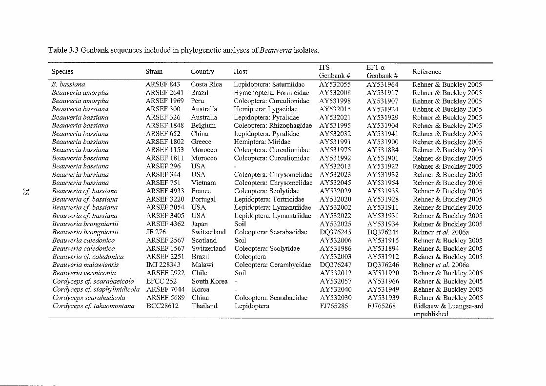
Table 3.3 Genbank sequences included in phylogenetic analyses of Beauveria isolates.
Species Strain Country Host ITS EFl-a
Reference Genbank# Genbank#
B. bassiana ARSEF 843 Costa Rica Lepidoptera: Saturniidae AY532055 AY531964 Rehner & Buckley 2005 Beauveria amorpha ARSEF 2641 Brazil Hymenoptera: Formicidae AY532008 AY531917 Rehner & Buckley 2005 Beauveria amorpha ARSEF 1969 Peru Coleoptera: Curculionidae AY531998 AY531907 Rehner & Buckley 2005 Beauveria bassiana ARSEF 300 Australia Hemiptera: Lygaeidae AY532015 AY531924 Rehner & Buckley 2005 Beauveria bassiana ARSEF 326 Australia Lepidoptera: Pyralidae AY532021 AY531929 Rehner & Buckley 2005 Beauveria bassiana ARSEF 1848 Belgium Coleoptera: Rhizophagidae AY531995 AY531904 Rehner & Buckley 2005 Beauveria bassiana ARSEF 652 China Lepidoptera: Pyralidae AY532032 AY531941 Rehner & Buckley 2005 Beauveria bassiana ARSEF 1802 Greece Hemiptera: Miridae AY531991 AY531900 Rehner & Buckley 2005 Beauveria bassiana ARSEF 1153 Morocco Coleoptera: Curculionidae AY531975 AY531884 Rehner & Buckley 2005 Beauveria bassiana ARSEF 1811 Morocco Coleoptera: Curculionidae AY531992 AY531901 Rehner & Buckley 2005 Beauveria bassiana ARSEF 296 USA AY532013 AY531922 Rehner & Buckley 2005 Beauveria bassiana ARSEF 344 USA Coleoptera: Chrysomelidae AY532023 AY531932 Rehner & Buddey 2005 Beauveria bassiana ARSEF 751 Vietnam Coleoptera: Chrysomelidae AY532045 AY531954 Rehner & Buckley 2005
\.)j Beauveria cf bassiana ARSEF 4933 France Coleoptera: Scolytidae AY532029 AY531938 Rehner & Buckley 2005 00
Beauveria cf bassiana ARSEF 3220 Portugal Lepidoptera: Tortricidae AY532020 AY531928 Rehner & Buckley 2005 Beauveria cf bassiana ARSEF 2054 USA Lepidoptera: Lymantriidae AY532002 AY531911 Rehner & Buckley 2005 Beauveria cf bassiana ARSEF 3405 USA Lepidoptera: Lymantriidae AY532022 AY531931 Rehner & Buckley 2005 Beauveria brongniartii ARSEF 4362 Japan Soil AY532025 AY531934 Rehner & Buckley 2005 Beauveria brongniartii JE276 Switzerland Coleoptera: Scarabaeidae DQ376245 DQ376244 Rehner et at. 2006a Beauveria caledonica ARSEF 2567 Scotland Soil AY532006 AY531915 Rehner & Buckley 2005 Beauveria caledonica ARSEF 1567 Switzerland Coleoptera: Scolytidae AY531986 AY531894 Rehner & Buckley 2005 Beauveria cf caledonica ARSEF 2251 Brazil Coleoptera AY532003 AY531912 Rehner & Buckley 2005 Beauveria malawiensis IMI228343 Malawi Coleoptera: Cerambycidae DQ376247 DQ376246 Rehner et at. 2006a Beauveria vermiconia ARSEF 2922 Chile Soil AY532012 AY531920 Rehner & Buckley 2005 Cordyceps cf scarabaeicola EFCC252 South Korea - AY532057 AY531966 Rehner & Buckley 2005 Cordyceps cf staphylinidicola ARSEF 7044 Korea AY532040 AY531949 Rehner & Buckley 2005 Cordyceps scarabaeicola ARSEF 5689 China Coleoptera: Scarabaeidae AY532030 AY531939 Rehner & Buckley 2005 Cordyceps cf takaomontana BCC28612 Thailand Lepidoptera FJ765285 FJ765268 Ridkaew & Luangsa-ard
unpublished
3.2.3 Insect bioassays
Tenebrio !nolitor
To examine host specificity in B. bassiana and B. malawiensis, strains isolated from various hosts
were tested in laboratory bioassays to determine their relative virulence towards a coleopteran
species. Strains isolated from Coleoptera, Hemiptera, and Hymenoptera were tested against larvae
of Tenebrio molitor (Coleoptera: Tenebrionidae). This species was used as a model organism
because large numbers of larvae were available from a commercial supplier (Biosuppliers Live
Insects, Auckland). Eight isolates from each of the host orders above were tested against T. molitor
in three separate sets ofbioassays, with each experiment repeated two times.
All isolates were cultured on PDA for 2-3 weeks at 25°C. Conidia were harvested from plates using
a sterile spatula and suspended in 5 ml of sterile aqueous 0.01 % Triton X-IOO. Conidial suspensions
were adjusted to 106 conidia/ml using a haemocytometer. Conidial viability of all isolates was
determined by spreading 100 ul of conidial suspension onto plates of PDA and incubating at 25°C
for 20 hours. The germination rate was assessed at 20 hours from counts of 300 conidia (Goettel &
Inglis 1997) observed from inverted plates at 100x magnification.
Each bioassay was conducted as a completely randomised design. Batches of six T. molitor larvae
were first sorted into separate 10 ml plastic containers. For each treatment, five replicate 90 mm
petri dishes were lined with filter paper and each inoculated with 1 ml of conidial suspension. One
ml of 0.01 % sterile Triton X-I 00 was added to the filter paper in each of two sets of five replicate
control plates. Each batch of six larvae was then randomly assigned to a treated petri dish. Plates
were placed in sealed polythene bags and incubated at room temperature (21°C) for 12 days. Larval
mortality was assessed and recorded every two days. To confirm infection, any dead larvae were
transferred to separate petri dishes lined with moist filter paper and incubated at room temperature
until sporulation was observed. Each experiment was repeated twice with freshly prepared conidial
suspensions and a new supply of T. molitor larvae. For each replicate, LTso values (time required to
kill half of the larvae) in days were recorded. If necessary, LT so times were estimated from linear
interpolation between the two assessment periods that straddled the value of '3 dead' (Glare et al.
2008). Cumulative mortality (arcsine-transformed) and LTso (log-transformed) data was analysed
by ANOV A. Means of LTso values were separated by Tukeys test to determine significant
differences between treatments. All statistical analyses were performed using SPSS version 11.00
(SPSS, Inc.).
39
Vespula vulgaris
To evaluate the potential of B. bassiana and B. malawiensis for wasp biocontrol, strains were tested
in laboratory bioassays to determine pathogenicity towards V. vulgaris larvae. Six strains originally
isolated from adult Vespula species were selected for testing. Conidial suspensions were prepared as
described above but were first diluted to a concentration of 108 conidia/ml. One millilitre of conidial
suspension was added to an equal volume of 0.1 % yeast extract to give a final concentration of 5 x
107 conidia conidia/ml. Conidial viability was determined as above.
Comb material was obtained from V. vulgaris nests collected from beech forests in the Nelson area.
Adults and pupae were removed and combs separated into sections (replicates) containing 20-25
final instar larvae and placed into separate plastic containers with lids. For each treatment three
replicates of 20-25 larvae were prepared. Each larva was inoculated on the head capsule with 5 f.ll
of conidial suspension. Larvae were incubated at 25°C and mortality was assessed every 2-3 days
for 10 days. Estimation of LT50 times and data analysis was performed as described for Tenebrio
molitor.
40
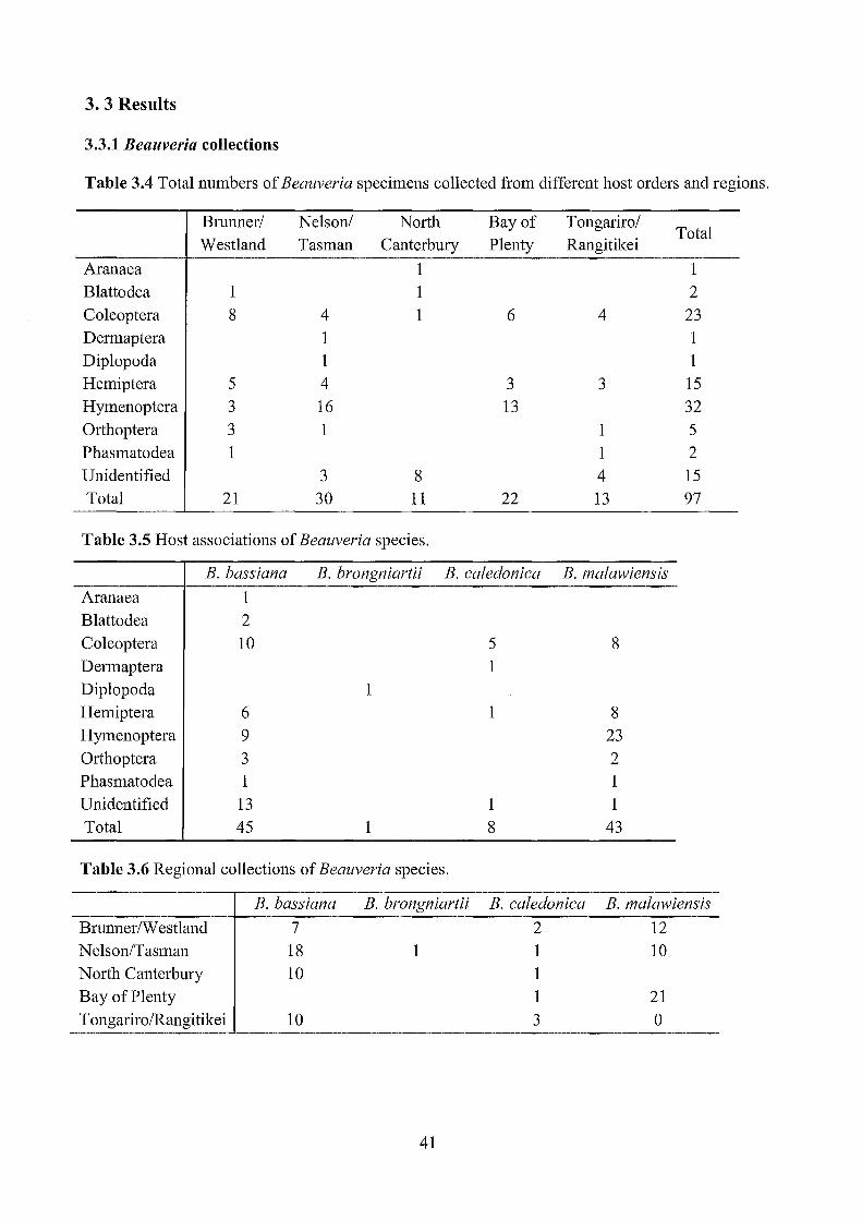
3.3 Results
3.3.1 Beauveria collections
Table 3.4 Total numbers of Beauveria specimens collected from different host orders and regions.
Brunnerl Nelson! North Bay of Tongarirol Total
Westland Tasman Canterbury Plenty Rangitikei
Aranaea 1 1 Blattodea 1 1 2 Coleoptera 8 4 1 6 4 23 Dermaptera 1 1 Diplopoda 1 1 Hemiptera 5 4 3 3 15 Hymenoptera 3 16 13 32 Orthoptera 3 1 1 5 Phasmatodea 1 1 2 Unidentified 3 8 4 15 Total 21 30 11 22 13 97
Table 3.5 Host associations of Beauveria species.
B. bassiana B. brongniartii B. caledonica B. malawiensis
Aranaea 1 Blattodea 2 Coleoptera 10 5 8 Dermaptera 1 Diplopoda 1 Hemiptera 6 1 8 Hymenoptera 9 23 Orthoptera 3 2 Phasmatodea 1 1 Unidentified 13 1 1 Total 45 1 8 43
Table 3.6 Regional collections of Beauveria species.
B. bassiana B. brongniartii B. caledonica B. malawiensis
Brunner/Westland 7 2 12 N elson/Tasman 18 1 1 10 North Canterbury 10 1 Bay of Plenty 1 21 Tongariro/Rangitikei 10 3 0
41
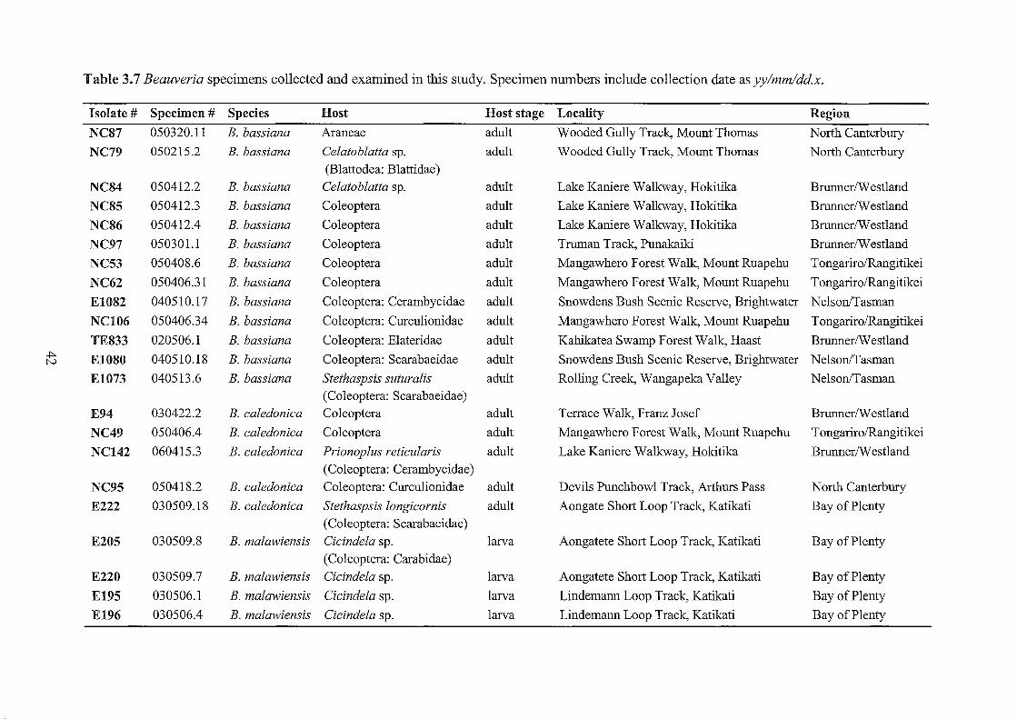
Table 3.7 Beauveria specimens collected and examined in this study. Specimen numbers include collection date as yy/mm/dd.x.
Isolate # Specimen # Species Host Host stage Locality Region
NC87 050320.11 B. bassiana Araneae adult Wooded Gully Track, Mount Thomas North Canterbury
NC79 050215.2 B. bassiana Celatoblatta sp. adult Wooded Gully Track, Mount Thomas North Canterbury (Blattodea: Blattidae)
NC84 050412.2 B. bassiana Celatoblatta sp. adult Lake Kaniere Walkway, Hokitika BrunnerlW estland
NC85 050412.3 B. bassiana Coleoptera adult Lake Kaniere Wallcway, Hokitika BrunnerlW est1and
NC86 050412.4 B. bassiana Coleoptera adult Lake Kaniere Walkway, Hokitika BrunnerlW estland
NC97 050301.1 B. bassiana Coleoptera adult Truman Track, Punakaiki BrunnerlW estland
NC53 050408.6 B. bassiana Coleoptera adult Mangawhero Forest Walk, Mount Ruapehu TongarirolRangitikei
NC62 050406.31 B. bassiana Coleoptera adult Mangawhero Forest Walle, Mount Ruapehu TongarirolRangitikei
EI082 040510.17 B. bassiana Coleoptera: Cerambycidae adult Snowdens Bush Scenic Reserve, Brightwater Nelson/Tasman
NCI06 050406.34 B. bassiana Coleoptera: Curcu1ionidae adult Mangawhero Forest Walk, Mount Ruapehu TongarirolRangitikei
TE833 020506.1 B. bassiana Coleoptera: Elateridae adult Kahikatea Swamp Forest Walk, Haast BrunnerlW estland .j:::..
EI080 040510.18 B. bassiana Coleoptera: Scarabaeidae adult Snowdens Bush Scenic Reserve, Brightwater Nelson/Tasman N
EI073 040513.6 B. bassiana Stethaspsis suturalis adult Rolling Creek, Wangapeka Valley Nelson/Tasman (Coleoptera: Scarabaeidae)
E94 030422.2 B. caledonica Coleoptera adult Terrace Walk, Franz Josef BrunnerlW estland
NC49 050406.4 B. caledonica Coleoptera adult Mangawhero Forest Walk, Mount Ruapehu TongarirolRangitikei
NC142 060415.3 B. caledonica Prionoplus reticularis adult Lake Kaniere Walkway, Hokitika BrunnerlW estland (Coleoptera: Cerambycidae)
NC95 050418.2 B. caledonica Coleoptera: Curculionidae adult Devils Punchbowl Track, Arthurs Pass North Canterbury
E222 030509.18 B. caledonica Stethaspsis longicornis adult Aongate Short Loop Track, Katikati Bay of Plenty (Coleoptera: Scarabaeidae)
E205 030509.8 B. malawiensis Cicindela sp. larva Aongatete Short Loop Track, Katikati Bay of Plenty (Coleoptera: Carabidae)
E220 030509.7 B. malawiensis Cicindela sp. larva Aongatete Short Loop Track, Katikati Bay of Plenty
E195 030506.1 B. malawiensis Cicindela sp. larva Lindemann Loop Track, Katikati Bay of Plenty
E196 030506.4 B. malawiensis Cicindela sp. larva Lindemann Loop Track, Katikati Bay of Plenty

Table 3.7 continued.
Isolate # Specimen # Species Host Host stage Locality Region
NC2I4 060511.3 B. malawiensis Coleoptera: Curculionidae adult Nile River Valley Walk, Charleston BrunnerIW estland
EI88 030506.12 B. malawiensis Pyronota festiva adult Aongatete Short Loop Track, Katikati Bay of Plenty
(Coleoptera: Scarabaeidae)
NC202 060508.3 B. malawiensis Coleoptera: Scarabaeidae adult Charming Creek Wallcway, Westport Nelson/Tasman
NCI88 060415.51 B. malawiensis Coleoptera: Scarabaeidae adult Lake Kaniere Wallcway, Hokitika BrunnerIW estland
EI079 040513.17 B. caledonica Dermaptera adult Rolling Creek, Wangapeka Valley Nelson/Tasman
NC225 060510.1 B. brongniartii Procyliosoma tuberculatum adult Oparara Arch Walk, Karamea Nelson/Tasman (Diplopoda: Sphaerotheriidae)
T875* n.a. B. bassiana Arachnocampa luminosa adult Waitomo Caves Waikato (Diptera: Keroplatidae)
NC123 050408.22 B. bassiana Hemiptera: Cicadidae adult Mangawhero Forest Walk, Mount Ruapehu TongarirolRangitikei
NC208 060509.6 B. bassiana Amphipsalta. zealandica adult Nikau Loop Walk, Karamea Nelson/Tasman
.j::.. (Hemiptera: Cicadidae) w
NC209 060509.7 B. bassiana A. zealandica adult Nikau Loop Walk, Karamea Nelson/Tasman
NC228 060509.1 B. bassiana A. zealandica adult Nikau Loop Walk, Karamea Nelson/Tasman
TE439 020417.6 B. bassiana A. zealandica adult Goldsborough (Shamrock)Track, Hokitika BrunnerIW estland
NCl1I 050404.11 B. bassiana Amphipsalta sp. adult Mangawhero Forest Walk, Mount Ruapehu TongarirolRangitikei
NC48 050408.2 B. caledonica Hemiptera: Cicadidae adult Mangawhero Forest Walk, Mount Ruapehu TongarirolRangitikei
NCI68 060415.27 B. malawiensis Hemiptera adult Lake Kaniere Walkway, Hokitika BrunnerIW estland
EI90 030509.34 B. malawiensis Hemiptera: Cicadidae adult Aongatete Short Loop Track, Katikati Bay of Plenty
E2I9 030507.1 B. malawiensis Hemiptera: Cicadidae adult Aongatete Short Loop Track, Katikati Bay of Plenty
E96 030422.6 B. malawiensis Hemiptera: Cicadidae adult Terrace Walk, Franz Josef BrunnerIW estland
TE672 020507.2 B. malawiensis Hemiptera: Cicadidae adult Hapuka Estuary Walk, Okuru BrunnerIW estland
NC205 060508.1 B. malawiensis A. zealandica adult Nikau Loop Walk, Karamea Nelson/Tasman
NC78 050315.16 B. malawiensis A. zealandica adult Truman Track, Punakaiki BrunnerIW estland
E221 030509.15 B. malawiensis Monteithiella humeralis adult Aongatete Short Loop Track, Katikati Bay of Plenty
(Hemiptera: Pentatomidae)

Table 3.7 continued.
Isolate # Specimen # Species Host Host stage Locality Region
El063 040513.3 B. bassiana Vespula germanica adult Rolling Creek, Wangapeka Valley Nelson/Tasman (Hymenoptera: Vespidae)
El069 040511.12 B. bassiana V. germanica adult Eves Valley Scenic Reserve, Brightwater Nelson/Tasman
El068 040511.27 B. bassiana V. vulgaris adult Eves Valley Scenic Reserve, Brightwater Nelson/Tasman
El070 040511.17 B. bassiana V. vulgaris adult Eves Valley Scenic Reserve, Brightwater Nelson/Tasman
E1175 040511.24 B. bassiana V. vulgaris adult Eves Valley Scenic Reserve, Brightwater Nelson/Tasman
El057 040514.9 B. bassiana Vespula vulgaris adult Loop Track, Lake Rotoiti Nelson/Tasman
El064 040514.6 B. bassiana Vespula sp. adult Loop Track, Lake Rotoiti Nelson/Tasman
El065 040510.10 B. bassiana Vespula sp. adult Snowdens Bush Scenic Reserve, Brightwater Nelson/Tasman
El067 040513.15 B. bassiana Vespula sp. adult Rolling Creek, Wangapeka Valley Nelson/Tasman
T884 000408.1 B. malawiensis Certonotus jractinervis adult Mount French Track, Lake Brunner BrunnerlW estland (Hymenoptera: Ichneumonidae)
..j:::.. E197 030506.13 B. malawiensis V. vulgaris adult Aongatete Short Loop Track, Katikati Bay of Plenty
..j:::..
E20l 030509.4 B. malawiensis V. vulgaris adult Aongatete Short Loop Track, Katikati Bay of Plenty
E206 030509.12 B. malawiensis V. vulgaris adult Aongatete Short Loop Track, Katikati Bay of Plenty
E207 030509.14 B. malawiensis V. vulgaris adult Aongatete Short Loop Track, Katikati Bay of Plenty
E208 030509.16 B. malawiensis V. vulgaris adult Aongatete Short Loop Track, Katikati Bay of Plenty
E2l0 030509.26 B. malawiensis V. vulgaris adult Aongatete Short Loop Track, Katikati Bay of Plenty
E2l5 030509.35 B. malawiensis V. vulgaris adult Aongatete Short Loop Track, Katikati Bay of Plenty
El059 040511.18 B. malawiensis V. vulgaris adult Eves Valley Scenic Reserve, Brightwater Nelson/Tasman
El060 040511.20 B. malawiensis V. vulgaris adult Eves Valley Scenic Reserve, Brightwater NelsonlTasman
El066 040511.19 B. malawiensis V. vulgaris adult Eves Valley Scenic Reserve, Brightwater Nelson/Tasman
El084 040510.10 B. malawiensis V. vulgaris adult Snowdens Bush Scenic Reserve, Brightwater Nelson/Tasman
E1176 040511.38 B. malawiensis V. vulgaris adult Eves Valley Scenic Reserve, Brightwater Nelson/Tasman
NC2l0 060509.8 B. malawiensis V. vulgaris adult Nikau Loop Walk, Karamea Nelson/Tasman
NC2l5 060511.4 B. malawiensis V. vulgaris adult Nile River Valley Walk, Charleston BrunnerlW estland

Table 3.7 continued.
Isolate # Specimen # Species Host Host stage Locality Region
E202 030509.5 B. malawiensis Vespula sp. adult Aongatete Short Loop Track, Katikati Bay of Plenty
E203 030509.6 B. malawiensis Vespula sp. adult Aongatete Short Loop Track, Katikati Bay of Plenty
E211 030509.27 B. malawiensis Vespula sp. adult Aongatete Short Loop Track, Katikati Bay of Plenty
E213 030509.29 B. malawiensis Vespula sp. adult Aongatete Short Loop Track, Katikati Bay of Plenty
E214 030509.30 B. malawiensis Vespula sp. adult Aongatete Short Loop Track, Katikati Bay of Plenty
E216 030509.2 B. malawiensis Vespula sp. adult Aongatete Short Loop Track, Katikati Bay of Plenty
T885 000408.5 B. malawiensis Vespula sp. adult Mount French Track, Lake Brunner Nelson/Tasman
NC222 060512.5 B. malawiensis Vespula sp. adult Oparara Arch Walk, Karamea BrunnerIW estland
NCI07 050408.11 B. bassiana Orthoptera adult Mangawhero Forest Walk, Mount Ruapehu TongarirolRangitikei
E1139 040510.1 B. bassiana Phaulacridium marginale adult Snowdens Bush Scenic Reserve, Brightwater Nelson/Tasman (Orthoptera: Acrididae)
NC219 0605012.1 B. bassiana Orthoptera: Tettigoniidae adult Truman Track, Punakaiki BrunnerIW estland ~ NC83 050412.1 B. malawiensis P. marginale adult Lake Kaniere Walkway, Hokitika BrunnerIW estland VI
NC220 060512.2 B. malawiensis Orthoptera: Tettigoniidae adult Truman Track, Punakaiki BrunnerIW estland
NC43 050405.8 B. bassiana Phasmatodea: Phasmatidae adult Mangawhero Forest Walk, Mount Ruapehu TongarirolRangitikei
TE445 n.a. B. malawiensis Clitarchus hookeri adult Hapuka Estuary Walk, Okuru BrunnerIW estland (phasmatodea: Phasmatidae)
NC96 050510.8 B. bassiana n.d adult Devils Punchbowl Track, Arthurs Pass North Canterbury
NC99 050418.1 B. bassiana n.d. adult Devils Punchbowl Track, Arthurs Pass North Canterbury
NCI00 050510.1 B. bassiana n.d. adult Devils Punchbowl Track, Arthurs Pass North Canterbury
NCI0l 050418.5 B. bassiana n.d adult Devils Punchbowl Track, Arthurs Pass North Canterbury
NCI04 050418.6 B. bassiana n.d. adult Devils Punchbowl Track, Arthurs Pass North Canterbury
NC82 050320.2 B. bassiana n.d. adult Wooded Gully Track, Mount Thomas North Canterbury
NC88 050320.10 B. bassiana n.d. adult Wooded Gully Track, Mount Thomas North Canterbury
NC92 050320.14 B. bassiana n.d. adult Wooded Gully Track, Mount Thomas North Canterbury
EI083 040510.15 B. bassiana n.d. adult Snowdens Bush Scenic Reserve, Brightwater Nelson/Tasman

../:::>. 0\
Table 3.7 continued.
Isolate # Specimen # Species Host Host stage Locality
E1174 040511.22 B. bassiana n.d. adult Eves Valley Scenic Reserve, Brightwater
NC41 050408.1 B. bassiana n.d. adult Mangawhero Forest Walk, Mount Ruapehu
NCll0 050408.5 B. bassiana n.d adult Mangawhero Forest Walk, Mount Ruapehu
NC119 050408.4 B. bassiana n.d. adult Mangawhero Forest Walle, Mount Ruapehu
NC44 050408.12 B. caledonica n.d. adult Mangawhero Forest Walk, Mount Ruapehu
EI077 040511.15 B. malawiensis n.d. adult Eves Valley Scenic Reserve, Brightwater
n.a., not available; n.d., not determined; * culture provided by N. Waipara, AgResearch, Ruakura, February 2000 .
Region
Nelson/Tasman
Tongariro/Rangitikei
TongarirolRangitikei
TongarirolRangitikei
TongarirolRangitikei
Nelson/Tasman
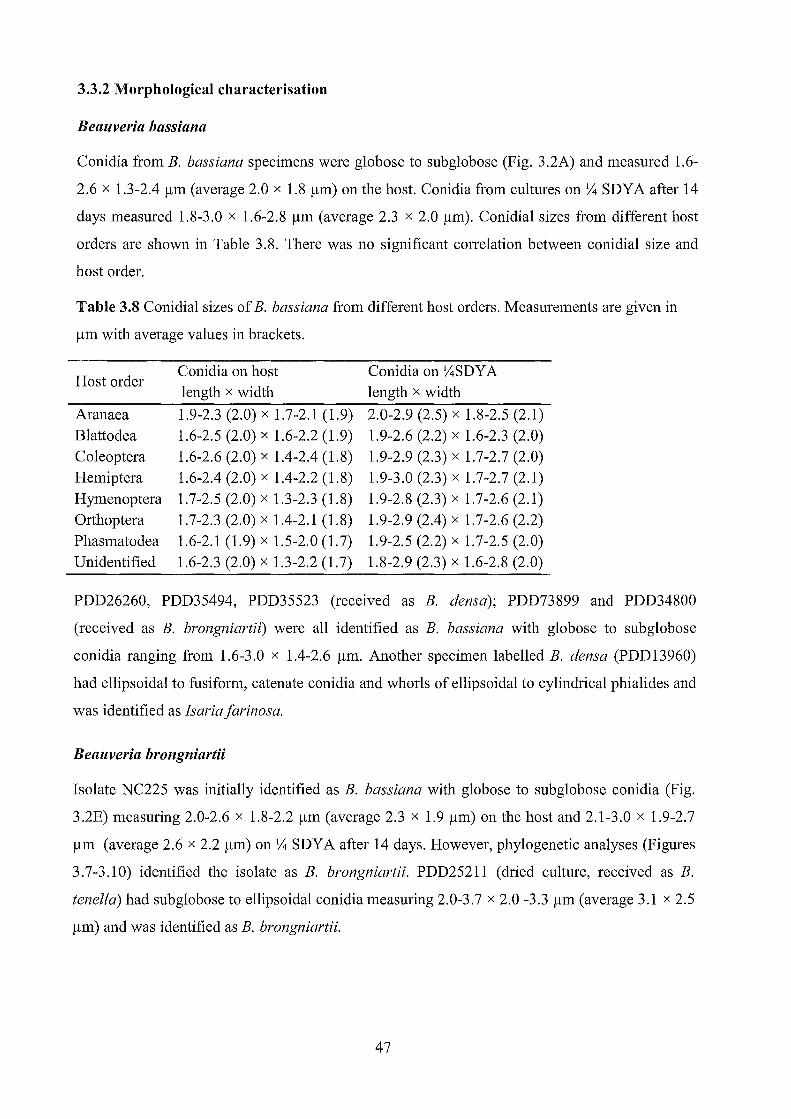
3.3.2 Morphological characterisation
Beauveria bassiana
Conidia from B. bassiana specimens were globose to subglobose (Fig. 3.2A) and measured 1.6-
2.6 x 1.3-2.4 !lm (average 2.0 x 1.8 !lm) on the host. Conidia from cultures on Y4 SDY A after 14
days measured 1.8-3.0 x 1.6-2.8 !lm (average 2.3 x 2.0 !lm). Conidial sizes from different host
orders are shown in Table 3.8. There was no significant correlation between conidial size and
host order.
Table 3.8 Conidial sizes of B. bassiana from different host orders. Measurements are given in
!lm with average values in brackets.
Host order
Aranaea Blattodea Coleoptera Hemiptera Hymenoptera Orthoptera Phasmatodea Unidentified
Conidia on host length x width
1.9-2.3 (2.0) x 1.7-2.1 (1.9) 1.6-2.5 (2.0) x 1.6-2.2 (1.9) 1.6-2.6 (2.0) x 1.4-2.4 (1.8) 1.6-2.4 (2.0) x 1.4-2.2 (1.8) 1.7-2.5 (2.0) x 1.3-2.3 (1.8) 1.7-2.3 (2.0) x 1.4-2.1 (1.8) 1.6-2.1 (1.9) x 1.5-2.0 (1.7) 1.6-2.3 (2.0) x 1.3-2.2 (1.7)
Conidia on Y4SDY A length x width
2.0-2.9 (2.5) x 1.8-2.5 (2.1) 1.9-2.6 (2.2) x 1.6-2.3 (2.0) 1.9-2.9 (2.3) x 1.7-2.7 (2.0) 1.9-3.0 (2.3) x 1.7-2.7 (2.1) 1.9-2.8 (2.3) x 1.7-2.6 (2.1) 1.9-2.9 (2.4) x 1.7-2.6 (2.2) 1.9-2.5 (2.2) x 1.7-2.5 (2.0) 1.8-2.9 (2.3) x 1.6-2.8 (2.0)
PDD26260, PDD35494, PDD35523 (received as B. densa); PDD73899 and PDD34800
(received as B. brongniartii) were all identified as B. bassiana with globose to subglobose
conidia ranging from 1.6-3.0 x 1.4-2.6 !lm. Another specimen labelled B. dens a (PDD13960)
had ellipsoidal to fusiform, catenate conidia and whorls of ellipsoidal to cylindrical phialides and
was identified as Isaria farinosa.
Beauveria brongniartii
Isolate NC225 was initially identified as B. bassiana with globose to sub globose conidia (Fig.
3.2E) measuring 2.0-2.6 x 1.8-2.2 !lm (average 2.3 x 1.9 !lm) on the host and 2.1-3.0 x 1.9-2.7
/lm (average 2.6 x 2.2 /lm) on Y4 SDYA after 14 days. However, phylogenetic analyses (Figures
3.7-3.10) identified the isolate as B. brongniartii. PDD25211 (dried culture, received as B.
tenella) had subglobose to ellipsoidal conidia measuring 2.0-3.7 x 2.0 -3.3 !lm (average 3.1 x 2.5
/lm) and was identified as B. brongniartii.
47
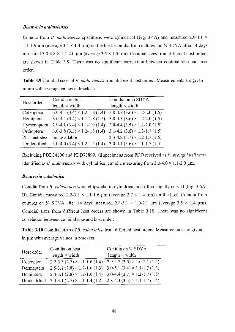
Beallveria malawiensis
Conidia from B. malawiensis specimens were cylindrical (Fig. 3.4A) and measured 2.9-4.1 x
1.1-1.9 /lm (average 3.4 x 1.4 /lm) on the host. Conidia from cultures on V4 SDY A after 14 days
measured 3.0-4.8 x 1.1-2.0 /lm (average 3.5 x 1.5 /lm). Conidial sizes from different host orders
are shown in Table 3.9. There was no significant correlation between conidial size and host
order.
Table 3.9 Conidial sizes of B. malawiensis from different host orders. Measurements are given
in /lm with average values in brackets.
Host order
Coleoptera Hemiptera Hymenoptera Orthoptera Phasmatodea Unidentified
Conidia on host length x width
3.0-4.1 (3.4) x 1.2-1.8 (1.4) 3.0-4.1 (3.4) x 1.1-1.8 (1.5) 2.9-4.1 (3.4) x 1.1-1.9 (1.4) 3.0-3.8 (3.3) x 1.2-1.8 (1.4) not available 3.0-4.0 (3.4) x 1.2-1.5 (1.4)
Conidia on V4 SDY A length x width
3.0-4.8 (3.6) x 1.2-2.0 (1.5) 3.0-4.3 (3.6) x 1.2-2.0 (1.5) 3.0-4.4 (3.5) x 1.2-2.0 (1.5) 3.1-4.2 (3.6) x 1.3-1.7 (1.5) 3.3-4.2 (3.7) x 1.2-1.7 (1.5) 3.0-4.1 (3.6) x 1.1-1.7 (1.4)
Excluding PDD34800 and PDD73899, all specimens from PDD received as B. brongniartii were
identified as B. malawiensis with cylindrical conidia measuring from 3.0-4.0 x 1.1-2.0 /lm.
Beallveria caledonica
Conidia from B. caledonica were ellipsoidal to cylindrical and often slightly curved (Fig. 3.6A
B). Conidia measured 2.2-3.3 x 1.1-1.6 /lm (average 2.7 x 1.4 /lm) on the host. Conidia from
cultures on V4 SDY A after 14 days measured 2.8-5.1 x 1.0-2.1 /lm (average 3.5 x 1.4 /lm).
Conidial sizes from different host orders are shown in Table 3.10. There was no significant
correlation between conidial size and host order.
Table 3.10 Conidial sizes of B. caledonica from different host orders. Measurements are given
in /lm with average values in brackets.
Host order
Coleoptera Dermaptera Hemiptera Unidentified
Conidia on host length x width
2.2-3.3 (2.7) x 1.1-1.6 (1.4) 2.5-3.1 (2.8) x 1.2-1.6 (1.3) 2.4-3.1 (2.8) x 1.2-1.6 (1.4) 2.4-3.1 (2.7) x 1.1-1.4 (1.2)
Conidia on V4 SDY A length x width
2.9-4.7 (3.5) x 1.0-2.1 (1.4) 3.0-5.1 (3.4) x 1.1-1.7 (1.3) 3.0-4.4 (3.7) x 1.2-1.7 (1.5) 2.8-4.3 (3.3) x 1.1-1.7 (1.4)
48

Table 3.11 Measurements of conidia from Beauveria specimens and cultures. All measurements are in /lm with averages (n=25) in brackets.
Isolate # Specimen # Species Host Region Conidia on 1;4 SDY A Conidia on host length x width length x width
NC87 050320.11 B. bassiana Araneae North Canterbury 2.0-2.9 (2.5) x 1.8-2.5 (2.1) 1.9-2.3 (2.0) x 1.7-2.1 (1.9) NC79 050215.2 B. bassiana Blattodea North Canterbury 1.9-2.4 (2.1) x 1.6-2.2 (1.9) 1.6-2.2 (2.0) x 1.6-2.1 (1.8) NC84 050412.2 B. bassiana Blattodea BrunnerlW estland 1.9-2.6 (2.2) x 1.6-2.3 (2.0) 1.8-2.5 (2.1) x 1.6-2.2 (1.9) NC85 050412.3 B. bassiana Coleoptera BrunnerlW estland 2.0-2.7 (2.3) x 1.8-2.4 (2.0) 1.7-2.2 (2.0) x 1.5-2.2 (1.8) NC86 050412.4 B. bassiana Coleoptera BrunnerlW estland 2.0-2.5 (2.3) x 1.8-2.4 (2.0) 1.9-2.3 (2.1) x 1.7-2.2 (2.0) NC97 050301.1 B. bassiana Coleoptera BrunnerlW estland 2.0-2.6 (2.2) x 1.7-2.4 (2.0) 1.8-2.2 (1.9) x 1.5-2.1 (1.8) NC53 050408.6 B. bassiana Coleoptera TongarirolRangitikei 2.0-2.5 (2.2) x 1.7-2.3 (2.0) 1.6-2.0 (1.8) x 1.4-1.9(1.6) NC62 050406.31 B. bassiana Coleoptera TongarirolRangitikei 1.9-2.6 (2.2) x 1.8-2.3 (2.0) 1.7-2.0 (1.8) x 1.5-1.8 (1.6)
E1082 040510.17 B. bassiana Coleoptera Nelson/Tasman 1.9-2.4 (2.2) x 1.7-2.2 (1.9) 1.9-2.3 (2.1) x 1.6-2.1 (1.8) NC106 050406.34 B. bassiana Coleoptera TongarirolRangitikei 2.0-2.9 (2.5) x 1.8-2.5 (2.1) 1. 7 -2.3 (1.9) x 1.4-1.9 (1.6)
~ TE833 020506.1 B. bassiana Coleoptera BrunnerlW estland 2.2-2.8 (2.4) x 1.8-2.7 (2.1) 2.0-2.6 (2.2) x 1.8-2.4 (2.0)
\0 E1080 040510.18 B. bassiana Coleoptera Nelson/Tasman 2.1-2.9 (2.3) x 1.8-2.3 (2.0) 2.0-2.5 (2.3) x 1.8-2.2 (2.0) E1073 040513.6 B. bassiana Coleoptera Nelson/Tasman 1.9-2.6 (2.2) x 1.7-2.4 (1.9) 1.9-2.5 (2.2) x 1.8-2.3 (2.1) E94 030422.2 B. caledonica Coleoptera BrunnerlW estland 3.0-4.7 (3.8) x 1.2-2.1 (1.7) 2.5-3.3 (2.8) x 1.2-1.6 (1.4) NC49 050406.4 B. caledonica Coleoptera TongarirolRangitikei 2.9-4.4 (3.5) x 1.0-1.8 (1.3) 2.3-3.1 (2.7) x 1.1-1.6 (1.4) NC142 060415.3 B. caledonica Coleoptera BrunnerlW estland 2.9-4.3 (3.3) x 1.1-1.6 (1.4) 2.2.2-9 (2.5) x 1.1-1.6 (1.4) NC95 050418.2 B. caledonica Coleoptera North Canterbury 2.9-4.5 (3.4) x 1.2-1.6 (1.3) 2.3-3.1 (2.7) x 1.1-1.5 (1.3) E222 030509.18 B. caledonica Coleoptera Bay of Plenty 2.9-4.3 (3.3) x 1.2-1.9 (1.5) 2.4-3.1 (2.7) x 1.2-1.6 (1.4) E205 030509.8 B. malawiensis Coleoptera Bay of Plenty 3.1-4.1 (3.6) x 1.3-2.0 (1.6) 3.0-4.1 (3.5) x 1.2-1.8 (1.4) E220 030509.7 B. malawiensis Coleoptera Bay of Plenty 3.1-3.9 (3.5) x 1.3-2.0 (1.6) 3.0-3.8 (3.4) x 1.3-1.6 (1.5) E195 030506.1 B. malawiensis Coleoptera Bay of Plenty 3.1-4.0 (3.5) x 1.2-1.8 (1.5) 3.2-4.0 (3.5) x 1.2-1.6 (1.4) E196 030506.4 B. malawiensis Coleoptera Bay of Plenty 3.0-4.0 (3.5) x 1.2-1.7 (1.3) 3.0-3.7 (3.3) x 1.2-1.8 (1.5) NC214 060511.3 B. malawiensis Coleoptera BrunnerlW estland 3.4-4.8 (4.0) x 1.3-1.8 (1.5) 3.0-3.8 (3.4) x 1.2-1.6 (1.5) E188 030506.12 B. malawiensis Coleoptera Bay of Plenty 3.1-4.2 (3.6) x 1.3-1.8 (1.6) 3.0-4.0 (3.4) x 1.3-1.6 (1.5)
NC202 060508.3 B. malawiensis Coleoptera Nelson/Tasman 3.2-3.9 (3.5) x 1.3-1.7 (1.4) 3.3-3.8 (3.6) x 1.2-1.6 (1.3)

Table 3.11 continued.
Isolate # Specimen # Species Host Region Conidia on 1f4 SDY A Conidia on host length x width length x width
NCl88 060415.51 B. malawiensis Coleoptera BrunnerlW estland 3.1-4.0 (3.6) x 1.2-1.9 (1.4) 3.0-3.8 (3.3) x 1.2-1.6 (1.3) EI079 040513.17 B. caledonica Dermaptera Nelson/Tasman 3.0-5.1 (3.4) x 1.1-1.7 (1.3) 2.5-3.1 (2.8) x 1.2-1.6 (1.3) NC225 060510.1 B. bassiana Diplopoda Nelson/Tasman 2.1-2.8 (2.4) x 1.8-2.2 (2.0) 2.0-2.6 (2.3) x 1.8-2.2 (1.9) T875 n.a. B. bassiana Diptera Waikato 2.1-2.5 (2.3) x 1.9-2.4 (2.1) n.a. NC123 050408.22 B. bassiana Hemiptera TongarirolRangitikei 2.1-3.0 (2.6) x 1.9-2.7 (2.2) 1.7-2.4 (2.1) x 1.6-2.2 (1.9) NC208 060509.6 B. bassiana Hemiptera Nelson/Tasman 2.0-2.5 (2.3) x 1.8-2.3 (2.1) 1.8-2.3 (2.0) x 1.5-2.1 (1.8)
NC209 060509.7 B. bassiana Hemiptera Nelson/Tasman 1.9-2.6 (2.2) x 1.9-2.3 (2.1) 1.7-2.1 (1.9) x 1.4-1.9 (1.7) NC228 060509.1 B. bassiana Hemiptera Nelson/Tasman 1.9-2.4 (2.2) x 1.7-2.2 (2.0) 1.8-2.3 (2.1) x 1.7-2.1 (1.9) TE439 020417.6 B. bassiana Hemiptera BrunnerlW estland 1.9-2.4 (2.2) x 1.8-2.1 (1.9) 1.9-2.4 (2.2) x 1.8-2.1 (2.0) NClll 050404.11 B. bassiana Hemiptera TongarirolRangitikei 2.0-2.7 (2.3) x 1.8-2.5 (2.1) 1.6-2.0 (1.8) x 1.4-2.0 (1.7)
VI NC48 050408.2 B. caledonica Hemiptera TongarirolRangitikei 3.0-4.4 (3.7) x 1.2-1.7 (1.5) 2.4-3.1 (2.8) x 1.2-1.6 (1.4) 0
NC168 060415.27 B. malawiensis Hemiptera BrunnerlW estland 3.2-4.2 (3.6) x 1.3-1.9 (1.5) 3.0-4.0 (3.4) x 1.2-1.6 (1.4) E190 030509.34 B. malawiensis Hemiptera Bay of Plenty 3.0-3.9 (3.4) x 1.2-1.8 (1.5) 3.0-3.8 (3.3) x 1.2-1.5 (1.3) E219 030507.1 B. malawiensis Hemiptera Bay of Plenty 3.2-4.2 (3.6) x 1.3-1.9 (1.5) 3.0-3.7 (3.3) x 1.3-1.6 (1.5) E96 030422.6 B. malawiensis Hemiptera BrunnerlW estland 3.0-3.8 (3.4) x 1.2-1.7 (1.5) 3.1-3.7 (3.4) x 1.1-1.7 (1.4) TE672 020507.2 B. malawiensis Hemiptera BrunnerlW estland 3.2-4.0 (3.6) x 1.2-1.8 (1.5) 3.0-3.6 (3.4) x 1.4-1.8 (1.6)
NC205 060508.1 B. malawiensis Hemiptera Nelson/Tasman 3.3-4.3 (3.6) x 1.2-1.7 (1.4) 3.0-3.9 (3.5) x 1.2-1.8 (1.5) NC78 050315.16 B. malawiensis Hemiptera BrunnerlW estland 3.2-4.2 (3.7) x 1.3-1.6 (1.4) 3.0-4.1 (3.5) x 1.2-1.7 (1.4) E221 030509.15 B. malawiensis Hemiptera Bay of Plenty 3.2-4.2 (3.7) x 1.5-2.0 (1.7) 3.0-3.8 (3.4) x 1.2-1.6 (1.4) E1063 040513.3 B. bassiana Hymenoptera Nelson/Tasman 2.1-2.7 (2.5) x 1.9-2.6 (2.2) 1.9-2.5 (2.1) x 1.7-2.0 (1.9) EI069 040511.12 B. bassiana Hymenoptera Nelson/Tasman 2.1-2.7 (2.4) x 1.8-2.6 (2.1) 1.9-2.3 (2.1) x 1.6-2.3 (1.8) EI068 040511.27 B. bassiana Hymenoptera Nelson/Tasman 2.1-2.8 (2.4) x 1.8-2.6 (2.2) 1.7-2.3 (2.1) x 1.6-2.2 (1.9) EI070 040511.17 B. bassiana Hymenoptera Nelson/Tasman 2.0-2.4 (2.2) x 1.7-2.3 (1.9) 1. 7 -2.0 (1.8) x 1.3-1.8 (1.6) E1l75 040511.24 B. bassiana Hymenoptera Nelson/Tasman 1.9-2.3 (2.1) x 1.8-2.2 (1.9) 1.9-2.3 (2.1) x 1.7-2.2 (2.0) EI057 040514.9 B. bassiana Hymenoptera Nelson/Tasman 2.0-2.6 (2.3) x 1.7-2.6 (2.1) 1.7-2.2 (1.9) x 1.5-2.0 (1.7)

Table 3.11 continued.
Isolate # Specimen # Species Host Region Conidia on y,. SDY A Conidia on host length x width length x width
E1064 040514.6 B. bassiana Hymenoptera Nelson/Tasman 2.0-2.8 (2.3) x 1.7-2.6 (2.0) 1.9-2.5 (2.1) x 1.7-2.2 (1.9) E1065 040510.10 B. bassiana Hymenoptera Nelson/Tasman 2.0-2.8 (2.3) x 1.7-2.5 (2.0) 1.7-2.4 (2.0) x 1.5-2.2 (1.8) E1067 040513.15 B. bassiana Hymenoptera Nelson/Tasman 2.2-2.8 (2.4) x 1.7-2.6 (2.1) 1.7-2.3 (2.0) x 1.5-2.2 (1.8) T884 000408.1 B. malawiensis Hymenoptera BrunnerlWestland 3.1-4.2 (3.7) x 1.3-1.9 (1.5) 3.0-3.7 (3.4) x 1.3-1.6 (1.5) E197 030506.13 B. malawiensis Hymenoptera Bay of Plenty 3.2-4.0 (3.6) x 1.2-1.6 (1.4) 2.9-3.5 (3.2) x 1.1-1.5 (1.3) E201 030509.4 B. malawiensis Hymenoptera Bay of Plenty 3.2-4.2 (3.5) x 1.3-1.8 (1.5) 2.9-3.5 (3.2) x 1.3-1.6 (1.4) E206 030509.12 B. malawiensis Hymenoptera Bay of Plenty 3.0-3.8 (3.3) x 1.2-1.8 (1.5) 3.0-3.7 (3.3) x 1.2-1.6 (1.3) E207 030509.14 B. malawiensis Hymenoptera Bay of Plenty 3.0-3.9 (3.4) x 1.3-1.8 (1.4) 3.0-3.8 (3.4) x 1.2-1.6 (1.4)
E208 030509.16 B. malawiensis Hymenoptera Bay of Plenty 3.2-4.1 (3.7) x 1.2-1.9 (1.6) 3.0-4.0 (3.5) x 1.2-1.6 (1.4) E210 030509.26 B. malawiensis Hymenoptera Bay of Plenty 3.0-3.6 (3.2) x 1 .2-1.8 (1.4) 3.0-3.6 (3.3) x 1.2-1.6 (1.4)
Vl E215 030509.35 B. malawiensis Hymenoptera Bay of Plenty 3.1-4.3 (3.3) x 1.2-1.7 (1.4) 2.9-3.7 (3.4) x 1.2-1.8 (1.5) ...... E1059 040511.18 B. malawiensis Hymenoptera Nelson/Tasman 3.1-4.4 (3.6) x 1.3-2.0 (1.5) 3.0-3.8 (3.3) x 1.1-1.6 (1.4) E1060 040511.20 B. malawiensis Hymenoptera Nelson/Tasman 3.0-4.3 (3.5) x 1.3-1.8 (1.5) 3.1-3.9 (3.5) x 1.2-1.7 (1.4) E1066 040511.19 B. malawiensis Hymenoptera Nelson/Tasman 3.2-4.2 (3.7) x 1.2-1.8 (1.5) 3.0-3.8 (3.4) x 1.1-1.5 (1.3) E1084 040510.10 B. malawiensis Hymenoptera Nelson/Tasman 3.0-4.0 (3.3) x 1.1-1.7 (1.4) 2.9-3.4 (3.1) x 1.2-1.6 (1.4) E1176 040511.38 B. malawiensis Hymenoptera Nelson/Tasman 3.1-4.2 (3.6) x 1.3-1.8 (1.6) 3.1-3.9 (3.5) x 1.2-1.7 (1.4) NC210 060509.8 B. malawiensis Hymenoptera Nelson/Tasman 3.2-4.2 (3.5) x 1.2-1.7 (1.4) 2.9-3.7(3.2) x 1.3-1.7 (1.5) NC215 060511.4 B. malawiensis Hymenoptera BrunnerlWestland 3.1-4.3 (3.6) x 1.2-1.7 (1.4) 3.0-3.7 (3.4) x 1.3-1.8 (1.5) E202 030509.5 B. malawiensis Hymenoptera Bay of Plenty 3.0-4.1 (3.5) x 1.2-1.7 (1.4) 3.1-3.7 (3.3) x 1.2-1.5 (1.3) E203 030509.6 B. malawiensis Hymenoptera Bay of Plenty 3.1-4.0 (3.5) x 1.2-1.7 (1.5) 3.1-4.0 (3.6) x 1.2-1.6 (1.3) E211 030509.27 B. malawiensis Hymenoptera Bay of Plenty 3.0-4.1 (3.4) x 1.2-1.6 (1.4) 3.0-3.6 (3.4) x 1.2-1.6 (1.4) E213 030509.29 B. malawiensis Hymenoptera Bay of Plenty 3.2-4.0 (3.5) x 1.2-1.7 (1.5) 3.0-3.5 (3.2) x 1.2-1.7 (1.5) E214 030509.30 B. malawiensis Hymenoptera Bay of Plenty 3.0-4.0 (3.5) x 1.2-1.8 (1.4) 2.9-3.4 (3.2) x 1.1-1.6 (1.3) E216 030509.2 B. malawiensis Hymenoptera Bay of Plenty 3.1-4.2 (3.5) x 1.3-1.8 (1.6) 2.9-3.8 (3.3) x 1.1-1.8 (1.4) T885 000408.5 B. malawiensis Hymenoptera BrunnerIW estland 3.1-4.1 (3.6) x 1.2-1.7 (1.4) 3.1-4.1 (3.6) x 1.3-1.7 (1.6)

Table 3.11 continued.
Isolate # Specimen # Species Host Region Conidia on ~ SDY A Conidia on host length x width length x width
NC222 060512.5 B. malawiensis Hymenoptera Nelson/Tasman 3.1-4.3 (3.7) x 1.3-2.0 (1.7) 3.2-4.0 (3.6) x 1.2 1.7 (1.5) NCl07 050408.11 B. bassiana Orthoptera Tongariro/Rangitikei 1.9-2.6 (2.3) x 1.7-2.2 (2.0) 1.7-2.1 (1.9) x 1.4-1.9 (1.6) Ell39 040510.1 B. bassiana Orthoptera Nelson/Tasman 1.9-2.8 (2.3) x 1.7-2.4 (2.1) 1.8-2.3 (2.1) x 1.6-2.1 (1.9) NC219 0605012.1 B. bassiana Orthoptera BrunnerIW estland 2.2-2.9 (2.6) x 2.0-2.6 (2.4) 1.7-2.3 (2.1) x 1.5-1.9 (1.8) NC83 050412.1 B. malawiensis Orthoptera BrunnerIW estland 3.1-4.2 (3.5) x 1.3-1.7 (1.5) 3.0-3.8 (3.2) x 1.2-1.5 (1.4) NC220 060512.2 B. malawiensis Orthoptera BrunnerIW estland 3.3-4.2 (3.7) x 1.3-1.7 (1.5) 3.0-3.7 (3.3) x 1.2-1.8 (1.4) NC43 050405.8 B. bassiana Phasmatodea TongarirolRangitikei 1.9-2.5 (2.2) x 1.7-2.5 (2.0) 1.6-2.1 (1.9) x 1.5-2.0 (1.7) TE445 n.a. B. malawiensis Phasmatodea BrunnerIW estland 3.3-4.2 (3.7) x 1.2-1.7 (1.5) n.a. NC96 050510.8 B. bassiana n.d North Canterbury 1.9-2.7 (2.2) x 1.7-2.5 (2.0) 1.9-2.3 (2.1) x 1.7-2.2 (1.9) NC99 050418.1 B. bassiana n.d. North Canterbury 2.0-2.6 (2.4) x 1.8-2.3 (2.1) 1. 7 -2.0 (1.9) x 1.4-1.8 (1.5)
Ul NCIOO 050510.1 B. bassiana n.d. North Canterbury 1.9-2.6 (2.2) x 1.7-2.6 (2.0) 1.8-2.2 (2.1) x 1.6-2.2 (1.9) tv
NC101 050418.5 B. bassiana n.d North Canterbury 2.1-2.9 (2.5) x 1.8-2.8 (2.2) 1.6-2.1 (1.8) x 1.4-2.0 (1.6)
NC104 050418.6 B. bassiana n.d. North Canterbury 2.0-2.7 (2.3) x 1.6-2.4 (1.9) 1.8-2.2 (2.0) x 1.5-1.9 (1.7) NC82 050320.2 B. bassiana n.d. North Canterbury 1.8-2.3 (2.0) x 1.6-2.3 (1.9) 1.7-2.1 (2.0) x 1.5-1.9 (1.7) NC88 050320.10 B. bassiana n.d. North Canterbury 1.8-2.5 (2.1) x 1.6-2.1 (1.9) 1.7-2.2 (1.9) x 1.5-1.9 (1.7) NC92 050320.14 B. bassiana n.d. North Canterbury 2.0-2.7 (2.3) x 1.9-2.5 (2.1) 1.8-2.1 (1.9) x 1.3-2.0 (1.7) EI083 040510.15 B. bassiana n.d. Nelson/Tasman 2.2-2.9 (2.5) x 2.0-2.6 (2.3) 1.9-2.3 (2.1) x 1.6-2.0 (1.8) E1l74 040511.22 B. bassiana n.d. Nelson/Tasman 2.0-2.7 (2.3) x 1.8-2.5 (2.0) 1.6-2.1 (1.9) x 1.5-2.0 (1.6) NC41 050408.1 B. bassiana n.d. TongarirolRangitikei 2.0-2.6 (2.2) x 1.8-2.5 (2.1) 1.7-2.2 (2.0) x 1.6-2.2 (1.8)
NC110 050408.5 B. bassiana n.d Tongariro/Rangitikei 2.0-2.9 (2.3) x 1.8-2.6 (2.1) 1.7-2.1 (1.9) x 1.5-2.0 (1.7) NCl19 050408.4 B. bassiana n.d. TongarirolRangitikei 1.9-2.9 (2.4) x 1.8-2.6 (2.1) 1.7-2.2 (2.0) x 1.6-2.1 (1.7) NC44 050408.12 B. caledonica n.d. TongarirolRangitikei 2.8-4.3 (3.3) x 1.1-1.7 (1.4) 2.4-3.1 (2.7) x 1.1-1.4 (1.2) E1077 040511.15 B. malawiensis n.d. Nelson/Tasman 3.0-4.1 (3.6) x 1.1-1.7 (1.4) 3.0-4.0 (3.4) x 1.2-1.5 (1.4)
n.a., not available; n.d., not determined

B
D
E
Figure 3.1 A·G Beauveria bassiana on various hosts: A Hymenoptera, 040511.27; B Coleoptera, 050406.34; C Blattodea, 050215.2; D Coleoptera, 040510.1; E Orthoptera, 040510.1; F Hemiptera, 050408.2; G Hemiptera, 020417.6. H Beauveria brongniartii on Sphaerotheriida, 060510.1.
53

Figure 3.2. A-D Beauveria bassiana: A conidia, 050408.22 (clade A); B conidia, 040511.24 (clade C); CaD conidiogenous cells, 040511.24. E-F Beauveria brongniarlii: E conidia, 060510.1; F conidiogenous cells, 060510.1. Scale bars indicate 5 )lm in A, S, C, E; and 10 )lm in D, F.
54

Figure 3.3. A·H Beauveria malawiensis: A 030509.4, B 000408.1 on Hymenoptera; C 030506.1, on Coleoptera; D 030509.15, E 060415.27, F 030507.1, on Hemiptera; G 060508.3, H 060511.3, on Coleoptera.
55
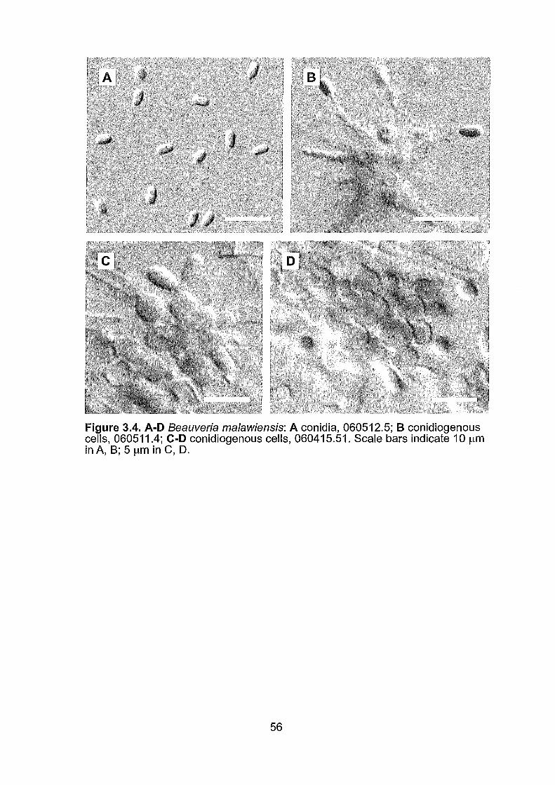
Figure 3.4. A·D Beauveria malawiensis: A conidia, 060512.5; B conidiogenous cells, 060511.4; C-D conidiogenous cells, 060415.51. Scale bars indicate 10 Ilm inA, 8; 51lm in C, D.
56

A B
Figure 3.5. A-F Beauveria caledonica: A 030422.2, B 030509.18, C 060415.3, D 050406.4, on Coleoptera; E 050408.2, on Hemiptera; F 040513.17, on Dermaptera.
57

Figure 3.6. A-F Beauveria caledonica: A conidia, 30422.2, B conidia from 1/4 SDYA culture NC94,C-F conidiogenous cells,060415.3.
58

3.3.3 Phylogenetic analyses
Sequences from the ITSl-5.8S-ITS2 region using primers AB28 and TW81 were approximately
534 nucleotides in length. Sequences were trimmed at each end to match the shortest Genbank
sequences included in the analyses. The final ITS dataset consisted of 506 aligned positions, of
which 52 were parsimony-informative sites. The optimal tree from the neighbour-joining
analysis of the full ITS dataset is shown in Fig. 3.7. Maximum parsimony (MP) analysis of the
smaller ITS dataset yielded 902 equally parsimonious trees with a length of 89 steps. Bayesian
likelihood analysis was conducted using the GTR+I+G model (general time reversible model
with a gamma distribution and a proportion of invariable sites). The consensus tree from
Bayesian analysis showed no significant conflict with the trees from MP analysis. One of the
most parsimonious trees from the maximum parsimony analysis of the ITS region is shown in
Fig. 3.8 with MP bootstrap values (BS) and posterior probabilities (PP) from the Bayesian
analysis.
Partial EF I-a sequences obtained usmg the pnmers 1777F and 2218R consisted of
approximately 529 nucleotides. These were trimmed in the final alignment to match the shortest
included Genbank sequence. The final EFl-a dataset had 470 aligned positions with 30
parsimony-infonnative sites. The optimal tree from the neighbour-joining analysis of the full
EF1-a dataset is shown in Fig. 3.9. Maximum parsimony analysis of the smaller EF1-a dataset
yielded 1012 equally parsimonious trees with a length of 61 steps. Bayesian likelihood analysis
was conducted using the GTR+I+G model. The consensus tree from Bayesian analysis showed
no significant conflict with the trees from MP analysis. One of the most parsimonious trees from
the maximum parsimony analysis ofEF1-a is shown in Fig. 3.10 with MP bootstrap values (BS)
and posterior probabilities (PP) from the Bayesian analysis.
Neighbour-joining, maximum parsimony and Bayesian analyses of the EF1-a dataset generally
supported the results from the ITS analyses, but usually with lower branch support for the
tenninal clades. However, the ITS and EFl-a phylogenies showed significant conflict in their
grouping of several B. caledonica isolates and the datasets were not considered suitable for
combined analysis.
In most cases analysis of the ITS and partial EF I-a regions tended to confinn speCIes
identifications based on conidial morphology. Beauveria bassiana isolates clustered in the two
main lineages (clades A and C) identified by Rehner and Buckley (2005). Differing levels of
support for clade C were provided by EF1-a (84% NJ BS, 75% MP BS, 53% PP) and ITS (98 %
NJ BS, 69% MP BS, unsupported in Bayesian). Analyses of the EF1-a region showed limited
59

support (54% NJ BS, 32% MP BS, 53% PP) for clade A compared with ITS (95% NJ BS, 71 %
MP BS, 100% PP).
A single isolate identified from morphology as B. bassiana grouped with B. brongniartii in the
NJ and MP analyses of ITS (99% NJ BS, 90% MP BS) and EF1-a (84% NJ BS, 64% MP BS).
Bayesian analysis of each region failed to resolve B. brongniartii, with the three included
sequences forming a polytomy at a basal node. The identification of B. malawiensis was strongly
supported from analysis of both ITS (99% NJ BS, 99% MP BS, 100% PP) and EF1-a (94% NJ
BS, 88% MP BS, 98% PP). In the ITS phylogeny New Zealand B. caledonica isolates formed
two distinct well-supported clades. One group corresponded with Scottish (ARSEF 2567) and
Swiss (ARSEF 1567) isolates of B. caledonica (97% NJ BS, 90% MP BS, 98% PP) while the
other group was closer to a South American isolate ARSEF 2251 (89% NJ BS, 70% MP BS,
55% PP). In contrast, partial EF1-a sequences grouped all of the New Zealand B. caledonica
with ARSEF 2251 (62 % NJ BS, 71% MP BS, 65% PP) while ARSEF 1567 and ARSEF 2567
formed a distinct and strongly supported (67% NJ BS, 82% MP BS, 95% PP) clade.
60

S. cf. bassiana S. cf. bassiana S. cf. bassiana S. cf. bassiana NC87 Aranaea NC79 Blattodea NC84 Blattodea E1073 Coleoptera E1080 Coleoptera E1082 Coleoptera NC53 Coleoptera
98 NC85 Coleoptera NC97 Coleoptera NC106 Coleoptera TE833 Coleoptera NC111 Hemiptera NC208 Hemiptera NC209 Hemiptera NC228 Hemiptera E1057 Hymenoptera E1067 Hymenoptera E1175 Hymenoptera E11390rthoptera NC107 Orthoptera NC2190rthoptera NC43 Phasmatodea
B. bassiana Clade C
B. caledonica S. caledonica
97 NC48 Hemiptera ....----1 E94 Coleoptera
63 E222 Coleoptera NC95 Coleoptera NC142 Coleoptera
99 S. cf. caledonica 2251 Brazil NC44 N/A ...---------1 NC49 Coleoptera E1079 Dermaptera
1----- S. vermiconia 1--___ ~99-1 S. amorpha 2641 Brazil
S. amorpha 1969 Peru B. brongniartii JE276 Switzerland
B. brongniartii 4362 Japan NC225 Sphaerotheriida
S. malawiensis ... ,,&&.,~,"'" E188 Coleoptera NC202 Coleoptera NC214 Coleoptera E96 HemiRtera E190 Hemiptera E219 Hemiptera E221 Hemiptera NC78 HemlRtera NC168 Hemiptera
99 TE672 Hemiptera ...-______ --1 E195 Hymenoptera
E201 Hymenoptera E213 Hymenoptera E1066 Hymenoptera E1084 Hymenoptera E1176 Hymenoptera NC210 Hymenoptera NC222 Hymenoptera T884 Hymenoptera T885 Hymemoptera NC83 Orthoptera NC220 Orthoptera TE445 Phasmatodea
69 Cordyceps cf. staphylinidicola 7044 '--___ -I Cordyceps sCl1rl1baeicoia 5689 China
94 Cordyceps cf. scarabaeicola EFCC252 S. bassiana 1153 Morocco S. basslana 300 Australia
65 S. bassiana 344 USA B. bassiana 1811 Morocco bassiana 296 USA
T875 Diptera B. bassiana 652 China S. bassiana 751 Vietnam S. bassiana 1848 Belgium B. bassiana
E1069 Hymenoptera Clade A 95 NC123 Hemiptera
E1063 Hymenoptera S. bassiana 1802 Greece S. bassiana 326 Australia S. bassiana 843 Costa Rica
NC25 /saria cicadae ...... ---------------------9::':9' Cordyceps cf. takaomontana BCC28612
f----------j
0.005
Figure 3.7 Optimal tree from neighbour-joining analysis of ITS sequences from Beauveria isolates. Genbank accession numbers for overseas isolates (in red) are given in Table 3.3. Bootstrap values (1000 replicates) and posterior probabilities 2:50% from Bayesian likelihood analysis are labelled at each branch, respectively.
61

71/100
I I 5 changes
57/54
99/100
99/100 E221 Hemiptera NC220 Orthoptera
,...------i TE445 Phasmatodea
E1057 Hymenoptera NC87 Araneae
69/- E11390rthoptera NC43 Phasmatodea NC209 Hemiptera NC84 Blattodea NC97 Coleoptera
brongniartii 4362 NC225 Sphaerotheriida
Ei88 Coleoptera NC222 .... "'Tu ... n
B. bassiana Clade C
B. bassiana Clade A
Cordyceps cf. takaomontana BCC28612 100/100 NC25 Isaria cicadae
Figure 3.8 One of the shortest trees from maximum parsimony analysis of ITS sequences from Beauveria isolates. Genbank accession numbers for overseas isolates (in red) are given in Table 3.3 Bootstrap values (1000 replicates) and posterior probabilities ::0:50% from Bayesian likelihood analysis are labelled at each branch, respectively.
62

99
54
60
E1069 Hymenoptera NC123 Hemiptera
63 E1063 Hymenoptera T875 Diptera
bassiana
B. nrc.nam;;rtll 1.---84-1 brongniartii Japan
NC225 Sphaerotheriida 65 NC168 Hemiptera
NC78 Hemiptera ma/ali\fler;.SIS IMI228343
E221 Hemiptera TE672 Hemiptera E188 Coleoptera NC202 Coleoptera NC214 Coleoptera
'------i E195 Coleoptera 94 E1176 Hymenoptera
NC222 Hymenoptera T884 Hymenoptera T885 Hymenoptera NC83 Orthoptera NC220 Orthoptera TE445 Phasmatodea
55
84
97
99
E1079 Dermaptera NC44 NC48 Hemiptera
62 NC49 Coleoptera E94 Coleoptera NC95 Coleoptera E222 Coleoptera NC142 Coleoptera
"",I,ori,>n/r"" 156"7
NC84 Blattodea E1073 Coleoptera TE833 Coleoptera NC111 Hemiptera
63 NC209 Hemiptera NC228 Hemiptera E1057 Hymenoptera E1067 Hymenoptera NC43 Phasmatodea
amorpha
B. bassiana Clade A
B. bassiana Clade C
L _____________ -C=======-~N~C~2:5~/s:a~r~ia. cicadae Cordyceps cf. takaomontana BCC28612
I------~.-j
0.005
Figure 3.9 Optimal tree from neighbour-joining analysis of EFl-a sequences from Beauveria isolates. Genbank accession numbers for overseas isolates (in red) are given in Table 3.3. Bootstrap values (1000 replicates) and posterior probabilities ::::50% from Bayesian likelihood analysis are labelled at each branch, respectively.
63

75/53
NC209 Hemiptera E1057 Hymenoptera E1073 Coleoptera NC43 Phasmatodea NC84 Blattodea
NC49 Coleoptera 71/65 NC142 Coleoptera
E1079 Dermaptera NC48 Hemiptera
E221 Hemiptera TE445 Phasmatodea
L-----".88",-:/9~8~ NC222 Hymenoptera E188 Coleoptera NC220 Orthoptera
B. bassiana Clade C
B. bassiana Clade A
L-----------1"()6i1oo-C======= NC251saria cicadae 100/100 Cordyceps cf. takaomontana BCC28612
I I 2 changes
Figure 3.10 One of the shortest trees from maximum parsimony analysis ofEFl-a sequences from Beauveria isolates. MP bootstrap values and posterior probabilities 2:50% from Bayesian likelihood analysis are labelled at each branch, respectively. Genbank accession numbers for overseas isolates (in red) are given in Table 3.3.
64

3.3.4 Tenebrio molitor bioassays
Bioassays confirmed that all tested strains of B. bassiana and B. malawiensis isolated from
coleopteran, hemipteran, and hymenopteran hosts were pathogenic towards T. molitor larvae. All
Beauveria strains caused significant mortality of T. molitor when compared to the controls after
12 days. Control mortality ranged between 0% and 5% after 12 days (Fig 3.11A-C). Mortality
trends were similar across all of the tested fungi with most larvae dying between 5 and 12 days.
Coleopteran isolates
Mean mortality caused by isolates from Coleoptera ranged from 93% to 100% after 12 days (Fig.
3.11A). Mean LTso values ranged from 5.14 to 7.49 days with a total mean LTso of 6.12 days.
Isolate NC106 (B. bassiana) had significantly higher LTso values compared to all other isolates
(Fig 3.l2A). Total means for each species were 6.17 days for B. bassiana and 6.03 days for B.
malawiensis. No significant difference was found between the total mean LTso values from each
speCIes.
Hemipteran isolates
Mean mortality caused by isolates from Hemiptera ranged from 63% to 100% after 12 days (Fig.
3.11B). Mean LTso values ranged from 6.43 to 10.8 days with a total mean of7.54 days. Isolate
TE672 (B. malawiensis) had significantly higher LTso values compared to all other isolates (Fig
3.l2B). Total means for each species were 7.09 days for B. bassiana and 7.98 days for B.
malawiensis. Total mean LTso values were significantly higher for B. malawiensis.
Hymenopteran isolates
Mean mortality caused by isolates from Hemiptera ranged from 67% to 100% after 12 days (Fig.
3.11C). Mean LTso values ranged from 6.98 to 11.13 days with a total mean of 8.38 days.
Isolates EI057 and EI069 (both B. bassiana) had significantly higher LTso values compared to
all other isolates (Fig. 3.12C). Total means for each species were 8.85 days for B. bassiana and
7.91 days for B. malawiensis. Total mean LTso values were significantly higher for B. bassiana.
65

A 100
80
>-.'!::: "'iii 60 .... .... 0 E .... 40 r: Q) u .... Q)
20 Q.
a
B 100
80
>-.'!::: "'iii 60 .... .... 0 E .... 40 r: Q) u ... Q) 20 Q.
a
C 100
.~ "'iii 1:: o E .... r: Q)
~ Q) Q.
80
60
40
20
a
a 2
a 2
a 2
4 6 8 10
Days after inoculation
4 6 8 10
Days after inoculation
4 6 8 10
Days after inoculation
12
12
12
B. bassiana
,E1080 B. bassiana
~E1082 B. bassiana
-+-NC85 B. bassiana
~::,~"NC106 B. bassiana
B. malawiensis
NC202 B. malawiensis
. ,NC214 B. malawiensis
Cantrall
Control 2
NC1ll B. bassiana
NC123 B. bassiana
NC208 B. bassiana
NC228 B. bassiana
E96 B. malawiensis
E219 B. malawiensis
NC78 B. malawiensis
TE672 B. malawiensis
Cantrall
Control 2
_E1057. bassiana
,E1063 B. bassiana
~E1067 B. bassiana
@~-E1069 B. bassiana
"r.~~.~E201 B. malawiensis
.E207 B. malawiensis
, E1066 B. malawiensis
E1084 B. malawiensis
Cantrall
Control 2
Figure 3.11 Cumulative mortality of Tenebrio molitor larvae after inoculation with Beauveria species isolated from A) Coleoptera; B) Hemiptera; C) Hymenoptera. Each graph shows means (n=60) from two replicate bioassays.
66

A 12
10
8
II)
> 6 ro C
4
2
0
E1073 E1082 E1080 NC8S NC106 E188 NC202 NC214
Isolate
B 12
10
8
II) B. bassiana
> 6 ro B. malawiensis c
4
2
0
NC123 NC208 NC228 NC1ll E96 E219 NC78 TE672
Isolate
C 12
10
8
II)
> 6 ro c
4
2
0
E1063 E1067 E10S7 E1069 E1066 E1084 E201 E207
Isolate
Figure 3.12 Mean LT 50 values from bioassays of Tenebrio mali tor larvae with Beauveria species isolated from: A) Coleoptera; B) Hemiptera; C) Hymenoptera. Each graph shows means (n=60) from two
replicate bioassays. Means with the same letter in each graph are not significantly different at P < 0.05.
67

3.3.5 Vespula vulgaris bioassay
Bioassays confirmed that all tested strains of B. bassiana and B. malawiensis were pathogenic towards V. vulgaris larvae. All isolates caused significant mortality when compared to the controls after 10 days. Mortality ranged from 93 to 100% after 10 days (Fig. 3.13A). There was no mortality in control larvae. Mean LT50 values ranged from 4.7 days to 6.02 days with a total mean of 5.46 days. A significant difference was found between the most virulent (B. bassiana
EI067) and least virulent (B. malawiensis EI066) isolates (Fig. 3.13B). Total means for each species were 5.13 days for B. bassiana and 5.79 days for B. malawiensis. Total mean LT50 values were significantly higher for B. malawiensis.
A
.08 III +' ... 0 E +' c: OJ u ... OJ
Co
B
VI
100
80
60
40
20
a a 2 4 6 8 10
Days after inoculation
8 T"~'·~~~--~-·~-·~~~·~~·"·~'-~--~···~-~~"~-····-··--·~-~~.----~-~.~~--.~.-
7 ·r-·---~-~··-----·'--·--"··'·~---~------'--··-··-·--·-.---.-------
6 4-·-~··--'---·----·--···-·~~--···-··-·--·----···-·-···-.--.. -.~-.-.-
5
~ 4 C
3
2
1
a El067 El063 El057 E201 El084 El066
Isolate
--- El057 B. bassiana
El063 B. bassiana
El067 B. bassiana
E201 B. malawiensis
El066 B. malawiensis
El084 B. malawiensis
Control
B. bassiana
B. malawiensis
Figure 3.13 A) Cumulative mortality of Vespula vulgaris larvae after inoculation with Beauveria species; B) Mean LT50 values from Vespula vulgaris bioassay. Means with the same letter are not significantly different at P < 0.05.
68
3.4 Discussion
Beauveria species were common in New Zealand native forests and were collected from sites in
all of the five main regions in this study (Tables 3.4, 3.6, 3.7). Insects were the most frequent
hosts, with collections including representatives from Blattodea, Coleoptera, Dermaptera,
Hemiptera, Hymenoptera, Orthoptera and Phasmatodea. Members of Hymenoptera (mainly
Vespula spp.) were the most commonly collected hosts (33%), followed by species in Coleoptera
(24%) and Hemiptera (16%, mainly Cicadidae). Beauveria species were also found on two non
insect arthropods in Aranaea and Diplopoda (Table 3.S). Beauveria bassiana and B. malawiensis
appeared to be the main Beauveria species in native forest, with each sharing a broad host range.
Fewer collections of B. brongniartii and B. caledonica were made, which may reflect more
stringent host requirements of these species.
Beauveria bassiana, B. malawiensis, and B. caledonica could be readily distinguished by their
conidial morphology (Tables 3.8-3.11), with sequence data from the ITS and EFl-a regions
(Figs. 3.7-3.10) generally supporting identifications based on conidial form. However, conidial
morphology did not fully indicate the phylogenetic affiliations of all isolates. Two previously
reported phylogenetic species within B. bassiana were identified but could not be reliably
separated by morphological data. Similarly, a B. brongniartii isolate (NC22S) did not show the
typical morphology of this species and could only be identified from molecular data. These
results confirm the necessity of molecular-level characterisation for unambiguous identification
of Beauveria species.
Beauveria bassiana was isolated from insect hosts in Blattodea, Coleoptera, Hemiptera,
Hymenoptera, Orthoptera, and Phasmatodea. Several collections represent new host records for
B. bassiana in New Zealand: Stethaspsis suturalis (Coleoptera), Phaulacridium marginale
(Orthoptera), and Cetatoblatta spp. (Blattodea). B. bassiana has not been previously reported in
this country from any Blattodean or Orthopteran hosts. A single collection of a spider infected
by B. bassiana represents an uncommon host association for this species. Most records of B.
bassiana from arachnids are associated with mites (Chandler et at. 2000; Li 1998), and only one
previous record of the species infecting spiders could be found (Petch 1931).
Specimens identified as B. bassiana typically had globose to subglobose conidia (Fig. 3.2A),
measuring from 1.6-2.6 x 1.3-2.4 flm on the host and 1.8-3.0 x 1.6-2.8 flm in culture. Conidial
sizes were comparable to those recorded for New Zealand (Glare et at. 1996a; Glare & Inwood
1998; Townsend et al. 1995) and overseas isolates (e.g. de Hoog 1972; Mugnai et al. 1989;
Rehner & Buckley 200S). No significant correlation was found between conidial size and host.
69
Sequences from the ITS and EF1-a region separated B. bassiana isolates into the two
phylogenetic species (clade A and clade C) identified by Rehner & Buckley (2005), confirming
an earlier report that both taxa are present in New Zealand (Reay et al. 2007). Generally,
representatives of each group were morphologically indistinguishable (e.g. Fig. 3.2A-B) and had
overlapping ranges of conidial size. However, one clade A isolate (NC123) had slightly larger
conidia than B. bassiana species identified as clade C in the phylogenetic analyses. Rehner &
Buckley (2005) also reported larger conidia in this clade, although similarly the difference was
not consistently shown in all isolates.
Beauveria bassiana isolates that grouped in clade C showed no variation in ITS sequences and
were identical with the European and North American representatives included in the analyses.
Slightly more variation was seen in the EF1-a region, but differences were limited to changes at
only one or two nucleotide positions. Clade A isolates were more variable in both ITS and EF1-
a sequences. Isolates E1063, E1069 and NC123 had similar ITS and EF1-a sequences, while
T875 was more divergent and differed by a total of seven nucleotides over the two regions.
These differences suggest that the two groups of isolates have different origins in New Zealand
and may reflect the outcome of separate historical dispersal events (see Rehner & Buckley 2005;
Rehner et al. 2006b). Sequences from isolate T875 consistently grouped with those from a
Chinese isolate (ARSEF652) in the ITS and EF1-a phylogenies, suggesting a close relationship
and shared evolutionary history of these isolates.
Currently, little information is available concerning the distribution of the two B. bassiana clades
in New Zealand. The presence of both groups in native forests was first demonstrated by Reay et
al. (2007) in association with Platypus spp. (Curculionidae: Coleoptera). In the present study,
while representatives of both clades were also found in native forests, the majority of isolates
grouped in clade C. In contrast, B. bassiana isolates from New Zealand pine plantations were all
found to belong to clade A (Reay et al. 2008). These findings suggest that clade C may be
restricted to undisturbed habitats in New Zealand. Populations of entomopathogenic fungi are
thought to be particularly sensitive to disturbance effects from human land use (Barker & Barker
1998; Hywel-Jones 2001; Samson et al. 1988). Molecular characterisation of B. bassiana isolates
from other modified environments (e.g. agricultural settings) in New Zealand may provide
further information on any habitat preferences shown by each clade.
A Beauveria specimen (NC225) from the pill millipede Procyliosoma tuberculatum (Diplopoda)
was initially identified as B. bassiana and formed globose-subglobose conidia (Fig. 3.2E)
measuring 2.0-2.6 x l.8-2.2 )Jm on the host and 2.1-3.0 x l.9-2.7 )Jm in culture. However,
70

phylogenetic analysis identified the isolate as B. brongniartii, with ITS and EF1-a sequences
each differing at a single nucleotide position from sequences of the closest overseas strain. While
this species has generally been differentiated from B. bassiana by larger (>3 /lm long) ellipsoidal
conidia (de Hoog 1972; Glare & Inwood 1998), in some cases strains that formed ellipsoidal
conidia on the hosts were shown to produce only spherical conidia in culture (Mugnai et al.
1989; Townsend et al. 1995). In other studies, strains forming ellipsoidal conidia in culture were
identified as B. bassiana based on DNA sequence data (Aquino de Muro et al. 2005; Rehner &
Buckley 2005). Isolate NC225 provides another example of the difficulties associated with
applying morphological criteria to distinguish between these two species. No other collections of
B. brongniartii were made in this study and the species appears to be rare in native forests,
despite earlier records of the fungus infecting a wide range of hosts in these habitats (Anon
2001-2009; Glare et al. 1993b).
Although millipedes (Diplopoda) are commonly infected by biotrophic fungal parasites in
Laboulbeniales (Rossi & Weir 1998; Weir & Beakes 1995), there are few records of true fungal
pathogens from these hosts. Beauveria brongniartii has not been previously recorded as infecting
diplopods, although Petch (1931) collected B. bassiana from a millipede in Ceylon. Verticillium
griseum (Gams 1971) and an undescribed Lecanicillium species (Kurihaya et al. 2008) have also
been reported from millipedes. Significantly, two surveys aimed at finding potential biocontrol
agents for millipede pests failed to find any entomopathogenic fungi among their natural enemies
(Baker 1985; Brito 1994). Diplopods are known to secrete defence compounds with antifungal
properties (Sierwald & Bond 2007), which could explain the lack of pathogenic fungi found in
association with these invertebrates.
Beauveria malawiensis is recorded infecting insects in New Zealand for the first time. The
species was easily distinguished from other Beauveria species by the characteristic straight,
cylindrical conidia (Fig. 3.4A-C). Conidia from cultures of New Zealand isolates were of slightly
different dimensions (3.0-4.8 x 1.1-2.0 /lm) to those recorded in the original description of B.
malawiensis (3.7-4.5 x 1.3-1.9 /lm), but shared the typical globose conidiophores (Fig. 3.4C-D)
and pink conidia in culture described for this species (Rehner et al. 2006a). No significant
correlation was found between conidial size and host. ITS sequences were identical to those from
the type specimen, as were most partial EF I-a sequences, apart from two isolates from
Hemiptera which differed at a single nucleotide position. A previous report identified an isolate
from a New Zealand pine forest soil as B. malawiensis based on analysis of a partial EF1-a
sequence (Reay et al. 2008). Another New Zealand soil isolate identified variously as B.
71

brongniartii (Glare 2004) and Cordyceps scarabaeicola (Reay et al. 2007), also represents B.
malawiensis based on the ITS sequence available for this isolate (Genbank DQ385618).
Rehner et al. (2006a) described B. malawiensis from a single culture isolated from the
coleopteran species Phoracantha semipunctata (Cerambycidae) and there have been no
subsequent records of this species from other hosts. The present study significantly extends the
known host range of B. malawiensis. In native forests, the species was collected on hosts in at
least ten different families in Coleoptera, Hemiptera, Hymenoptera, Orthoptera and
Phasmatodea. Hymenopteran. Vespula species were particularly common as hosts of B.
malawiensis, and large numbers of wasps killed by this species were observed at sites in Nelson
and the Bay of Plenty.
Beauveria malawiensis was first described from an isolate which had been originally identified
as B. brongniartii (Rehner et al. 2006a). The apparent rarity of B. brongniartii in native forests
observed in this study suggests that many earlier records of this species in these habitats may
have actually been B. malawiensis. This conclusion is also supported by the wide host range
previously recorded for B. brongniartii in this country (Anon 2001-2009; Glare et al. 1993b),
which tends to correlate with that of B. malawiensis. Examination of PDD herbarium specimens
originally identified as B. brongniartii confirmed that these are all B. malawiensis or B.
bassiana. Based on conidial morphology, the only PDD specimen likely to represent B.
brongniarti is PDD25211 (received as B. teneZla). However, the material examined consisted of
a dried agar culture, and any identification must remain tentative considering previous reports of
the variability of B. bassiana and B. brongniartii in culture (Aquino de Muro et al. 2005; Mugnai
et al. 1989; Rehner & Buckley 2005; Townsend et al. 1995). While B. brongniartii seems to be
rare in native forests, several isolates from scarabaeid hosts in New Zealand pastures appear to
be 'authentic' B. brongniartii based on morphological and molecular data (Glare & Inwood
1998), so the species may be more common in agricultural habitats.
To investigate host specificity in B. malawiensis and B. bassiana, laboratory bioassays were
conducted to examine the pathogenicity of strains isolated from various hosts towards the
coleopteran species Tenebrio molitor. B. bassiana and B. malawiensis isolates from Coleoptera,
Hymenoptera and Hemiptera were all shown to be pathogenic towards T. molitor larvae (Fig.
3.11). All of the isolates tested caused significantly higher mortality than the controls after 12
days. These results would seem to indicate that the examined strains of both B. malawiensis and
B. bassiana are generalists with no strict host preference. However, differences in virulence as
expressed by LT50 times were observed among isolates from each host group. Although isolates
72

from non-coleopteran hosts generally had similar virulence to those derived from Coleoptera,
single isolates of B. bassiana from the hemipteran group and B. malawiensis from the
hymenopteran groups each caused significantly lower mortality (Fig. 3.11) and had higher LT 50
times (Fig. 3.12). Although this could suggest a degree of specialisation towards the hosts from
which they were originally isolated, they could instead represent strains that are simply less
virulent towards insects in general. Further studies testing comparative virulence towards a wide
range of insect species are needed to more fully investigate any specific host preferences shown
by individual isolates of these species.
The potential of B. bassiana and B. malawiensis strains for control of Vespula wasps was also
examined. The latter species has not been previously tested against these insects. B. bassiana and
B. malawiensis strains isolated from wasp hosts all caused significant mortality compared with
the controls when tested against V. vulgaris larvae (Fig. 3.13A). Isolates varied in virulence
towards V. vulgaris. Shorter LT50 times were shown by B. bassiana isolates indicating that these
strains may make the best candidates for wasp control (Fig. 3.13B). Further characterisation of
the efficacy of these strains against Vespula species is suggested.
Beauveria caledonica was also isolated from insects in native forests. The species has only been
previously recorded in New Zealand in association with Coleoptera in pine plantations (Glare et
al. 2008; Reay et al. 2008). While most collections of B. caledonica from native forest were also
from Coleoptera, single collections of an infected cicada and a dermapteran (earwig) species
were also made. The species has not been previously recorded from non-coleopteran hosts.
Morphologically, B. caledonica specimens were characterised by ellipsoidal to cylindrical
conidia, measuring 2.2-3.3 x 1.1-1.6 )lm on the host (Fig. 3.6A) and 2.8-5.1 x1.0-2.1 )lm in
culture (Fig. 3.6B), closely matching the description by Bisset & Widden (1988). While conidial
dimensions tend to overlap those of B. malawiensis, conidia of B. caledonica were often
distinctively flattened on one side or slightly curved, sometimes with an almost reniform
appearance. Conidia of B. amorpha also have a similar shape but are larger, measuring 3.5-5 x
1.5-2.0 )lm on the host and 5-6 x 1.5-1.7 )lm in culture (Samson & Evans 1982). In the ITS
phylogeny, New Zealand isolates of B. caledonica formed two separate lineages. One group had
identical ITS sequences Scottish (ARSEF 2567) and Swiss (ARSEF 1567) strains of B.
caledonica while those of the other group were identical to a South American strain, ARSEF
2251. In contrast, EF I-a sequences grouped all of the New Zealand isolates with ARSEF 2251.
No consistent morphological differences were observed between the two groups. Glare et al.
(2008) also reported New Zealand B. caledonica isolates with ITS identical to ARSEF 2567 and
73
ARSEF 1567, and EF1-a identical to ARSEF2251. Rehner & Buckley (2005) suggested that
ARSEF 2251 may represent a separate species from B. caledonica based on its differing ITS and
EF1-a sequences and a slight difference in conidial size. However, until the taxonomic status of
this strain is clarified it seems advisable to classify all of the New Zealand strains discussed here
as B. caledonica.
Glare et al. (2008) suggested that B. caledonica may have been introduced to New Zealand from
Britain with Hylastes ater and Hylurgus ligniperda, based on the occurrence of the fungus on
similar hosts in both countries. In this case, the present study shows that B. caledonica has now
become established in native forest, possibly through migration of infected hosts from pine
forests. The discovery of two distinct ITS haplotypes in New Zealand strains suggests that two
separate introductions of B. caledonica have taken place.
Increasing molecular evidence has shown that many elements of the New Zealand biota (see
McDowall 2007; Perrie & Brownsey 2007), including fungal species (Moncalvo & Buchanan
2008; Moyersoen et al. 2003) have originated in New Zealand through long distance dispersal.
This is particularly relevant to explaining geographically disjunct distributions of fungal taxa, as
fungal spores have been shown to travel for thousands of kilometres on wind currents (Brown &
Hovmoller 2002; McKenzie 2000). Entomopathogenic fungi may also migrate via living,
infected insects; or through accidental transportation of diseased insects by human activities
(Bidochka & Small 2005). Future studies using additional, higher resolution molecular markers
(e.g. Rehner et al. 2006b) may provide insight into the phylogeographic history and likely
origins of Beauveria species in New Zealand.
74

CHAPTER FOUR: THE GENUS ISARIA IN NATIVE FORESTS
4.1 Introduction
The genus Isaria has had a complex taxonomic history. The name Isaria was first used by Hill in
1791 for three species that are now recognised as representing a myxomycete, a basidiomycete
and a rust (Petch 1934). Throughout the nineteenth and early twentieth century, many species
were added to the genus, which ultimately came to include over 200 species of mainly
entomopathogenic and mycoparasitic fungi (Hodge et a1. 2005). Generally, species were
classified in Isaria based on the presence of simple or branched synnemata producing one-celled
hyaline conidia, with no consideration given to differences in conidiogenous structures (Mains
1955). Petch (1934) also noted that in some cases species were also included in the genus solely
on the basis of their association with insects.
Members of Isaria were later redistributed amongst diverse fungal groups, with
entomopathogenic species transferred to several genera including Akanthomyces, Gibellu1a,
Hirsutella, Hymenostilbe and Spicaria (e.g. Speare 1920; Petch 1932a, 1932b, 1936, 1937;
Mains 1950). Clements & Shear (1931) proposed the entomopathogenic Isaria farinosa as the
type species for Isaria, and this decision was also supported by Mains (1955). However, Petch
(1934) reviewed the early nomenclatural history and identified varying concepts of the genus
among early authors, leading to confusion over the correct typification. Petch suggested that
Isaria should not be used as a generic name and followed Vuillemin who had earlier regarded 1.
farinosa as a member of Spicaria (Brown & Smith 1957).
The genus Spicaria was formerly used to accommodate species with verticillate conidiophores
and phialides, bearing chains of conidia. However, as noted by Vuillemin (1912) and Owen
(1919), these characteristics differed from the original description of the genus which had been
misinterpreted by subsequent authors. Hughes (1951) suggested that because of this taxonomic
confusion Spicaria should be abandoned and transferred two species to the similar but little
known genus Paecilomyces, established by Bainier in 1907 for the thermophilic species P.
variotii. Paecilomyces was described as resembling Penicillium but is primarily distinguished by
short cylindrical phialides tapering into long thin necks (Pitt & Hocking 1985). The genus was
monographed by Brown & Smith (1957), who followed Hughes (1951) and included many
species previously classified in Spicaria and Isaria. Onions & Barron (1967) also included
species with solitary phialides in Paecilomyces, but most of these were later transferred to
Acremonium (Gams 1971).
75

Samson (1974) restricted Paecilomyces to speCIes with verticillate conidiophores producing
divergent whorls of branches and phialides. Phialides were characterised by a cylindrical or
inflated base and a long distinct neck, bearing chains of hyaline, one-celled conidia. Several
species classified in Paecilomyces by Brown & Smith (1957) with deviating conidiogenous
structures (e.g. awl-shaped phialides or basitonous branching) were assigned to other genera.
Samson also separated Paecilomyces into two sections: sect. Paecilomyces and sect. Isarioidea.
Section Paecilomyces included P. variotii and other thermophilic or thermotolerant fungi, many
of which are commonly found as food spoilage organisms. Paecilomyces farinosus and other
invertebrate-pathogenic species were placed in section Isarioidea which included all species
with flask shaped phialides and catenate conidia formerly classified in Isaria. Several new
species from insects collected in tropical forests in Ghana (see Evans 1974) were also included
in this section. In addition to being ecologically distinct, the two sections within Paecilomyces
recognised by Samson (1974) were also characterised by disparate teleomorphic affiliations,
indicating that the genus was polyphyletic and did not reflect a natural taxonomic group. Species
in section Paecilomyces were known to produce teleomorphs in the eurotialean genera
Byssochlamys, Talaromyces and Thermoascus (Brown & Smith 1957; Samson 1974; Stolk &
Samson 1972) , while entomopathogenic species in section Isarioidea had been linked with the
hypocrealean genera Cordyceps and Torrubiella (e.g. Kobayasi 1941; Mains 1949; Petch 1937).
The phylogenetic placement of Paecilomyces section Isarioidea within the Hypocreales was
confirmed for one member of the section, P. tenuipes, from analysis of 18S (Fukatsu et al.1997)
and 28S rDNA sequences (Nikoh & Fukatsu 2000; Suh et al. 1998,). Obomik et al. (2001)
analysed 28S sequences from four invertebrate-pathogenic species in sect. Isarioidea and
demonstrated that this section was polyphyletic. Phylogenetic relationships based on 18S
sequences among a larger group of Paecilomyces species were examined by Luangsa-ard et al.
(2004) who included representatives from both sections. The genus was found to be polyphyletic
at the order level; species in section Paecilomyces were shown to form a group within the
Eurotiales, while those in section Isarioidea grouped in Hypocreales. The authors concluded that
the genus Paecilomyces should be used only for eurotialean taxa related to the type species P.
variotii and with a Byssochlamys teleomorph. The 18S phylogeny also confirmed the
polyphyletic status of section Isarioidea. Luangsa-ard et al. (2005) further examined
relationships in this group using ITS and ~-tubulin tubulin sequences. Species in section
Isarioidea were separated into four monophyletic groups. Significantly, a distinct clade of
entomopathogenic species based around P. farinosus was recognized. These species were
reclassified in Isaria (see Table 4.1) following a thorough review of the nomenclatural history
76
and proposal to conserve the genus as typified by 1. farinosa (Hodge et al. 2005; Gams et al.
2005). Luangsa-ard et al. (2005) suggested that two additional species placed in section
Isarioidea by Samson (1974), (P. ramosus and P. xylariiformis) probably also belong in Isaria,
although cultures were not available for molecular analyses. It seems likely that other
entomopathogenic Paecilomyces species with comparable morphology such as P. breviramosus
(Bisset 1979b) and P. rariramus (Liang et al. 2003) should also be included in the genus.
Although a close phylogenetic affiliation with Cordyceps has been well established by molecular
evidence (e.g. Luangsa-ard et al. 2004,2005; Nikoh & Fukatsu 2000, 2001; Stensrud et al. 2005;
Sung et al. 2007a), few specific teleomorph connections are known for the species currently
assigned to Isaria. A link with Cordyceps was originally suggested in the mid-nineteenth century
by Tulasne who stated that 1. farinosa was the te1eomorph of Cordyceps militaris (Gray 1858).
This connection was later refuted by Petch (1936) who determined from ascospore isolations
that C. militaris produces a 'Cephalosporium' anamorph (now included in Lecanicillium; Zare &
Gams 2001). Isaria farinosa has also been associated with the spider-pathogenic species
Torrubiella gonylepticida (Petch 1937) and T. pulvinata (Mains 1949). Both species were
described with Spicaria anamorphs that were synonymised with P. farinosus by Samson (1974).
More recently, Pacioni & Frizzi (1978) demonstrated from cultural studies that Isariafarinosa is
the conidial state of the rarely collected species Cordyceps memorabilis. However, the direct
association of 1. farinosa with a teleomorphic state has not been confirmed by any subsequent
authors. The most convincingly demonstrated anamorph-te1eomorph connection in Isaria
appears to be that between 1. tenuipes and Cordyceps takaomontana. The association was first
made by Kobayasi (1941) who observed that C. takaomontana produced perithecial stromata
concurrently with an 1. tenuipes conidial state when grown on a rice medium and also
occasionally on the host. A close relationship between the two taxa was also suggested from
analysis of 18S rDNA sequences (Nikoh & Fukatsu 2001). The relationship was further
substantiated by Luangsa-ard et al. (2005) who found that a Thai isolate provisionally identified
as C. takaomontana grouped with 1. tenuipes in both p-tubulin and ITS-based phylogenies.
Isaria species show varying degrees of host specificity (Table 4.1). The broadest host range is
seen in 1. farinosa and 1. fumosorosea. Both species are most commonly associated with
Lepidoptera but each also infects hosts in several insect orders (see Zimmermann 2008). Both
species are known to infect mites (Acari) (Chandler et al. 2000), with 1. farinosa also occurring
on other arachnids including spiders (Aranaea), harvestmen (Opiliones) and pseudoscorpions
(Chelonethida) (Cokendolpher 1993; Leatherdale 1970; Samson 1974; Samson & Evans 1977;
77

Sosnowska et al. 2004). Other members of the genus appear to have more restricted host ranges
and in some cases are only found in association with a single host order or family. For example,
1. ghanensis and 1. coleopterorum have only been recorded as pathogens of Lepidoptera and
Coleoptera, respectively, while 1. cicadae is restricted to immature stages of the single family
Cicadidae in Hemiptera.
Table 4.1. Host ranges of species accepted in Isaria by Luangsa-ard et al. (2005).
Species
1. amoenerosea
Host range
Chelonethida, Coleoptera, Lepidoptera, Hemiptera, Hymenoptera
I. cateniannulata Coleoptera, Diptera, Lepidoptera, Hymenoptera
1. cateniobliqua Lepidoptera 1. cicadae Hemiptera (Cicadidae) 1. coleopterorum Coleoptera
1. farinosa Acari, Aranea, Coleoptera, Chelonethida, Diptera, Hemiptera, Hymenoptera, Lepidoptera, Opiliones,
1. jillnosorosea
1.javanica
1. ghanensis I. tenuipes
Thysanoptera. Acari, Blattodea, Coleoptera, Diptera, Hemiptera, Hymenoptera, Isoptera, Lepidoptera, N europtera, Thysanoptera. Coleoptera, Lepidoptera, Aranaea Lepidoptera Coleoptera, Lepidoptera
References
Samson 1974; Samson & Evans 1977
Liang 1981; Shimazu 2001
Liang 1981 Samson 1974; Liang et al. 2005 Samson 1974; Samson & Evans 1977
see Zimmermann 2008
see Zimmermann 2008
Samson 1974; Samson & Evans 1977; Luangsa-ard et al. 2005 Samson 1974; Samson & Evans 1977 Mains 1955; Samson 1974; Luangsa-ard 2004
Currently the genus Isaria is poorly known in New Zealand. Two species recorded from this
country, 1. sulphurea and 1. jelina, appear to be saprophytic fungi and are not included in the
current circumscription of Isaria. Among species now included in the genus, 1. cicadae is the
most frequently reported in New Zealand. The species was first recorded in this country by
Taylor (1855) and has been known under several synonyms including Sphaeria basili,
Cordyceps sinclairii and Isaria sinclairii. Although 1. farinosa was first reported (as Spicaria
farinosa) from New Zealand early last century (Kirk 1909), there have been few subsequent
records (see Anon 2001-2009; Glare et al. 1993b) and information on the host range of the
78
species in this country is limited. Other Isaria species which are relatively common in other
parts of the world have been only rarely collected in this country, with 1. tenuipes, 1. javanica,
and I. jitmosorosea each represented by a single New Zealand collection (Anon 2001-2009).
This chapter discusses the morphological and molecular characterisation of Isaria species
collected and isolated from arthropod hosts in native forests.
79

4.2 Methods
4.2.1 Morphological characterisation
Isaria species were collected and isolated from native forest as described in chapter two.
Specimens and corresponding cultures were identified as Isaria according to Samson (1974) and
Luangsa-ard et at. (2005). Material was identified as Isaria based on conidiogenous structures
i.e. verticillately branched conidiophores bearing dense whorls of phialides with a swollen base
and distinct neck, producing conidia in chains. For species determination, measurements were
made from conidia taken directly from infected hosts and from cultures grown at 25°C for 14
days in darkness on 2% malt extract agar (MEA: 20g/L malt extract, 15g/L agar). For each
specimen and culture, length and width of 25 conidia was recorded. Length and width of 25
phialides was measured directly from host material. Methods used to obtain measurements are
given in chapter two. Species were determined by comparing phialide and conidial morphology
with the descriptions given by Samson (1974). To determine any significant relationship
between host affiliation and conidia/philaide size within Isaria jarinosa, mean conidia and
phi ali de sizes were square-root transformed and analysed by ANOV A. Means were separated by
Tukeys test. Statistical analyses were performed using SPSS version 11.00
4.2.2 Molecular characterisation
To confirm species identifications and examine phylogenetic relationships in New Zealand
Isaria, representative isolates were selected for sequencing based on the range of morphological
diversity and host affiliations observed. All methods used for DNA extraction, PCR, sequencing,
and phylogenetic analysis are given in Chapter Two.
For initial phylogenetic analysis the entire ITS region was amplified and sequenced using the
primers ITS4 and ITS5 (White et at. 1990). Sequences were aligned with Isaria sequences from
Genbank. Cordyceps cardinalis and Cordyceps pseudomilitaris were used as outgroup taxa
based on their basal relationship to Isaria species in the phylogeny of Sung et at. 2007a.
Phylogenetic analysis was conducted using maximum parsimony and Bayesian likelihood
inference. To provide further support for phylogenetic groups indicated from the ITS phylogeny,
a subset of isolates was selected for analysis of ~-tubulin and EF1-a gene regions. Primers and
PCR conditions are described in chapter two. For each region sequences were aligned with
Isaria sequences from Genbank. As few sequences from the Bt2a/Bt2b region for were
available, Paecitomyces lilacinus and Cordyceps cylindrica were selected as the closest usable
outgroup taxa for the ~-tubulin analysis. Sil11plicilliu1111anosoniveum and Simplicillium
80

lamellicola were selected as the closest usable outgroup taxa for the EF1-a analysis.
Phylogenetic analysis for ~-tubulin and EFl-a was conducted as above. Genbank accession
numbers and strain details for overseas isolates are shown in Table 4.2.
81
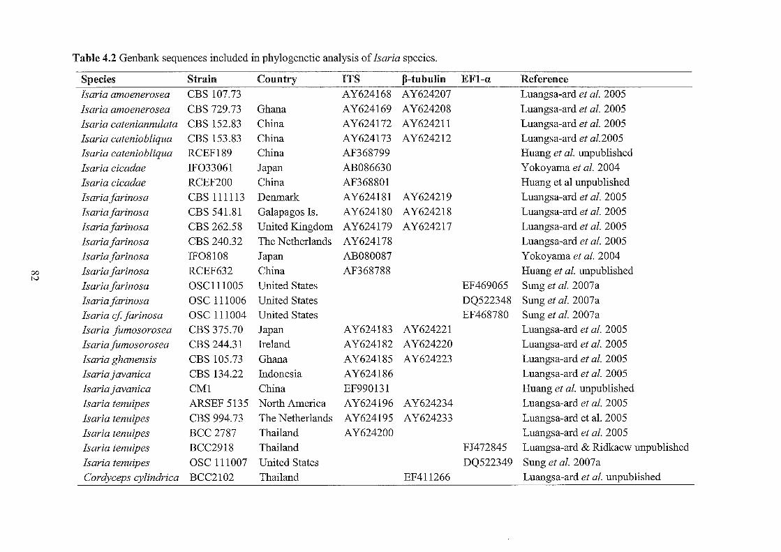
Table 4.2 Genbank sequences included in phylogenetic analysis of Isaria species.
Species Strain Country ITS p-tubulin EF1-a Reference Isaria amoenerosea CBS 107.73 AY624168 AY624207 Luangsa-ard et al. 2005
Isaria amoenerosea CBS 729.73 Ghana AY624169 AY624208 Luangsa-ard et al. 2005
Isaria eateniannulata CBS 152.83 China AY624172 AY624211 Luangsa-ard et al. 2005
Isaria eateniobliqua CBS 153.83 China AY624173 AY624212 Luangsa-ard et al.2005
Isaria eateniobliqua RCEF189 China AF368799 Huang et al. unpublished
Isaria deadae IF033061 Japan AB086630 Yokoyama et al. 2004
Isaria deadae RCEF200 China AF368801 Huang et al unpublished
Isaria farinosa CBS 111113 Denmark AY624181 AY624219 Luangsa-ard et al. 2005
Isaria farinosa CBS 541.81 Galapagos Is. AY624180 AY624218 Luangsa-ard et al. 2005
Isaria farinosa CBS 262.58 United Kingdom AY624 I 79 AY624217 Luangsa-ard et al. 2005
Isaria farinosa CBS 240.32 The Netherlands AY624178 Luangsa-ard et al. 2005
Isaria farinosa IF08108 Japan AB080087 Yokoyama et al. 2004
00 Isaria farinosa RCEF632 China AF368788 Huang et al. unpublished tv
Isaria farinosa OSCl11005 United States EF469065 Sung et al. 2007a
Isaria farinosa OSC 111006 United States DQ522348 Sung et al. 2007 a
Isaria ef farinosa OSC 111004 United States EF468780 Sung et al. 2007 a
Isaria fumosorosea CBS 375.70 Japan AY624183 AY62422I Luangsa-ard et al. 2005
Isaria jilmosorosea CBS 244.31 Ireland AY624 I 82 AY624220 Luangsa-ard et al. 2005
Isaria ghanensis CBS 105.73 Ghana AY624185 AY624223 Luangsa-ard et al. 2005
Isaria javaniea CBS 134.22 Indonesia AY624186 Luangsa-ard et al. 2005
Isaria javaniea CM1 China EF990131 Huang et al. unpublished
Isaria tenuipes ARSEF 5135 North America AY624196 AY624234 Luangsa-ard et al. 2005
Isaria tenuipes CBS 994.73 The Netherlands AY624 I 95 AY624233 Luangsa-ard et aL 2005
Isaria tenuipes BCC 2787 Thailand AY624200 Luangsa-ard et al. 2005
Isaria tenuipes BCC2918 Thailand FJ472845 Luangsa-ard & Ridkaew unpublished
Isaria tenuipes OSC 111007 United States DQ522349 Sung et al. 2007a
Cordyeeps eylindriea BCC2102 Thailand EF411266 Luangsa-ard et al. unpublished

00 U-l
Table 4.2 continued
Species Cordyceps cardinalis
Cordyceps pseudomilitaris
Paecilomyces lilacinus Simplicillium lanosoniveum
Simplicillium lamellicola
Strain BCMUCCOI NHJ6 CBS284.36 CBS 704.86 CBS 116.25
Country ITS p-tubulin EFt-a Reference Japan AB237660 Yokoyama et al. unpublished
Thailand AJ786589 Stensrud et al. 2005
AY624227 Luangsa-ard et al. 2005
DQ522358 Spatafora et al. 2007
DQ522356 Spatafora et al. 2007
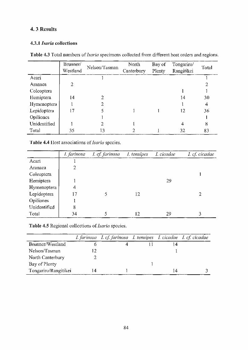
4.3 Results
4.3.1 Isaria collections
Table 4.3 Total numbers of Isaria specimens collected from different host orders and regions.
Brunner/ Nelson/Tasman
North Bay of Tongariro/ Total
Westland Canterbury Plenty Rangitikei
Acari 1 1 Aranaea 2 2 Coleoptera 1 1 Hemiptera 14 2 14 30 Hymenoptera 1 2 1 4 Lepidoptera 17 5 1 1 12 36 Opiliones 1 1 Unidentified 1 2 1 4 8 Total 35 13 2 1 32 83
Table 4.4 Host associations of !saria species.
1.farinosa I. cf farinosa 1. tenuipes 1. cicadae 1. cf cicadae
Acari 1 Aranaea 2 Coleoptera 1 Hemiptera 1 29 Hymenoptera 4 Lepidoptera 17 5 12 2 Opiliones 1 Unidentified 8 Total 34 5 12 29 3
Table 4.5 Regional collections of Isaria species.
1.farinosa 1. cf farinosa 1. tenuipes 1. cicadae 1. cf cicadae
Brunner/Westland 6 4 11 14 Nelson/Tasman 12 1 North Canterbury 2 Bay of Plenty 1 Tongariro/Rangitikei 14 1 14 3
84
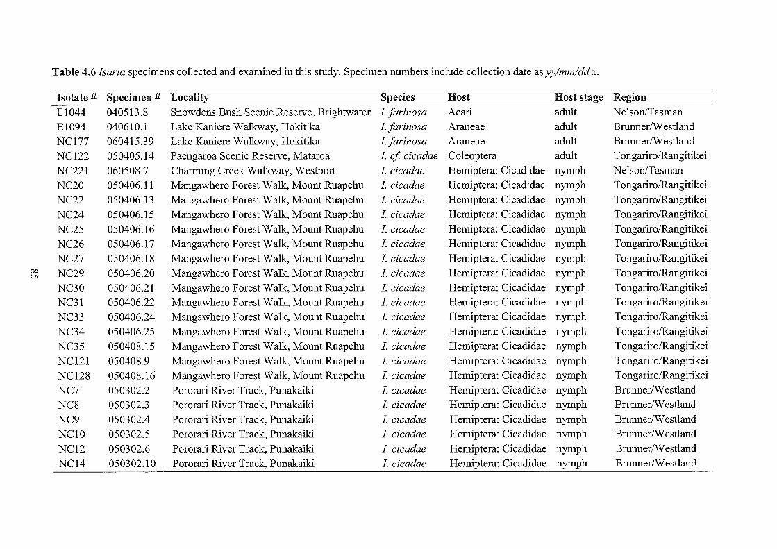
Table 4.6 Isaria specimens collected and examined in this study. Specimen numbers include collection date as yy/mm/dd.x.
Isolate # Specimen # Locality Species Host Host stage Region
E1044 040513.8 Snowdens Bush Scenic Reserve, Brightwater 1.farinosa Acari adult Nelson/Tasman
E1094 040610.1 Lake Kaniere Walkway, Hokitika 1.farinosa Araneae adult BrunnerlW estland
NC177 060415.39 Lake Kaniere Walkway, Hokitika 1.farinosa Araneae adult BrunnerlW estland
NC122 050405.14 Paengaroa Scenic Reserve, Mataroa 1. cf cicadae Coleoptera adult TongarirolRangitikei
NC221 060508.7 Charming Creek Walkway, Westport 1. cicadae Hemiptera: Cicadidae nymph Nelson/Tasman
NC20 050406.11 Mangawhero Forest Walk, Mount Ruapehu 1. cicadae Hemiptera: Cicadidae nymph Tongariro/Rangitikei
NC22 050406.13 Mangawhero Forest Walle, Mount Ruapehu 1. cicadae Hemiptera: Cicadidae nymph TongarirolRangitikei
NC24 050406.15 Mangawhero Forest Walk, Mount Ruapehu 1. cicadae Hemiptera: Cicadidae nymph TongarirolRangitikei
NC25 050406.16 Mangawhero Forest Walk, Mount Ruapehu 1. cicadae Hemiptera: Cicadidae nymph TongarirolRangitikei
NC26 050406.17 Mangawhero Forest Walle, Mount Ruapehu 1. cicadae Hemiptera: Cicadidae nymph TongarirolRangitikei
NC27 050406.18 Mangawhero Forest Walk, Mount Ruapehu 1. cicadae Hemiptera: Cicadidae nymph Tongariro/Rangitikei 00 NC29 050406.20 Mangawhero Forest Walk, Mount Ruapehu 1. cicadae Hemiptera: Cicadidae nymph TongarirolRangitikei Vl
NC30 050406.21 Mangawhero Forest Walle, Mount Ruapehu 1. cicadae Hemiptera: Cicadidae nymph TongarirolRangitikei
NC31 050406.22 Mangawhero Forest Walk, Mount Ruapehu 1. cicadae Hemiptera: Cicadidae nymph TongarirolRangitikei
NC33 050406.24 Mangawhero Forest Walk, Mount Ruapehu 1. cicadae Hemiptera: Cicadidae nymph TongarirolRangitikei
NC34 050406.25 Mangawhero Forest Walk, Mount Ruapehu 1. cicadae Hemiptera: Cicadidae nymph TongarirolRangitikei
NC35 050408.15 Mangawhero Forest Walk, Mount Ruapehu 1. cicadae Hemiptera: Cicadidae nymph Tongariro/Rangitikei
NC121 050408.9 Mangawhero Forest Walle, Mount Ruapehu 1. cicadae Hemiptera: Cicadidae nymph TongarirolRangitikei
NC128 050408.16 Mangawhero Forest Walk, Mount Ruapehu 1. cicadae Hemiptera: Cicadidae nymph TongarirolRangitikei
NC7 050302.2 Pororari River Track, Punakaiki 1. cicadae Hemiptera: Cicadidae nymph BrunnerlW estland
NC8 050302.3 Pororari River Track, Punakaiki 1. cicadae Hemiptera: Cicadidae nymph BrunnerlW estland
NC9 050302.4 Pororari River Track, Punakaiki 1. cicadae Hemiptera: Cicadidae nymph BrunnerlW estland
NC10 050302.5 Pororari River Track, Punakaiki 1. cicadae Hemiptera: Cicadidae nymph BrunnerlW estland
NC12 050302.6 Pororari River Track, Punakaiki 1. cicadae Hemiptera: Cicadidae nymph BrunnerlW estland
NC14 050302.10 Pororari River Track, Punakaiki 1. cicadae Hemiptera: Cicadidae nymph BrunnerlW estland

Table 4.6 continued.
Isolate # Specimen # Locality Species Host Host stage Region
NC15 050302.11 Pororari River Track, Punakaiki 1. cicadae Hemiptera: Cicadidae nymph BrunnerlW estland NC16 050302.12 Pororari River Track, Punakaiki 1. cicadae Hemiptera: Cicadidae nymph BrunnerlW estland NC17 050302.13 Pororari River Track, Punakaiki 1. cicadae Hemiptera: Cicadidae nymph BrunnerlW estland NC18 050302.14 Pororari River Track, Punakaiki 1. cicadae Hemiptera: Cicadidae nymph BrunnerlW estland
NC19 050302.15 Pororari River Track, Punakaiki 1. cicadae Hemiptera: Cicadidae nymph BrunnerlW estland NC37 050302.8 Pororari River Track, Punakaiki 1. cicadae Hemiptera: Cicadidae nymph BrunnerlW estland
NC38 050302.9 Pororari River Track, Punakaiki 1. cicadae Hemiptera: Cicadidae nymph BrunnerlW estland NC6 050302.1 Truman Track, Punakaiki 1. cicadae Hemiptera: Cicadidae nymph BrunnerlW estland E1051 040510.6 Snowdens Bush Scenic Reserve, Brightwater 1.farinosa Hemiptera adult Nelson/Tasman TE443 020417.10 Lake Kaniere Walkway, Hokitika 1.farinosa Hymenoptera adult BrunnerlW estland NC126 050408.17 Mangawhero Forest Walle, Mount Ruapehu 1.farinosa Hymenoptera adult Tongariro/Rangitikei
00 EI048 040514.3 Loop Track, Lake Rotoiti 1.farinosa Hymenoptera: Vespidae adult Nelson/Tasman 0\
E1054 040513.7 Rolling Creek, Wangapeka Valley 1.farinosa Hymenoptera: Vespidae adult Nelson/Tasman NCl12 050406.7 Mangawhero Forest Walk, Mount Ruapehu 1. cf cicadae Lepidoptera pupa Tongariro/Rangitikei NCl27 050406.35 Mangawhero Forest Walle, Mount Ruapehu 1. cf cicadae Lepidoptera pupa Tongariro/Rangitikei TE02 010420.1 Lake Kaniere Walkway, Hokitika 1.farinosa Lepidoptera larva BrunnerlW estland NC76 050302.16 Truman Track, Punakaiki 1.farinosa Lepidoptera adult BrunnerlW estland
NC1l7 050408.25 Mangawhero Forest Walk, Mount Ruapehu 1.farinosa Lepidoptera larva Tongariro/Rangitikei NC124 050408.13 Mangawhero Forest Walk, Mount Ruapehu 1.farinosa Lepidoptera larva Tongariro/Rangitikei NC131 050408.7 Mangawhero Forest Walk, Mount Ruapehu 1.farinosa Lepidoptera larva Tongariro/Rangitikei NC63 050405.11 Paengaroa Scenic Reserve, Mataroa 1.farinosa Lepidoptera larva Tongariro/Rangitikei EI049 040510.14 Snowdens Bush Scenic Reserve, Brightwater 1.farinosa Lepidoptera pupa Nelson/Tasman E1056 040510.13 Snowdens Bush Scenic Reserve, Brightwater 1.farinosa Lepidoptera pupa Nelson/Tasman E1053 040514.7 Loop Track, Lake Rotoiti 1.farinosa Lepidoptera pupa Nelson/Tasman
NC203 060508.5 Charming Creek Walkway, Westport 1.farinosa Lepidoptera pupa Nelson/Tasman
NC94 050510.7 Devils Punchbowl Track, Arthurs Pass 1.farinosa Lepidoptera pupa North Canterbury
Table 4.6 continued.
Isolate # Specimen # Locality Species Host Host stage Region
NC125 050404.18 Mangawhero Falls Walk, Mount Ruapehu 1.farinosa Lepidoptera pupa TongarirolRangitikei
NC55 050408.8 Mangawhero Forest Walk, Mount Ruapehu 1.farinosa Lepidoptera pupa TongarirolRangitikei
NC56 050408.11 Mangawhero Forest Walk, Mount Ruapehu 1.farinosa Lepidoptera pupa TongarirolRangitikei
NC116 050408.21 Mangawhero Forest Walk, Mount Ruapehu 1.farinosa Lepidoptera pupa TongarirolRangitikei
NC129 050408.19 Mangawhero Forest Walle, Mount Ruapehu 1.farinosa Lepidoptera pupa TongarirolRangitikei
NC206 060509.2 Nikau Loop Walk, Karamea 1.farinosa Lepidoptera pupa BrunnerlW estland
NC181 060415.42 Lake Kaniere Walkway, Hokitika 1. cffarinosa Lepidoptera pupa BrunnerlW estland
NC184 060415.46 Lake Kaniere Walkway, Hokitika 1. cffarinosa Lepidoptera pupa BrunnerlW estland
NC180 060415.41 Lake Kaniere Walkway, Hokitika 1. cf farinosa Lepidoptera pupa BrunnerlW estland
NC212 060511.10 Nile River Valley Walle, Charleston 1. cffarinosa Lepidoptera pupa BrunnerlW estland
NC108 050405.12 Paengaroa Scenic Reserve, Mataroa 1. cf farinosa Lepidoptera pupa TongarirolRangitikei 00 E378 030506.6 Lindemann Loop Track, Katikati 1. tenuipes Lepidoptera pupa Bay of Plenty -....l
TE677 020507.3 Cascade Valley, Haast 1. tenuipes Lepidoptera pupa BrunnerlW estland
TE433 020417.0 Goldsborough (Shamrock) Track, Hokitika 1. tenuipes Lepidoptera pupa BrunnerlW estland
TE434 020417.1 Goldsborough (Shamrock) Track, Hokitika 1. tenuipes Lepidoptera pupa BrunnerlW estland
TE497 020509.1 Hapuka Estuary Walk, Okuru 1. tenuipes Lepidoptera pupa BrunnerlW estland
E1095 040610.2 Lake Kaniere Walkway, Hokitika 1. tenuipes Lepidoptera pupa BrunnerlW estland
E1096 040610.3 Lake Kaniere Walkway, Hokitika 1. tenuipes Lepidoptera pupa BrunnerlW estland
NC182 060415.45 Lake Kaniere Walkway, Hokitika 1. tenuipes Lepidoptera pupa BrunnerlW estland
TE435 020417.2 Lake Kaniere Walkway, Hokitika 1. tenuipes Lepidoptera pupa BrunnerlW estland
TE436 020417.3 Lake Kaniere Walkway, Hokitika 1. tenuipes Lepidoptera pupa BrunnerlW estland
E090 030421.1 Lake Kaniere Walkway, Hokitika 1. tenuipes Lepidoptera pupa BrunnerlW estland
NC200 060508.1 Nile River Valley Walle, Charleston 1. tenuipes Lepidoptera pupa BrunnerlW estland
E1045 040510.21 Snowdens Bush Scenic Reserve, Brightwater 1.farinosa Opiliones adult Nelson/Tasman
NC80 050215.3 Devils Punchbowl Track, Arthurs Pass 1.farinosa n.d. n.d North Canterbury

00 00
Table 4.6 continued.
Isolate # Specimen #
TE437 020417.4 EI046 040510.4
EI047 040510.20 NC54 050404.26
NCI13 050406.8
NC118 050406.27 NC61 050405.5
n.d. not determined.
Locality
Lake Kaniere Walkway, Hokitika Snowdens Bush Scenic Reserve, Brightwater Snowdens Bush Scenic Reserve, Brightwater Mangawhero Falls Walle, Mount Ruapehu
Mangawhero Forest Walk, Mount Ruapehu Mangawhero Forest Walle, Mount Ruapehu Paengaroa Scenic Reserve, Mataroa
Species Host Host stage Region
Lfarinosa n.d. n.d. BrunnerIW estland Lfarinosa n.d. n.d. Nelson/Tasman Lfarinosa n.d. n.d. Nelson/Tasman Lfarinosa n.d. n.d TongarirolRangitikei Lfarinosa n.d. n.d. TongarirolRangitikei
Lfarinosa n.d n.d. TongarirolRangitikei Lfarinosa n.d. n.d. TongarirolRangitikei

4.3.2 Morphological characterisation
/saria jarinosa
Conidia from 1.farinosa were ellipsoidal to fusiform (Fig. 4.2A) and measured 2.0-2.9 x 1.1-1.9
flm (average 2.4 x 1.4 flm) on the host. Conidia from cultures on MEA after 14 days measured
2.1-3.2 x 1.1-2.0 flm (average 2.6 x 1.5 flm). Phialides from the host (Fig. 4.2C-E) measured
3.8-9.8 x 1.9-3.5 flm (average 5.8 x 2.5 flm). Conidia and phialide sizes from different host
orders are shown in Table 4.7. Conidia and phialides were significantly longer on non-insect
(arachnid) hosts.
Table 4.7 Conidia and phialide sizes of!. farinosa from different host orders.
Conidia on host Conidia on MEA Phialides on host length x width length x width length x width
Acari 2.1-2.6 (2.4) x 1.1-1.5(1.3) 2.5-3.1 (2.7) x 1.1-1.7 (1.3) 4.5-7.3 (6.0) x 2.4-3.1 (2.8)
Aranaea 2.3-2.9 (2.7) x 1.3-1.8 (1.6) 2.3-3.0 (2.7) x 1.2-1.9 (1.6) 4.5-7.9 (6.1) x 2.0-2.9 (2.5)
Hemiptera 2.3-2.6 (2.4) x 1.2-1.7 (1.5) 2.3-3.0 (2.6) x 1.3-1.8 (1.5) 3.9-7.0 (5.5) x 1.9-2.7 (2.2)
Hymenoptera 2.1-2.9 (2.4) x 1.1-1.7 (1.4) 2.1-3.2 (2.6) x 1.2-1.9 (1.4) 4.1-7.2 (5.9) x 1.9-3.0 (2.5)
Lepidoptera 2.0-2.9 (2.4) x 1.1-1.9 (1.4) 2.1-3.1 (2.6) x 1.1-2.0 (1.5) 3.8-8.3 (5.6) x 2.0-3.5 (2.5)
Opiliones 2.3-2.9 (2.6) x 1.4-1.7 (1.6) 2.3-3.1 (2.7) x 1.1-1.7 (1.4) 5.4-9.8 (7.3) x 2.4-3.3 (2.9)
Unidentified 2.0-2.9 (2.4) x 1.1-1.7 (1.4) 2.1-3.1 (2.6) x 1.1-1.9 (1.4) 4.1-8.7(5.8) x.1.9-3.2(2.5)
Isaria cf. jarinosa
Conidia were ellipsoidal to fusiform (Fig 4.2B) and measured 2.5-3.8 x 1.5-2.5 flm (average 3.1
x 1.9 flm) on the host. Conidia from cultures on MEA after 14 days measured 2.6-4.0 x 1.8-2.7
flm (average 3.2 x 2.0 flm). Phialides from the host (Fig 4.2F-H) measured 4.6-11.4 x 2.3-4.0
(average 6.6 x 3.1 flm).
/saria tenuipes
Conidia were ellipsoidal to cylindrical, often slightly curved or allantoid (Fig 4.4A-C). Conidia
measured 3.0-6.9 x 1.2-2.4 flm (average 4.7 x 1.7 flm) on the host. Conidia from cultures on
MEA after 14 days measured 3.9-9.1 x 1.4-3.5 flm (average 6.0 x 2.0 flm). Phialides from the
host (Fig. 4.4D-H) measured 3.6-7.2 x 2.5-5.3 flm (average 5.0 x 3.5 flm).
89

Isaria cicadae
Conidia were cylindrical, usually highly curved (Fig. 4.6A), and measured 3.2-6.5 x 1.1-2.7 )lm
(average 4.2 x 1.5 )lm) on the host. Conidia from cultures on MEA after 14 days measured 3.7-
12.3 x 1.4-2.7 )lm (average 7.1 x 2.0 )lm). Phialides from the host (Fig. 4.6C-D) measured 3.5-
6.6 x 2.4-5.1 )lm (average 4.9 x 3.4 )lm).
Isaria cf. cicadae
Conidia were cylindrical, curved (Fig. 4.6B), and measured 5.7-9.0 x 1.6-2.5 )lm (average 7.0 x
2.0 )lm) on the host. Conidia from cultures on MEA after 14 days measured 6.2-13.7 x 1.4-3.0
)lm (average 9.2 x 2.1 )lm). Phialides from the host (Fig. 4.6E-F) measured 4.4-6.9 x 3.2-5.4 )lm
(average 5.7 x 4.2 )lm).
90

Table 4.8 Measurements of conidia from host material and 14 day old MEA cultures, and phialide measurements from host materiaL All measurements are given in /lm with averages (n=25) in brackets.
Isolate # Specimen # Species Host Conidia on MEA Conidia on host Phialides on host length x width length x width length x width
EI044 040513.8 Lfarinosa Acari 2.5-3.1 (2.7) x 1.1-1.7 (1.3) 2.1-2.6 (2.4) x 1.1-1.5(1.3) 4.5-7.3 (6.0) x 2.4-3.1 (2.8) EI094 040610.1 Lfarinosa Araneae 2.3-3.0 (2.7) x 1.2-1.9 (1.5) 2.3-2.8 (2.6) x 1.3-1.7 (1.5) 4.7-7.9 (6.5) x 2.4-2.9 (2.6) NC177 060415.39 Lfarinosa Araneae 2.3-3.0 (2.6) x 1.4-1.9 (1.6) 2.3-2.9 (2.7) x 1.4-1.8 (1.6) 4.5-7.0 (5.7) x 2.0-2.7 (2.3) NC122 050405.14 L ef deadae Coleoptera 6.2-10.9 (8.0) x 1.4-2.2 (1.8) 6.0-7.9 (6.8) x 1.6-2.5 (1.9) 4.4-6.9 (5.8) x 3.2-5.4 (4.2) NC221 060508.7 L deadae Hemiptera 3.7-10.8 (6.1) x 1.4-2.5 (1.8) 3.8-5.4 (4.5) x 1.4-1.9 (1.6) 3.7-6.4 (4.8) x 3.0-4.2 (4.8) NC20 050406.11 L deadae Hemiptera 4.1-10.0 (6.4) x 1.5-2.4 (1.9) 3.5-5.5 (4.3) x 1.2-1.7 (1.4) 4.0-5.6 (4.8) x 2.5-4.0 (3.0) NC22 050406.13 L deadae Hemiptera 4.0-11.9 (7.0) x 1.5-2.6 (2.1) 3.4-4.6 (3.8) x 1.3-1.7 (1.4) 4.2-6.1 (5.1) x 2.7-3.6 (3.3) NC24 050406.15 L deadae Hemiptera 4.0-10.9 (6.4) x 1.5-2.5 (2.0) 3.2-5.0 (4.0) x 1.2-2.0 (1.4) 4.2-6.1 (5.1) x 3.2-4.1 (3.6) NC25 050406.16 L deadae Hemiptera 3.8-9.7 (6.6) x 1.7-2.6 (2.1) 3.2-4.5 (4.0) x 1.1-1.7 (1.4) 3.5-5.3 (4.6) x 3.1-3.7 (3.3)
'0 NC26 050406.17 L deadae Hemiptera 3.8-11.5 (7.3) x 1.6-2.6 (2.1) 3.5-5.0 (4.3) x 1.4-1.7 (1.6) 4.0-6.0 (4.9) x 3.0-4.1 (3.4) >--'
NC27 050406.18 L deadae Hemiptera 4.1-11.4 (7.0) x 1.5-2.3 (2.0) 3.5-4.9 (4.0) x 1.2-1.8 (1.4) 3.7-5.8 (4.7) x 2.9-3.6 (3.1) NC29 050406.20 L deadae Hemiptera 4.2-11.2 (6.4) x 1.5-2.7 (2.1) 3.5-5.1 (4.2) x 1.2-1.8 (1.4) 4.4-5.8 (5.2) x 2.7-4.0 (3.5) NC30 050406.21 L deadae Hemiptera 3.7-9.4 (6.6) x 1.7-2.4 (2.1) 3.7-4.7 (4.1) x 1.4-1.8 (1.6) 4.1-6.1 (4.8) x 2.7-3.8 (3.4)
NC31 050406.22 L deadae Hemiptera 3.8-10.8 (7.2) x 1.5-2.4 (1.9) 3.5-4.9 (4.1) x 1.3-1.8 (1.5) 3.7-5.4 (4.6) x 2.8-3.9 (3.4) NC33 050406.24 L deadae Hemiptera 3.9-11.4 (6.7) x 1.5-2.3 (1.8) 3.4-4.2 (3.7) x 1.2-1.6 (1.4) 4.1-6.0 (4.8) x 2.7-3.5 (3.1) NC34 050406.25 L deadae Hemiptera 4.1-11.9 (7.3) x 1.5-2.5 (2.1) 3.2-5.9 (4.0) x 1.4-1.9 (1.6) 4.2-5.5 (4.8) x 2.9-3.7 (3.3) NC35 050408.15 L deadae Hemiptera 4.0-10.0 (7.1) x 1.5-2.5 (2.0) 3.7-5.0 (4.3) x 1.4-1.8 (1.6) 4.1-5.3 (4.6) x 2.9-3.4 (3.1) NC121 050408.9 L deadae Hemiptera 3.9-11.5 (7.1) x 1.6-2.5 (2.0) 3.7-5.7 (4.3) x 1.4-2.1 (1.7) 4.1-5.7 (4.9) x 3.4- 4.5 (3.9) NC128 050408.16 L deadae Hemiptera 3.9-12.3 (7.0) x 1.5-2.5 (1.9) 3.5-5.0 (4.1) x 1.3-1.7 (1.5) 4.0-6.0 (5.2) x 2.7-4.0 (3.4) NC7 050302.2 L deadae Hemiptera 4.0-11.2 (6.6) x 1.5-2.5 (1.9) 3.4-4.5 (3.7) x 1.1-1.6 (1.4) 3.8-6.0 (4.8) x 2.5-4.2 (3.2) NC8 050302.3 L deadae Hemiptera 3.9-11.9 (8.2) x 1.4-2.5 (2.1) 3.7-5.1 (4.2) x 1.4-1.9 (1.6) 4.4-6.4 (5.1) x 2.7-3.7 (3.1) NC9 050302.4 L deadae Hemiptera 4.1-11.9 (7.8) x 1.5-2.6 (2.1) 3.8-6.5 (4.8) x 1.6-2.7 (2.0) 3.7-5.4 (4.5) x 2.4-3.6 (3.0) NCI0 050302.5 L deadae Hemiptera 4.1-11.5 (7.1) x 1.6-2.6 (2.1) 3.5-5.0 (4.2) x 1.3-1.7 (1.5) 4.1-6.3 (5.0) x 3.1- 5.1 (3.8) NC12 050302.6 L deadae Hemiptera 4.2-12.1 (8.7) x 1.5-2.5 (2.0) 3.5-5.5 (4.4) x 1.2-1.8 (1.5) 4.0-6.6 (5.0) x 3.1-4.0 (3.6)

Table 4.8. continued.
Isolate # Specimen # Species Host Conidia on MEA Conidia on host Phialides on host length x width length x width length x width
NC14 050302.10 1. cicadae Hemiptera 4.5-10.3 (7.4) x 1.7-2.5 (2.1) 3.6-5.3 (4.4) x 1.2-1.7 (1.4) 4.2-5.9 (5.1) x 2.9-4.2 (3.6) NC15 050302.11 1. cicadae Hemiptera 3.8-11.8 (7.5) x 1.5-2.6 (2.0) 3.6-5.4 (4.4) x 1.1-1.8 (1.5) 4.2-5.8 (5.0) x 2.9-4.1 (3.6) NC16 050302.12 1. cicadae Hemiptera 3.5-11.3 (8.1) x 1.4-2.6 (2.0) 3.6-4.7 (4.1) x 1.2-1.7 (1.5) 4.1-6.1 (5.1) x 3.2-4.1 (3.6)
NC17 050302.13 1. cicadae Hemiptera 3.9-11.3 (6.9) x 1.5-2.3 (2.0) 3.3-5.5 (4.2) x 1.4-1.8 (1.6) 4.2-5.8 (5.1) x 2.9-4.9 (3.6) NC18 050302.14 1. cicadae Hemiptera 3.9-10.3 (6.6) x 1.6-2.9 (2.2) 3.2-4.7 (3.8) x 1.3-1.9 (1.6) 3.9-5.8 (4.8) x 3.0-4.1 (3.5) NC19 050302.15 1. cicadae Hemiptera 3.9-10.7 (7.2) x 1.3-2.2 (1.9) 3.3-6.0 (4.3) x 1.3-1.9 (1.7) 4.4-6.4 (5.2) x 3.0-4.2 (3.5) NC37 050302.8 1. cicadae Hemiptera 3.6-10.3 (6.7) x 1.5-2.6 (2.0) 3.7-4.8 (4.2) x 1.3-1.8 (1.6) 3.9-6.1 (4.5) x 2.7-3.6 (3.3) NC38 050302.9 1. cicadae Hemiptera 3.8-10.1 (7.5) x 1.5-2.2 (1.8) 3.6-5.1 (4.3) x 1.2-1.7 (1.5) 4.0-5.1 (4.6) x 3.0-4.2 (3.5) NC6 050302.1 1. cicadae Hemiptera 4.2-11.2 (8.0) x 1.4-2.5 (2.0) 3.5-5.3 (4.3) x 1.2-1.8 (1.5) 3.9-5.6 (4.7) x 2.6-3.8 (3.2) E1051 040510.6 1.farinosa Hemiptera 2.3-3.0 (2.6) x 1.3-1.8 (1.5) 2.3-2.6 (2.4) x 1.2-1.7 (1.5) 3.9-7.0 (5.5) x 1.9-2.7 (2.2)
\0 TE443 020417.10 1.farinosa Hymenoptera 2.2-2.9 (2.6) x 1.2-1.7 (1.5) 2.2-2.9 (2.5) x 1.2-1.7 (1.4) 5.5-7.1 (6.4) x 1.9-2.7 (2.3) tv
NC126 050408.17 1.farinosa Hymenoptera 2.4-2.9 (2.7) x 1.2-1.6 (1.4) 2.1-2.5 (2.3) x 1.2-1.6 (1.4) 4.1-7.0 (5.3) x 2.0-2.8 (2.5) E1048 040514.3 1.farinosa Hymenoptera 2.3-3.2 (2.7) x 1.2-1.9 (1.4) 2.3-2.8 (2.5) x 1.1-1.7 (1.4) 5.6-7.2 (6.3) x 2.4-3.0 (2.6) E1054 040513.7 1.farinosa Hymenoptera 2.1-3.0 (2.5) x 1.2-1.6 (1.4) 2.2-2.7 (2.4) x 1.2-1.6 (1.4) 4.5-7.1 (5.6) x 2.3-3.0 (2.6) NCl12 050406.7 1. cf cicadae Lepidoptera 6.9-11.8 (9.4) x 1.7-2.9 (2.1) 5.7-7.8 (6.6) x 1.8-2.2 (2.0) 4.6-6.6 (5.6) x 3.4-4.8 (4.1) NC127 050406.35 1. cf cicadae Lepidoptera 7.2-13.7 (10.3) x 1.7-3.0 (2.3) 6.1-9.0 (7.7) x 1.7-2.5 (2.1) 5.1-6.7 (5.7) x 3.6-4.8 (4.2) NC76 050302.16 1.farinosa Lepidoptera 2.3-2.9 (2.6) x 1.1-1.4 (1.2) 2.1-2.7 (2.4) x 1.2-1.7 (1.4) 4.7-7.6 (6.0) x 2.2-3.0 (2.6) TE02 010420.1 1.farinosa Lepidoptera 2.3-2.1(2.8) x 1.2-1.6 (1.4) 2.1-2.8 (2.3) x 1.1-1.6 (1.3) 4.2-6.7 (5.6) x 2.3-3.4 (2.9) NC117 050408.25 1.farinosa Lepidoptera 2.2-2.8 (2.5) x 1.3-1.7 (1.5) 2.2-2.6 (2.4) x 1.3-1.7 (1.5) 4.6-7.1 (5.6) x 2.1-3.0 (2.6) NC124 050408.13 1.farinosa Lepidoptera 2.1-2.8 (2.4) x 1.2-1.7 (1.5) 2.1-2.6 (2.3) x 1.1-1.6 (1.4) 4.3-6.4 (5.4) x 2.2-2.8 (2.5) NC131 050408.7 1.farinosa Lepidoptera 2.4-3.0 (2.7) x 1.4-2.0 (1.7) 2.0-2.5 (2.3) x 1.1-1.5 (1.3) 4.5-6.4 (5.4) x 2.0-2.8 (2.4) NC63 050405.11 1.farinosa Lepidoptera 2.1-2.8 (2.4) x 1.1-1.6 (1.4) 2.0-2.7 (2.3) x 1.2-1.6 (1.4) 4.3-6.9 (5.4) x 2.3-2.9 (2.5) E1049 040510.14 1.farinosa Lepidoptera 2.2-2.9 (2.6) x 1.2-1.6 (1.4) 2.0-2.5 (2.3) x 1.2-1.7 (1.4) 4.5-7.9 (5.8) x 2.1-3.1 (2.6) E1056 040510.13 1.farinosa Lepidoptera 2.2-3.1 (2.6) x 1.2-1.7 (1.5) 2.1-2.6 (2.3) x 1.2-1.6 (1.4) 5.9-7.9 (6.6) x 2.5-3.2 (2.8) E1053 040514.7 1.farinosa Lepidoptera 2.3-2.9 (2.7) x 1.2-1.8 (1.5) 2.2-2.7 (2.4) x 1.3-1.6 (1.5) 4.8-8.3 (5.8) x 2.4-3.3 (2.8)

Table 4.8. continued.
Isolate # Specimen # Species Host Conidia on MEA Conidia on host Phialides on host length x width length x width length x width
NC203 060508.5 Ifarinosa Lepidoptera 2.3-3.1 (2.6) x 1.3-1.7 (1.5) 2.2-2.8 (2.4) x 1.3-1.7 (1.5) 4.7-6.9 (5.5) x 2.5-3.5 (2.9) NC94 050510.7 Ifarinosa Lepidoptera 2.5-2.9 (2.7) x 1.2-1.7 (1.5) 2.1-2.6 (2.3) x 1.2-1.6 (1.4) 3.8-7.2 (5.4) x 2.0-2.9 (2.3) NC125 050404.18 Ifarinosa Lepidoptera 2.1-2.6 (2.4) x 1.2-1.7 (1.5) 2.1-2.7 (2.4) x 1.2-1.5 (1.3) 4.1-6.9 (5.5) x 2.1-2.9 (2.5) NC55 050408.8 Ifarinosa Lepidoptera 2.1-2.8 (2.5) x 1.2-1.7 (1.5) 2.1-2.7 (2.4) x 1.1-1.6 (1.4) 4.4-6.9 (5.7) x 2.0-3.1 (2.4) NC56 050408.11 Ifarinosa Lepidoptera 2.3-3.0 (2.6) x 1.2-1.7 (1.4) 2.2-2.7 (2.4) x 1.4-1.8 (1.6) 4.7-7.1 (5.9) x 2.2-3.2 (2.5) NCl16 050408.21 Ifarinosa Lepidoptera 2.2-2.8 (2.5) x 1.2-1.7 (1.5) 2.2-2.7 (2.4) x 1.2-1.7 (1.5) 4.2-6.5 (5.6) x 2.2-2.9 (2.5) NC129 050408.19 Ifarinosa Lepidoptera 2.5-3.1 (2.8) x 1.2-1.7 (1.4) 2.2-2.9 (2.6) x 1.2-1.6 (1.4) 4.2-6.9 (5.8) x 2.0-2.9 (2.3) NC206 060509.2 Ifarinosa Lepidoptera 2.4-3.0 (2.7) x 1.5-1.9 (1.6) 2.2-2.9 (2.6) x 1.2-1.9 (1.5) 3.8-7.2 (5.4) x 2.0-2.9 (2.3) NC181 060415.42 I cffarinosa Lepidoptera 2.8-3.9 (3.2) x 1.8-2.5 (2.1) 2.6-3.6 (3.0) x 1.6-2.2 (1.9) 4.9-8.1 (6.3) x 2.6-4.0 (3.3) NC184 060415.46 I cffarinosa Lepidoptera 2.6-3.6 (3.2) x 1.8-2.3 (2.0) 2.5-3.5 (2.9) x 1.8-2.3 (2.0) 5.1-8.5 (6.6) x 2.3-3.1 (2.8)
\.0 NC180 060415.41 I cffarinosa Lepidoptera 2.7-3.8 (3.2) x 1.8-2.7 (2.1) 2.7-3.5 (3.2) x 1.5-2.0 (1.7) 4.7-7.6 (5.9) x 2.4-3.2 (2.9) w
NC212 060511.10 I cffarinosa Lepidoptera 2.9-4.0 (3.3) x 1.7-2.5 (2.0) 2.5-3.8 (3.1) x 1.7-2.5 (2.1) 4.6-8.6 (6.7) x 2.9-4.0 (3.4) NC108 050405.12 I cffarinosa Lepidoptera 2.8-3.6 (3.3) x 1.8-2.4 (2.0) 2.7-3.5 (3.1) x 1.8-2.3 (2.0) 5.4-11.4 (7.6) x 2.7-3.8 (3.4) E378 030506.6 I tenuipes Lepidoptera 4.8-7.7 (6.1) x 2.1-3.2 (2.5) 3.8-5.2 (4.4) x 1.3-1-8 (1.5) 3.6-5.8 (4.7) x 2.7-3.8 (3.4) TE677 020507.3 I tenuipes Lepidoptera 4.6-7.8 (5.7) x 1.9-2.7 (2.2) 3.2-5.1 (3.9) x 1.3-1.8 (1.5) 4.0-6.5 (5.1) x 3.0-4.0 (3.5) TE433 020417.0 I tenuipes Lepidoptera 5.1-8.0 (6.4) x 1.5-2.5 (1.9) 4.0-5.4 (4.5) x 1.4-1.8 (1.6) 4.4-6.2 (5.3) x 3.0-4.1 (3.5) TE434 020417.1 I tenuipes Lepidoptera 4.0-8.3 (6.1) x 1.4-2.3 (1.9) 3.7-5.3 (4.3) x 1.5-1.9 (1.7) 4.0-6.6 (5.0) x 3.0-5.0 (3.4) TE497 020509.1 I tenuipes Lepidoptera 4.2-8.5 (5.7) x 1.4-2.5 (1.9) 4.0-5.3 (4.5) x 1.4-1.9 (1.6) 4.0-5.9 (4.8) x 2.8-4.0 (3.3) E1095 040610.2 I tenuipes Lepidoptera 3.9-8.0 (5.4) x 2.0-3.1 (2.3) 4.0-6.2 (5.3) x 1.2-1.9 (1.6) 4.4-6.1 (5.1) x 2.5-3.6 (3.2) E1096 040610.3 I tenuipes Lepidoptera 4.2-7.9 (5.6) x 1.9-3.5 (2.3) 4.1-6.6 (5.4) x 1.2-1.9 (1.6) 4.0-6.5 (5.2) x 3.0-4.0 (3.4) NC182 060415.45 I tenuipes Lepidoptera 4.5-8.1 (6.1) x 1.7-2.9 (2.3) 4.0-6.9 (5.6) x 1.5-2.3 (1.9) 4.7-7.2 (5.7) x 3.5-5.3 (4.1) TE435 020417.2 I tenuipes Lepidoptera 4.6-9.1 (5.9) x 1.9-2.5 (2.1) 4.7-6.6 (5.5) x 1.5-2.0 (1.7) 3.9-5.8 (4.8) x 2.9-3.9 (3.4) TE436 020417.3 I tenuipes Lepidoptera 4.5-8.0 (6.1) x 1.4-2.3 (1.8) 3.5-5.0 (4.2) x 1.4-1.9 (1.6) 4.1-6.0 (4.9) x 3.1-3.8 (3.5) E090 030421.1 I tenuipes Lepidoptera 4.9-9.0 (6.7) x 1.9-3.0 (2.4) 3.0-4.9 (4.2) x 1.3-1.9 (1.5) 4.3-5.9 (5.3) x 3.0-4.4 (3.7) NC200 060508.1 I tenuipes Lepidoptera 4.4-7.7 (6.2) x 2.0-3.2 (2.6) 3.7-5.3 (4.5) x 1.7-2.4 (2.0) 4.0-5.5 (4.5) x 2.8-4.2 (3.5)

Table 4.8. continued.
Isolate # Specimen # Species Host Conidia on MEA Conidia on host Phialides on host length x width length x width length x width
EI045 040510.21 I.farinosa Opiliones 2.3-3.1 (2.7) x 1.1-1.7 (1.4) 2.3-2.9 (2.6) x 1.4-1.7 (1.6) 5.4-9.8 (7.3) x 2.4-3.3 (2.9) NC80 050215.3 I.farinosa n.d. 2.3-3.1 (2.6) x 1.3-1.6 (1.4) 2.1-2.8 (2.4) x 1.1-1.6 (1.4) 4.2-7.7 (5.8) x 2.0-2.9 (2.4) TE437 020417.4 I.farinosa n.d. 2.2-3.0 (2.5) x 1.1-1.5 (1.3) 2.2-2.6 (2.4) x 1.2-1.7 (1.5) 4.2-7.1 (5.3) x 2.1-3.0 (2.6) EI046 040510.4 I.farinosa n.d. 2.2-3.1 (2.7) x 1.3-1.9 (1.5) 2.1-2.8 (2.4) x 1.2-1.6 (1.4) 4.5-8.7 (6.3) x 1.9-3.2 (2.5) E1047 040510.20 I.farinosa n.d. 2.3-3.1 (2.7) x 1.2-1.9 (1.5) 2.3-2.9 (2.5) x 1.3-1.7 (1.5) 5.1-7.2 (6.2) x 2.3-3.0 (2.6) NC54 050404.26 I.farinosa n.d. 2.1-2.8 (2.5) x 1.3-1.7 (1.5) 2.0-2.6 (2.3) x 1.2-1.6 (1.4) 4.3-7.2 (5.4) x 2.3-2.8 (2.5) NCl13 050406.8 I.farinosa n.d. 2.1-2.8 (2.4) x 1.2-1.7 (1.4) 2.1-2.6 (2.4) x 1.3-1.7 (1.5) 4.7-7.4 (5.7) x 2.2-2.9 (2.7) NCl18 050406.27 I.farinosa n.d 2.2-2.9 (2.5) x 1.2-1.7 (1.4) 2.1-2.9 (2.4) x 1.2-1.7 (1.3) 4.1-7.0 (5.8) x 2.0-2.9 (2.5) NC61 050405.5 I.farinosa n.d. 2.3-3.0 (2.6) x 1.3-1.7 (1.5) 2.1-2.8 (2.3) x 1.2-1.6 (1.4) 5.2-6.9 (6.0) x 2.0-2.6 (2.3)
n.d., not determined. 1.0 .,J:>.
D
F
H
Figure 4.1. A-G /saria farinosa: A 050408.17, 8 050413.7, on Hymenoptera; C 040510.21 on Opiliones; D 050408.25, E 050408.21 on Lepidoptera; F 060415.39, on Aranaea; G 060508.5, on Lepidoptera. H: /saria cf. farinosa, 060415.36 on Lepidoptera.
95

Figure 4.2. /saria farinosa: A conidia, 060415.39; CaE phialides, 040610.1. /saria ct. farinosa: B conidia, 060415.46; F-H phialides, 060511.10. Scale bars indicate 1 0 ~m.
96

Figure 4.3. A-F: Isaria tenuipes on lepidopteran pupae. A 060415.45, B 030421.1, C 020417.1, D 060508.1, E 040610.2, F 020417.2.
97

B
Figure 4.4. A-H: /saria tenuipes. A conidia, 060508.1; B conidia, 020417.0; C conidia, 040610.3; D-E phial ides, 030506.6; F phialides, 060415.45; G conidiophore and phialides, 060415.45; (H) conidiophore and phialides, 030506.6. Scale bars indicate 10llm, except E-F which are 5 Ilm.
98

E E o N
A
v
B
,j
F
Figure 4.5. A-D Isaria cicadae on cicada nymphs: A 050302.3, 050302.4; B 050302.10; C 060508.7; D 050408.16. E-F Isaria ct. cicadae: E 050406.7, on lepidopteran pupa; F 050405.14, on Coleoptera.
99

A
c
Figure 4.6. /saria cicadae: A conidia, 050406.16; CaD conidiophores and phialides 050406.16. /saria cf. cicadae: B conidia, 050405.14; E-F conidiophores and phial ides, 050406.35. Scale bars indicate 10 Ilm.
100

4.3.3 Phylogenetic analyses
Sequences from the ITSl-5.8S-ITS2 region using primers ITS4 and ITS5 were approximately
534 nucleotides in length. The final ITS alignment consisted of 456 aligned positions, of which
63 were parsimony-informative sites. Maximum parsimony (MP) analysis of the ITS dataset
generated 540 equally parsimonious trees with a length of 148 steps. Bayesian likelihood
analysis was conducted using the GTR + I+G model. One of the most parsimonious trees from the
maximum parsimony analysis of the ITS region is shown in Fig. 4.7 with MP bootstrap values
(BS) and posterior probabilities (PP) from the Bayesian analysis indicating support for each
clade.
The ITS phylogeny generally supported species identifications based on morphology, with New
Zealand Isaria isolates clustering in four main clades. Isolates identified as 1. farinosa grouped
in a well supported basal clade (99% BS, 100% PP) with most overseas representatives of the
taxon, but in a different clade from the type strain of 1. farinosa from Denmark (CBS 111113).
Four isolates that were morphologically comparable to 1. farinosa but characterised by larger
spores (designated here as 1. cf farinosa) formed a distinct clade (80% BS, 100% PP) that
grouped strongly (95% BS, 100% PP) with CBS111113. New Zealand isolates of 1. cicadae-like
fungi clustered with strains of 1. cicadae from China and Japan, although MP analysis showed
only limited bootstrap support (67% BS) for this clade and it was not resolved in the Bayesian
analysis. MP and Bayesian analyses indicated a distinction between New Zealand strains of 1.
cicadae from cicadas and larger-spored strains (designated 1. cf cicadae) from non-cicada hosts,
although again this was not strongly supported by bootstrap values (64%) or posterior
probabilities (87%). New Zealand isolates of 1. tenuipes grouped in a well supported clade (90%
BS, 100% PP) with overseas strains of the species.
Partial sequences from the p-tubulin and EF1-a gene regions were analysed to further confirm
the phylogenetic groupings indicated from ITS analysis. Sequences from the p-tubulin region
using primers Bt2a and Bt2b were approximately 330 nucleotides in length. The final alignment
had 299 aligned positions with 65 parsimony-informative sites. Maximum parsimony analysis of
the ITS dataset yielded 106 equally parsimonious trees with a length of 142 steps. Bayesian
likelihood analysis was conducted using the GTR+I model (general time reversible model with a
proportion of invariable sites. One of the most parsimonious trees from maximum parsimony
analysis of the partial p-tubulin region is shown in Figure 4.8A with MP bootstrap values (BS)
and posterior probabilities (PP) from the Bayesian analysis indicating support for each clade.
101

Partial EFl-a sequences obtained usmg the primers 1777F and 221SR consisted of 529
nucleotides. The final alignment had 493 positions, including lOS parsimony-informative sites.
Maximum parsimony (MP) analysis of the EFl-a dataset generated 107 equally parsimonious
trees with a length of ISO steps. Bayesian likelihood analysis was conducted using the GTR+G
model (general time reversible model with a gamma distribution). One of the most
parsimonious trees from maximum parsimony analysis of the EF1-a region is shown in Fig.
4.SB with MP bootstrap values (BS) and posterior probabilities (PP) from the Bayesian analysis
indicating support for each clade.
Analysis of partial EF1-a and ~-tubulin regions also supported the distinction between the two
groups of I farinosa-like strains. In the ~-tubulin phylogeny the larger spored isolates again
formed a separate group (96% BS, 100% PP) in a clade with CBS 111113 (99% BS, 100% PP).
The more typical 1. farinosa isolates were clearly separated in a different clade (SO% BS, 100%
PP) with other overseas representatives. Although no sequence of CBSl11113 was available for
comparison in the EF1-a analysis two distinct groups were again supported with the larger
spored isolates forming a separate clade (99% BS, 100% PP) from a similarly well-supported (
99% BS, 100% PP) group ofNZ and overseas Ifarinosa.
No EF1-a or ~-tubulin sequences from overseas I cicadae were available for phylogenetic
comparison with NZ strains. Analysis of the partial ~-tubulin region did not show any genetic
variation among 1. cicadae-like isolates from different hosts. However, the EF1-a phylogeny
indicated (69% BS) that 1. cicadae isolates from cicadas formed a distinct subgroup from the
larger spored examples associated with Coleopteran and Lepidopteran hosts. However, as in the
ITS analysis this distinction showed only limited bootstrap support (69% BS) in the MP analysis
and was not resolved by Bayesian inference.
102

99/100
97/100
91/88
74/-641
90/100
97/-
I. tenuipes E90 Lepidoptera I. tenuipes E1 095 Lepidoptera I. tenuipes E1096 Lepidoptera I. tenuipes TE436 Lepidoptera I. tenuipes TE497 Lepidoptera I. tenuipes TE677 Lepidoptera I. tenuipes NC182 Lepidoptera
1--____ I. '411"",<'1''''''''
87 I. cf. cicadae NC112 Lepidoptera I. cf. cicadae NC122 Coleoptera I. cf. cicadae NC127 Lepidoptera
I. cicadae NC6 Hemiptera 67/- I. cicadae NC17 Hemiptera
I. cicadae NC25Hemiptera I. cicadae NC121 Hemiptera
99/100 I. cf. farinosa NC108 Lepidoptera
99/100 80/100 I. cf. farinosa NC180 Lepidoptera r-------I I. cf. farinosa NC212 Lepidoptera
I. cf. farinosa NC184 Lepidoptera
71/100
I. farinosa NC76 Lepidoptera I. farinosa NC177 Aranaea I. farinosa NC203 Lepidoptera
I. farinosa E1044 Acari 1--____ --1 I. farinosa E1045 Opiliones
5 changes
99/100 I. farinosa E1051 Hemiptera I. farinosa E1094 Aranaea I. farinosa NC117 Lepidoptera I. farinosa NC118 I. farinosa NC131 Lepidoptera I. farinosa TE443 Hymenoptera
I. farinosa TE02 Lepidoptera 65/97 I. farinosa E1048 Hymenoptera
Figure 4.7 One of the most parsimonious trees showing relationships of New Zealand Isaria species with overseas strains (in red). The numbers at each branch represent bootstrap values based on lOOO replicates and posterior probabilities from Bayesian analysis, respectively. Only values over 50% are shown. Phylogenetic distance is indicated by the scale bar at the base of the figure.
103

A
B
99/100
I I 5 changes
82/93
65/-
54/-
63/-
75/67
I. cicadae NC6 Hemiptera I. cicadae NC25 Hemiptera I. cicadae NC128 Hemiptera I. cf. cicadae NC122 Coleoptera
51/- I. cf. cicadae NC127 Lepidoptera ...--il'--- 05.73
I. farinosa E1048 Hymenoptera 99/99 I. farinosa E1051 Hemiptera
.--------1 I. farinosa E1094 Aranaea
80/97
78/77
81/56
I. farinosa NC76 Lepidoptera I. farinosa NC177 Aranaea I. farinosa NC203 Lepidoptera
69/- I. cicadae NC25 Hemiptera 99/100 I. cicadae NC128 Hemiptera
99/100
72/-
I. cf. cicadae NC127 Leoidootera I. cf. cicadae NC122 Coleoptera
I. farinosa E1048 Hymenoptera I. farinosa E1094 Aranaea
I. farinosa E1051 Hemiptera I. farinosa NC76 Lepidoptera I. farinosa NC177 Aranaea I. farinosa NC203 Lepidoptera
I. cf. farinosa NC184 Lepidoptera 1...-_____________ -1 I. cf. farinosa NC108 Lepidoptera
99/100 I. cf. farinosa NC180 Lepidoptera I. cf. farinosa NC212 Lepidoptera
CBS 99/100
10 changes
Figure 4.8 Relationships of New Zealand Isaria species with overseas strains (in red) based on analysis of (A) partial p-tubulin and (B) partial EFl-a sequences. Each tree is one of the shortest trees from maximum parsimony analysis with numbers at each branch denoting bootstrap values based on 1000 replicates and posterior probabilities from Bayesian analysis, respectively. Only values over 50% are shown. Phylogenetic distance is indicated by the scale bar at the base of each figure.
104
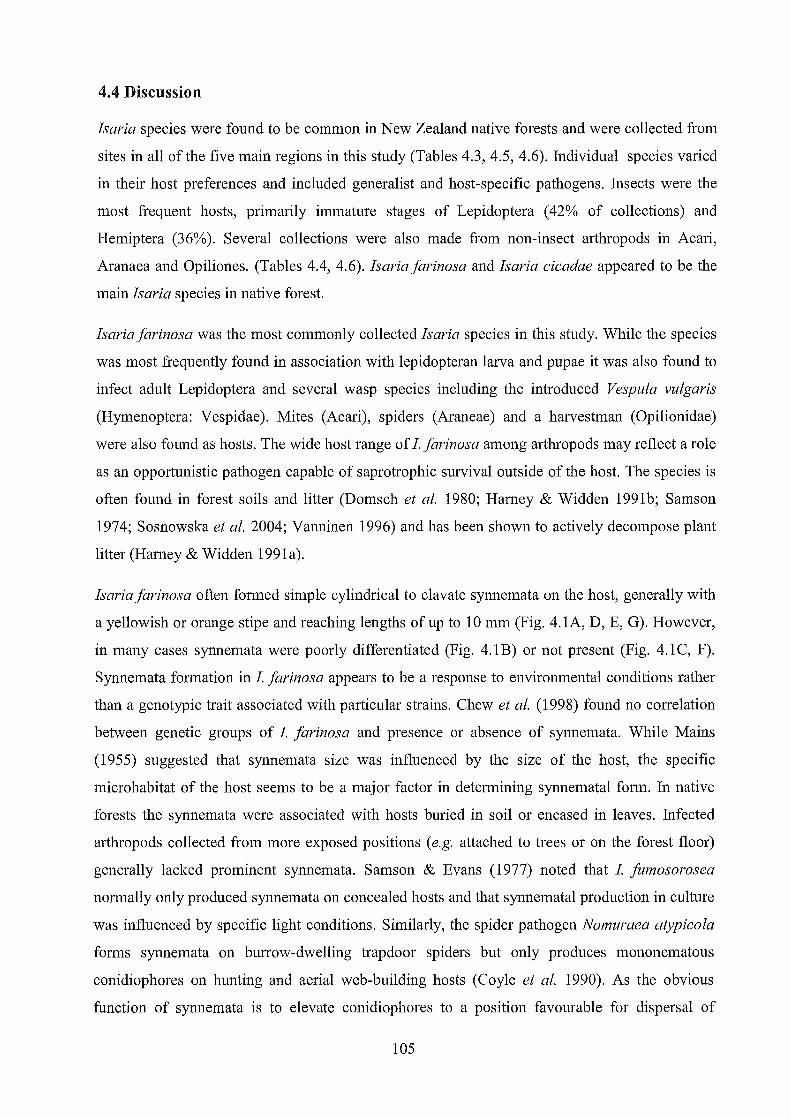
4.4 Discussion
Isaria species were found to be common in New Zealand native forests and were collected from
sites in all of the five main regions in this study (Tables 4.3, 4.5, 4.6). Individual species varied
in their host preferences and included generalist and host-specific pathogens. Insects were the
most frequent hosts, primarily immature stages of Lepidoptera (42% of collections) and
Hemiptera (36%). Several collections were also made from non-insect arthropods in Acari,
Aranaea and Opiliones. (Tables 4.4, 4.6). Isaria farinosa and Isaria cicadae appeared to be the
main Isaria species in native forest.
Isaria farinosa was the most commonly collected Isaria species in this study. While the species
was most frequently found in association with lepidopteran larva and pupae it was also found to
infect adult Lepidoptera and several wasp species including the introduced Vespula vulgaris
(Hymenoptera: Vespidae). Mites (Acari), spiders (Araneae) and a harvestman (Opilionidae)
were also found as hosts. The wide host range of 1. farinosa among arthropods may reflect a role
as an opportunistic pathogen capable of saprotrophic survival outside of the host. The species is
often found in forest soils and litter (Domsch et al. 1980; Harney & Widden 1991b; Samson
1974; Sosnowska et al. 2004; Vanninen 1996) and has been shown to actively decompose plant
litter (Harney & Widden 1991a).
Isaria farinosa often formed simple cylindrical to clavate synnemata on the host, generally with
a yellowish or orange stipe and reaching lengths of up to 10 mm (Fig. 4.1A, D, E, G). However,
in many cases synnemata were poorly differentiated (Fig. 4.1B) or not present (Fig. 4.1 C, F).
Synnemata formation in 1. farinosa appears to be a response to environmental conditions rather
than a genotypic trait associated with particular strains. Chew et al. (1998) found no correlation
between genetic groups of 1. farinosa and presence or absence of synnemata. While Mains
(1955) suggested that synnemata size was influenced by the size of the host, the specific
microhabitat of the host seems to be a major factor in determining synnematal form. In native
forests the synnemata were associated with hosts buried in soil or encased in leaves. Infected
arthropods collected from more exposed positions (e.g. attached to trees or on the forest floor)
generally lacked prominent synnemata. Samson & Evans (1977) noted that 1. jil1nosorosea
normally only produced synnemata on concealed hosts and that synnematal production in culture
was influenced by specific light conditions. Similarly, the spider pathogen Nomuraea atypicola
forms synnemata on burrow-dwelling trapdoor spiders but only produces mononematous
conidiophores on hunting and aerial web-building hosts (Coyle et al. 1990). As the obvious
function of synnemata is to elevate conidiophores to a position favourable for dispersal of
105

conidia (Evans 1982), their production has become an ecological requirement for species which
infect hosts that are buried or otherwise hidden (Samson & Evans 1977).
Isaria farinosa was primarily distinguished from other members of the genus examined in this
study by its ellipsoidal to fusiform conidia (Fig. 4.2A) measuring 2.0-2.9 x 1.1-1.9 /lm on the
host and 2.1-3.2 x 1.1-2.0 /lm in culture. Phialides had ellipsoidal bases which tapered to a thin
neck (Fig. 4.2C-E), and measured 3.8-9.8 x 1.9-3.5 /lm. Although phialides and conidia were
significantly longer on non-insect hosts, this was not demonstrated in measurements from
cultures. This may suggest the differences are related to variation in the nutritional composition
of the two groups of hosts. Morphological characteristics of New Zealand representatives
matched closely with the description of Paecilomyces farinosus by Samson (1974) who
recorded conidia as measuring 2.0- 3.0 x 1.0- 1-8 /lm and phialides as 5.0-15.0 x 1.2-2.5 /lm.
Isaria farinosa was first described as Ramaria farinosa by Holm from Denmark in 1780,
although none of his original specimens were preserved (Hodge et al. 2005). In the recent
establishment of I farinosa as the type species for Isaria, Hodge et al. (2005) designated Holm's
original illustration as the lectotype for the species. However, as the figure lacks microscopic
detail and could easily also represent I tenuipes, a specimen and corresponding culture of I
farinosa from Denmark were selected as an epitype. A sequence from the epitype culture was
included in the ITS phylogeny of Luangsa-ard et al. (2005), but did not group closely with other
included representatives of the species, demonstrating that I farinosa is not monophyletic within
Isaria.
In the ITS phylogeny presented in this study, New Zealand isolates identified as I farinosa
grouped with examples of the species from various countries, forming a well-supported clade at
a basal position in the tree that was distinct from the epitype strain (CBS 111113). Analysis of
partial B-tubulin sequences also clustered New Zealand isolates with overseas strains of I
farinosa in a different clade from the type. New Zealand isolates again grouped with overseas
strains of I farinosa in the EF1-a analysis, although a sequence from the type culture was not
available for comparison. The majority of New Zealand ITS sequences were identical to most
sequences from overseas strains, although others diverged by several nucleotides. Three isolates
(NC76, NC177, NC203) formed a well supported subclade in the ITS and EF1-a phylogenies
but this did not correlate with any differences in conidial size or host affiliation. All B-tubulin
sequences from examined isolates ofNZ Ifarinosa were identical.
Four isolates from lepidopteran pupae (NC108, NC180, NC184, NC212) were morphologically
similar to I farinosa with ellipsoidal to fusiform conidia (Fig. 4.2B), but conidia were found to
106
be larger, measuring 2.5-3.8 x 1.5-2.5 !lm on the host material and 2.6-4.0 x 1.8-2.7 !lm in
culture. The arrangement of conidiogenous structures was similar to that observed in specimens
identified as 1. jarinosa, although phialides typically had a wider base, ranging from 2.3-4.0 !lm
(Fig 4.2F-H). All of the collected material was characterised by the production of simple white
synnemata on the host (Fig. 4.1H). Molecular analyses placed these apart from other isolates in
a distinct clade which was well-supported in the ITS, EF1-a, and p-tubulin phylogenies. A close
affiliation with the epitype strain of 1. jarinosa (CBS 111113) was shown from analysis of the
ITS and p-tubulin regions. Both morphological and molecular data clearly indicates that these
isolates should be placed in a separate species from the more commonly encountered 1. farinosa
with smaller spores. However, the taxonomic status of "1. jarinosa" seems uncertain and
molecular data from this study and from Luangsa-ard et at. (2005) indicates that the strain
selected as the type may not be an ideal representative of the species. Currently, the taxon
appears to be a species complex that requires further phylogenetic revision.
Isaria tenuipes was collected and isolated from lepidopteran pupae buried under moss or in leaf
litter. The species appeared to be less common than 1. jarinosa or 1. cicadae, which may explain
the scarcity of previous New Zealand records. Typically, the species produced characteristic
synnemata which were highly branched with bright yellow stipes, extending up to about 20 mm
from the host (Fig. 4.3). Synnemata of 1. tenuipes have often been described as resembling 1.
farinosa (Bissett 1979a; Mains 1955; Samson 1974). However, as also noted by Petch (1937),
the examined specimens had conidial heads that were composed of distinct terminal branches
and were less compact than in 1. jarinosa, giving a characteristic plumose appearance. This was
especially pronounced in older specimens that had lost most of their conidia.
Conidia of 1. tenuipes were ellipsoidal to cylindrical, often slightly curved or allantoid (Fig.
4.4A-C). Conidial dimensions showed some variation on the host, with conidia of most
specimens ranging from 3.2-5.4 x 1.2-2.4 !lm, while several collections had conidia up to 6.9
!lm long. In culture, conidia measured 3.9-9.1 x 1.4-3.5 !lm. Samson (1974) recordedP. tenuipes
strains as having two distinct types of conidia: either one-celled and measuring 3-7.5 x 2.0-2.5
!lm or 6-12 !lm long with one or two cells. No two-celled conidia were seen in the New Zealand
isolates. Phialides generally measured 3.6-6.6 x 2.7-5.3 !lm and had a globose-subglobose base,
tapering abruptly or gradually to a short, thin neck (Fig. 4.4D-F). Metulae were typically
globose-subglobose and borne on swollen, densely branched conidiophores (Fig. 4.4G-H).
Morphology closely matched the description of the species in Samson (1974). ITS sequences of
the New Zealand isolates showed no variation and were identical to sequences from overseas
strains, including a culture derived from the type locality in North America (ARSEF5135).
107

All Isaria species on cicada nymphs collected in native forest were identified as Isaria cicadae.
The species was often observed in large numbers, and at one site over 50 individual specimens
were counted in an area covering about 25 square metres. In most cases 1. cicadae could be
easily identified before microscopic examination due to its specific host and characteristic stout,
branched synnemata which reached up to 70 mm long with conidia aggregated in compact,
cauliflower-like heads (Fig. 4.5A-D). Conidia from host material were cylindrical and usually
highly curved (Fig. 4.6A), measuring from 3.2-6.5 x 1.1-2.7 ).tm, which corresponds closely
with 1. cicadae according to Samson (1974). Conidia in culture were extremely variable; some
were similar in size and shape to those from the host while others were cylindrical and straight
or irregularly curved, sometimes with a tunicate (hook-shaped) or sigmoid appearance, and
measuring up to 12.3 ).tm long. As shown by Kobayasi (1939, 1941), Kobayasi & Shimizu
(1963), and Samson (1974), conidiogenous structures (Fig. 4.6C-D) strongly resembled those of
1. tenuipes, with both species producing phialides with a globose-subglobose base and a short,
thin neck from globose-sub globose metulae and densely branched, highly swollen
conidiophores.
Three isolates (NC122, NC112, NC127) from hosts other than cicadas (lepidopteran pupae and a
coleopteran species) produced cylindrical, curved conidia (Fig. 4.6B) measuring 5.7-9.0 x 1.6-
2.5 ).tm on the host and 6.2-13.7 x 1.4 -3.0 ).tm in culture. Phialides were similar to those
observed on cicada specimens (Fig. 4.6E-F). While these also tend to fit with Samson's
description of 1. cicadae they were clearly different from the New Zealand specimens described
from cicada nymphs. Although one specimen had similar synnemata to those produced on
cicadas, the other collections had greatly reduced synnemata that extended only a few
millimeters from the host (Fig. 4.5E-F). Analysis ofITS and EF1-a sequences also supported the
distinction between the two groups of New Zealand 1. cicadae-like isolates, although differences
were limited to two nucleotides in each region.
Isaria cicadae was first described by Miquel in 1838 for a fungus infecting buried cicada
nymphs in Brazil (Petch 1933). The species received little attention until it was considered by
Petch to be an earlier synonym of Isaria sinclairii which had been originally described from
New Zealand material as Cordyceps sinclairii by Berkeley (1855). Although the original
description of C. sinclairii did not include a figure the species was later illustrated in Berkeley
(1857) and also (in more detail) by Gray (1858). Berkeley (1855) recorded the fungus as
occurring on an "orthopterous insect"; however Gray (1858) was more definite in stating that the
host was a cicada. Torrubia caespitosa, described from cicadas by Tulasne in 1865, was
apparently based on the same set of specimens examined by Berkeley (Cooke 1892; Lloyd
108
1915). A fungus illustrated by Taylor (1855) from New Zealand and (invalidly) named as
Sphaeria basili also appears to be the same species. The host was given by Taylor as a "locust",
a term often used in the nineteenth century to describe cicadas (Kritsky 2001).
Massee (1895) and Cunningham (1921) both noted that C. sinclairii had never been shown to
produce perithecia and the species was accordingly recombined in Isaria by Lloyd (1923). The
description by Berkeley (1855) of Cordyceps sinclairii gave little detail of microscopic
characters, although spores were given as about 7 /lm long and oblong-shaped. Petch (1924)
provided the first detailed description of Isaria sinclairii from specimens collected in Ceylon.
Conidia were described as oblong-oval measuring 8.0-10.0 x 2.0-3.0 /lm. Petch (1933) later
examined specimens of 1. sinclairii from New Zealand and Mexico, and deciding that these were
the same as the species earlier described by Miquel, synonymised 1. sinclairii and several other
species with 1. cicadae. Kobayasi (1939) however, disagreed with Petch and preferred to retain
the name 1. sinclairii, stating that 1. cicadae was "obscurely known with inadequate description
based on the sterile and dried specimen". Conidia of 1. sinclairii were recorded from Japanese
material by Kobayasi (1939, 1941) and Kobayasi & Shimizu (1963) as ovoid, elongate
ellipsoidal, or fusiform; frequently curved; and measuring 5.0-9.0 x 2.0-3.0 /lm.
In transferring entomopathogenic Isaria species to Paecilomyces, Samson (1974) followed Petch
(1933) and made the combination Paecilomyces cicadae, giving spore dimensions as 3.5-8.0 x
1.5-3.5 /lm. Samson examined specimens from various countries including a New Zealand
collection of Cordyceps sinclairii from Berkeley'S herbarium. Samson (1974 fig. 20) illustrated
three sets of conidia for P. cicadae: two groups equate to the large cylindrical-ellipsoidal conidia
as described for 1. sinclairii from Ceylon (Petch 1924) and Japan (Kobayasi 1939), while the
third set are identical to the smaller, highly curved conidia observed for New Zealand 1. cicadae
in the present study. Although Samson does not reference these conidial types to any particular
specimen it seems likely that the smaller conidia illustrated are from the New Zealand specimens
he examined.
It is suggested that New Zealand strains identified as Isaria cicadae, with conidia measuring 3.2-
6.5 x 1.1-2.0 /lm may represent a separate species to similar fungi with larger conidia recorded
from cicadas in other parts of the world. New Zealand isolates with large conidia from hosts
other than cicadas appear to be an allied species that may be more closely related to overseas 1.
cicadae. The morphological distinction between the two groups is also supported to a limited
extent by phylogenetic data, although a lack of available sequences prevented a detailed
molecular comparison with strains from other countries. Further examination of New Zealand
109

and overseas isolates using both morphology and multiple, high-resolution molecular markers is
clearly necessary to clarify the taxonomy of this species.
110

CONCLUSIONS
Species of Beauveria and Isaria were found to be frequently occurring in native forests and must
function as important natural regulators of arthropod populations in these habitats. The two
recognized phylogenetic species (clades A and C) within Beauveria bassiana were both found to
be present in native forests. Clade C has not been identified from disturbed habitats in this
country and it is suggested that the group is restricted to natural forests in New Zealand.
Beauveria malawiensis, previously only known from coleopteran hosts, was found to have a
broad host range in native forests, infecting representatives of the insect orders Hemiptera,
Hymenoptera, Orthoptera and Phasmatodea. It is suggested that many previous records of
Beauveria brongniartii in this country may have been B. malawiensis. Insect bioassays generally
indicated that individual strains of B. malawiensis and B. bassiana are not highly host-specific
and demonstrated the potential of the both species for the biological control of Vespula wasps.
Beauveria caledonica, previously known only from pine forests in New Zealand, was also found
to be established in native forests. The species was also recorded for the first time from non
coleopteran hosts. Isaria farinosa and 1. tenuipes, although poorly recorded in New Zealand,
were both present in native forests. Molecular data supported previous findings that 1. farinosa is
not monophyletic and may consist of an assemblage of morphologically similar species. Two
distinct groups of 1. cicadae-like fungi were identified in native forests. It is suggested that
species from cicada nymphs in New Zealand previously classified as I. cicadae may represent a
separate, possibly endemic species.
Molecular analyses presented in this study have indicated several unique lineages in New
Zealand representatives of Beauveria and Isaria species. Future studies using additional
molecular markers are necessary to provide further insight into the diversity and
phylogeographic origins of these species in New Zealand.
111
REFERENCES
Andersen, S., Hojrup, P. & Roepstorff, P. (1995). Insect cuticular proteins. Insect Biochemistry
and Molecular Biology 25, 153-176.
Anon (1892). Entomological. White grub culture. Otago Witness 2411111892, p5.
Anon (1893). Entomological. Sporotrichum globuliferum. Otago Witness 4/4/1893, p5.
Anon (2001-2009) [Herbarium PDD]. NZFUNGI Database of New Zealand Fungi.
http://nzfungi.1andcareresearch.co.nz/htmllmyco1ogy.asp.
Aquino de Muro, M.A., Elliott, S., Moore, D., Parker, B.L., Skinner, M., Reid, W. & E1 Bouhssini, M. (2005). Molecular characterisation of Beauveria bassiana isolates obtained from overwintering sites of SUllll Pests (Eurygaster and Aelia species).
Mycological Research 109,294-306.
Aquino de Muro, M.A., Mehta, S. & Moore, D. (2003). The use of amplified fragment length polymorphism for molecular analysis of Beauveria bassiana isolates from Kenya and other countries, and their correlation with host and geographical origin. FEMS
Microbiology Letters 229, 249-257.
Baker, G. (1985). Parasites of the millipede Ommatoiulus moreletti (Lucas) (Dip10poda: Lu1idae) in Portugal, and their potential as biological control agents in Australia.
Australian Journal of Zoology 33,23-32.
Baker, R. (1981). Natural mortality of currant clearwing, Synanthedon salmachus (Lepidoptera: Sesiidae), in New Zealand. New Zealand Journal of Zoology 8,529-530.
Barker, G. & Addison, P. (1989). Sampling Argentine stem weevil, Listronotus bonariensis
(Kuschel), populations in pasture: the soil-dwelling stages. New Zealand Journal of
Agricultural Research 32, 95-103
Barker, C.W. & Barker, G.M. (1998). Generalist entomopathogens as biological indicators of deforestation and agricultura11and use impacts on Waikato soils. New Zealand Journal of
Ecology 22, 189-196.
Barker, G.M., Goh, H.H., Lyons, S.N. & Addison, P.J. (1991). Comparative pathogenicity to Argentine stem weevil of Beauveria bassiana from various hosts. Proceedings of the NZ
Weed and Pest Control Conference 44,214-215.
Barlow, N. & Goldson, S. (2002). Alien invertebrates in New Zealand. In Biological invasions -
Environmental impacts (ed. D. Pimentel) pp. 195-217. CRC Press: Boca Raton.
Baverstock, l, Clark, S.l & Pell, lK. (2008). Effect of seasonal abiotic conditions and field margin habitat on the activity of Pandora neoaphidis inoculum on soil. Journal of
Invertebrate Pathology 97, 282-290.
Beggs, J.R. (2001). The ecological consequences of social wasps (Vespula spp.) invading an ecosystem that has an abundant carbohydrate resource. Biological Conservation 99, 17-
28.
112
Beggs, J.R., Rees, lS. & Harris, R.I. (2002). No evidence for establishment of the wasp parasitoid, Sphecophaga vesparum burra (Cresson) (Hymenoptera: Ichneumonidae) at
two sites in New Zealand. New Zealand Journal o/Zoology 29,205-211.
Beggs, J.R., Rees, lS., Toft, R.J., Dennis, T.E. & Barlow, N.D. (2008). Evaluating the impact of a biological control parasitoid on invasive Vespula wasps in a natural forest ecosystem.
Biological Control 44, 399-407.
Beggs, J.R., Toft, R.I., Malham, J., Rees, lS., Tilley, J., Moller, H. & Alspach, P.A. (1998). The difficulty of reducing introduced wasp (Vespula vulgaris) populations for conservation gains. New Zealand Journal o/Ecology 22,55-63.
Beggs, J.R. & Wilson, P. (1991). The kaka, Nestor meridionalis, a New Zealand parrot endangered by introduced wasps and mammals. Biological Conservation 56, 23-38.
Berkeley, M. (1855). Nat. Ord. ClI. Fungi. In The Botany o/the Antarctic Voyage II. Flora Novae-Zelandiae. Part II. Flowerless Plants (eds. J. Hooker) pp. 172-210. Lovell Reeve:
London.
Berkeley, M. (1857). Introduction to cryptogamic botany. H. Bailliere: London.
Bidochka, M.J, Kamp, A.M., Lavender, T.M., Dekoning, J., & De Croos, IN. (2001). Habitat association in two genetic groups of the insect-pathogenic fungus Metarhizium anisopliae: uncovering cryptic species? Applied and Environmental Microbiology 67,
1335-1342.
Bidochka, M.l, McDonald, M.A., St Leger, R.l & Roberts, D.W. (1994). Differentiation of species and strains of entomopathogenic fungi by random amplification of polymorphic
DNA(RAPD). Current Genetics 25, 107-113.
Bidochka, M.I., Menzies, F.V. & Kamp, A.M. (2002). Genetic groups of the insect-pathogenic fungus Beauveria bassiana are associated with habitat and thermal growth preferences.
Archives 0/ Microbiology 178, 531-537.
Bidochka M.l & Small, C. (2005). Phylogeography of Metarhizium, an insect pathogenic fungus. In Insect-fungal associations: ecology and evolution (eds. F.E. Vega & M. Blackwell) pp. 28-50. Oxford University Press: Oxford.
Bissett, J. (1979a). Paecilomyces tenuipes. Fungi Canadenses 158, 1-2.
Bissett, J. (1979b). Paecilomyces breviramosus. Fungi Canadenses 159, 1-2.
Bissett, J. & Widden, P. (1988). A new species of Beauveria isolated from Scottish moorland soil. Canadian Journal of Botany 66, 361-362.
Booth, C. (1961). Studies ofPyrenomycetes: VI. Thielavia, with notes on some allied genera.
Mycological Papers 83, 1-15.
Boucias, D.G. & Pendland, J.C. (1984). Nutritional requirements for conidial germination of several host range pathotypes of the entomopathogenic fungus, Nomuraea rileyi. Journal
0/ Invertebrate Pathology 43, 288-292.
113

Boucias, D.G. & Pendland, lC. (1991). Attachment of my cop at hog ens to cuticle: the initial
event of mycoses in arthropod hosts. In The jimgal spore and disease initiation in plants and animals (eds. G. Cole & H. Hoch) pp. 101-128. Plenum Press: New York.
Boucias, D.G., Pendland, J.C. & Latge, J.P. (1988). Nonspecific Factors Involved in Attachment
of Entomopathogenic Deuteromycetes to Host Insect Cuticle. Applied and Environmental Microbiology 54, 1795-1805.
Bridge, P., Abraham, Y., Cornish, M., Prior, C. & Moore, D. (1990). The chemotaxonomy of
Beauveria bassiana (Deuteromycotina: Hyphomycetes) isolates from the coffee berry
borer Hypothenemus hampei (Coleoptera: Scolytidae). Mycopathologia 111, 85-90.
Bridge, P.D., Spooner, B. & Roberts, P. (2005). The impact of molecular data in fungal
systematics. Advances in Botanical Research 42,33-67.
Brito, M. (1994). The millipede of Santo Antao, Spinotarsus caboverdus: survey for pathogenic
microorganisms, bioassay tests against S. caboverdus and Melanoplus sanguinipes. Unpublished M.Sc. thesis, Montana State University, Boseman.
Brown, A. & Smith, G. (1957). The genus Paecilomyces Bainier and its perfect stage
Byssochlamys Westling. Transactions of the British Mycological Society 40, 17-89.
Brown, J. & Hovmoller, M. (2002). Aerial dispersal of pathogens in the global and continental
scales and its impact on plant disease. Science 297,537-541.
Brownbridge, M., Nelson, T.L., Hackell, D.L., Eden, T.M., Wilson, D.J, Willoughby, B.E. &
Glare, T.R. (2006). Field application of biopolymer-coated Beauveria bassiana F418 for
clover root weevil (Sitona lepidus) control in Waikato and Manawatu. New Zealand Plant Protection 59, 304-31.
Bruns, T. (2001). ITS reality. Inoculum 52, 2-3.
Bruns, T. & Shefferson, R. (2004). Evolutionary studies of ectomycorrhizal fungi: recent
advances and future directions. Canadian Journal of Botany 82, 1122-1132.
Bruns, T., White, T. & Taylor, l (1991). Fungal molecular systematics. Annual Review of Ecology and Systematics 22, 525-564.
Buchanan, P.K., Beever, R.E., Galloway, D.J., Glare, T.R., Johnston, P.R., McKenzie, E.H.C.,
di Menna, M.E., Pennycook, S.R., Ridley, G.S., Smith, J.M.B. & Stephenson, S.L.
(2004). The fungi of New Zealand: an introduction. In Introduction to Fungi of New
Zealand (ed. E. McKenzie) pp. 1-48. Fungal Diversity Press: Hong Kong.
Butt, T.M. (2002). Use of entomogenous fungi for the control of insect pests. In The Mycota XI. Agricultural applications (ed. F. Kempen) pp. 111-134. Springer-Verlag: Berlin.
Bye, N. & Charnley, A. (2008). Regulation of cuticle-degrading subtilisin proteases from the
entomopathogenic fungi, Lecanicillium spp.: implications for host specificity. Archives of Microbiology 189, 81-92.
Cameron, P.l & Walker, G.P. (1989). Acyrthosiphon kondoi Shinji, blue-green lucerne aphid and Acyrthosiphon pisum (Harris), pea aphid (Homoptera: Aphididae). In A review of biological control of invertebrate pests and weeds in New Zealand 1874-1987 (eds.
114

P.l. Cameron, R.L. Hill, 1. Bain & W.P. Thomas) pp. 3-8. CAB International, Wallingford.
Carruthers, R., Sawyer, A. & Hural, K. (1991). Use of fungal pathogens for biological control of
insect pests. In Sustainable agriculture research and education in the field (eds. B. Rice)
pp. 336-372. National Academy Press: Washington.
Castellanos-Mogue1, 1., Gonzales-Barajas, M., Mier, T., del Rocio Reyes-Montes, M., Aranda, E. & Toriello, C. (2007). Virulence testing and extracellular subtilisin-like (Prl) and
trypsin-like (Pr2) activity during propagule production of Paecilomyces jitmosoroseus isolates from whiteflies (Homoptera: Aleyrodidae). Revista Iberoamericana de Micologia 26,62-68.
Castrillo, L.A., Vandenberg, J.D. & Wraight, S.P. (2003). Strain-specific detection of introduced
Beauveria bassiana in agricultural fields by use of sequence-characterized amplified
region markers. Journal o/Invertebrate Pathology 82, 75-83.
Castrillo, L.A., Wiegmann, B.M. & Brooks, W.M. (1999). Genetic variation in Beauveria bassiana populations associated with the darkling beetle, Alphitobius diaperinus. Journal o/Invertebrate Pathology 73, 269-275.
Chandler, D., Davidson, G., Grant, W., Greaves, G. & Tatchel, G. (2008). Microbial
biopesticides for integrated crop management: an assessment of environmental and
regulatory stability. Trends in Food Science and Technology 19, 275-283.
Chandler, D., Davidson, G., Pell, 1.K., Ball, B., Shaw, K. & Sunderland, K. (2000). Fungal biocontrol of Acari. Biocontrol Science and Technology 10, 357-384.
Charnley, A. (1991). Microbial pathogens and insect pest control. Letters in Applied Microbiology 12, 149-157.
Charnley, A. (2003). Fungal pathogens of insects: cuticle degrading enzymes and toxins.
Advances in Botanical Research 40, 241-321.
Charnley, A. & Collins, S. (2007). Entomopathogenic fungi and their role in pest control. In The Mycota IV. Environmental and microbial relationships (eds. C. Kubicek & 1. Druzhinina) pp. 159-187. Springer-Verlag: Berlin.
Chaverri, P., Bischoff, J.F., Evans, H.C. & Hodge, K.T. (2005). Regiocrella, a new
entomopathogenic genus with a pycnidial anamorph and its phylogenetic placement in the Clavicipitaceae. Mycologia 97, 1225-1237.
Chaverri, P., Liu, M. & Hodge, K.T. (2008). A monograph of the entomopathogenic genera
Hypocrella, Moelleriella, and Samuelsia gen. nov. (Ascomycota, Hypocreales,
Clavicipitaceae), and their aschersonia-like anamorphs in the Neotropics. Stud Mycol60, 1-66.
Chew, J.S.K., Strongman, D.B. & Mackay, R.M. (1998). Comparisons of twenty isolates ofthe
entomopathogen Paecilomyces /arinosus by analysis of RAPD markers. Mycological Research 102, 1254-1258.
Cist, 1. (1824). Notice ofthe Melolontha or may bug. American Journal 0/ Science and Arts 8, 269-271.
115
Clapperton, B., Lo, P.L., Moller, H., Sandlant, G.R. (1989). Variation in colour markings of
German wasps Vespula germanica (F.) and common wasps Vespula vulgaris (L.)
(Hymenoptera: Vespidae) in New Zealand. New Zealand Journal o/Zoology 16,303-313.
Clarkson, J.M. & Charnley, A (1996). New insights into the mechanisms of fungal pathogenesis
in insects. Trends in Microbiology 4, 197-203.
Clements, F.E. & Shear, C.L. (1931). The genera o/jimgi. Hafner Press: New York.
Close, R. (1956). A muscardine fungus on a caterpillar of Antheraea eucalypti Scott
(Lepidoptera, Satumiidae). New Zealand Entomologist 2, 1-2.
Cokendolpher, l (1993). Pathogens and parasites of Opiliones (Arthropoda: Arachnida). The Journal 0/ Arachnology 21, 120-146.
Cook, R.l, Bruckart, W.L., Coulson,lR., Goettel, M.S., Humber, R.A, Lumsden, R.D.,
Maddox., J.V., McManus., M.L., Moore M., L., Meyer, S.F., Quimby, P.C., Stack lP.,
Vaughn, J.L. (1996). Safety of microorganisms intended for pest and plant disease
control: a framework for scientific evaluation. Biological Control 7, 333-351.
Cooke, M.e. (1892). Vegetable wasps and plant worms. A popular history 0/ entomogenous jimgi, or jimgi parasitic upon insects. Society for promoting Christian knowledge:
London.
Coyle, F.A, Goloboff, P.A & Samson, R.A (1990). Actinopus trapdoor spiders (Araneae,
Actinopodidae) killed by the fungus, Nomuraea atypicola (Deuteromycotina). Acta Zoologica Fennica 190, 89-93.
Crosby, T., Dugdale, J. & Watt, J. (1976). Recording specimen localities in New Zealand: an
abitrary system of areas and codes defined. New Zealand Journal o/Zoology 3,69.
Crosby, T., Dugdale, l & Watt, l (1998). Area codes for recording specimen localities in the
New Zealand subregion. New Zealand Journal o/Zoology 25, 175-183.
Crowe, A. (2002). Which New Zealand insect? Penguin Books: Auckland.
Cruz, L.P., Gaitan, AL., Gongora, C.E., AF Cruz, L.P., Gaitan, AL. & Gongora, C.B. (2006).
Exploiting the genetic diversity of Beauveria bassiana for improving the biological
control of the coffee berry borer through the use of strain mixtures. Applied Microbiology and Biotechnology 71, 918-926.
Cunningham, G. (1921). The genus Cordyceps in New Zealand. Transactions and Proceedings o/the New Zealand Institute 53,372-382.
Cunningham, G. (1922). A singular Cordyceps from Stephen Island, New Zealand. Transactions o/the British Mycological Society 8, 72-75.
Curran, J., Driver, F., Ballard, l & Milner, R. (1994). Phylogeny of Metarhizium: analysis of
ribosomal DNA sequence data. Mycological Research 98,547-552.
de Faria, M. & Wraight, S. (2007). Mycoinsecticides and mycoacaricides: A comprehensive list
with worldwide coverage and international classification of formulation types. Biological Control 43, 237-256.
116

de Hoog, G. (1972). The genera Beauveria, isaria, Tritirachium and Acrodontium gen. nov.
Studies in Mycology 1, 1-41.
de Hoog, G. (1978). Notes on some fungicolous Hyphomycetes and their relatives. Persoonia
10,33-81.
de Hoog, G. & Rao, V. (1975). Some new hyphomycetes. Persoonia 8,207-212.
Dingley, J. (1951). The Hypocreales of New Zealand II. The genus Nectria. Transactions of the
Royal Society of New Zealand 79, 177-202.
Dingley, J. (1953). The Hypocreales of New Zealand V. The genera Cordyceps and Torrubiella. Transactions of the Royal Society of New Zealand 81,329-343.
Dingley, J. (1954). The Hypocreales of New Zealand.VI. The Genera Hyp 0 crella, Barya, Claviceps and Podonectria. Transactions of the Royal Society of New Zealand 81,489-499.
Domsch, K, Gams, W. & Anderson, T. (1980). Compendium ofsoilfimgi. Academic Press:
London.
Dromph, K & Vestergaard, S. (2002). Pathogenicity and attractiveness ofentomopathogenic
hyphomycete fungi to collembolans. Applied Soil Ecology 21, 197-210.
Edwards, G. (1764). Gleanings of natural history, containingfigures of quadrupeds, birds, insects, plants, etc. Vol. 3. Royal College of Physicians: London.
Edwards, J.S. (1965). Entomogenous micro-organisms of Prionoplus reticularis (White). New Zealand Entomologist 3,21.
Eilenberg, J., Hajek, A. & Lomer, C. (2001). Suggestions for unifying the terminology in
biological control. BioControl46, 387-400.
Ekesi, S., Shah, P., Clark, S. & Pell, J. (2005). Conservation biological control with the fungal pathogen Pandora neoaphidis; implications of aphid species, host plant and predator
foraging. Agricultural and Forest Entomology 21-30.
Enkerli, J., Widmer, F., Gessler, C. & Keller, S. (2001). Strain-specific micro satellite markers in the entomopathogenic fungus Beauveria brongniartii. Mycological Research 105, 1079-
1087.
Estrada, M., Camacho, M. & Benito, C. (2007). The molecular diversity of different isolates of Beauveria bassiana (Bals.) Yuill. as assessed using intermicrosatellites (ISSRs). Cellular & Molecular Biology Letters 12, 240-252.
Evans, H.C. (1974). Natural control of arthropods, with special reference to ants (Formicidae), by fungi in tropical high forest of Ghana. Journal of Applied Ecology 11,37-49.
Evans, H.C. (1982). Entomogenous fungi in tropical forest ecosystems - an appraisal. Ecological
Entomology 7, 47-60.
Evans, H.C. (1988). Coevolution of entomogenous fungi and their insect hosts. In Coevolution of Fungi with Plants and Animals (eds. K Pirozynski & D. Hawksworth) pp. 149-171.
Academic Press: London.
117

Evans, H.C. (1989). Mycopathogens of insects in epigeal and aerial habitats. In Insect-fimgus
interactions (eds. N. Wilding, N. Collins, P. Hammond & J. Webber) pp. 205-237.
Academic Press: London.
Evans, RC. & Samson, R.A. (1982a). Cordyceps species and their anamorphs pathogenic on
ants (Formicidae) in tropical forest ecosystems 1. The Cephalotes (Myrmicinae) complex.
Transactions of the British Mycological Society 79, 431-453.
Evans, H.C. & Samson, R.A. (1982b). Entomogenous fungi from the Galapagos islands.
Canadian Journal of Botany 60, 2325-2333.
Evans, RC. & Samson, R.A. (1984). Cordyceps species and their anamorphs pathogenic on ants
(Formicidae) in tropical forest ecosystems II. The Camponotus (Formicinae) complex.
Transactions of the British Mycological Society 81, 127-150.
Fargues, J. & Remaudiere, G. (1977). Considerations on the specificity of entomopathogenic
fungi. Mycopathologia 62,31-37.
Federici, B. (1999). Naturally occuring baculoviruses for insect pest control. In Biopesticides:
use and delivery (eds. F. Hall & J. Menn) pp. 301-320.
Felsenstein, J. (1985). Confidence limits on phylogenies: An approach using the bootstrap.
Evolution 39, 783-791.
Ferguson, A.M. & Fletcher, J.D. (1991). Greedy scale and the fungus Fusarium stilboides.
Proceedings of the New Zealand Weed and Pest Control Conference 44, 260-161.
Fraser, K. (1994). John Hill and the Royal Society in the Eighteenth Century. Notes and Records
of the Royal Society of London 48, 43-67.
Freimoser, F., Hu, G. & St Leger, R.J. (2005). Variation in gene expression patterns as the insect
pathogen Metarhizium anisopliae adapts to different host cuticles or nutrient deprivation
in vitro. Microbiology 151, 361-371.
Fukatsu, T., Sato, H. & Kuriyama, R (1997). Isolation, inoculation to insect host, and molecular
phylogeny of an entomogenous fungus Paecilomyces tenuipes. Journal of Invertebrate
Pathology 70, 203-208.
Fuller, R., Buchanan, P. & Roberts, M. (2004). Maori Knowledge of Fungi / Matauranga 0 Nga Horere. In Introduction to Fungi of New Zealand (ed. E. McKenzie) pp. 81-118. Fungal Diversity Press: Hong Kong.
Fuxa, J. (1987). Ecological considerations for the use of entomopathogens in IPM. Annual
Review of En tomo logy 32,225-251.
Gams, W. (1971). Cephalosporium-artige Schimmelpilze. Gustav Fischer Verlag: Stuttgart.
Gams, W., Hodge, K.T., Samson, R.A., Korf, R.P. & Seifert, K.A. (2005). (1684) Proposal to
conserve the name Isaria (anamorphic fungi) with a conserved type. Taxon 54, 537-539.
Gams, W., O'Donnell, K., Schroers, H.-J. & Christensen, M. (1998). Generic classification of
some more hyphomycetes with solitary conidia borne on phialides. Canadian Journal of
Botany 76, 1570-1583.
118

Geiser, D. (2004). Practical molecular taxonomy of fungi. In Advances infungal biotechnology
for industry, agriculture and medicine (eds. J. Tkacz & L. Lange) pp. 3-14. Kluwer
Academic/Plenum Publishers: New York
Geiser, D., del Mar Jiminez-Gasco, M., Kang, S., Makalowska, 1., Veeraraghavan, N., Ward, T.,
Zhang, N., Kuldau, G. & O'Donnell, K. (2004). FUSARIUM-ID v. 1.0: A DNA
Sequence Database for Identifying Fusarium. European Journal of Plant Pathology 11 0,
473-479.
Gillespie, J., Bailey, A, Cobb, B. & Vilcinskas, A (2000). Fungi as elicitors of insect immune
responses. Archives of Insect BiochemistlY and Physiology 44, 49-68.
Glare, T. (2004). Molecular characterisation in the entomopathogenic fungal genus Beauveria.
Laimburg Journal 1, 286-298.
Glare, T.R. & Inwood, A.J. (1998). Morphological and genetic characterisation of Beauveria
spp. from New Zealand. Mycological Research 102, 250-256.
Glare, T.R., Inwood, AJ., van der Wielen, P. & Nelson, T. (1996a). Identification of novel
starins of Beauveria and Metarhizium in New Zealand. In Proceedings of the Third
International Workshop on Microbial Control of Soil Dwelling Pests (eds. T.A Jackson
& T.R. Glare) pp. 39-49.
Glare, T.R., Jackson, T.A & Cistemas, E. (1993a). Beauveria vermiconia is an
entomopathogenic fungus. Mycological Research 97, 336-338.
Glare, T.R., Milner, R.J. & Beaton, C.D. (1996b). Variation in Metarhizium, a genus of fungal
pathogens attacking Orthoptera: is phialide morphology a useful taxonomic criterion?
Journal of Or tho pte ran Research 5, 19-27.
Glare, T., O'Callaghan, M. & Wigley, P.J. (1993b). Checklist of naturally-occurring
entomopathogenic microbes and nematodes in New Zealand. New Zealand Journal of
Zoology 20,95-120.
Glare, T.R., Reay, S.D., Nelson, T.L. & Moore, R. (2008). Beauveria caledonica is a naturally
occurring pathogen of forest beetles. Mycological Research 112,352-360.
Glass, N.L. & Donaldson, G.C. (1995). Development of primer sets designed for use with the
PCR to amplify conserved genes from filamentous ascomycetes. Applied and
environmental microbiology 61, 1323-1330.
Goettel, M.S. & Inglis, G.D. (1997). Fungi: Hyphomycetes. In Manual of Techniques in Insect
Pathology (eds. L.A Lacey) pp. 213-249. Academic Press: San Diego.
Goettel, M.S., Poprawski, T., Vandenberg, J.D., Li, Z. & Roberts, D.W. (1990). Safety to
nontarget invertebrates of fungal biocontrol agents. In Safety of microbial insecticides
(eds. L. Marshall & L.A. Lacey) pp. 209-231. CRC Press: Boca Raton.
Goh, H.H., Barker, G.M., Addison, PJ., Lyons, S.N. & Firth, A.C. (1991a). Comparative
pathogenicity of Beauveria bassiana isolates to adult Argentine stem weevil in the
laboratory. Proceedings of the New Zealand Weed and Pest Control Conference 44, 185-
188.
119
Goh, H.H., Willoughby, B., Prestidge, R.A. & Lyons, S.N. (1991b). Pathogenicity of Beauveria,
Metarhizium, Paecilomyces and Fusarium isolates against early instar grass grub
(Costelytra zealandica) larvae. Proceedings of the NZ Weed and Pest Control Conference 44, 209-211.
Goldson, S., Rowarth, J. & Caradus, J. (2005). The impact of invasive invertebrate pests in
pastoral agriculture: a review. New Zealand Journal of Agricultural Research 48, 401-415.
Gray, G. (1858). Notices of insects that are known to form the bases of fungoid parasites. Privately printed: London.
Grehan, J.R. (1982). Infection of Aenetus virescens (Lepidoptera: Hepialidae) larvae by the
fungus Beauveria bassiana. New Zealand Entomologist 7,327-329.
Grehan, J.R. & Wigley, P.l. (1984). Fungal and bacterial diseases ofpuriri moth, Aenetus
virescens (Lepidoptera: Hepialidae), larvae. New Zealand Entomologist 8, 61-63.
Hadapad, A., Reineke, A. & Zebitz, C.P.W. (2006). Genetic variability among Beauveria
brongniartii (Saccardo) Petch isolates from various geographical and host origins based
on AFLP analysis. Mitteilungen der Deutschen Gesellschaft fiir Allgemeine und Angewandte Entomologie 15, 71-76.
Hajek, A.E. (1997). Ecology of terrestrial fungal entomopathogens. Advances in Microbial Ecology 15, 193-249.
Hajek, A.E. (2004). Natural enemies: an introduction to biological control. Cambridge University Press: New York.
Hajek, A.E., Delalibera Junior, 1. & McManus, M.L. (2007a). Introduction of exotic pathogens
and documentation of their establishment and impact. In Field manual of techniques in
invertebrate pathology (eds. L.A. Lacey & H.K. Kaya) pp. 299-325. Springer: Dordrecht.
Hajek, A.E. & Goettel, M.S. (2007). Guidelines for evaluating effects of entomopathogens on
non-target organisms. In Field manual of techniques in invertebrate pathology (eds. L.A. Lacey & H.K. Kaya) Springer: Dordrecht.
Hajek, A.E., McManus, M.L. & Delalibera Junior, 1. (2007b). A review of introductions of
pathogens and nematodes for classical biological control of insects and mites. Biological Control 41, 1-13.
Hajek, A.E. & St Leger, R.A. (1994) Interactions between fungal pathogens and their hosts.
Annual review of En tom 0 logy 39,293-322.
Harney, S. & Widden, P. (1991a). Physiological properties of the entomopathogenic
hyphomycete Paecilomyces farinosus in relation to its role in the forest ecosystem. Canadian Journal of Botany 69,1-5.
Harney, S. & Widden, P. (199lb). The ecology of Paecilomycesfarinosus in two balsam fir
forests infested with spruce budworm. Canadian Journal of Botany 69,512-515.
Harris, R.l. & Etheridge, N. (2001). Comparison of baits containing fipronil and sulfluramid for
the control of Vespula wasps. New Zealand Journal of Zoology 28,39-48.
120

Harris, Rl., Harcourt, S.l, Glare, T.R, Rose, E. & Nelson, T.l. (2000). Susceptibility of
Vespula vulgaris (Hymenoptera: Vespidae) to generalist entomopathogenic fungi and their potential for wasp control. Journal of Invertebrate Pathology 75, 251-258.
Harris, R.l. & Rees, 1.S. (2000). Aerial poisoning of wasps. Science for Conservation 162, 1-26.
Hegedus, D. & Khachatourians, G. (1996). Identification and differentiation of the
entomopathogenic fungus Beauveria bassiana using polymerase chain reaction and single-strand conformation polymorphism analysis. Journal of Invertebrate Pathology
67,289-299.
Helson, G. (1965). Entomogenous microorganisms of New Zealand. Transactions of the Royal
Society of New Zealand Zoology 6, 1-6.
Hibbett, D.S., Binder, M., Bischoff, 1.F., Blackwell, M., Cannon, P.F., Eriksson, O.E., Huhndorf, S., lames, T., Kirk, P.M., Lucking, R., Thorsten Lumbsch, H., Lutzoni, F., Matheny, P.B., McLaughlin, D.l, Powell, M.l, Redhead, S., Schoch, C.S., Spatafora, 1.W., Stalpers, lA., Vilgalys, R., Catherine Aime, M.,Aptroot, A., Bauer, R., Begerow, D., Benny, G.L., Castlebury, L.A., Crous, P., Yu-Cheng Dai, P.W., Gams, W., Geiser, Griffith, G.W., Gueidan, C. C., Hawksworth, D.L., Hestmark, G., Hosaka, K, Humber, R.A, Hyde, KD., Ironside, 1.D, Ljalg, U.K, Kurtzman, C.P., Larsson, KH., Lichtwardt, R., Longcore, 1., Dlikowska, 1.M., Miller, A., Moncalvo, Mozley-Standridge, 1., Oberwinkler, F., Parmasto, Reeb, V., Rogers, lD., Roux, C., Ryvarden, L., Paulo Sampaio, 1., Schubler, A, Sugiyama, 1., Thorn, R.G., Tibell, L., Untereiner, W.A, Walker, c., Wang, Z., Weir, W., Weiss, M., White, M.M.,Winka, K, Yao, Y. (2007). A
higher-level phylogenetic classification of the Fungi. Mycological Research 111, 509-547.
Hill, R., Cumber, R. & Allan, D. (1985). Parasitoids introduced to attack larvae of the Noctuidae
(Lepidoptera) and their establishment in New Zealand, 1968-1978. DSIR Entomology
Division Report 5, 1-24.
Hodge, K.T. (1998). Revisionary studies in Hirsutella (Anamorphic Hypocreales: Clavicipitaceae). Unpublished Ph.D. thesis, Cornell University, Ithaca.
Hodge, K.T., Gams, W., Samson, R.A, Korf, R.P. & Seifert, K.A. (2005). Lectotypification and status of Isaria Pers. : Fr. Taxon 54, 485-489.
Holdcanen, H.M.T., Bigler, F., Burgio, G., van Lenteren, lC. & Thomas, M.B. (2003). Ecological risk assesment framework for biological control. In Environmental impacts of
microbial insecticides: need and methods for risk assessment. (eds. H.M.T. Holdcanen &
AE. Hajek) pp. 1-14. Kluwer Academic Publishers: Dordrecht.
Holder, D. & Keyani, N. (2005). Adhesion of the entomopathogenic fungus Beauveria
(Cordyceps) bassiana to substrata. Applied and Environmental Microbiology 71,5260-
5266.
Holloway, B. (1956). Revision of the New Zealand pill millipedes (Oniscomorpha, Sphaerotheridae). Transactions of the Royal Society of New Zealand 84, 431-446.
Hooker, W. (1837). leones Plantarum Series I: Volumes I-V. Longman, Rees, Orme, Brown,
Green and Longman: London.
121

Huang, B., Li, C.R., Li, Z.G., Fan, M.Z. & Li, Z.Z. (2002). Molecular identification of the
teleomorph of Beauveria bassiana. Mycotaxon 81, 229-236.
Huelsenbeck, J. & Ronquist, F. (2001). MRBAYES: Bayesian inference of phylogeny.
Bioinformatics 17, 754-755.
Hughes, S. (1951). Studies on micro-fungi. XI. Some Hyphomycetes, which produce phialides.
Mycological Papers 45, 1-36
Hughes, S. (1953). Conidiophores, conidia, and classification. Canadian Journal o/Botany 31,
577-659.
Humber, R.A. (2000). Fungal pathogens and parasites of insects. In Applied Microbial
Systematics (eds. F. Preist & M. Goodfellow) pp. 203-230. Kluwer Academic Publishers:
Dordrecht.
Humber, R.A. (2008). Evolution of entomopathogenicity in fungi. Journal 0/ Invertebrate
Pathology 98, 262-266.
Hummel, R., Walgenbach, J., Barbercheck, M., Hoyt, G. & Arellano, C. (2002). Effects of
production practices on soil-borne entomopathogens in western North Carolina vegetable
systems. 31, 84-91.
Hywel-Jones, N. (1995a). Cordyceps sphecocephala and a Hymenostilbe sp. infecting wasps and
bees in Thailand. Mycological Research 99, 154- 158.
Hywel-Jones, N.L. (1995b). Cordyceps brunneapunctata sp. nov. infecting beetle larvae in
Thailand. Mycological Research 99, 1195- 1198.
Hywe1-Jones, N.L. (1996). Cordyceps myrmecophila-like fungi infecting ants in the leaflitter of
tropical forest in Thailand. Mycological Research 100, 613- 619.
Hywel-Jones, N. (1997a). The biological diversity of invertebrate pathogenic fungi. In
Biodiversity o/tropical microfimgi (ed. K. Hyde) pp. 107-119. Hong Kong University
Press: Hong Kong.
Hywel-Jones, N. (1997b). Torrubiella petchii, a new species of scale insect pathogen from
Thailand. Mycological Research 101, 143-145.
Hywel-Jones, N. (2001). A review of invertebrate pathogenic Clavicipitaceae from Thailand.
BRT Research Reports 2001, 34-41.
Hywel-Jones, N. (2002). The importance of invertebrate-pathogenic fungi from the tropics. In
Tropical Mycology, Vol.2, Micromycetes (eds. R. Watling, J.C. Frankland, A.M.
Ainsworth, S. Isaac & C.H. Robinson) pp. 133-144. CAB International: Wallingford.
Hywel-Jones, N. (2004). The relationship between the entomopathogenic fungal genera
Cordyceps and Beauveria. Laimburg Journal 1, 299-304.
Hywel-Jones, N.L. & Sivichai, S. (1995). Cordyceps cylindrica and its association with
Nomuraea atypicola in Thailand. Mycological Research 99, 809- 812.
Inglis, G.D., M.S., G., Butt, T.M. & Strasser, H. (2001). Use ofhyphomycetous fungi for
managing insect pests. In Fungi as biocontrol agents: progress, problems and potential
(eds. T.M. Butt, C. Jackson & N. Magan) pp. 23-69. CAB International: Wallingford.
122

Inglis, P. & Tigano-Milani, M. (2006). Identification and taxonomy of some entomopathogenic
Paecilomyces spp. (Ascomycota) isolates using rDNA-ITS Sequences. Genetics and
Molecular Biology 29, 132-136.
Isaka, M., Kittakoop, P., Kirtikara, K., Hywel-Jones, N.L. & Thebtaranonth, Y. (2005).
Bioactive substances from insect pathogenic fungi. Accounts of Chemical Research 38,
813-823.
James, T.Y., Kauff, F., Schoch, C.L., Matheny, P.B., Hofstetter, V., Cox C.J., Celio, G.,
Gueidan, C., Fraker, E., Miadlikowska, J., Lumbsch, H.T., Rauhut, A., Reeb, V., Arnold,
A.E., Amtoft, A., Stajich, J.E., Hosaka, K., Sung, G.H., Johnson, D., O'Rourke, B.,
Crockett, M., Binder, M., Curtis, J.M., Slot, lC., Wang, Z., Wilson, A.W., Schubler, A.,
Longcore, J.E., O'Donnell, K., Mozley-Standridge, S., Porter, D., Letcher, P.M., Powell,
M.l, Taylor, lW., White, M.M., Griffith, G.W., Davies, D.R., Humber, R.A., Morton,
J.B., Sugiyama, J., Rossman, A.Y., Rogers, lD., Pfister, D.H., Hewitt, D., Hansen, K,
Hambleton, S., Shoemaker, R.A., Kohlmeyer, l, Volkmann-Kohlmeyer, B., Spotts,
R.A., Serdani, M., Crous, P.W., Hughes, KW., Matsuura, K, Langer, E., Langer, G.,
Untereiner, W.A., Lucking, R., Budel, B., Geiser, D.M., Aptroot, A., Diederich, P.,
Schmitt, 1., Schultz, M., Yahr, R., Hibbett, D.S., Lutzoni, F., McLaughlin, D.J.,
Spatafora, lW. & Vilgalys, R. (2006). Reconstructing the early evolution of Fungi using
a six-gene phylogeny. Nature 443, 818-822.
Jarrold, S., Moore, D., Potter, U. & Charnley, A. (2007). The contribution of surface waxes to
pre-penetration growth of an insect fungus on host cuticle. Mycological Research 111,
240-249.
Johnson, D., Sung, G.H., Hywel-Jones, N., Luangsa-Ard, J.J., Bischoff, J.F., Kepler, R. &
Spatafora, J.W. (2009). Systematics and evolution of the genus Torrubiella
(Hypocreales, Ascomycota). Mycological Research 113,279-289.
Kadlec, Z., Simek, P., Heydova, A., Jegorov, A., Matha, V., Landa, Z. & Eyal, J. (1994).
Chemotaxonomic discrimination among the fungal genera Tolypocladium, Beauveria and
Paecilomyces. Biochemical Systematics and Ecology 22, 803-808.
Kaya, H.K. & Lacey, L.A. (2007). Introduction to microbial control. In Field manual of
techniques in invertebrate pathology: Application and evaluation of pathogens for
control of insects and other invertebrate pests (eds. L.A. Lacey & H.K Kaya) pp. 3-7.
Springer: Dordrecht.
Keller, S. & Zimmermann, G. (1989). Mycopathogens of soil insects. In Insect-fungus
interactions (eds. N. Wilding, N. Collins, P. Hammond & J. Webber) pp. 239-270.
Academic Press: London.
Kerwin, J. & Washino, R. (1986). Cuticular regulation of host recognition and spore germination
by entomopathogenic fungi. In Fundamental and applied aspects of invertebrate
pathology (eds. R.A. Samson, J. Vlak & D. Peters) pp. 423-425. Ponsen and Looijen:
Wageningen.
Kikuchi, H., Takahashi, N. & Oshima, Y. (2004). Novel aromatics bearing 4-0-methylglucose
unit isolated from the oriental crude drug Bombyx batryticatus. Tetrahedron Letters 45,
367-370.
123

Kirby, W. (1826). Diseases of insects. In An introduction to entomology or elements of the
natural history of insects (eds. W. Kirby & W. Spence) pp. 197-232. Longman, Rees,
Orme, Brown and Green: London.
Kirk, T.W. (1909). Biology division. Report ofT.W. Kirk, F.L.S., biologist. In Report o/the Department of Agriculture of New Zealand (ed. anon.) pp. 44-89.
Kirschner, R. (2001). Diversity of filamentous fungi in bark beetle galleries in Central Europe.
In Trichomycetes and other fungal groups (eds. R. Lichtwardt, J. Misra & B. Horn) pp.
175-196. Science publishers: Enfield.
Kobayasi, Y (1939). On the genus Cordyceps and its allies on cicadae from Japan. Bulletin of
the Biogeographical Society 0/ Japan 9, 145-176.
Kobayasi, Y (1941). The genus Cordyceps and its allies. Science Report of the Tokyo Bunrika Daigaku, Section B. No. 845, 53-260.
Kobayasi, Y (1982). Keys to the taxa of the genera Cordyceps and Torrubiella. Transactions of the Mycological Society of Japan 23, 329-364.
Kobayasi, Y & Shimizu, D. (1963). Monographic studies of Cordyceps 2. Group parasitic on
Cicadae. Bulletin o/the National Science Museum, Tokyo 6, 286-314.
Kobayasi, Y & Shimizu, D. (1982). Monograph of the genus Torrubiella. Bulletin of the
National Science Museum, Tokyo, Series B 8, 43-78.
Kouvelis, V., Ghikas, D., Edgington, S., Typas, M. & Moore, D. (2008). Molecular characterization of isolates of Beauveria bassiana obtained from overwintering and summer populations of Sunn Pest Eurygaster integriceps. Letters in Applied Microbiology 46, 414-420.
Kritsky, G. (2001). Periodical revolutions and the early history of the "locust" in American
cicada terminology. American Entomologist 47, 186-188.
Kurihara, Y, Sukarno, N., llyas, M., Yuniarti, E., Mangunwardoyo, W., Saraswati, R., Park,J.Y.,
Inaba, S., Widyastuti, Y & Ando, K. (2008). Entomopathogenic fungi isolated from
suspended-soil-inhabiting arthropods in East Kalimantan, Indonesia. Mycoscience 49, 241-249.
Lacey, L.A., Frutos, R., Kaya, H.K. & Vail, P. (2001). Insect pathogens as biological control
agents: Do they have a future? Biological Control 21, 230-248.
Lacey, L.A. & Shapiro-llan, D. (2003). The potential role for microbial control of orchard insect
pests in sustainable agriculture. Food, Agriculture and Environment 1, 326-331.
Lariviere, M.-C. (1995). Cydnidae, Acanthosomatidae, and Pentatomidae (Insecta:
Heteroptera): systematics, geographical distribution, and bioecology. Fauna of New Zealand 35, 1-112.
Lariviere, M.-C. , Rhode, B. & Larochelle, A. (2006). New Zealand Cicadas (Hemiptera:
Cicadidae): A virtual identification guide. The New Zealand Hemiptera Website,
NZHW. http://hemiptera.1andcareresearch. co .nz/.
Leatherdale, D. (1970). The arthropod hosts of entomogenous fungi in Britain. Entomophaga 15, 419-435.
124

Li, Z. (1988). List on the insect hosts of Beauveria bassiana. In Study and Application of
Entomogenous Fungi in China, Vol. 1 (eds. Y. Li & Z. Li et al. ) pp. 241-255. Academic Periodical Press: Beijing.
Li, Z.Z., Li, C.R., Huang, B. & Fan, M.Z. (2001). Discovery and demonstration of the
teleomorph of Beauveria bassiana (Bals.) Vuill., an important entomogenous fungus.
Chinese Science Bulletin 46,751-753.
Liang, Z. (1981). Two new species of Paecilomyces from insects. Acta Microbiologica Sinica 21,31-34.
Liang, Z., Han, Y., Chu, H. & Liu, A.Y. (2005). Studies on the genus Paecilomyces in China 1. Fungal Diversity 20, 83-101.
Liang, Z., Wang, B. & Kang, J.C. (2003). Several rare entomopathogenic fungi from the
Western Sichaun mountains. Fungal Diversity 12, 129-34.
Lieckfeldt, E. & Seifert, K. (2000). An evaluation of the use ofITS sequences in the taxonomy
of the Hypocreales. Studies in Mycology 45,35-44.
Liu, Z., Liang, Z., Liu, A.Y., Yao, Y.J., Hyde, K.D. & Yu, Z. (2002). Molecular evidence for
te1eomorph-anamorph connections in Cordyceps based on ITS-5.8S rDNA sequences.
Mycological Research 106, 1100-1108.
Lloyd, C. (1915). Mycological notes, no 39. Mycological Writings 4,525-540.
Lloyd, C. (1919). Mycological notes, no. 61. Mycological Writings 5,877-903.
Lloyd, C. (1920). Mycological notes, no. 62. Mycological Writings 6, 904-944.
Lloyd, C. (1923). Mycological notes, no. 68. Mycological Writings 7, 1169-1184.
Lockwood, J. (1993a). Environmental issues involved in biological control of rangeland
grasshoppers (Orthoptera: Acrididae) with exotic agents. Environmental Entomology 22, 503-518.
Lockwood, J. (1993b). Benefits and costs of controlling rangeland grasshoppers (Orthoptera: Acrididae) with exotic organisms: Search for a null hypothesis and regulatory
compromise. Environmental Entomology 22,904-914.
Luangsa-ard, J.J. (2004). A phylogenetic study of Paecilomyces and related genera. Unpublished Ph.D. thesis. Kasetsart University, Bangkok.
Luangsa-ard, J.J., Hywel-Jones, N.L., Manoch, L. & Samson, R.A. (2005). On the relationships
of Paecilomyces sect. Isarioidea species. Mycological Research 109, 581-589.
Luangsa-ard, J.J., Hywel-Jones, N. & Samson, R. (2004). The polyphyletic nature of
Paecilomyces sensu lato based on 18S-generated rDNA phylogeny. Mycologia 96, 773-
780.
Lutzoni, F., Kauff, F., Cox, C.J., McLaughlin, D., Celio, G., Dentinger, B., Padamsee, M.,
Hibbett, D., James, T.Y., Baloch, E., Grube, M., Reeb, V., Hofstetter, V. & Schoch,
Arnold, A.E., Miadlikowska, J., Spatafora, J., Johnson, D., Hambleton, S., Crockett, M.,
Shoemaker, R., Sung, G-H., Lucking, R., Lumbsch, T., O'Donnell, K., Binder, M.,
Diederich, P., Ertz, D., Gueidan, C., Hansen, K., Harris, R.C., Hosaka, K., Lim, Y-W.,
125
Matheny, P.B., Nishida, R., Pfister, D., Rogers, l, Rossman, A., Schmitt, 1., Sipman, R.,
Stone, l, Sugiyama, J., Yahr, R. & Vilgalys, R. (2004). Assembling the fungal tree of life: progress, classification, and evolution of subcellular traits. American Journal of
Botany 91, 1446-1480.
Luz, C., Tigano, M.S., Silva, 1.G., Cordeiro, C.M. & Aljanabi, S.M. (1998). Selection of Beauveria bassiana and Metarhizium anisopliae Isolates to Control Triatoma infestans.
Memorias do Instituto Oswaldo Cruz 93,839-846.
MacLeod, D.M. (1954). Investigations on the genera Beauveria Vuill. and Tritirachium Limber. Canadian Journal of Botany 32,818-890.
Mains, E. (1940). Species ofCordyceps. Mycologia 32,310-320.
Mains, E. (1947). New and interesting species of Cordyceps. Mycologia 39,535-545
Mains, E. (1949). New species of Torrubiella, Hirsutella and Gibellula. Mycologia 41,304-310.
Mains, E. (1950). Entomogenous species of Akanthomyces, Hymenostilbe and Insecticola in
North America. Mycologia 42,567-589.
Mains, E. (1955). Some entomogenous species of Isaria. Papers of the Michigan Academy of
Science, Arts, and Letters 40, 23-32.
Major, R. (1944). Agostino Bassi and the parasitic history of disease. Bulletin of the History of
Medicine 16, 97-107.
Manocha, M.S. & Chen, Y. (1990). Specificity of attachment of fungal parasites to their hosts. Canadian Journal of Microbiology 36, 69-76.
Marshall, R.K., Lester, M.T., Glare, T.R. & Christeller, IT. (2003). The fungus, Lecanicillium
muscarium, is an entomopathogen of passion vine hopper (Scolypopa australis). New
Zealand Journal of Crop and Horticultural Science 31, 1-7.
Massee, G. (1895). A revison of the genus Cordyceps. Annals of Botany 9, 1-44.
Matheny, P.B., Wang, Z., Binder, M., Curtis, J.M., Lim, Y.W., Nilsson, R.H., Hughes, KW., Hofstetter, V., Ammirati, J.F., Schoch, C.L., Langer, E., Langer, G., McLaughlin, D.l, Wilson, A.W., Froslev, T., Ge, Z.W., Kerrigan, R.W., Slot J. C., Yang Z.-L., Baroni T. J., Fischer M., Hosaka K., Matsuura K., Seidl M. T., Vauras l & Hibbett D. S. (2007). Contributions ofrpb2 and tefl to the phylogeny of mushrooms and allies (Basidiomycota, Fungi). Molecular Phylogenetics and Evolution 43, 430-451.
Maurer, P., Couteaudier, Y., Girard, P.A., Bridge, P.D. & Riba, G. (1997). Genetic diversity of Beauveria bassiana and relatedness to host insect range. Mycological Research 101, 159-
164.
McCoy, C. & Boucias, D.G. (1989). Selection of Beauveria bassiana pathotypes as potential microbial control agents of soil-inhabiting citrus weevils. Memorias do Instituto Oswaldo
Cruz 84, 75-80.
McDonald, B.A. (1997). The population genetics of fungi: tools and techniques. Phytopathology
87,448-453.
126
McDowall, R. (2007). Process and pattern in the biogeography of New Zealand. Journal of Biogeography 35, 197-212.
McKenzie, E.H.C. (2000). Uromyces transversalis, rust fungus infecting Iridaceae in New
Zealand. New Zealand Journal of Crop and Horticultural Science 28,289-292.
Meyling, N.V. (2008). PCR-based characterisation of entomopathogenic fungi for ecological
studies. Deliverable 5.2from VegQure. (http://orgprints/14345/IIDS_2Ynal.pdj) 1-14.
Meyling, N.V. & Eilenberg, J. (2006). Occurrence and distribution of soilborne
entomopathogenic fungi within a single organic agroecosystem. Agriculture, Ecosystems and Environment 113, 341.
Meyling, N.V. & Eilenberg, J. (2007). Ecology of the entomopathogenic fungi Beauveria bassiana and Metarhizium anisopliae in temperate agroecosystems: Potential for conservation biological control. Biological Control 43, 145-155.
Meyling, N.V., Pell, J.K. & Eilenberg, J. (2006). Dispersal of Beauveria bassiana by the activity
of nettle insects. Journal of Invertebrate Pathology 93, 121-126.
Micales, J., Bonde, M. & Peterson, G. (1986). The use of isozyme analysis in fungal taxonomy
and genetics. Mycotaxon 27, 405-449.
Milner, R.J. (1997). Prospects for biopesticides for aphid control. Entomophaga 42, 227-239.
Milner, R.J., Soper, R. & Lutton, G. (1982). Field release of an Israeli strain of the fungus
Zoophthora radicans (Brefeld) Batko for the biological control of Theiroaphis trifolii (Monell) f maculata. Journal of the Australian Entomological Society 21, 113-118.
Minter, D., Brady, B. & Hall, R. (1983). Five hyphomycetes isolated from eriophyid mites.
Transactions of the British Mycological Society 81, 455-471.
Mitchill, S. (1827). Views of the process in nature, by which, under particular circumstances,
vegetables grow on the bodies of living animals. The American Journal of Science and Arts 12, 21-28.
Moncalvo, J. & Buchanan, P.K. (2008). Molecular evidence for long distance dispersal across
the Southern Hemisphere in the Ganoderma applanatum-australe species complex
(Basidiomycota). Mycological Research 112, 425-436.
Moyersoen, B., Beever, R.E. & Martin, F. (2003). Genetic diversity of Pisolithus in New
Zealand indicates multiple long-distance dispersal from Australia. New Phytologist 160,
569-579.
Mugnai, L., Bridge, P.D. & Evans, H.C. (1989). A chemotaxonomic evaluation of the genus
Beauveria. Mycological Research 92, 199-209.
Nei, M. & Kumar, S. (2000). Molecular Evolution and Phylogenetics. Oxford University Press:
New York.
Nelson, T.L., Willoughby, B.E., Wilson, D., Eden, T. & Glare, T.R. (2004). Establishing the
fungus Beauveria bassiana in pasture for clover root weevil (Sitona lepidus) control.
New Zealand Plant Protection 57, 314-318.
127

Neuveglise, C., Brygoo, Y., Vercambre, B. & Riba, G. (1994). Comparative analysis of
molecular and biological characteristics of strains of Beauveria brongniartii isolated from insects. Mycological Research 98, 322-328.
Nikoh, N. & Fukatsu, T. (2000). Interkingdom host jumping underground: phylogenetic analysis
of entomoparasitic fungi of the genus Cordyceps. Molecular Biology and Evolution 17,
629-638.
Nikoh, N. & Fukatsu, T. (2001). Evolutionary dynamics of multiple group I introns in nuclear
ribosomal RNA genes of endoparasitic fungi of the genus Cordyceps. Molecular Biology
and Evolution 18, 1631-1642.
Nilsson, R.H., Kristiansson, E., Ryberg, M., Hallenberg, N. & Larsson, K (2008). Intraspecific
ITS variability in the kingdom Fungi as expressed in the international sequence databases
and its implications for molecular species identification. Evolutionary Bioinformatics 4, 193-201.
Norup, T., Berg, T., Stenholm, H., Andersen, S. & Hojrup, P. (1996). Purification and
characterization of five cuticular proteins from the spider Araneus diadematus. Insect
Biochemistry and Molecular Biology 26,907-915.
Nylander, J. A. A (2004). MrModeltest v2. Program distributed by the author. Evolutionary
Biology Centre, Uppsala University.
O'Donnell, K. & Cigelnik, E. (1997). Two divergent intragenomic rDNA ITS2 types within a
monophyletic lineage of the fungus Fusarium are nonorthologous. Molecular
Phylogenetics and Evolution 7, 103-116.
Obornik, M., Jirku, M. & Dolezel, D. (2001). Phylogeny of mit os po ric entomopathogenic fungi:
is the genus Paecilomyces polyphyletic? Canadian Journal of Microbiology 47, 813-819.
Onions, A. H. S. & Barron, G.L. (1967). Monophialidic species of Paecilomyces. Mycological
Papers. 107, 1-25.
Owen, M. (1919). The skin spot disease of potato tubers (Oospora pustulans.). Bulletin of
Miscellaneous Information (Royal Gardens, Kew) 8,289-301.
Pacioni, G. & Frizzi, G. (1978). Paecilomyces farinoslls, the conidial state of Cordyceps
memorabilis. Canadian Journal of Botany 56,391-394.
Payne, C., Hafez, M., Jones, K, Harpaz, 1., Akingbohungbe, A, Paterson, R. & Hag Amed, S.
(1988). Pathogens for the control of insects: where next? [and discussion]. Philosophical
Transactions of the Royal Society of London. Series B, Biological Sciences 318, 225-248.
Pearson, W. (1989). Coleophorafrischella L., whitetipped clover casebearer , C. spissicornis
Haworth, banded clover casebearer (Lepidoptera: Coleophoridae). In A Review of
biological control of invertebrate pests and weeds in New Zealand 1874-1987 (eds. P. J.
Cameron, R.L. Hill, J. Bain & W.P. Thomas) pp. 73-85. CAB International:
Wallingford.
Pell, lK, Eilenberg, l, Hajek, A & Steinkraus, D. (2001). Biology, ecology and pest
management potential of Entomophthorales. In Fungi as biocontrol agents: progress,
128

problems and potential (eds. T.M. Butt, C. Jackson & N. Magan) pp. 71-167. CAB International: Wallingford.
Pemberton, R. (1999). Insects and other arthropods used as drugs in Korean traditional medicine. Journal of Ethnopharmacology 65, 207-216.
Pennycook, S.R. & Galloway, DJ. (2004). Checklist of New Zealand "Fungi". In Introduction to
jimgi of New Zealand (eds. E.H.C. McKenzie) pp. 401-488. Fungal Diversity Press: Hong Kong.
Perrie, L. & Brownsey, P. (2007). Molecular evidence for long-distance dispersal in the New Zealand pteridophyte flora. Journal of Biogeography 34,2028-2038.
Petch, T. (1924). Studies in entomogenous fungi. IV. Some Ceylon Cordyceps. Transactions of
the British Mycological Society 10, 28-45.
Petch, T. (1926). Studies in entomogenous fungi. VIII. Notes on Beauveria. Transactions of the
British Mycological Society 10, 244-271Petch, T. (1931). Notes on entomogenous fungi.
Transactions of the British Mycological Society 16, 55-76.
Petch, T. (1931). Notes on entomogenous fungi. Transactions of the British Mycological Society 16,55-76.
Petch, T. (1932a). British species of Hirsutella. The Naturalist 1932,45-49.
Petch, T. (1932b). Gibellula. Annales Mycologici 30,386-393.
Petch, T. (1933). Notes on entomogenous fungi. Transactions of the British Mycological Society
18,48-75.
Petch, T. (1934). Isaria. Transactions of the British Mycological Society 19, 34-38.
Petch, T. (1936). Cordyceps militaris and Isariafarinosa. Transactions of the British
Mycological Society 20,216-224.
Petch, T. (1937). Notes on entomogenous fungi. Transactions of the British Mycological Society
21,34-67.
Piatti, P., Cravanzola, F., Bridge, P.D. & Ozino, 0.1. (1998) Molecular characterization of Beauveria brongniartii isolates obtained from Melolontha melolontha in Valle d'Aosta (Italy) by RAPD-PCR. Letters in Applied Microbiology 26, 317-324.
Pillai, J. & Smith, J. (1968). Fungal Pathogens of Mosquitoes in New Zealand 1. Coelomomyces opifexi sp. n., on the Mosquito Opifex ji{SCllS Hutton. Journal of Invertebrate Pathology
11,316-320.
Pitt, J.I. & Hocking, A. (1985). Interfaces among genera related to Aspergillus and Penicillium.
Mycologia 77,810-824.
Poprawski, T., Riba, G., Jones, W. & Aioun, A. (1988). Variation in isoesterase profiles of geographical populations of Beauveria bassiana (Deuteromycotina: Hyphomycetes)
isolated from Sitona weevils (Coleoptera: Curculionidae). Environmental Entomology
17,275-279.
129

Prior, C. (1992). Discovery and characterization of fungal pathogens for locust and grasshopper
control. In Biological control of locusts and grasshoppers (eds. C. Lomer & C. Prior) pp. 159-180. CAB International: Wallingford.
Rakotonirainy, M.S., Dutertre, M., Brygoo, Y. & Riba, G. (1991). rRNA sequence comparison
of Beauveria bassiana, Tolypocladium cylindrosporum, and Tolypocladium extinguens.
Journal of Invertebrate Pathology 57, 17-22.
Ramsbottom, J. (1941). The expanding knowledge of mycology since Linnaeus. Proceedings of the Linnean Society of London 151,280-367.
Rath, A.C., Carr, C.J. & Graham, B.R. (1995). Characterization of Metarhizium anisopliae
strains by carbohydrate utilization (API50CH). Journal of Invertebrate Pathology 65,
152- 161.
Reay, S.D., Brownbridge, M., Cummings, N.J., Nelson, T.L., Souffre, B., Lignon, C. & Glare,
T.R. (2008). Isolation and characterization of Beauveria spp. associated with exotic bark
beetles in New Zealand Pinus radiata plantation forests. Biological Control 46, 484-494.
Reay, S., Hachet,C., Nelson, T.L., Brownbridge, M. & Glare, T. (2007). Persistence of conidia
and potential efficacy of Beauveria bassiana against pinhole borers in New Zealand
southern beech forests. Forest Ecology and Management 246,232-239.
Reeb, V, Lutzoni, F. & Roux, C. (2003). Contribution ofRPB2 to multilocus phylogenetic
studies of the Pezizomycotina (Euascomycetes, Fungi) with special emphasis on the
lichen-forming Acarosporaceae and evolution ofp01yspory. Molecular Phylogenetics
and Evolution 32, 1036-1060.
Rehner, S.A. (2005). Phy10genetics of the insect pathogenic genus Beauveria. In Insect-fimgal
associations: ecology and evolution (eds. F.E. Vega & M. Blackwell) pp. 3-27. Oxford
University Press: New York.
Rehner, S.A. (2009) Molecular systematics of entomopathogenic fungi. In Insect pathogens:
molecular approaches and techniques (eds. S.P. Stock, J. Vandenberg, I Glazer, N.
Boemare) pp. 145-158. CABI international, Wallingford.
Rehner, S.A., Aquino de Muro, M.A. & Bischoff, J.F. (2006a). Description and phylogenetic
placement of Beauveria malawiensis sp. nov. (Clavicipitaceae, Hypocrea1es). Mycotaxon 98, l37-145.
Rehner, S.A. & Buckley, E.P. (2003). Isolation and characterization of micro satellite loci from
the entomopathogenic fungus Beauveria bassiana (Ascomycota : Hypocreales). Molecular Ecology Notes 3, 409-411.
Rehner, S.A. & Buckley, E. (2005). A Beauveria phylogeny inferred from nuclear ITS and EF1-
alpha sequences: evidence for cryptic diversification and links to Cordyceps teleomorphs. Mycologia 97, 84-98.
Rehner, S.A., Posada, F., Buckley, E.P., Infante, F., Castillo, A. & Vega, F.E. (2006b).
Phylogenetic origins of African and Neotropical Beauveria bassiana s.l. pathogens of the
coffee berry borer, Hypothenemus hampei. Journal of Invertebrate Pathology 93, 11-21.
130

Renker, C., Otto, P., Schneider, K., Zimdars, B., Maraun, M. & Buscot, F. (2005). Oribatid
mites as potential vectors for soil microfungi: study of mite-associated fungal species.
Microbial Ecology 50, 518-528.
Reynolds, D. (1993). The fungal holomorph: an overview. In Thefungal holomorph: mitotic,
meiotic, and pleomorphic speciation infimgal systematics (eds. D. Reynolds & J. Taylor)
pp. 15-25. CAB International: Wallingford.
Riley, M. (1994). Maori Healing and Herbal. New Zealand Ethonobotanical Sourcebook. Viking Sevenseas NZ: Paraparaumu.
Ronquist, F. & Huelsenbeck, J. (2003). MRBAYES 3: Bayesian phylogenetic inference under
mixed models. Bioinformatics 19, 1572-1574.
Rossi, W. &.Weir, A.(1998). Triainomyces, a new genus ofLaboulbeniales on the pill-millipede,
Procyliosoma tuberculatum from New Zealand. Mycologia 90, 282-289.
Roy, H., Steinkraus, D., Eilenberg, J., Hajek, A. & Pell, lK. (2006). Bizzare interactions and
endgames: entomopathogenic fungi and their arthropod hosts. Annual Review of Entomology 51,331-357.
Samson, RA. (1974). Paecilomyces and some allied hyphomycetes. Studies in Mycology 6, 1-
119.
Samson, RA. (1995). Constraints associated with taxonomy ofbiocontrol fungi. Canadian Journal of Botany 73, S83-S88.
Samson, RA. & Evans, H. (1973). Entomogenous fungi from Ghana 1. The genera Gibellula
and Pseudogibellula. Acta Botanica Neerlandica 22, 522-528.
Samson, RA. & Evans, H.C. (1974). Notes on entomogenous fungi from Ghana II. The genus
Akanthomyces. Acta Botanica Neerlandica 23,28-35.
Samson, RA. & Evans, H.C. (1975). Notes on entomogenous fungi from Ghana III. The genus
Hymenostilbe. Proceedings of the Koninklijke Nederlandse Akademie Van
Wetenschappen Series C-Biological and Medical Sciences 78, 73-80.
Samson, R & Evans, H. (1977). Notes on entomogenous fungi from Ghana IV. The genera
Paecilomyces and Nomuraea. Proceedings of the Koninklijke Nederlandse Akademie Van
Wetenschappen Series C-Biological and Medical Sciences 80, 127-134.
Samson, RA. & Evans, H.C. (1982). Two new Beauveria spp. from South America. Journal of
Invertebrate Pathology 39,93-97
Samson, RA., Evans, H.C. & Latge, J.P. (1988). Atlas of entomopathogenic fimgi. SpringerVerlag: Berlin.
Samson, RA., McCoy, C. & O'Donnell, K. (1980). Taxonomy of the acarine parasite Hirsutella
thompsonii. Mycologia 72,359-377.
Samson, R.A. & Soares, G. (1984). Entomopathogenic species of the hyphomycete genus
Tolypocladium. Journal of Invertebrate Pathology 43, 133-139.
Sasaki, F., Miyamoto, T., Tarnai, Y. & Yajima, T. (2007). Note on Cordyceps brongniartii
Shimazu collected from the wild in Japan. Mycoscience 48,312-315.
131
Scott, R.R. (1975). A study of the biology and population dynamics of Synanthedon tipuliformis
(Clerck) (Lepidoptera: Sesiidae) in Canterbury, New Zealand. Unpublished Ph.D. thesis, Lincoln University, Lincoln.
Seifert, K. & Crous, P. (2008). The all-fungi DNA barcoding campaign (FunBOL). Persoonia
20, 106.
Seifert, K. & Gams, W. (200 1). The taxonomy of anamorphic fungi. In The Mycota VII. Part A. (eds. D.l McLaughlin, E.G. McLaughlin & P.A. Lemke) pp. 307-347. Springer-Verlag: Berlin.
Seifert, K.A., Wingfield, B.D. & Wingfeld, MJ. (1995). A critique of DNA sequence analysis in the taxonomy of filamentous Ascomycetes and ascomycetous anamorphs. 73, S760-S767. Shah, P.A. & Pell, J.K. (2003). Entomopathogenic fungi as biological control agents. Applied Microbiology and Biotechnology 61, 413-423.
Shimazu, M. (2001). Paecilomyces cateniannulatus, a commonly found, but an unrecorded entomogenous fungus in Japan. Applied Entomology and Zoology 36, 283-288.
Shimazu, M., Mitsuhashi, W. & Hashimoto, B. (1988). Cordyceps brongniartii sp. nov., the teleomorph of Beauveria brongniartii. Transactions of the Mycological Society of Japan
29,323-330.
Sierwald, P. & Bond, l (2007). Current status of the myriapod class Diplopoda (millipedes): Taxonomic diversity and ecology. Annual Review of Entomology 52,401-420.
Sigler, L., Frances, S. & Panter, C. (1987). Culicinomyces bisporalis, a new entomopathogenic hyphomycete from larvae of the mosquito Aedes kochi. Mycologia 79,493-500.
Sitch, J. & Jackson, C. (1997). Pre-penetration events affecting host specificity of Verticillium
lecanii. Mycological Research 101, 535-541.
Smith, R. & Gru1a, E. (1981). Nutritional requirements for conidial germination and hyphal growth of Beauveria bassiana. Journal of Invertebrate Pathology 37, 222-230.
Sogonov, M.V., Schroers, H.J., Gams, W., Dijksterhuis, l & Summerbell, R.C. (2005). The hyphomycete Teberdinia hygrophila gen. nov., sp. nov. and related anamorphs of Pseudeurotium species. Mycologia 97, 695-709.
Soper, R. (1974). The genus Massospora, entomopathogenic for cicadas, Part 1, taxonomy of the genus. Mycotaxon 13-40.
Sosnowska, D., Balazy, S., Prishchepa, L. & Mikulskaya, N. (2004). Biodiversity of arthropod pathogens in the Bialowieza Forest. Journal of Plant Protection Research 44, 313-321.
Spatafora, lW., Sung, G.B., Sung, lM., Hywe1-Jones, N.L. & White, W.l (2007). Phylogenetic evidence for an animal pathogen origin of ergot and the grass endophytes. Molecular
Ecology 16, 1701-1711.
Speare, A. (1920). On certain entomogenous fungi. Mycologia 12, 62-76.
St Leger, R.J. (1991). Integuement as a barrier to microbial infections. In Physiology of the
insect epidermis (eds. K. Binnington & A. Retnakaran) pp. 285-306. CSIRO: Canberra.
132

St Leger, RJ. (1993). Biology and mechanisms of insect-cuticle invasion by Deuteromycete
fungal pathogens. In Parasites and pathogens of insects (eds. N. Beckage, S. Thompson & B. Federici) pp. 211-229. Academic Press: San Diego.
St Leger, R.J. (1995). The role of cuticle-degrading proteases in fungal pathogenesis of insects. Canadian Journal of Botany 73, S 1119-S 1125.
St Leger, R., Allee, L., May, B., Staples, R & Roberts, D.W. (1992a). World-wide distribution of genetic variation among isolates of Beauveria spp. Mycological Research 96, 1007-
1015.
St Leger, R., Bidochka & Roberts, D.W. (1994). Germination triggers of Metarhizium anisopliae conidia are related to host species. Microbiology 140, 1651-1660.
St Leger, R., Staples, R. & Roberts, D. (1992b). Genetic differences in allozymes and in formation of infection structures among isolates of the entomopathogenic fungus Metarhizium anisopliae. Journal of Invertebrate Pathology 60, 89-101.
Steinhaus, E. (1956). Microbial control - the emergence of an idea. A brief history of insect pathology through the nineteenth century. Hilgardia 26, 107-160.
Steinhaus, E. (1975). Disease in a minor chord. Ohio State University Press: Columbus.
Stensrud, 0., Hywel-Jones, N.L. & Schumacher, T. (2005). Towards a phylogenetic classification of Cordyceps: ITS nrDNA sequence data confirm divergent lineages and
paraphyly. Mycological Research 109,41-56.
Stolk, A.C. & Samson, RA. (1972). The genus Talaromyces. Studies on Talaromyces and
related genera II. Studies in Mycology 2, 1-65.
Suh, S.O., Spatafora, lW., Ochiel, G., Evans, H.C. & Blackwell, M. (1998). Molecular phylogenetic study of a termite pathogen Cordycepioideus bisporus. Mycologia 90, 611-
617.
Sung, G.H., Hywel-Jones, N.L., Sung, J.M., Luangsa-ard, J.1., Shrestha, B. & Spatafora, J.W. (2007a). Phylogenetic classification of Cordyceps and the clavicipitaceous fungi. Studies in Mycology 5-59.
Sung, G.H., Spatafora, J.W., Zare, R., Hodge, K.T. & Gams, W. (2001). A revision of Verticillium sect. Prostrata. II. Phylogenetic analyses of SSU and LSU nuclear rDNA sequences from anamorphs and teleomorphs of the Clavicipitaceae. Nova Hedwigia 72,
29-46.
Sung, G.H., Sung, lM., Hywel-Jones, N.L. & Spatafora, lW. (2007b). A multi-gene phylogeny of Clavicipitaceae (Ascomycota, Fungi): Identification of localized incongruence using a combinational bootstrap approach. Molecular Phylogenetics and Evolution 44, 1204-
1223.
Sutton, B. (1996). Conidiogenesis, classification and correlation. InA century ofmycology (ed. B. Sutton) pp. 137-160. Cambridge University Press: Port Chester.
Swofford, D. (2002). PAUP: Phylogenetic analysis using parsimony (and other methods),
Version 4.0b10. Sinauer Associates: Sunderland.
133
Takatsuka, J. (2007). Characterization of Beauveria bassiana isolates from Japan using inter
simple-sequence-repeat-anchored polymerase chain reaction (ISSR-PCR) amplification.
Applied Entomology & Zoology 42,563-571
Tamura, K., J., D., Nei, M. & Kumar, S. (2007). MEGA4: Molecular Evolutionary Genetics
Analysis (MEGA) software version 4.0. Molecular Biology and Evolution 24, 1596-
1599.
Taylor, J. (1993). A contemporary view of the holomorph: nucleic acid sequence and computer
databases are changing fungal classification. In The fitngal holomorph: mitotic, meiotic,
and pleomorphic speciation infimgal systematics (eds. D. Reynolds & J. Taylor) pp. 3-
13. CAB International: Wallingford.
Taylor, R. (1855). Te ika a Malli; or, New Zealand and its inhabitants. Wertheim & Macintosh: London. 490 pp.
Thomas, C., Moller, H., Plunkett, G. & Harris, R.J. (1990). The prevalence of introduced
Vespula vulgaris wasps in a New Zealand beech forest community. New Zealand Journal of Ecology 13, 63-72.
Todorova, S.l., Cote, J.e. & Coderre, D. (1998). Distinction between Beauveria and
Tolypocladium by carbohydrate utilization. Mycological Research 102, 81-87.
Toft, R.J. & Rees, J.S. (1998). Reducing predation of orb-web spiders (Araneidae) by controlling
common wasps (Vespula vulgaris) in a New Zealand beech forest. Ecological Entomology 23,90-95.
Townsend, R.J., Glare, T.R. & Willoughby, B.E. (1995). The fungi Beauveria spp. cause
epizootics in grass grub populations in Waikato. Proceedings of the New Zealand Plant Protection Conference 48,237-241.
Tulloch, M. (1976). The genus Metarhizium. Transactions of the British Mycological Society 66, 407-411.
Vma Devi, K., Padmavathi, J., Vma Maheswara Rao, C., Ali P. lilian, A & Mohan, C.M ..
(2008). A study of host specificity in the entomopathogenic fungus Beauveria bassiana
(Hypocreales, Clavicipitaceae). Biocontrol Science and Technology 18, 975-989.
Vma Devi, K., Reineke, A, Nageswara Rao Reddy, N., Vma Maheswara Rao, C. & Padmavathi,
J. (2006). Genetic diversity, reproductive biology, and speciation in the
entomopathogenic fungus Beauveria bassiana (Balsamo) Vuillemin. Genome 49,495-504.
Vrtz, B.E. & Rice, W.C. (1997). RAPD-PCR characterization of Beauveria bassiana isolates
from the rice water weevil Lissorhoptrus oryzophilus. Letters in Applied Microbiology
25, 405-409.
Kouvelis, V.N., Ghikas, D.V., Edgington, S., Typas, M.A & Moore, D. (2008). Molecular
characterization of isolates of Beauveria bassiana obtained from overwintering and
summer populations of Sunn Pest Eurygaster integriceps. Letters in Applied
Microbiology 46,414-420.
134

Vanninen,1. (1996). Distribution and occurence of four entomopathogenic fungi in Finland.
Mycological Research 100, 93-101.
Vestergaard, S., Cherry, A., Keller, S. & Goettel, M.S. (2003). Safety ofhyphomycete fungi as
microbial control agents. In Environmental impacts of microbial insecticides (eds. H. Hokkanen & A. Hajek) pp. 35-62. Kluwer Academic Publishers: Dordrecht.
Vey, A., Fargues, J. & Robert, P. (1982). Histological and ultrastructural studies of factors
determing the specificity of pathotypes of the fungus Metarhizium anisopliae for scarabeid larvae. Entomophaga 27,387-397.
von Arx, J. (1986). Tolypocladium, a synonym of Beauveria. Mycotaxon 25, 153-158.
Vuillemin, P. (1912). Beauveria, nove au genre de Verticilliacies. Bulletin de la Societe Botanique de France 59, 34-40.
Wang, Y. (1987). Mycology in ancient China. The Mycologist 1,59-61.
Wang, S.B., Miao, X.X., Zhao, W.G., Huang, B., Fan, M.Z., Li, Z.Z. & Huang, Y.P. (2005).
Genetic diversity and population structure among strains of the entomopathogenic
fungus, Beauveria bassiana, as revealed by inter-simple sequence repeats (ISSR).
Mycological Research 109, 1364-1372.
Watson, W. (1764). XLIV. An account of the insect called the vegetable fly. Philosophical
Transactions of the Royal Society of London 53, 271-274.
Weir, A. & Beakes, G. (1995). An introduction to the Laboulbeniales: a fascinating group of entomogenous fungi. The Mycologist 9,6-10.
White, T., Bruns, T., Lee, S. & Taylor, J. (1990). Amplification and direct sequencing of fungal
ribosomal RNA genes for phylogenetics. In PCR Protocols: A guide to methods and
applications (ed. M.A. Innis) pp. 315-322. Academic Press: San Diego
Willoughby, B., Glare, T., Kettlewell, F. & Nelson, T.L. (1998). Beauveria bassiana as a
potential biocontrol agent against the clover root weevil Sitona lepidus. Proceedings of
the New Zealand Plant Protection Coriference 51,9-15.
Woods, S. & Grula, E. (1984). Utilizable surface nutrients on Heliothis zea available for growth
of Beauveria bassiana. Journal of Invertebrate Pathology 43, 259-269.
Yokoyama, E., Yamagishi, K. & Hara, A. (2004). Development ofa PCR-based mating-type assay for Clavicipitaceae. FEMS Microbiology Letters 237, 205-212.
Zare, R. & Gams, W. (2001). A revision of Verticillium section Prostrata. N. The genera
Lecanicillium and Simplicillium gen. nov. Nova Hedwigia 73, 1-50.
Zimmermann, G. (2007a). Review on safety of the entomopathogenic fungi Beauveria bassiana
and Beauveria brongniartii. Biocontrol Science and Technology 17, 553-596.
Zimmermann, G. (2007b). Review on safety of the entomopathogenic fungus Metarhizium
anisopliae. Biocontrol Science and Technology 17,879-920.
Zimmermann, G. (2008). The entomopathogenic fungi Isariafarinosa (formerly Paecilomyces
farinosus) and the Isariafumosorosea species complex (formerly Paecilomyces
135

filmosoroseus): biology, ecology and use in biological control. Biocontrol Science and
Technology 18, 865-901.
136

APPENDIX: Authorities for generic and specific fungal names used in the text
All authorities below were obtained from Mycobank entries (Robert, Stegehuis & Stalpers 2005. The MycoBank engine and related databases. http://www.mycobank.org).
Acremoniu111 Link 1809
Akanthomyces Lebert 1858
Aschersonia Endlicher 1842
Aschersonia duplex Berkeley 1855
Beauveria Vuillemin 1912
Beallveria alba (Limber) Saccas 1948
Beauveria amorpha (Hahnel) Samson & H.C. Evans1982
Beallveria bassiana (Balsamo-Crivelli) Vuillemin 1912
Beallveria brongniartii (Saccardo) Petch 1926
Beallveria caledonica Bissett & Widden 1988
Beallveria densa (Link) F. Picard 1914
Beallveria malawiensis S.A. Rehner & Aquino de Muro 2006
Beallveria ten ella (Saccardo) Siemaszko 1954
Beallveria velata Samson & H.C. Evans 1982
Beallveria vermiconia de Hoog & V. Rao 1975
Botrytis P. Micheli ex Haller 1768
Botrytis bassiana Balsamo-Crivelli 1835
Botrytis paradoxa Balsamo-Crivelli 1835
Botrytis ten ella (Saccardo) Delacroix 1891
Byssochlamys Westling 1909
137
Cephalosporium Corda 1839
Clavaria sobolifera Hill ex Watson 1763
Coelomomyces opifexi Pillai & lM.B. Smith 1968,
Conoideocrella D. Johnson, G.H. Sung, Hywel-Jones & Spatafora 2009
Cordyceps Fries 1833
Cordyceps bassiana Z.Z. Li, C.R. Li, B. Huang & M.Z. Fan 2001
Cordyceps brongniartii Shimazu 1988
Cordyceps cardinalis G.H. Sung & Spatafora Sung, G.H. & Spatafora, J.W 2004,
Cordyceps cylindrica Petch 1937
Cordyceps memorabilis (Cesati) Cesati1861
Cordyceps militaris (Linnaeus) Link 1833
Cordyceps pseudomilitaris Hywel-Jones & Sivichai 1994,
Cordyceps robertsii (Hooker) Berkeley 1855
Cordyceps scarabaeicola Kobayasi 1976
Cordyceps sinclairii Berkeley 1855
Cordyceps sobolifera (Hill ex Watson) Berkeley & Broome 1875
Cordyceps staphylinidicola Kobayasi & Shimizu 1982
Cordyceps takaomontana Yakushiji & Kumazawa 1941
Engyodontium de Hoog 1978
Fusarium Link 1809
Gibellula Cavara 1894
Hirsutella Patouillard 1892
Hirsutella thompsonii F.E. Fisher 1950,
138

Hymenostilbe Petch 1931
Hypocrella Saccardo 1878
Isaria Persoon 1794
Isaria amoenerosea Hennings 1902
Isaria amorpha Hohnel 1909
Isaria cateniannlllata (Z.Q. Liang) Samson & Hywel-Jones 2005
Isaria cateniobliqlla (Z.Q. Liang) Samson & Hywel-Jones 2005
Isaria cicadae Miquel1838
Isaria coleopterorllm (Samson & H.C. Evans) Samson & Hywel-Jones 2005
Isaria farinosa (Holmskjold) Fries 1832
Isaria felina (DeCandolle) Fries 1832
Isaria fllmosorosea Wize 1904
Isaria ghanensis (Samson & H.C. Evans) Samson & Hywel-Jones2 005
Isariajavanica (Friedrichs & Bally) Samson & Hywel-Jones 2005
Isaria orthopterorltm Petch 1933
Isaria sinclairii (Berkeley) Lloyd 1923
Isaria slllphllrea Fiedler 1859
Isaria tenllipes Peck 1879
Lecanicillillm W. Gams & Zare 2000
Massospora.Peck 1879
Metarhizillm Sorokin1879
Metarhizillm anisopliae (Metschnikoff) Sorokin 1883
Moelleriella Bresadola 1896
139

Nomuraea Maublanc 1903
Nomllraea atypicola (Yasuda) Samson 1974
Nomllraea rileyi (Farlow) Samson 1974
Orbiocrella D. Johnson, G.H. Sung, Hywel-Jones & Spatafora 2009
Ophiocordyceps Petch 1931
Ophiocordyceps robertsii (Hooker) G.H. Sung, lM. Sung, Hywel-Jones & Spatafora 2007
Ophiocordyceps sinensis (Berkeley) G.H. Sung, J.M. Sung, Hywel-Jones & Spatafora 2007
Paecilomyces Bainier 1907
Paecilomyces breviramoslls Bissett1979
Paecilomyces cicadae (Miquel) Samson 1974
Paecilomycesfarinoslls (Holmskjold) A.R.S. Brown & G. Smith 1957
Paecilomyces lilacinus (Thorn) Samson 1974
Paecilomyces ramOSliS Samson & R. C. Evans 1974
Paecilomyces rariramlls Z.Q. Liang & B. Wang 2003
Paecilomyces tenllipes (Peck) Samson 1974
Paecilomyces variotii Bainier 1907
Paecilomyces xylariiformis (Lloyd) Samson 1974
Penicillium Link 1809
Pselldellrotillm bakeri C. Booth 1961
Pselldellrotium J.F.R. Beyma 1937
Ram aria farinosa Holmskjold 1781
Regiocrella Chaverri & K. T. Hodge 2005,
Samuelsia P. Chaverri & K.T. Hodge 2008
140

Simplicillium lamellicola (F.E.V. Smith) Zare & W. Gams 2001
Simplicillium lanosonivellm (J.F.R. Beymct) Zare & W. Gams 2001
Sphaeria basili Taylor 1855 a
Sphaeria Haller 1768
Spicaria farinosa (Holmskjold) Vuillemin 1911
Spicaria Harting 1846
Sporotrichum Link 1809
Sporotrichum globliliferlim Spegazzini 1880
Strongwellsea A. Batko & Weiser 1965,
Talaromyces C.R. Benjamin 1955,
Teberdinia Sogonov, W. Gams, Summerbell & Schroers 2005
Thermoasclls Miehe 1907
Tolypocladium W. Gams 1971
Tolypocladillm extingllens Samson & Soares 1984
Torrllbia caespitosa Tulasne & C. Tulasne 1865
Torrllbiella Boudier 1885
Torrubiella gonylepticida (A. M011er) Petch 1937b
Torrllbiella plilvinata Mains 1949
Verticillilllll N ees 1817
Verticillilllll grisellm (Petch) W. Gams 1971
Verticillillm lecanii (Zimmermann) Viegas 1939
a Date given incorrectly as 1844; b Date not given.
141

ACKNOWLEDGEMENTS
Many thanks to my senior supervisor Dr Tony Cole for his patience throughout this project.
Also thanks to Tony for giving me the initial opportunity to work in mycology.
I am extremely grateful to Dr Travis Glare and AgResearch for funding and the use of laboratory
facilities.
Dr Peter Buchanan, Dr Peter Johnson, Dr Shaun Pennycook, Dr David Orlovich, Dr Jerry Cooper and others for organising the Landcare Fungal Forays.
Dr Nigel Hywel-Jones for his warm hospitality in Thailand and ongoing advice.
Matt Walters for his superb photography and help with digital imaging and printing.
David Tattle, Rennie Bishop and Dr Gerhard Lang for field assistance.
Tracey Nelson and Nicky Richards for their instruction in molecular techniques.
Dr Michael Brownbridge and Dr Steve Reay for help with bioassays, and many useful
discussions.
Dr Cor Vink for his advice on phylogenetics.
Dr Alexis Guerin and Manfred Ingerfeld for French and German translation, respectively.
Dr Ermin Schadich for his kind assistance on various levels.
I would also like to thank my family for their wonderful support and patience.
142