Enterobacteriaceae Opportunistic pathogens Escherichia coli Klebsiella pneumoniae Enterobacter...
46
Enterobacteriace ae Opportunistic pathog ens Escherichia coli Klebsiella pneumoniae Enterobacter aerogenes Serratia marcescens Proteus spp. Providencia spp. Citrobacter spp. Obligate pathogens Salmonella spp. Shigella spp. Yersinia spp. Some E. coli strains Sepsis Meningiti s UTI Diarrhe a Pneumonia
-
Upload
ilene-mosley -
Category
Documents
-
view
222 -
download
1
Transcript of Enterobacteriaceae Opportunistic pathogens Escherichia coli Klebsiella pneumoniae Enterobacter...

EnterobacteriaceaeOpportunistic pathogens
Escherichia coli
Klebsiella pneumoniae
Enterobacter aerogenes
Serratia marcescens
Proteus spp.
Providencia spp.
Citrobacter spp.
Obligate pathogens
Salmonella spp.
Shigella spp.
Yersinia spp.
Some E. coli strains
Sepsis
Meningitis
UTI
Diarrhea
Pneumonia

Morphology and Physiology
Short gram-negative rods.
Facultative anaerobes.
Grow readily and rapidly
on simple media.
K. pneumoniae
Klebsiella spp. have large capsule
(form large and very mucoid coloni
es); those of Enterobacter have sm
aller capsule; the others produce di
ffusible slime layers (form circular,
convex and smooth colonies).

Proteus spp.
Some enteric bacteria are
motile. Klebsiella species are
not motile, while Proteus
species move very actively by
means of peritrichous flagella,
resulting in "swarming" on solid
medium.
Some strains of E. coli
produce hemolysis on blood
plates.

Enterobacteriaceae is characterized biochemically by the a
bility to reduce nitrates to nitrites and to ferment glucose.
Cytochrome oxidase-negative.
Enterobacteriaceae species differ in their ability to ferment
lactose. Some ferment lactose rapidly, some does it slowly
and the others (e.g., Salmonella and Shigella) do not
ferment lactose at all.
Some Enterobacteriaceae pathogens (e.g., Salmonella and
Shigella) are resistant to bile salts, and this property can be
used to select them from commensal organisms that are
inhibited by bile salts.

Antigenic StructureO antigens
O-specific polysaccharides located in LPS. Heat-stable and resistant to alcohol. A single organism may carry several O antigens.
(Core polysaccharide of LPS: enterobacterial common antigen)
K antigens
External to O antigens in some strains. Mostly are capsular antigens (polysaccharides). K antigens of Klebsiella can be identified by capsular swelling test.
H antigen
Flagellin. Heat-labile and denatured by alcohol. May be absent or undergo phase variation in different species.

ECA
O antigen
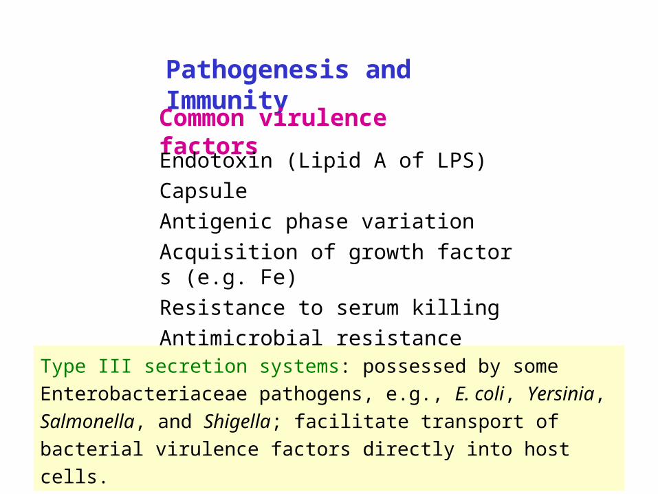
Pathogenesis and ImmunityCommon virulence factors
Type III secretion systems: possessed by some Enterobacteriaceae
pathogens, e.g., E. coli, Yersinia, Salmonella, and Shigella;
facilitate transport of bacterial virulence factors directly into host
cells.
Endotoxin (Lipid A of LPS)
Capsule
Antigenic phase variation
Acquisition of growth factors (e.g. Fe)
Resistance to serum killing
Antimicrobial resistance

Toll-like receptor 4(TLR-4)
Pathogenesis of sepsis caused by gram-negative bacteria

Pathophysiological effects of LPS
Activation of complement, release of cytokines, fe
ver, leukocytosis, thrombocytopenia, impaired org
an perfusion, disseminated intravascular coagulati
on (DIC), hypotension, shock and death.

Escherichia coli
Sepsis
For people with inadequate host defenses, e.g. the newborns. Usually originates from UT or GI infections. Many of this type of infection are endogenous.
Pathogenesis and clinical diseases
Meningitis
E. coli (particularly K1 strains) and S. agalactiae are the leading causes of meningitis in neonates.
Bacteremia

Urinary tract infection
E. coli is the most common cause of urinary tract infection.
Community- vs. hospital-acquired UT infection
Most infections originate from colon; the bacteria contaminate t
he urethra, ascend into the bladder, and may migrate into the
kidney or prostate.
Symptoms: urinary frequency, dysuria, hematuria, and pyuria.
Can result in bacteremia and sepsis.
Many uropathogenic E. coli strains produce P (pyelonephritis-a
ssociated) pili, which is associated with renal colonization, and
hemolysin HlyA.
Escherichia coli
Pathogenesis and clinical diseases

Escherichia coliPathogenesis and clinical diseases
Gastroenteritis (Diarrhea)
Caused by various virotypes:
Enterotoxigenic E. coli
Enteroaggregative E. coli
Enteropathogenic E. coli
Enterohemorrhagic E. coli
Enteroinvasive E. coli
Table 30-2
EAST & PET
STx
Escherichia coliPathogenesis and clinical diseases
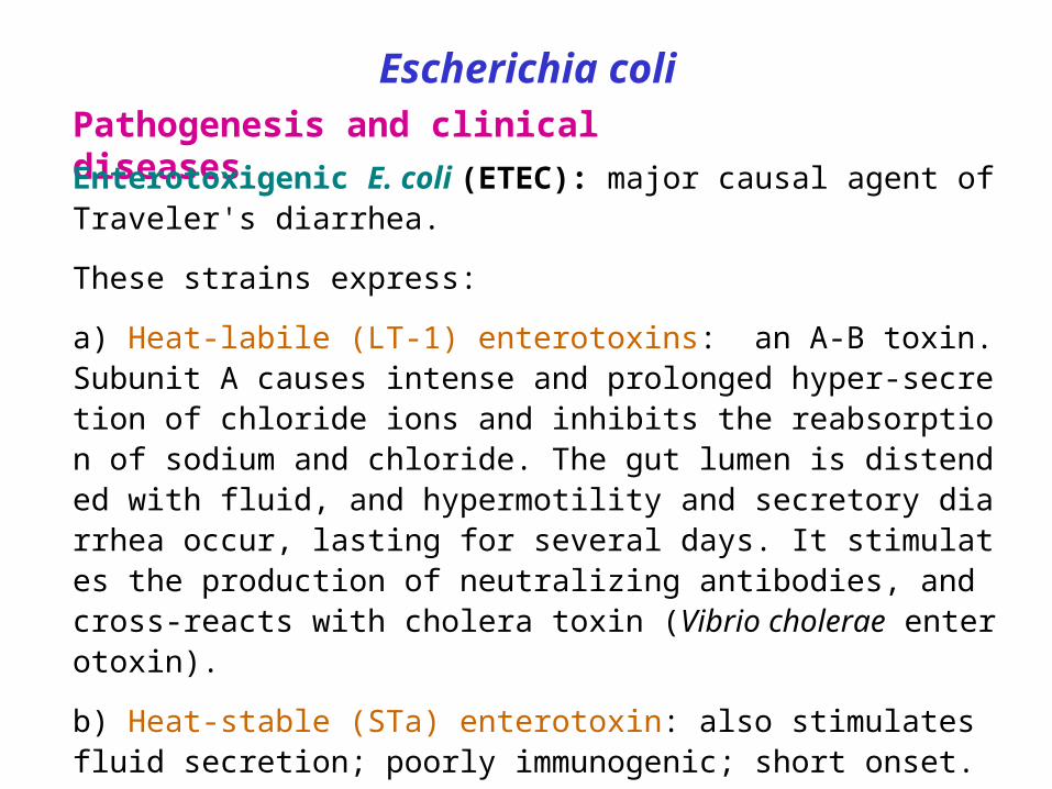
Enterotoxigenic E. coli (ETEC): major causal agent of Traveler's diarrhea.
These strains express:
a) Heat-labile (LT-1) enterotoxins: an A-B toxin. Subunit A causes intense and prolonged hyper-secretion of chloride ions and inhibits the reabsorption of sodium and chloride. The gut lumen is distended with fluid, and hypermotility and secretory diarrhea occur, lasting for several days. It stimulates the production of neutralizing antibodies, and cross-reacts with cholera toxin (Vibrio cholerae enterotoxin).
b) Heat-stable (STa) enterotoxin: also stimulates fluid secretion; poorly immunogenic; short onset.
c) Colonization factors (CFAs): facilitate the attachment of E. coli strains to intestinal epithelium. Usually are pili in nature.

ADP-ribosylation
Enhance chloride secretion
Decrease sodium and chloride absorption

Escherichia coliPathogenesis and clinical diseases
Enteropathogenic E. coli (EPEC): causes infant diarrhea in poor countries. Watery diarrhea results from microvilli destruction. Spread by person-to-person contact.
Enteroinvasive E. coli (EIEC): closely related to Shigella in pathogenic properties.
Enteroaggregative E. coli (EAEC): causes chronic diarrhea and growth retardation in infants in developing countries.
Escherichia coliPathogenesis and clinical diseases
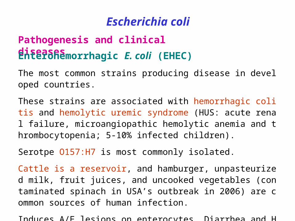
Enterohemorrhagic E. coli (EHEC)
The most common strains producing disease in developed countries.
These strains are associated with hemorrhagic colitis and hemolytic uremic syndrome (HUS: acute renal failure, microangiopathic hemolytic anemia and thrombocytopenia; 5-10% infected children).
Serotpe O157:H7 is most commonly isolated.
Cattle is a reservoir, and hamburger, unpasteurized milk, fruit juices, and uncooked vegetables (contaminated spinach in USA’s outbreak in 2006) are common sources of human infection.
Induces A/E lesions on enterocytes. Diarrhea and HUS may be associated with the Shiga toxins, which are A-B toxins that bind to 28S rRNA and disrupt protein synthesis.

歐洲腸菌元凶 疑來自埃及2011-7-1〔編譯陳成良/綜合報導〕歐洲腸道出血性大腸桿菌( O104:H4)疫情的罪
魁禍首,又出現了新的元凶!據歐洲科學家初步調查,埃及出口至法國與德國的香苜蓿( fenugreek ,又稱葫蘆巴)種子,可能是引爆歐洲 48 人死亡的腸道出血性大腸桿菌疫情的元凶。
5 月初爆發的這一波奪命大腸桿菌疫情,至今已造成歐洲及北美地區 4000 多人感染,幾乎所有病患都是居住在德國或最近曾往當地旅遊。德國的疫情以及法國波爾多地區的較小群聚感染,都被認為與發芽的種子有
關。 歐洲疾病預防管制中心( ECDC )與歐洲食品安全局( EFSA )表示,初步
調查顯示,芽菜的食用,疑為德國與法國的大腸桿菌食物中毒疫情的感染媒介,目前的追查結果顯示,其源頭有可能是 2009 年及去年從埃及進口的香苜蓿種子。聲明指出, 2009 年埃及出口的種子與法國疫情有關,去年出口的種子則與德
國疫情有關。但該局也強調,相關檢驗仍未出現陽性結果,還需要進行更多調查,才能確定元凶。這兩大機構已要求緊急調查這兩批種子在德國及歐洲的銷售情形,並警告說,
種子可能是在生產、運輸、包裝及分銷此一漫長又複雜供應鏈的任何一階段受到污染,不排除還有受污染貨品仍在歐盟甚至世界其他地區流通。

Klebsiella
K. pneumoniae and K. oxytoca are the most commonly isolated.
Can cause community-acquired primary lobar pneumonia (frequently involves necrotic destruction of alveolar space), and infections of wound, soft tissue, and urinary tract.
Risk factors for pneumonia: alcoholism; compromised pulmonary function.
*In Taiwan: liver abscess is commonly seen in infection of diabetes patients by K. pneumoniae.
K. granulomatis may cuase granuloma inguinale, a sexually transmitted disease, in some countries.
K. rhinoscleromatis: granulomatous disease of the nose.
K. ozaenae: chronic atrophic rhinitis.
Other opportunistic Enterobacteriaceae

Proteus
Most common isolates: P. mirabilis.
Cause urinary tract infections and bacteremia.
Produce urease, making the urine of the patients of UT infection with Proteus alkaline, promoting kidney stone formation by precipitating Mg and Ca.
Enterobacter, Citrobacter, Morganella, Serratia
Opportunistic pathogens causing nosocomial infections in neonates and immunocompromised patients.
These genera, particularly Enterobacter, are resistant to multiple antibiotics.

Escherichia coli and other opportunistic
EnterobacteriaceaeLaboratory diagnosis
Smears: the Enterobacteriaceae pathogens resemble each othe
r. The presence of large capsules is suggestive Klebsiella.
Culture: blood agar and selective differential media (e.g., MacC
onkey agar), the latter is useful for preliminary identification. Co
mmercial biochemical test systems can be used for identificatio
n of Enterobacteriaceae members.
Serologic tests are used for determining the clinical significance
of an isolate and for epidemiologic purpose.

Treatment
Diarrhea patients usually need only symptomatic relief.
Antibiotic treatment may prolong the fecal carriage or
increase the risk of secondary complications.
Treatment of bacteremia and septic shock: prompt
antibiotic treatment, restoration of fluid and electrolyte
balance, and treatment of disseminated IV coagulation.
Variation in drug susceptibility is great in opportunistic
infections, and antibiotic sensitivity tests are essential.
E. coli and other opportunistic Enterobacteriaceae
Prevention and control
Enterobacteriaceae are a major part of normal flora and
a common contaminant of the environment. Food safety
plays an important role in prevention of GI infections.
In hospitals, opportunistic Enterobacteriaceae are comm
only transmitted by personnel, instruments, or parenteral
medications. Their control depends on hand washing, rig
orous asepsis, sterilization of equipment, disinfection, re
straint in IV therapy, and strict precautions in keeping the
urinary tract sterile.
E. coli and other opportunistic Enterobacteriaceae

Salmonella
Classification of salmonellae into groups and species is traditionall
y based on serogrouping and serotyping of O and H antigens (> 2,
500 serotypes). However, Salmonellae have been reclassified bas
ed on DNA homology. Therefore, the correct name of S. typhi is S.
enterica, serovar. Typhi or S. Typhi. They can be identified by bioc
hemical tests and serogrouping and serotyping.
Salmonella spp. do not ferment lactose.
Most species of Salmonella are motile with peritrichous flagella.
Some Salmonellae have capsular antigens; that of S. Typhi is refer
red to as Vi antigen.

SalmonellaEpidemiology
S. Typhi and S. Paratyphi are primarily infective for humans.
Other salmonellae are chiefly pathogenic in animals (poultry, pigs, rodents, cattle, pets etc.) that constitute the reservoir for human infection.
Humans usually become infected by ingestion of contaminated food or drink (mean infective dose: 106-108, but that of S. typhi is lower). In children, infections can result from direct fecal-oral spread.
The most common sources of human infections: poultry, eggs, dairy products, and foods prepared on contaminated work surfaces. However, the major source of infection for enteric fever is the carriers (convalescent or healthy permanent).

SalmonellaPathogenesis and Immunity
Invasion
Acid tolerance response (ATR) gene protects the organism from gastric acid.
The bacteria invade into (by inducing membrane ruffling) and multiply in the M cells and enterocytes of the small intestine. They can also be transported across the enterocytes and released into the blood and lymphatic circulation.
Inflammatory response confines the infection to the GI tract in non-typhoid salmonellosis.
Survival in macrophages
Salmonellae are facultative intracellular pathogen.

Salmonella
Clinical diseases
1. Enteritis
Incubation period: 6-48 hours.
Symptoms: nausea, headache, vomiting, nonbloody prof
use diarrhea, with few leukocytes in the stools. Low-grad
e fever, abdominal cramp, myalgia, and headache are al
so common. Episode resolves in 2-7 days.
Inflammatory lesions of the small and large intestine are
present. Stool cultures remain positive for several weeks
after clinical recovery.

SalmonellaClinical diseases
2. Bacteremia
Most common causal species: S. Choleraesuis, S Typhi
and S. Paratyphi.
Symptoms: like sepsis caused by other gram-negative b
acteria. 10% of patients may have localized suppurative i
nfections, e.g., osteomyelitis, endocarditis, arthritis, etc.
High risk population: pediatric and geriatric patients; AID
S patients.
SalmonellaClinical diseases
3. Enteric fever (typhoid fever)
Causal species: S. Typhi, S. Paratyphi A, S. Schottmuelleri,
and S. Hirschfeldii.
Mouth small intestine lymphatics and bloodstream
infect liver, spleen and bone marrow mul
tiply and pass into the blood second and heavier bactere
mia onset of clinical illness colonization of ga
llbladder invasion of the intestine typhoid ulcers a
nd severe illness.
Chronic carriers (1%-5% of patients): bacteria persist in the g
allbladder and the biliary tract for more than one year.

Symptoms: incubation time: 10-14 days. Gradually increasing fever, malaise, headache, myalgias, and anorexia, which persist for a week or longer. In severe cases: intestinal hemorrhage and perforation.
Principal lesions: hyperplasia and necrosis of lymphoid tissue, hepatitis, focal necrosis of the liver, and inflammation of the gallbladder, periosteum, lungs and other organs.

SalmonellaTreatment
Enteric fever and bacteremia require antibiotic treatment: chlo
ramphenicol, ampicillin, trimethoprim-sulfamethoxazole. Surg
ical drainage of metastatic abscesses may be required.
Salmonella enterocolitis needs only supportive therapy (antibi
otic treatment may prolong the symptoms and excretion of th
e salmonellae). Drugs to control hypermotility of the gut shou
ld be avoided because it is easy to transform a trivial gastroe
nteritis into a life-threatening bacteremia by paralyzing the bo
wel.
Chronic carriers of S. Typhi may be cured by antibiotics alone
or combined with cholecystectomy.

Salmonella
Prevention and control
Sanitary measures.
Carriers must not be allowed to work as food handlers.
Strict hygienic precautions for food handling.
Vaccines against S. Typhi:
Purified Vi antigen
Oral, live attenuated vaccine.

National salmonella death toll rises to 7(Staff writer Ridgely Ochs contributed to this story. January 24, 2009)
A seventh death was linked Friday to a nationwide outbreak of salmonella associated with tainted peanut butter and paste sourced to the Peanut Corp. of America's plant in Blakely, Ga., authorities confirmed.
Although their exact causes of death have not been determined, all seven people have died after being infected with the bacterial strain Salmonella Typhimurium, the Centers for Disease Control and Prevention said on its Web site.
There have been 493 cases reported in 43 states and one Canadian province of people sickened, though authorities stress the numbers sickened are likely far in excess of that as many cases go unreported. Known patients ranged in age from 1 to 98, and 22 percent of the those have been hospitalized.
Another 10 firms Friday recalled products that use PCA peanut butter or paste - bringing to roughly 360 the number of products affected - as it emerged that the Peanut Corp. of America's plant in Blakely, Ga. laid off most of its roughly 50 workers. The outbreak has triggered a congressional inquiry and renewed calls for reform of food safety laws.
http://www.newsday.com/services/newspaper/printedition/saturday/health/ny-lisalm246010666jan24,0,5876138.story

ShigellaS. dysenteriae, S. flexneri , S. sonnei , & S. boydii: bacillary dysentery
> 45 O serotypes; have no H antigen; do not ferment lactose.
Pathogenesis and Immunity
Shigellosis is primarily a pediatric disease, and is restricted to the GI tract.
Mean infective dose: 103.
Mouth colon invade M cells and subsequently spread to mucosal epithelial cells cause microabscess in the wall of colon and terminal ileum necrosis of the mucous membrane, superficial ulceration, bleeding, and formation of pseudomembrane.
Shiga toxin
An A-B toxin inhibiting protein synthesis.
Damages intestinal epithelium and glomerular endothelial cells (associated with HUS) .

Internalized shigellae induce apoptosis of macrophage and release of the bacteria
Attracted by the cytokines released by macrophage
Destablize the intestinal wall
Activates the invasion genes on the virulence plasmid
M cell

Shigella
Clinical diseases
Incubation period: 1-3 days
Sudden onset of abdominal pain, fever and watery diarrhea
number of stools increase, less liquid, often contain mucus
and blood, rectal spasms with resulting lower abdominal pain (t
enesmus) symptoms subside spontaneously in 2-5 days in
adult cases, but loss of water and electrolytes frequently occur
in children and the elderly a small number of patients rem
ain chronic carriers.
Some cases were accompanied by hemolytic uremic syndrom
e (HUS).

Shigella
Laboratory diagnosis
Specimens: fresh stool, mucus flecks, and rectal swabs. Large
numbers of fecal leukocytes and some RBC may often be seen
microscopically.
Culture: differential and selective media as used for salmonellae.
Treatment
Antibiotic treatment: chloramphenicol, ampicillin, tetracycline, an
d trimethoprim-sulfamethoxazole. Drug resistance is common.
Opiates should be avoided.

Shigella
Prevention and control
Humans are the only reservoir for shigellae.
Transmission of shigellae: water, food, fingers, feces, and flies.
Most cases occur in children under 10 years of age.
Prevention and control of dysentery:
1. Sanitary control of water, food and milk; sewage disposal;fly control.
2. Isolation of patients and disinfection of excreta.
3. Detection of subclinical cases and carriers.

Yersinia Y. pestis: plague ("black death")
Y. pseudotuberculosis and Y. enterocolitica: gastroenteritis
Grows more rapidly in media containing blood or tissue fluids an
d fastest at 30 oC. Some species (e.g. Y. enterocolitica) can gro
w in refrigerated food.
Pathogenesis
The Yersinia pathogens are able to resist phagocytic killing by se
creting proteins into the phagocyte and result in inhibition of killin
g by phagocyte, apoptosis of macrophage, and suppression of c
ytokine production.
Y. pestis produces a protein capsule (Fraction 1) and Pla (plasmi
nogen activator protease) that degrades C3b, C5a and fibrin clot
(enhances spread of bacteria into blood stream).

Yersinia pestisCauses zoonotic infections; humans are accidental hosts.
Three major pandemics have occurred in 541 AD, 1320s and 1860s.
Two forms of infections:
Urban plague
Rats as natural reservoirs.
Spread among rats or between rats and humans by infected flea.
Can be eliminated by effective control of rats and better hygiene.
Sylvatic plague: infections of rodents and domestic cats.
Y. pestis are widely distributed in mammalian reservoirs and flea vectors and produces fatal infections in animal reservoirs.
Human infections are acquired by contacting the reservoir population.

Yersinia pestisPathogenesis
Bubonic plague
Y. pestis enters a flea when it feeds on an infected animal the bacteria multiply in the gut of the flea flea becomes hungry and bites ferociously the bacteria passes from the flea into the bite wound bacteria are phagocytised, but can multiply intracellularly or extracellularly bacteria reach the lymphatics, and an intense hemorrhagic inflammation develops in the enlarged lymph nodes, which may undergo necrosis the bacteria may reach the blood stream and disseminated. Hemorrhagic and necrotic lesions may develop in all organs.
Primary pneumonic plague
Results from inhalation of infective droplets (usually from a coughing patient), with hemorrhagic consolidation of the lung, sepsis and death.

Yersinia pestisClinical Diseases
Bubonic plague
Incubation period: 2-7 days.
High fever and painful lymphoadenopathy with greatly enlarged, tender lymph nodes (buboes) in the groin and axilla sepsis (early stage: vomiting and diarrhea; late stage: hypotension, renal and cardiac failure; terminal stage: pneumonia and meningitis). Mortality: 75% if untreated.
Pneumonic plague
Incubation time: 2-3 days.
Fever and malaise, pulmonary signs develop within 1 day. Patients are highly infectious. Mortality: 90% if untreated.

Yersinia pestisTreatment
Patients have to be promptly treated with antibiotics (drug of choice: streptomycin).
Epidemiology and control
Plague is an infection of wild rodents that still occurs in many parts of the world (enzootic areas: India, Southeast Asia, Africa, and North and South America).
Control of plague requires surveys of infected animals, vectors, and human contacts, and by destruction of infected animals.
All patients with suspected plague should be isolated.
Contacts of patients with suspected pneumonic plague should receive tetracycline as chemoprophylaxis.

Y. enterocolitica and Y. pseudotuberculosisCause zoonotic infections.
Y. enterocolitica is a common cause of enteritis in cold areas during the cold months. Y. pseudotuberculosis infection is relatively uncommon.
They are found in the intestine of a variety of animals, and are transmissible to humans through contaminated food, drink or fomites, resulting in diarrhea, fever and abdominal pain that last for 1-2 weeks or, in some cases, months. Most are self-limited.
Y. enterocolitica infection can cause pseudoappendicitis (enlarged mesenteric lymph nodes) in children, and blood-transfusion related sepsis in those who used blood products stored for at least 4 weeks.

Y. enterocolitica grows slowly at 37 oC and prefers cooler temperatures. The fecal specimen can be mixed with saline and then stored at 4 oC for 2 weeks or more to facilitate isolation of this organism (cold enrichment).

How does Proteus swarm?

Back
Lipopolysaccharide (LPS)
is also called endotoxin.
LPS is composed of lipid A, cor
e polysaccharide, and O-specif
ic polysaccharide.
Lipid A anchors LPS in the lipid
bilayer of outer membrane. It c
auses symptoms associated wi
th endotoxin.
O-specific polysaccharide can
be used to identify certain spec
ies and strains.