EnhancedHealingofDiabeticWoundsbyTopical ...downloads.hindawi.com/journals/sci/2011/304562.pdf ·...
12
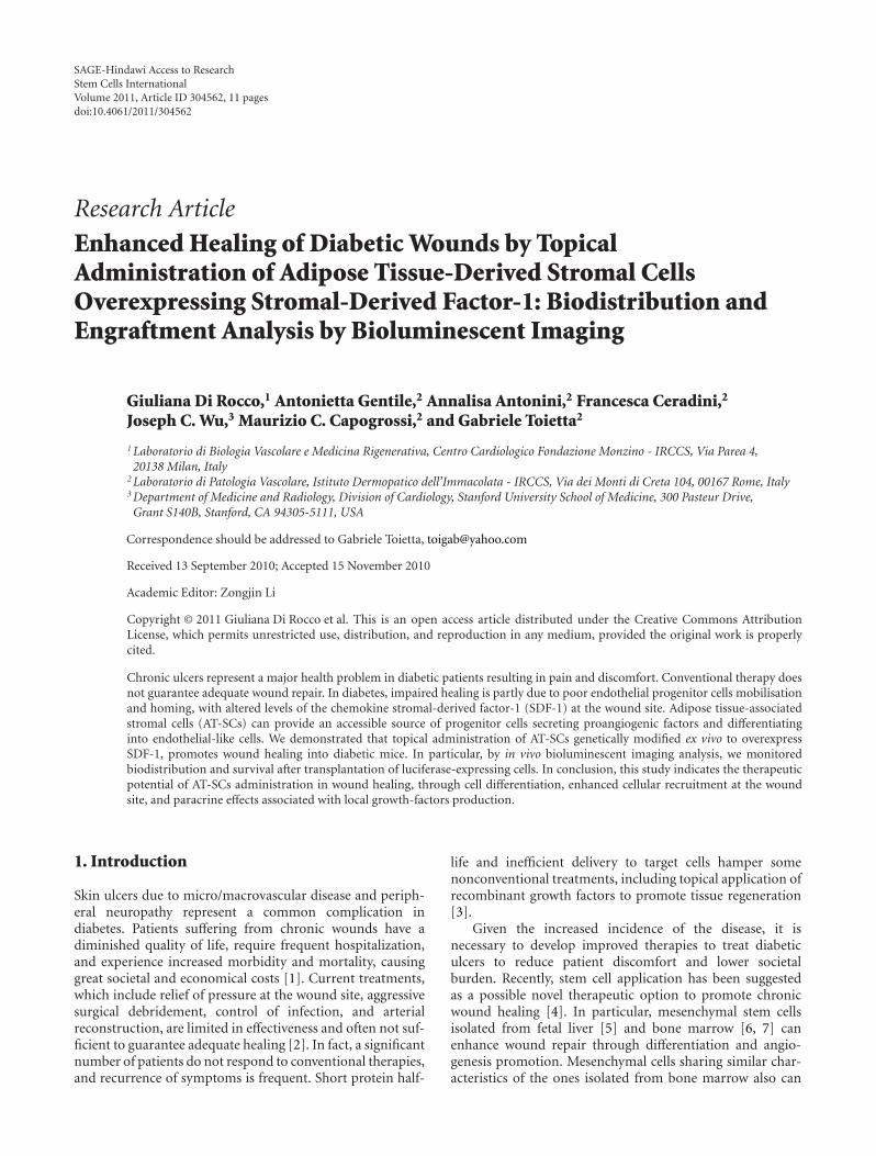
SAGE-Hindawi Access to Research Stem Cells International Volume 2011, Article ID 304562, 11 pages doi:10.4061/2011/304562 Research Article Enhanced Healing of Diabetic Wounds by Topical Administration of Adipose Tissue-Derived Stromal Cells Overexpressing Stromal-Derived Factor-1: Biodistribution and Engraftment Analysis by Bioluminescent Imaging Giuliana Di Rocco, 1 Antonietta Gentile, 2 Annalisa Antonini, 2 Francesca Ceradini, 2 Joseph C. Wu, 3 Maurizio C. Capogrossi, 2 and Gabriele Toietta 2 1 Laboratorio di Biologia Vascolare e Medicina Rigenerativa, Centro Cardiologico Fondazione Monzino - IRCCS, Via Parea 4, 20138 Milan, Italy 2 Laboratorio di Patologia Vascolare, Istituto Dermopatico dell’Immacolata - IRCCS, Via dei Monti di Creta 104, 00167 Rome, Italy 3 Department of Medicine and Radiology, Division of Cardiology, Stanford University School of Medicine, 300 Pasteur Drive, Grant S140B, Stanford, CA 94305-5111, USA Correspondence should be addressed to Gabriele Toietta, [email protected] Received 13 September 2010; Accepted 15 November 2010 Academic Editor: Zongjin Li Copyright © 2011 Giuliana Di Rocco et al. This is an open access article distributed under the Creative Commons Attribution License, which permits unrestricted use, distribution, and reproduction in any medium, provided the original work is properly cited. Chronic ulcers represent a major health problem in diabetic patients resulting in pain and discomfort. Conventional therapy does not guarantee adequate wound repair. In diabetes, impaired healing is partly due to poor endothelial progenitor cells mobilisation and homing, with altered levels of the chemokine stromal-derived factor-1 (SDF-1) at the wound site. Adipose tissue-associated stromal cells (AT-SCs) can provide an accessible source of progenitor cells secreting proangiogenic factors and differentiating into endothelial-like cells. We demonstrated that topical administration of AT-SCs genetically modified ex vivo to overexpress SDF-1, promotes wound healing into diabetic mice. In particular, by in vivo bioluminescent imaging analysis, we monitored biodistribution and survival after transplantation of luciferase-expressing cells. In conclusion, this study indicates the therapeutic potential of AT-SCs administration in wound healing, through cell differentiation, enhanced cellular recruitment at the wound site, and paracrine effects associated with local growth-factors production. 1. Introduction Skin ulcers due to micro/macrovascular disease and periph- eral neuropathy represent a common complication in diabetes. Patients suffering from chronic wounds have a diminished quality of life, require frequent hospitalization, and experience increased morbidity and mortality, causing great societal and economical costs [1]. Current treatments, which include relief of pressure at the wound site, aggressive surgical debridement, control of infection, and arterial reconstruction, are limited in effectiveness and often not suf- ficient to guarantee adequate healing [2]. In fact, a significant number of patients do not respond to conventional therapies, and recurrence of symptoms is frequent. Short protein half- life and inefficient delivery to target cells hamper some nonconventional treatments, including topical application of recombinant growth factors to promote tissue regeneration [3]. Given the increased incidence of the disease, it is necessary to develop improved therapies to treat diabetic ulcers to reduce patient discomfort and lower societal burden. Recently, stem cell application has been suggested as a possible novel therapeutic option to promote chronic wound healing [4]. In particular, mesenchymal stem cells isolated from fetal liver [5] and bone marrow [6, 7] can enhance wound repair through differentiation and angio- genesis promotion. Mesenchymal cells sharing similar char- acteristics of the ones isolated from bone marrow also can
Transcript of EnhancedHealingofDiabeticWoundsbyTopical ...downloads.hindawi.com/journals/sci/2011/304562.pdf ·...
SAGE-Hindawi Access to ResearchStem Cells InternationalVolume 2011, Article ID 304562, 11 pagesdoi:10.4061/2011/304562
Research Article
Enhanced Healing of Diabetic Wounds by TopicalAdministration of Adipose Tissue-Derived Stromal CellsOverexpressing Stromal-Derived Factor-1: Biodistribution andEngraftment Analysis by Bioluminescent Imaging
Giuliana Di Rocco,1 Antonietta Gentile,2 Annalisa Antonini,2 Francesca Ceradini,2
Joseph C. Wu,3 Maurizio C. Capogrossi,2 and Gabriele Toietta2
1 Laboratorio di Biologia Vascolare e Medicina Rigenerativa, Centro Cardiologico Fondazione Monzino - IRCCS, Via Parea 4,20138 Milan, Italy
2 Laboratorio di Patologia Vascolare, Istituto Dermopatico dell’Immacolata - IRCCS, Via dei Monti di Creta 104, 00167 Rome, Italy3 Department of Medicine and Radiology, Division of Cardiology, Stanford University School of Medicine, 300 Pasteur Drive,Grant S140B, Stanford, CA 94305-5111, USA
Correspondence should be addressed to Gabriele Toietta, [email protected]
Received 13 September 2010; Accepted 15 November 2010
Academic Editor: Zongjin Li
Copyright © 2011 Giuliana Di Rocco et al. This is an open access article distributed under the Creative Commons AttributionLicense, which permits unrestricted use, distribution, and reproduction in any medium, provided the original work is properlycited.
Chronic ulcers represent a major health problem in diabetic patients resulting in pain and discomfort. Conventional therapy doesnot guarantee adequate wound repair. In diabetes, impaired healing is partly due to poor endothelial progenitor cells mobilisationand homing, with altered levels of the chemokine stromal-derived factor-1 (SDF-1) at the wound site. Adipose tissue-associatedstromal cells (AT-SCs) can provide an accessible source of progenitor cells secreting proangiogenic factors and differentiatinginto endothelial-like cells. We demonstrated that topical administration of AT-SCs genetically modified ex vivo to overexpressSDF-1, promotes wound healing into diabetic mice. In particular, by in vivo bioluminescent imaging analysis, we monitoredbiodistribution and survival after transplantation of luciferase-expressing cells. In conclusion, this study indicates the therapeuticpotential of AT-SCs administration in wound healing, through cell differentiation, enhanced cellular recruitment at the woundsite, and paracrine effects associated with local growth-factors production.
1. Introduction
Skin ulcers due to micro/macrovascular disease and periph-eral neuropathy represent a common complication indiabetes. Patients suffering from chronic wounds have adiminished quality of life, require frequent hospitalization,and experience increased morbidity and mortality, causinggreat societal and economical costs [1]. Current treatments,which include relief of pressure at the wound site, aggressivesurgical debridement, control of infection, and arterialreconstruction, are limited in effectiveness and often not suf-ficient to guarantee adequate healing [2]. In fact, a significantnumber of patients do not respond to conventional therapies,and recurrence of symptoms is frequent. Short protein half-
life and inefficient delivery to target cells hamper somenonconventional treatments, including topical application ofrecombinant growth factors to promote tissue regeneration[3].
Given the increased incidence of the disease, it isnecessary to develop improved therapies to treat diabeticulcers to reduce patient discomfort and lower societalburden. Recently, stem cell application has been suggestedas a possible novel therapeutic option to promote chronicwound healing [4]. In particular, mesenchymal stem cellsisolated from fetal liver [5] and bone marrow [6, 7] canenhance wound repair through differentiation and angio-genesis promotion. Mesenchymal cells sharing similar char-acteristics of the ones isolated from bone marrow also can
2 Stem Cells International
be derived from adipose tissue [8]. Adipose tissue-derivedstromal cells (AT-SCs), also known as stromal vascularfraction cells (AT-SVFs), are collected from adipose tissueby collagenase digestion and differential centrifugation [9].Since liposuction involves only local anaesthesia, cells may beobtained from the patient with a repeatable, nondebilitatingoperation and autologously transplanted at the site of tissueregeneration. The procedure can be performed in a large-scale, reproducible manner according to GMP regulations,allowing AT-SCs to be used in preclinical studies andexperimental clinical trials [10].
Adipose tissue-derived multipotential precursor cells areable to differentiate in several cells types of both meso-dermal and nonmesodermal origins, including adipocytes,chondrocytes, osteocytes, myocytes, hepatocytes, endocrinepancreatic cells, neurons, and endothelial cells (for review see[9]).
AT-SCs secrete angiogenic molecules, including HGF,VEGF, PlGF, IGF, and KGF [11, 12]. Moreover, transcrip-tional profiling has demonstrated the expression of proan-giogenic genes [13]. Transplantation of AT-SCs has beenshown to promote therapeutic angiogenesis in hind limbischemia in mice [14–16]. This may be due to integrationof AT-SCs into vessels in vivo [17] and to the contributionto neovascularisation mediated by a paracrine effect [18].Moreover, AT-SCs can promote human dermal fibroblastproliferation [12] and wound healing in a mouse model[19, 20] both via direct cell-to-cell contact and by a paracrineeffect. In addition, a protective effect of AT-SCs and theirsecretory factors during oxidative injury has been described[21]. Improved wound healing of adipose tissue extracts wasdetermined in a porcine experimental model [6], while AT-SCs extract did not affect murine wound repair [19].
Taken together, these studies support the developmentof approaches using AT-SCs administration to promotewound healing. Nonetheless, we know very little about howtransplanted cells behave in vivo, as well as their ability ofengraftment, persistence, differentiation, and biodistributionafter in vivo administration. In vivo studies in animal modelshave the potential to demonstrate the clinical potential ofstem cell therapy by elucidating cell biology and physiologyof the transplanted cells and their progeny. Current methodsof studying stem cell fate after administration mainly rely onhistochemical evaluation of samples obtained from sacrificedanimals. This approach is time consuming, laborious, andexpensive, requiring sectioning analysis of multiple samplesderiving from a large number of experimental animals. Tobetter understand stem cell activity in vivo, rapid, affordable,and noninvasive imaging techniques are needed.
In order to evaluate engraftment of AT-SCs in amurine model of diabetic impaired wound healing, weused bioluminescent imaging to track transplanted cells.Furthermore, we were interested in determining whetherconcomitant overexpression of SDF-1 may further promotewound healing and cell engraftment. In fact, in the diabeticcondition, impaired healing is at least in part due to SDF-1 downregulation [22], which affects migration and homingat the wound site of circulating endothelial progenitor cells(EPCs) [23]. On the other hand, exogenous administration
at the lesion site of recombinant SDF-1 [24] or of a lentiviralvector expressing SDF-1 [25] reversed the impaired EPChoming in a murine model of diabetic wound healing.Conversely, SDF-1 inhibition in diabetic animals resulted infurther impairment of wound repair [26]. Moreover, SDF-1 overexpression increased survival and growth of CXCR4-expressing mesenchymal stem cells both in vitro [27] and invivo [28, 29].
Here, we show that topical administration of AT-SCspromotes wound healing in diabetic mice and determine byBLI the biodistribution and the kinetic of engraftment ofadministered cells.
2. Material and Methods
2.1. Experimental Animals. Mice used in the study were6- to 8-week-old wild-type Swiss CD1 males, transgenicmice expressing ubiquitously GFP [30], or GFP and fireflyluciferase [31] from colonies maintained in our institutionalanimal facility. All experimental procedures were performedaccording to the guidelines of the Italian National Institutesof Health and were approved by the Institutional AnimalCare and Use Committee.
Induction of diabetes was obtained by intraperitonealinjection of 50 mg/kg streptozotocin (Sigma-Aldrich, St.Louis, MO) in 0.05 M Na citrate, pH 4.5, for 5 consecutivedays. Two weeks after the last streptozotocin injection,animals were fasted for 2 hours and blood glucose levelmeasured using an Ascensia Confirm glucometer (BayernHealthCare, Basel, Switzerland). Mice with glycaemia above200 mg/dl were selected for further studies. Three weeks later,diabetic animals were used for wound-healing experiments.
2.2. Cells Isolation. AT-SCs were obtained from 6-week-old mice as previously described [32]. Briefly, inguinalsubcutaneous fat pads were digested for 45 minutes in ashaking water bath at 37◦C in PBS containing 2% BSAand 2 mg/ml collagenase A (Roche Diagnostics, Mannheim,Germany). After tissue disaggregation, cells were filteredthrough a 40 μm cell strainer (BD Falcon, Franklin Lakes,NJ) and collected by centrifugation at low speed (500 g) toremove floating mature adipocytes. Cells were then washedin PBS, counted, and used for wound healing experiments.With this procedure, we isolated approximately 0.8–1.2× 10⁶
cells from each 6-week-old mouse.To obtain adipose-derived adherent stromal (ADAS)
[13], sometimes referred to also as adipose issue-derivedmesenchymal cells (AT-MSCs) [33], cells were plated inDMEM 20% FCS in tissue culture dishes. After an overnightincubation, the adherent cell population was obtained.
Skin fibroblasts were obtained from the same animals aspreviously described [34]. Briefly, skin samples were washedin PBS, then the subcutaneous tissues were eliminated andthe epidermis removed by enzymatic digestion. After a washin PBS, dermal samples were cut into small pieces and treatedwith 0.3% trypsin in PBS for 30 min in a 37◦C water bath.Ice-cold complete medium (DMEM 10% FCS) was thenadded and the samples vigorously mixed on a vortex. The
Stem Cells International 3
suspension was passed through a 40 μm cell strainer, and cellswere collected by centrifugation (10 min at 150 × g, 4◦C).Cells were then washed in PBS and counted and either usedas control for angiogenesis plug assay (see below) or platedon tissue culture dishes (0.4 × 10⁵ cells/cm2) in completemedium.
2.3. Construction of Viral Vectors. Recombinant first-generation E1-E3-deleted adenoviral (Ad) vectors forexpression of SDF-1 and GFP were obtained using the Ad-Easy system as previously described [35]. After amplification,the adenoviral vectors were purified by CsCl2 gradientultracentrifugation [36] and dialyzed against a solutioncontaining 3% sucrose, 10 mM Tris, pH 7.8, 150 mM NaCl,and 10 mM MgCl2. Viral titers were estimated by serialdilution of the viral stocks in 293 cells and expressed asplaque-forming units per ml (PFU/ml). Viral stocks weretested for absence of replication-competent adenovirus onA549 cells.
2.4. In Vitro Lesion Repair Assay. The assay was performedas described by Bilic et al. [37] in triplicate. Second passagemesenchymal cells derived from adipose tissue were grownto confluence in complete medium (DMEM 20% FCS). Cellswere then transduced by adenoviral-mediated gene transferfor 1 hour at 37◦C at multiplicity of infection of 10. Cellswere washed with PBS then grown with serum-free mediumsupplemented with 0.1% bovine serum albumin (BSA). Theday after gene transfer cell monolayer was lesioned using a2 mm cell scraper without damaging the dish surface. Lesionareas were recorded with a digital camera at time zero andafter 1 and 2 days. Analysis was performed by tracing thelesion edges and calculating the pixel area using the ImageAnalysis System (IAS, Delta Sistemi, Rome, Italy). In vitrolesion repair is expressed as percent of the value calculatedat time zero.
2.5. In Vivo Angiogenesis Gel Plug Assay. The assay is basedon the implant of Matrigel plugs [38]. Freshly isolated AT-SCs resuspended in PBS were mixed with Cultrex growthfactor-reduced basement membrane extract (Trevigen Inc.,Gaithersburgh, MD) maintained at 4◦C (liquid state). Analiquot of 400 μl of Cultrex containing 7 × 10⁵ cells wasinjected subcutaneously into Swiss CD1 mice gathered inexperimental groups of 5 animals each near the abdominalmidline. The solution and the syringe were kept on icebefore the injection to prevent gelling in the needle. Aweek later, mice were sacrificed and gel plugs removed.Samples were fixed in 4% formaldehyde and embedded inparaffin for sectioning and histological analysis. Sectionswere then processed with Masson Trichromic stain andcapillary density determined.
2.6. Cell Administration at the Wound Site. Hyperglycaemicanimals were sedated by intraperitoneal administration ofAvertin (200 mg/kg, Sigma-Aldrich) and shaved on thedorsum, and a full-thickness wound was performed onthe dorsal midline using a 3.5 mm-diameter biopsy punch
(Stiefel, Offenbach am Main, Germany). Right after woundinduction, freshly isolated AT-SCs were then administeredsite in a single dose (7.5 × 10⁵ cells/mouse dissolved in 40 μlof saline solution) topically at the lesion. Control animalsreceived the same volume of saline solution. Woundedanimals were then housed individually.
2.7. Wound Analysis. At different time points (0, 3, 5, 7, 10,and 14 days) after wounding, lesion closure was documentedusing a digital camera. Images were processed and analyzedby tracing the wound margin and calculating the pixel areausing the Image Analysis System (IAS). Re-epithelialisationis reported as the percentage of the initial wound area andcalculated as re-epithelialisation percentage = [1 − (areaon day of analysis/area on day 0)] × 100 [39]. The day inwhich the full-thickness wound was seen to be completelyclosed was taken as the day of complete healing. Moreover,images of haematoxylin and eosin stained slides of eachwound obtained from maximal cross-sections were digitallyacquired, then the epithelial gap (distance between the twoepithelial edges) was measured to assess re-epithelialization.
2.8. Ex Vivo and In Vivo Optical Bioluminescent Imaging.Ex vivo and in vivo imaging analysis was performed usingthe IVIS Lumina from Caliper Life Sciences (Caliper LifeSciences, Hopkinton, MA). For ex vivo imaging, AT-SCsisolated from inguinal fad pads from luciferase-expressingmice were plated into clear bottom tissue culture dishesand incubated in a solution of D-luciferin (Caliper LifeSciences) dissolved in prewarmed tissue culture medium(150 μg/ml) before analysis. For in vivo analysis, mice wereanesthetized with Avertin and D-luciferin dissolved in PBS(150 mg/kg body weight), was administered i.p. 10 minutesbefore analysis. Photons emitted from luciferase-expressingAT-SCs transplanted into the animals were collected withfinal accumulation times of 1 to 5 minutes, depending on theintensity of the bioluminescence emission. The same micewere analyzed at different time points after transplantationwith the same procedure, providing longitudinal data oftransplanted cells biodistribution and survival.
2.9. Histological Analysis. Biopsies were fixed in 4%formaldehyde for 48 hours, embedded in paraffin,and serially sectioned (7 μm) perpendicularly to thewound surface. Every eighth section of each wound washaematoxylin and eosin stained to perform morphologicanalyses. Immunohistochemistry on formalin fixed,paraffin-embedded tissue was performed with antibodiesagainst GFP (Abcam Ltd., Cambridge, UK),
2.10. Statistical Analysis. Results are expressed as means ±SEM. Data analysis and comparisons between control andtreated groups were done with INSTAT (GraphPad, SanDiego). The significance of differences was assessed witha two-tailed Student t-test for unpaired data; statisticalsignificance level was set at P < .05.

4 Stem Cells International
Ad-GFP
Ad-SDF
0
25
50
75
100
Lesi
onsi
ze(%
ofda
y0)
NT Ad GFP Ad SDF-1 FCS
∗
Figure 1: In vitro lesion repair assay. Lesion closure (expressed as % of the area at day 0) in an in vitro lesion repair assay. NontransducedAT-SCs (NT) or transduced with an adenoviral vector expressing GFP (Ad-GFP) or expressing SDF-1 and GFP (Ad-SDF-1) were maintainedin serum-free medium supplemented by 0.1% BSA. Control cells were grown in complete medium (DMEM-20% FCS). Data are expressedas mean ± SEM, and experiment was performed in triplicate. Statistical significance level was set at P < .05. No statistically significantdifference was assessed between the NT and Ad-GFP-transduced groups. At 48 hours, lesion repair in the Ad-SDF-1 group was improved incomparison with both NT group (P = .018) and Ad-GFP (∗P = .006). Representative fluorescence micrographs of lesion area (dottedlines) at day 2. Original magnification 40X.
3. Results and Discussion
3.1. SDF-1 Overexpression Promotes Lesion Repair in AT-SCs Monolayer Cultures In Vitro. We determine that AT-SCs have a repair potential in an in vitro lesion repair assayperformed as previously described [37]. AT-SCs culturedin 20% FCS containing filled a 2 mm gap performed onthe cell monolayer in less than 3 days. Non-transducedand mock-transduced AT-SCs maintained in serum-freemedium were not able to repair the scratch. Non-transducedcells performed better than mock-transduced (Ad-GFP),possibly for toxicity associated with viral exposure, but thedifference was not statistically significant at 48 hours afterlesion induction. On the other hand, cells after adenoviral-mediated gene transfer of SDF-1 had an improved lesionrepair potential after 48 hours from the lesion, comparedboth with non-transduced and mock-transduced AT-SCs(Figure 1).
AT-SCs have been shown to express the CXCR4 alpha-chemokine receptor specific for stromal-derived factor-1,and its overexpression increases AT-SCs migration andproliferation [40]. Accordingly, these results indicate thatSDF-1 may act as an autocrine factor on AT-SCs after a viral-mediated gene transfer that promotes cell proliferation andmigration.
3.2. AT-SCs Promotes In Vivo Angiogenesis in a Gel PlugAssay. We found that the stromal fraction isolated fromadipose tissue contained a population of cells capable ofdifferentiating into endothelial-like structures in a Matrigelplug assay [14]. To perform this assay, we isolated AT-SCsfrom transgenic mice ubiquitously expressing GFP [30] andthen transplanted the plugs containing the GFP-positivecells in GFP-negative animals. We were then able to detectendothelial-like structures derived from implanted GFP-positive AT-SCs (Figure 2(b)), while we failed to detect
similar structure in control mice (Figure 2(a)). In this exper-imental condition, more than 99% of endothelial-like struc-tures were derived from GFP-positive cells (Figure 2(d)).We also performed the assay using AT-SCs transducedwith an adenoviral mock vector and AT-SCs transducedwith an adenoviral vector expressing SDF-1. We observedthat even in normoglycemic conditions, the number ofendothelial-like structures formation was slightly improved(5%) by SDF-1 overexpression (Figure 2(h)); albeit in theexperimental setting used to perform the analysis (n = 5per group), the difference versus the control group did notachieve statistical significance (P-value ≥ .05).
Matrigel plugs containing an equal number of skinfibroblasts obtained from the same animals used for isolationof AT-SCs were used as controls and failed to organize intoendothelial-like structures (data not shown).
In addition, we also performed a Matrigel plug assayusing a conditioned medium obtained from an overnightculture of AT-SCs and found that secreted soluble fac-tors produced by AT-SCs may promote angiogenesis(Figure 2(f)). This is in accordance with several studieswhich have proved that AT-SCs secrete a panel of angiogenicmolecules [21], which have the potential to be, at least inpart, responsible for the therapeutic effect we and others haveobserved on wound healing.
Collectively these sets of data indicate that AT-SCs areable to promote vessel formation in vivo in a Matrigel assayby taking part in vessel-like structure organization and bysecretion of proangiogenic factors.
3.3. AT-SCs Transplantation Promotes Wound Healing in Dia-betic Animals. For in vivo studies, we used streptozotocin-induced CD1 diabetic mice receiving a full-thickness woundon the dorsal midline. We observed that topical adminis-tration at the lesion site of 7.5 × 10⁵ AT-SCs significantlyenhanced wound healing in diabetic mice (Figure 3).

Stem Cells International 5
(a) (b)
(c) (d)
(e) (f)
(g)
25 μm
(h)
Figure 2: In vivo angiogenesis gel plug assay. AT-SCs isolated from ubiquitously GFP-expressing mice were mixed with Cultrex and injectedsubcutaneously into CD1 recipient. After 1 week, gel plugs were analyzed and capillary density was determined by Masson Trichromic stain(b) and anti-GFP immunohistochemistry (d). Control animals received gel plugs containing saline solution (a)–(c). Masson trichromic stainof implanted gel plugs containing nonconditioned serum-free medium (e) and conditioned medium from AT-SCs cultures (f); AT-SCs weregenetically modified by Ad-GFP (g) and Ad-SDF-1 (h). The number of endothelial-like structures formation was slightly improved by SDF-1overexpression, but the difference versus the control group (Ad-GFP) did not achieve statistical significance (P-value ≥ .05).
Mesenchymal cells isolated from adipose tissue representa suitable target for cell-mediated gene therapy as they havebeen proven to be prone to viral-mediated gene transfer [41].We then assessed whether overexpression of SDF-1, whosedownmodulation plays a pivotal role in the pathophysiology
of diabetic wounds, might result in a better therapeu-tic profile of the AT-SCs-mediated treatment. Indeed, weachieved further improvement by adenoviral-mediated SDF-1 gene transfer into AT-SCs before topical administration.Wound area and epithelial gap were significantly reduced

6 Stem Cells International
0
25
50
75
100
125
Wou
nd
clos
ure
(%of
day
0)
0 5 10 15
Days after wounding
SalineAT-SCs
AT-SCs GFPAT-SCs SDF-1
∗
∗∗
(a)
Saline
AT-SCs
AT-SCs GFP
AT-SCs SDF-1
5 mm
0 3 5 7 10 14
(b)
Figure 3: Wound closure in diabetic animals. Effect of topicaladministration of AT-SCs at the lesion site. Four groups of woundeddiabetic mice were treated with saline solution (Saline), AT-SCs notgenetically modified (AT-SCs) or AT-SCs after adenoviral-mediatedgene transfer of GFP (AT-SCs GFP), and SDF-1 (AT-SCs SDF-1).At different time points after wounding, the area of the lesion wasdetermined using image analysis. Re-epithelialization is reported asthe percentage of the initial wound area (a). Representative imagesfrom an animal from each group were taken immediately afterwounding (day 0) and at different time points after injury (b).Data are expressed as mean ± SEM indicated by error bars; n =8–12 wounds for each group. Values of the AT-SCs, AT-SCs GFP,and AT-SCs SDF-1 were significantly different (P < .05) whencompared to the saline group at the time points indicated by anasterisk. Moreover, at 3 and 5 days, wound closure was improved inAT-SCs SDF-1-treated animals in comparison with the AT-SCs GFPgroup (P < .05).
in treated animals. In particular, the percentage of woundclosure 3 days after injury was 24 in control mice 41 and58%, respectively, in animals receiving AT-SCs and AT-SCsexpressing SDF-1. At 5 days, values were 45, 68, and 78%,respectively. Full-thickness wounds were completely closedin 7 to 10 days in all AT-SCs-treated mice, while in controls,complete healing was achieved later than 14 days after thepunch.
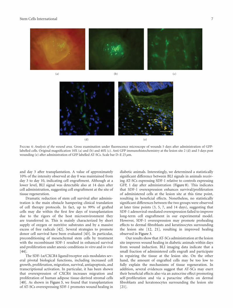
By gross examination and immunohistochemical anal-ysis, we determined the presence at the site of tissueregeneration of GFP-positive administered cells several daysafter administration (Figure 4). Further studies to determinebiodistribution and persistence of administered cells wereperformed using in vivo bioluminescent imaging techniques(see below).
3.4. Tracking Transplanted AT-SCs by BLI. After in vivoadministration of AT-SCs expressing luciferase, we mon-itored luciferase expression by real time in vivo imagingin the whole animal, together with a light photograph, toprovide for anatomical references. The intensity of the signaldetected by in vivo imaging can be precisely quantifiedand correlates with the presence of luciferase-expressingcells, and therefore with effective cell engraftment afteradministration. Since luciferin metabolism requires ATP,only living cells expressing luciferase are able to produce asignal. The ability to perform repeated analysis on the sameanimal at different time points allowed us to determine thekinetic of AT-SCs biodistribution and engraftment at thewound site.
AT-SCs were isolated from inguinal fad pads fromluciferase-expressing mice. Figure 5 shows the BLI signaloriginated from 7.5×10⁵ cells placed on a 96-well cell cultureplate and of the same cells right after topical administrationat the wound site. Quantification of the BLI signal did notshow any statistically significant difference between non-transduced cells and AT-SCs transduced with adenoviralvectors expressing GFP and SDF-1 (Figure 5) at the time ofadministration. Differences in BLI signal observed from cellsplaced in a 96 well tissue culture and from the same cells afteradministration to the dorsum of the mouse are dependent,at least in part, on the approximately 3-fold difference in thearea of a 96-well compared to the area of the biopsy punch.
To determine biodistribution of delivered cells, someanimals were sacrificed 1, 10, and 15 days after woundinduction and topical administration at the lesion site ofAT-SCs expressing luciferase. BLI was performed before andafter sacrifice of the mouse and dissection of the woundedarea. As shown in Figure 6, cells were mainly located atthe lesion site, with minimal spreading from the site ofdelivery. Moreover, performing BLI on the dissected tissuewith the skin facing the CCD camera produced a signal thatwas consistently lower than the one obtained with flippingthe piece (Figure 6). This reflects the fact that administeredcells mainly contributed to dermal and subdermal tissueregeneration and engrafted only in small part in the skin.We detected a positive signal in the region of the skin andin the corresponding subdermal area even at day 15, whenthe wound was entirely healed (Figure 6(c)). This clearlyindicates engraftment and permanence of administered cellsin the regenerated tissue even after complete healing.
The kinetics of engraftment of administered cells,expressed as percent of the BLI signal at day 0 is reportedon Figure 7, and it inversely correlates with the kinetics ofwound healing as observed in Figure 3. Most of the signalassociated with transplanted cells was lost between day 1
Stem Cells International 7
(a) (b) (c)
(d) (e)
Figure 4: Analysis of the wound area. Gross examination under fluorescence microscope of wounds 5 days after administration of GFP-labelled cells. Original magnification 10X (a) and (b) and 40X (c). Anti GFP immunohistochemistry at the lesion site 2 (d) and 5 days postwounding (e) after administration of GFP labelled AT-SCs. Scale bar D–E 25 μm.
and day 3 after transplantation. A value of approximately10% of the intensity observed at day 0 was maintained fromday 3 to day 10, indicating cell engraftment. Although at alower level, BLI signal was detectable also at 14 days aftercell administration, suggesting cell engraftment at the site oftissue regeneration.
Dramatic reduction of stem cell survival after adminis-tration is the main obstacle hampering clinical translationof cell therapy protocols. In fact, up to 99% of graftedcells may die within the first few days of transplantationdue to the rigors of the host microenvironment theyare transferred in. This is mainly characterized by shortsupply of oxygen or nutritive substrates and by a massiveexcess of free radicals [42]. Several strategies to promotedonor cell survival have been evaluated [43]. In particular,preconditioning of mesenchymal stem cells by treatmentwith the recombinant SDF-1 resulted in enhanced survivaland proliferation under anoxic conditions in vitro and in vivo[44].
The SDF-1α/CXCR4 ligand/receptor axis modulates sev-eral pivotal biological functions, including increased cellgrowth, proliferation, migration, survival, antiapoptosis, andtranscriptional activation. In particular, it has been shownthat overexpression of CXCR4 increases migration andproliferation of human adipose tissue-derived stromal cells[40]. As shown in Figure 3, we found that transplantationof AT-SCs overexpressing SDF-1 promotes wound healing in
diabetic animals. Interestingly, we determined a statisticallysignificant difference between BLI signals in animals receiv-ing AT-SCs expressing SDF-1 relative to controls expressingGFP, 1 day after administration (Figure 8). This indicatesthat SDF-1 overexpression enhances survival/proliferationof administered cells at the lesion site at this time point,resulting in beneficial effects. Nonetheless, no statisticallysignificant differences between the two groups were observedat later time points (3, 5, 7, and 14 days), suggesting thatSDF-1 adenoviral-mediated overexpression failed to improvelong-term cell engraftment in our experimental model.However, SDF-1 overexpression may promote prohealingeffects to dermal fibroblasts and keratinocytes surroundingthe lesion site [12, 21], resulting in improved healingobserved in Figure 3.
Our results show that AT-SCs administration at the lesionsite improves wound healing in diabetic animals within daysfrom wound induction. BLI imaging data indicate that asmall fraction of administered cells engraft and participatein repairing the tissue at the lesion site. On the otherhand, the amount of engrafted cells may be too low tofully explain the mechanisms of tissue regeneration. Inaddition, several evidences suggest that AT-SCs may exerttheir beneficial effects also via an autocrine effect promotingself-profileration and via a paracrine effects on dermalfibroblasts and keratonocytes surrounding the lesion site[21].

8 Stem Cells International
(a) (b)
43
2
105
97543
2
104
9754
P/sec/cm2/sr
(c)
0
2
4
6
8
10
BL
(105
p/se
c/cm
2/s
r)
NT Ad-Mock Ad-SDF
On plate
On wound
(d)
Figure 5: Ex vivo and in vivo imaging of AT-SCs expressing luciferase at time 0. Freshly isolated AT-SCs from luciferase-expressing mice weresubjected to adenoviral-mediated gene transfer ((a) non-transduced; (b) Ad-mock-transduced; (c) SDF-1-transduced cells). The AT-SCswere placed in a 96-well cell culture plate and BLI imaging was performed (insert of each figure panel). The same cells were then administeredto hyperglycemic mice right after performing a dorsal lesion and BLI was repeated. Panel (d) shows quantification of the signals. Note. Thediameter of a well of a 96-well plate is 6.4 mm; the diameter of the biopsy punch used to induce wounding is 3.5 mm. Data are expressed asmean ± SEM indicated by error bars; n = 3 for each group.
2
105
9876
5
4
3
2
P/sec/cm2/sr(a) (b) (c)
1 cm
a′ a′′ b′ b′′ c′ c′′
Figure 6: Biodistribution after topical administration at the wound site of AT-SCs expressing luciferase. BLI imaging of representative animalssacrificed 1 (a), 10 (b), and 15 (c) days after topical administration of luciferase-expressing cells at the wound site (n = 3). The regionsurrounding the wound was excided and BLI performed both with the skin facing the CCD camera (a′, b′, and c′) and on the oppositeorientation (a′′, b′′, and c′′). At all time points, cells are mainly located in the lesion area with minimal spreading from the site of topicaladministration.

Stem Cells International 9
1
10
100
BLI
(%of
day
0)
0 3 5 7 10 14
Days after wounding
(a)
3 2 104
9 8 7 6 5 4 3
P/s
ec/c
m2/s
r 1 cm
0 3 5 7 10 14
(b)
Figure 7: Persistence and engraftment of AT-SCs expressing luciferaseafter topical administration at the wound. Diabetic mice were treatedafter wounding with AT-SCs isolated from luciferase-expressingmice. Longitudinal analysis performed by BLI at different timepoints after the treatment indicates persistence of signal throughoutthe process of wound healing. Representative images from anexperimental animal at different time points (n = 6). Data areindicated as the percentage of the value immediately after topicaladministration of the cells (day 0). Values are expressed as mean ±SEM indicated by error bars.
4. Conclusions
We have demonstrated that topical administration of AT-SCs improves impaired wound healing in diabetic mice.Moreover, using BLI, we were able to follow biodistributionand the kinetics of engraftment, survival, and proliferation ofadministered cells, which proved relatively permanent in theregenerated tissue even after complete healing. In addition,our data suggest that concomitant genetic manipulation oftransplanted AT-SCs designed to promote overexpressionof SDF-1 may further improve diabetic impaired woundhealing.
Acknowledgments
This work was supported by EU FP6 STREP LSHB-CT-2005-512102 and by the Italian Ministry of Health (no.
0
2
4
BL
Isi
gnal
fold
indu
ctio
n
AT-SCs GFP AT-SCs SDF-1
∗
Day 0
Day 1
Figure 8: Topically administration of AT-SCs expressing luciferaseand overexpressing SDF-1. SDF-1 overexpression promotesenhanced survival/proliferation of administered cells at the lesionsite 1 day after wounding and cell administration, as assessed byBLI. Values are expressed as mean ± SEM indicated by error bars;n = 3, ∗P < .001.
RF 351467). J.C.W. was supported by the NIH grants nos.HL099117 and EB009689. The authors thank S. Artuso, I.Manni, G. Piaggio, and S. Straino for assistance.
References
[1] A. O’Loughlin, C. McIntosh, S. F. Dinneen, and T. O’Brien,“Review paper: basic concepts to novel therapies: a reviewof the diabetic foot,” International Journal of Lower ExtremityWounds, vol. 9, no. 2, pp. 90–102, 2010.
[2] W. J. Jeffcoate, B. A. Lipsky, A. R. Berendt et al., “Unresolvedissues in the management of ulcers of the foot in diabetes,”Diabetic Medicine, vol. 25, no. 12, pp. 1380–1389, 2008.
[3] S. Barrientos, O. Stojadinovic, M. S. Golinko, H. Brem, andM. Tomic-Canic, “Growth factors and cytokines in woundhealing,” Wound Repair and Regeneration, vol. 16, no. 5, pp.585–601, 2008.
[4] Y. Wu, R. C. H. Zhao, and E. E. Tredget, “Concise review: bonemarrow-derived stem/progenitor cells in cutaneous repair andregeneration,” Stem Cells, vol. 28, no. 5, pp. 905–915, 2010.
[5] A. T. Badillo, R. A. Redden, L. Zhang, E. J. Doolin, and K.W. Liechty, “Treatment of diabetic wounds with fetal murinemesenchymal stromal cells enhances wound closure,” Cell andTissue Research, vol. 329, no. 2, pp. 301–311, 2007.
[6] X. Fu, L. Fang, H. Li, X. Li, B. Cheng, and Z. Sheng, “Adiposetissue extract enhances skin wound healing,” Wound Repairand Regeneration, vol. 15, no. 4, pp. 540–548, 2007.
[7] E. V. Badiavas and V. Falanga, “Treatment of chronic woundswith bone marrow-derived cells,” Archives of Dermatology, vol.139, no. 4, pp. 510–516, 2003.
[8] P. A. Zuk, M. Zhu, P. Ashjian et al., “Human adipose tissue is asource of multipotent stem cells,” Molecular Biology of the Cell,vol. 13, no. 12, pp. 4279–4295, 2002.
10 Stem Cells International
[9] A. Schaffler and C. Buchler, “Concise review: adipose tissue-derived stromal cells—basic and clinical implications for novelcell-based therapies,” Stem Cells, vol. 25, no. 4, pp. 818–827,2007.
[10] A. Sterodimas, J. de Faria, B. Nicaretta, and I. Pitanguy, “Tissueengineering with adipose-derived stem cells (ADSCs): currentand future applications,” Journal of Plastic, Reconstructive andAesthetic Surgery, 2009.
[11] J. Rehman, D. Traktuev, J. Li et al., “Secretion of angiogenicand antiapoptotic factors by human adipose stromal cells,”Circulation, vol. 109, no. 10, pp. 1292–1298, 2004.
[12] W. S. Kim, B. S. Park, J. H. Sung et al., “Wound healing effectof adipose-derived stem cells: a critical role of secretory factorson human dermal fibroblasts,” Journal of DermatologicalScience, vol. 48, no. 1, pp. 15–24, 2007.
[13] A. J. Katz, A. Tholpady, S. S. Tholpady, H. Shang, and R.C. Ogle, “Cell surface and transcriptional characterizationof human adipose-derived adherent stromal (hADAS) cells,”Stem Cells, vol. 23, no. 3, pp. 412–423, 2005.
[14] V. Planat-Benard, J. S. Silvestre, B. Cousin et al., “Plasticityof human adipose lineage cells toward endothelial cells:physiological and therapeutic perspectives,” Circulation, vol.109, no. 5, pp. 656–663, 2004.
[15] M. H. Moon, S. Y. Kim, Y. J. Kim et al., “Human adiposetissue-derived mesenchymal stem cells improve postnatalneovascularization in a mouse model of hindlimb ischemia,”Cellular Physiology and Biochemistry, vol. 17, no. 5-6, pp. 279–290, 2006.
[16] M. Sumi, M. Sata, N. Toya, K. Yanaga, T. Ohki, and R. Nagai,“Transplantation of adipose stromal cells, but not matureadipocytes, augments ischemia-induced angiogenesis,” LifeSciences, vol. 80, no. 6, pp. 559–565, 2007.
[17] A. Miranville, C. Heeschen, C. Sengenes, C. A. Curat, R.Busse, and A. Bouloumie, “Improvement of postnatal neo-vascularization by human adipose tissue-derived stem cells,”Circulation, vol. 110, no. 3, pp. 349–355, 2004.
[18] H. Nakagami, K. Maeda, R. Morishita et al., “Novel autologouscell therapy in ischemic limb disease through growth factorsecretion by cultured adipose tissue-derived stromal cells,”Arteriosclerosis, Thrombosis, and Vascular Biology, vol. 25, no.12, pp. 2542–2547, 2005.
[19] J. S. Lim and G. Yoo, “Effects of adipose-derived stromal cellsand of their extract on wound healing in a mouse model,”Journal of Korean Medical Science, vol. 25, no. 5, pp. 746–751,2010.
[20] T. G. Ebrahimian, F. Pouzoulet, C. Squiban et al., “Cell therapybased on adipose tissue-derived stromal cells promotes phys-iological and pathological wound healing,” Arteriosclerosis,Thrombosis, and Vascular Biology, vol. 29, no. 4, pp. 503–510,2009.
[21] W. S. Kim, B. S. Park, and J. H. Sung, “The wound-healingand antioxidant effects of adipose-derived stem cells,” ExpertOpinion on Biological Therapy, vol. 9, no. 7, pp. 879–887, 2009.
[22] H. Brem and M. Tomic-Canic, “Cellular and molecular basisof wound healing in diabetes,” Journal of Clinical Investigation,vol. 117, no. 5, pp. 1219–1222, 2007.
[23] O. M. Tepper, J. Carr, R. J. Allen Jr. et al., “Decreasedcirculating progenitor cell number and failed mechanismsof stromal cell-derived factor-1α mediated bone marrowmobilization impair diabetic tissue repair,” Diabetes, vol. 59,no. 8, pp. 1974–1983, 2010.
[24] K. A. Gallagher, Z. J. Liu, M. Xiao et al., “Diabetic impairmentsin NO-mediated endothelial progenitor cell mobilization andhoming are reversed by hyperoxia and SDF-1α,” Journal ofClinical Investigation, vol. 117, no. 5, pp. 1249–1259, 2007.
[25] A. T. Badillo, S. Chung, L. Zhang, P. Zoltick, and K. W. Liechty,“Lentiviral gene transfer of SDF-1α to wounds improvesdiabetic wound healing,” Journal of Surgical Research, vol. 143,no. 1, pp. 35–42, 2007.
[26] D. M. Bermudez et al., “Inhibition of Stromal Derived Factor-1α (SDF-1α) in diabetic dermal wounds further prolongsthe diabetic wound healing impairment,” Journal of SurgicalResearch, vol. 151, no. 2, pp. 268–269, 2009.
[27] A. Kortesidis, A. Zannettino, S. Isenmann, S. Shi, T. Lapidot,and S. Gronthos, “Stromal-derived factor-1 promotes thegrowth, survival, and development of human bone marrowstromal stem cells,” Blood, vol. 105, no. 10, pp. 3793–3801,2005.
[28] M. Zhang, N. Mal, M. Kiedrowski et al., “SDF-1 expression bymesenchymal stem cells results in trophic support of cardiacmyocytes after myocardial infarction,” FASEB Journal, vol. 21,no. 12, pp. 3197–3207, 2007.
[29] T. Zhao, D. Zhang, R. W. Millard, M. Ashraf, and Y. Wang,“Stem cell homing and angiomyogenesis in transplantedhearts are enhanced by combined intramyocardial SDF-1α delivery and endogenous cytokine signaling,” AmericanJournal of Physiology, vol. 296, no. 4, pp. H976–H986, 2009.
[30] M. Okabe, M. Ikawa, K. Kominami, T. Nakanishi, and Y.Nishimune, “’Green mice’ as a source of ubiquitous greencells,” FEBS Letters, vol. 407, no. 3, pp. 313–319, 1997.
[31] YU. A. Cao, M. H. Bachmann, A. Beilhack et al., “Molec-ular imaging using labeled donor tissues reveals patternsof engraftment, rejection, and survival in transplantation,”Transplantation, vol. 80, no. 1, pp. 134–139, 2005.
[32] G. Di Rocco, M. G. Iachininoto, A. Tritarelli et al., “Myogenicpotential of adipose-tissue-derived cells,” Journal of CellScience, vol. 119, no. 14, pp. 2945–2952, 2006.
[33] S. Kern, H. Eichler, J. Stoeve, H. Kluter, and K. Bieback,“Comparative analysis of mesenchymal stem cells from bonemarrow, umbilical cord blood, or adipose tissue,” Stem Cells,vol. 24, no. 5, pp. 1294–1301, 2006.
[34] T. Takashima, “Preparation and isolation of cells: preparingfibroblasts,” in Current Protocols in Cell Biology, J. S. Bonifa-cino, M. Dasso, J. B. Harford, J. Lippincott-Schwartz, and K.M. Yamada, Eds., John Wiley & Sons, New York, NY, USA,1999.
[35] T. C. He, S. Zhou, L. T. Da Costa, J. Yu, K. W. Kinzler, and B.Vogelstein, “A simplified system for generating recombinantadenoviruses,” Proceedings of the National Academy of Sciencesof the United States of America, vol. 95, no. 5, pp. 2509–2514,1998.
[36] J. Luo, Z. L. Deng, X. Luo et al., “A protocol for rapidgeneration of recombinant adenoviruses using the AdEasysystem,” Nature Protocols, vol. 2, no. 5, pp. 1236–1247, 2007.
[37] G. Bilic, N. Ochsenbein-Kolble, H. Hall, R. Huch, and R. Zim-mermann, “In vitro lesion repair by human amnion epithelialand mesenchymal cells,” American Journal of Obstetrics andGynecology, vol. 190, no. 1, pp. 87–92, 2004.
[38] A. Passaniti, R. M. Taylor, R. Pili et al., “Methods in laboratoryinvestigation: a simple, quantitative method for assessingangiogenesis and antiangiogenic agents using reconstitutedbasement membrane, heparin, and fibroblast growth factor,”Laboratory Investigation, vol. 67, no. 4, pp. 519–528, 1992.

Stem Cells International 11
[39] B. Cheng, H. W. Liu, X. B. Fu, T. Z. Sun, and Z. Y.Sheng, “Recombinant human platelet-derived growth factorenhanced dermal wound healing by a pathway involving ERKand c-fos in diabetic rats,” Journal of Dermatological Science,vol. 45, no. 3, pp. 193–201, 2007.
[40] H. H. Cho, K. M. Kyoung, M. J. Seo, Y. J. Kim, Y. C. Bae, andJ. S. Jung, “Overexpression of CXCR4 increases migration andproliferation of human adipose tissue stromal cells,” Stem Cellsand Development, vol. 15, no. 6, pp. 853–864, 2006.
[41] K. Morizono, D. A. De Ugarte, M. Zhu et al., “Multilineagecells from adipose tissue as gene delivery vehicles,” HumanGene Therapy, vol. 14, no. 1, pp. 59–66, 2003.
[42] R. Das, H. Jahr, G. J. van Osch, and E. Farrell, “The roleof hypoxia in bone marrow-derived mesenchymal stem cells:considerations for regenerative medicine approaches,” Tissueengineering. Part B, Reviews, vol. 16, no. 2, pp. 159–168, 2010.
[43] H. K. Haider and M. Ashraf, “Strategies to promote donor cellsurvival: combining preconditioning approach with stem celltransplantation,” Journal of Molecular and Cellular Cardiology,vol. 45, no. 4, pp. 554–566, 2008.
[44] Z. Pasha, Y. Wang, R. Sheikh, D. Zhang, T. Zhao, and M.Ashraf, “Preconditioning enhances cell survival and differ-entiation of stem cells during transplantation in infarctedmyocardium,” Cardiovascular Research, vol. 77, no. 1, pp. 134–142, 2008.
Submit your manuscripts athttp://www.hindawi.com
Hindawi Publishing Corporationhttp://www.hindawi.com Volume 2014
Anatomy Research International
PeptidesInternational Journal of
Hindawi Publishing Corporationhttp://www.hindawi.com Volume 2014
Hindawi Publishing Corporation http://www.hindawi.com
International Journal of
Volume 2014
Zoology
Hindawi Publishing Corporationhttp://www.hindawi.com Volume 2014
Molecular Biology International
GenomicsInternational Journal of
Hindawi Publishing Corporationhttp://www.hindawi.com Volume 2014
The Scientific World JournalHindawi Publishing Corporation http://www.hindawi.com Volume 2014
Hindawi Publishing Corporationhttp://www.hindawi.com Volume 2014
BioinformaticsAdvances in
Marine BiologyJournal of
Hindawi Publishing Corporationhttp://www.hindawi.com Volume 2014
Hindawi Publishing Corporationhttp://www.hindawi.com Volume 2014
Signal TransductionJournal of
Hindawi Publishing Corporationhttp://www.hindawi.com Volume 2014
BioMed Research International
Evolutionary BiologyInternational Journal of
Hindawi Publishing Corporationhttp://www.hindawi.com Volume 2014
Hindawi Publishing Corporationhttp://www.hindawi.com Volume 2014
Biochemistry Research International
ArchaeaHindawi Publishing Corporationhttp://www.hindawi.com Volume 2014
Hindawi Publishing Corporationhttp://www.hindawi.com Volume 2014
Genetics Research International
Hindawi Publishing Corporationhttp://www.hindawi.com Volume 2014
Advances in
Virolog y
Hindawi Publishing Corporationhttp://www.hindawi.com
Nucleic AcidsJournal of
Volume 2014
Stem CellsInternational
Hindawi Publishing Corporationhttp://www.hindawi.com Volume 2014
Hindawi Publishing Corporationhttp://www.hindawi.com Volume 2014
Enzyme Research
Hindawi Publishing Corporationhttp://www.hindawi.com Volume 2014
International Journal of
Microbiology